Embed Size (px)
Citation preview

Frontiers in Environmental Microbiology 2018; 4(2): 55-70
http://www.sciencepublishinggroup.com/j/fem
doi: 10.11648/j.fem.20180402.13
ISSN: 2469-7869 (Print); ISSN: 2469-8067 (Online)
Biological Soil Crusts and Extremophiles Adjacent to Native Plants at Sabkhas and Rawdahs, Qatar: The Possible Roles
Roda Fahad Al-Thani, Bassam Taha Yasseen*
Department of Biological and Environmental Sciences, College of Arts & Sciences, Qatar University, Doha, the State of Qatar
Email address:
*Corresponding author
To cite this article: Roda Fahad Al-Thani, Bassam Taha Yasseen. Biological Soil Crusts and Extremophiles Adjacent to Native Plants at Sabkhas and Rawdahs,
Qatar: The Possible Roles. Frontiers in Environmental Microbiology. Vol. 4, No. 2, 2018, pp. 55-70. doi: 10.11648/j.fem.20180402.13
Received: February 1, 2018; Accepted: February 25, 2018; Published: March 21, 2018
Abstract: Several studies have investigated the biology of native plants (halophytes and xerophytes), biological soil crusts
(BSC), as well as the associated and adjacent microorganisms. These studies concentrated on their eco-physiological aspects as
well as the possible roles they might play in various life activities and industries. This article presents the mechanisms adopted
by these living organisms to cope with the harsh environment, and the possible roles microorganisms might play to support
native plants, some important examples from the flora of Qatar are presented to show these mechanisms. Special attention has
been paid to the senescence and death of leaves or whole plants, since this strategy has been considered as an important option
for escaping extreme environmental conditions. Discussion about solute accumulation (organic and inorganic) in all native
living organisms in general and in microorganisms in particular, could result to further investigation to clarify their roles in
such an ecosystem. Some case studies have been reported to discuss the ecological aspects at two main types of habitats in
Qatar; Rawdahs and Sabkhas were chosen to show how native living organisms adapt in these habitats. This study discussed
the general identification of BSC and microorganisms, as well as their possible roles in the soil biota. The possible methods
and mechanisms exhibited by extremophiles to support native plants in coping with saline and arid lands have also been
reported in this article. This study also discussed the recent outlook about the origin of metabolic activities in these living
organisms to support the growth of native plants under stress conditions.
Keywords: Biological Soil Crust, Extremophiles, Halophytes, Adaptation, Microorganisms Role,
Horizontal Transfer Gene (HTG)
1. Introduction
The State of Qatar is a small country located at the middle of
the western part of the Arabian Gulf, it is an extension from the
Arabia desert. It covers an area of 11,437 km2, measuring 180
km long and 85 km wide, and is situated between 24° 27' and
26° 10' north and at 50° 45' and 51° 40' east. Qatar is surrounded
on three sides by the Arabian Gulf, and only the south of Qatar
has an extension with the land of Saudi Arabia (Figure 1). The
land of Qatar is characterized as flat to wavy, sandy soil is
common in the northeastern coastline area while rocky hills and
sand dunes are common in the southern parts with a saline
coastline and patches of Sabkhas and Rawdahs at many other
locations across the country.
This country has no rivers and lakes, hence ground water
is the primary source of fresh water. As such, the soil is
vulnerable to more desertification and salinization, receives
very little rainfall as the annual rainfall may be below 80
mm, ECe may reach above 200 dSm-1
for various reasons,
and temperatures may exceed 50°C during summer. Thus, the
climate of this region is considered as warm and humid
throughout the year and the land is arid or semi-arid with
highly saline soils [1]. The physiography of the State of
Qatar is based on a number of studies which started in the
mid-seventies of the last century [2, 3] and recognized at
least seven regions and soil types: (1) Rocky desert, (2)
Rawdahs, (3) Sabkhas, (4) Sand formations, (5) Intertidal
areas, (6) Islands, and (7) Cultivated lands.

56 Roda Fahad Al-Thani and Bassam Taha Yasseen: Biological Soil Crusts and Extremophiles Adjacent to Native Plants at
Sabkhas and Rawdahs, Qatar: The Possible Roles
Figure 1. Map of Qatar showing the main locations of case studies.
This article concentrated mainly on two extremes of these
regions:
First: Rawdahs represent the area with good quality of
water and soil, and are found normally in the northern half of
the country. There are about 850 surface depressions
(Rawdahs) in Qatar, which are produced as a result of the
collapse in the earth surface below and / or as a result of
evaporation from the soil surface [4]. The area of each
Rawdah ranged from a few square hundred meters to three
square kilometers, and circular in shape and lie up to 20 m
below the surrounding landscape. Soils in Rawdahs are
enriched with calcareous loams, clay loams, silt and often
interspersed with sand and minerals that are washed down
from the surrounding higher areas during rainy seasons, and
therefore, are considered as good rangelands and can be
transformed into farmlands as well as lands for the grazing of
camels and cattle.
Second: Sabkhas are salt flats (or salt pans) and are the
most prominent feature of the Arabian Gulf region; it might
be vegetated or barren. They are characterized by high soil
salinity, and encompass most halophytes and extremophiles
which have different mechanisms to deal with such highly
saline and arid lands. Most halophytic plants are well adapted
and inhabit in such an ecosystem; however, other halophytic
taxa might not be able to cope with Sabkhas, thereby leaving
Sabkhas barren (Figure 2). Most studies were conducted in
sabkhas in the eastern coast from Doha city northwards to
Al-Khor, Ras-Al-Matbakh, Al-Dhakhira and some Rowdahs
in the mid-land of northern Qatar (Figures 1, 3), which
concentrate on the eco-physiological aspects of halophytic
plants and Halo-thermophilic microorganisms. More
discussions and elucidations can be obtained from research
books [5], and several published articles [6-12].
Figure 2. Soils at Sabkhas could become barren. Very few plants can survive
arid lands and high saline soils.
It has been reported that numerous Sabkhas in Qatar cover
about 7% of the land surface [13], and occupy a total area of
701 square kilometers [3, 14]. The largest one occurs in the

Frontiers in Environmental Microbiology 2018; 4(2): 55-70 57
vicinity of Dukhan (near the richest oil wells in Qatar) with
few species which are highly stressed by both salinity and
aridity.
In addition to these two regions (Sabkhas and Rawdahs),
rocky desert and sand formations are the most common over
the vast areas in Qatar, and sand dunes are estimated to cover
1500 square kilometers representing 12.92% of the total area
of Qatar [4]. This article presents these locations along with
the roles of the native plants, biological soil crusts and
extremophiles in this ecosystem.
Figure 3. A Sabkha as seen from one of the study locations at-high tide.
2. Wild Plants Versus Microorganisms:
Adaptation to Arid Environments
The main features of most wild plants in this region can be
grouped under three main aspects: (1) water, (2) solutes, and
(3) structures. The main mechanisms of wild plants for
resisting severe environmental conditions (e.g. salinity,
drought and high temperatures) at the Arabian Gulf region
have recently been discussed [1, 10]. There are three main
mechanisms which enable plants to cope with those factors:
(a) Avoidance mechanisms: Plants avoid salt stress by
many secondary mechanisms, for example, the extrusion
mechanism is an efficient method of removing the extra toxic
ions from the active sites of plant tissues to the outside
surface of the plant body. Many halophytes from Qatari
habitats such as Limonium axillare, Avicennia marina,
Aeluropus lagopoides, Tamarix spp. and Atriplex spp. have
been reported to have salt glands (Figure 4) and salt bladders.
There are two types of salt glands and they differ in the
number of constitutive cells: (1) salt glands of 2 cells, and (2)
salt glands of 4-10 cells. The first type is found in some
native plants like A. lagopoides [15], and Atriplex spp., and
possibly other native plants including monocot plants, while
the second type of salt glands is found in Tamarix, Limonium,
and Frankenia. Several authors have described and discussed
the structure and function of these salt glands [1, 7].
Figure 4. SEM image of the adaxial leaf surface (the upper side) of L.
axillare showing salt glands with some salt crystals and stomata.
Magnification ×400.
Some of these halophytes have salt glands capable of
excreting a substantial amount of Na+ outside the plant
body as compared to K+, with the best-known Cl
--ATPase
pump to channel Cl- outside the plant body. This situation
protects plants from toxic ions in the growth medium [16].
An ion exclusion mechanism is found in Date Palm
(Phoenix dactylefera) trees and mangroves (Avicennia
marina) by keeping low levels of toxic ions like Na+ and Cl
-
at the top of plants, while most of these ions were retained
in the root system or might be excluded to the soil
environment [17, 18]. Other mechanisms are also operating in glycophytes and
some halophytes that do not have salt glands or salt bladders.
Ions are compartmentalized in non-active cell organelles with
little metabolic activities such as cell walls and vacuoles.
Halophytes like Suaeda maritima could have such a
mechanism to avoid toxic ions as the activity of enzymes in
the active cell organelles like chloroplasts are inhibited by
salts in a similar manner to that in glycophytes. Despite the
apparent compartmentalization process found in this plant,
this mechanism might be considered as a tolerant
mechanism, since plant tissues still contain substantial
amounts of salts with the appropriate management of extra
ions. When the barriers at the root system failed to exclude
toxic ions like Na+
and Cl-, some other plants of the flora of
Qatar like Ricinus communis adopted other mechanisms,
these ions might be re-translocated back from the top of the
plant to the root tissues or even to the root environment.
Much studies are still required about other halophytes in the
Qatari habitats and other parts of the Arabian Gulf region.
The third avoidance mechanism in these plants include salt
dilution as found in halophytes like Salsola soda (Figure 5).
Extra accumulated salts in the tissues of these plants can be
diluted to keep the cytoplasmic salinity below the toxic
levels, and these plants are considered as salt includers.
In native plants, drought avoidance mechanisms can be
seen through conservation of water in their leaves;
Tetraena qatarense is a good example [1], or by

58 Roda Fahad Al-Thani and Bassam Taha Yasseen: Biological Soil Crusts and Extremophiles Adjacent to Native Plants at
Sabkhas and Rawdahs, Qatar: The Possible Roles
acceleration of water absorption, as observed in
Helianthemum lipii (Figure 6). More details can be found
in some recent articles [1, 10] with good examples from
the flora of Qatar.
Figure 5. Salsola soda. Salt includer.
Figure 6. Helianthemum lipii. This plant extends deep roots into the soil to
facilitate water absorption.
(b) Tolerance mechanism: Plants tolerate severe
environmental conditions mainly by the accumulation of
organic and inorganic solutes. Compatible osmolytes such as
proline, gylycinebetaine, soluble sugars such as trehalose and
some organic acids are accumulated in plant tissues to cope
with the harsh environments of salinity, drought and high
temperatures [1, 12]. Ascorbic acid (vitamin C) as an organic
acid, accumulates under extreme saline conditions, and it is
worth discussing its role here; as an enzyme cofactor might
play many functions especially those related to
photosynthesis and growth [19]. In fact, saline habitats
encourage the production of reactive oxygen species (ROS)
in plants [20, 21], and the last step of the pathway (Figure 7)
is to show how native plants produce this organic acid as an
antioxidant agent to ease the oxidative stress, thereby
providing help in terms of coping with the harsh
environments of osmotic stress.
Figure 7. Ascorbic acid biosynthesis in plants under stress.
Halophytes are capable of finding cheap methods of
coping with salt stress, the sequestration of inorganic ions
mainly Na+ and Cl
- in the vacuoles might save plants a lot of
energy and metabolites. Osmoregulation methods involve the
internal distribution of toxic ions and organic compatible
osmolytes around the cell; this is unlike the exclusion
mechanism mentioned above. Conversely, xerophytes are
able to accumulate various organic and inorganic solutes to
achieve osmotic adjustment, osmoregulation, and experience
changes in the physical properties of the cell wall. Certainly,
such activities could lower the osmotic and water potentials
so as maintain the water balance between plants and their
environment, and to resist the internal water shortage.
Tetraena qatarense and Ochradenus baccatus are good
examples of xerophytes which accumulate organic solutes
such as proline and soluble sugars to withstand water stress
[6, 9]. Some desert plants open their stomata even under
severe soil water shortage to maintain photosynthesis with
low transpiration rate [1, 17]. In the last four decades, several
articles and monographs have discussed these dehydration
avoidance mechanisms [9, 10, 16, 22, 23]. Moreover,
dehydration tolerance methods such as: uncoupling of
photosynthesis from transpiration, low respiration rate,
changes in protein synthesis and hydrolysis, etc., are
additional mechanisms which allow wild plants to succeed in
such harsh habitats of the Arabian Gulf region [17].
Haloxylon salicornicum, Arthrocnemum macrostachyum and
Seidlitzia rosmarinus are good examples from the Qatari
habitats, these plants are well adapted to arid and semi-arid
environments. These plants are typically tolerant to both dry

Frontiers in Environmental Microbiology 2018; 4(2): 55-70 59
lands and saline soils. Figure 8 shows that these plants
survive very dry sandy soils and perhaps thrive in such
habitats.
Figure 8. Haloxylon salicornicum thrives on dry sandy soil. This plant is
tolerant to drought and salinity.
(c) Escaping mechanism: Living organisms escape harsh
environmental conditions by different methods, activities and
behaviours. For example, quickly completing their life cycle
when the environmental conditions are in favour of growth,
and then imposing a quiescent period (dormant stage) to pass
the harsh episode of environmental stresses, or, committing
death partially or totally when those environmental
conditions worsen. Some desert herbs (ephemerals) complete
their life cycle in a very short period of time when water is
available after heavy rains. Their dormant seeds, germinate,
grow, flower and reproduce before the dry season, and
produce more dormant seeds for the next rainy season. Two
examples (Senecio desfontainei and Polycarpaea spicata)
can be found in the flora of Qatar (Figure 9, and [1]. Thus,
under the arid conditions of the Arabian Gulf region, there is
early maturation of herb plants and when the produced seeds
are left dormant for a period of time before the next rainy
season, this could be a good strategy for escaping severe
environmental conditions. The traits in these plants might be
experimental materials for modern molecular biology and
biotechnology for the development of crop plants that can
complete their life cycle within a short period of time, in
order to escape periods of severe water shortage [24].
Figure 9. Polycarpaea spicata. Drought-escaping herbs. (Courtesy of
Abdel-Bari, E. M. M, 26 May 2011).
The death of leaves or whole plants has been considered as
a strategy of escaping adverse environmental conditions. One
interesting example is the Halocnemum strobilaceum
community at some Sabkhas in Qatar; plants die at the center
(Figure 10, A, B) showing bare patches of dead branches and
green growth at the periphery. These gradually spread
towards the edges of the mat (Figure 10, C). Another
example is Halopeplis perfoliate which accelerates leaf death
to reduce the number of active green leaves, so as to maintain
the plant’s life (Figure 11). In some sabkhas, at very high salt
stress the land could be left as barren. This kind of death is
considered as the last desperate method by some native plant
species to escape the severe osmotic stress of salinity and
drought. As discussed above, many reports have concluded
that while roots serve as the real barrier against the excessive
uptake of salts, plants have many other options to redistribute
toxic ions around the plant body like excluding these ions to
less functioning organs (leaf sheaths and petioles), or re-
translocating toxic ions to roots and finally to the root
environment. For example, the sodium ion (Na+) may be
transported from leaves to roots and then to the rhizosphere
[25].
Figure 10. The death of leaves and plants of Halocnemum strobilaceum
could be a strategy for escaping stress conditions.
Many halophytic plants can extrude or include extra salts by
their salt glands or dilution mechanisms respectively. Also,
some plants have the ability to exclude toxic ions to less active
organelles like the apoplastic systems (cell walls) or vacuoles.
Such a situation keeps the active machinery of the cell
(cytoplasm) safe and away from the harmful ions, and when

60 Roda Fahad Al-Thani and Bassam Taha Yasseen: Biological Soil Crusts and Extremophiles Adjacent to Native Plants at
Sabkhas and Rawdahs, Qatar: The Possible Roles
salts accumulate inside the cells, cells might be killed unless
the salts are compartmentalized in vacuoles and other non-
active organelles [26]. Such strategies could minimize or delay
the toxic effects of ions like Cl- and Na
+. However, if plants
fail to achieve all these processes, the plant's last option to
ensure survival is the partial death of some parts (changing
colour of leaves and / or death of leaves) (Figure 12),
otherwise the whole plant will die as a result of failure to cope
with the harsh environmental conditions [27].
Leaf senescence and death are important events in
determining the total leaf area of plants, thereby affecting
their productivity. Plants normally tend to keep the number of
living leaves relatively constant [28], and environmental
conditions may have a great impact on the rate and timing of
leaf death. Salt stress, for example, might cause the
accumulation of toxic ions like Na+ and Cl
- in leaves which
induce shedding them [29-31]. Earlier studies on Mexican
wheats [32] showed that leaves die as the concentration of
NaCl increased in the growth medium, and chloride was
found in large amounts in the dead leaves under salt stress.
Figure 11. The death of Halopeplis perfoliate leaves could be a strategy for
reducing the number of active leaves, so as to save the life of the whole
plant.
Figure 12. Desert plants getting different colours due to the severe
environmental conditions, dying partially or totally, while some others are
staying green and thrive under such dry lands.
In fact, the timing and the rate of leaf death could be
regarded as a reliable criterion in the varietal differences
between crops, as their ability differ in the accumulation of
chloride in the mature leaves, a process which could lead to
the premature senescence of mature leaves and perhaps,
result to the death of these leaves thereby determining the
number of active leaves and reduce the plant surface
responsible for photosynthesis, growth and productivity [33,
34]. Previous studies on the physiology and biochemistry of
these activities showed that the chlorophyll and protein
contents were reduced in intact leaves of crops (such as
Phaseolus vulgaris) as NaCl increased in the growth medium
and resulted to a rapid senescence [35]. The hormone
imbalance under NaCl stress could have an adverse role in
the process of leaf senescence and death [36, 37]. Then, more
investigations were carried out to confirm the impact of
environmental conditions (temperature, drought, salinity,
nutrient deficiency, insufficient light) on growth regulators,
productive growth, and the consequent events which
culminated to senescence and death. Molecular investigations
have come to the conclusion that signals from these factors
could cause changes in gene expression and up-regulation of
many senescence-associated genes that cause various types
of physiological and biochemical changes: decline in
photosynthesis, degradation of macromolecules, mobilization
of nutrients and ultimately cell death [38, 39]. The question
that must be raised is whether leaf death is considered as a
process of escaping the severe environmental conditions. The
inability of plants to cope with such conditions, might get rid
of leaves partially or totally by accumulating toxic ions in the
mature leaves, a process which facilitates their shedding by
accelerating the death of these leaves to reduce the active
plant surface and maintain the potentiality of plants. Studies
have confirmed that the accumulation of excess salts in older
leaves may cause shedding and these leaves were replaced by
new leaves with low salt content [40, 41]. In fact, this
strategy of replacing older leaves by new leaves during the
life span of the plant, is a strategy found in almost all
perennial and annual plants [25]. Some authors [42] have
confirmed that such accumulation could be regarded as a
mechanism in controlling ion homeostasis and thereby salt
resistance in plants such as halophytes. Moreover, some
theories have suggested suspending the ageing of younger
leaves as a strategy of resistance against harsh environmental
conditions [32, 43, 44]. Recently, some authors [45] stated
that the leaves of some plants do well in maintaining
greenness and their photosynthetic function than others, in
the presence of high levels of Na+ in tissues under salt stress,
which clearly proved the earlier theories. Further studies are
required to investigate the above theories about the
mechanism of senescence under stress conditions. Finally,
the general conclusion is that plant death is the final stage of
escape from the worst fate facing plants under severe
conditions.
Mechanisms operating in microorganisms: The methods
and processes adopted by microorganisms to deal with the
harsh environments can be grouped under the umbrella of
Frontiers in Environmental Microbiology 2018; 4(2): 55-70 61
regulation of inorganic ions and the accumulation of
organic solute. These organisms cope with severe
environments as individual cells rather than complex multi-
cellular organisms through two main mechanisms; 1) the
salt-in strategy and 2) organic-osmolyte mechanism [46].
The salt-in-strategy means that the dominant ion in the
cytoplasm is K+, an inorganic ion that has long been
considered as the main compatible inorganic ion found in
the cytoplasm. K+ plays very important roles in maintaining
water balance and ionic strength inside the cell and also
serves as an enzyme activator for many metabolic
processes. In fact, the regulation of all types of solutes
depends on the characteristics of the plasma membranes,
types of protein transporters and the enzyme systems of the
cell wall and the cytoplasm. Published articles and reports
on this subject showed that microorganisms differ in their
regulation of inorganic solutes. For example, halophilic
archaea keep the cytoplasm with very little accumulated
sodium, while potassium along its counterion chloride
accumulates substantially to conduct various types of
functions and activities. Other halophilic anaerobic bacteria
use different tactics, Haloanaerobium praevalens showed
that both K+ and Na
+ are the dominating cations at the
stationary phase as well as in the exponentially growing
cells. Such a situation imposes some kind of preferential
accumulation of organic residues that balance the ionic
strength inside the cytoplasm [47]. The second mechanism
is the organic-osmolyte mechanism, and it involves the
accumulation of various organic solutes mainly compatible
solutes to balance the osmotic stress of the growth medium.
Extremophilic microorganisms like those in the Sabkhas of
Qatari habitats have adopted this mechanism to survive the
harsh environment of high temperature and salinity under
arid land. They accumulate organic low molecular weight
compounds like ectoines and others such as a range of
extremolytes with heterogenous chemical structures [48],
and other compatible solutes such as quaternary ammonium
compounds (glycinebetaine), amino acids (proline),
carbohydrates or their derivatives, sugars and polyols. One
peculiarity of organic solutes is that their accumulation can
be achieved by either de novo synthesis or by uptake from
the surrounding environment, and the ability to influx and /
or efflux inorganic toxic ions regulate the mechanisms of
resistance against harsh environmental conditions. These
organic solutes offer many functions to these
microorganisms, including physiological and biochemical
functions, and act as protectors against heat, desiccation,
freezing and salinity. The main functions of these organic
solutes include: (1) maintaining osmotic equilibrium across
plasma membranes, and (2) stabilizing proteins and the
whole cell. Moreover, they have various biotechnological
applications [49]. These include: (1) protein and/or enzyme
stabilizers, (2) cosmetic uses, (3) therapeutic agents, (4)
bioremediation, and (5) improving the salinity tolerance of
crop plants by HGT [12].
As a general conclusion, microorganisms and plants have
common cellular basics of physiological and biochemical
activities in dealing with extreme environmental conditions.
However, water conservation and structural modifications
against drought and salinity differ in both major groups.
Microorganisms are able to conserve and maintain their
activities and life through the formation of spores, while
plants are capable of undergoing more structural
modifications and activities to avoid and tolerate harsh
environments.
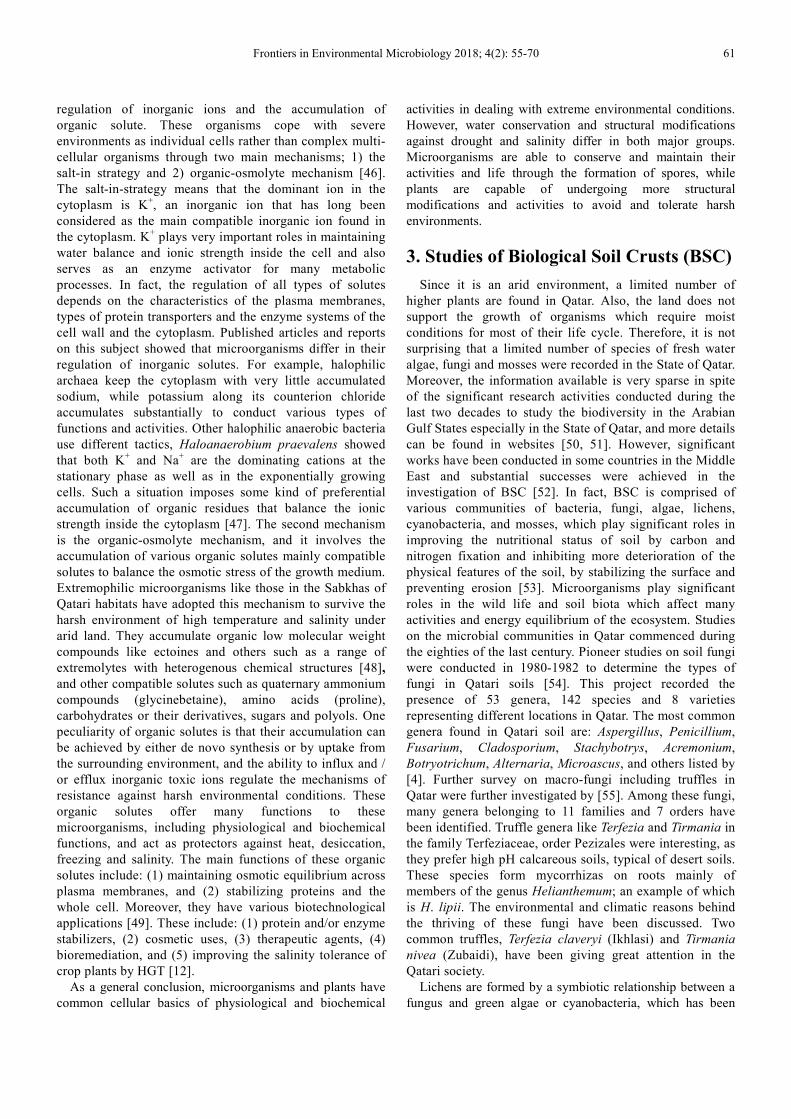
3. Studies of Biological Soil Crusts (BSC)
Since it is an arid environment, a limited number of
higher plants are found in Qatar. Also, the land does not
support the growth of organisms which require moist
conditions for most of their life cycle. Therefore, it is not
surprising that a limited number of species of fresh water
algae, fungi and mosses were recorded in the State of Qatar.
Moreover, the information available is very sparse in spite
of the significant research activities conducted during the
last two decades to study the biodiversity in the Arabian
Gulf States especially in the State of Qatar, and more details
can be found in websites [50, 51]. However, significant
works have been conducted in some countries in the Middle
East and substantial successes were achieved in the
investigation of BSC [52]. In fact, BSC is comprised of
various communities of bacteria, fungi, algae, lichens,
cyanobacteria, and mosses, which play significant roles in
improving the nutritional status of soil by carbon and
nitrogen fixation and inhibiting more deterioration of the
physical features of the soil, by stabilizing the surface and
preventing erosion [53]. Microorganisms play significant
roles in the wild life and soil biota which affect many
activities and energy equilibrium of the ecosystem. Studies
on the microbial communities in Qatar commenced during
the eighties of the last century. Pioneer studies on soil fungi
were conducted in 1980-1982 to determine the types of
fungi in Qatari soils [54]. This project recorded the
presence of 53 genera, 142 species and 8 varieties
representing different locations in Qatar. The most common
genera found in Qatari soil are: Aspergillus, Penicillium,
Fusarium, Cladosporium, Stachybotrys, Acremonium,
Botryotrichum, Alternaria, Microascus, and others listed by
[4]. Further survey on macro-fungi including truffles in
Qatar were further investigated by [55]. Among these fungi,
many genera belonging to 11 families and 7 orders have
been identified. Truffle genera like Terfezia and Tirmania in
the family Terfeziaceae, order Pezizales were interesting, as
they prefer high pH calcareous soils, typical of desert soils.
These species form mycorrhizas on roots mainly of
members of the genus Helianthemum; an example of which
is H. lipii. The environmental and climatic reasons behind
the thriving of these fungi have been discussed. Two
common truffles, Terfezia claveryi (Ikhlasi) and Tirmania
nivea (Zubaidi), have been giving great attention in the
Qatari society.
Lichens are formed by a symbiotic relationship between a
fungus and green algae or cyanobacteria, which has been

62 Roda Fahad Al-Thani and Bassam Taha Yasseen: Biological Soil Crusts and Extremophiles Adjacent to Native Plants at
Sabkhas and Rawdahs, Qatar: The Possible Roles
paid a great deal of attention and given substantial efforts
since such combination serves the ecosystem by offering
many important functions, especially in the State of Qatar. It
seemed BSC at the Arabian Gulf region developed as a result
of the interaction between microorganisms like cyanobacteria
and the physiochemical characteristics of soil [56], as desert
crusts harbor a substantial diversity of microorganisms like
fungi [57] which play critical roles in the ecosystem by
improving the soil structure and fertility, water status, soil
stabilization, water retention, as well as carbon and nitrogen
fixation [58]. In Qatar, the contribution of microorganisms in
the soil crust has been poorly studied and investigated. In
fact, in 1997 four lichens species were reported for the first
time as addition to the flora of Qatar [59, 60], these were
Ramalina farinosa Ach (fruticose lichens), Acarospora sp.,
Buellia spp., and Caloplaca spp. (crustose lichens).
Thereafter, other authors [61] reported twelve species of
lichens; composed of ascomycetes as the main fungal part
while cyanobacteria served as the autotrophic part [53]. In
fact, three main groups of lichens have been recognized and
reported in Qatar: (1) crustose lichens, (2) fruticose lichens,
and (3) foliose lichens [4]. There are many methods of
identification, however, the morphology and ultrastructure
have been widely utilized around the globe [62].
Non-vascular plants including mosses are confined to a
very limited number of species, and no taxonomic studies
have been conducted to identify this group. Authors like [63]
reported one moss genus (Funaria); collected from the soil
surface of potted plants and from various moisty soils after
heavy rains (Figure 13). One fern species (Ophioglossum
polyphyllum) which occurs in many parts of the country, has
been recorded in Qatar [60, 63].
Case study of lichens at Rawdahs-Northern Qatar: In
this location, the mean annual temperature during summer
time is about 42°C and the absolute temperature may rise
up to 50°C, while during winter period the mean annual
temperature is about 23°C, with high humidity during
most months of the year. The expected rainy season
extends from October to March with a well-defined dry
period in July. The soil is almost shallow, sandy and
rocky, and show different levels of alkalinity and salinity
depending on the influence of geological substrates at
each site.
Figure 13. Little species of mosses found in Qatari habitats. This image was
taken in a private garden with ample moisture in its soil.
Samples were collected from a private farm in Northern-
Qatar, and these samples of BSC were collected from areas
of 50 m2 each during the dry season, June 2016 (Figure 14
A). To preserve their integrity, samples of BSC were taken
from the central part of the area and placed in plastic boxes
with care, and kept until they were transported to the
laboratory for analysis. Samples were collected again after
heavy rains in April 2017 (Figure 14 B) adjacent to the
plants which thrived in that period. Table 1 shows some
physical and chemical properties of the soil samples
collected from this Rawdha as compared to those from
different locations of Sabkhas, which clearly indicated the
presence of much clay, high field capacity, and acceptable
pH values (between 6 and 8), and low salinity which ranged
between 6 -8 dSm-1
.
Table 1. Some physical and chemical properties of soil samples collected from different locations of Sabkhas and inland Rawdahs.
Location Soil texture Field capacity (%) Soil water content
(of FC%) Absolute water content (%)
pH (soil
extract) ECe (dSm-1)
Doha Sandy-Sandy loam 26* 7 - 61 2 – 18 7 – 10 31 - 177
Al-Khor Sandy loam- Loamy sand 37 6 - 10 2 – 3 8 – 9 > 200
Ras-Al-Matbakh Sandy-Silty loam 38 11 - 51 2 – 28 6 - 8 12 - 49
Al-Dhakhira Sandy loam 30 43 - 71 12 – 26 8 - 9 > 200
Inland Rawdahs Loamy sand-Sandy clay loam 40 9 - 12 3 – 4 6 - 8 4-6
*All data are mean or a range of 10 readings. ECe was measured according the method described by [64].

Frontiers in Environmental Microbiology 2018; 4(2): 55-70 63
Figure 14. The sampling site before (A), and after rainy (B) seasons.
The vegetation of the area at that period consisted mainly
of xerophytic shrubs and Ephemerals which is dominated by
Suaeda aegyptiaca, Stipa capensis, Anastatica hierochuntica,
Fagonia indica, Lycium shawii, Pulicaria gnaphalodes,
Plantago ovata as well as other grasses and herbs (Figures 9
and 15).
Figure 15. Ephemerals found in the study location, which are supported by
BSC including lichens. These herbs germinate, grow and reproduce before
the dry season.
During the dry season, soil surface of the study location
shows cracks and a substantial number of lichens colonies of
different types (Figure 16). These colonies play a significant
role in the biological activities of the soil and contributed
actively to the thriving the biodiversity [57, 58]. Such
colonies might be found on rocks of different types and
shapes which serve as growth substrate and provide minerals
to them, lichens on rocks play a significant role in rock
weathering and breakdown in arid and semi-arid
environments [4], and to support the growth of many desert
plants. Soils of the study site contain some thermo-halophilic
bacteria which belong to the genus Bacillus and other
microorganisms like the cyanobacteria Microcoleus, with
significant presence of many species of Nostoc and
Anabaena, and possibly other genera that play crucial roles to
support the soil with various types of biological activities
[11]. Green algae are found on the rocks, and, in particular
some unicellular green algae are found especially after heavy
rains.
Figure 16. Various types of lichens colonies found on different patches of the
study site appeared on the soil crust.
The many possible important roles played by BSC in the
desert environments include: (a) increasing the nutrient
content of soils: nitrogen fixation is one example especially
in Qatar, as the soil suffers from low nitrogen availability [7,
65], (b) stabilizing soil surfaces and preventing soil erosion
by their contribution to soil formation and structure. For
example, lichens can help in providing organic matter, water
retention and in the collection of soil components like dust
and silt which enrich the soil with some nutrients [4, 53], (c)
monitor atmospheric pollution [66, 67] as a result of
expansion in the industrial activities of the oil and gas sectors
in Qatar [61], (d) some other benefits and advantages can be
obtained for any future plans to use lichens in the human life;
these include food preparation and diets, textile industry,
chemistry, medicine, pharmacology as well as cleaning the
environment from various types of pollution … etc [68-72].
Studies on bacterial communities in Qatar:
Studies on bacterial communities did not commence until
the beginning of the current century, our records showed that
these studies did not exceed reporting the presence of some
genera and species at various habitats. For example, studies
like [73, 74] have shown that the decomposition rates in the
shoot system (leaves) of Avicennia marina L. were highest in
the summer season, and the bacterial groups that
predominated during these processes were those that do not
form spores (non-spore forming bacteria) and were in the
order: Amylolytic > proteolytic > cellulolytic, and the least
ones were the spore forming bacteria. The follow up
investigation of the heterotrophic aerobic marine bacteria of
the above groups revealed the bacterial counts during the
period April 1998 to February 1999 which showed some
differences depending on the atmospheric temperatures, such
conditions might have had a significant role in releasing
substances from the leaves of that plant, thereby affecting the
bacterial counts. These studies did not go further to identify
these groups, genera and species. This is the first time that
the ecology and biodiversity of Streptomyces is being studied
in the soil of Qatar, in many sites around the country. The

64 Roda Fahad Al-Thani and Bassam Taha Yasseen: Biological Soil Crusts and Extremophiles Adjacent to Native Plants at
Sabkhas and Rawdahs, Qatar: The Possible Roles
morphology of the isolates of this genus have a common
form of colony, they are spherical, smooth, elevated with
diameter mainly ranging between 1-5 mm [75]. The bacterial
inhabitants in the soil (Figure 17) adjacent to many
halophytes and in the rhizosphere of these plants at the
coastal zone and at the inland zones were investigated [76].
The results of this study showed that the total bacterial counts
were higher in the rhizosphere of plants at the coastal line as
compared to those of the non-rhizosphere soil and at the
inland areas as well. Gram positive cocci predominated in
isolates from the rhizosphere and non-rhizosphere soil, while
Gram-positive bacilli were predominant in aqueous washings
of phyllospheres of the green and senescing parts of these
plants. One important result that should be reported here is
the presence of low bacterial colonization on the
phyllosphere of the halophytes which have a salt extrusion
mechanism than those which adopt dilution mechanism to
avoid salinity.
Escherichia coli was investigated in some ponds around
Doha city [77]. The study revealed that this bacterium is
prominently present in untreated ponds (Abu-Hamour pond)
compared to treated ones (Abu-Nakhla pond). Other bacteria
species were also found, and these included: Aeromonas
hydrophilia, Pseudomonas aeruginosa, Klebsiella
pneumoniae and Chromobacterium violaceum.
Figure 17. Diversity of bacterial species isolated from Sabkhas in Qatar.
The other bacterial species which were also found in the
wet soil around these ponds included: Streptomyces sp.,
Bacillus sp. and Macrococcus sp. The isolation of some
native bacterial strains like Bacillus sp., Pseudomonas
geniculata and Achromobacter xylosoxidans from different
locations around Qatar has also been confirmed. The above
mentioned strains have the ability to degrade contaminants
such as phenol derivatives and polycyclic aromatic
hydrocarbons [78, 79]. Several bacterial strains have been
identified during the phytoremediation processes of soil
contaminated with petroleum hydrocarbons [80, 81] and to
improve the quality of industrial waste water produced
during oil and gas operations. These efforts were introduced
as one of the objectives to save the ecosystems in Qatar from
the real risks and consequences of the expansion in the
industrial activities [1]. Bacterial communities have been
studied through modern methods including direct counts, and
also by some advanced techniques to gain more
understanding of microbial abundance, diversity, and
potential metabolic capabilities in various locations in Qatar
[82]. Extremophiles like some bacilli species (Bacillus
thuringenses and Bacillus cereus) isolated from Qatari soils
(Figure 18) adjacent to native plants or associated with them
(Figure 19) might play significant roles in supporting or
promoting the growth of these plants.
Figure 18. Bacteria of Sabkhas: colonies represent Bacillus sp.; the most
common bacteria isolated from Sabkhas in Qatar (A), spore forming Bacillus
bacteria as seen under the light microscope (B). (magnification: x1000).
Recently, a new approach has emerged which supports the
growth and development of plants under severe
environmental conditions by microorganisms which
inhabitant the soil. One of the main issues that should be
addressed is the possible roles of the presence of
extremophiles in salt glands and on the plant body in
providing some important resistance traits to the native plants
as well as other biological needs [12, 83]. Microorganisms

Frontiers in Environmental Microbiology 2018; 4(2): 55-70 65
could be a solution to increase the plant’s resistance to
various harsh abiotic stresses [84], and the following are the
methods and mechanisms adopted by microbes which have
recently been discussed [11, 12]: (a) biofilm formation, (b)
polymers production, (c) chemotaxis, (d) phytohormone
production, (e) nitrogen fixation, (f) phosphate solubilization,
(g) production phytohormone-degrading enzymes and (h)
osmolytes biosynthesis. All these methods could provide
some kind of protection for plants against various types of
environmental stresses (Figure 20).
In fact, halotolerant and halo-thermophilic bacteria found
in Qatari soils could offer many applications in
biotechnology. These include: the production of compatible
solutes [12], salt antagonists, or stress-protective agents, and
other useful molecules like bio-surfactants,
exopolysaccharides and enzymes which have important
applications in food biotechnology, bioremediation … etc
[85]. Moreover, the advances in the modern biological
techniques could offer great opportunity to study the ecology
and function of microbial communities in hot desert
ecosystems [86]. In fact, many studies have demonstrated
that microbes might provide support for non-host plants
through the adoption of a resistance mechanism that
improves their ability to conduct various functions and
activities in their natural habitats [87, 88].
Figure 19. The SEM image shows a huge number of bacterial cells cover the
leaf surface of native plants in Qatari desert.
HTG: Horizontal Gene Transfer
Figure 20. The different methods and mechanisms by which microorganisms protect plants against abiotic stresses. This figure was modified from Figure (1)
of [12].

66 Roda Fahad Al-Thani and Bassam Taha Yasseen: Biological Soil Crusts and Extremophiles Adjacent to Native Plants at
Sabkhas and Rawdahs, Qatar: The Possible Roles
In the meantime, more promising achievements have been
made during the last twenty years around the world. This is
because a great deal of attention has been placed on the
production of antibiotics, since natural soils are rich with a
large number of microbes capable of producing various types
of antibiotics. Significant studies, especially in the third
world (developing countries), have shown that a large
number of bacterial isolates and fungi have antimicrobial
activity. Based on several experimental works, researchers
[89] have collected dozens of soil bacteria genera, including
eubacteria and actinomycetes, which have different and
powerful abilities to produce antibiotics. In India [90],
screening was performed for potent antimicrobial compounds
and at least one strain with antagonistic abilities against
many test microorganisms was isolated. A study in Nigeria
[91] is of interest, because genetic techniques were used to
show that microorganisms can synthesize useful organic
compounds such as antibiotics during their activity with soil
contaminants, although only a limited number of the
investigated Bacilli species exhibited antibacterial activity.
More achievements have been reported from Ethiopia, as one
successful breakthrough isolated actinomycetes that produce
antibiotics active in fighting against antibiotic-resistant
pathogens [92].
Again, the issue of the origin of activities in both native
plants and their associated microorganisms is still
outstanding, and the mystery of life has been strongly
addressed by scientists and research centers. Also, the
relationships between living organisms and their activities
are still the core of their thought and interest. For example,
[93] concluded that the HGT (Horizontal Gene Transfer) of
antibiotic resistance traits played a crucial role in the
evolution of bacteria and their diversity. In fact, since the
review of [94] in evaluating the possibility of HGT from
plants to microorganisms, only a few cases of HGT from
eukaryotes to bacteria have been reported. There is still a
possibility of introducing genes into the environment and
subsequently transferring them to other organisms. More
reports are available about the possibilities of HGT between
living organisms and it is worth mentioning here. In their
review, [95] discussed the role of HGT in eukaryotic
evolution, as this approach has been more successful than
traditional genetic modification methods. In fact, this
approach has expanded rapidly in the last decade since many
eukaryotic genes have been found to have microbial origins
[96]. In a review on the early evolution of photosynthesis,
[97] stated that the evidence clearly showed that
photosynthesis in eukaryotic organisms originated from the
endosymbiosis of cyanobacteria-like organisms, which were
later converted to chloroplasts. This conclusion means that
the origin of photosynthesis began in prokaryotic organisms.
HGT appears to have played a crucial role in the evolution of
life on Earth, and is supported by numerous events which
have been observed in the genomes of the three domains of
life [98]. However, although the traditional thought is that
most genes in eukaryotes were derived from mitochondria
and plastids, these organelles cannot explain the presence of
bacterial genes in eukaryotes. Historically, HGT from
Agrobacterium to plants has been considered the first event
in the transfer of genes between living organisms, as this
event has been considered a novel research finding of the
transfer and integration of genetic materials from bacteria
into eukaryotic organisms [99]. This event opened the door to
expand our knowledge of modern genetic manipulations to
improve the resistance of crops to environmental stresses
[100].
HGT: Horizontal Gene Transfer
Figure 21. The possible hypotheses of horizontal gene transfer (HGT) between living organisms.

Frontiers in Environmental Microbiology 2018; 4(2): 55-70 67
In recent review [12], some objectives were outlined for
any future plans: (1) ecophysiology of native plants, the
associated microorganisms and the type of relationships, (2)
study the salt glands and the plants surface with their
associated microbes, (3) study the compatible solutes in
plants and microbes, and (4) molecular studies to elucidate
the origin of activities in plants and microorganisms. Three
possibilities have been suggested about the origin of
metabolic activities (Figure 21), these are: (a) plant specific
genes, with possibilities of HGT to other living organisms,
(b) microbe specific genes, with possibilities of HGT to other
living organisms, and (c) independent origin that are found in
either plants or microorganisms with no possibilities of HGT
to other living organisms. Certain examples have been
reported in some websites [101, 102]. More discussions and
explanations can be found in several other reports and
articles [12, 103-105].
4. Conclusion
The different physiological and biochemical
characteristics of the huge number of living organisms
(BSC, plants or others), and the mechanisms they adopt in
coping with harsh environments can be considered as
experimental materials for more advanced and deep
research. In fact, the tremendous evolution in molecular
biology and biotechnology could be utilized to solve many
of the problems facing humanity in the agricultural and
health sectors as well as the understanding of many secrets
and mysteries about the creation of life. The gene bank of
these living organisms, especially those living under
extreme environmental conditions, requires substantial
efforts for modern manipulation to develop living
organisms capable of improving, increasing and sustaining
production in the agricultural and pharmaceutical sectors.
Also, looking at the system: Soil-BSC-Plants, all living
organisms seem to be living in an organized harmony of
cooperation and support of each other. Each group, or even
individual organism, has its roles to support the life and
activities of mankind. In recent years, many facts have been
revealed about the reality of these roles in human life as
well as the methods and approaches adopted by these living
organisms to bring about a sustainable support of each
other. The evidence presented so far support the idea of the
unity of life, and the reasons and objectives behind their
creation, and such task still requires more studies for proper
understanding.
Acknowledgements
The authors would like to thank the Department of
Biological and Environmental Sciences, College of Arts and
Sciences, as well as the Environmental Studies Centre, Qatar
University, for the financial and technical support provided
during the case studies.
References
[1] B. T. Yasseen, and R. F. Al-Thani, “Ecophysiology of wild plants and conservation perspectives in the state of Qatar,” In: M. Stoytcheva, and R. Zlatev (Eds.), Agricultural chemistry, Chapter 3, ISBN: 978-953-51-1026-2, InTech. 2013; 37-70. DOI: 10.5772/55305, 2013.
[2] State of Qatar / Ministry of Industry and Agriculture / UNDP / FAO, “Reconnaissance soil classification map,” 1976.
[3] K. H. Batanouny, “Ecology and flora of Qatar,” The Alden Press Ltd., Oxford, U. K, The Scientific and Applied Research Centre, University of Qatar, 1981.
[4] H. A. Abulfatih, E. M. M. Abdel-Bari, A. Alsubaey, and Y. M. Ibrahim, “Vegetation of Qatar,” Scientific and Applied Research Center (SARC), University of Qatar, Doha, Qatar, 2001.
[5] E. M. M. Abdel-Bari, B. T. Yasseen, and R. F. Al-Thani,” Halophytes in the state of Qatar,” Environmental Studies Centre, Qatar University, Doha, Qatar, ISBN. 99921-52-98-2, 2007.
[6] B. T. Yasseen, and R. F. Al-Thani, “Halophytes and associated properties of natural soils in the Doha area, Qatar,” AEHMS, vol. 10 (3), pp. 320–326, 2007.
[7] B. T. Yasseen, and M. A. Abu-Al–Basal, “Ecophysiology of Limonium axillare and Avicennia marina from the coastline of Arabian Gulf-Qatar,” Journal of Coastal Conservation: Planning and Management, vol. 12 (1), pp. 35-42, 2008.
[8] B. T. Yasseen, and M. A. Abu-Al-Basal, “Ecophysiology of chenopodiaceae at the coastline of Arabian Gulf-Qatar: possible destruction and prespective conservation,” European Journal of Scientific Research, vol. 39 (1), pp. 90–104, 2010.
[9] B. T. Yasseen, “Urban development threatening wild plants in Doha City-Qatar: Ecophysiology is a Prerequisite for Ecological Restoration,” Journal of Plant Sciences, vol. 6 (3), pp. 113–123, 2011.
[10] B. T. Yasseen, “Traits of wild plants in Qatar peninsula and research perspectives,” J. Biology & Nature, vol. 5 (2), pp. 52–66, 2016.
[11] R. F. Al-Thani, and B. T. Yasseen, “Halo-thermophilic bacteria and heterocyst cyanobacteria found adjacent to halophytes at Sabkhas, Qatar: Preliminary study and possible roles,” African J. Microbiology Research, vol. 11 (34), pp. 1346-1354, 2017.
[12] R. F. Al-Thani, and B. T. Yasseen, “Solutes in native plants in the Arabian Gulf region and the role of microorganisms: future research,” J. Plant Ecology, rtx066, https://doi.org/10.1093/jpe/rtx066, 2017.
[13] M. M. Ashour, “Sabkhas in Qatar peninsula, landscape and geodiversity,”1/2013, pp. 10-35, ISSN 2286-0177 ICCS, Spiru Haret University, Bucharest, Romania, 2013.
[14] I. A. Mahasneh, R. F. Al-Thani, and G. Brown,” The microorganisms of Sabkhat in Qatar,” In: M. Ajmal Khan, B. Boer, G. S. Kust, and H-J. Barth, Sabkha Ecosystems, vol. 2, West and Central Asia, Tasks for Vegetation Science 42, H. Leith, University of Osnabruck, Germany. Springer, The Netherlands, 2006.

68 Roda Fahad Al-Thani and Bassam Taha Yasseen: Biological Soil Crusts and Extremophiles Adjacent to Native Plants at
Sabkhas and Rawdahs, Qatar: The Possible Roles
[15] F. M. Salama, S. M. El-Naggar, and T. Ramadan, “Salt glands of some halophytes in Egypt,” Phyton-Annales Rei Botanicae, vol. 39 (1), pp. 91-105, 1999.
[16] D. M. Orcutt, and E. T. Nilsen, “Physiology of plants under stress, soil and biotic factors, John Wiley & Sons, Inc NY, 2000.
[17] J. Levitt, “Responses of plants to environmental stresses, water, radiation, salt, and other stresses,” vol. II, Academic Press, New York, London, 1980.
[18] H. J. Aljuburi, and H. H. Al-Masry, “Effects of salinity and indole acetic acid on growth and mineral content of date palm seedlings,” Fruits, vol. 55, pp. 315-323, 2000.
[19] K. Nahar, S. M. Ullah, and N. Islam,” Osmotic adjustment and quality response of five tomato cultivars (Lycopersicon esculentum Mill) following water deficit stress under subtropical climate,” Asian Journal of Plant Sciences, vol. 10, pp. 153-157, 2011.
[20] P. Sharma, A. B. Jha, R. S. Dubey, and M. Pessarakli, “Reactive oxygen species, oxidative damage, and antioxidative defence mechanism in plants under stressful conditions,” Journal of Botany, vol. 2012, Article ID 217037, 26 pages, http://dx.doi.org/10.1155/2012/217037, 2012.
[21] R. Ksouri, A. Smaoui, H. Isoda, and C. Abdelly, “Utilization of halophytes species as new source of bioactive substances,” Journal of Arid Land Studies, vol. 22 (1), pp. 41-44, 2012.
[22] L. G. Paleg, and D. Aspinall, “The physiology and biochemistry of drought resistance in plants,” Academic Press, Sydney, New York, London, pp. 492, 1981.
[23] W. Larcher, “Physiological plant ecology, Ecophysiology and stress physiology of functional groups,” 4th ed., Springer, Berlin, 2003.
[24] M. K. Omara, “Selection of early maturing barley with improved response to drought stress,” Aust. J. Agric. Res, vol. 38, pp. 835-845, 1987.
[25] P. Adam, “Saltmarsh ecology, “Cambridge University Press, 1993.
[26] R. Munns, “The impact of salinity stress: (1) The environmental and physiological nature of salinity,” CSIRO Division of Plant Industry Canberra ACT, Australia. http://plant stress.com/articles/salinity_i/salinity_i.htm, 2017, Accessed on 29th October 2017).
[27] R. Munns, “Comparative physiology of salt and water stress,” Plant Cell & Environ, vol. 28, pp. 239-250, 2002.
[28] R. H. M. Langer, “How grasses grow,” 2nd edition, Studies in biology No. 34, Edward Arnold Ltd, 1979.
[29] H. O. Kostina, “Gradient distribution of chloride and ash in plants and its ecological significance,” Ukr. Bot. Zh, vol. 31, pp. 322-326, 1974.
[30] E. V. Maas, and R. H. Niemen, “Physiology of plant tolerance to salinity,” In: Crop tolerance to suboptimal land conditions, ASA, Special publication number 32, American Society of Agronomy, Medison, USA, 1978.
[31] R. Munns, and M. Tester, “Mechanisms of salinity tolerance,” Annu. Rev. Plant Biol, Vol. 59, pp. 651-81, doi: 10.1146/annurev. arplant. 59.032607.092911, 2008.
[32] B. T. Yasseen, “An analysis of the effects of salinity on leaf growth in Mexican wheats,” Thesis, The University of Leeds, UK, 1983.
[33] G. R. Cramer, and R. S. Nowak, “Supplemental manganese improves the relative growth, net assimilation and photosynthetic rates of salt-stressed barley,” Physiol. Plant, vol. 84 (4), pp. 600-605, 1992.
[34] P. Carillo, M. G. Annunziata, G. Pontecorvo, A. Fuggi, and P. Woodrow, “Salinity Stress and Salt Tolerance,” In: A. Shanker, (Ed.), Abiotic Stress in Plants - Mechanisms and Adaptations, InTech, http://www.intechopen.com/books/abioticstress-in-plants-mechanisms-and-adaptations/salinity-stress-and-salt-tolerance, 2011.
[35] J. T. Prisco, and J. W. O’Leary, “Enhancement of intact bean leaf senescence by NaCl salinity,” Physiol. Plant, vol. 27, pp. 95-100, 1972.
[36] C. Itai, and Y. Vaadia, “Cytokinin activity in water -stressed shoots,” Plant Physiol, vol. 47, pp. 87-90, 1971.
[37] M. I. Khan, M. A. Khan, and T. Khizar, “Plant growth regulators from species differing in salt tolerance as affected by soil salinity,” Plant & Soil, vol. 45, pp. 267-271, 1976.
[38] Y. Guo, and S. Gan, “Leaf senescence: Signals, execution, and regulation,” Current Topics in Developmental Biology, vol. 71, pp. 83–112, 2005.
[39] J. P. Vainonen, P. Jaspers, M. Wrzaczek, A. Lamminmäki, R. A. Reddy, L. Vaahtera, M. Brosché, and J. Kangasjärvi, “RCD1–DREB2A interaction in leaf senescence and stress responses in Arabidopsis thaliana,” Biochemical Journal, vol. 442 (3), pp. 573-58, 2012.
[40] A. T. Ayoub, “Sodium and cation accumulation by senna (Cassia acutifolia),” J. Exp. Bot, vol. 26, pp. 891–896, 1975.
[41] R. Albert, “Salt regulation in halophytes,” Oecologia, vol. 21, pp. 57–71, 1975.
[42] Y. Waisel, “Biology of Halophytes,” Academic Press, New York and London, 1972.
[43] M. M. Ludlow, and T. T. Ng, “Water stress suspends leaf ageing,” Plant Sci. Lett, vol. 3, pp. 235-240, 1974.
[44] R. Munns, C. J. Brady, and E. W. R. Barlow, “Solute accumulation in the apex and leaves of wheat during water stress,” Aust. J. Plant Physiol, vol. 6, pp. 379-389, 1979.
[45] S. Negrao, S. M. Schmockel, and M. Tester, “Evaluating physiological responses of plants to salinity stress,” Annals of Botany, vol. 119, pp. 1–11. doi:10.1093/aob/mcw191, available online at www.aob.oxfordjournals.org, 2017.
[46] S. P. Singh, V. Raval, and M. K. Purohit, “Strategies for the salt tolerance in bacteria and archeae and its implications in developing crops for adverse conditions,” In: N. Tuteja, and S. Singh Gill, (Eds.), Plant Acclimation to Environmental Stress, Springer, New York, NY, 2013.
[47] H. J. Kunte, “K+ Transport and its role for osmoregulation in a halophilic member of the bacteria domain: Characterization of the K+ uptake systems from Halomonas elongate,” In: N. Gunde-Cimerman et al. (Eds.), Adaptation to Life at High Salt Concentrations in Archaea, Bacteria, and Eukarya, pp. 287-300. Springer, The Netherlands, 2005.

Frontiers in Environmental Microbiology 2018; 4(2): 55-70 69
[48] P. Babu, A. K. Chandel, and O. V. Singh, “Survival mechanisms of extremophiles,” In: Extremophiles and Their Applications in Medical Processes. Springer Briefs in Microbiology, Springer, Cham, pp. 9-23, 2015.
[49] P. Shivanand, and G. Mugeraya, “Halophilic bacteria and their compatible solutes–osmoregulation and potential applications,” Current Science, vol. 100 (10), pp. 1516–1521, 2011.
[50] https://www.cbd.int/doc/world/qa/qa-nbsap-01-en.pdf.
[51] https://www.cbd.int/doc/world/qa/qa-nbsap-v2-en.pdf.
[52] M. Galun, and J. Garty, “Biological soil crusts of the Middle East,” In: J. Belnap, andO. L. Lange, (Eds.), Biological soil crusts: structure, function, and management, Berlin, Springer, pp. 95–106, 2003.
[53] R. Richer, D. Anchassi, I. El-Assaad, M. El-Matbouly, F. Ali, I. Makki, and J. S. Metcalf, “Variation in the coverage of biological soil crusts in the State of Qatar,” Journal of Arid Environments, vol. 78, pp. 187–190, 2012.
[54] A. H. Moubasher, and A. A. T. Al-Subai, “Soil fungi in the State of Qatar,” The Scientific and Applied Research Council, University of Qatar, pp. 108, 1987.
[55] R. F. Al-Thani, “Survey of macrofungi (including truffles) in Qatar,” KBM Journal of Biology, vol.1 (2), pp. 26-29, 2010.
[56] F. I. Khalaf, and A. A. Al-Shuaibi, “Occurrence and characteristics of quaternary calcareous microbiatitic crust in the southern desert in Kuwait, Arabian Gulf,” J. Arid Environments, vol. 79, pp. 48–55, 2012.
[57] R. M. M. Abed, A. M. Al-Sadi, M. Al-Shehi, S. Al-Hinai, and M. D. Robinson, “Diversity of free-living and lichenized -fungal communities in biological soil crusts of the Sultanate of Oman and their role in improving soil properties,” Soil Biology & Biochemistry, vol. 57, pp. 695–705, 2013.
[58] E. Redfield, S. M. Barns, J. Belnap, L. L. Daane, and C. R. Kuske, “Comparative diversity and composition of cyanobacteria in three predominant soil crusts of the Colorado Plateau,” FEMS Microbiol. Ecol, vol. 40, pp. 55–63, 2002.
[59] E. M. M. Abdel-Bari, “Record of a fruiticose lichen Ramalina farinacea (L.) Ach. for Qatar,” Qatar University Science Journal, vol. 17 (2), pp. 265-270, 1997.
[60] E. M. M. Abdel-Bari, “Addition to the flora of Qatar,” Qatar University Science Journal, vol. 17 (2), pp. 303-312, 1997.
[61] R. F. Al-Thani, and H. A. Al-Meri, “Study of some lichens of Qatar,” Atlas Journal of Biology, vol 1 (3), pp. 41- 46, 2011.
[62] C. Hartl, A. R. Schmidt, J. Heinrichs, L. J. Seyfullah, N. Schäfer, C. Gröhn, J. Rikkinen, and U. Kaasalainen, “Lichen preservation in amber: morphology, ultrastructure, chemofossils, and taphonomic alteration,” Foss. Rec, vol. 18, pp. 127–135, 2015.
[63] E. M. M. Abdel Bari, “The flora of Qatar, The Dicotyledons, The Monocotyledons, Environmental Studies Centre, Qatar University, Doha, Qatar, Volumes:1-2, pp.700 (Volume 1), pp.199 (Volume 2), 2012.
[64] L. A. Richards, “U. S. Salinity Laboratory Staff, Diagnosis and Improvement of Saline and Alkaline Soils,” U. S. Department of Agriculture Hand Book No. 60, P 160, 1954.
[65] M. M. Ashore, “Sabkhas in the peninsula of Qatar–geomorphologic and geological and biological studies–University of Qatar,” Centre of Documentation and Humanitarian Studies, Doha, Qatar, 1991.
[66] P. L. Nimis, C. Scheidegger, P. A. Wolseley, “Monitoring with Lichens–Monitoring Lichens,” Kluwer Academic Publishers, The Netherlands, pp. 1-4, 2002.
[67] J. Nascimbene, P. L. Nimis, and L. Marini, “Testing indicators of epiphytic lichen diversity: a case study in N Italy,” Biodivers. Conserv, vol. 16, pp. 3377–3383, 2007.
[68] P. William, “Lichens. The Natural History Museum,” London, UK. pp. 112. 2000.
[69] K. Müller, “Pharmaceutically relevant metabolites from lichens,” Applied Microbiology and Biotechnology, vol. 56, pp. 9-16. 2002.
[70] M. Bačkor, K. Paulíková, A. Geralská, and R. Davidson, “Monitoring of air pollution in Košice (Eastern Slovakia) using lichens,” Polish Journal of Environmental Studies, vol. 12 (2), pp.141-150, 2003.
[71] J. Boustie, and M. Grube, “Lichens-a promising source of bioactive secondary metabolites,” Plant Genetic Resources, vol. 3, pp. 273-278, 2005.
[72] M. Melgarejo, O. Sterner, J. Vila-Castro, and P. Mollinedo, “More investigations in potent activity and relationship structure of the Lichen antibiotic (+)- using acid its derivate dibenzoylusnic acid,” Revista Boliviana De Quimica, vol. 25 (1), pp. 24-29, 2008.
[73] A. M. Mahasneh, “Bacterial decomposition of Avicennia marina leaf litter from AlKhore (Qatar-Arabian Gulf),” On line Journal of Biological Sciences, vol. 1 (8), pp. 717-719, 2001.
[74] A. M. Mahasneh, “Heterotrophic marine bacteria attached to leaves of Avicennia marina L. along the Qatari coast (Arabian Gulf),” On line Journal of Biological Sciences, vol. 2 (11), pp. 740-743, 2002.
[75] R. F. Al-Thani, and I. A. Mahasneh, “Ecology and distribution of Streptomycets in the soil of Qatar,” The Scientific and Applied Research Center (SARC), Qatar University, Doha, Qatar, ISBN: 99921-52-56-7, 2002.
[76] G. M. Fahmy, and R. F. Al-Thani, “Ecology of halophytes and their bacterial inhabitants in the coastal salt marsh of Al-Dhakhira, Qatar,” Environmental Studies Centre (ESC), University of Qatar, P. O. Box: 2713, Doha, Qatar, ISBN: 99921-751-0-9. 2006.
[77] R. F. Al-Thani, “Coliform bacteria of wastewater ponds in Qatar,” In: H. A. Abulfatih, R. F. Al-Thani, I. S. Al-Naimi, J. A. Sweeileh, E. A. Elhag, and M. M. Kardousha, Ecology of wastewater ponds in Qatar, Scientific and Applied Research Center (SARC), University of Qatar, Doha, Qatar, pp.129–145. 2002.
[78] R. F. Al-Thani, D. A. Abd-El-Haleem, and M. Al- Shammri, “Isolation, biochemical and molecular characterization of 2-chlorophenol degrading Bacillus Isolates,” Afric. J. Biotec, vol. 6 (23), pp. 2675-2681, 2007.
[79] R. F. Al-Thani, D. A. Abd-El-Haleem, and M. Al-Shammri, “Isolation and characterization of polyaromatic hydrocarbons-degrading bacteria from different Qatar soils,” African J. Micro. Res, vol. 3 (11), pp. 761-766, 2009.

70 Roda Fahad Al-Thani and Bassam Taha Yasseen: Biological Soil Crusts and Extremophiles Adjacent to Native Plants at
Sabkhas and Rawdahs, Qatar: The Possible Roles
[80] M. Y. Al-Sulaiti, I. M. Al-Shaikh, B. T. Yasseen, S. Ashraf, and H. M. Hassan, “Ability of Qatar’s native plant species to phytoremediate industrial wastewater in an engineered wetland treatment system for beneficial water re-use,” Qatar Foundation Annual Research Forum Proceedings, Vol. 2013, EEO 010, DOI: 10.5339/qfarf.2013. EEO-010, Published online: 22 Nov 2013.
[81] B. T. Yasseen, “Phytoremediation of industrial wastewater from oil and gas fields using native plants: The research perspectives in the State of Qatar,” Central European Journal of Experimental Biology, vol. 3 (4), pp. 6-23, 2014.
[82] S. Abdul Majid, M. F. Graw, A. D. Chatziefthimiou, H. Nguyen, R. Richer, M. Louge, et al., “Microbial characterization of Qatari Barchan sand dunes,” PLoS ONE, vol. 11 (9), e0161836. https://doi.org/10.1371/journal.pone.0161836, 2016.
[83] C. Gougoulias, J. M. Clark, L. J. Shaw, “The role of soil microbes in the global carbon cycle: tracking the below-ground microbial processing of plant-derived carbon for manipulating carbon dynamics in agricultural systems,” J Sci Food Agric, vol. 94 (12), pp. 2362-2371, 2014.
[84] M. Hanin, C. Ebel, M. Ngom, L. Laplaze, and K. Masmoudi, “New insights on plant salt tolerance mechanisms and their potential use for breeding,” Frontiers in Plant Sciences, vol. 7, Article 1787, 2016.
[85] R. Margesin, and F. Schinner, “Potential of halotolerant and halophilic microorganisms for biotechnology,” Extremophiles, vol. 5 (2), pp. 73–83. 2001.
[86] T. P. Makhalanyane, A. Valverde, E. Gunnigle, A. Frossard, J. B. Ramond, and D. A. Cowan, “Microbial ecology of hot desert edaphic systems,” FEMS Microbiol. Rev, vol. 39 (2), pp. 203-221, 2015.
[87] J. W. Whitaker, G. A. McConkey, and D. R. Westhead, “The transforme of metabolic genes explored: analysis of the horizontal transfer of enzyme encoding genes in unicellular eukaryotes,” Genome Biology, vol. 10 (4), Article R36, 2009.
[88] Z. Yuan, I. S. Druzhinina, J. Labbé, R. Redman, Y. Qin, R. Rodriguez, C. Zhang, G. A. Tuskan, and F. Lin, “Specialized microbiome of a halophyte and its role in helping non-host plants to withstand salinity,” Scientific Reports, vol. 6, p. 32467, 2016.
[89] T. A. Huck, N. Porter, M. E. Bushell, “Positive selection of antibiotic-producing soil isolates,” Journal of General Microbiology, vol. 137, pp. 2321-2329, 1991.
[90] A. R. Srividya, G. S. Saitha, and B. Suresh, “Study of the soil isolates for antimicrobial activity,” Indian J. Pharm Sci, vol. 70 (6), pp. 812–815, 2008.
[91] R. N. Ahmed, A. Sani, A. K. Ajijolakewu, and F. B. Alamu, “Soil screening for antibiotic - producing microorganisms,” Advances in Environmental Biology, vol. 7 (1), pp. 7-11, 2013.
[92] A. Bizuye, F. Moges, and B. Andualem, “Isolation and screening of antibiotic producing actinomycetes from soils in Gondar town, North West Ethiopia,” Asian Pac J Trop Dis, vol. 3 (5), pp. 375-381, 2013.
[93] J. Wiedenbeck, and F. M. Cohan, “Origins of bacterial diversity through horizontal genetic transfer and adaptation to new ecological niches,” FEMS Microbiol Rev, vol. 35, pp. 957–976. 2011.
[94] K. M. Nielsen, “Barriers to horizontal gene transfer by natural transformation in soil bacteria,” APMIS, vol. 106, pp. 77-84, 1998.
[95] P. J. Keeling, and J. D. Palmer, “Horizontal gene transfer in eukaryotic evolution,” Nature Reviews Genetics, vol. 9, pp. 605-618, 2008.
[96] J. Huang, “Horizontal gene transfer in eukaryotes: The weak-link model,” Bioessays, vol. 35 (10), pp. 868-875, 2013.
[97] R. E. Blankenship, “Future perspectives in plant biology early evolution of photosynthesis,” Plant Physiol, vol. 154, pp. 434–438, 2010.
[98] R. I. Aminov, “Horizontal gene exchange in environmental microbiota,” Frontiers in Microbiology, vol. 2 / Article 158, 2011.
[99] B. Lacroix, and V. Citovsky, “Transfer of DNA from bacteria to eukaryotes,” mBio, vol. 7 (4), e00863-16. doi:10.1128/mBio.00863-16. V, 2016.
[100] T. V. Matveeva, and L. A. Lutova, “Horizontal gene transfer from Agrobacterium to plants,” Front. Plant Sci, vol. 5, p. 326. doi: 10.3389/fpls.2014.00326, 2014.
[101] https://en.wikipedia.org/wiki/Plant_secondary_metabolism.
[102] https://en.wikipedia.org/wiki/Microbial_metabolism.
[103] H. B. Bode, and R. Müller, “Possibility of bacterial recruitment of plant genes associated with the biosynthesis of secondary metabolites,” Plant Physiol, vol. 132, pp. 1153–1161, 2003.
[104] N. Empadinhas, and M. S. da Costa, “Osmoadaptation mechanisms in prokaryotes: distribution of compatible solutes,” International Microbiology, vol. 11, pp. 151-161, 2008.
[105] N. Empadinhas, and M. S. da Costa, “Diversity, distribution and biosynthesis of compatible solutes in prokaryotes,” Contributions to Science, vol. 5 (1), pp. 95–105. 2009.