Embed Size (px)
Citation preview
Al-Azhar Journal of Agricultural Research V. (45) No. (2) December (2020) 15-32 El-Damaty et al.
15
Biological evaluation of some synthetic and natural food colorants
A. M. El-Damaty 1, M. E. Abd-ElGhany 2, F. O. F. Abou- Zaid 1 and M. M. Salama 1,*
1 Department of Food Science and Technology, Faculty of Agriculture, Al-Azhar University, Cairo, Egypt 2 Department of Plant Production, Desert Research Center, Cairo, Egypt
* Correspondence: [email protected] (M. Salama)
ABSTRACT
This study was carried out to evaluate and compare the possible toxic effect among some synthetic and natural
food colorants on biochemical parameters as well as liver and kidney histological of experimental rats. A total
number of 45 young albino male rats (weight about 100-120 g) were used in this study. All rats were divided
randomly into nine groups and fed on tested diets for four weeks (carmoisine, raspberry, sunset-yellow, and fast
green as synthetic colorants; and anthocyanin, betalain, carotenoids, and chlorophyll as natural food colorants; as
well as a control group). The results showed a significant increase in levels of serum alanine aminotransferase,
aspartate aminotransferase, urea, creatinine, total protein, and albumin, with decreased levels of immune-globulins
were observed in all groups treated by synthetic colorants groups compared to the groups treated by natural food
colorants and control group. Histological examinations revealed alterations in kidneys including congestion and
hemorrhage with infiltration, and deformation of the glomeruli structure. Whereas, alterations in the liver
including congestion, hemorrhage, and dilatation of sinusoids and central vein with microvascular steatosis.
Therefore, it is advisable to limit the uses of synthetic food colorants or replacing them with natural ones, especially
for children’s foods.
Keywords: Natural food colorants; Synthetic food colorants; Biochemical parameters; Liver histology; Kidney
histology.
INTRODUCTION
Color is the most important characteristic of food. Based on the color of the food, first impressions are made. For example, the color indicates the ripeness degree of the fruit, food freshness, and toast burning. Based on these first impressions, a judgment is made whether the food is safe to eat or not and whether it can be expected to taste good or not. Since color is closely associated with expectations, the addition of color to food is a way to fulfill these expectations (Aberoumand, 2011).
Colorants are added to food to serve several purposes, including accounting for the loss during processing or storage, to enhance color already present, to minimize batch to batch variations, and color otherwise uncolored food (Lakshmi, 2014). Food colors can be divided, in respect of their origin, into natural, identical to natural and synthetic ones (Janiszewska-Turak et al., 2016).
Synthetic food colorings have been associated with adverse reactions in some individuals, with claims of links to hyperactivity, asthma, and other allergic reactions (Larsen, 2008).
These claims have generated much debate as well as scientific research and have led to negative consumer perceptions of synthetic colors .The current consumer perception of synthetic food ingredients and food colorants, in particular, is that they have negative health
implications and that foods containing the natural ingredient are safer, healthier, and hence a better choice (Crino et al., 2013).
Regarding food color industry trends, the use of natural colorants has increased in foods and beverages as substitutes for their synthetic counterparts. This is mainly due to the growing awareness of the environmental hazards and the potential side-effects of the chemicals used in the synthesis of food colorants (Carocho et al., 2014). Also, to satisfy consumers who demand natural ingredients, major food and beverage companies have committed to removing synthetic substances, including synthetic colors, from their products. This marketing strategy is in line with the so-called “clean label” trend (Cortez et al., 2017) .
In Egypt, there has been a sharp increase in the use of synthetic food colorants in the past few years and additionally, there is uncontrolled use of synthetic color particularly in food mostly consumed by children (Salah, 1994). More attention must be focused on the physiological and pathological effects of color additives (Helal et al., 2000).
The purpose of this study is to compare and illustrate the health effects of four natural (anthocyanin, betalain, carotenoids, and chlorophyll) and four synthetic food colorants (carmoisine, raspberry, sunset yellow, and fast green) on biochemical and histological parameters of experimental rats.
Al-Azhar Journal of Agricultural Research V. (45) No. (2) December (2020) 15-32 El-Damaty et al.
16
MATERIAL AND METHODS
Materials
Natural colorants
Anthocyanin, betalain, carotenoids, and chlorophyll pigments were extracted from different plant sources includes: Roselle (Hibiscus sabdariffa), red beetroot (Beta Vulgaris), orange navel peels (Citrus aurantium) and green peas peel (Pisum sativum), respectively by ultrasound-assisted extraction (UAE) according to the methods of Sivakumar et al. (2009); Almahy et al. (2017) and Boukroufa et al. (2017). Encapsulation of pigment extracts was prepared by mixing 100 ml of the pigment extract with 20 g of matrix consist maltodextrin DE 20 and Arabic gum (at ratio 19: 1), then were mixed homogeneously. The mixture was constantly and thoroughly stirred to ensure homogeneity during freeze-drying process as described by Ravichandran et al. (2012).
Synthetic colorants
The synthetic colorants (carmoisine, raspberry, sunset yellow, and fast green) were purchased from Kamena Company, Kafr Tohormous, Giza, Egypt.
Methods
Experimental design
A total number of 45 young albino male rats (weighing about 100-120 g) were used in the present study. All rats were fed on a balanced diet (AIN-93) for one week according to Reeves et al. (1993). Such feed contained 15% casein, 5% cellulose, 3.5% mineral mixture, 1% vitamin mixture, 8% oil, 10% sugar and 57.5% starch. The rats were then divided randomly into nine groups (five rats each) and were fed on the following diets for 4 weeks:
Group 1: rats fed on the basal diet (as a control group), the standard diet was prepared according to Reeves et al. (1993). Group 2: rats fed on the basal diet and ingested orally with a dose of anthocyanin microcapsules (50 mg/kg body weight). Group 3: rats fed on basal diet and ingested orally with a dose of carmoisine (10 mg/kg body weight). Group 4: rats fed on basal diet and ingested orally with a dose of betalain microcapsules (50 mg/kg body weight). Group 5: rats fed on basal diet and ingested orally with a dose of raspberry (10 mg/kg body weight). Group 6: rats fed on basal diet and ingested orally with a dose of carotenoids microcapsules (50 mg/kg body weight). Group 7: rats fed on basal diet and ingested orally with a dose of sunset yellow (10 mg/kg body weight). Group 8: rats fed on basal
diet and ingested orally with a dose of chlorophyll microcapsules (50 mg/kg body weight). Group 9: rats fed on basal diet and ingested orally with a dose of fast green (10 mg /kg body weight).
The biological investigation was carried out at the Animal House Lab, Food Science and Technology, Faculty of Agriculture, Al-Azhar University, Cairo, Egypt.
At the end of the experiment, the rats were fasted overnight, and total food intake, final rat's body weights, and body weight gain were recorded.
The food efficiency ratio was calculated according to this equation:
Food efficiency ratio = body weight gain/food intake (Proll et al., 1998).
Blood Samples
At the end of the experimental period (28 days), blood samples were obtained after fasting (overnight) using the orbital sinus technique of Soltan and Shehata (2012). The blood samples were collected in a dry tube and were left to clot, then centrifuged at 3000 rpm for ten min. Serum was separated and frozen at -20 °C for the subsequent analyses.
Afterward, the rats were decapitated and some internal organs such as liver and kidneys were dissected out, trimmed of excess fat, and weighted before performing histological studies. The organ weight was presented as relative organ weight and calculated according to Ping et al. (2006) and Elgawish and Ghanem (2014), as follows:
Relative organ weight = (Absolute organ weight/ whole body weight) × 100.
Biochemical Investigation
Measuring blood glucose
Blood glucose levels were measured according to Dafallah et al. (2015).
Determination of liver function enzymes activity
Serum aspartate –aminotransferase (AST) and alanine-amino transferase (ALT) were determined according to the method described by Abd El-Wahab and Moram (2012).
Determination of total protein and albumin concentration
Total protein and albumin concentration were measured by the method described by Helal et al. (2000).
Al-Azhar Journal of Agricultural Research V. (45) No. (2) December (2020) 15-32 El-Damaty et al.
17
Determination of kidney functions
Blood urea and creatinine concentration were estimated by the enzymatic method as described by Abd El-Wahab and Moram (2012).
Lipid profile
Total lipids, total cholesterol, triglycerides, HDL-cholesterol, and LDL-cholesterol were determined according to the method described by Ibrahim (2001).
Determination of plasma immune-system (Ig G, Ig A, and Ig M)
The kit is intended for measuring plasma Ig G, Ig A, and Ig M according to the method described by Dafallah et al. (2015).
Histological Examination
The liver and kidneys of rats were dissected out and fixed instantaneously in 10% formal saline for 24 h. The specimens were washed in tap water, dehydrated in ascending grades of ethanol, cleared in xylene, embedded in paraffin wax (melting point of 50-56 °C). Paraffin sections were cut at 6μm thicknesses using a rotary microtome (Model MR 60, Russian), then the sections were stained with Harris' hematoxylin and eosin. The observations were performed using a light microscope (Zeiss Axiophot, Germany) and photographs were taken with an automatic photomicrographic system (Sarkar et al., 2005).
Statistical Analysis
The data obtained from three replicates were analyzed by (ANOVA) using the SPSS statistical package program, and the differences among the means were compared using the Duncan's Multiple Range test of SPSS (1998). A significant level of 0.05 was chosen.
Results and Discussion
Effect of oral ingestion of colorants on the behavior of rats
It was noticed that after oral administration of synthetic colorants, rats became more active, nervous, and aggressive. Also, a skin irritation was noticed after the administration of sunset yellow. These additives are a common cause of chronic urticarial, angioedema in rats, and also caused positive skin and intestinal reaction irritability (Soltan and Shehata, 2012). These results are nearly in agreement with the findings of Helal et al. (2000) and Soltan and Shehata (2012).
Effect of oral ingestion of the colorants on weights of rats
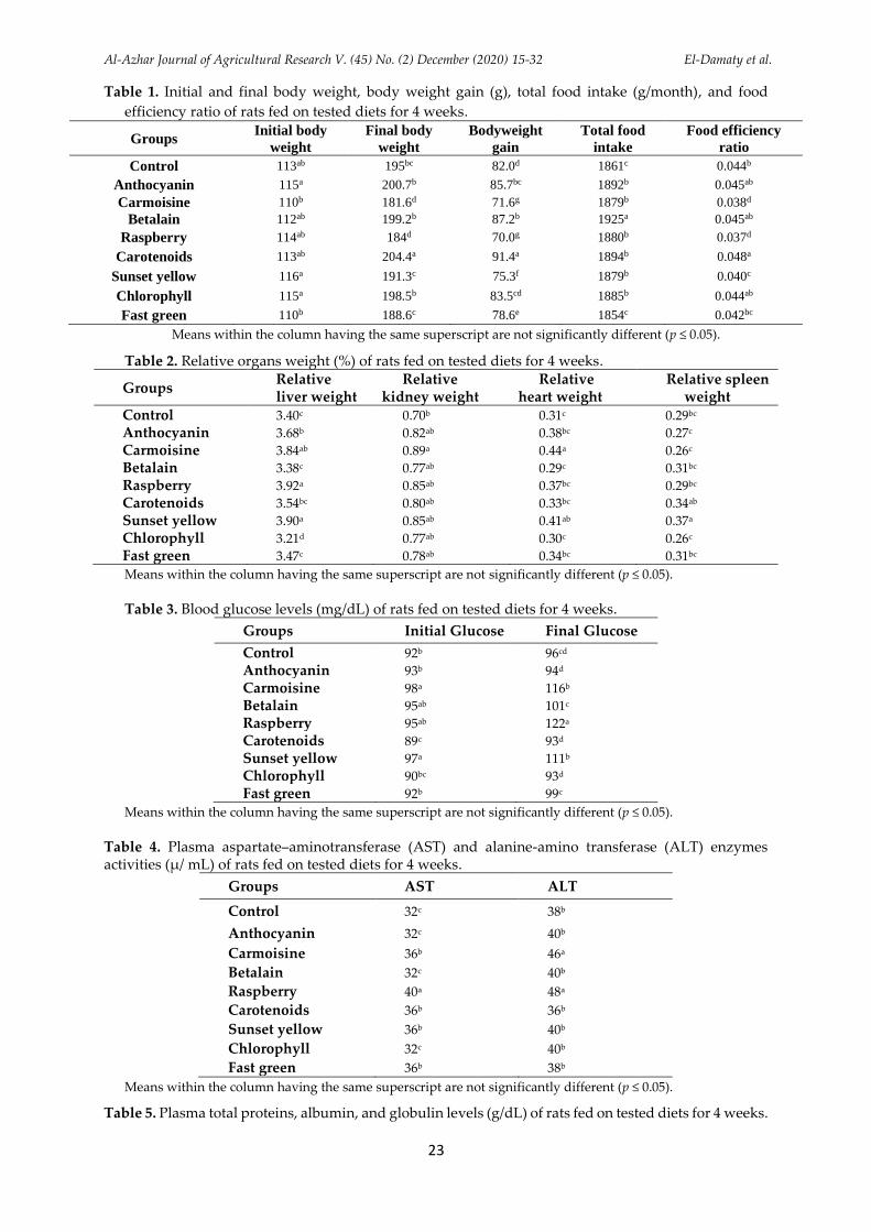
The data given in Table (1) shows that the bodyweight of rats ingested oral synthetic colorants (carmoisine, raspberry, sunset yellow, and fast green) was significantly lower than the control and the other groups ingested natural colorants (anthocyanin, betalain, carotenoids, and chlorophyll).
Also, Table (1) illustrates that, except fast green group, rats fed synthetic colorants recorded a significant increase (p < 0.05) in feed intake values compared to the control group, but the amount of increase in feed intake differed among groups. Thus, the decrease in gaining body weight was not due to the decrease in feed consumption. For these reasons, the feed efficiency of the diet was calculated.
The results of the food efficiency ratio indicated that the rats that ingested oral natural colorants and control had higher (p ≤ 0.05) food efficiency ratios than the other groups. The changes in food intake were not parallel to the growth rate, feed efficiency of the different colorants. These results supported the hypothesis that, the digestion of diet may be inhibited in a certain manner by the addition of synthetic colorants.
The data revealed that the synthetic food colorants had a negative effect of on growth rate (body weight gain) which may be related to the disturbance effects of colorants on different metabolic systems (Abd El-Wahab and Moram, 2012). These results are in harmony with the results of EFSA (2009), ANS (2009), and Abd El-Wahab and Moram (2012).
Relative organs weight
From Table (2), it can be found that the relative weight of tested organs showed an increase in the relative liver, kidney, and heart weights in the rats that had oral synthetic colorants compared to all the other treatments. These results are in agreement with the results of Abd El-Wahab and Moram (2012) who recorded a significant increase in the relative weight of the liver for some treatments that had diets containing synthetic colorants mixed with synthetic flavors.
Effect of oral ingestion of colorants on biochemical properties
Blood glucose levels
Results in Table (3) show that the highest increase (p ≤ 0.05) in blood glucose level was noticed in treatments that treated with synthetic colorants, especially in the case of raspberry
Al-Azhar Journal of Agricultural Research V. (45) No. (2) December (2020) 15-32 El-Damaty et al.
18
treatment which amounted to (122 mg/dL), compared to the control (96 mg/dL). The elevation of glucose level can be explained by stimulation of glycogenolysis and gluconeogenesis in the liver with a temporary loss of endocrine functions of the pancreas which leading to hyperglycemia (Al-Shinnawy, 2009). These results are nearly in agreement with the reports of Amin et al. (2010), Himri et al. (2011), Dafallah et al. (2015), and Wopara et al. (2019) who indicated that the blood glucose in rats treated by synthetic colorants was significantly (p ≤ 0.05) increased compared to all the other treatments.
Liver functional enzyme activity
Data in Table (4) revealed a marked increase (p ≤ 0.05) in serum AST level of all groups treated by synthetic colorants as compared to the control group. On the other hand, rats treated with natural colorants (except rats fed on carotenoids) showed a significant decrease (p ≤ 0.05) in AST level compared with rats fed on synthetic colorants.
As found in Table (4), the groups treated by carmoisine and raspberry had the highest levels (p ≤ 0.05) of ALT (46 and 48 µ/mL, respectively) compared to the other groups (ranged between 36 – 40 µ/mL). The significant increase in the activities of serum AST and ALT for rats treated with synthetic colorants may be due to the haptic potency of these colors resulting in destructive changes in the hepatic cells. This increase is considered to be a result of related to destruction of the liver cells, while leads to liberation of these enzymes in the blood stream. Similarly, Tameda et al. (2005) considered the increase in ALT and AST activities as sensitive indicators of a liver cell injury. These results are approximately in agreement with the results of Helal et al. (2000), Amin et al. (2010), Abd El-Wahab and Moram, (2012), and Soltan and Shehata (2012) who indicated that rats that consumed high dose synthetic color showed a significant increase in serum ALT and AST compared to the control rats.
Total proteins, albumin, and globulin levels
Data in Table (5) revealed that the oral administration of carmoisine and raspberry induced a significant change in serum total proteins, while other groups showed insignificant differences (p > 0.05) between them. Also, the results showed that the levels of the plasma albumin (g/dL) were increased (p < 0.05) in rats fed on carmoisine, raspberry, sunset yellow, and chlorophyll compared to the other groups. The present data illustrated a significant decrease (p ≤ 0.05) of globulin in
groups treated by sunset yellow and chlorophyll. These results are in agreement with the report of Helal et al. (2000).
Creatinine and urea levels
Concerning the kidney functions, the present data in Table (6). The experimental rats treated by raspberry showed the highest increase (p ≤ 0.05) in serum creatinine level (1.40 mg/dL) compared to other groups. This may be indicating protein catabolism, kidney dysfunction, or both actions (Abdel-Wahhab and Aly, 2003). Also, the experimental rats that orally ingested with raspberry showed a significant increase (p ≤ 0.05) in serum urea level (66.20 mg/dL), compared all the other groups.
Also, the synthetic colorants, especially raspberry induced alteration in kidney functions which had a highly significant increase (p < 0.05) in serum urea and creatinine levels. These changes may indicate a reduction in the glomerular filtration rate as a result of acute renal dysfunction. The serum level of these two parameters depends largely on the glomerular filtration (Abd El-Wahab and Moram, 2012). These results are partially in agreement with those obtained by Abd El-Wahab and Moram (2012), Soltan and Shehata (2012), and Dafallah et al. (2015).
Total lipids, total cholesterol, and triglyceride levels
The data in Table (7) shows that a significant decrease (p ≤ 0.05) in total lipids and total cholesterol for all groups that treated by natural colorants compared to synthetic colorants. The highest values were 430 and 211 mg/dL, respectively for sunset yellow, while the lowest values were 260 and 165 mg/dL, respectively in anthocyanin treatment. Also, the results showed that there was a slight significant decrease in triglyceride level in rats treated by natural colorants, except for chlorophyll compared to those groups treated by synthetic colorants. The highest value of the triglyceride level was 136 mg/dL in carmoisine, while the lowest value was 108 mg/dL in the group treated by anthocyanin.
These results are in agreement with those reported by Abou El-Zahab et al. (1997), Himri et al. (2011), Dafallah et al. (2015), and Wopara et al. (2019) who observed significant increases in serum total lipids, cholesterol, and triglycerides in rats had supplemented with some food colorants in different concentrations. The current results are contrary to the results of Sharma et al. (2006) who reported that two doses of tomato red (blend of carmoisine and
Al-Azhar Journal of Agricultural Research V. (45) No. (2) December (2020) 15-32 El-Damaty et al.
19
ponceau 4R) showed a significant decrease in serum total cholesterol and triglycerides when Swiss albino mice consumed these colorants for 21 days as short term or 42 days as long-term. Also, these results are opposite to those reported by Ashour and Abdelaziz (2009) who noticed a significant reduction in serum total cholesterol and triglycerides levels when food color azo dye (fast green) was orally consumed by male albino rats for 35 days.
Serum HDL-cholesterol, LDL-cholesterol levels
Data in Table (8) showed a significant increase (p ≤ 0.05) in HDL and a decrease in serum LDL cholesterol for all groups treated by natural colorants compared to synthetic colorants. The highest value of LDL cholesterol was observed in the group treated by sunset yellow (134.6 mg/dL), while the lowest value was noticed in the group treated by anthocyanin (86.8 mg/dL). On the other hand, the current data showed a significant increase (p ≤ 0.05) in HDL/LDL ratios for all groups treated by natural colorants compared to synthetic colorants. The improvement of HDL/LDL ratios were related to a significant increase in HDL levels as well as to the significant decrease in LDL levels in all groups treated by natural colorants compared to synthetic colorants. These results are approximately in agreement with the findings of Dafallah et al. (2015).
Immune-System
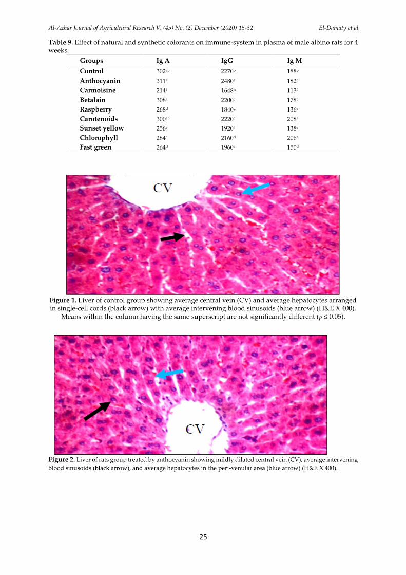
Regarding the effect of oral administration of colorants on the plasma immune-system, the data obtained in Table (9) revealed that, a significant decrease (p ≤ 0.05) in Ig A, Ig G, and Ig M was observed in the groups treated with synthetic colorants compared to the control and natural colorants. These results are nearly in agreement with those reported by Dafallah et al. (2015).
Histological alterations in some functional organs of rats as affected by oral administration of colorants
Histological alterations of the liver
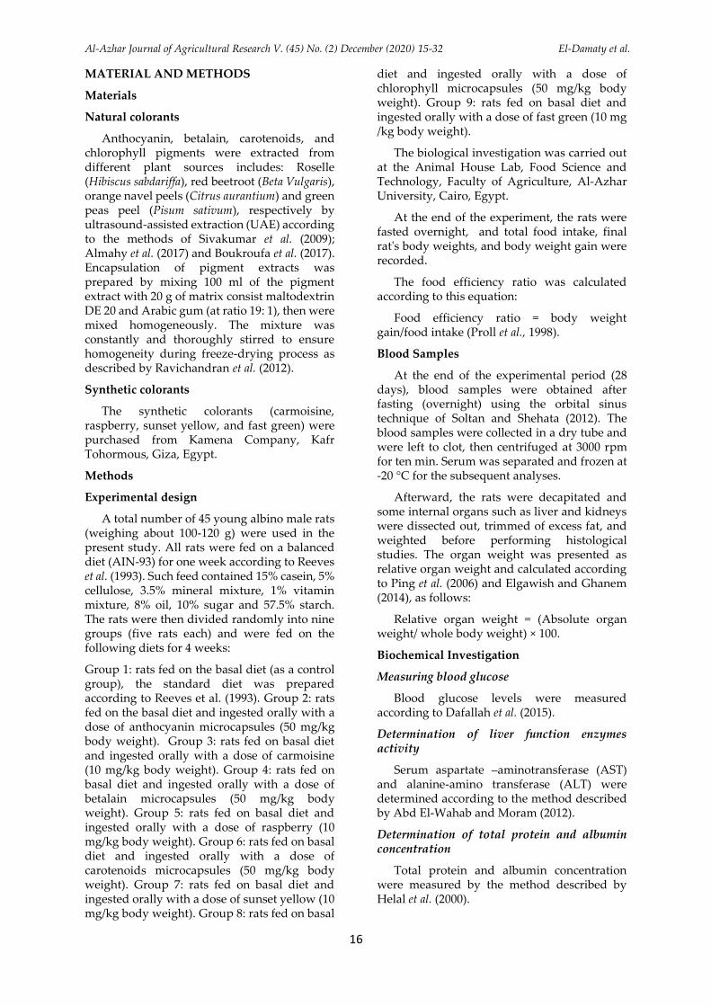
Data presented in Figure (1) indicated that the histological examination of the liver sections in the control group shows a normal histological photomicrograph. The liver of the control group showed average portal tract, portal veins and average hepatocytes in peri-portal area, central veins, hepatocytes arranged in single-cell cords with average intervening blood sinusoids. While, the liver in the rats treated with anthocyanin showed average portal tracts, portal veins and average
hepatocytes in the peri-portal area and mildly dilated central veins with average intervening blood sinusoids and average hepatocytes in the peri-venular area (Figure 2). Anthocyanin is a functional food factor and plays an important role in the prevention of liver diseases (Bendokas et al., 2020).
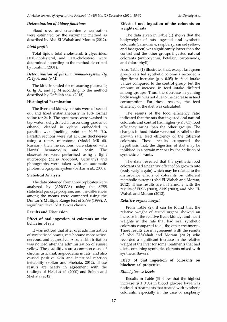
On the other hand, the liver sections of rats treated with an oral dose of carmoisine showed mildly edematous portal tracts with mildly dilated congested portal veins and average hepatocytes in the peri-portal area, and mildly dilated central veins with mildly congested blood sinusoids and average hepatocytes in the peri-venular area (Figure 3). Montaser and Alkafafy (2013) found that with increasing the doses of carmoisine, there were degenerative changes of hepatocytes.
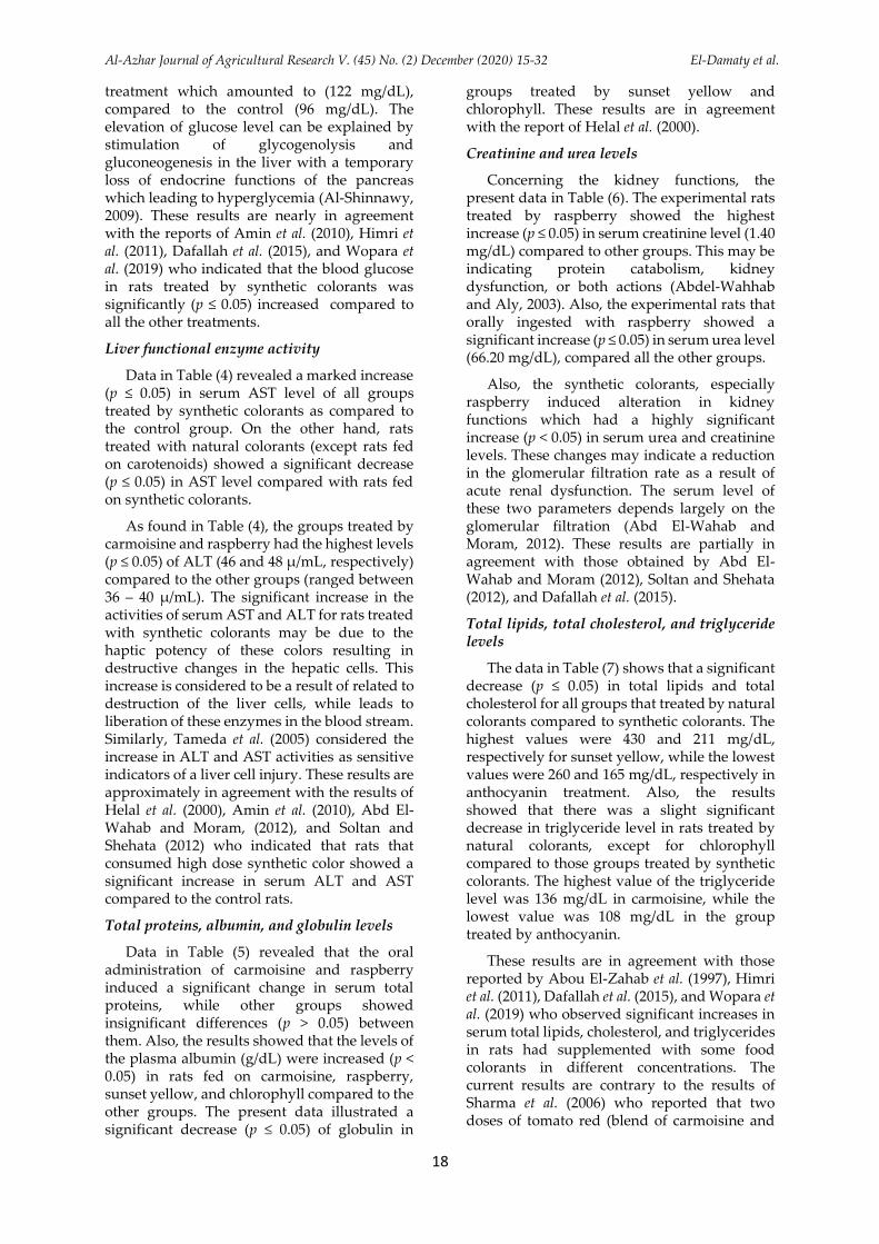
Also, the liver sections of rats treated with an oral dose of betalain showed mildly edematous portal tracts and average hepatocytes in peri-venular area, and mildly dilated central veins (Figure 4). Betalain was demonstrated as having a close relationship with hepatic tissue protection (da Silva et al., 2019).
The liver of rats treated by an oral dose of raspberry showed mildly edematous portal tracts with mildly dilated congested portal veins and average hepatocytes in peri-portal area, mildly dilated central veins with detached lining with moderate micro-vesicular steatosis of hepatocytes in peri-venular area, and markedly dilated congested blood sinusoids (Figure 5). These results are in agreement with those obtained by Sharma et al. (2008) and Soltan and Shehata (2012) who reported that the synthetic color has an adverse effect on the vital organs. At a low dose of synthetic color, the liver exhibited a disruption of hepatic cells near the central vein, and hepatocellular damage.
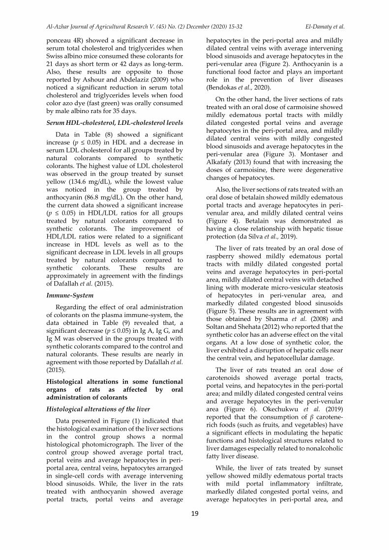
The liver of rats treated an oral dose of carotenoids showed average portal tracts, portal veins, and hepatocytes in the peri-portal area; and mildly dilated congested central veins and average hepatocytes in the peri-venular area (Figure 6). Okechukwu et al. (2019) reported that the consumption of β carotene-rich foods (such as fruits, and vegetables) have a significant effects in modulating the hepatic functions and histological structures related to liver damages especially related to nonalcoholic fatty liver disease.
While, the liver of rats treated by sunset yellow showed mildly edematous portal tracts with mild portal inflammatory infiltrate, markedly dilated congested portal veins, and average hepatocytes in peri-portal area, and

Al-Azhar Journal of Agricultural Research V. (45) No. (2) December (2020) 15-32 El-Damaty et al.
20
mildly dilated central veins with mild micro-vesicular steatosis of hepatocytes in the peri-venular area (Figure 7). Al-Dahhan et al. (2014) observed fatty degeneration of rat liver in the sunset yellow group, in a histological examination.
Also, the liver of rats treated by chlorophyll showed average portal tracts, portal veins, hepatocytes in the peri-portal area, and mildly dilated central veins with average hepatocytes in the peri-venular area (Figure 8). These results are nearly in agreement with the results of Suparmi et al. (2016) who noted thet the structure was insignificantly changed to normal values in a histological study in the liver of rats treated with chlorophyll from Sauropus androgynus L. Merr. leaf.
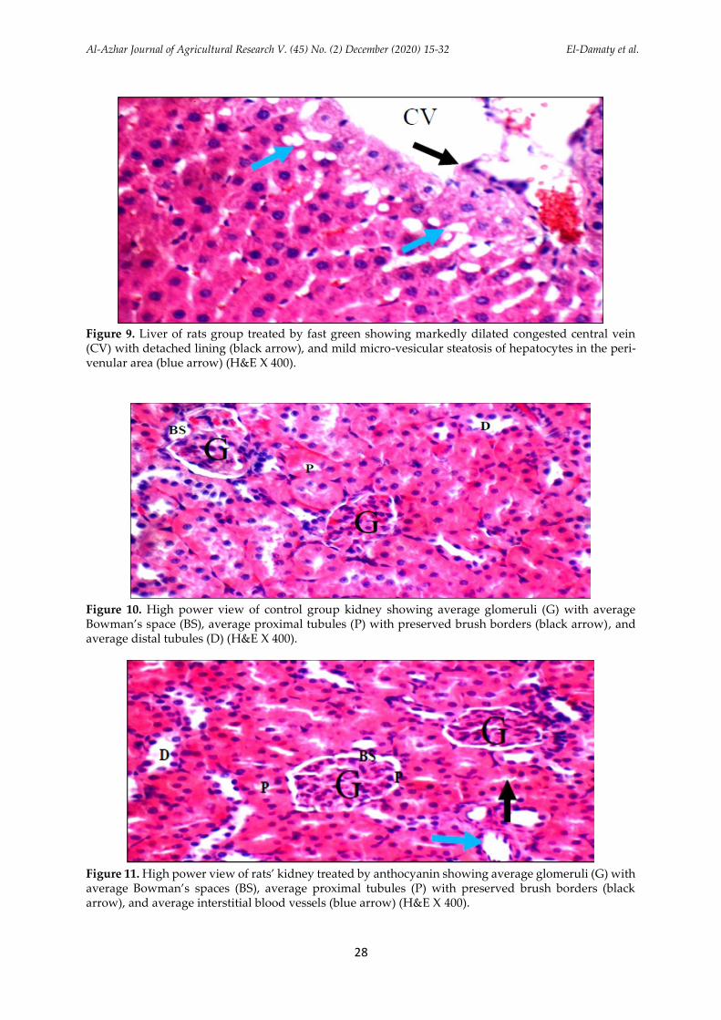
The liver of rats treated by the fast green group showed portal tracts with mildly dilated congested portal veins and average hepatocytes in the peri-portal area, and markedly dilated congested central veins with detached lining and mild micro-vesicular steatosis of hepatocytes in the peri-venular area (Figure 9). Mekkawy et al. (1998) found that synthetic colorants (ponceau, carmoisine, erythrosine, sunset yellow, tartrazine, fast green, indigotin, brilliant blue, and brilliant black) had a toxic effect which appeared as hepatocellular damage indicated by vacuolation, swelling, necrosis, and pyknosis in liver cells.
Histopathological alterations of the kidney
Data presented in Figure (10) showed the kidney section of the control group. The data indicated average renal capsule, glomeruli, Bowman’s spaces, proximal tubules with preserved brush borders, distal tubules, and renal medulla showed average collecting tubules with an average interstitium. Under the light microscope, the kidney sections from the control group did not show any damage, with the glomeruli and tubuli having a normal appearance.
The kidney of the rats treated by anthocyanin showed average renal capsule, average glomeruli with average Bowman’s spaces, average proximal tubules with preserved brush borders, and renal medulla showed average collecting tubules with average interstitium (Figure 11). Yarijani et al. (2019) found that anthocyanins reduces the renal toxicity which was induced by gentamicin and can improve kidney function and a decrease tissue injuries.
On the other hand, kidney sections of rats treated with an oral dose of carmoisine showed
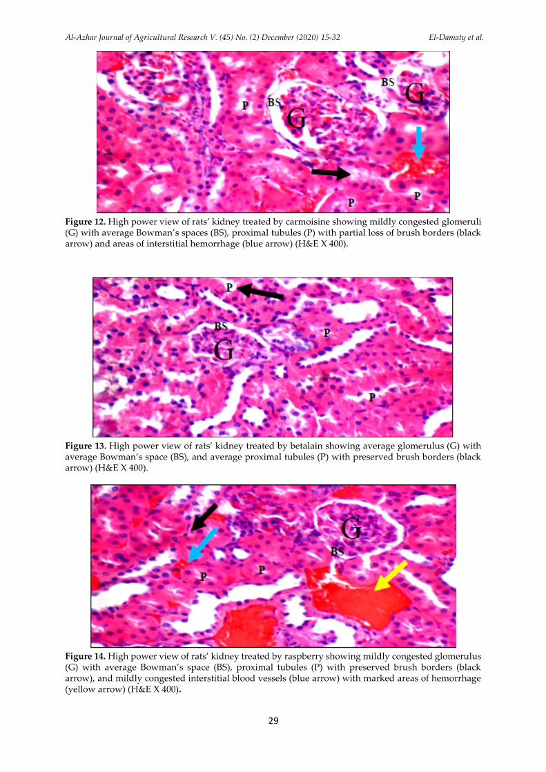
average renal capsule, mildly congested glomeruli with average Bowman’s spaces, proximal tubules with scattered apoptotic epithelial lining and partial loss of brush borders, mildly dilated interstitial blood vessels with areas of hemorrhage, marked peri-glomerular and peri-tubular inflammatory infiltrate, and the renal medulla showed collecting tubules with scattered apoptotic epithelial lining and mildly congested peri-tubular capillaries (Figure 12). Rus et al. (2010) observed phenomena represented by stasis and edema, congestion, hepatocyte and kidney apoptosis, with atrophy of renal corpuscles.
While the kidney of rats treated by betalain showed average renal capsule, average glomeruli with average Bowman’s spaces, average proximal tubules with preserved brush borders, and the renal medulla showed collecting tubules with average epithelial lining and mildly congested peri-tubular capillaries (Figure 13). These results are nearly in agreement with the results of Almeer et al. (2019) who found that the kidney tissue of the control rats and rats treated with red beetroot extract had normal kidney structure with normal renal tubules and glomeruli.
From Figure (14), the kidney of rats treated with raspberry showed average renal capsule, mildly congested glomeruli with average Bowman’s spaces, average proximal tubules with preserved brush borders, mildly congested interstitial blood vessels with marked areas of hemorrhage, and the renal medulla showed collecting tubules with average epithelial lining and mildly congested peri-tubular capillaries. Sharma et al. (2008) reported that the presence of synthetic color in food causes kidney injuries.
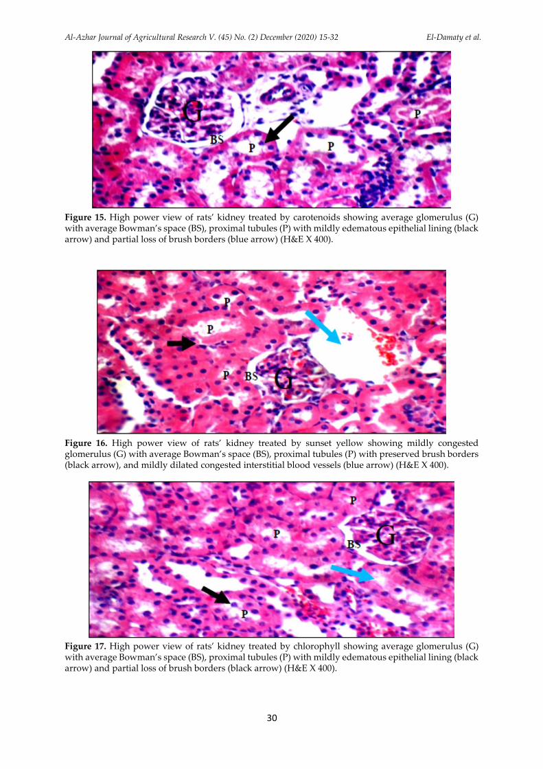
Also, the kidney section of rats treated with an oral dose of carotenoids showed average renal capsule, average glomeruli with average Bowman’s spaces, proximal tubules, mildly dilated interstitial blood vessels, and the renal medulla showed average collecting tubules with average interstitium (Figure 15). These results are in agreement with the results of Ezejindu et al. (2014), who found that the consumption of carotenoids at low or high doses is not a risk factor for kidney disorders.
The kidney sections of rats treated an oral dose of sunset yellow showed average renal capsule, mildly congested glomeruli with average Bowman’s spaces, average proximal tubules with preserved brush borders, mildly dilated congested interstitial blood vessels with areas of hemorrhage, and the renal medulla
Al-Azhar Journal of Agricultural Research V. (45) No. (2) December (2020) 15-32 El-Damaty et al.
21
showed collecting tubules with average epithelial lining and mildly congested peri-tubular capillaries (Figure 16). Al-Dahhan et al. (2014) observed nephritis in the kidney of rats treated by sunset yellow, in a histological study.
The kidney of the rats treated by chlorophyll group showed average renal capsule, average glomeruli with average Bowman’s spaces with mildly edematous epithelial lining, mildly dilated interstitial blood vessels, and the renal medulla showed average collecting tubules with average interstitium (Figure 17). These results are nearly in agreement with the results of Suparmi et al. (2016) who noted that rats treated with chlorophyll from Sauropus androgynus (L) Merr were insignificantly changed compared to the normal values.
Also, the kidney of the rats treated by fast green showed average renal capsule, average glomeruli with average Bowman’s spaces, average proximal tubules with preserved brush borders, markedly dilated congested interstitial blood vessels, and the renal medulla showed average collecting tubules with mildly congested peri-tubular capillaries (Figure 18). Ghonimi and Elbaz (2015) found that the examined kidney sections of rats treated with synthetic colorants showed degenerative changes in the renal tubules and glomeruli. The same authors observed an enlargement of renal glomeruli, with separation and vacuolation of its lining epithelial cells as well as congestion of its blood capillaries.
CONCLUSION
It could be concluded that the synthetic colorants adversely affected hepatic and renal parameters compared to natural colorants. Synthetic colorants caused increases in the levels of blood glucose, AST, ALT, creatinine, urea, total lipids, total cholesterol, and LDL. While, such synthetic colorants decreased each of body weight gain, globulin, HDL, Ig A, Ig M, and Ig A. The histological examinations revealed alterations in kidneys that included congestion and hemorrhage with infiltration and deformation of the structure of glomeruli. Also, alterations in the liver included congestion, hemorrhage, and dilatation of sinusoids and central vein with microvesicular steatosis occurred. Therefore, it is necessary to be aware of the hazardous effects of consuming such synthetic food colorants. More attention should be paid for using natural colorants. If these results have applied to humans, it is advised that the application of synthetic food colorants consumption should be drastically
minimized as these colorants possess detrimental physiological effects.
REFERENCES
Abd El-Wahab, H.M.F., Moram, G.S.E.D., 2012. Toxic effects of
some synthetic food colorants and/or flavor additives on
male rats. Toxicol. Ind. Health, 29 (2), 224-232.
Abdel-Wahhab, M.A., Aly, S.E., 2003. Antioxidants and radical
scavenging properties of vegetable extracts in rats fed
aflatoxin-contaminated diet. J. Agric. Food Chem., 51 (8),
2409-2414.
Aberoumand, A., 2011. A review article on edible pigments
properties and sources as natural colorants in foodstuff and
food industry. World J. Dairy Food Sci., 6 (1), 71-78.
Abou El-Zahab, H.S.H., El-Khyat, Z.A., Awadallah, R., Mahdy,
K.A., 1997. Physiological effects of some synthetic food
coloring additives on rats. Boll. Chim. Farm, 136 (10), 615-
627.
AL-Dahhan, M.A.H., AL-Samawy, E.R.M., AL-Kaisei, B.I.,
Jarad, A.S., 2014. Effect of synthetic colorants (Sunset yellow
and Ponceau 4R) in some biochemical and histopathological
parameters of albino rats. AL-Qadisiya J. Vet. Med. Sci., 13
(1), 80 -84.
Almahy, H.A., Abdel-Razik, H.H., El-Badry, Y.A., Ibrahim,
A.M., 2017. Ultrasound-assisted extraction of anthocyanin
pigments from Hibiscus sabdariffa (Rosella) and its
phytochemical activity at Kingdom of Saudi Arabia. Int. J.
Chem. Sci., 15 (4), 196.
Almeer, R., Alarifi, S., Alkhtani, S., Farhood, M., Al-Otibi,
F.O., Alkubaisi, N., Rizwana, H. 2019. Nephroprotective
role of Beta vulgaris L. root extract against chlorpyrifos-
induced renal injury in rats. Evid. Based Complement.
Alternat. Med., 2019, 3595761.
Al-Shinnawy, M.S., 2009. Physiological effect of a food additive
on some haematological and biochemical parameters of
male albino rats. Egypt. Acad. J. Biol. Sci., 2 (1), 143-151.
Amin, K.A., Hameid, H.A., Abd Elsttar, A.H., 2010. Effect of
food azo dyes tartrazine and carmoisine on biochemical
parameters related to renal, hepatic function and oxidative
stress biomarkers in young male rats. Food Chem. Toxicol.,
48 (10), 2994-2999.
Ashour, A.A., Abdelaziz, I., 2009. Role of fast green on the blood
of rats and the therapeutic action of vitamins C or E. Int. J.
Integr. Biol., 6 (1), 6-11.
Bendokas, V., Stanys, V., Mazeikiene, I., Trumbeckaite, S.,
Baniene, R., Liobikas, J., 2020. Anthocyanins: from the field
to the antioxidants in the body. Antioxidants, 9, 819.
Boukroufa, M., Boutekedjiret, C., Chemat, F., 2017.
Development of a green procedure of citrus fruits waste
processing to recover carotenoids. Resour. Efficient
Technol., 3 (3), 252-262.
Carocho, M., Barreiro, M.F., Morales, P., Ferreira, I.C.F.R., 2014.
A review on synthetic and natural food additives. Compr.
Rev. food Sci. food Saf., 13, 377-399.
Cortez, R., Luna-Vital, D.A., Margulis, D., de Mejia, E.G., 2017.
Natural pigments: Stabilization methods of anthocyanins
for food applications. Compr. Rev. Food Sci. Food Saf., 16,
180-198.
Crino, M.A., Heenan, C.N., Nguyen, M.H., Stathopoulos, C.E.,
2013. The stability of natural red/pink food colors in
ultrahigh-temperature (UHT) products. J. Sci. Food Agric.,
93, 2022-2027.
Dafallah, A.A., Abdellah, A.M., Abdel-Rahim, E.A., Ahmed,
S.H., 2015. Physiological effects of some artificial and
Al-Azhar Journal of Agricultural Research V. (45) No. (2) December (2020) 15-32 El-Damaty et al.
22
natural food coloring on young male albino rats. J. Food
Technol. Res., 2 (2), 21-32.
da Silva, D.V.T., Pereira, A.D., Boaventura, G.T., Ribeiro, R.S.A.,
Vericimo, M.A., de Carvalho-Pinto, C.E., Baiao, D.D., Del
Aguila, E.M., Paschoalin, V.M.F., 2019. Short-term betanin
intake reduces oxidative stress in Wistar rats. Nutrients, 11,
1978.
European Food Safety Authority (EFSA) panel on Food
Additives and Nutrient Sources added to Food (ANS). 2009.
Scientific opinion on the reevaluation tartrazine (E102) on
request from the European Commission. European Food
Safety Authority, 7 (11), 1331–1383.
Elgawish, R.A.R., Ghanem, M.E., 2014. Effect of long term
cadmium chloride exposure on testicular functions in male
albino rats. Am. J. Anim. Vet. Sci., 9 (4), 182-188.
Ezejindu D.N., Tochukwu, N., Akingboye A.J., 2014.
Hepatoprotective effects of carotenoids on the kidneys of
adult Wistar rats. Int. J. Sci. Res. Public., 4 (2), 1-7.
Ghonimi, W.A.M., Elbaz, A. 2015. Histological changes of
selected Westar rat tissues following the ingestion of
tartrazine with special emphasis on the protective effect of
royal jelly and cod liver oil. J. Cytol. Histol., 6, 346.
Helal, E.G.E., Zaahkouk, S.A.M., Mekkawy, H.A., 2000. Effect
of some food colorants (synthetic and natural products) of
young Albino rats (I- liver and kidney functions). Egypt. J.
Hospital Med., 1, 103-113.
Himri, I., Bellahcen, S., Souna, F., Belmekki, F., Aziz, M.,
Bnouham, M., Berkia, Z., Mekhfi, H., Saaluri, E., 2011. A 90-
day oral toxicity study of tartrazine, a synthetic food dye, in
Wister rats. Int. J. Pharm. Pharmaceut. Sci., 3 (2), 159-169.
Ibrahim, A.A., 2001. Effects of single doses of Bitis arietans crude
venom on serum biochemical parameters in rats. Sci. J. King
Faisal Univ., 2, 103-111.
Janiszewska-Turak, E., Pisarska, A., Krolczyk, J.B., 2016.
Natural food pigments application in food products. Nauka
Przyr. Technol., 10 (4), 51.
Lakshmi, C.G., 2014. Food coloring: The natural way. Res. J.
Chem. Sci., 4 (2), 87-96.
Larsen, J.C., 2008. Legal and illegal colors. Trends Food Sci.
Technol., 19 (1), 64-69.
Mekkawy, H.A., Ali, M.O., El-Zawahry, A.M., 1998. Toxic effect
of synthetic and natural food dyes on renal and hepatic
functions in rats. Toxicol. Lett. 95 (Suppl. 1), 155.
Montaser, M.M., Alkafafy, M.E., 2013. Effects of synthetic food
color (carmoisine) on expression of some fuel metabolism
genes in liver of male Albino rats. Life Sci. J., 10 (2), 2191-
2198.
Okechukwu, G.N., Nweke, O.B., Nwafor, A.J., Godson, A.G.,
Kenneth, E.U., Ibegbu, A.O., 2019. Beta (β)-carotene-
induced effects on the hepato-biochemical parameters in
Wistar rats fed dietary fats. Jordan J. Biol. Sci., 12 (3), 283-
288.
Ping, X.U., Xing-Guo, Z., You-Ming, L.I., Chao-Hui, Y.U., Lei,
X.U., Gen-Yun, X.U., 2006. Research on the protection effect
of pioglitazone for non-alcoholic fatty liver disease
(NAFLD) in rats. J. Zhejiang Univ. Sci. B, (8), 627-633.
Proll, J., Petzke, J., Ezeagu, E., Metges, C., 1998. Low nutrition
quality of unconventional tropical crop seeds in rats. J.
Nutr., 128, 2014-2022.
Ravichandran, K., Ahmed, A.R., Knorr, D., Smetanska, I., 2012.
The effect of different processing methods on phenolic acid
content and antioxidant activity of red beet. Food Res. Int.,
48, 16-20.
Reeves, P.G., Nielsen, F.H., Fahey, V., 1993. AIN-93 purified
diets for laboratory rodents: final report of the American
Institute of Nutrition ad hoc writing committee on the
reformulation of the AIN-76A rodent diet. J. Nutr., 123 (11),
1939-1951.
Rus, V., Gherman, C.M., Miclauş, V., Mihalca, A. D., Nadaş,
G.C., 2010. Comparative toxicity of food dyes on liver and
kidney in guinea pigs: A histopathological study. Ann.
Rom. Soc. Cell Biol., 15 (1), 161-165.
Salah, S.H., 1994. Biochemical Studies on Some Synthetic Food
Colorants. M.Sc.. Fac. Agric. Cairo Univ., Cairo, Egypt.
Sarkar, B., Chatterjee, A., Adhikari, S., Ayyappan, S., 2005.
Carbofuran and cypermethrin induced histopathological
alterations in the liver of Labeo rohita and its recovery. J.
Appl. Ichthyol., 21, 131-135.
Sharma, S., Goyal, R.P., Chakravarty, G., Sharma, A., 2006.
Tomato red toxicity: Haematological and serological
changes in the blood of swiss albino mice, Mus musculus.
Ind. J. Environ. Sci, 10 (2), 145-148.
Sharma, S., Goyal, R.P., Chakravarty, G., Sharma, A., 2008.
Toxicity of tomato red, a popular food dye blend on male
Albino mice. Exper. Toxicol. Pathol., 60 (1), 51-57.
Sivakumar, V., Anna, J.L., Vijayeeswarri, J., Swaminathan, G.,
2009. Ultrasound assisted enhancement in natural dye
extraction from beetroot for industrial applications and
natural dyeing of leather. Ultrason. Sonochem., 16, 782-789.
Soltan, S.S.A., Shehata, M.M.E.M., 2012. The effects of using
color foods of children on immunity properties and liver,
kidney on rats. Food Nutr. Sci., 3, 897-904.
SPSS, 1998. Statistical Package for the Social Sciences, for
windows. Release, 9.0.0, Standard Version SPSS. Inc.
Suparmi, S., Fasitasari, M., Martosupono, M., Mangimbulude,
J.C., 2016. Comparisons of curative effects of chlorophyll
from Sauropus androgynus (L) Merr leaf extract and cu-
chlorophyllin on sodium nitrate-induced oxidative stress in
rats. J. Toxicol., 2016, 8515089.
Tameda, M., Shiraki, K., Ooi, K., Takase, K., Kosaka, Y., Nobori,
T., Tameda, Y., 2005. Aspartate aminotransferase-
immunoglobulin complexes in patients with chronic liver
disease. World J. Gastroenterol., 11 (10), 1529-1531.
Wopara, I., Mounmbenga, P.E., Amanda, I., Modo, E.U., 2019.
Hepatotoxic Effect of tartrazine and erythrosine on male
wistar rats. Eur. J. Pharmaceut. Med. Res., 6 (7), 107-110.
Yarijani, Z.M., Najafi, H., Shackebaei, D., Madani, S.H.,
Modarresi, M., Jassemi, S.V., 2019. Amelioration of renal
and hepatic function, oxidative stress, inflammation and
histopathologic damages by malva sylvestris extract in
gentamicin induced renal toxicity. Biomed. Pharmacother.,
112, 108635.
Al-Azhar Journal of Agricultural Research V. (45) No. (2) December (2020) 15-32 El-Damaty et al.
23
Table 1. Initial and final body weight, body weight gain (g), total food intake (g/month), and food
efficiency ratio of rats fed on tested diets for 4 weeks.
Groups Initial body
weight
Final body
weight
Bodyweight
gain
Total food
intake
Food efficiency
ratio
Control 113ab 195bc 82.0d 1861c 0.044b
Anthocyanin 115a 200.7b 85.7bc 1892b 0.045ab
Carmoisine 110b 181.6d 71.6g 1879b 0.038d
Betalain 112ab 199.2b 87.2b 1925a 0.045ab
Raspberry 114ab 184d 70.0g 1880b 0.037d
Carotenoids 113ab 204.4a 91.4a 1894b 0.048a
Sunset yellow 116a 191.3c 75.3f 1879b 0.040c
Chlorophyll 115a 198.5b 83.5cd 1885b 0.044ab
Fast green 110b 188.6c 78.6e 1854c 0.042bc
Means within the column having the same superscript are not significantly different (p ≤ 0.05).
Table 2. Relative organs weight (%) of rats fed on tested diets for 4 weeks.
Groups Relative liver weight
Relative kidney weight
Relative heart weight
Relative spleen weight
Control 3.40c 0.70b 0.31c 0.29bc
Anthocyanin 3.68b 0.82ab 0.38bc 0.27c
Carmoisine 3.84ab 0.89a 0.44a 0.26c
Betalain 3.38c 0.77ab 0.29c 0.31bc
Raspberry 3.92a 0.85ab 0.37bc 0.29bc
Carotenoids 3.54bc 0.80ab 0.33bc 0.34ab
Sunset yellow 3.90a 0.85ab 0.41ab 0.37a
Chlorophyll 3.21d 0.77ab 0.30c 0.26c
Fast green 3.47c 0.78ab 0.34bc 0.31bc
Means within the column having the same superscript are not significantly different (p ≤ 0.05).
Table 3. Blood glucose levels (mg/dL) of rats fed on tested diets for 4 weeks.
Groups Initial Glucose Final Glucose
Control 92b 96cd
Anthocyanin 93b 94d
Carmoisine 98a 116b
Betalain 95ab 101c
Raspberry 95ab 122a
Carotenoids 89c 93d
Sunset yellow 97a 111b
Chlorophyll 90bc 93d
Fast green 92b 99c
Means within the column having the same superscript are not significantly different (p ≤ 0.05).
Table 4. Plasma aspartate–aminotransferase (AST) and alanine-amino transferase (ALT) enzymes activities (µ/ mL) of rats fed on tested diets for 4 weeks.
Groups AST ALT
Control 32c 38b
Anthocyanin 32c 40b
Carmoisine 36b 46a
Betalain 32c 40b
Raspberry 40a 48a
Carotenoids 36b 36b
Sunset yellow 36b 40b
Chlorophyll 32c 40b
Fast green 36b 38b
Means within the column having the same superscript are not significantly different (p ≤ 0.05).
Table 5. Plasma total proteins, albumin, and globulin levels (g/dL) of rats fed on tested diets for 4 weeks.

Al-Azhar Journal of Agricultural Research V. (45) No. (2) December (2020) 15-32 El-Damaty et al.
24
Groups Total proteins Albumin Globulin
Control 6.6bc 3.6c 3.0b
Anthocyanin 7.0b 3.2d 3.8a
Carmoisine 7.8a 4.1b 3.7a
Betalain 7.0b 3.7c 3.3b
Raspberry 7.5a 4.0b 3.5ab
Carotenoids 6.7b 3.6c 3.1b
Sunset yellow 7.2ab 4.8a 2.4c
Chlorophyll 6.4c 4.0b 2.4c
Fast green 7.0b 3.5c 3.5ab
Means within the column having the same superscript are not significantly different (p ≤ 0.05).
Table 6. Plasma creatinine, urea levels (mg/dL) of rats fed on tested diets for 4 weeks:
Groups Creatinine Urea
Control 0.80c 52.60e
Anthocyanin 0.80c 57.40cd
Carmoisine 1.20ab 63.40ab
Betalain 1.00bc 56.20cd
Raspberry 1.40a 66.20a
Carotenoids 1.00bc 51.60e
Sunset yellow 0.80c 61.20bc
Chlorophyll 0.80c 57.80cd
Fast green 1.00bc 59.80bcd
Means within the column having the same superscript are not significantly different (p ≤ 0.05).
Table 7. Plasma total lipids, total cholesterol, and triglyceride (mg/dL) of rats fed on tested diets for 4 weeks.
Groups Total lipids Total
cholesterol Triglyceride
Control 310e 181cd 120cd
Anthocyanin 260f 165f 108e
Carmoisine 400b 195b 136a
Betalain 320d 174e 118d
Raspberry 350c 186c 130b
Carotenoids 320d 169ef 122c
Sunset yellow 430a 211a 132b
Chlorophyll 342c 176de 129b
Fast green 422a 201b 130b
Means within the column having the same superscript are not significantly different (p ≤ 0.05).
Table 8. Plasma HDL-cholesterol, LDL-cholesterol levels (mg/dL), and HDL/LDL ratios of rats fed on tested diets for 4 weeks.
Groups HDL-Cholesterol LDL-Cholesterol HDL/LDL ratio
Control 50bc 91.6ef 0.545bc
Anthocyanin 56a 86.8f 0.645a
Carmoisine 45ef 117b 0.384de
Betalain 52b 90.4ef 0.575ab
Raspberry 47de 111.2c 0.422d
Carotenoids 52b 93e 0.559ab
Sunset yellow 43f 134.6a 0.319e
Chlorophyll 48cd 103d 0.466cd
Fast green 46de 115.4bc 0.398de
Means within the column having the same superscript are not significantly different (p ≤ 0.05).
Al-Azhar Journal of Agricultural Research V. (45) No. (2) December (2020) 15-32 El-Damaty et al.
25
Table 9. Effect of natural and synthetic colorants on immune-system in plasma of male albino rats for 4 weeks.
Groups Ig A IgG Ig M
Control 302ab 2270b 188b
Anthocyanin 311a 2480a 182c
Carmoisine 214f 1648h 113f
Betalain 308a 2200c 178c
Raspberry 268d 1840g 136e
Carotenoids 300ab 2220c 208a
Sunset yellow 256e 1920f 138e
Chlorophyll 284c 2160d 206a
Fast green 264d 1960e 150d
Figure 1. Liver of control group showing average central vein (CV) and average hepatocytes arranged in single-cell cords (black arrow) with average intervening blood sinusoids (blue arrow) (H&E X 400).
Means within the column having the same superscript are not significantly different (p ≤ 0.05).
Figure 2. Liver of rats group treated by anthocyanin showing mildly dilated central vein (CV), average intervening
blood sinusoids (black arrow), and average hepatocytes in the peri-venular area (blue arrow) (H&E X 400).

Al-Azhar Journal of Agricultural Research V. (45) No. (2) December (2020) 15-32 El-Damaty et al.
26
Figure 3. Liver of rats group treated by carmoisine showing mildly dilated central vein (CV) with mildly dilated
blood sinusoids (black arrow), and average hepatocytes in the peri-venular area (blue arrow) (H&E X 400).
Figure 4. Liver of rats group treated by betalain showing mildly dilated central vein (CV), average
hepatocytes in the peri-venular area (black arrow) (H&E X 400).
Figure 5. Liver of rats group treated by raspberry showing mildly dilated central vein (CV) with
detached lining (black arrow), mildly congested blood sinusoids (blue arrow), and moderate micro-
vesicular steatosis of hepatocytes in the peri-venular area (yellow arrow) (H&E X 400).

Al-Azhar Journal of Agricultural Research V. (45) No. (2) December (2020) 15-32 El-Damaty et al.
27
Figure 6. Liver of rats group treated by carotenoids showing mildly dilated congested central vein (CV), mildly congested blood sinusoids (blue arrow), and average hepatocytes in the peri-venular area (black arrow) (H&E X 400).
Figure 7. Liver of rats group treated by sunset yellow showing mildly dilated central vein (CV) and mild micro-vesicular steatosis of hepatocytes in the peri-venular area (black arrow) (H&E X 400).
Figure 8. Liver of rats group treated by chlorophyll showing mildly dilated central vein (CV) with average hepatocytes in the peri-venular area (blue arrow) (H&E X 400).
Al-Azhar Journal of Agricultural Research V. (45) No. (2) December (2020) 15-32 El-Damaty et al.
28
Figure 9. Liver of rats group treated by fast green showing markedly dilated congested central vein (CV) with detached lining (black arrow), and mild micro-vesicular steatosis of hepatocytes in the peri-venular area (blue arrow) (H&E X 400).
Figure 10. High power view of control group kidney showing average glomeruli (G) with average Bowman’s space (BS), average proximal tubules (P) with preserved brush borders (black arrow), and average distal tubules (D) (H&E X 400).
Figure 11. High power view of rats’ kidney treated by anthocyanin showing average glomeruli (G) with average Bowman’s spaces (BS), average proximal tubules (P) with preserved brush borders (black arrow), and average interstitial blood vessels (blue arrow) (H&E X 400).
Al-Azhar Journal of Agricultural Research V. (45) No. (2) December (2020) 15-32 El-Damaty et al.
29
Figure 12. High power view of rats’ kidney treated by carmoisine showing mildly congested glomeruli (G) with average Bowman’s spaces (BS), proximal tubules (P) with partial loss of brush borders (black arrow) and areas of interstitial hemorrhage (blue arrow) (H&E X 400).
Figure 13. High power view of rats’ kidney treated by betalain showing average glomerulus (G) with average Bowman’s space (BS), and average proximal tubules (P) with preserved brush borders (black arrow) (H&E X 400).
Figure 14. High power view of rats’ kidney treated by raspberry showing mildly congested glomerulus (G) with average Bowman’s space (BS), proximal tubules (P) with preserved brush borders (black arrow), and mildly congested interstitial blood vessels (blue arrow) with marked areas of hemorrhage (yellow arrow) (H&E X 400).
Al-Azhar Journal of Agricultural Research V. (45) No. (2) December (2020) 15-32 El-Damaty et al.
30
Figure 15. High power view of rats’ kidney treated by carotenoids showing average glomerulus (G) with average Bowman’s space (BS), proximal tubules (P) with mildly edematous epithelial lining (black arrow) and partial loss of brush borders (blue arrow) (H&E X 400).
Figure 16. High power view of rats’ kidney treated by sunset yellow showing mildly congested glomerulus (G) with average Bowman’s space (BS), proximal tubules (P) with preserved brush borders (black arrow), and mildly dilated congested interstitial blood vessels (blue arrow) (H&E X 400).
Figure 17. High power view of rats’ kidney treated by chlorophyll showing average glomerulus (G) with average Bowman’s space (BS), proximal tubules (P) with mildly edematous epithelial lining (black arrow) and partial loss of brush borders (black arrow) (H&E X 400).

Al-Azhar Journal of Agricultural Research V. (45) No. (2) December (2020) 15-32 El-Damaty et al.
31
Figure 18. High power view of rats’ kidney treated by fast green showing average glomerulus (G) with average Bowman’s space (BS), average proximal tubules (P) with preserved brush borders (black arrow) and markedly dilated congested interstitial blood vessels (blue arrow) (H&E X 400).
Al-Azhar Journal of Agricultural Research V. (45) No. (2) December (2020) 15-32 El-Damaty et al.
32
والطبيعية الصناعية الغذائية الملونات التقييم البيولوجى لبعض
,* 1 ، مختار مبروك أ بو سلامة 2 ، فؤاد عمر فؤاد أ بوزيد 1 ، ماهر الس يد عبدالغن 1 ي الس يد محمد الدماط
مص ،القاهرة ،جامعة ال زهر ،كلية الزراعة ،قسم علوم وتكنولوجيا ال غذية 1
مص ،القاهرة ،مركز بحوث الصحراء ،قسم الإنتاج النباتي 2
[email protected] :للباحث الرئيسى البريد الاليكتروني*
العرب الملخص
ومقارنتها ( السريع وال خضر الشمس أ صفرغروب الرازبرى، الكارموزين،) الغذائية الصناعية الملونات لبعض المحتمل الضار التأ ثي ومقارنة لتقييم الدراسة هذه أ جريت
كذلك و لفئران التجارب البيوكيميائية العوامل على( والكلوروفيل والكاروتينويدات والبيتالي ال نثوس ياني )ببعض الملونات الغذائية الطبيعية المس تخلصة من مصادر نباتية
للكبد النس يجية الفئران الكلى و الفحوصات زيادة. لتلك وجود النتائج أ وضحت ترانسفييز، أ مينو ال س بارتات ، ترانسفييز أ مينو أ لني مس تويات في معنوية وقد
مع بالمقارنة المختبرة الصناعية الفئران التى تم تغذيتها على الملونات مجموعات في المناعي الجلوبيولي مس تويات انخفاض مع وال لبيومي، الكلي البروتي الكرياتيني، اليوريا،
احتقان : تشمل الكلى في حدوث تغيات عن النس يجية الفحوصات كما أ ظهرت. التحكم )الكنترول( ومجموعة الطبيعية الغذاء مجموعات الفئران التى تم تغذيتها على ملونات
في التغيات كانت بينما. الكبيبات الكلوية ةبني في وتشوه ارتشاح مع ونزيف حدثت والوريد الجيوب أ ش باه وتورم والنزيف الاحتقان: الكبد التى مع المركزي الكبدية
دهنية لجزيئات تراكم في حدوث الحجم الملونات مجموعات دقيقة على تغذيتها تم التى الملوناتمجموعات مع بالمقارنة المختبرة الصناعية الفئران على تغذيتها تم التى الفئران
الملونات اس تخدام من بالحد ينُصح لذلك، . التحكم )الكنترول( ومجموعة الطبيعية الغذائية الطبيعية استبدالها سابقة الذكر أ و الصناعية تلك فى أ غذية خاصة بالملونات
. ال طفال
للكلية. النس يجى فحصال للكبد؛ النس يجى فحصال البيوكيميائية؛ المعايي الصناعية؛ الغذائية الملونات الطبيعية؛ الغذائية الملونات: الإسترشادية الكلمات