Embed Size (px)
Citation preview

NISTIR 7996
Biological and Environmental Monitoring and Archival of Sea Turtle
Tissues (BEMAST): Rationale, Protocols, and Initial Collections of
Banked Sea Turtle Tissues
Jennifer M. Keller Rebecca S. Pugh
Paul R. Becker
http://dx.doi.org/10.6028/NIST.IR.7996

NISTIR 7996
Biological and Environmental Monitoring and Archival of Sea Turtle
Tissues (BEMAST): Rationale, Protocols, and Initial Collections of
Banked Sea Turtle Tissues
Jennifer M. Keller Rebecca S. Pugh
Paul R. Becker Chemical Sciences Division
Material Measurement Laboratory
http://dx.doi.org/10.6028/NIST.IR.7996
April 2014
U.S. Department of Commerce
Penny Pritzker, Secretary
National Institute of Standards and Technology Patrick D. Gallagher, Under Secretary of Commerce for Standards and Technology and Director

ii
ABSTRACT
The National Institute of Standards and Technology (NIST) expanded its analytical measurement and specimen banking capabilities into the U.S. Pacific Islands region in 2010. As part of this Program, NIST established the Biological and Environmental Monitoring and Archival of Sea Turtle Tissues (BEMAST) project in 2011 to archive tissues from sea turtles primarily for health and contaminant studies. This report describes the scientific rationale behind BEMAST and detailed protocols for sample collection and processing during sea turtle live captures, necropsies, and nest excavations as well as protocols for sampling ingested plastics, homogenizing scutes, and cataloging tissues. Additionally, this report summarizes the collection of samples archived by BEMAST and already analyzed for certain laboratory studies. Today the collection includes 854 tissue samples from 288 individual sea turtles and 38 sea turtle nests. All five species inhabiting the U.S. Pacific Island region are included, and locations encompass the Hawaiian Islands, Palmyra Atoll, Saipan, Tinian, San Diego Bay, and pelagic waters of the tropical Pacific Ocean. Tissues include blood, scute, muscle, liver, fat, fibropapilloma lesions, bile, follicles, ingested plastics, eggs, and mouth algae and are stored at -150° C at the NIST Marine Environmental Specimen Bank at the Hollings Marine Laboratory in Charleston, South Carolina. Analysis of subsamples has begun primarily for green sea turtles, including contaminant concentration measurements, as well as metabolomics, lipidomics, and proteomics. Collections and analyses continue with expansion into additional Pacific Islands and focus on more endangered species, such as leatherback and hawksbill sea turtles.
KEYWORDS
Specimen banking, environmental contaminants, analytical chemistry, sea turtle, marine turtle, marine organism health, reptile, pollution, Pacific Islands

iii
TABLE OF CONTENTS
LIST OF TABLES ........................................................................................................................ iv
LIST OF FIGURES ...................................................................................................................... iv
1. INTRODUCTION ................................................................................................................. 1
1.1. ENVIRONMENTAL SPECIMEN BANKING .................................................................. 1
1.2. RATIONALE FOR BANKING SEA TURTLE TISSUES ..................................................... 2
1.3. PROJECT GOAL AND OBJECTIVES ............................................................................. 5
1.4. BANKING SEA TURTLE TISSUES ..................................................................................... 5
2. MANAGEMENT SYSTEM ................................................................................................... 8
3. METHODS ........................................................................................................................... 10
3.1. LIVE CAPTURE COLLECTION FIELD PROTOCOL .................................................... 12
3.2. NECROPSY FIELD PROTOCOL ......................................................................................... 23
3.3. INGESTED PLASTIC FIELD PROTOCOL ........................................................................ 43
3.4. EGG COLLECTION PROTOCOL ....................................................................................... 48
3.5. SCUTE HOMOGENIZATION PROTOCOL........................................................................ 57
3.6. TISSUE CATALOGING PROTOCOL ................................................................................. 61
4. RESULTS ............................................................................................................................. 62
4.1. COLLECTIONS REPORT .................................................................................................... 62
4.2. HOMOGENIZATION REPORT........................................................................................... 64
4.3. ANALYSIS REPORT ........................................................................................................... 64
5. ACKNOWLEDGMENTS ..................................................................................................... 66
6. DISCLAIMER ..................................................................................................................... 67
7. REFERENCES .................................................................................................................... 67
APPENDIX A ............................................................................................................................... 70

iv
LIST OF TABLES Table 1. Locations and field sampling partners for three access categories of five sea turtle species in the Pacific Ocean. ............................................................................................................................................... 7 Table 2. Current BEMAST collaborating field project codes. ................................................................... 10 Table 3. Current BEMAST tissue and blood aliquot codes. ....................................................................... 11 Table 4. Aliquoting procedure for 10 mL of blood. ................................................................................... 16 Table 5. Aliquoting procedure for 20 mL of blood. ................................................................................... 16 Table 6. Summary of routine necropsy steps. ............................................................................................. 25 Table 7. Scute method choice to use during necropsy of each sea turtle species. ...................................... 27 Table 8. Target masses for aliquots from scute samples of different approximate masses. ....................... 58 Table 9. Summary of sea turtle samples collected and archived as a part of the BEMAST project. .......... 62
LIST OF FIGURES Figure 1. U.S. States and Territories in the Pacific Islands Region. ............................................................ 2 Figure 2. Leatherback sea turtle with longline hook in right front flipper; obvious example of fisheries by-catch. ....................................................................................................................................................... 2 Figure 3. Green sea turtle with fibropapilloma (FP) tumors from Kapoho, Hawaii. ................................ 3 Figure 4. Proper restraint and incline of a green sea turtle for blood sampling. ........................................ 15 Figure 5. Making a blood smear. ................................................................................................................ 16 Figure 6. Reading a hematocrit tube ........................................................................................................... 17 Figure 7. 5 mL Teflon jar with graduations for estimating volumes. ......................................................... 17 Figure 8. Shaving motion and ribbons of scute during scute scraping of 5th central scute. ....................... 20 Figure 9. Collection of a scute sample using a biopsy punch tool by the Rusty Day method. ................... 28 Figure 10. Collection of a scute sample using a knife blade by the George Balazs method. ...................... 29 Figure 11. Location of leatherback blubber sampling on right carapace. ................................................... 32 Figure 12. Example of a photograph of ingested plastics found from within one turtle’s gastrointestinal tract. ............................................................................................................................................................ 45 Figure 13. Bag of unhatched eggs from one nest covered in sand and white puffy eggs chosen and rinsed in beaker of water. ....................................................................................................................................... 50 Figure 14. Examples of egg contents from loggerhead sea turtle eggs with no development (No), early (E), middle (M), and late (L)-stage embryos. Note the eye spot among the E egg contents ...................... 51 Figure 15. Egg contents being homogenized in the BagMixer. .................................................................. 51 Figure 16. Grinding sea turtle scute samples with mortar and pestle and transferring into cryovial aliquots. .................................................................................................................................................................... 58 Figure 17. Locations of sea turtles samples collected and archived for BEMAST project. ....................... 63

1
BIOLOGICAL AND ENVIRONMENTAL MONITORING AND ARCHIVAL OF SEA TURTLE TISSUES (BEMAST): Rationale, Protocols, and Initial Collections of Banked Sea
Turtle Tissues
1. INTRODUCTION 1.1. ENVIRONMENTAL SPECIMEN BANKING Archiving biological and environmental samples for retrospective analysis is a major component of systematic environmental monitoring. The long-term storage of carefully selected, representative samples in an environmental specimen bank is an important complement to the real-time monitoring of the environment. These archived samples permit:
1. The use of subsequently developed innovative analytical technology that was not available at the time the samples were archived, for clear state-of-art identification and quantification of chemical contaminants and health-related research studies,
2. The identification and quantification of chemicals or health-related measures that are of
subsequent interest but that were not of interest at the time the samples were archived, 3. The comparison of present and past analytical techniques and values, providing continued
credibility of past analytical values, and allowing flexibility in environmental monitoring programs, and
4. The exploration of temporal and spatial trends in chemical contaminants and health-
related measures. The National Institute of Standards and Technology (NIST) operates an internationally-recognized, state-of-the-art environmental specimen bank cryogenically maintaining collections of tissues from several marine species, including marine mammals, seabirds, fish, and bivalves, that date back to 1976 for purposes of retrospective studies on environmental contaminants and health, among other uses. Specimen bank design for long-term cryogenic storage has been described by Wise and Koster [1]. The NIST facility, the Marine Environmental Specimen Bank (Marine ESB), is located in ISO certified Class 5-7 clean rooms in the Hollings Marine Laboratory, Charleston, South Carolina. The Marine ESB is a result of 30+ years of experience involving cooperative efforts between NIST and many other federal agencies, including the Environmental Protection Agency (EPA), U.S. Department of Agriculture (USDA), Food and Drug Administration (FDA), National Cancer Institute (NCI), National Oceanic and Atmospheric Administration (NOAA), U.S. Fish and Wildlife Service (USFWS), and U.S. Geological Survey (USGS), as well as several years of comparative studies with specimen banking programs in Germany, Japan, Sweden, and Canada.
2
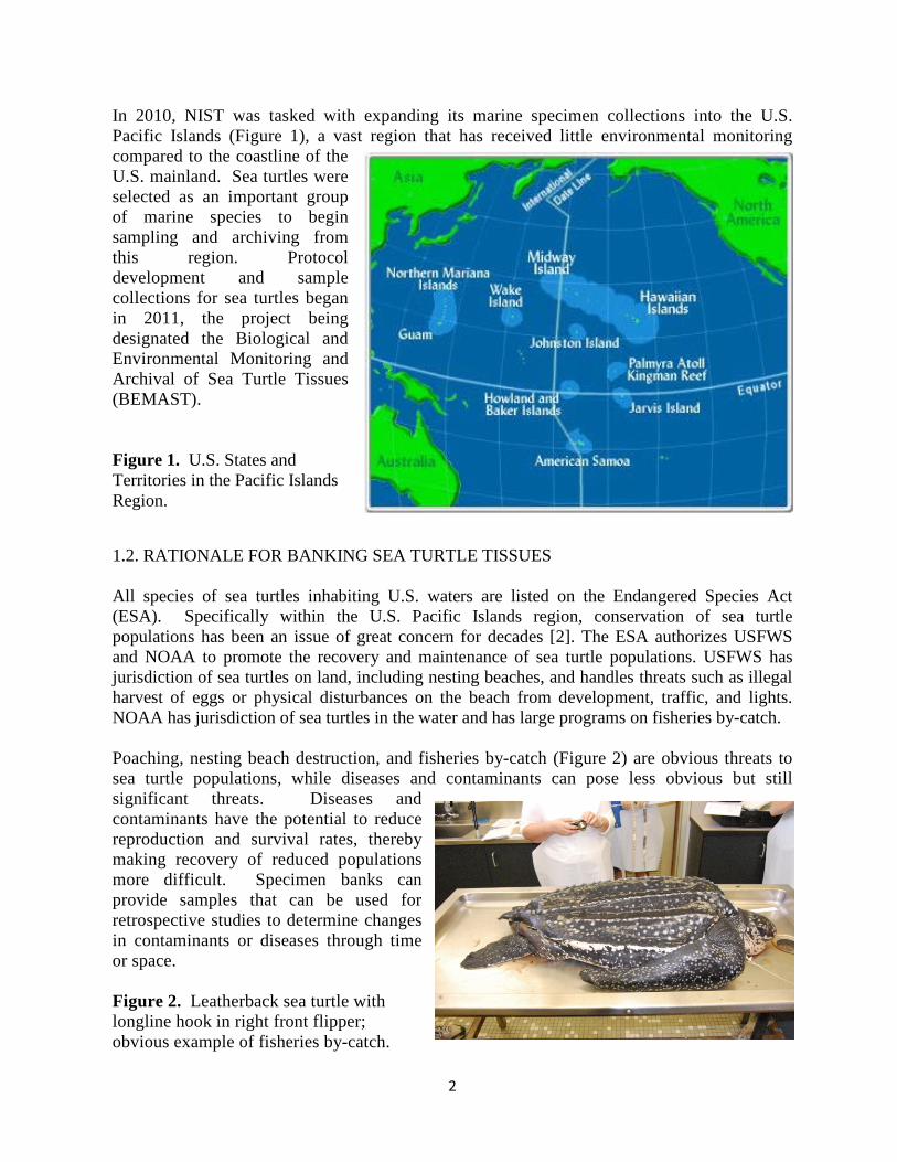
In 2010, NIST was tasked with expanding its marine specimen collections into the U.S. Pacific Islands (Figure 1), a vast region that has received little environmental monitoring compared to the coastline of the U.S. mainland. Sea turtles were selected as an important group of marine species to begin sampling and archiving from this region. Protocol development and sample collections for sea turtles began in 2011, the project being designated the Biological and Environmental Monitoring and Archival of Sea Turtle Tissues (BEMAST). Figure 1. U.S. States and Territories in the Pacific Islands Region.
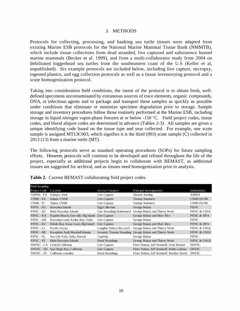
1.2. RATIONALE FOR BANKING SEA TURTLE TISSUES All species of sea turtles inhabiting U.S. waters are listed on the Endangered Species Act (ESA). Specifically within the U.S. Pacific Islands region, conservation of sea turtle populations has been an issue of great concern for decades [2]. The ESA authorizes USFWS and NOAA to promote the recovery and maintenance of sea turtle populations. USFWS has jurisdiction of sea turtles on land, including nesting beaches, and handles threats such as illegal harvest of eggs or physical disturbances on the beach from development, traffic, and lights. NOAA has jurisdiction of sea turtles in the water and has large programs on fisheries by-catch. Poaching, nesting beach destruction, and fisheries by-catch (Figure 2) are obvious threats to sea turtle populations, while diseases and contaminants can pose less obvious but still significant threats. Diseases and contaminants have the potential to reduce reproduction and survival rates, thereby making recovery of reduced populations more difficult. Specimen banks can provide samples that can be used for retrospective studies to determine changes in contaminants or diseases through time or space. Figure 2. Leatherback sea turtle with longline hook in right front flipper; obvious example of fisheries by-catch.

3
For example, one of the first questions asked after the Deepwater Horizon oil spill occurred in April, 2010, was “who has samples from sea turtles from the Gulf of Mexico prior to the spill?” Scientists needed a baseline and a specimen bank is a critical resource for high quality samples to answer such questions. One well-documented disease is green sea turtle fibropapillomatosis (FP) (Figure 3). FP is characterized by benign tumors that are associated with a herpesvirus that obstruct vision, movement, and/or swallowing. The disease emerged in Hawaii in the 1980s, increased in incidence by 1995, and has declined since, although different regions of Hawaii have different temporal patterns [3]. For four decades scientists have speculated that environmental contaminants may contribute to FP [4], yet little to no research has been dedicated to this subject. A specimen bank, if it existed, would allow researchers to go back in time to examine sea turtle tissues for viruses, health indicators, or contaminants prior to or during fluctuations in disease or contaminant use. Figure 3. Green sea turtle with fibropapilloma (FP) tumors from Kapoho, Hawaii.
Data on environmental contaminant exposure is limited for sea turtles. For example, less than 30 samples from sea turtles have been analyzed for persistent organic pollutant concentrations within the U.S. Pacific Islands region (see review [5]); thus, resource managers cannot make informed decisions that consider the relative risk of chemical threats compared to other threats like fisheries by-catch. Monitoring for known contaminants and diseases in eggs and tissues of sea turtles in this region is critical for understanding these threats; and archiving samples for determining contaminants and diseases that are not yet identified is equally as important. NIST has played a primary role in documenting sea turtle contaminant exposure with over 20 publications. All sea turtle species are long-lived, and most take 20 or more years to reach maturity, allowing them a long time to accumulate chemical pollutants. They are highly migratory, with most species crossing entire ocean basins at least once in their lifetime. However, they are also known to return multiple times to a specific region to forage or nest during each life stage. These life history traits make them good integrators of contaminant and disease patterns present in their chosen habitats. Most contaminant monitoring in sea turtles has focused on the loggerhead sea turtles in the southeastern region of the U.S., principally because this is the most abundant species co-located with the largest density of sea turtle biologists. This research has documented that sea turtles are good indicators of regional-scale coastal contamination [5]. Because of their strong site fidelity for a specific annual foraging area, juvenile loggerhead turtles from different U.S. states exhibit different contaminant concentrations and patterns of exposure. For example, turtles foraging in coastal regions of eastern Florida have a different accumulation pattern and concentration than turtles foraging off

4
of South Carolina [6]. Additionally, contaminant concentrations measured in this species have been related to poorer health indicators, suggesting that man-made chemicals are accumulating in the marine environment to levels that may be causing sub-lethal toxicological effects on these threatened and endangered species [7-9]. The collection and archival of sea turtle tissues over a period of several years from a new region will be a resource for future analyses, providing samples that can be used to determine baseline contaminant levels as well as health indicators and biomarkers. Five sea turtle species inhabit the U.S. Pacific Islands region (green sea turtle, Chelonia mydas; hawksbill sea turtle, Eretmochelys imbricata; olive ridley sea turtle, Lepidochelys olivacea; Pacific leatherback sea turtle, Dermochelys coriacea and loggerhead sea turtle, Caretta caretta) and all are listed as either threatened or endangered under the Endangered Species Act (ESA) because of past population declines. The five species vary in dietary habits and migratory pathways. They each use the region in a unique way and have their own particular conservation and health concerns. Thus all five species are important for sample archival and real-time contaminant and health-related studies. Details about the life history strategies and population abundances can be found in the Recovery Plan documents for each species and stock [10-14]. The following describe relevant facts about the diet and habits of each species that provide rationale for banking samples for health and contaminants research:
1. The green sea turtle is the most abundant species found in the U.S. Pacific Islands Region. The genetically-distinct Hawaiian population nests predominantly in the Northwest Hawaiian Islands. This Hawaiian population has increased to the point that NOAA is considering removing it from the list of endangered species under the ESA. The population rebound happened despite the high prevalence of FP in certain locations around Hawaii [3]. The cause and co-factors associated with FP could be investigated using banked specimens.
2. The hawksbill sea turtle has smaller populations, is listed as “endangered” under the ESA throughout the Pacific Ocean, and is approaching extinction [10]. This species can be considered a toxicological novelty as they eat predominantly marine sponges, which are known to produce toxic natural products, suggesting that hawksbills have unidentified, unique detoxifying biochemical pathways. Additionally, they were recently found to have the highest levels of persistent organic pollutants (POPs) compared to four other sea turtle species in the southeastern coast of the U.S. [9]. This higher exposure and high conservation concern elevates the priority of studying and archiving hawksbill samples.
3. The olive ridley sea turtle nests in the Pacific Ocean primarily along Central America, is a very pelagic species, feeds on invertebrates, and is often caught in the longline fisheries, the rate of catch being second only to the green turtle. Little to nothing is known about their health or contaminant exposure globally; however, they are known to ingest plastic marine debris (Balazs, unpublished data).
4. The leatherback sea turtle is critically endangered in the Pacific Ocean. It has only small nesting populations in Central America and Southeast Asia (e.g., Papua New Guinea and Indonesia) and none in the U.S. Their use of U.S. waters for foraging is also limited, although adults forage off of central California in the summer, and juvenile to adult life stages are captured as by-catch in the Hawaiian and American Samoan longline fisheries, which has fueled large conflict between conservation and the fishery. The Pacific leatherback sea turtle faces a real extinction risk due to bycatch in fisheries and intentional harvest. Chemical exposure and health status of the highly endangered Pacific leatherback

5
is poorly understood and should be studied to put these issues in context with the other more obvious threats from fisheries and harvest.
5. Likewise, the Pacific loggerhead sea turtle does not nest on U.S. lands, instead it nests mainly in Japan, but falls victim to the longline fleets during trans-Pacific migrations. The loggerhead turtle is the most studied species for environmental contaminant exposure and effects, mainly along the southeast coast of the U.S. and in the Mediterranean Sea [5]. Little to nothing is known about its contaminant exposure in the Pacific Ocean and a comparison of these distant locations is warranted for this species.
In summary, all five sea turtle species utilize the U.S. Pacific Island region or come in contact with U.S. fisheries and have unique characteristics that makes them important for environmental and health research. 1.3. PROJECT GOAL AND OBJECTIVES The goal of BEMAST is to archive a representative collection of tissues annually from prioritized sea turtles species and geographic locations for real-time and retrospective contaminant and health-related research studies. The Project is currently funded for sampling only in the U.S. Pacific Islands. BEMAST has four objectives:
1. Receive specimens from permitted field studies from representative species suitable for real-time and retrospective monitoring of chemical contaminants and health-related research studies. Preference is given to animals, locations, or projects that have the advantage of multiple collaborators that are acquiring additional information on the turtles (e.g., mark/recapture tagging, satellite tagging, time/depth recording, foraging studies, stable isotope, genetics, population abundance studies, aging, etc). This objective is meant to obtain the most information from each turtle “take”.
2. Transport, inventory, homogenize, subsample, and archive samples in conditions that are suitable for long-term storage and eventual analysis for contaminants and health-related research.
3. Analyze samples for contaminants and health-related research. 4. Make the collection inventory accessible to other researchers and provide samples to
researchers from approved research projects consistent with the goals of BEMAST. Sample collection, packaging, transport, cataloging, homogenizing, archiving, and sample sharing are performed according to protocols established by the Marine ESB [15]. Storage is at liquid nitrogen vapor-phase temperatures (-150 °C), which provides the best conditions for minimizing sample degradation. Samples will be available to project partners for future contaminant and health-related research studies. Requests for archived samples by other researchers and agencies will be considered by project partners on a case-by-case basis as described below in the Management Section. 1.4. BANKING SEA TURTLE TISSUES Before collections for BEMAST began, efforts were made to network with wildlife veterinarians, sea turtle field biologists, analytical chemists, and protected resource managers to identify on-going sea turtle projects for sample collection, to prioritize tissue types for banking, and to develop collection and storage protocols.

6
Because of the protected status of sea turtles, sampling must be non-lethal, non-invasive, or opportunistically taken from animals found dead. Three categories of access to samples include 1) turtles that are captured alive and released (“live captures”), 2) turtles that are stranded, fresh-dead or by-caught in fisheries, and 3) turtles or eggs on nesting beaches. All sampling categories have trade-offs that must be considered. From live turtles, whether they are captured at sea, held in captivity, or sampled on the nesting beach, only non-lethal and relatively non-invasive sampling is permitted. Typical routine samples taken during live capture and release projects include blood, scute scrapings, prey collected from the mouth, and skin biopsies. Occasionally, cloacal swabs and mouth/stomach contents are collected. Beyond those sample types, qualified trained personnel (e.g., veterinarians) are required to collect biopsies of internal tissues, like fat, liver, or gonad [16]. Small sample masses or volumes available from live animals create challenges for long-term storage of multiple subsamples for future uses. From fresh dead turtles, numerous tissues of larger mass can be collected, but these animals can be biased towards more diseased animals and blood is not usually available for comparison to live animals. As for eggs, the collection of unhatched, addled eggs after live hatchlings have emerged and during routine nest inventories is preferred by field personnel instead of sacrificing a fresh egg that has the potential to develop. Although unhatched eggs are not fresh, they are usually abundant and easy to collect and have been shown to have the same concentration of POPs as fresh loggerhead eggs [17]. As for tissue choices, Dr. Thierry Work (USGS) suggested archiving sea turtle fat, liver, blood components (plasma and red blood cells), scute scrapings, FP lesions, skin, bile, urine, kidney and gonad tissues. Additional advice was provided to archive sea turtle eggs and muscle tissue as these are the tissues most often eaten by people who harvest turtles internationally. Moreover, NMFS and NIST convened a workshop in August 2012 attended by wildlife veterinarians, analytical chemists, and specimen banking experts to prioritize marine mammal tissue collected for the National Marine Mammal Tissue Bank (NMMTB) [18]. Sea turtles were included in these discussions and future protocol modifications may be made based on the workshop report, which is in preparation. As for turtle species and field project choices, early networking efforts identified sea turtle field projects in the Pacific Ocean (Table 1). Most projects are closely connected to the Marine Turtle Research Program at PIFSC, the National Wildlife Health Center at USGS, the Protected Resources Division at PIRO, the Marine Mammal and Turtle Molecular Research Sample Collection, a specimen bank for genetics and stable isotope research, at the Southwest Fisheries Science Center (SWFSC), and the American Museum of Natural History (AMNH). Table 1 shows the field collection partners and locations for the five sea turtle species and three access categories. The only intensive live captures that collect blood currently in U.S. Pacific waters are NOAA/PIFSC’s Marine Turtle Research Program in Hawaii, AMNH’s program in Palmyra, SWFSC’s programs in California, and the Department of Land and Natural Resources (DLNR) of the Commonwealth of the Northern Mariana Islands (CNMI) program out of Saipan funded by PIRO. Additional projects include Grupo Tortuguero de las Californias’ project along the Baja Peninsula in Mexico and SWFSC cruises throughout the Eastern Tropical Pacific that sample marine mammals and sea turtles. Live capture projects are also taking place in more U.S. Territories, such as a nesting beach at Rose Atoll (American Samoa) funded by PIFSC. All five species cannot be sampled in one location because of their differing habitat choices.

7
Some can only rarely be sampled in U.S. waters (leatherback turtles). To collect representative samples of all species, several locations are necessary, some of which are foreign. The locations and field sampling partners are shown in Table 1. Although all sea turtle species are represented by our current collaborators, these projects cover only a portion of the vast geographic region and represent a diversity of age classes. This diversity in ages may cause difficulty in comparing one site to another. It may be important to focus on one age class or representatively sample several age classes per species. Table 1. Locations and field sampling partners for three access categories of five sea turtle species in the Pacific Ocean. The locations (partners) shown in bold and underlined are already collaborating with NIST for sample collection. Non-bolded but underlined partners are willing to collaborate with NIST.
HI = Hawaii; PIFSC = Pacific Islands Fisheries Science Center; AMNH = American Museum of Natural History; CA = California; SWFSC = Southwest Fisheries Science Center; CMNI = Commonwealth of the Northern Marianas Islands; DLNR (Department of Land and Natural Resources); FFS = French Frigate Shoals; USFWS = U.S. Fish and Wildlife Service; HWF = Hawaii Wildlife Fund; EPHI = Eastern Pacific Hawksbill Initiative; PNG = Papua New Guinea; ETP = Eastern Tropical Pacific cruise.
Live Captures
Species Reason to bank research captures strandings by-catch eggsmaternal
blood
Green Tumors, abundant
HI (PIFSC), Palmyra (AMNH), CA
(SWFSC), CMNI (DLNR), Guam
HI (PIFSC), CNMI (DLNR),
Samoa
HI & Samoa (PIFSC), Japan
HI (PIFSC), Palmyra (USFWS),
Solomon Islands (AMNH), CNMI
(DLNR), FFS, Samoa
Palmyra (USFWS),
Samoa (Rose Atoll; PIFSC),
HI
HawksbillUniquely sponge eating, high conservation concern
Palmyra (AMNH), CNMI (DLNR), HI (HWF), Central America (EPHI)
HI (PIFSC), CNMI (DLNR),
Samoa
HI (PIFSC), Solomon Islands (AMNH), Central America, Samoa
HI, Central America
LeatherbackCritical conservation concern CA (SWFSC), Japan CA (SWFSC)
HI & Samoa (PIFSC), Japan
Solomon Islands (AMNH), Indonesia
(Tapilatu), PNG, Costa Rica
PNG, Costa Rica
Olive Ridley No baseline data ETP (SWFSC) HI (PIFSC) HI & Samoa (PIFSC) Mexico, Costa Rica Mexico, Costa Rica
LoggerheadConservation concern, no data in Pacific
Baja Mexico (Peckham; Wang),
Japan
Baja Mexico (Peckham)
HI & Samoa (PIFSC), Mexico (Peckham),
JapanJapan Japan
Nesting beachesNecropsies

8
2. MANAGEMENT SYSTEM The BEMAST specimens are collected by other agencies (e.g., NOAA, USGS, AMNH, USFWS, state agencies, private institutions, or non-profit organizations) during routine and permitted field projects often with field assistance by NIST personnel. The ownership of samples should be determined before collection through a formal written agreement between the lead of BEMAST and the permitted sample collector. An example agreement is provided in Appendix A. This agreement should clearly state the role of the permitted sample collector in reviewing requests for sample release and use as well as expectations for authorship and acknowledgements. If no formal agreement is in place, ownership of samples from sea turtles collected under a NMFS permit remains with the permitted sample collector and agency. Occasionally, ownership of samples from sea turtles collected under a USFWS permit (e.g., Palmyra Atoll) remains with USFWS. BEMAST samples are transported to, processed by, and banked at NIST. Hawaii Pacific University (HPU) provides logistical support and handling of samples (e.g., liquid nitrogen vapor-phase freezers (-150 ºC), -80 ºC freezers, shipping, supply preparation, sampling assistance, and sample processing) on Oahu. Samples are archived long-term at NIST’s Marine ESB at the Hollings Marine Laboratory in Charleston, South Carolina, where they are stored at temperatures of -150 ºC in liquid nitrogen vapor-phase freezers and homogenized and subsampled into multiple aliquots for analysis. After arrival at the Marine ESB, the samples are checked-in, inventoried, and banked. The Marine ESB inventorying and archiving procedures are consistent with those employed by NIST for the NMMTB [19]. The lead of BEMAST will provide reports to permit holders at their request. For all samples collected from turtles within NOAA NMFS jurisdiction (turtle in the water or on high seas), NIST is currently applying for a NMFS permit for the possession, storage, analysis, and release/transfer of those samples to third party recipients for approved research proposals. This permit will alleviate the need to add NIST as a sample recipient on field collectors’ permits (when field collectors sign an agreement stating that they permanently relinquish samples to NIST) and will alleviate the need to add third party recipients to the original field sampling permits. Researchers associated with NIST or the permitted sample collector can request banked samples through written communication (e.g., email) with the lead of BEMAST and the permitted sample collector. In cases where the BEMAST lead or permitted sample collector is no longer employed/associated with the original agency or does not respond to the request for three weeks or longer, the successor of their position or another researcher identified by a supervisor within the same agency may be assigned to make decisions and take up discussions. Discussions should explain scientific purpose of using the banked samples, brief methods, and an agreement upon authorship or acknowledgements before samples are analyzed. Requests from researchers not associated with NIST or the permitted sample collector will be considered by application only. The release of the tissues to outside investigators will be contingent upon the approval of all of the following personnel:
1. the lead of BEMAST;

9
2. the permitted sample collector, if the collector selected the role to “review requests for sample release” on the initial formal agreement, or the successor of their position or another researcher identified by a supervisor within the same agency may be assigned;
3. a person from the NMFS, Office of Protected Resources, if the samples were collected under a NMFS permit.
These personnel have a maximum of three weeks to complete the review process. A lack of response will be considered approval of the request. Sample release will depend on a determination that a surplus of requested sample material exists beyond anticipated analytical needs of BEMAST or permitted sample collectors. Requests for samples must include a clear and concise statement of the proposed work and be consistent with the goal and purposes of BEMAST. The following specific information should be included in the request application:
1. Name of principal investigator and affiliated research or academic organization 2. List of specific samples and quantities desired 3. Rationale and objectives of proposed research to be conducted, including funding source 4. Justification for use of banked tissue 5. Description of research facility where analyses will be conducted 6. Name of researcher conducting analyses and qualifications (short CV) 7. Analytical quality control procedures to be used and agreement to participate in NIST-
directed interlaboratory comparison exercises for quality assurance (QA) purposes 8. Estimated date for completion of research, and schedule/date of subsequent reports 9. Agreement that all results and findings, including analytical data, be provided to the Marine
ESB and BEMAST (this includes a data submission schedule) 10. Agreement that remaining samples must be destroyed and not used for other research
purposes or given to another researcher for use without prior written consent of the personnel listed above
11. Agreement to authorship and/or acknowledgment requirements determined during the review process. Appropriate credit must be given to sample collectors, NIST, and BEMAST for use of banked tissues in all publications, abstracts, or presentations. These requirements will be provided in the final approval letter.
Shipping charges will be borne by the individual or institution requesting the samples. Additional charges to cover costs associated with sample preparation and distribution may also be required. BEMAST will publish tissue inventory reports. These reports will provide information on the status of sea turtle tissue samples archived at the Marine ESB and the results of any chemical analyses that may have been conducted on them.
10
3. METHODS Protocols for collecting, processing, and banking sea turtle tissues were adapted from existing Marine ESB protocols for the National Marine Mammal Tissue Bank (NMMTB), which include tissue collections from dead stranded, live captured and subsistence hunted marine mammals (Becker et al. 1999), and from a multi-collaborator study from 2004 on debilitated loggerhead sea turtles from the southeastern coast of the U.S (Keller et al, unpublished). Six example protocols are included below, including live capture, necropsy, ingested plastics, and egg collection protocols as well as a tissue inventorying protocol and a scute homogenization protocol. Taking into consideration field conditions, the intent of the protocol is to obtain fresh, well-defined specimens uncontaminated by extraneous sources of trace elements, organic compounds, DNA, or infectious agents and to package and transport these samples as quickly as possible under conditions that eliminate or minimize specimen degradation prior to storage. Sample storage and inventory procedures follow those routinely performed at the Marine ESB, including storage in liquid nitrogen vapor-phase freezers at or below -150 °C. Field project codes, tissue codes, and blood aliquot codes are determined in advance (Tables 2-3). All samples are given a unique identifying code based on the tissue type and year collected. For example, one scute sample is assigned MT13C003, which signifies it is the third (003) scute sample (C) collected in 2013 (13) from a marine turtle (MT). The following protocols serve as standard operating procedures (SOPs) for future sampling efforts. However, protocols will continue to be developed and refined throughout the life of the project, especially as additional projects begin to collaborate with BEMAST, as additional tissues are suggested for archival, and as tissues need homogenization prior to analysis. Table 2. Current BEMAST collaborating field project codes.
Field Sampling Project Code Location Access Category Principal Investigator(s) Institution(s)AMNH - PA Palmyra Atoll Live Capture Eleanor Sterling AMNHCNMI - SA Saipan, CNMI Live Capture Tammy Summers CNMI DLNRCNMI - TI Tinian, CNMI Live Capture Tammy Summers CNMI DLNRPIFSC - EG Hawaiian Islands Egg Collection George Balazs PIFSCPIFSC - EU Main Hawaiian Islands Live Strandings/Euthanized George Balazs and Thierry Work PIFSC & USGSPIFSC - KA Kapoho Beach, East side, Big Island Live Capture George Balazs and Marc Rice PIFSC & HPAPIFSC - KB Kawainui canal, Kailua Bay, Oahu Live Capture George Balazs PIFSCPIFSC - KC Kiholo Bay, Kona Coast, Big Island Live Capture George Balazs and Marc Rice PIFSC & HPAPIFSC - LL Pacific Ocean Longline Fishery By-catch George Balazs and Thierry Work PIFSC & USGSPIFSC - MI Kwajalein Atoll, Marshall Islands Acoustic Trauma Stranding George Balazs and Thierry Work PIFSC & USGSPIFSC - SL Sea Life Park, Oahu, Hawaii Captivity George Balazs PIFSCPIFSC - ST Main Hawaiian Islands Dead Strandings George Balazs and Thierry Work PIFSC & USGSSWFSC - CA Central California Live Capture Peter Dutton, Jeff Seminoff, Scott Benson SWFSCSWFSC - SD San Diego Bay, California Live Capture Peter Dutton, Jeff Seminoff, Robin LaRoux SWFSCSWFSC - ST California coastline Dead Strandings Peter Dutton, Jeff Seminoff, Heather Harris SWFSC

11
Table 3. Current BEMAST tissue and blood aliquot codes.
Tissue Code TissueS Whole blood spun and frozen uprightB Whole blood unspun frozenW Whole blood spun and aliquoted when freshC ScuteL LiverF FatU BlubberI Fibropapilloma lesionD SkinM MuscleG BileK KidneyH BrainE EggA Gut contents (plastic, mucosa, undigested prey)
Blood Aliquot Code Blood ComponentP Plasma
WWhite blood cells plus red blood cells
R Red blood cellsB Whole bloodPAX PAXgene tube

12
3.1. LIVE CAPTURE COLLECTION FIELD PROTOCOL Overview: Sea turtles will be captured by hand or net by collaborating, permitted field biologists. The typical target sample size for each sampling event is 20 turtles. Blood and scute sampling should occur on the same turtles. As much capture and biological data should be recorded as possible, including capture method, date/time, water temperature, standard morphometrics to include straight carapace length (defined as notch to tip) and weight, degree and severity of fibropapillomatosis, trauma, and emaciation, tag identification numbers, capture history, and other samples collected. Because the samples collected for the BEMAST project are primarily intended for contaminants and health research, inadvertent contamination during sampling and sample handling is a major concern. The four major sources of contamination are from 1) airborne chemicals (e.g., cigarette smoke, dust, vehicle exhaust), 2) chemicals in or on surfaces that touch the samples or touch the supplies that will later touch the samples (e.g., sunscreen, insect repellent, plastic gloves, rain, sand carried by wind), 3) cross-contamination from one sample to another, and 4) supplies that directly touch the samples. The fourth source is inevitable but is minimized by pre-cleaning certain supplies at NIST and by preparing water “blanks” from unused supplies and storing these with the samples for analysis of background contamination. For this reason, only NIST-provided supplies should be used, as NIST will make 6 complete sets of blanks using supplies of the identical lot number provided. During sampling and processing, care should be taken to minimize residues from windblown sand, rainwater, saltwater drips, indoor and outdoor dust, fuel, engine exhaust, sunscreen, insect repellent, cigarette smoke, skin, hair, and any other chemicals. The most likely problems occur from touching NIST-provided supplies to gloves, bench-tops, rain or saltwater, or beach sand prior to or during their use in processing samples. These problems must be avoided by either processing samples in an indoor environment or taking care to protect samples from the elements. NIST-provided supplies must remain covered in original packaging and only removed immediately before its use. If a pipette tip touches anything but the intended sample, it should be considered contaminated and be discarded. To mimic possible contamination during storage, water blanks are to be stored alongside the samples. Blood Collection Preface: Timing matters. Blood sampling should occur as quickly as possible after capture (preferably within 15 min of capture). Sample processing should occur as quickly as possible after sample collection with no longer than a 12 h delay but not if it will jeopardize the integrity of the supplies or samples (see above). Blood volumes collected will range from 12.5 mL to 22.5 mL depending upon turtle mass and permitted volumes (Permits typically allow up to 3 mL per kg of turtle). The blood will be split into several aliquots to maximize the utility of the sample. Intended collection and uses of aliquots: Aliquot ID Vol (mL) Sample Type Container Intended purpose WB not saved 0.1 whole blood any vial hematocrit and smears B001 1.0 whole blood Cryovial Inorganic compounds *B002-3 1.0 whole blood Cryovial Bank
13
P001 4 to 5 plasma Teflon jar Organic contaminants P002 0.5 to 1.8 plasma Cryovial Plasma chemistry P003-n 0.5 to 1.8 Remaining plasma Cryovials PFCs/Bank W001 as needed Buffy coat with RBC Cryovial Bank/gene expression R001 0.5 RBCs Cryovial genetics R002 1.0 to 1.5 RBCs Cryovial Inorganic compounds R003-n 1.0 to 1.5 RBCs Cryovial Bank * Only if 20+ mL of blood collected. Pre-Cleaning Materials (used at NIST only) • Hexane: Burdick and Jackson Cat # GC215-4 for trace analysis • Methanol: Burdick and Jackson Cat # GC230-4 for trace analysis • Nitric acid: Optima ultra-pure, Fisherbrand Cat # A4672 diluted to 3 % with Millipore
water from the laminar flow hood in the NIST Inorganic Chemistry of the Hollings Marine Laboratory
• Hydrochloric acid: 36.5 % to 38 % VWR Cat # JT9530-33, diluted 2:1 with Millipore water 18.2 resistivity (MΩ·cm)
• Nitric acid: 69.0 % to 70.0 % VWR Cat # BDH3046-2.5LPC, diluted 2:1 with Millipore water 18.2 resistivity (MΩ·cm)
• Ethanol: Sigma Aldrich Cat # 459844, ACS reagent, 99.5 % (200 proof), absolute • Chloroform: Sigma Aldrich Cat # 319988 Blood Collection Materials: • Watch • Thermometer for water temperature (VWR Cat # 89095‐572) • Powder free vinyl gloves (Kimtech Cat # 61002 for medium) • Clipboard with pens, pencils, and sharpies inside • Data sheet printed on Rite in the Rain paper (Cat # 8511) • Soft brush to clean turtle neck (Fusion décor aquarium cleaning brush) • Glass bottle of tap water for neck cleaning • freshwater from a hose or sea water for rinsing turtle • 1 L Nalgene LDPE bottles or 2 L Teflon bottle with Millipore water • LDPE plastic squirt bottle for Millipore water • Isopropanol (70 % Ricca cubitainer, Fisher Cat # 4210‐2.5) • LDPE plastic squirt bottle for 70 % isopropanol • Paper towels • Double-ended needle (1.5 in, 21 g, Vacuette Cat # 450076) • Vacutainer 10 mL glass sodium heparin blood collection tube (Becton Dickinson Cat #
366480) • PAXgene blood RNA tube (Becton Dickinson Cat # 762165) • Vacutainer hubs (Becton Dickinson Cat # 364815) • Small cooler with frozen gel packs and bubble wrap bag • Sharps container, 1 L (1 quart, VWR Cat # 19001‐001) • Medium cooler for supplies, 47 L (50 quart) • Millipore water (18 MΩ·cm) from NIST Inorganic Laboratory in Teflon bottle that was pre-
cleaned with acid, water, hexane, acetone, and water for making blanks

14
• Millipore water (18 MΩ·cm) from NIST Inorganic Laboratory in plastic (LDPE) bottle that was pre-cleaned with acid and water for making blanks
• Trash bag for in field Blood Processing Materials: • Powder free vinyl gloves-medium (Kimtech Cat # 61002) • Centrifuge (preferably swinging bucket rotor, LW Scientific C5) • Cryovials (Corning 2 mL self-standing, conical bottom, silicone washer, external threading,
Cat # 430659; not pre-cleaned by NIST; 11/animal) • Cryo-Babies labels, pre-printed for labeling cryovials (Cat # LCRY-1700-G) • Teflon jars (5 mL vial, conical inside with fin-type bottom, Savillex Cat # 200-005-32; pre-
cleaned in the Marine ESB; 1/animal) • Teflon jar lids (for 5 mL and 7 mL vials, recessed lid, 24 mm threaded closure, Savillex Cat
# 600-024-71; pre-cleaned in the Marine ESB; 1/animal) • Teflon jar labels (Savillex Cat # 730-0100 cut for 5 mL jars; 1/animal) • 30x30 cm (12x12 inch) Teflon bags for storage of Teflon jars in vapor shipper or freezer
(KNF Cleanroom Cat # LB602:1212; 2 per event) • Cable ties to seal Teflon bags (2 per event) • ≈2.0 mL plastic tubes for hematocrit and smears (any brand) • Pasteur pipettes, 23 cm (9 inch), borosilicate glass, non-sterile (Fisherbrand Cat # 13-678-
20C pre-cleaned at NIST) • Pipette bulbs (latex, VWR Cat # 82024-554) • Microscope slides (Fisher Cat # 12‐544‐2) • Slidebox (VWR Cat # 82024‐608) • Hematocrit tubes (BD Clay Adams SurePrep plain capillary tubes, 75 mm self sealing with
mylar wrapping, ref 420314 2x100 or 420315 10x100) • Hematocrit tube carrier for centrifuge (Jorgensen CRIT Carrier Cat # J0501C) • Microhematocrit card reader (Jorgensen ZIPocrit card reader Cat # J0501RC) • Tube racks (4 for cryovials Corning Cat # 431131; 4 for blood tubes VWR Cat # 89215‐
768) • 5.1 cm (2 inch) tall cryovial freezer boxes with 81-place dividers • Liquid nitrogen vapor shipper (Doble 34 30-day 5 canister) • Cryogloves • Freezer, -80 oC or liquid nitrogen vapor-phase • Datasheet • Pens, pencils, and sharpies • Glass disposal box or 1 gallon sharps container (VWR Cat # 19001‐003) • Trash bags • Packing tape Blood Supply Pre-cleaning Procedure: • Teflon jars and lids provided by NIST are pre-cleaned using the Marine ESB cleaning
protocol and the jars are air dried in the ISO Class 5 clean room to ensure contaminant free storage containers [15].

15
• “A” pipettes are broken at the neck to make a wider bore, immersed in 3 % nitric acid for 24 h in an LDPE bottle, rinsed 3x rinse with Millipore water, allowed to dry on cleanroom wipers in a Hepa hood, and stored in the same LDPE bottle.
• “B” pipettes are pre-rinsed 3x hexane with Teflon squirt bottle and placed into a hexane-rinsed foil package stored in a cardboard box.
Blood Collection Procedure (performed by permitted collaborators): • Wear gloves. Label Vacutainer tubes with a temporary turtle ID number specific for this
turtle and an “A” on the first tube; “B” on the second and so forth. Note this number on the datasheet.
• Scrub debris and algae from blood collection site on the dorsal surface of the neck with a soft brush and tap water from the glass bottle. Rinse neck with Millipore water, wipe with a paper towel. Rinse neck with 70 % isopropanol making sure to elevate eyes so as not to get alcohol into the eyes.
• Restrain turtle on an incline with head down (Figure 4) and collect blood as described elsewhere [20]. Apply a hub to the Vacutainer needle for safety, insert the long end of the needle into the neck, then push the 10 mL Vacutainer tube A onto the outer needle.
Figure 4. Proper restraint and incline of a green sea turtle for blood sampling.
• Collect a full 10 mL of blood into green-top tube A. Collect a full green-top tube B, if
permitted. Collect 2.5 mL of blood into the PAXgene tube, making sure it is full. • Remove the tube from the needle prior to removing the needle from the turtle. Rinse neck
with isopropanol. • After collection, slowly and smoothly invert the blood tubes 8 times to mix the blood with
the anticoagulants (10 times for PAXgene tube). Do not shake. • Place the green-top tubes in a cooler with frozen gel packs. Do not put the blood tubes
directly in contact with frozen objects as hemolysis can occur. Use a bubble wrap bag or plastic cup lined with paper towels as a barrier. Keep the PAXgene tube at room temperature for at least 2 h.
• Dispose of the needle safely in the sharps container. • Note the turtle ID number on the datasheet (e.g., left rear flipper PIT tag).
Blood Processing Procedure: • At an indoor location, wear clean vinyl gloves. • Wipe outsides of PAXgene tubes with isopropanol, apply a green Cryobaby label (aliquot
code is PAX), and freeze tubes upright at -20 ºC within 72 h of blood collection. After more than 1 h, transfer to -80 °C or below. Record freezing times on the datasheet.
• Use Tables 4 or 5 to set up and label aliquot tubes expected for use from 10 mL or 20 mL of blood in green-top tubes, respectively.

16
Table 4. Aliquoting procedure for 10 mL of blood.
Table 5. Aliquoting procedure for 20 mL of blood.
• Invert tubes slowly and smoothly 8 times to homogenize the blood. Using a clean “A”
Pasteur pipette with a bulb, transfer 1.0 mL of whole blood to the B001 cryovial. Avoid returning blood to the original tube and don’t waste sample. Take three aliquots of whole blood if 20 mL of blood were collected.
• Place 0.1 mL blood into the 2 mL plastic tube (any brand) for hematocrit.
• Make a smear with the remaining drop in pipette A by sliding the edge of one slide into and out of the drop of blood at a 45 degree angle (Figure 5).
Figure 5. Making a blood smear.
Tissue Pipet Aliquot ID Vial type Volume (mL) CommentsWhole blood A (short) B001 2 mL cryovial 1.0Plasma B (long) P001 teflon jar 4.0Plasma B (long) P002 2 mL cryovial 1.0 split remaining betweenPlasma B (long) P003 2 mL cryovial 0.6 P002 and P003WBC&RBC B (long) W001 2 mL cryovial 0.5-1.0 as much as it takes to get rid of WBCRBCs A (short) R001 2 mL cryovial 0.5RBCs A (short) R002 2 mL cryovial 1.0 split remaining betweenRBCs A (short) R003 2 mL cryovial 1.0 R002 and R003
Tissue Pipet Aliquot ID Vial type Volume (mL) Tube CommentsWhole blood A (short) B001 2 mL cryovial 1.0 AWhole blood A (short) B002 2 mL cryovial 1.0 BWhole blood A (short) B003 2 mL cryovial 1.0 mix equalize volumes in tubes for spinningPlasma B (long) P001 teflon jar 5.0 A or most hemolyzedPlasma B (long) P002 2 mL cryovial 1.1 mix finish tube A, add more from BPlasma B (long) P003 2 mL cryovial 1.1 BPlasma B (long) P004 2 mL cryovial 1.1 BPlasma B (long) P005 2 mL cryovial 1.1 BWBC&RBC B (long) W001 2 mL cryovial 1.0 mix as much as it takes to get rid of WBCRBCs A (short) R001 2 mL cryovial 0.5 ARBCs A (short) R002 2 mL cryovial 1.0 ARBCs A (short) R003 2 mL cryovial 1.0 BRBCs A (short) R004 2 mL cryovial 1.0 B

17
• From this 2 mL plastic tube (any brand), fill two hematocrit tubes about 75 % full and spin them at 524 rad/s (5000 rpm, 4000 x gn, setting 50 on LW Scientific C5) for 8 min, noting color of carrier and position on the datasheet. Read hematocrit by placing the hematocrit onto the card reader as shown in Figure 6, and record hematocrit on datasheet. When using a new centrifuge, spin tube again for 2 min to make sure reading does not decrease. Continue spinning for additional 2 min until the reading stabilizes.
Figure 6. Reading a hematocrit tube. Place tube so that 1) the bottom of the red blood cell layer is at the top of the bottom red line and 2) the meniscus of the plasma is in the center of the top red line. Read the percentage at the interface of the red blood cells and the buffy coat (37 % in this sample).
• Recap blood tubes and centrifuge for 10 min at 314 rad/s (3000 rpm, setting 30 on LW Scientific C5) to separate plasma from red blood cells and other cellular material.
• Using a clean “B” Pasteur pipette with a bulb, pull up plasma from different layers with each suction (don’t just sip off the top) but also avoid cellular material from the buffy coat getting into the plasma by not taking plasma too close to this layer. Transfer 4 mL to 5 mL of plasma into a 5 mL Teflon jar and label P001 (see Figure 7 for volume reference; tube with most hemolysis is used for P001). Next transfer 1.0 mL of plasma into a cryovial labeled P002, and then the remaining plasma should be split into 0.5 mL to 1 mL aliquots in additional cryovials. Plasma should not be removed from the Teflon jar into a cryovial or vice versa.
• Using the same “B” pipette, transfer the remaining thin layer of plasma and the buffy coat (greatly contaminated with red blood cells) into the W001 cryovial. The goal is to remove all plasma and white blood cells from the underlying red blood cells.
• Using a clean “A” pipette, transfer 0.5 mL RBCs into R001 cryovial and then 0.5 mL to 1.5 mL aliquots of RBCs into additional cryovials labeled R002-R00n.
• Ensure all Teflon jars and cryovials are tightly capped and freeze at the coolest possible temperature available (preferably -80 °C or below). Store the blood smears in the slide box at room temperature.
• Complete datasheet making sure to include all pertinent information.
Figure 7. 5 mL Teflon jar with graduations for estimating volumes.
543
2
mLEnd of top thread

18
*NOTE* If the protocol is not followed, please note all modifications on the datasheet. Blanks for blood: Collect three field blanks each using the Millipore water from both the pre-cleaned Teflon bottle and the pre-cleaned LDPE bottle. Spread out the making of blanks over several days of the sampling event. Below is an example of six blanks made during one sampling event. Blank Lots ID Water source Date made Vacuette Needle Green top Cryovial Teflon jar 1 Teflon Kapoho 11-11 10 Jul 2012 11D06B 1243785 17910025F fin bottom 2 Teflon Kapoho 11-11 11 Jul 2012 11D06B 1243785 17910025F fin bottom 3 Teflon Kapoho 11-11 12 Jul 2012 11D06B 1243785 17910025F fin bottom 4 LDPE Kiholo 4-20-11 10 Jul 2012 11D06B 1243785 17910025F fin bottom 5 LDPE Kiholo 4-20-11 11 Jul 2012 11D06B 1243785 17910025F fin bottom 6 LDPE Kiholo 4-20-11 12 Jul 2012 11D06B 1243785 17910025F fin bottom Water blanks should be made in the same manner as the blood samples were collected and processed. One blank set will be made with NIST Teflon water (Teflon bottle) and another set will be made with NIST non-Teflon water (LDPE bottle): • At the sampling location, using a Vacutainer needle, draw deionized, Millipore water from
the Teflon bottle into 2 green-top blood tubes. Invert tubes 8 times as done with the blood sample to mix the water with the anticoagulants.
• Follow the steps in the Processing Procedure Section above (skip the PAXgene, smears, and hematocrit steps).
• Label the cryovials and Teflon jar for the blanks according to the Label Section. • Repeat the steps above two more times using new needles and blood tubes. Label these
appropriately Blank 1, 2, and 3 for each set of blanks. • Repeat the steps above three more times using water from the LDPE bottles for Blanks 4, 5,
and 6. • Fill out a separate datasheet for each blank. NOTE: A new set of blanks will need to be made when a different needle, blood tube, or cryovial lot number is used. Scute Collection Preface: The protocol that will be used to collect scute samples from green, hawksbill, and loggerhead live captured turtles was suggested by George Balazs (unpublished method). This method should be used cautiously on sea turtles with thin, flexible scute keratin, like the eastern Pacific green turtle (aka black turtle) and olive ridley sea turtles. The major sources of contamination to avoid while scute sampling are from epibiota on the carapace and from the external environment (exhaust, dust, rain, saltwater, sand). In general, keratin will be shaved from the entire surface of the 5th central (posterior vertebral) scute. The top layer penetrated with algae will be discarded. Underlying clean layers approximately 1 mm to 1.5 mm deep will be collected. Avoid portions of the scute that have previous injury, avoid scute seams, and avoid shaving too deeply to avoid injury to the turtle. Sample Mass Requirements: 0.3 g to 1.0 g

19
Materials: • Freshwater from a hose or sea water • plastic scrubbing pad (Scotchguard Dobie) • Plastic container to keep scrubbing pad in 70 % isopropanol between turtles • Millipore water in LDPE squirt bottle • 70 % Isopropanol in LDPE squirt bottle (Ricca cubitainer, Fisher Cat # 4210‐2.5) • 10x10 cm (4x4 inch) cotton cleanroom wipers (Texwipe Cat # TX304) • Stainless steel knife (Promar bait knife) • 30x30 cm (12x12 inch) Teflon bag heat-sealed at NIST with the bottom in a V-shape (KNF
clean room products Cat # 300045-20) • Cable tie • Green Tyvec labels (Uline Cat # S-5984G) • Powder free vinyl gloves, medium (Kimtech Cat # 61002) • Small cooler with frozen gel pack • Liquid nitrogen vapor shipper (Doble 34 30-day 5 canister) • Cryogloves • Freezer, -80 oC or liquid nitrogen vapor • Datasheet • Pens, pencils, and sharpies • Scissors • Lab tape • Bucket or bowl for alcohol waste catchment • Umbrella • Stool • Trash bag for in field Scute Collection Procedure (performed by permitted collaborators): • Wearing vinyl gloves, rinse plastic scrubbing pad with isopropanol and tap water prior to
use. • Clean the 5th central scute (Fig. 8) as well as 5 cm (2 inches) anterior to this scute and the
two posterior marginals (dorsal and ventral sides). Remove sloughing keratin and epiphytic/epibiotic organisms using a plastic scrubbing pad and tap water from hose or sea water in the glass bottle.
• Move turtle to elevated table with tail hanging over table. • Rinse scute with isopropanol, Millipore water, and wipe dry with a cleanroom wiper to
remove any remaining foreign matter and debris. Use this same method to clean the knife. • Use the knife to shave the top layer (<0.5 mm) of keratin off of the entire surface of the 5th
central scute until all epibiota is removed. Reclean knife with isopropanol and then Millipore water and wipe dry with cleanroom wiper. Use the same wiper to brush away algae-penetrated keratin from the turtle.
• Place the umbrella upwind. Have an assistant hold the Teflon bag under the turtle while you use the knife to shave keratin layers (<2.0 mm) off of the entire surface of the 5th central scute (Figure 8). Avoid scute seams and going too deep so as not to harm the turtle.
• Allow shavings to fall into bag.

20
• Seal bag with a cable tie and Tyvec label that is properly labeled, and cut top of bag to make it less bulky.
• Store the sample with frozen gel packs until frozen at -80 °C or below later.
• Fill out the datasheet. Figure 8. Shaving motion and ribbons of scute during scute scraping of 5th central scute.
Labeling All samples should be labeled following a standard procedure. Each tube label should include the following information: Animal ID Aliquot ID (blood aliquot IDs above, PAX = PAXgene, C = scute)
Date (dd Mmm yyyy) NIST/ Project code
SAMPLE: Turtle ID used by field collaborator P001
12 Jul 2012 NIST / PIFSC – KB Storage Samples will be stored temporarily in a -80 oC or liquid nitrogen vapor-phase freezer at Hawaii Pacific University with oversight by Brenda Jensen. Samples will be shipped to Jennifer Keller/Rebecca Pugh, NIST, Hollings Marine Laboratory, 331 Fort Johnson Road, Charleston, SC 29412 for long term storage at the NIST Marine ESB until a facility is built in Hawaii. Smears will be stored at room temperature by Jennifer Keller at NIST in Charleston. Before transferring, relocating, or shipping samples, or if storage conditions fail, contact all of the following personnel: Jennifer Keller ([email protected]) 843-725-4822 or 843-442-2188 George Balazs ([email protected]) 808-983-5733 Rebecca Pugh ([email protected]) 843-762-8952 or 843-709-0145 If you have questions about the sample or processing protocol contact: Jennifer Keller ([email protected])
843-725-4822 (work) 843-442-2188 (cell phone)

21
LIVE CAPTURE DATASHEET EXAMPLE

22

23
3.2. NECROPSY FIELD PROTOCOL Overview: This protocol is a modification of Appendix B in NIST IR 6279 “National Marine Mammal Tissue Bank and Quality Assurance Program: Protocols, Inventory, and Analytical Results” [18] and protocols previously used to sample during sea turtle necropsies, such as the Debilitated Loggerhead protocol (Keller et al. unpublished). Necropsies are to be authorized, organized, and overseen by permitted field biologists and veterinarians. Selection criteria for sampling sea turtles during necropsy for the BEMAST project should include: freshness of tissues post-mortem (euthanized or freshly dead), priority species, and priority locations. Turtles captured in longline fisheries, killed during acoustic traumas, or stranded dead, frozen quickly, and thawed within 48 h of necropsy should be the targeted samples. In addition, turtles that strand alive and are subsequently euthanized should be sampled. As much data should be recorded as possible, including species, discovery method, date/time of discovery/freezing/necropsying/sample storing, standard morphometrics to include straight carapace length (notch to tip) and weight, degree and severity of fibropapillomatosis (FP), trauma and emaciation, tag identification numbers, capture history, and a list of other samples collected. Turtles that strand in Hawaii with severe fibropapillomatosis (FP) are reported to the Marine Turtle Research Program (MTRP) of the NOAA Pacific Island Fisheries Science Center (PIFSC). Veterinarians and the MTRP use guidelines (Morris et al. unpublished report) to determine whether or not to euthanize the animal. Approximately 20 euthanized turtles should be sampled for the NIST BEMAST project. Blood should be collected before euthanasia (use Blood Collection Procedures as described above in the “Live Capture Field Protocol”) and a necropsy should be performed in which samples described in this “Necropsy Field Protocol” should be collected. All sampling for this protocol is to be authorized, organized, and overseen by the MTRP and Dr. Thierry Work at the USGS. Because the samples collected for the BEMAST project are primarily intended for contaminants and health research, inadvertent contamination during sampling and sample handling is a major concern. The four major sources of contamination are from 1) airborne chemicals (e.g., cigarette smoke, dust, vehicle exhaust), 2) chemicals in or on surfaces that touch the samples or touch the supplies that will later touch the samples (e.g., sunscreen, insect repellent, plastic gloves, rain, sand carried by wind), 3) cross-contamination from one sample to another, and 4) supplies that directly touch the samples. The fourth source is inevitable but is minimized by pre-cleaning NIST supplies using standard and tested protocols. For this reason, only NIST-provided supplies should be used when collecting the samples intended for banking. During sampling and processing, care should be taken to minimize residues from all of the above sources. The most likely problems occur from touching NIST-provided supplies unnecessarily to gloves and bench-tops, or from carryover from one turtle tissue to the next. NIST-provided supplies must remain covered in original packaging and only removed immediately before its use. If supplies touch anything but the intended sample, it should be considered contaminated and be discarded. Touching tissues with the NIST-supplied gloves should also be avoided.

24
Tissue Collection – order of collection, tissue locations, knife re-rinsing • All external assessments and measurements (see datasheet) should be taken prior to any
sampling. • If the turtle is to be euthanized, blood sampling should follow the “Live Capture Field
Protocol” (described above). • See Table 6 for a summary of routine necropsy steps. • Scute scrapings and external FP lesions should be collected prior to opening the body
cavity. • Body fat should be collected from the left inguinal region if available, if not then it should
be taken from between the plastron and body cavity. • The plastron should be cut open with a stainless steel scalpel blade and then removed using
a stainless steel knife both pre-cleaned with Millipore water and high purity ethanol (both in Teflon bottles). A crowbar may be used to pry open the plastron from one edge, but care should be taken not to touch any internal organs with this implement. Internal organs should not be touched with any object (e.g., gloves, additional blades) before the NIST sampling is complete.
• Pectoral muscle should be collected from the right on big turtles or both sides for smaller turtles, followed by bile, and liver from the posterior marginal edge of the right lobe.
• Gastrointestinal (GI) tracts should be tied off at the highest possible position (esophagus or end of stomach) and at the anus and placed in a plastic bag.
• Follicles and eggs should be collected if present. • From leatherbacks, a full-depth rectangle of blubber should be collected from between the
two ribs at the widest portion of the carapace. • Gloves should be changed between each tissue and animal. • The titanium knife will be used for multiple tissues and multiple turtles, but requires
cleaning between tissues and animals. It should be rinsed using Millipore water in the Teflon bottle. While rinsing and with gloved hands, run fingers and a cleanroom wiper over the blade and handle to help remove any adhering blood or tissue. This is best done before any fluid or tissue has a chance to dry on the knife. Rinse the knife again with Millipore water and then with 200 proof ethanol (both in Teflon squirt bottles). The knife should then be placed on a clean surface like a Teflon sheet or Bytac (do not touch the blade) and allowed to air dry, preferably in a laminar flow hood. The knife should then be placed in a Teflon bag for storage and transport to the next sampling site. The implement should at no time be touched with ungloved hands.

25
Table 6. Summary of routine necropsy steps.
Materials Pre-Cleaning Materials (used at NIST only) • Hexane: Burdick and Jackson Cat # GC215-4 for trace analysis • Nitric acid: Optima ultra-pure, Fisherbrand Cat # A4672 diluted to 3 % with Millipore
water (>18 MΩ·cm) from the laminar flow hood in the NIST Inorganic Chemistry of the Hollings Marine Laboratory
• Hydrochloric acid: 36.5 % to 38 % VWR Cat # JT9530-33, diluted 2:1 with Millipore water 18.2 resistivity (MΩ·cm)
• Nitric acid: 69.0 % to 70.0 % VWR Cat # BDH3046-2.5LPC, diluted 2:1 with Millipore water 18.2 resistivity (MΩ·cm)
• Ethanol: Sigma Aldrich Cat # 459844, ACS reagent, 99.5 % (200 proof), absolute • Chloroform: Sigma Aldrich Cat # 319988 • Scalpel handles and forceps for fat sample are pre-cleaned by rinsing three times with
hexane and wrapped in hexane-rinsed foil Re-Cleaning Materials (used at necropsy site) • Millipore water (>18 MΩ·cm) in 1 L Nalgene LDPE bottles or carboy • Millipore water (>18 MΩ·cm) in 2 L Teflon bottle
Order Tissue location on turtle ImplementImplement rinsed
with Tissue rinsed with Sample containerTissue
ID
Scute5th central scute with Balazs
method (all spp but leatherback)stainless steel
knifeplastic isopropanol;
plastic waterplastic isopropanol;
plastic water 12x12 Teflon bag C8 most posterior marginal scutes with Day method (Olive, black,
loggerhead only)6 mm disposable
biopsy punch noneplastic isopropanol;
plastic water 12x12 Teflon bag C
FP lesion1 external, 1 oral, 1 internal as
available scalpel bladehandle pre-cleaned at
NISTTeflon water;
texwipe6x7 Teflon bags / cardboard tubes I
Fat Preferred - left inguinalscalpel blade and
forcepshandle & forceps pre-
cleaned at NISTTeflon water;
texwipe skin 1st 15 mL Teflon jar F
Secondary - under plastronscalpel blade and
forcepshandle & forceps pre-
cleaned at NIST nonesame 15 mL Teflon
jar as above F
Open plastronscalpel blade and
knifeTeflon water; Teflon
ethanol
Muscle Right Pectoral Titanium knifeTeflon water; Teflon
ethanol none2 x 90 mL Teflon
jars M
Bile Gallbladder60 mL plastic
syringe & needle none none3 x 5 mL Teflon jars; ≤ 7 x 5 mL cryovials G
Liver right lobe marginal to gall bladder Titanium knifeTeflon water; Teflon
ethanol none2 x 90 mL Teflon
jars L
Blubberright carapace; 5 cm dorsal to
widest part Titanium knifeTeflon water; Teflon
ethanolTeflon water;
texwipe skin 1st2 x 90 mL Teflon
jars ULeatherback skin
shaved from blubber sample above Titanium knife
Teflon water; Teflon ethanol
Teflon water; texwipe
Teflon bag / cardboard tube D
Follicles (>1 cm) Ovaries Titanium knife
Teflon water; Teflon ethanol none
2 x 90 mL Teflon jars E
3 shelled eggs OviductTeflon bag-
wrapped hand none none Teflon Bag E
GI tract anterior esophagus to anus any knife none none cable ties/trash bag A

26
• Ethanol, absolute 99.5 % (Sigma Cat # 459844-4L) • 23x23 cm (9x9 inch) Cleanroom wipers (Texwipe Cat # TX309) • Bytac Teflon surface protector (VWR Cat # 54112-100) Sample Collection Materials: • Safety glasses • Implements to open turtle: scalpel handle, stainless steel knife, crowbar for prying open
plastron, and sterile scalpel blade (provided by field personnel) • Powder free vinyl gloves (Kimtech Cat # 61002 for medium) • Datasheet • Clipboard with pens and sharpies • Freshwater from hose • plastic scrubbing pad (Scotchguard Dobie) • Plastic container to keep scrubbing pad in isopropanol between turtles • 500 mL plastic squirt bottle pre-cleaned with acid and water containing Millipore water
from Nalgene bottles • 70 % Isopropanol (Ricca cubitainer, Fisher Cat # 4210‐2.5) • 500 mL LDPE squirt bottle pre-cleaned with acid and water containing 70 % isopropanol • Cleanroom wipers (Texwipe Cat # TX304 and TX309) • Disposable stainless steel biopsy tool for Day scute method (Jorgensen Cat # J163D6) • Stainless steel knife for Balazs scute method (unpublished; Promar bait knife) • 15x18 cm (6x7 inch) Teflon bag (KNF Cleanroom Cat # LB602:0607; 3 max/animal) • 30x30 cm (12x12 inch) Teflon bag (KNF Cleanroom Cat # LB602:1212) heat-sealed with
V-shape • 30x30 cm (12x12 inch) Teflon bag (KNF Cleanroom Cat # LB602:1212) • Bag tie closure • Green Tyvec labels (Uline Cat # S-5984G) • Small cooler with frozen gel packs and bubble wrap • Medium cooler with supplies • 500 mL Teflon squirt bottle containing Millipore water from 2 L Teflon bottle • 500 mL Teflon squirt bottle containing 200 proof ethanol • Bytac Teflon surface protector (VWR Cat # 54112-100) • Pre-cleaned, foil-wrapped stainless steel scalpel handle; size 3 • Sterile stainless steel scalpel blade; size 10 (Miltex Cat # 4-310) • Pre-cleaned, foil-wrapped stainless steel forceps • 60 mL plastic syringe luer-lok tip (Becton Dickinson Cat # 309653) • PrecisionGlide stainless steel needle (Becton Dickinson Cat # 305165) • Cryovials – Corning 5 mL self-standing, conical bottom, silicone washer, external
threading, Corning # 430663 lot #22510013; not pre-cleaned (7/animal; NIST) • Cryovial rack • Teflon jars with lid, pre-cleaned in the Marine ESB (NIST) o 5 mL jar, Savillex Cat # 200-005-32 or 200-005-30 & lid 600-024-71 (3/animal) o 15 mL jar, Savillex Cat # 200-015-12 & lid 600-033-71 (1/animal) o 90 mL jar, Savillex Cat # 100-0090-01 & lid 600-053-71 (4/turtle or 6/leatherback) • Teflon jar labels
27
• Titanium knife (1/necropsy session) • Cable ties for GI tract (2/animal) • Large trash bags for GI tract (2/animal) • Balance • Sharps container • Cardboard tube (RBL Industries Custom Cat # “NIST 3-piece cardboard tube”) with Avery
labels and Scienceware Lab polyester tape (VWR Cat # 36442-248) (1/animal; NIST) • Freezer, -80 oC or liquid nitrogen vapor-phase (-150 C) • Box to store samples in freezer • Lab tape • Scissors • Cryogloves • Camera Sample Collection Pre-cleaning Procedure: • Teflon jars and lids provided by NIST are pre-cleaned using the Marine ESB cleaning
protocol and the jars are air dried in the ISO Class 5 clean room to ensure contaminant free storage containers [15].
• Titanium knives provided by NIST are occasionally pre-cleaned between necropsy sessions using the Marine ESB cleaning protocol specific for the titanium knives and are air dried in the ISO Class 5 clean room to ensure a contaminant free implement [15].
• Scalpel handles and forceps are pre-cleaned in the NIST Organic Chemistry Laboratory by soap and water sonication for 20 min followed by rinsing three times each with tap water, Millipore water, and hexane in a Teflon squirt bottle. Handles and forceps are individually wrapped in hexane-rinsed foil.
Scute Collection There are two methods for scute collection, follow Table 7 for which method to use on which species or subspecies. Table 7. Scute method choice to use during necropsy of each sea turtle species.
Olive Ridleys, Blacks, Loggerheads - Rusty Day Method [21] Preface: The major sources of contamination while scute sampling are from epibiota on the carapace and external environment (exhaust, dust, rain, saltwater, sand). Never reuse a biopsy punch on different animals. Keratin will be scraped from the outermost edge of scutes within a
Species (subspecies)5th central scute
with Balazs method8 most posterior marginal scutes with Day method
Green Yes NoBlack (aka East Pacific green or Mexican turtle) Yes YesOlive Ridley Yes YesLoggerhead Yes YesHawksbill Yes NoLeatherback No No

28
standardized area comprised of the eight most posterior marginal scutes of the carapace (Fig. 9). Scutes sampled will be those most free of fouling organisms, and those that appear to have keratin of sufficient thickness and texture to provide a sufficient sample mass while minimizing the risk of penetrating through the keratin layer. This most often occurs where the keratin from the dorsal and ventral surfaces of a scute meet. This area can form a relatively thin edge, especially on the posterior corner, where the keratin and underlying bone can be discriminated. This avoids scraping too deeply and causing injury to live turtles and it also prevents contaminating the sample with untargeted tissues. Sample Mass Requirements: 0.3 g to 1.0 g Procedure: • Wearing supplied vinyl gloves, rinse plastic scrubbing pad with isopropanol and Millipore
water in LDPE squirt bottle prior to use. • Move turtle to elevated table with tail hanging over table. • Clean the 2 cm of carapace dorsal and ventral to the edge of the eight most posterior
marginal scutes. Remove sloughing keratin and epiphytic/epibiotic organisms using a plastic scrubbing pad and water hose or sea water.
• Use cleanroom wipers, isopropanol, and then Millipore water from plastic squirt bottle to remove any remaining foreign matter and debris. Blot dry the region with a clean cleanroom wiper.
• Move the biopsy tool horizontal along the prepared carapace edge to obtain 0.3 g or more of superficial keratin (Figure 9). Allow the small shavings or splinters of keratin < 1 mm in thickness to drop directly into a Teflon bag that was previously heat-sealed with a V-shape at the bottom to funnel the shavings into a concentrated area of the bag.
• Label Tyvec label and use cable tie to seal bag tightly with label attached.
• Store the sample on frozen gel packs until frozen at -80 °C or below.
• Fill out the datasheet. Figure 9. Collection of a scute sample using a biopsy punch tool by the Rusty Day method.
Greens, Blacks, Olive Ridleys, Loggerheads, Hawksbill Turtles - George Balazs Method (unpublished) Preface: The major sources of contamination while scute sampling are from epibiota on the carapace and external environment (exhaust, dust, rain, saltwater, sand). Keratin will be shaved from the entire surface of the 5th central (posterior vertebral) scute (Fig. 10). The top layer if penetrated with algae will be discarded (not for black and olive ridley turtles), and underlying clean layers approximately 1 mm to 1.5 mm deep will be collected. Avoid portions of the scute that have previous injury, avoid scute seams, and avoid shaving too deeply so as not to

29
contaminate the sample with underlying tissue. Sample Mass Requirements: 0.3 g to 1.0 g Procedure: • Wearing supplied vinyl gloves, rinse plastic scrubbing pad with isopropanol and high purity
water from LDPE squirt bottles prior to use. • Move turtle to elevated table with tail hanging over table. • Clean the 5th central scute as well as 5 cm (2 inches) anterior to this scute and the two
posterior marginals (dorsal and ventral sides). Remove sloughing keratin and epiphytic/epibiotic organisms using a plastic scrubbing pad and water from a hose or sea water.
• Use cleanroom wipers, isopropanol, and then Millipore water from the LDPE squirt bottle to remove any remaining foreign matter and debris. Blot dry the region with a clean cleanroom wiper.
• Use the knife to shave the top layer (<0.5 mm) of keratin off of the entire surface of the 5th central scute until all epibiota is removed. Re-clean knife with isopropanol and then Millipore water from the LDPE squirt bottle and wipe dry with cleanroom wiper. Use this same wiper to brush away algae-penetrated keratin from turtle. Do not perform this step on pelagic and clean black and olive ridley turtles as the keratin is too thin to discard any shavings.
• Place the Teflon bag under the turtle while the knife shaves underlying layers (<2.0 mm) off of the entire surface of the 5th central scute (Figure 10). Avoid scute seams and going too deeply.
• Allow shavings to fall into bag. • Label Tyvec label and use cable tie to
seal bag tightly with label attached. • Store the sample on frozen gel packs
until frozen at -80 °C or below. • Fill out the datasheet. Figure 10. Collection of a scute sample using a knife blade by the George Balazs method.
FP Lesion Collection • Avoid lesions with necrotic tissue. Take photos. Note location and size of lesion(s) being
taken. • Wearing provided vinyl gloves and safety glasses, rinse lesion(s) with Teflon water and
wipe with cleanroom wiper to remove debris from skin. • Tare 15x18 cm (6x7 inch) Teflon bag on a balance. • Wrap gloved hand into the inverted Teflon bag so that the lesion can be handled with only
the internal portions of the bag. • Holding the lesion(s) with the bagged hand, cut it using a stainless steel scalpel blade on a
pre-cleaned stainless steel handle (initial samples were collected using the titanium knife)

30
into pieces that will fit and fill one cardboard tube, allowing the pieces to accumulate in the Teflon bag.
• If there are multiple lesions, collect one bag/cardboard tube for each of the three types of lesion: external, oral and internal tumors. More than one lesion from the same lesion type can be placed in the same bag/tube.
• Record weight of tissue. • Seal bag with a tie closure, place it into the cardboard tube so that the tube will close
completely, label the outside of the tube with an Avery label sticker and tightly wrap polyester tape around tube two times so that the tape covers the label and adheres to itself (otherwise the label and tape will fall off at cryogenic temperatures).
• Freeze tube at -80 °C or below. Body Fat Collection • Wearing provided vinyl gloves, rinse left inguinal region with Millipore water from Teflon
squirt bottle and wipe with cleanroom wiper to remove debris from skin. • Tare 15 mL Teflon jar on a balance. • Using the scalpel blade make a 5 cm (2 inch) incision and transfer inguinal fat using the
forceps into the Teflon jar. • If no fat is present here (or it looks like a teardrop shape when held with the forceps), then
collect it from under the plastron. This is why it is important to open the plastron using the ethanol-rinsed stainless steel knife. Mix fat from both locations if need be to get a full sample in the jar.
• Record weight of tissue and tightly close the lid onto the jar. • Keep tissue cool on frozen gel packs, label, and freeze at -80 °C or below. Muscle Collection • Wrap gloved hand into Teflon bag so that the muscle can be handled with only the internal
portions of the bag. • Holding the right pectoral muscle with the bagged hand, cut a portion of the muscle using
the titanium knife, allowing the tissue to accumulate in the Teflon bag. • Collect more muscle from the left pectoral if first sample won’t fill two 90 mL jars. • Tare a 90 mL Teflon jar. • Cut chunks of muscle inside the bag using the titanium knife. • Transfer the chunks using the titanium knife into two tared 90 mL Teflon jars. • Record weight of tissue and tightly close the lid onto the jar. • Keep tissue cool on frozen gel packs, label, and freeze at -80 °C or below. Bile Collection • Due to the need to quickly collect tissues, bile and liver collection should involve two
people working together simultaneously. • Wearing supplied vinyl gloves, invert a Teflon bag onto a gloved hand so that the liver can
be handled with only the internal portions of the bag. • Holding the right lobe of the liver with the bagged hand, the bile sampler will insert the
needle connected to a 60 mL plastic syringe into the gall bladder near the anterior end and collect all of the bile (or no more than 60 mL) in one pull. The liver should immediately be processed by another person following the procedure below.

31
• Bile sampler will expel bile from the needle into 5 mL aliquots: fill three 5 mL Teflon jars first followed by up to seven 5 mL Corning cryovials. Use vial racks for ease of transfer.
• Tightly place lids onto each jar/vial and record volume of bile in each aliquot onto datasheet.
• Keep tissue cool on frozen gel packs, label, and freeze at -80 °C or below. • Make blanks with Teflon NIST water each time new lots of syringes, needles, and cryovials
are used. Pull water from a NIST-supplied bottle of Millipore water through a new set of needle and syringe and expel water from needle into one 5 mL Teflon jar and two 5 mL Corning cryovials. Record blank preparation and storage on a datasheet and repeat three times.
Liver Collection • Due to the need to quickly collect tissues, bile and liver collection should involve two
people working together simultaneously. • Wearing supplied vinyl gloves, invert a Teflon bag onto a gloved hand so that the liver can
be handled with only the internal portions of the bag. • Holding the right lobe of the liver with the bagged hand, cut the portion of the liver marginal
from the gall bladder (starting from the anterior edge move towards and alongside the gall bladder towards the posterior end) using the titanium knife, allowing the tissue to accumulate in the Teflon bag. Avoid breaking the gall bladder.
• Tare a 90 mL Teflon jar. • Cut chunks of liver inside the bag using the titanium knife. • Wipe excess blood from the tissue along a raised portion of the Teflon bag as you transfer
the chunks using the titanium knife into two tared 90 mL Teflon jars. • Record weight of tissue and place lids tightly onto the jars. • Keep tissue cool on frozen gel packs, label, and freeze at -80 °C or below. Egg or Follicle Collection • If follicles >1 cm or whole eggs are present, Wearing supplied vinyl gloves, invert a Teflon
bag onto a gloved hand so that the follicles can be handled with only the internal portions of the bag.
• Gently pull the follicles with the bagged hand until they come free from other tissue allowing the follicles to accumulate in the Teflon bag.
• Tare a 90 mL Teflon jar. • Transfer follicles into two tared 90 mL Teflon jar and seal the lid tightly. • Record the weight on the datasheet. • Keep follicle samples cool on frozen gel packs, label, and freeze at -80 °C or below. • If shelled eggs are present, collect three whole shelled eggs in one Teflon bag and seal with
tie closure and green Tyvec label. • Keep shelled eggs refrigerated and process using the Sea Turtle Egg Processing Protocol. Gastrointestinal Tract Collection • Wearing supplied vinyl gloves, tie off the GI tract at the anus with a cable tie. • Tie off the GI tract with a cable tie at the most anterior section as possible based on needs of
field collectors.

32
• Place entire GI tract and any macroplastics found by field collectors into the supplied large trash bag and double bag.
• Either process the GI tract the same day using the “Ingested Plastics Field Protocol” (described below) or freeze entire GI tract at -80 °C or below.
Leatherback Blubber/Skin Collection • Wearing the supplied vinyl gloves, rinse the right carapace at the widest spot across the
turtle (Figure 11). Rinse skin with Teflon water and wipe with cleanroom wiper to remove debris from skin.
• Using the titanium knife cut rectangular full-depth blubber pieces (a total of 5x15 cm [2x6 inches]) from the carapace starting 5 cm dorsal from the widest margin (Figure 11). Collect from between two ribs at the widest part of the carapace. If blubber is thin, take a larger cube, enough to fill two 90 mL Teflon jars.
• Handle blubber pieces with gloved hand until they are removed from turtle. Rinse with Teflon water and place them in a clean 30x30 cm (12x12 inch) Teflon bag.
• Holding the blubber with the Teflon bag, slice away the skin using the titanium knife and place skin into a tared 15x18 cm (6x7 inch) Teflon bag.
• Record mass of skin sample, cable tie the bag, and place it in a cardboard tube labeled with an Avery label-sticker (skin code = D) and tightly wrap polyester tape around the tube two times so that the tape covers the label and adheres to itself (otherwise the label and tape will fall off at cryogenic temperatures).
• Rinse away excess blood from the blubber with Teflon bottle of water. • Tare a 90 mL Teflon jar. • Cut the blubber into full-depth pieces and transfer them to two 90 mL jars using the knife. • Tighten lids and record the weights on the datasheet. • Keep tissue cold, label appropriately, and freeze at -80 °C or below.
Figure 11. Location of leatherback blubber sampling on right carapace.
Dorsal view of carapace
Widest section of carapace
2 in or 5 cm
~2x6 inch full depth chunks between ribs and other bone (not to scale)
Turtle’s right side

33
Labeling All samples should be labeled following a standard procedure. Each tube/jar label should include the following information:
Animal ID Tissue code and aliquot number (See Table 6 for tissue codes) Necropsy Date (dd Mmm yy) NIST/ Project code
SAMPLE: LL022301 or AS002323 or 070912A
L001 12 Jul 2012
NIST / PIFSC – LL (or NIST/PIFSC – ST, NIST/PIFSC – MI, PIFSC – EU)
Storage Samples will be stored temporarily in a -80 oC or liquid nitrogen vapor-phase freezer at Hawaii Pacific University with oversight by Brenda Jensen. Samples will be shipped to Jennifer Keller/Rebecca Pugh, NIST, Hollings Marine Laboratory, 331 Fort Johnson Road, Charleston, SC 29412 for long term storage in the NIST Marine ESB until a facility is built in Hawaii. Before transferring, relocating, or shipping samples, or if storage conditions fail, contact all of the following personnel: Jennifer Keller ([email protected]) 843-725-4822; 843-442-2188 George Balazs ([email protected]) 808-983-5733 Thierry Work ([email protected]) 808-792-9520 Rebecca Pugh ([email protected]) 843-762-8952; 843-709-0145 If you have questions about the protocol or sampling, contact: Jennifer Keller ([email protected]) 843-725-4822 (work) 843-442-2188 (cell phone)

34
NECROPSY FIELD DATASHEET – LONGLINE CAUGHT
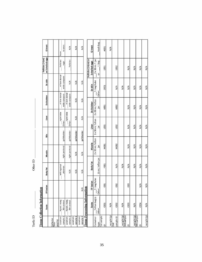
35

36

37
NECROPSY FIELD DATASHEET – DEAD STRANDED

38

39

40
NECROPSY FIELD DATASHEET - LIVE STRANDED/EUTHANIZED

41

42

43
3.3. INGESTED PLASTIC FIELD PROTOCOL Overview: This protocol is used to collect and analyze macroplastics ingested by sea turtles that have undergone necropsy. The objectives are to collect all macroplastics while also weighing the entire gut contents. Care must be taken to avoid contamination of the ingested plastics with other items. Because the samples collected for the BEMAST project of the NIST specimen bank are primarily intended for contaminants and health research, inadvertent contamination during sampling and sample handling is a major concern. The four major sources of contamination are from 1) airborne chemicals (e.g., cigarette smoke, dust, vehicle exhaust), 2) chemicals in or on surfaces that touch the samples or touch the supplies that will later touch the samples (e.g., sunscreen, insect repellent, plastic gloves, rain, sand carried by wind), 3) cross-contamination from one sample to another, and 4) supplies that directly touch the samples. The fourth source is inevitable but is minimized by pre-cleaning NIST supplies using standard and tested protocols. For this reason, only NIST-provided supplies should be used when collecting the samples intended for banking. During sampling and processing, care should be taken to minimize residues from all of the above sources. The most likely problems occur from touching NIST-provided supplies to gloves and bench-tops, or from carryover from one turtle tissue to the next. NIST-provided supplies must remain covered in original packaging and only removed immediately before its use. If supplies touch anything but the intended sample, it should be considered contaminated and be discarded. Unnecessarily touching tissues with the NIST-supplied gloves should also be avoided, but the supplied gloves are better than other gloves or bare hands. Materials Pre-Cleaning Materials (used at NIST only) • Hexane: Burdick and Jackson Cat # GC215-4 for trace analysis • Forceps are pre-cleaned by rinsing three times with hexane from a Teflon squirt bottle and
wrapped in hexane-rinsed foil • Foil squares (30x30 cm) are pre-cleaned by rinsing three times with hexane from a glass
pipettetaken from a glass jar • 100 mL to 400 mL glass jars with Teflon-lined lids are pre-cleaned by rinsing three times
with hexane from a glass pipette taken from a glass jar Re-Cleaning Materials (used at necropsy site) • Millipore (18 MΩ·cm) water in a 2 L Teflon bottle • Ethanol, absolute 99.5 % (Sigma Cat # 459844-4L) in Teflon squirt bottle • 23x23 cm (9x9 inch) Cleanroom wipers (Texwipe Cat# TX309) Sample Collection Materials: • Stainless steel knife to open GI tract • Powder free vinyl gloves (Kimtech Cat # 61002 for medium) • Data sheet • Clipboard with pens and sharpies • Tap water from hose

44
• Plastic bin • Pre-cleaned, foil-wrapped stainless steel forceps • 500 mL Teflon squirt bottle containing Millipore water from 2 L Teflon bottle • 500 mL Teflon squirt bottle containing 200 proof ethanol absolute 99.5 % (Sigma Cat #
459844-4L) • 23x23 cm (9x9 inch) Cleanroom wipers (Texwipe Cat # TX309) • Two balances to weigh bin (to 1 gram) and to weigh plastics (to 0.1 gram) • Large cooler to thaw and transport GI tracts • Small cooler with frozen gel packs and bubble wrap • Medium cooler for supplies • Large trash bags • Paper towels • Ruler to measure plastics in cm or mm • First aid kit, bug spray, headlamps • 30x30 cm (12x12 inch) Teflon bag (KNF Cat#300045-20; 6 max/animal) • Bag tie closure (2/animal) • Green Tyvec labels (Uline Cat # S-5984G) • Foil squares (30x30 cm), pre-cleaned with hexane • 100 mL to 400 mL glass jars with Teflon-lined lids, pre-cleaned with hexane • Sticker labels • Scienceware Lab polyester tape (VWR Cat # 36442-248) • Freezer -80 oC or LN2 vapor freezer/shipper Procedure: • Thaw GI tract in a shaded protected area away from scavengers and direct sunlight
overnight, if previously frozen. • When just thawed or while still partially frozen, open GI tract with knife cleaned between
turtles with tap water, Millipore water and ethanol in Teflon squirt bottles. • Record the tare mass of the bin, two pieces of foil, and one glass jar, separately. • Allow gut contents to fall into the plastic bin. Avoid losing any contents. • Remove plastics from gut contents with forceps before they fall into the bin. Place them
onto a clean cleanroom wiper. Record the location in the GI tract of observed macroplastics noting their color, size, shape (e.g., fragment, line, sheet, foam, or nurdle), and opacity. Rinse pieces with Millipore water from the Teflon squirt bottle while holding them with the forceps, and blot them dry on a clean cleanroom wiper.
• Take a photo of all plastics collected from one turtle’s gut on a 23x23 cm (9x9 inch) cleanroom wiper from directly above the sample. Include the entire cleanroom wiper in the photo with little background outside of the wipe (Figure 12).
• Transfer plastic pieces to one of the tared foil squares (if found, this should be sample A001) with forceps and fold 3 edges to make a sealed pouch.
• Collect cream-colored mucosa from upper intestines by dripping it into the pre-tared glass jar (if plastic is found, this should be sample A002, otherwise it is A001).
• Collect undigested prey items from the stomach or upper intestines using forceps into the other pre-tared foil square (if plastic is found, this should be sample A003, otherwise it is A002). Fold 3 edges to make a sealed pouch.

45
• Record the contents found in each section of the GI tract on the datasheet (see example below).
• Record the full masses of the bin, two pieces of foil, and glass jar, separately. • Label both foil pieces as shown below with a sharpie directly onto the foil. Be careful not to
puncture through the foil while writing. Place both into one 30x30 cm (12x12 inch) Teflon bag and label (as shown below) the Tyvec label and cable tie close the bag.
• Seal the glass jar tightly. Label the outside with an Avery label sticker (as shown below) and tightly wrap polyester tape around tube two times so that the tape covers the label and adheres to itself (otherwise the label and tape will fall off at LN2 temperatures).
• Place samples into cooler with frozen gel packs until freezing at -80 oC or below.
Figure 12. Example of a photograph of ingested plastics found from within one turtle’s gastrointestinal tract.
Labeling All samples should be labeled following a standard procedure. Each tube label should include the following information: Animal ID Aliquot ID (A = macroplastic GI tract sampling)
Date (dd Mmm yyyy) NIST/ Project code
SAMPLE: LL022301 or AS002323 or 070912A A001
12 Jun 2013 NIST / PIFSC – LL (or NIST/PIFSC – ST or NIST/PIFSC – MI) Storage Samples will be stored temporarily in a -80 oC or liquid nitrogen vapor-phase freezer at Hawaii Pacific University on Oahu with oversight by Brenda Jensen. Samples will be shipped to Jennifer Keller/Rebecca Pugh, NIST, Hollings Marine Lab, 331 Ft. Johnson Rd., Charleston, SC 29412 for long term storage in the NIST Marine ESB until a facility is built in Hawaii. Before transferring, relocating, or shipping samples, or if storage conditions fail, contact all of the following personnel: Jennifer Keller ([email protected]) 843-725-4822 or 843-442-2188 George Balazs ([email protected]) 808-983-5733 Thierry Work ([email protected]) 808-792-9520 Rebecca Pugh ([email protected]) 843-762-8952 or 843-709-0145 If you have questions about the sample or processing protocol contact: Jennifer Keller ([email protected]) 843-725-4822 (work) 843-442-2188 (cell phone)

46
INGESTED PLASTIC FIELD DATASHEET

47

48
3.4. EGG COLLECTION PROTOCOL Overview: Eggs are a common sample archived in environmental specimen banks because they are often analyzed for environmental contaminants. Sea turtle eggs, primarily green turtle and hawksbill sea turtles, will be collected from Hawaiian nests by nesting beach projects in collaboration with the Marine Turtle Research Program (MTRP) of the Pacific Islands Fisheries Science Center. Because the samples collected for the BEMAST project of the NIST Marine Environmental Specimen Bank (ESB) are primarily intended for contaminants and health research, inadvertent contamination during sampling and sample handling is a major concern. The four major sources of contamination are from 1) airborne chemicals (e.g., cigarette smoke, dust, vehicle exhaust), 2) chemicals in or on surfaces that touch the samples or touch the supplies that will later touch the samples (e.g., sunscreen, insect repellent, plastic gloves, rain, sand carried by wind), 3) cross-contamination from one sample to another, and 4) supplies that directly touch the samples. The fourth source is inevitable but is minimized by pre-cleaning NIST supplies using standard and tested protocols. For this reason, only NIST-provided supplies should be used when collecting the samples intended for banking. During sampling and processing, care should be taken to minimize residues from all of the above sources. The most likely problems occur from touching NIST-provided supplies to gloves and bench-tops, or from carryover from one turtle tissue to the next. NIST-provided supplies must remain covered in original packaging and only removed immediately before its use. If supplies touch anything but the intended sample, it should be considered contaminated and be discarded. Unnecessarily touching tissues with the NIST-supplied gloves should also be avoided, although preferred to other gloves or bare hands. Egg Collection (performed by permitted collaborators) 1. Conduct nest excavation and inventory post-emergence. 2. Count the number of hatched empty eggshells, live and dead hatchlings inside the nest and
outside of the nest, pipped, depredated, and unhatched eggs, preferably according to Miller JD. 1999. Determining clutch size and hatching success. In Eckert KL, Bjorndal KA, Abreu-Grobois FA, Donnelly M, eds, Research and Management Techniques for the Conservation of Sea Turtles. IUCN/SSC Marine Turtle Specialist Group Publication 4, Washington, DC, USA, pp 124–129.
3. Place unhatched eggs from a single nest into a single plastic baggie (not provided by NIST). 4. Label baggie with nest ID and date of excavation. 5. Freeze baggie as soon as possible. 6. Transport frozen eggs to the PIFSC Marine Turtle Research Program who will sort through
what they need to keep for other studies and will provide the remainder to NIST. Pre-Cleaning Materials (used at NIST only) • Hydrochloric acid: 36.5 % to 38 % VWR Cat# JT9530-33, diluted 2:1 with Millipore water
18.2 resistivity (MΩ·cm) • Nitric acid: 69.0 % to 70.0 % VWR Cat# BDH3046-2.5LPC, diluted 2:1 with Millipore
water 18.2 resistivity (MΩ·cm) • Ethanol: Sigma Aldrich Cat#459844, ACS reagent, 99.5 % (200 proof), absolute

49
• Chloroform: Sigma Aldrich Cat#319988
Processing Materials • Fume Hood • Millipore Water (≥18 MΩ·cm) • Interscience Bag Mixer • Balance • 15x18 cm (6x7 inch) Teflon bag 2 mm thick (KNF Cleanroom Cat# LB602:0607) • 18x25 cm (7x10 inch) Teflon bag 5 mm thick (KNF Cleanroom Cat# LB605:0710) • Heavy Duty Low Form 250 mL Glass Beakers (VWR Cat# 89000-200) • 1000 mL Plastic Beakers (VWR Cat#13890-148) • Stainless steel medical scissors • 7 mL Teflon jars with recessed lids (Savillex Cat# 200-007-10 for jar; Cat# 600-024-71),
pre-cleaned in the Marine ESB • Savillex Lid labels • Powder free vinyl gloves (Kimtech Cat# 61002 for medium) • Cryo-Babies labels, pre-printed for labeling cryovials (Cat# LCRY-1700-G) • 23x23 cm (9x9 inch) cleanroom wipers (Texwipe Cat# TX309) • 10x10 cm (4x4 inch) cleanroom wipers (Texwipe Cat# TX304) • Bytac Teflon surface protector (VWR Cat# 54112-100; Lab Pure) • Hexane GC/GC-MS Solvent (VWR Cat# BJGC215-4 for trace analysis) • Styrofoam Cryovial rack • 5.1 cm (2 inch) tall cryovial freezer boxes with or without 81-place dividers • Cryovials –2 mL self-standing, conical bottom, silicone washer, external threading (Corning
Cat# 430659), not pre-cleaned, lot # recorded on datasheet • 6" Electronic Digital Calipers (0.001" reading, Chicago Brand Model# 50001) • Teflon Squirt Bottle • Cardboard tubes • Heat sealer
Supply Pre-cleaning Procedure • Teflon jars and lids provided by NIST are pre-cleaned using the Marine ESB cleaning
protocol and the jars are air dried in the ISO Class 5 clean room to ensure contaminant free storage containers [15].
• Rinse glass beakers and stainless steel scissors three times with hexane from a Teflon squirt bottle.
Egg Processing Procedure 1) Wearing supplied vinyl gloves , choose the three best looking eggs per nest that are white
and puffy (not dessicated and sunken in; Figure 13).

50
Figure 13. Bag of unhatched eggs from one nest covered in sand and white puffy eggs chosen and rinsed in beaker of water. NOTE: Eggs should be placed on a cleanroom wiper not foil as shown in photo.
2) Clean the outside of the eggs, prior to opening: a) Pour 200 mL of Millipore water into a pre-cleaned glass beaker. b) Submerge eggs in water to remove sand (use same beaker of water for 3 eggs from the
same nest) c) Used gloved hands to wipe sand from shell d) Blot water from outside of egg with a large cleanroom wiper (use a new wiper for each
egg). 3) Weigh (g) each whole egg on a clean, dry, tared cleanroom wiper and record this value on
the datasheet. 4) To open the egg:
a) Place a 6” x 7” 2 mm Teflon bag in a clean beaker on a scale and tare (g). Foil the top of the bag over once to keep the open edge clean and help the bag stay open.
b) Carefully open egg with gloved fingers over the top of the tared Teflon bag seated in the beaker.
(1) **This is the tricky part.** Make a small pinched hole at the top of the egg, invert the egg over the bag to collect all of the egg contents only inside the bag.
(2) The most important things to keep in mind: (a) All of the egg contents must be in the bag, so squeeze the egg like a roll of
toothpaste. (b) Egg contents should not touch anything except a little bit of the outside of the
eggshell and the inside of the bag (especially not your gloves, table surface, or sand).
(c) Do not pick up dropped egg contents and place them into the jar. Record estimated loss on the datasheet, if over 10 % discard this entire egg and choose another one from the remaining and repeat above steps.
5) Record the egg contents mass on the datasheet and stage the embryo. The identification of early (E), middle (M), and late (L) embryonic stages are based on the following criteria: E = small white embryo (< 1 cm), perhaps a black eye spot, without an obvious carapace; M = white embryo with a carapace, without dark scutes (perhaps 1 cm to 2 cm long); L = large brown or white (amelaninic) embryo with fully formed scutes (perhaps 2 cm to 4 cm long) (Figure 14). An egg without visible development of an embryo is classified as “No” embryo.

51
Figure 14. Examples of egg contents from loggerhead sea turtle eggs with no development (No), early (E), middle (M), and late (L)-stage embryos. Note the eye spot among the E egg contents. The contents shall be inside the Teflon bag, not a glass beaker or on foil as shown.
6) If you see a middle or late stage embryo try not to let the contents fall into the Teflon bag. Instead allow the contents to collect in another beaker. Remove the embryo from the yolk sac and all other egg contents. Weigh the embryo alone and measure its flattened carapace length with calipers. Discard this sample or save it for the MTRP. Development that has progressed beyond ‘early development’ cannot be used by NIST.
7) If the egg contained no development or early development, then its contents should be in the Teflon bag. Record mass of egg contents on datasheet (pg. 2).
8) Repeat process until you have 3 eggs in separate bags (the last bag can be a 5 mm 7” x 10” Teflon bag), or no more eggs remain for that nest.
9) Clean (not thoroughly, just to remove sand), weigh, open, and stage all remaining eggs. Measure and weigh all middle to late stage embryos. Record all of this on the datasheet, but discard these samples.
10) Combine all of the eggs from a clutch together in one tared 5 mm thick (7x10 inch) Teflon bag for use in the Bag Mixer. Record mass of combined sample. a) Two eggs should be in 2 mm Teflon bags. b) The last or 3rd egg should be in a tared 5 mm Teflon bag. c) Use the mixer on the two thinner bags, for 10 s at speed 6 (see bag mixer instructions in
step 11). d) Then pour the 2 eggs into the thicker
bag to combine with the 1st egg. e) Record combined sample mass and
number of eggs pooled on datasheet (pg 2).
f) Then mix all 3 eggs as described here with the bag mixer.
Figure 15. Egg contents being homogenized in the BagMixer.
11) When all eggs from one nest are combined in a 5 mm Teflon bag, homogenize egg contents using the Bag Mixer (Figure 15). BagMixer Instructions: Speeds & time intervals may need to be modified as the viscosity of the turtle egg may differ from albatross eggs.
No E M L

52
a) Pull the latch on the back and turn the crank clockwise until stops and paddles are closest to the door. Turn the crank counterclockwise 5 turns (moves paddles back 5 mm).
b) Set Speed Dial at 6 and Time Dial at 30 s. c) Remove Teflon bag with egg contents from beaker. Unfold Teflon bag and gently
squeeze out air while placing between paddles and door of BagMixer. Depending on size of the sample, the paddles may need to be adjusted. Close door and check that paddles slightly compress sample, if not move crank clockwise a turn to move paddles closer, if compressed greatly, move crank counterclockwise a turn to move paddles back (if paddles are too close, the Teflon bag may crack).
d) Turn BagMixer on, hold the outside edge of the sample bag and latch door. BagMixer will automatically start and stop.
e) When completed Set Speed Dial to 7 and Time Dial to 10 s. f) Open and close latch again. When completed, check for visual homogenization, if can
still see albumin “chunks”, try again at Speed 6 and Time 10 s. The goal is complete visual homogenization without a large amount of foaming. The timings may need to be adjusted (speed 8 for 30 s has been used for stubborn samples).
12) Label (see instructions below), stand into racks, and open Teflon jars and cryovials. a) For samples of only one egg, fill at least two 7ml Teflon jars and six 2ml cryovials. b) For samples of two pooled eggs, fill at least four 7ml Teflon jars and eight 2ml
cryovials. c) For samples with three pooled eggs, fill six 7ml teflon jars and ten 2ml cryovials.
13) Record the lot number of the 2mL cryovial on the datasheet (find lot # imprinted on the bottom of the Corning bag).
14) Once homogenized, remove from BagMixer and stand up in a beaker. 15) Lift and position the bag so that the majority of the egg contents are in the opposite corner
from the bottom corner you will clean. Clean bottom corner with a cleanroom wiper, then squirt the corner with hexane, three times on each side of the bag. Allow solvent to dry while holding bag, do not allow the clean corner to touch any surface.
16) Using hexane-rinsed, air-dried scissors, cut open the very tip of the clean bag corner, to create a “pastry bag” type dispenser.
17) Using the “pastry bag”, fill jars and cryovials. Be certain not to fill past the 1.8 mL mark on cryovials and not past the top thread of the Teflon jar to allow for expansion when frozen.
18) If sample remains in the Teflon bag, you may heat seal it and place it into a cardboard tube. Note this on the datasheet.
19) Label jars and vials with the following information: NIST Nest ID (Species code, beach code, island code, sequential nest #)*
Tissue code and Aliquot number Processing Date (dd Mmm yyyy) NIST/Project code
*Assign the NIST Nest ID by using the running Excel file of previously processed eggs. A copy of this table is appended to the end of this protocol.
SAMPLE: CMPBOA01 (green turtle, Police Beach, Oahu, #01) E001
12Jul2014 NIST / PIFSC – EG

53
20) Organize and split Teflon jars and cryovials in cardboard freezer boxes. The Teflon jars
should be evenly split into 2 different boxes (without 81-place divider) for each nest (ex. E001, E002 in Box A; E003, E004 in Box B). Record box number for each jar on the data sheet. Cryovials should be organized the same way, using 2 different boxes (with the 81-place divider). Fill all boxes to capacity for each type of vial before moving to a new box.
21) Place samples in the -80 oC freezer and record date and time of freezing on datasheet. Allow cryovials and Teflon jars to remain in the upright position until frozen.
Storage and Shipping Samples will be stored temporarily in a -80 oC freezer at Hawaii Pacific University’s (HPU’s) Oceanic Institute on Oahu or a liquid nitrogen vapor freezer at HPU’s Hawaii Loa campus with oversight by Brenda Jensen. Samples will be shipped to Jennifer Keller, NIST, Hollings Marine Lab, 331 Ft. Johnson Rd., Charleston, SC 29412 for long term storage in the NIST Environmental Specimen Bank until a facility is built in Hawaii. Before transferring, relocating, or shipping samples, or if storage conditions fail, contact all of the following personnel: Jennifer Keller ([email protected]) 843-725-4822; 843-442-2188 George Balazs ([email protected]) 808-983-5733 Rebecca Pugh ([email protected]) 843-762-8952; 843-709-0145
If you have questions about the protocol or sampling, contact: Jennifer Keller ([email protected]) 843-725-4822 (work) 843-442-2188 (cell phone)

54
EGG PROCESSING DATASHEET

55

56

57
3.5. SCUTE HOMOGENIZATION PROTOCOL Overview: Scute samples were collected from sea turtles during live captures or from necropsies. Two methods of collection were used: Rusty Day’s method of moving a biopsy tool along the 8 posterior marginal scutes and George Balazs’s method of shaving the dorsal surface of one entire scute using a knife blade. Samples consist of very small masses (less than 1 g) of shavings that tend to static cling, and need to be homogenized and aliquotted for metals (approximately 0.1 g), mercury (0.1 g), stable isotopes (0.001 g), and also banked for future research. NIST research chemists discussed the homogenization method options and agreed that a mortar/pestle method would be best. The mortar and pestle method was tested at two temperatures, liquid nitrogen vapor (-150 °C) and at room temperature (22 °C). The room temperature method was determined to be the only feasible method even through the scute material will undergo thawing during the homogenization process. Homogenization Protocol Materials: • 100 % ethanol (Sigma Cat # 459844-4L) in Teflon squirt bottle • Millipore water (≥18 MΩ·cm) in Teflon squirt bottle • HEPA-filtered hood or cleanroom environment • Powder free vinyl gloves (Kimtech Cat # 61002 for medium) • Coorstek porcelain mortar (145 mL Cat # 60316) and pestle (Cat # 60317) pre-cleaned by
sonicating in warm soapy water for 20 min, scrubbed with a bristle brush, rinsed three times with tap water, three times with Millipore water, and cleaned with acids and solvents in the Marine ESB according to Pugh et al. [15]
• Teflon sheet (KNF Cleanroom Cat # LS0602:1212) • Scissors, stainless steel, pre-cleaned with ethanol and Millipore water, dried with cleanroom
wiper • Balance that weighs to 0.0001 g • Funnels, disposable polypropylene (VWR Cat # 414004-289), pre-cleaned with ethanol and
Millipore water, dried on cleanroom wiper in HEPA hood • Spatulas, disposable polypropylene (VWR Cat # 80081-190), not pre-cleaned • Cryovials –2 mL self-standing, conical bottom, silicone washer, external threading (Corning
Cat # 430659), not pre-cleaned • Cryo-Babies labels (Cat # LCRY-1700-G), pre-printed • Cryovial tube racks • 23x23 cm (9x9 inch) Cleanroom wipers (Texwipe Cat # TX309; NIST) • 5.1 cm (2 inch) tall cryovial freezer boxes with 81-place dividers • Liquid nitrogen biological dry shipper or vapor-phase freezer • Datasheet • Pens and Sharpies Scute Homogenization Procedure: • On an electronic tracking sheet, enter the turtle Field ID, tissue types, and tissue aliquots and
assign a specimen bank Storage ID (MTyearC###) to each tissue collected (See Tissue Cataloguing Protocol).
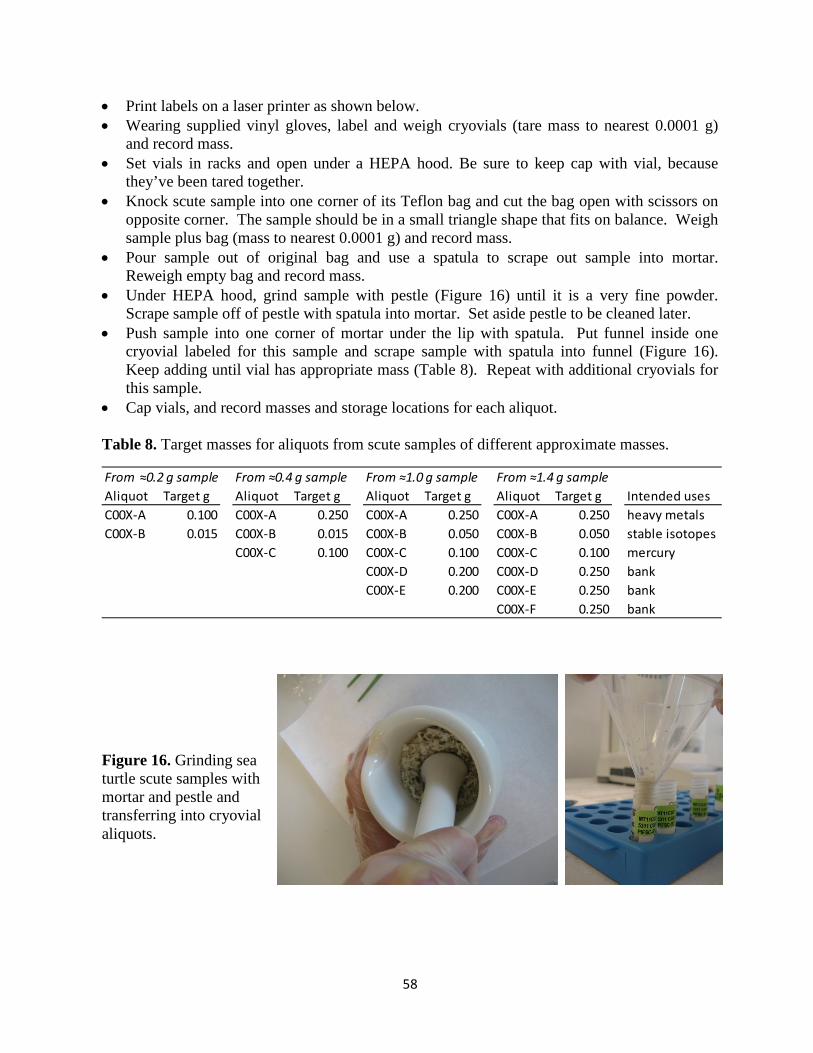
58
• Print labels on a laser printer as shown below. • Wearing supplied vinyl gloves, label and weigh cryovials (tare mass to nearest 0.0001 g)
and record mass. • Set vials in racks and open under a HEPA hood. Be sure to keep cap with vial, because
they’ve been tared together. • Knock scute sample into one corner of its Teflon bag and cut the bag open with scissors on
opposite corner. The sample should be in a small triangle shape that fits on balance. Weigh sample plus bag (mass to nearest 0.0001 g) and record mass.
• Pour sample out of original bag and use a spatula to scrape out sample into mortar. Reweigh empty bag and record mass.
• Under HEPA hood, grind sample with pestle (Figure 16) until it is a very fine powder. Scrape sample off of pestle with spatula into mortar. Set aside pestle to be cleaned later.
• Push sample into one corner of mortar under the lip with spatula. Put funnel inside one cryovial labeled for this sample and scrape sample with spatula into funnel (Figure 16). Keep adding until vial has appropriate mass (Table 8). Repeat with additional cryovials for this sample.
• Cap vials, and record masses and storage locations for each aliquot. Table 8. Target masses for aliquots from scute samples of different approximate masses.
Figure 16. Grinding sea turtle scute samples with mortar and pestle and transferring into cryovial aliquots.
From ≈0.2 g sample From ≈0.4 g sample From ≈1.0 g sample From ≈1.4 g sample Aliquot Target g Aliquot Target g Aliquot Target g Aliquot Target g Intended usesC00X-A 0.100 C00X-A 0.250 C00X-A 0.250 C00X-A 0.250 heavy metalsC00X-B 0.015 C00X-B 0.015 C00X-B 0.050 C00X-B 0.050 stable isotopes
C00X-C 0.100 C00X-C 0.100 C00X-C 0.100 mercuryC00X-D 0.200 C00X-D 0.250 bankC00X-E 0.200 C00X-E 0.250 bank
C00X-F 0.250 bank

59
Labeling:
All samples should be labeled following a standard procedure. After entering the turtle ID and tissue types into the tracking sheet, assign each sample of tissue a specimen bank Storage ID. Each tube label should include the following information:
Specimen bank sample ID (assigned as explained in the Tissue Cataloging Protocol) Original turtle ID – aliquot ID NIST/Project code SAMPLE: MT11C025 LL0059 C001-A
NIST/PIFSC-LL Storage: Samples are predominantly homogenized in the NIST Marine ESB and stored in the liquid nitrogen vapor freezers. If samples are homogenized elsewhere, they should be stored at -80 oC or below and shipped to Jennifer Keller/Rebecca Pugh, NIST, Hollings Marine Laboratory, 331 Fort Johnson Road, Charleston, SC 29412 for long term storage in the NIST Marine ESB until a facility is built in Hawaii. Before transferring, relocating, or shipping samples, or if storage conditions fail, contact all of the following personnel: Jennifer Keller ([email protected]) 843-725-4822; 843-442-2188 George Balazs ([email protected]) 808-983-5733 Rebecca Pugh ([email protected]) 843-762-8952; 843-709-0145 If you have questions about the protocol or sampling, contact: Jennifer Keller ([email protected]) 843-725-4822 (work) 843-442-2188 (cell phone)

60
SCUTE HOMOGENIZATION DATASHEET
MT# Cryovial lotTurtle ID Date/NameScute ID C001 Scute mass (g)
Aliquot ID C001-A C001-B C001-C C001-D C001-E C001-F
Cryovial tare (g)
Cryovial final (g)
Storage location C001-A C001-B C001-C C001-D C001-E C001-F
Freezer
Rack
Box
XY

61
3.6. TISSUE CATALOGING PROTOCOL After receipt of samples at NIST, tissues are inventoried and entered into Excel spreadsheets and Freezerworks Unlimited (Dataworks Development, Inc.) sample management database with data from the field datasheets as well as the freezer location information. Every tissue sample is given a unique BEMAST Storage ID, similar to other projects of the Marine ESB, whether it was fresh homogenized at the field site (e.g., blood) or is destined for homogenization at NIST (e.g., liver). For example, the entire blood sample from one turtle is given one ID (e.g., MT11W001).
MT 11 W 001
Subsequent aliquots from that specific sample are given an Aliquot ID, such as B001 (1st whole blood aliquot), P001 (1st plasma aliquot), W001 (1st buffy coat aliquot), R001 (1st red blood cell sample), and R002 (2nd red blood cell sample). Blood samples may or may not be relabeled with the original Storage ID (MT ID). For samples that are homogenized at NIST, each subsample is given an original Storage ID (MT ID). For example, if four scute samples were taken from different locations on one turtle carapace, these were labeled in the field as C001, C002, C003, and C004 along with the field turtle ID. Each of these four samples would have a different MT ID. After homogenization at NIST, containers are labeled with the new MT ID as well as the original sample ID (C001) plus a letter designating the aliquot (C001-A). This will become the Aliquot ID. A future bar code labeling system will be developed in association with the Marine ESB specimen tracking database, Freezerworks Unlimited.
Program: MT-Marine Turtle
Year sample collected
Tissue Type: Refer to Table 3 –
Tissue Code
nth sample of this tissue type collected in the given year

62
4. RESULTS 4.1. COLLECTIONS REPORT Using the protocols detailed above, the BEMAST project collections now include 854 samples from 288 individual sea turtles and 38 sea turtle nests (Table 9). These samples come from live captures, necropsies, and egg collections. Samples from all five species have been collected and locations encompass the Hawaiian Islands, Palmyra Atoll, Saipan and Tinian, and pelagic waters of the tropical Pacific Ocean (Figure 17). Table 9. Summary of sea turtle samples collected and archived as a part of the BEMAST project.
BEMAST collections began in the Main Hawaiian Islands in 2011 in close collaboration with the PIFSC Marine Turtle Research Program, the USGS National Wildlife Health Center, and
Species Capture Method Location FP Tumor Status No. of Animals Collection Date
(Sample Type)
Green Live capture Kailua Bay, Oahu 0% 20 (blood, scute) Mar 2011
Green Live capture Kiholo Bay, Hawaii 0% 20 (blood, scute) May 2011
Green Live capture Kapoho Bay, Hawaii 34% 22 (blood, scute) Nov 2011
Green Live capture Kailua Bay, Oahu 5% 21 (blood, scute) Jul 2012
Green Live capture Palmyra Atoll 0% 20 (blood, scute) Jul 2012
Green Live capture Palmyra Atoll 0 22 (blood, scute, mouth algae)
Jun-Jul 2013
Green Live capture San Diego, CA 0% 7 (blood) Jun-Aug 2013
Green Live capture Kailua Bay, Oahu 5% 20 (blood, scute, mouth algae)
Jul 2013
Green Live capture Kiholo Bay, Hawaii 0% 21 (blood, scute, mouth algae)
Dec 2013
Green Live capture Saipan & Tinian, CNMI 0% 20 (blood, scute, mouth algae)
Dec 2013
Green Live strandings euthanized
Main Hawaiian Islands 100% 20 (blood, scute, fat, liver, FP lesion)
Jul 2011 – Jul 2012
Green Captivity Sea Life Park, Oahu 0% 6 (blood, scute) Aug 2012
Green Longline caught and drowned
>200 km from American Samoa & Hawaiian Is.
0% 14 (scute, fat, muscle, bile, liver)
Mar 2011 - Jan 2014
Green Dead stranding Main Hawaiian Islands 0% 7 (scute) May 2011 - May 2013
Green Dead stranding San Diego, CA 0% 1 (scute, fat, muscle, bile, liver)
Jan 2013
Green Acoustic dead Kwajalein Atoll, Marshall Islands; 1992
0% 3 (scute, fat, liver, muscle, bile)
Jul 2012
Green Unhatched eggs Main Hawaiian Islands N/A 26 nests Jan 2013 - Jan 2014
Olive Ridley Longline caught and drowned
>200 km from American Samoa & Hawaiian Is.
0% 22 (scute, fat, muscle, bile, liver, GI tract)
Mar 2011 - Jan 2014
Olive ridley Dead stranding Main Hawaiian Islands 0% 4 (scute, fat, liver, muscle, bile, GI tract)
Nov 2011 - Jul 2013
Hawksbill Live capture Palmyra Atoll 0% 2 (blood, scute) Jun-Jul 2013
Hawksbill Live capture Saipan & Tinian, CNMI 0% 4 (blood, scute) Dec 2013
Hawksbill Dead stranding Main Hawaiian Islands 0% 3 (scute, fat, muscle, bile, liver)
Nov 2011 - Jul 2013
Hawksbill Acoustic dead Kwajalein Atoll, Marshall Islands; 1992
0% 2 (scute, fat, muscle, bile, liver)
Jul 2012
Hawksbill Unhatched eggs Main Hawaiian Islands N/A 12 nests Dec 2012 - Jan 2014
Leatherback Longline caught and drowned
>200 km from American Samoa & Hawaiian Is.
0% 4 (blubber, fat, skin, liver, muscle)
Mar 2011 - Jul 2013
Loggerhead Longline caught and drowned
>200 km from American Samoa & Hawaiian Is.
0% 3 (scute, fat, muscle, bile, liver, GI tract)
Mar 2011 - Jan 2014

63
Hawaii Preparatory Academy. The live capture field protocol was developed and tested initially on green sea turtles in Hawaii with a parallel objective to assess real-time contaminant exposure at three sites with varying incidence of FP. Twenty green turtles were sampled for blood and scute shavings near the Kawainui Canal in Kailua Bay on Oahu in March 2011. Although this site has had a moderate incidence of FP in the past, no turtle sampled in March exhibited the disease. In May 2011, twenty turtles were sampled on the Kona coast of Hawaii (the “Big Island”) at Kiholo Bay. This site has historically been FP-free and no turtles captured during this sampling event had the disease. In November 2011, 24 green turtles were sampled at a third Hawaiian site, Kapoho Beach on the eastern coast of the Big Island, which has a higher FP rate (34 % during our sampling event).
Figure 17. Locations of sea turtles samples collected and archived for BEMAST project.
Kailua Bay has been identified as the site for annual live capture sampling in Hawaii, because of the intermediate FP rates and historical and current human land use in this watershed. This site has been sampled for a second (July 2012) and third year (July 2013) in which 5 % of the turtles captured exhibited external FP tumors. Because of interesting hydroxylated halogenated compounds found in plasma of Kiholo turtles, this site was sampled again in December 2013, in which algae was also collected from the turtles’ mouths and the reef. In July 2012, the live capture protocol was also tested successfully in a more remote, logistically difficult field site at Palmyra Atoll in collaboration with American Museum of Natural History (AMNH). Twenty green turtles were sampled in July to August 2012 with no FP documented in the five years of the AMNH-conducted project. Sampling at Palmyra Atoll during the summer of 2013 repeated the green turtle captures and successfully added two hawksbill turtles. In addition, live captures of green turtles in San Diego Bay, California, in collaboration with the Southwest Fisheries Science Center (SWFSC) began in June 2013 and is on-going, and sampling of 20 greens and 4 hawksbills was successful in CNMI off of Saipan and Tinian in December 2013. The live capture field protocol was also used to sample blood and scute shavings from six adult green turtles maintained in captivity for decades at Sea Life Park. Equal sample sizes of males and females were selected to determine if contaminant levels differ between sexes, since the

64
females lay eggs on a small nesting beach and could potentially offload contaminants into those eggs. Pellet food was also collected and analysis of these samples will determine if food provided in captivity leads to higher levels of contaminants in captive turtles than wild turtles. The live capture and necropsy field protocols were used to sample 20 green turtles that stranded live in the Main Hawaiian Islands with end-stage FP and required euthanasia. Blood, scute, FP lesion, fat, and liver were collected from these animals. Future collections will not be scheduled until contaminants are measured in the preliminary sample set. The necropsy field protocol was used to sample several species of sea turtles during multiple necropsy session events. The protocol began with only scute, fat, and liver sampling (plus skin and blubber from leatherbacks) but expanded in July 2012 to include muscle and bile and again in December 2012 to include gastrointestinal tracts for ingested plastics. Turtles came primarily from the PIRO Fisheries Observer Program that maintains 100 % coverage on the Hawaiian and American Samoan longline fishery fleet. Observers document the by-catch of the fishery and collect turtles that die. These turtles, refrigerated or frozen on the fishing boats, are transferred to the PIFSC Marine Turtle Research Program, which conducts necropsies with the USGS National Wildlife Health Center. They have agreed to freeze the whole turtle and conduct necropsy sessions that are concurrent with BEMAST sampling. Necropsies were also performed in 2012 and 2013 on a select few fresh dead strandings in the Main Hawaiian Islands (rare species, rare age classes, interesting disease states) and on five turtles that died due to acoustic trauma in Kwajalein Atoll in the Republic of the Marshall Islands in 1992 in collaboration with the PIFSC Marine Turtle Research Program. In addition, the necropsy field protocol was used to sample one green turtle from California in collaboration with SWFSC. Egg collections began in December 2012. Unhatched eggs were collected by numerous nesting beach biologists in the main Hawaiian Islands from 26 green and 12 hawksbill turtle nests and transferred to the PIFSC Marine Turtle Research Program. Selected eggs were then transferred to Hawaii Pacific University for processing. 4.2. HOMOGENIZATION REPORT Scute samples from all turtles sampled in and before December 2013 have been homogenized in the NIST Marine Environmental Specimen Bank using the scute homogenization protocol. Three to four aliquots are common from live captured samples, while up to nine aliquots have been possible from samples taken during necropsy. All egg samples have been homogenized at Hawaii Pacific University using the egg collection protocol.
4.3. ANALYSIS REPORT Analysis of certain samples is underway or planned. Seven projects have begun analysis, but results are either too preliminary to include in full detail in this report or are being reported elsewhere. The project objectives and brief results summary are provided here: 1. Plasma samples from non-banked green turtles with and without FP and those that were
basking versus swimming from Hawaii were analyzed by Tracey Schock in the NIST nuclear magnetic resonance (NMR) facility at the HML in Charleston for metabolomics.

65
The NMR metabolome was not significantly different between turtle categories but was different between locations. The paper was published by Current Metabolomics [22].
2. Scute samples from three green turtles (stranded and/or longline) collected at four locations across the carapace were homogenized as described above and analyzed by Colleen Bryan in the NIST inorganic chemistry lab in Charleston for a suite of trace elements. The homogenization technique did not introduce additional trace elements into the sample, as determined by analyzing three replicates of NIST SRM 2586 Trace Elements in Soil (contains lead from paint) that was homogenized using this protocol. Trace element concentrations differed between the Day and Balazs scute collection techniques, and the Balazs method has been chosen for the majority of future scute collection because it allows for a greater sample mass to be collected and in less time.
3. Scute samples from four locations on the 5th central scute of six stranded green turtles were collected to assess differences in trace element concentrations across the anterior versus posterior portion as well as the outer versus inner layer of this scute. Samples were homogenized and are awaiting analysis by Colleen Bryan in the NIST inorganic chemistry lab.
4. Plasma subsamples from 13 green turtles from each of the three Hawaiian live capture locations, from 14 FP-stranded green turtles that required euthanasia, and from 5 green turtles from Palmyra Atoll (n=58 total) were analyzed for persistent organic pollutants (POPs) and hydroxylated polychlorinated biphenyls (PCBs) and polybrominated diphenyl ethers (PBDEs) by Jennifer Keller and Frances Nilsen in the NIST organic chemistry lab at the HML. Concentrations of the POPs were greatest in the euthanized FP animals, probably due to lipid mobilization of these compounds into the blood during weight loss. Kiholo green turtles had greater levels of hydroxylated compounds that may be naturally produced by their prey, marine algae. These data are in a manuscript in review by a journal (Keller et al. Investigating the Potential Role of Persistent Organic Pollutants in Hawaiian Green Sea Turtle Fibropapillomatosis. Submitted to Environmental Science and Technology).
5. Plasma subsamples from three green turtles from Kailua Bay, Mar 2011, were provided to John Bowden in the NIST organic chemistry lab at the HML to gain preliminary results for a lipidomics study comparing various species. Other species include the loggerhead sea turtle (samples from South Carolina) and American alligators.
6. Ingested plastic amounts, types and geographic comparisons to the described gyres of marine debris in the Pacific Ocean are being assessed in longline captured olive ridley sea turtles by a University of Hawaii graduate student, Katharine Clukey.
7. Plasma subsamples from 13 green turtles from each of the three Hawaiian live capture locations, 14 FP-stranded green turtles that required euthanasia, 10 green and 2 hawksbill turtles from Palmyra Atoll, 12 green and 4 hawksbill turtles from Saipan and Tinian, and eggs from 12 Hawaiian hawksbill nests were analyzed for perfluoroalkyl contaminants (PFCs) by Jennifer Keller and John Brooker in the NIST organic chemistry lab at the HML. Perfluorooctane sulfonate (PFOS) was the predominant PFC. Differences between groups, locations, and species will be investigated.
Planned or prospective analyses include the following projects: 1. Algae samples collected from the mouths of green turtles live captured in Kailua Bay,
Kiholo Bay, and Palmyra Atoll will be analyzed by the NIST organic chemistry lab for POPs and hydroxylated compounds.

66
2. Blood or scute samples will be analyzed from live captured and euthanized turtles for trace elements by the NIST inorganic chemistry lab to understand baseline exposure as well as the influence of these elements on FP.
3. Ingested plastics, mucosa, and undigested prey items will be analyzed by Katharine Clukey for POPs and plastic-additive pollutants.
4. A prospective National Research Council post-doctoral fellow, Lisa Komoroske, has requested to perform a gene expression (transcriptomics) study on the PAXgene blood tubes that were collected from live captured green and hawksbill turtles in Hawaii, Palmyra, San Diego and Saipan.
5. Hawksbill fat and egg samples may be assessed in the NIST organic chemistry lab at the HML for POPs and natural halogenated compounds.
5. ACKNOWLEDGMENTS The authors would like to acknowledge the following individuals and institutions for their work and support in developing the Biological and Environmental Monitoring and Archival of Sea Turtle Tissues (BEMAST). Field sampling was conducted by the following individuals: George Balazs (National Oceanic and Atmospheric Administration’s [NOAA’s] Pacific Islands Fisheries Science Center [PIFSC]) and his staff (Stacy Hargrove, Shawn Murakawa, Shandell Brunson, Tyler Bogardus, Wendy Marks, Devon Franke, Irene Nurzia Humburg, and Emily Walker), Thierry Work (National Wildlife Health Center, U.S. Geological Survey [USGS]) and his staff (Bob Rameyer, Renee Breeden), Marc Rice, Laura Jim, and Arjun Clarry (Hawaii Preparatory Academy) and their students, Eleanor Sterling (American Museum of Natural History) and her staff and colleagues (Erin Vintinner Bentley, Felicity Arengo, Kevin Frey, Kate McFadden [Clemson University], Cheryl King [Hawaii Wildlife Fund], Alexander Gaos [Eastern Pacific Hawksbill Initiative], Kristen Hart and Thomas Selby [USGS]), Jeff Seminoff, Heather Harris, Scott Benson, Tomo Eguchi, and Camryn Allen (NOAA Southwest Fisheries Science Center), and Tammy Summers, Jessy Hapdei, (Department of Land and Natural Resources, Commonwealth of the Northern Marianas Islands) and their staff and volunteers. Amber Fong (Ph.D. student from Taiwan) assisted with sample processing in Kapoho Bay, Hawaii, Nov 2011. Dolphin Quest in the Hilton Waikoloa Village provided laboratory space and logistical support during the Kiholo Bay sampling events. The Nature Conservancy provided logistical support for the Palmyra Atoll sampling and access to Kiholo Bay. Katharine Clukey (University of Hawaii master’s student) assisted with sample collection and processing. Long-line incidentally caught turtles were provided by the Fisheries Observer Program of the NOAA Pacific Islands Regional Office (PIRO). Turtle eggs were provided by several nesting programs in the Hawaiian Islands, including Skippy Hau (Division of Aquatic Resources, State of Hawaii) and Will Seitz and Lauren Kurpita (Hawaii Island Hawksbill Turtle Recovery Project). Logistical support and sample collection/processing were provided by Brenda Jensen (Hawaii Pacific University [HPU]) and her staff/students (Frannie Nilsen, Angela Hansen, Melannie Bachman, Jessica Jacob, Elise Kohli, Adam Kurtz, Kerry Foltz, Julia Smith). Frannie Nilsen deserves a special thank you for tremendous hours of help with sample collection at numerous locations, scute and egg homogenizations, egg protocol development, supply organization, data

67
entry, sample transport and shipping, and assistance in the NIST organic chemistry laboratory for the POPs measurements in green turtle plasma. Assistance for scute homogenization was also provided by Melannie Bachman (HPU), Harry Quedenfeld (Wofford College), James Dean (Winthrop University), and John Brooker (College of Charleston). The authors would also like to thank Thierry Work (USGS), Peter Dutton, Jeff Seminoff, Scott Benson, and Heather Harris (NOAA Southwest Fisheries Science Center), Kyle Van Houtan and T. Todd Jones (PIFSC), Irene Kelly, Karen Frutchey, and Kim Maison (NOAA PIRO), Asuka Ishizaki (Western Pacific Fisheries Management Council), Will Seitz and Lauren Kurpita (National Park Service), Karen Arthur, Brian Bowen, Jason Turner, Karla McDermid, Jo-Ann Leong, Jay Murdock, and Yuanan Lu (University of Hawaii), David Hyrenbach (HPU) for their advice in the development of this project. Fernanda Colabuono (Brazil) and Christopher Lepczyk (UH) provided advice and support for the plastic ingestion protocol and study. We thank the other members of the NIST Pacific Islands Team for their advice and support: Colleen Bryan, Stacy Vander Pol, Amanda Moors, Rusty Day, and John Kucklick. Colleen Bryan provided preliminary trace element screening of green turtle scute samples. Tracey Schock and Dan Bearden (NIST) provided metabolomics data from Hawaiian green turtles.
6. DISCLAIMER Certain commercial equipment or instruments are identified in this paper to specify adequately the experimental procedures. Such identification does not imply recommendations or endorsement by the National Institute of Standards and Technology nor does it imply that the equipment or instruments are the best available for the purpose.
7. REFERENCES
[1] S. A. Wise and B. J. Koster, Considerations in the design of an environmental specimen bank - Experiences of the National Biomonitoring Specimen Bank Program, Environ. Health Perspect. 103, 61-67 (1995). DOI 10.2307/3432562 [2] P. H. Dutton, D. Squires, and M. Ahmed, Conservation of Pacific Sea Turtles. University of Hawaii Press, Honolulu (2011) p. 496. [3] K. S. Van Houtan, S. K. Hargrove, and G. H. Balazs, Land use, macroalgae, and a tumor-forming disease in marine turtles, PLoS ONE. 5 (9), e12900 (2010). DOI 10.1371/journal.pone.0012900 [4] A. A. Aguirre and P. L. Lutz, Marine turtles as sentinels of ecosystem health: Is fibropapillomatosis an indicator?, EcoHealth 1, 275-283 (2004). [5] J. M. Keller, Exposure to and effects of persistent organic pollutants, in The Biology of Sea Turtles Volume III, J. Wyneken, K. J. Lohmann, and J. A. Musick, Editors. CRC Press, Boca Raton, FL (2013) p. 285-328.

68
[6] S. G. O'Connell, M. Arendt, A. Segars, T. Kimmel, J. Braun-McNeill, L. Avens, B. Schroeder, L. Ngai, J. R. Kucklick, and J. M. Keller, Temporal and spatial trends of perfluorinated compounds in juvenile loggerhead sea turtles (Caretta caretta) along the east coast of the United States, Environ. Sci. Technol. 44 (13), 5202-5209 (2010). DOI 10.1021/Es9036447 [7] J. M. Keller, J. R. Kucklick, M. A. Stamper, C. A. Harms, and P. D. McClellan-Green, Associations between organochlorine contaminant concentrations and clinical health parameters in loggerhead sea turtles from North Carolina, USA, Environ. Health Perspect. 112 (10), 1074-1079 (2004). [8] J. M. Keller, P. D. McClellan-Green, J. R. Kucklick, D. E. Keil, and M. M. Peden-Adams, Effects of organochlorine contaminants on loggerhead sea turtle immunity: comparison of a correlative field study and in vitro exposure experiments, Environ. Health Perspect. 114 (1), 70-76 (2006). [9] J. M. Keller, L. Ngai, J. Braun McNeill, L. D. Wood, K. R. Stewart, S. G. O'Connell, and J. R. Kucklick, Perfluoroalkyl contaminants in plasma of five sea turtle species: Comparisons in concentration and potential health risks, Environ. Toxicol. Chem. 31 (6), 1223–1230 (2012). [10] NMFS and USFWS, Recovery plan for U.S. pacific populations of the hawksbill turtle (Eretmochelys imbricata), National Marine Fisheries Service, Silver Spring, MD, 1998, 83 pp. [11] NMFS and USFWS, Recovery plan for U.S. pacific populations of the leatherback turtle (Dermochelys coriacea), National Marine Fisheries Service, Silver Spring, MD, 1998, 65 pp. [12] NMFS and USFWS, Recovery Plan for U.S. Pacific Populations of the East Pacific Green Turtle (Chelonia mydas), National Marine Fisheries Service, Silver Spring, MD, 1998, 51 pp. [13] NMFS and USFWS, Recovery plan for U.S. pacific populations of the olive ridley turtle (Lepidochelys olivacea), National Marine Fisheries Service, Silver Spring, MD, 1998, 52 pp. [14] NMFS and USFWS, Recovery plan for U.S. pacific populations of the loggerhead turtle (Caretta caretta), National Marine Fisheries Service, Silver Spring, MD, 1998, 59 pp. [15] R. S. Pugh, M. B. Ellisor, A. J. Moors, B. J. Porter, and P. R. Becker, Marine Environmental Specimen Bank: Clean room and specimen bank protocols, National Institute of Standards and Technology IR 7389, Gaithersburg, MD, 2007, 113 pp. [16] J. M. Keller, J. R. Kucklick, C. A. Harms, and P. D. McClellan-Green, Organochlorine contaminants in sea turtles: Correlations between whole blood and fat, Environ. Toxicol. Chem. 23 (3), 726-738 (2004). [17] J. M. Keller, Forty-seven days of decay does not change persistent organic pollutant levels in loggerhead sea turtle eggs, Environ. Toxicol. Chem. 32, 747-756 (2013). [18] P. R. Becker, B. J. Porter, E. A. Mackey, M. M. Schantz, R. Demiralp, and S. A. Wise,

69
National Marine Mammal Tissue Bank and Quality Assurance Program: Protocols, inventory, and analytical results, National Institute of Standards and Technology IR 6279, Gaithersburg, MD, 1999 [19] P. R. Becker, S. A. Wise, L. Thorsteinson, B. J. Koster, and T. Rowles, Specimen banking of marine organisms in the United States: Current status and long-term prospective, Chemosphere 34 (9-10), 1889-1906 (1997). [20] NMFS, Sea turtle research techniques manual, NOAA Technical Memorandum NMFS-SEFSC-579, 2008, 92 pp.
[21] R. D. Day, S. J. Christopher, P. R. Becker, and D. W. Whitaker, Monitoring mercury in the loggerhead sea turtle, Caretta caretta, Environ. Sci. Technol. 39, 437-446 (2005). [22] T. B. Schock, J. M. Keller, M. Rice, G. H. Balazs, and D. W. Bearden, Metabotyping of a protected non-model organism, green sea turtle (Chelonia mydas), using 1H NMR spectroscopy and optimized plasma methods for metabolomics, Current Metabolomics 1, 279-290 (2013).

70
APPENDIX A EXAMPLE OF FORMAL OWNERSHIP AND TRANSFER AGREEMENT OF SEA TURTLE SAMPLES Sea turtle samples will be collected in collaboration between the National Institute of Standards and Technology (NIST) and _______________________________________________. This agreement determines the permitted field collectors’ requirements for ownership and release of samples that are stored at the NIST’s Marine ESB. Sample origination: NIST project code ____________________ Dates of collection ____________________ Location(s) of collection ____________________ Permit agencies and numbers _____________________ (agreement remains in effect upon renewals and additional permits) Ownership of samples: remains with the field collector’s agency remains with ________________________ is permanently loaned to NIST is permanently transferred to NIST 1) Release of samples to researchers at NIST, to field collector’s agency, or to either agency’s
associated partners (e.g., Hollings Marine Laboratory, Hawaii Pacific University): Field collector expects defined subsamples to be returned to them within a defined
timeframe_________________________________________________________________. Field collector allows release of samples to be determined by the BEMAST lead only after
written notification and discussion of authorship and acknowledgement expectations. Field collector allows release of samples without notification. 2) Release of samples to researchers outside of NIST, field collector’s agency, or either
agency’s associated partners: Field collector requires review of research proposal and will provide requirements for
authorship and acknowledgement. Reviews of proposals are due within 3 weeks of submission.
Field collector allows release of samples to be determined by the BEMAST lead only after written notification and discussion of authorship and acknowledgement expectations.
Field collector allows release of samples without notification and review. Supervisory management declaration at time of signing: Name Email Phone Lead of BEMAST Jennifer Keller [email protected] 843-725-4822 Program Coordinator of NIST Marine Environmental Specimen Bank
Rebecca Pugh [email protected] 843-762-8952

71
Group Leader Paul Becker [email protected] 843-725-4815 Agency of permitted field collector
Lead of field collections Supervisor of lead collector The signed parties have agreed upon the ownership and release procedures and requirements of sea turtle samples transferred and archived by the National Institute of Standards and Technology’s (NIST’s) Biological and Environmental Monitoring and Archival of Sea Turtle Tissues (BEMAST) project. Signed Parties: _____________________________________ ______________________ Lead of field collections Date _____________________________________ ______________________ Supervisor of lead collector Date _____________________________________ ______________________ Lead of BEMAST Date _____________________________________ ______________________ Program Coordinator for the Marine ESB Date _____________________________________ ______________________ Group Leader for the Marine ESB Date