Embed Size (px)
Citation preview

full title:
An Introduction to Multiple Sequence Alignment — and the T-Coffee Shop. Beyond just
aligning sequences: how good can you make your alignment, and so what?
running head:
Multiple Sequence Alignment and T-Coffee
author:
Steven M. Thompson
School of Computational Science, Florida State University, Tallahassee, FL, 32306-4120
e-mail: [email protected]
mailing address and phone:
2538 Winnwood Circle, Valdosta, GA, 31601-7953, 229-249-9751
1

abstract:
I begin the chapter with a discussion of the fundamental principles of multiple sequence
alignment starting with pairwise dynamic programming, then I move onto significance and
similarity statistics, then amino acid scoring matrices, and I end the introduction with multiple
sequence alignment algorithms themselves. Reliability issues, complications, and applications
of multiple sequence alignment are next discussed. The chapter finishes off with a description
and tutorial on using the T-Coffee multiple sequence alignment package.
keywords:
multiple sequence alignment, dynamic programming, significance, reliability, T-Coffee, M-
Coffee, 3DCoffee
2

1. Introduction
What can we know about a biological molecule, given its nucleotide or amino acid sequence?
How does it fit into a particular system in some organism? What is its role in some network?
We may be able to learn about it by searching for particular patterns within it that may reflect
some function, such as the many motifs ascribed to catalytic activity; we can look at its overall
content and composition, such as do several of the gene finding algorithms; we can map its
restriction enzyme or protease cut sites; and on and on. However, what about comparisons
with other sequences? Is this worthwhile? Yes, naturally it is: inference through homology is
fundamental to all the biological sciences. We can learn a tremendous amount by comparing
and aligning our sequence against others.
Furthermore, the power and sensitivity of sequence based computational methods dramatically
increase with the addition of more data. More data yields stronger analyses — if done
carefully! Otherwise, it can confound the issue. The patterns of conservation become ever
clearer by comparing the conserved portions of sequences amongst a larger and larger
dataset. Those areas most resistant to change are most important to the molecule, and to the
system that molecule interacts with. The basic assumption is that those portions of sequence
of crucial structural and functional value are most constrained against evolutionary change.
They will not tolerate many mutations. Not that mutation does not occur in these regions, just
that most mutation in the area is lethal, so we never see it. Other areas of sequence are able
to drift more readily, being less subject to this evolutionary pressure. Therefore, sequences
end up a mosaic of quickly and slowly changing regions over evolutionary time.
However, in order to learn anything by comparing sequences, we need to know how to
compare them. We can use those constrained portions as ‘anchors’ to create a sequence
alignment allowing comparison, but this brings up the alignment problem and ‘similarity.’ It is
3

easy to see that sequences are aligned when they have identical symbols at identical
positions, but what happens when symbols are not identical, or the sequences are not the
same length. How can we know when the most similar portions of our sequences are aligned,
when is an alignment optimal, and does optimal mean biologically correct?
A ‘brute force,’ naive approach just won’t work. Even without considering the introduction of
gaps, the computation required to compare all possible alignments between just two
sequences requires time proportional to the product of the lengths of the two sequences.
Therefore, if two sequences are approximately the same length (N), this is a N 2 problem. The
calculation would have to repeated 2N times to examine the possibility of gaps at each
possible position within the sequences, now a N4N problem. Waterman (1) pointed out that
using this naïve approach to align two sequences, each 300 symbols long, would require1088
comparisons, more than the number of elementary particles estimated to exist in the universe,
and clearly impossible to solve! Part of the solution to this problem is the dynamic
programming algorithm, as applied to sequence alignment, and this will be reviewed next.
1.1. Dynamic programming
Dynamic programming is a widely applied computer science technique, often used in many
disciplines whenever optimal substructure solutions can provide an optimal overall solution.
Let’s begin with an overview of sequence alignment dynamic programming with just two
sequences. I’ll use an incredibly oversimplified example first: we’ll consider matching symbols
to be worth one point, and will not consider gapping at all. The solution occurs in two stages.
The first begins very much like dot matrix methods; the second is totally different. Instead of
calculating the ‘score matrix’ on the fly, as is often taught as one proceeds through the graph, I
like to completely fill in an original ‘match matrix’ first, and then add points to those positions
that produce favorable alignments next. I also like to illustrate the process working through the
4

cells, many authors prefer to work through the edges; they are equivalent. Points are added
based on a “looking-back-over-your-left-shoulder” algorithm rule where the only allowable
trace-back is diagonally behind and above. The illustration follows below in Table 1.
Here are the two alignments from the path graph (f) in Table 1. They both have a score of
three, the three matches found by the algorithm, and the highest score in the bottom row of the
solved matrix:
SCATS SCA.TS | || | ||AC.TS ..ACTS
Most software will arbitrarily (based on some internal rule) choose one of these to report as
optimal. Some programs offer a HighRoad/LowRoad option to help explore this solution
space.
The next example will be slightly more difficult. Unlike the previous example without gap
penalties, I will now impose a very simple gap penalty function. Matching symbols will still be
worth one point, non-matching symbols will still be worth zero points, but we will penalize the
scoring scheme by subtracting one point for every gap inserted, unless they are at the
beginning or end of the sequence. In other words, end gaps will not be penalized; therefore,
both sequences do not have to begin or end at the same point in the alignment.
This zero penalty end-weighting scheme is the default for most alignment programs, but can
often be changed with a program option, if desired. However, the linear gap function
described here, and used in the example below, is a much simpler gap penalty function than
normally used in alignment programs. Normally (2) an ‘affine,’ function is used, the standard ‘y
= mx + b’ equation for a line that does not cross the X,Y origin, where ‘b,’ the Y intercept,
describes how much initial penalty is imposed for creating each new gap:
5

total penalty = ( [ length of gap ] * [ gap extension penalty ] ) + gap opening penalty
To run most alignment programs with the type of simple linear DNA gap penalty used in my
example below, you would have to designate a gap ‘creation’ or ‘opening’ penalty of zero, and
a gap ‘extension’ penalty of whatever counts in that particular program as an identical base
match for DNA sequences.
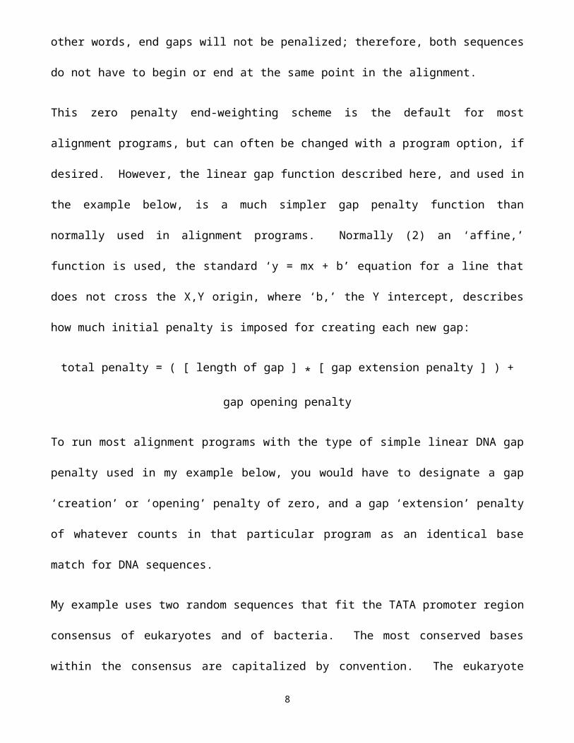
My example uses two random sequences that fit the TATA promoter region consensus of
eukaryotes and of bacteria. The most conserved bases within the consensus are capitalized
by convention. The eukaryote promoter sequence is along the X-axis, and the bacterial
sequence is along the Y-axis in the following example. The solution illustration begins in the
left panel in Table 2 below.
There may be more than one best path through the matrix. This time, starting at the top and
working down as we did, then tracing back, I found two optimal alignments, each with a final
score of 5, using our example’s zero/one scoring scheme. This score is the highest, bottom-
right value in the trace-back path graph, the sum of six matches minus one interior gap in one
path, and the sum of five matches minus no interior gaps in the other. This score is the
number optimized by the algorithm, not any type of a similarity or identity percentage! This first
path is the GCG Wisconsin Package (3) Gap program HighRoad alignment found with this
example’s parameter settings (note that GCG uses a score of 10 for a base match here, not 1):
GAP of: Euk_Tata.Seq to: Bact_Tata.Seq
Euk_Tata: A random Eukaryotic promoter TATA BoxPreferred region: center between -36 and -20.Bact_Tata: A random sequence that fits the consensus from thestandard E. coli RNA polymerase promoter ‘Pribnow’ box -10 region.
Gap Weight: 0 Average Match: 10.000 Length Weight: 10 Average Mismatch: 0.000
6
HighRoad option LowRoad option
Quality: 50 Quality: 50
Ratio: 6.250 Ratio: 6.250 Percent Similarity: 75.000 Percent Similarity: 62.500 Length: 10 Length: 10 Gaps: 2 Gaps: 0 Percent Identity: 75.000 Percent Identity: 62.500
1 cTATAtAagg 10 1 cTATAtAagg 10 | ||||| ||||| 1 cg.TAtAaT. 8 1 .cgTAtAaT. 8
The GCG LowRoad alignment is my second, equivalent path. Notice that even though it has
62.5% identity as opposed to 75% identity in the HighRoad alignment, it has exactly the same
score! Another way to explore dynamic programming’s solution space is to reverse the entire
process. This can often discover other alignments, so reverse the sequences to see
alternatives.
To recap, and for those people that like mathematics, an optimal pairwise alignment is defined
as an arrangement of two sequences, 1 of length i and 2 of length j, such that:
1) you maximize the number of matching symbols between 1 and 2;
2) you minimize the number of gaps within 1 and 2; and
3) you minimize the number of mismatched symbols between 1 and 2.
Therefore, the actual solution can be represented by the following recursion:
Si-1 j-1 or max Si-x j-1 + wx-1 or Sij = sij + max 2 < x < i max Si-1 j-y + wy-1 2 < y < i
where Sij is the score for the alignment ending at i in sequence 1 and j in sequence 2,
sij is the score for aligning i with j,
wx is the score for making a x long gap in sequence 1,
7
wy is the score for making a y long gap in sequence 2,
allowing gaps to be any length in either sequence.
However, just because dynamic programming guarantees an optimal alignment, it is not
necessarily the only optimal alignment. Furthermore, the optimal alignment is not necessarily
the ‘right’ or biologically relevant alignment! Significance estimators, such as Expectation
values and Monte Carlo simulations can give you some handle on this, but always question the
results of any computerized solution based on what you know about the biology of the system.
The above example illustrates the Needleman and Wunsch (4) global solution. Later
refinements (5) demonstrated how dynamic programming could also be used to find optimal
local alignments. To solve dynamic programming using local alignment (without going into all
the gory details) programs use the following two tricks:
1) Mismatches are penalized by using a match function that assigns negative numbers to
them. Therefore, bad paths quickly become very bad. This leads to a trace-back path
matrix with many alternative paths, most of which do not extend the full length of the
graph.
2) The best trace-back within the overall graph is chosen. This does not have to begin or
end at the edges of the matrix — it’s the best segment of alignment.
1.2. Significance
A particularly common misunderstanding regards the concept of homology versus similarity:
there is a huge difference! Similarity is merely a statistical parameter that describes how much
two sequences, or portions of them, are alike according to some set scoring criteria. It can be
normalized to ascertain statistical significance as in database searching methods, but it’s still
just a number. Homology, in contrast and by definition, implies an evolutionary relationship —
more than just the fact that all life evolved from the same primordial ‘slime.’ You need to be
8
able to demonstrate some type of evolutionary lineage between the organisms or genes of
interest in order to claim homology. Better yet, demonstrate experimental evidence, structural,
morphological, genetic, or fossil, that corroborates your assertion. There really is no such
thing as percent homology; something is either homologous or it’s not. Walter Fitch (personal
communication) explains with the joke, “homology is like pregnancy — you can’t be 45%
pregnant, just like something can’t be 45% homologous. You either are or you are not.” Do
not make the mistake of calling any old sequence similarity homology. Highly significant
similarity can argue for homology, not the other way around.
So, how do you tell if a similarity, in other words, an alignment discovered by some program,
means anything? Is it statistically significant, is it truly homologous, and even more
importantly, does it have anything to do with real biology? Many programs generate percent
similarity scores; however, as seen above, these really don’t mean a whole lot. Don’t use
percent similarities or identities to compare sequences except in the roughest way. They are
not optimized or normalized in any manner. Quality scores mean a lot more but are difficult to
interpret. At least they take the length of similarity, all of the necessary gaps introduced, and
the matching of symbols all into account, but quality scores are only relevant within the context
of a particular comparison or search. The quality ratio is the metric optimized by dynamic
programming divided by the length of the shorter sequence. As such it represents a fairer
comparison metric, but it also is relative to the particular scoring matrix and gap penalties used
in the procedure.
A traditional way of deciding alignment significance relies on an old statistics trick — Monte
Carlo simulations. This type of significance estimation has implicit statistical problems;
however, few practical alternatives exist for just comparing two sequences, and they are fast
and easy. Monte Carlo randomization options in dynamic programming alignment algorithms
compare an actual score, in this case the quality score of an alignment, against the distribution
9

of scores of alignments of a randomized sequence. These options randomize your sequence
at least 100 times after the initial alignment and then generate the jumbled alignment scores
and a standard deviation based on their distribution. Comparing the mean of the randomized
sequence alignment scores to the original score using a ‘Z score’ calculation can help you
decide significance. An old ‘rule-of-thumb’ is if the actual score is much more than three
standard deviations above the mean of the randomized scores, the analysis may be
significant; if it is much more than five, than it probably is significant; and if it is above nine,
than it definitely is significant. Many Z scores measure this distance from the mean using a
simplistic Monte Carlo model assuming a normal Gaussian distribution, in spite of the fact that
‘sequence-space’ actually follows an ‘extreme value distribution;’ however, this simplistic
approximation estimates significance quite well:
Z score = [ ( actual score ) - ( mean of randomized scores ) ] ( standard deviation of randomized score distribution )
When the two TATA sequences from the previous dynamic programming example are
compared to one another using the same scoring parameters as before, but incorporating a
Monte Carlo Z score calculation, their similarity is found to be not at all significant. The mean
score based on 100 randomizations was 41.8 +/- a standard deviation of 7.4. Plugged into the
formula: ( 50 – 41.8 ) / 7.4 = 1.11, i.e. there is no significance to the match in spite of 75%
identity! Composition can make a huge difference — the similarity is merely a reflection of the
relative abundance of A’s and T’s in the sequences!
The FastA (6 and 7), BLAST (8 and 9), Profile (10), and HMMer (11) search algorithms, all use
a similar approach but base their statistics on the distance of the query matches from the
actual, or a simulated, extreme value distribution from the rest of the, ‘insignificantly similar,’
members of the database being searched. For alignments without gaps, the math generalizes
such that the Expectation value E relates to a particular score S through the function E =
10

Kmnes (12 and see http://www.ncbi.nlm.nih.gov/BLAST/tutorial/Altschul-1.html). In a
database search m is the length of the query and n is the size of the database in residues. K
and are supplied by statistical theory, dependent on the scoring system and the background
amino acid frequencies, and calculated from actual or simulated database alignment
distributions. Expectation values are printed in scientific notation and the smaller the number,
i.e. the closer it is to 0, the more significant the match. Expectation values show us how often
we should expect a particular alignment to occur merely by chance alone in a search of that
size database. In other words, in order to assess whether a given alignment constitutes
evidence for homology, it helps to know how strong an alignment can be expected from
chance alone. Rough, conservative guidelines to Z scores and Expectation values from a
typical protein search follow in Table 3.
Be very careful with any guidelines such as these, though, because they are entirely
dependent on both the size and content of the database being searched as well as how often
you perform the search! Think about it: the odds are way different for rolling dice depending
on how many dice you roll, whether they are ‘loaded’ or not, and how often you try.
Another very powerful empirical method of determining significance is to repeat a database
search with the entry in question. If that entry finds more significant ‘hits’ with the same sorts
of sequences as the original search, then the entry in question is undoubtedly homologous to
the original entry. That is, homology is transient. If it finds entirely different types of
sequences, then it probably is not. Modular proteins with distinctly separate domains confuse
issues considerably, but the principles remain the same, and can be explained through domain
swapping and other examples of non-vertical transmission. And, finally, the ‘gold-standard’ of
homology is shared structural folds — if you can demonstrate that two proteins have the same
structural fold, then, regardless of similarity, at least that particular domain is homologous
between the two.11
1.3. Scoring matrices
However, what about protein sequences — conservative replacements and similarities, as
opposed to identities? This is certainly an additional complication. Particular amino acids are
very much alike, structurally, chemically, and genetically. How can we take advantage of
amino acid similarity of in our alignments? People have been struggling with this problem
since the late 1960’s. Dayhoff (13) unambiguously aligned closely related protein datasets (no
more than 15% difference, and in particular cytochrome c) available at that point in time and
noticed that certain residues, if they mutate at all, are prone to change into certain other
residues. As it works out, these propensities for change fell into the same categories that
chemists had known for years — those same chemical and structural classes mentioned
above — conserved through the evolutionary constraints of natural selection. Dayhoff’s
empirical observation quantified these changes. Based on the multiple sequence alignments
that she created and the empirical amino acid frequencies within those alignments, the
assumption that estimated mutation rates in closely related proteins can be extrapolated to
more distant relationships, and matrix and logarithmic mathematics, she was able to
empirically specify the relative probabilities at which different residues mutated into other
residues through evolutionary history, as appropriate within some level of divergence between
the sequences considered. This is the basis of the famous PAM (corrupted acronym of
‘accepted point mutation’) 250 (meaning that the matrix has been multiplied by itself 250 times)
log odds matrix.
Since Dayhoff’s time other biomathematicians (esp. see 14’s BLOSUM series of matrices, and
for a somewhat controversial matrix see 15) have created matrices regarded more accurate
than Dayhoff’s original, but the concept remains the same. Furthermore, Dayhoff’s original
PAM 250 matrix remains a classic as historically the most widely used amino acid substitution
matrix. Collectively these types of matrices are known as symbol comparison tables, log odds
12

matrices, and substitution or scoring matrices, and they are fundamental to all sequence
comparison techniques.
The default amino acid scoring matrix for most protein similarity comparison programs is now
the BLOSUM62 table (14). The “62” refers to the minimum level of identity within the
ungapped sequence blocks that went into the creation of the matrix. Lower BLOSUM numbers
are more appropriate for more divergent datasets. The BLOSUM62 matrix follows below in
Table 4; values whose magnitude is 4 are drawn in shadowed characters to make them
easier to recognize.
Notice that positive identity values range from 4 to 11, and negative values for rare
substitutions go as low as -4. The most conserved residue is tryptophan with an identity score
of 11; cysteine is next with a score of 9; histidine gets 8; both proline and tyrosine get scores of
7. These residues get the highest scores because of two biological factors: they are very
important to the structure and function of proteins, and they are the rarest amino acids found in
nature. Also check out the hydrophobic substitution triumvirate — isoleucine, leucine, valine,
and to a lesser extent methionine — all easily swap places. So, rather than using the zero/one
match function that we used in the previous dynamic programming examples, protein
sequence alignments use the match function provided by an amino acid scoring matrix. The
concept of similarity becomes very important with some amino acids being way ‘more similar’
than others!
1.4. Multiple sequence dynamic programming
Dynamic programming reduces the pairwise alignment problem’s complexity to order N2. But
how do you work with more than just two sequences at a time? It becomes a much harder
problem. You could manually align your sequence data with an editor, but some type of an
automated solution is desirable, at least as a starting point to manual alignment. However,
13

solving the dynamic programming algorithm for more than just two sequences rapidly becomes
intractable. Dynamic programming’s complexity, and hence its computational requirements,
increases exponentially with the number of sequences in the dataset being compared
(complexity=[sequence length]number of sequences). Mathematically this is an N-dimensional
matrix, quite complex indeed. Pairwise dynamic programming solves a two-dimensional
matrix, and the complexity of the solution is equal to the length of the longest sequence
squared. Well, a three-member standard dynamic programming sequence comparison would
be a matrix with three axes, the length of the longest sequence cubed, and so forth. You can
at least draw a three-dimensional matrix, but more than that becomes impossible to even
visualize. It quickly boggles the mind!
Several different heuristics have been employed over the years to simplify the complexity of
the problem. One program, MSA (16), attempts to simultaneously solve the N-dimensional
matrix recursion using a bounding box trick. However, the algorithm’s complexity precludes its
use in most situations, except with very small datasets. One way to simultaneously solve the
algorithm and yet reduce its complexity is to restrict the search space to only the most
conserved ‘local’ portions of all the sequences involved. This approach is used by the program
PIMA (17). MSA and PIMA are both available through the Internet at several bioinformatics
servers (in particular see 18), or they can be installed on your own machine.
1.5. How the algorithms work
Most implementations of automated multiple alignment modify dynamic programming by
establishing a pairwise order in which to build the alignment. This heuristic modification is
known as pairwise, progressive dynamic programming. Originally attributed to Feng and
Doolittle (19), this variation of the dynamic programming algorithm generates a global
alignment, but restricts its search space at any one time to a local neighborhood of the full
14
length of only two sequences. Consider a group of sequences. First all are compared to each
other, pairwise, using some quick variation of standard dynamic programming. This
establishes an order for the set, most to least similar, a ‘guide-tree’ if you will. Subgroups are
clustered together similarly. The algorithm then takes the top two, most similar sequences,
and aligns them. Then it creates a quasi-consensus of those two and aligns that to the third
sequence. Next create the same sort of quasi-consensus of the first three sequences and
align that to the forth most similar. The way that the program makes and uses this ‘consensus’
sequence is one of the big differences between the various implementations. This process, all
using standard, pairwise dynamic programming, continues until it has worked its way through
all of the sequences and/or sets of clusters, to complete the full multiple sequence alignment.
The pairwise, progressive solution is implemented in several programs. Perhaps ClustalW
(20) and its multi-platform, graphical user interface ClustalX (21) is the most popular. ClustalW
made the first major advances beyond the basic Feng and Doolittle algorithm by incorporating
variable sequence weighting, dynamically varying gap penalties and substitution matrices, and
a neighbor-joining (NJ, 22) guide-tree. The programs can be downloaded from the ClustalX
homesite, ftp://ftp-igbmc.u-strasbg.fr/pub/ClustalX/, to install on your own machine, or they can
be run through the World Wide Web (WWW) at several sites. ClustalX is available for most
windowing Operating Systems: most UNIX flavors, Microsoft Windows, and Macintosh.
Complete documentation comes with the program and is accessed through a “Help” menu.
The GCG program PileUp implements a similar method, but without the later innovations, and
ClustalW is now included in the GCG package as well.
Several more variations on the theme have come along in recent years. T-Coffee (Tree-based
Consistency Objective Function For alignment Evaluation, 23) was one of the first after
ClustalW, and it has gained much favor. It will be presented in further detail in this chapter’s
final section. Its biggest innovation is the use of a preprocessed, weighted library of all the
15

pairwise global alignments between the sequences in your dataset plus the ten best local
alignments associated with each pair of sequences. This helps build the NJ guide-tree and the
progressive alignment both. Furthermore, the library is used to assure consistency and help
prevent errors, by allowing ‘forward-thinking’ to see whether the overall alignment will be better
one way or another after particular segments are aligned one way or another. Notredame (24)
makes the apt analogy of school schedules: everybody, students, teachers and
administrators, with some folk being more important than others, i.e. the weighting factor, puts
the schedule they desire in a big pile, i.e. T-Coffee’s library, with the trick being to best fit all
the schedules to one academic calendar, so that everybody is happiest, i.e. T-Coffee’s final
multiple sequence alignment. T-Coffee is one of the most accurate multiple sequence
alignment methods available because of this rationale, but it is not the fastest.
Muscle (25) is another relatively new multiple sequence alignment program. It is incredibly
fast, yet nearly as accurate as T-Coffee. Muscle is an iterative method that uses weighted log-
expectation profile scoring along with a slew of optimizations. It proceeds in three stages: draft
progressive using k-mer counting, improved progressive using a revised tree from the previous
iteration, and refinement by sequential deletion of each tree edge with subsequent profile
realignment. Another fairly new program, MAFFT (26), can be run in either fast, approximate
mode, using a Fast Fourier Transformation, where its capability to handle large datasets and
its speed is similar to Muscle; or in a slow, iteratively refined, optimized mode, where its results
and capabilities are similar to T-Coffee. Perhaps the most accurate new multiple sequence
alignment program is ProbCons (27). It uses Hidden Markov Model (HMM) techniques and
posterior probability matrices that compare random pairwise alignments to expected pairwise
alignments. Probability consistency transformation is used to reestimate the scores, and a
guide-tree is then constructed, which is used to compute the alignment, which is then
16

iteratively refined. These methods and more are all tied into T-Coffee as external modules, as
long as they are all installed on your system.
1.6. Coding DNA issues
All of these alignment algorithms, pairwise, multiple, and database similarity searching, are far
more sensitive at the amino acid level than at the DNA level. Twenty match symbols are just
much easier to align then only four; the signal to noise ratio is so much better. And, the
concept of similarity applies to amino acids, but generally not to nucleotides. Furthermore,
many DNA base changes (especially third position changes) do not change the encoded
protein. All of these factors drastically increase the ‘noise’ level of DNA; typically giving protein
searches a much greater ‘look-back’ time, at least doubling it. Therefore, database searching
and sequence alignment should always be done on a protein level, unless you are dealing with
noncoding DNA, or if the sequences are so similar as to not cause any problems. Therefore,
usually, if dealing with coding sequences, translate the DNA to its protein counterpart, before
performing multiple sequence alignment.
Even if you are dealing with very similar coding sequences, where the DNA can be directly
aligned, it is often best to align the DNA along with its corresponding proteins. In addition to
the much more easily achieved alignment, this also insures that alignment gaps are not placed
within codons. Phylogenetic analysis can then be performed on the DNA rather than on the
proteins. This is especially important when dealing with datasets that are quite similar, since
the proteins may not reflect many differences hidden in the DNA. Furthermore, many people
prefer to run phylogenetic analyses on DNA rather than protein regardless of how similar they
are — the multiple substitution models have a long and well-accepted history, and yet are far
simpler. In fact, some phylogenetic inference algorithms do not even take advantage of amino
acid similarity when dealing with protein sequences; they only count identities, though many
17
others can use PAM style models. However, the more diverged a dataset becomes, the more
random third and eventually first codon positions become, which introduces noise (error) into
the analysis. Therefore, often third positions and sometimes first positions are masked out of
datasets. Just like in most of computational molecular biology, one is always balancing signal
against noise. Too much noise or too little signal, both degrade the analysis to the point of
nonsense.
Several scripts and programs, as well as some Web servers, can perform this sort of codon-
based alignment, but they can be a bit tricky to run. Examples include mrtrans (28) (also
available in EMBOSS [29] as tranalign and in BioPerl [30] as aa_to_dna_aln), transAlign (31),
RevTrans (32), protal2dna (33), and PAL2NAL (34). Dedicated sequence analysis editors,
such as GCG’s SeqLab (based on Smith’s Genetic Data Environment [GDE], 35), can also be
used for this in a manual process. The logic to this manual paired protein and DNA codon
alignment approach follows:
1) The easy case is where you can align the DNA directly. If the DNA sequences are
directly alignable because they are quite similar, then use whatever automated tool you
want to create your DNA alignment and load it into your multiple sequence editor. Next
use your editor’s align translation function to create aligned corresponding protein
sequences. Select the region to translate based on the CDS reference in each DNA
sequence’s annotation. Be careful of CDS entries that do not begin at position 1 — the
GenBank CDS feature annotation “/codon_start=” identifies which position the
translation begins within the first codon listed for each gene. You may also have to trim
sequences down to just the relevant gene or exons, especially if they’re genomic.
Group each protein to its corresponding DNA sequence, if the option is available, so
that subsequent manipulations will keep them together.
18
2) The way more difficult case is where you need to use the protein sequences to create
the alignment because the DNA is not directly alignable. In this case you need to create
the protein sequence alignment first, and then load their corresponding DNA sequences
into the editor. You can find the DNA sequence accession codes in the annotation of
the protein sequence entries. Next translate the unaligned DNA sequences into new
protein sequences with the align translation function and group these to their
corresponding DNA sequences, just as above. However, this time the DNA along with
their translated sequences are not aligned as a set, just the other protein set is aligned.
Also, group all of the aligned protein dataset together, separately from the DNA/aligned
translation set. Now comes the manual part; slide the original aligned protein sequence
set apart to match the codons of the DNA along with its aligned translation, inserting
gaps in whichever set need them to recreate the alignment. Merge the newly aligned
sequences into the existing alignment group as you go, and then start on the next one.
It sounds difficult, but since you’re matching up two identical protein sequences, the
DNA translation and the original aligned protein, it’s really not too bad.
Multiple sequence alignment is much more difficult if you are forced to align nucleotides
because the region does not code for a protein. Automated methods may be able to help as a
starting point, but they are certainly not guaranteed to come up with a biologically correct
alignment. The resulting alignment will probably have to be extensively edited, if it works at all.
Success will largely depend on the similarity of the nucleotide dataset.
2. Reliability?
One liability of most global progressive, pairwise methods is they are entirely dependent on the
order in which the sequences are aligned. Fortunately ordering them from most similar to least
similar usually makes biological sense and works quite well. However, the techniques are very
19

sensitive to the substitution matrix and gap penalties specified. Some programs allow ‘fine-
tuning’ areas of an alignment by realignment with different scoring matrices and/or gap
penalties; this can be extremely helpful. However, any automated multiple sequence
alignment program should be thought of as only a tool to offer a starting alignment that can be
improved upon, not the ‘end-all-to-meet-all’ solution, guaranteed to provide the ‘one-true’
answer. Although, in this post-genomics era, when having to deal with Giga bases of data, it
does make sense to start with the ‘best’ solution possible. This is the premise of using a very
accurate multiple sequence alignment package, such as T-Coffee (23).
Regardless of the program used to create an alignment, always use comparative approaches
to help assure its reliability. After the program has offered its best guess, try to improve it
further. Think about it — a sequence alignment is a statement of positional homology — it is a
hypothesis of evolutionary history. It establishes the explicit homologous correspondence of
each individual sequence position, each column in the alignment. Therefore, insure that you
have prepared a good one — be sure that it makes sense — devote considerable time and
energy toward developing the best alignment possible.
Editing alignments to insure that all columns are truly homologous should be encouraged.
Dedicated sequence alignment editing software such as GCG’s SeqLab (3), Jalview (36), Se-
Al (37), and SeaView (38) are great for this, but any editor will do, as long as the sequences
end up properly formatted afterwards. Use your understanding of the system to help guide
your judgment. Look for conserved functional sites and other motifs — they should all line up.
Searches of the PROSITE Database of protein families and domains (39) for catalogued
structural, regulatory, and enzymatic consensus patterns or ‘signatures’ in your dataset can
help, as can de novo motif discovery tools like the MEME (40), MotifSearch (41) program pair.
20

Make subjective decisions. Is it good enough; do things line up the way that they should?
Assure that known enzymatic, regulatory, and structural elements all align. Look for columns
of strongly conserved residues such as tryptophans, cysteines, and histidines; important
structural amino acids such as prolines, tyrosines and phenylanines; and those conserved
isoleucine, leucine, valine substitutions. If, after all else, you decide that you just can’t align
some region, or an entire sequence, then get rid of it. Another alternative is to use the mask
function available in some programs. Cutting an entire sequence out of an alignment may
leave columns of gaps across the entire alignment that will need to be removed. The extreme
amino- and carboxy-termini (5’ and 3’ in DNA) seldom align nicely; they are often jagged and
uncertain, and should probably be excluded. The results of subsequent analyses are
absolutely dependent upon the quality of your alignment assessment.
Researchers have successfully used the conservation of co-varying sites in ribosomal and
other structural RNA alignments to assist in alignment refinement. That is, as one base in a
stem structure changes the corresponding Watson-Crick paired base will change in a
corresponding manner. This principle has guided the assembly of rRNA structural alignments
at the Ribosomal Database Project at Michigan State University (42) and at the University of
Gent, Belgium, at the European Ribosomal RNA database (43).
Be sure an alignment makes biological sense — align things that make sense to align!
Beware of comparing ‘apples and oranges.’ Be particularly suspect of sequence datasets
found through text-based database searches such as Entrez (44) and GCG’s LookUp (based
on the Sequence Retrieval System [SRS] of Etzold and Argos [45]). For example, don’t try to
align receptors and/or activators with their namesake proteins. Be wary of trying to align
genomic sequences with cDNA when working with DNA; the introns will cause all sorts of
headaches. Similarly, aligning mature and precursor proteins, or alternate splicing forms, from
the same organism and locus, doesn’t make evolutionary sense, as one is not evolved from
21
the other, rather one is the other. Watch for redundant sequences; there are tons of them in
the databases. If creating alignments for phylogenetic inference, either make paralogous
comparisons (i.e. evolution via gene duplication) to ascertain gene phylogenies within one
organism, or orthologous (within one ancestral loci) comparisons to ascertain gene
phylogenies between organisms (which should imply organismal phylogenies). Try not to mix
them up without complete data representation. Otherwise, confusion can mislead
interpretation, especially if the sequences’ nomenclature is inconsistent. These are all easy
mistakes to make; try your best to avoid them.
Remember the old adage “garbage in — garbage out!” Some general guidelines to remember
include the following (46):
• If the homology of a region is in doubt, then throw it out.
• Avoid the most diverged parts of molecules; they are the greatest source of systematic
error.
• Do not include sequences that are more diverged than necessary for the analysis at
hand.
3. Complications
Sequence data format is a huge problem in computational molecular biology. The major
databases all have their own distinct format, plus many of the different programs and packages
require their own. Clustal (47) has a specific format associated with it. The FastA database
similarity-searching package (6) uses a very basic sequence format. The National Center for
Biotechnology Information (NCBI) uses a library standard called ASN.1 (Abstract Syntax
Notation One), plus it provides GenBank flatfile format for all sequence data. GCG uses three
sequence formats: Single Sequence Format (SSF), Multiple Sequence Format (MSF), and
SeqLab’s Rich Sequence Format (RSF) that contains both sequence data and annotation.
22

PAUP* (Phylogenetic Analysis Using Parsimony [and other methods, pronounced “pop star”]
48), MrBayes (49), and many other phylogenetic analysis packages, have a required format
called the NEXUS file. The PAUP* interface in the GCG Package, PAUPSearch, generates
NEXUS format directly from GCG alignments. Even PHYLIP (PHYLogeny Inference
Package, 50) has its own unique input data format. Format standards have been argued over
for years, such as using XML for everything, but until everybody agrees, which is not likely to
happen, it just won’t happen. Fortunately several freeware programs are available to convert
formats back and forth between the required standards, but it all can get quite confusing.
BioPerl’s SeqIO system (30) and ReadSeq (51) are two of the best. T-Coffee (23) comes with
one built in named “seq_reformat.”
Alignment gaps are another problem. Different program suites may use different symbols to
represent them. Most programs use hyphens, “-”; the GCG Package uses periods, “.”, for
interior gaps, and tildes, “~”, for placeholder gaps. Furthermore, not all gaps in sequences
should be interpreted as deletions. Interior gaps are probably okay to represent this way, as
regardless of whether a deletion, insertion or a duplication event created the gap, logically they
are treated the same by the algorithm. These are known as ‘indels.’ However, end gaps
should not be represented as indels, because a lack of information before or beyond the length
of any given sequence may not be due to a deletion or insertion event. It may have nothing to
do with the particular stretch being analyzed at all. It just may not have been sequenced!
These gaps are just placeholders for the sequence. Therefore, it is safest to manually edit an
alignment to change leading and trailing gap symbols to “x”’s which mean “unknown amino
acid,” or “n”’s which mean “unknown base,” or “?”’s which is supported by many programs, but
not all, and means “unknown residue or indel.” This will assure that the programs don’t make
incorrect assumptions about your sequences.
4. Applicability?23

Now that we understand some of the principles and problems of multiple sequence alignment,
what’s so great about doing it anyway; why would anyone want to bother? Multiple sequence
alignments are:
• very useful in the development of PCR primers and hybridization probes;
• great for producing annotated, publication quality, graphics and illustrations;
• invaluable in structure/function studies through homology inference;
• essential for building HMM profiles for remote homology similarity searching and
alignment; and
• required for molecular evolutionary phylogenetic inference programs.
A multiple sequence alignment is useful for probe and primer design by allowing you to
visualize the most conserved regions of an alignment. This technique is invaluable for
designing phylogenetic specific probes as it clearly localizes areas of high conservation and
high variability in an alignment. Depending on the dataset that you analyze, any level of
phylogenetic specificity can be achieved. Pick areas of high variability in the overall dataset
that correspond to areas of high conversation in phylogenetic category subset datasets to
differentiate between universal and phylo-specific potential probe sequences. After localizing
general target areas on the sequence, you can then use any of several primer discovery
programs, such as GCG’s Prime, or MIT’s Primer3 (52), or the commercial Oligo program
(National Biosciences, Inc.), to find the best primers within those regions, and to test those
potential probes for common PCR conditions and problems. (See my workshop tutorial
illustrating this technique using GCG and SeqLab at
http://bio.fsu.edu/~stevet/PrimerDesign.pdf if you are interested.) The technique is illustrated
in Figure 1 below where I identify potential primer locations that should differentiate between
the major capsid protein genes (L1) of the highly carcinogenic Human Papillomavirus (HPV)
Type 16 strains from the rest of the Type 16 relatives.
24

Graphics prepared from multiple sequence alignments can dramatically illustrate functional
and structural conservation. These can take many forms of all or portions of an alignment —
shaded or colored boxes or letters for each residue (e.g. BoxShade [53] and the various
PostScript output options in GCG’s SeqLab), cartoon representations (e.g. WebLogos [54] and
GCG’s SeqLab graphical feature representation), running line graphs of overall similarity (as
seen above with GCG’s PlotSimilarity and as displayed by ClustalX), overlays of attributes,
various consensus representations, etc. — all can be printed with high-resolution equipment,
usually in color or gray tones. These can make a big difference in a poster or manuscript
presentation. Figure 2 shows a multiple sequence alignment of the most conserved portion of
the HMG DNA-binding domain from several paralogous members of the human HMG-box
superfamily.
Conserved regions of an alignment are important. In addition to the conservation of primary
sequence, structure and function is also conserved in these crucial regions. In fact,
recognizable structural conservation between true homologues extends way beyond
statistically significant sequence similarity. An oft-cited example is in the serine protease
superfamily. S. griseus protease A demonstrates remarkably little sequence similarity when
compared to the rest of the superfamily (Expectation values E()101.8 in a typical search) yet
its three-dimensional structure clearly shows its allegiance to the serine proteases (RMSD of
less than 3 Å with most of the family) (Pearson, W.R., personal communication). These
principles are the premise of ‘homology modeling’ and it works remarkably well. An automated
homology modeling tool is even available on the ExPASy server in Switzerland. Supported by
the Swiss Institute of Bioinformatics (SIB) and GlaxoSmithKline, Swiss-Model (55) has
dramatically changed the homology modeling process. It is a relatively painless way to get a
theoretical model of a protein structure. While not always successful, the minimal amount of
effort involved in making the attempt makes it an excellent time investment. It won’t always
25

generate a homology model for your sequence, depending on how similar the closest
sequence with an experimentally solved structure is to it; however, it is a very reasonable first
approach and will often lead to remarkably accurate representations. I submitted a Giardia
lamblia Elongation Factor 1 sequence to Swiss-Model in “First Approach mode.” The results
were e-mailed back to me in less than five minutes. Figure 3 displays a RasMac (56)
“Strands” graphic of the Giardia EF-1 structural model from Swiss-Model superimposed over
the eight most similar solved structural templates.
Profiles are a position specific scoring matrix (PSSM) description of an alignment or a portion
of an alignment. Gap insertion is penalized more heavily in conserved areas of the alignment
than it is in variable regions, and the more highly conserved a residue is, the more important it
becomes. Profiles are created from an existing alignment of related sequences, and then they
are used to search for remote sequence similarities and/or to build larger multiple sequence
alignments. Originally described by Gribskov (10), and then automated by NCBI’s PSI-BLAST
(9), later refinements have added more statistical rigor (see e.g. Eddy’s Hidden Markov Model
profiles [11]). The original Gribskov style profiles require a lot of time and skill to prepare and
validate, and they are heuristics based. An excess of subjectivity, and a lack of formal
statistical rigor also contribute as drawbacks. Eddy’s HMMer (pronounced “hammer”) package
uses Hidden Markov modeling, with a formal probabilistic basis and consistent gap insertion
theory, to overcome these limitations. The HMMer package can build and manipulate HMMer
profiles and profile databases, search sequences against HMMer profile databases and visa
versa, and easily create multiple sequence alignments using HMMer profiles as a ‘seed.’ This
ability to easily create larger and larger multiple sequence alignments is incredibly powerful
and way faster than starting all over each time you want to add another sequence to an
alignment. The ‘take-home’ message is HMMer profiles are much easier to build than
traditional profiles, and they do not need to have nearly as many sequences in their alignments
26

in order to be effective. Furthermore, they offer a statistical rigor not available in Gribskov
profiles, plus they have all the sensitivity of any profile technique. In effect, they are like the
‘old-fashioned’ profiles pumped up on steroids! One big difference between HMMer profiles
and others is when the profile is built you need to specify the type of eventual alignment it will
be used with, rather than when the alignment is built. The HMMer profile will either be used for
global or local alignment, and it will occur multiply or singly on a given sequence. All profile
techniques are tremendously powerful; they can provide the most sensitive, albeit extremely
computationally intensive, database similarity search possible.
Finally, we can use multiple sequence alignments to infer phylogeny. Based on the assertion
of homologous positions in an alignment, many, many different methods can estimate the most
reasonable evolutionary tree for that alignment. A few of the packages that incorporate these
methods were mentioned earlier in the complications sections with regard to format issues:
PAUP* (48), MrBayes (49), and PHYLIP (50). This is a huge, complicated, and highly
contentious field of study. (See the Woods Hole Marine Biological Laboratory’s excellent
summer short course, the Workshop on Molecular Evolution, at
http://workshop.molecularevolution.org/.) However, always remember that regardless of the
algorithm used, any form of parsimony, all of the distance methods, all maximum likelihood
techniques, and even all types of Bayesian phylogenetic inference, they all make the absolute
validity of your input alignment matrix their first and most critical assumption (but see 57).
Therefore, the accuracy of your multiple sequence alignment is the most important factor in
inferring reliable phylogenies; your interpretations are utterly dependent on its quality.
Structural alignments are the ‘gold-standard,’ but the luxury of having homologous solved
structures is not always available. In fact, many experts recommend not using any
questionable portions of sequence data at all. These highly saturated regions have the
property known as ‘homoplasy.’ This is a region of a sequence alignment where so many
27
multiple substitutions have occurred at homologous sites that it is impossible to know if those
sites are properly aligned, and thus, impossible to ascertain relationships based on those sites.
Phylogenetic inference algorithms’ primary assumption is most violated in these regions, and
this phenomenon increasingly confounds evolutionary reconstruction as divergence between
the members of a dataset increases. Because of this, only analyze those sequences and
those portions or your alignment that assuredly do align. If any sequences or portions are in
doubt, exclude them. This usually means trimming down or masking the alignment’s terminal
ends and may require internal trimming or masking as well. These decisions are somewhat
subjective by nature, experience helps, and some software, such as ASaturA (58) and T-
Coffee (23), has the ability to evaluate the quality of particular regions of your alignment as
well. Biocomputing is always a delicate balance — signal against noise — and sometimes it
can be quite the balancing act!
5. The T-Coffee shop
I call this section the T-Coffee shop because T-Coffee (23) is much more than merely a
program for doing multiple sequence alignment. Much like a Starbucks coffee shop offers
many different flavors and types of coffee drinks, the T-Coffee command line offers an entire
suite of multiple sequence alignment tools. Notredame (24) has done a very good job of
providing documentation with the package’s distribution. In particular be sure to read the
entire Tutorial and FAQ. It’s quite good, and I can’t do it justice in my description here.
Therefore, I will attempt to distil out the most vital portions of the documentation, and illustrate
a subset of T-Coffee’s potential in a ‘bare-bones’ manner, just enough to get a novice user
started in exploring the package.
As mentioned in the algorithm section, T-Coffee is one of the most accurate multiple sequence
alignment tools around, and it does this in its default mode. It achieves its accuracy by
28

producing the multiple sequence alignment that has the highest consistency level with a library
of preprocessed, global and local pairwise alignments. However, it can do much more than
that. In addition to merely aligning a sequence dataset, it can combine preexisting alignments,
evaluate the consistency of alignments, extract a series of motifs to create a local alignment,
perform all sorts of data manipulation and format operations, and with SAP (Structure
Alignment Program, 59) installed it can even use structural information to make the most
accurate protein alignment possible.
T-Coffee’s “seq_reformat” tool can perform standard data reformatting operations and change
the appearance of your alignment, but it is incredible, as well, for extracting or combining
subsets of your data based on sequence names, patterns, coordinates in the alignment, and/or
level of consensus. It even has the ability to translate DNA sequences into their
corresponding protein sequences, or to generate DNA alignments based on the corresponding
protein alignment, either using the actual DNA sequences (ala mrtrans and relatives) or using
a random back-translation procedure. Furthermore, “seq_reformat” can read phylogenetic
trees in Newick format to compare two trees or to prune tips off of a tree. Another practical
utility in T-Coffee is “extract_from_pdb;” it allows you to download either the three-dimensional
coordinates or the FastA format sequence of structures held at the PDB (Protein Data Bank,
60) using UNIX’s “wget” command. There is little that cannot be done with the T-Coffee utility
tools when it comes to data organization and manipulation. One of my favorites is using it to
remove sequences that are, or are nearly, redundant. Pages 11 through 25 of Notredame’s
tutorial (24) cover all these operations very well, and I encourage you to work through the
examples there; I will not take the space to review them here.
Okay, how do we begin? I’m making the presumption that you already have T-Coffee installed
on either your own computer or on a server that you have access to. If this is not the case,
then refer to Notredame’s (24) Technical Documentation, and either install it yourself, or get a
29

local systems administrator to do it for you. I’m using version 5.05 here. I’ll be consistent in
my command syntax in these examples, but realize that the Notredame’s tutorial mixes up
command syntax a bit, freely replacing equal signs and commas with spaces in some
examples and not in others, and T-Coffee doesn’t mind. Let’s first look at T-Coffee’s default
mode. Issue the following command to see all of T-Coffee’s default parameter settings:
Prompt% t_coffee -help
The list is huge; scroll back to skim over the entire thing. Notice the “–seq” parameter usage:
“List of sequences in any acceptable format.” T-Coffee will accept several input formats, but it
works most reliably if you have your input files in FastA format. If I have a file containing
unaligned sequences in FastA format named “unaligned.fa,” then the following command will
run T-Coffee without any options or parameter specifications:
Prompt% t_coffee unaligned.fa
This will produce a screen trace of the program’s progress; an output alignment named
“unaligned.aln” in Clustal Aln format; another alignment file named “unaligned.html” in HTML
format for Web browsers, with columns color-coded based on reliability; and a file named
“unaligned.dnd,” that contains the Newick format tree used to guide the alignment. If you have
your dataset spread around in more than one file, then you can use the “–seq” option followed
by a comma-delimited list of input files. This option will also strip the gaps out of any input file
that might already be an alignment. And if you don’t like Clustal Aln format, use the “–output”
option. Here I’ll use it to generate a FastA format output alignment file named
“unaligned1.fasta_aln” from two FastA input files (T-Coffee uses the first file’s primary name to
identify the output file). The only output alignment file produced this way is in FastA format:
Prompt% t_coffee -seq=unaligned1.fa,unaligned2.fa -output=fasta_aln
30

The T-Coffee “–output” option supports alignment formats with the following identifiers:
“msf_aln” (for GCG MSF), “pir_aln” (for PIR), “fasta_aln” (for FastA), “phylip” (for PHYLIP), as
well as its default “clustalw_aln” and “html”. You can produce output files in more than one
format by comma separating the identifiers. So, you can’t get directly to NEXUS format, but
PAUP* has the ability to import GCG MSF, PIR, or PHYLIP format with the ToNEXUS
command. Plus, if you don’t like T-Coffee’s default output file naming convention, you can use
the “–outfile” option to specify any name you might want.
5.1. Alignment parameters
As in nearly all sequence alignment programs, the substitution matrix and the gap penalties
are very important run parameters. In most cases the T-Coffee matrix defaults, the
BLOSUM62 and 50 matrices for its global and local pairwise alignment steps, respectively, will
work just fine. And, in fact, T-Coffee only uses these matrices in its first pass through your
dataset, when it builds its consistency library. It replaces the usual BLOSUM style matrix when
building its final multiple sequence alignment with the optimal position specific scores of all the
potential pairwise matings in its library. Regardless, using the optional “–matrix=blosum30mt”
flag (or blosum40mt or blosum45mt depending on your data’s level of divergence) is a great
idea, whenever dealing with sequences that are quite dissimilar. Furthermore, gap penalties
can be messed with, if you really want to, but the default gap opening penalty of -50 and gap
extension penalty of zero, changed through the “–gapopen” and “–gapext” options are, as
Notredame (24) says, only “cosmetic,” changing the final alignment’s appearance by changing
how residues slide around in ‘unalignable’ regions, since all the ‘alignable’ regions are found
from the previously built library. It’s much more complicated if you want to change how the
library alignments are built, and I don’t suggest messing with it. If you insist, the parameters
are specified through the “–method” option, and two methods build the pairwise alignment
library by default (several others are available by option for special cases): a global,
31

“slow_pair,” one, and a local, “lalign_id_pair,” one. To change their respective default
behaviors a combination of “MATRIX” specification and “GOP” and “GEP” parameters are
used. The defaults for the global library alignments are a “GOP” of -10 with a “GEP” of -1; and
for the local alignment library a “GOP” of -10, with a “GEP” of -4. For instance, if I have a
really lousy dataset, with barely discernible homology, and with no structural homologues at
all, then perhaps using a combination of parameters such as the following would produce a
more accurate and more pleasing looking multiple sequence alignment:
Prompt% t_coffee -seq=lousydata.fa -matrix=blosum30mt
-gapopen=-100 -gapext=0 -output=fasta_aln,clustalw_aln,html
-method=slow_pair@EP@GOP@-5@GEP@-1,lalign_id_pair@EP@GOP@-5@GEP@-
4
Notice the bizarre syntax: the at sign, “@,” is used as a method parameter separator, and “EP”
stands for “Extra Parameter.” This command would run both the global and local library builds
with the BLOSUM30 matrix, would double the ‘cosmetic’ gap opening penalty, would cut the
penalties in half for opening a gap in both the gloabal and local library alignments, and would
keep all the extension penalties at their default levels. Additionally it would produce output
alignments in FastA, ClustalW, and HTML formats. If you wanted to use different substitution
matrices for the different methods, then you would add e.g. “MATRIX@blosum45mt” after
“@EP@” and before “@GOP@ for the appropriate method. But, will this actually produce a
‘better’ alignment?
5.2. Quality
This brings up the heart of T-Coffee: consistency. T-Coffee’s method relies on reconciling its
internal pairwise alignment library as best as it can with its eventual multiple sequence
alignment; the more these agree, the more consistent is the alignment and, we assume, the
32
more accurate. This premise allows us to use T-Coffee to evaluate and compare alignments.
The easiest way to see how accurate T-Coffee ‘thinks’ its alignment is, is to look at the
“SCORE” it receives in its ClustalW or HTML format output. The higher this score value is, the
more closely the alignment overall agrees with the internal pairwise alignment library.
Notredame (24) says that values above 40 are “usually pretty good.” Every T-Coffee ClustalW
and HTML format output alignment has this value associated with it. However, what if you
don’t have the right output format, or you want to see how the output from some other
alignment program ranks, or you are interested in how different alignments compare to each
other, or you are interested in what portions of an alignment are good and what portions are
bad? T-Coffee can do all of this.
I’ll discuss methods that do not rely on structure first. Structural methods will follow where I
discuss T-Coffee’s ability to integrate sequence and structure. T-Coffee’s CORE index is the
local consistency level of each position within your alignment. All T-Coffee HTML format
output alignments represent this index with color-coding, plus there are specific score output
file options. Position colors range across the spectrum from blue, to green, to yellow, to
orange, and finally red, corresponding to an increase in consistency level from none to
absolute. These colors correspond to local consistency values of 0 through 9. To test a
preexisting alignment with the CORE index use the “–infile” specification for your alignment,
the “–evaluate” (replaces the deprecated “–score” flag, and in default mode equivalent to “–
special_mode=evaluate”) option, and minimally specify HTML output format:
Prompt% t_coffee -infile=lousydata.fasta_aln -evaluate -output=html
Notice we need to use “–infile” rather than “–seq” in order to run T-Coffee in this manner.
There are even ways to automatically filter unreliable columns from your alignment based on
33

the CORE index; however, the various commands’ syntax are quite complicated, and I refer
you to page 45 of Notredame’s (24) tutorial.
5.3. Comparing alignments
T-Coffee has several ways to compare existing alignments of the same sequences beyond just
looking at their consistency scores. The “aln_compare” module is one of the more powerful,
and can tell you how different two alignments are. It needs to be launched with the “–
other_pg” option, which tells T-Coffee that you want to use an external module. This option
must be the first parameter on the command line after “t_coffee.” “aln_compare” supports
several further options that can help with visualization. Here the “aln_compare” module is
used to analyze the difference between two existing alignments with the “–al1” and “–al2”
options to produce an output screen trace of the first alignment where all residues with less
than 50% of their pairing partners in the other alignment are represented as an “x:”
Prompt% t_coffee -other_pg=aln_compare -al1=trial1.fasta_aln
-al2=trial2.fasta_aln -output_aln -output_aln_threshold 50
-output_aln_modif x
The same command without the “–output_align” parameters will produce a summary statistic
of the percentage of similarity between the two alignments counting the sum of all pairs of
residues in those alignments. Type the command without any parameters to see all that
“aln_compare” offers:
Prompt% t_coffee -other_pg=aln_compare
All of T-Coffee’s built in external modules support this help syntax, versus its standard “–help”
option.
34

Another way to compare alignments is to turn one into T-Coffee’s library and leave the other
an alignment. You need to use the “–aln” option to do this. This option tells T-Coffee to use
the specified input alignment file to build its library. The following command will show how well
the alignment “somedata1.fasta_aln” agrees with the library produced from the alignment
“somedata2.fasta_aln:”
Prompt% t_coffee -infile=somedata1.fasta_aln
-aln=somedata2.fasta_aln -evaluate -output=html
The HTML alignment output will highlight those residues that are in agreement or not between
the two input alignments using T-Coffee’s standard CORE index coloring scheme.
5.4. Combining alignments
Again, there’s a slew of ways to combine alignments with T-Coffee. One neat way is to not
really worry how alignments compare and just turn them all into a T-Coffee library so that they
will combine together yielding one optimal alignment that best agrees with all the input
alignments. They do not even need to all have the same sequences to do this. Turn the three
specified alignments into a library and produce an output alignment in Clustal Aln, FastA, and
HTML format with the command below:
Prompt% t_coffee -aln=one.fasta_aln,two.fasta_aln,three.fasta_aln
-output=clustal_aln,fasta_aln,html
And, of course, you could easily add some unaligned input sequences to the mix with the “–
seq” option as well.
As discussed in this chapter under multiple sequence alignment applications, profiles are a
very powerful technique for building larger and larger alignments. T-Coffee can deal with
profiles in several ways, though they are not quite the same sort of profile as, for instance,
35
Gribskov (10) or Eddy (11) envisioned. T-Coffee defines profiles as multiple sequence
alignment matrices that will not have their gaps removed, rather than a true PSSM where
residues receive higher weights in more conserved regions. Regardless, T-Coffee can take as
many different profiles and sequences as you want to specify, and combine them all into one
alignment (given that it is biologically correct to attempt to align them):
Prompt% t_coffee
-profile=one.fasta_aln,two.fasta_aln,three.fasta_aln
-seq=lousydata.fa,evenworsedata.fa
-output=clustal_aln,fasta_aln,html
This command will feed three alignments to T-Coffee such that the sequences within them will
not have their gaps removed, it will add more gaps to reconcile those three alignments, and it
will add two more sequences to the resulting alignment in the most consistent manner.
And to get the most accurate profile alignment add “–profile_comparison=full,” which runs the
profile alignment in a slower, more exact mode “on a library that includes every possible pair of
sequences between the two profiles,” as opposed to the above command, which “vectorizes”
the multiple sequence alignments designated as profiles (24).
5.5. Combining methods
T-Coffee has a special ‘Meta’ mode named M-Coffee (61). This gives T-Coffee an incredible
amount of power. M-Coffee is amazing for those situations where you just don’t know what
alignment tools to trust, and you don’t want to have to build and test a bunch of alternatives. It
automatically runs up to eight different multiple alignment programs (by default, more external
methods can be added) on your data, and combines the best parts of each, to come up with
one, most consistent, consensus alignment. Your system needs to have ClustalW (20), POA
(62), Muscle (25), ProbCons (27), MAFFT (26), Dialign-T (63), PCMA (64), and T-Coffee (23) 36
all installed for this to work. If I want to see how M-Coffee handles that lousy data FastA
format file I have, then I would issue the following command to run M-Coffee in its default
mode:
Prompt% t_coffee -seq=lousydata.fa -special_mode=mcoffee
-output=clustal_aln,fasta_aln,html
The output Clustal and HTML format files will list the alignment’s overall score as a percentage
of consistency between all the methods. The HTML format will additionally provide T-Coffee’s
usual color-coded position consistencies. Or, If you prefer some methods to others, you can
select particular methods to combine with the “–method” option, with syntax like the following:
Prompt% t_coffee -seq=lousydata.fa
-method=t_coffee_msa,mafft_msa,muscle_msa -output=fasta_aln,html
Here’s some general guidelines as to which of T-Coffee’s integrated external multiple
sequence alignment methods are best in which situations (based on 24 and 65):
clustalw_msa neither the fastest nor the most accurate, but a reasonable ‘industry-
standard.’
probcons_msa uses consistency and Bayesian inference to provide ultra-accurate, but
very slow runs.
muscle_msa very, very fast for large datasets, especially; uses weighted log-
expectation scoring.
mafft_msa in fast mode (FFT-NS-i) screaming quick on large datasets, but not
incredibly accurate; in slow mode (L-INS-i) very accurate but quite slow,
especially with large datasets.
pcma_msa combines ClustalW and T-Coffee strategies.
poa_msa very accurate local alignments using partial order graphs.37

dialignt_msa accurate local, segment-based, progressive alignment.
Pick and choose among the most appropriate methods and let M-Coffee combine the best
aspects of each.
5.6. Local multiple sequence alignments
If you know the coordinates of some predefined sequence pattern in one sequence, you can
use T-Coffee’s mocca routine (Multiple OCCurrences Analysis, 66) to find all the occurrences
of similar patterns in other sequences and assemble a local multiple sequence alignment of
them. Mocca is a perl script that launches T-Coffee, computes a T-Coffee library from the
input sequences, and then prompts you with an interactive menu to extract the homologous
motifs and assemble the alignment. It is designed to find and align motifs of 30% and greater
identity that are at least 30 amino acids long. The interactive menu can be confusing, so I
recommend that you place the sequence with your identified motif first in the dataset, and
specify the beginning and the length of your motif on the command line, rather than in the
menu. I’ll use mocca here to prepare a local sequence alignment of the motifs in a dataset
named “motifdata.fa,” where I know the first occurrence of the motif is at absolute position 35
and runs for 65 residues in my first sequence of the dataset:
Prompt% t_coffee -other_pg=mocca motifdata.fa -start=35 -len=65
You cannot use the “–seq” option with mocca to specify your input file in this command.
Specifying your motif coordinates is also a bit tricky, since all the input sequences have their
gaps removed (if it was an alignment) and are then concatenated together. That’s why it’s
easiest if you put your known motif sequence first. After mocca computes the optimal local
alignment with your motif it pauses and displays its menu. Type a capital “X” to exit the
program and write the default Clustal Aln and HTML alignment files.
38

5.7. The ‘gold-standard:’ creating structure based alignments
As mentioned earlier, you need to minimally have SAP (59) installed on your system for T-
Coffee’s structure based alignment mode to actually use structural information. And, even
better yet, have the FUGUE package (67) installed as well. Additionally you need “wget” on
your system, to access PDB files over the Internet, but most all UNIX/Linux installations should
include this utility. T-Coffee uses a special mode named 3DCoffee (68) to create structure-
based alignments. By default 3DCoffee uses four methods to create the T-Coffee consistency
library, if you specify an input sequence dataset: the standard T-Coffee global “slow_pair” and
local “lalign_id_pair,” SAP’s “sap_pair,” and FUGUE’s “fugue_pair.” If I have a dataset that
includes some PDB structures, and those sequences are named using PDB’s identifier with a
chain name (e.g. 1EFTA for chain A of the Thermus aquaticus elongation factor Tu structure
[69]), then the following command will produce the most consistent alignment of them based
on all available structural pairs and T-Coffee’s usual pairs:
Prompt% t_coffee -seq=elongation.fa -special_mode=3dcoffee
You’ll get three output files by default with this command: “elongation.aln,” “elongation.html,”
and “elongation.dnd”. The alignment files will have T-Coffee’s standard reliability index. If you
specify your input dataset is already an alignment, then the local “lalign_id_pair” will not be
used, and the alignment will be turned directly into T-Coffee’s library along with the SAP and
FUGUE pairs:
Prompt% t_coffee -aln=elongation.fasta_aln -special_mode=3dcoffee
These 3DCoffee analyses will even produce output alignments if you don’t have SAP or
FUGUE installed, but they will report error warnings for every pair, and, naturally, no structural
information will be used in the production of the alignment. If you don’t like the warning
messages, just specify the particular methods you have available, e.g:39

Prompt% t_coffee -aln=elongation.fasta_aln
-method=sap_pair,slow_pair
A nice trick is to combine two existing, related, but only distantly so, alignments with 3DCoffee,
if they both have at least one sequence whose structure has been solved, and they follow
proper naming conventions. Suppose I have one alignment of elongation factor Tu sequences
containing the sequence for the solved structure for Thermus aquaticus, and another
alignment of elongation factor 1 sequences containing the sequence of the solved human
structure. Do this analysis with the “–profile” input specification:
Prompt% t_coffee
-profile=elongation1a.fasta_aln,elongationtu.fasta_aln
-special_mode=3dcoffee
The output alignment will combine the two existing alignments in the most consistent manner
with the structural alignment of the human and Thermus aquaticus sequences.
5.8. Using structure to evaluate alignments
Your existing alignment needs to have at least two members with solved structures in order to
evaluate it using T-Coffee’s structural method, and, as above, those sequences need to be
named according to their PDB identifiers as well as their chain. T-Coffee uses a special
version of Root Mean Square Distance Deviation analysis not dependent on specific carbon
backbone superpositioning called iRMSD (the “i” stands for intra-catener [70]) for structural
alignment evaluation. It also reports a normalized NiRMSD not dependent on the alignment’s
length, and it reports an older measure, APDB, not as powerful as iRSMD, based on the
fraction of residue pairs with ‘correct’ structural alignments. The smaller the iRMSD Å
numbers are, the bigger the APDB percent will be, and the better the alignment corresponds to
structural ‘reality.’ As with the other integrated external methods in the T-Coffee package,
40

iRMSD is launched with the “–other_pg” option. Specify your alignment’s file name with the “–
aln” option, and specify an output file with the “–apdb_outfile” option, otherwise the output will
just scroll to screen. Optionally generate an HTML alignment output as well with “–outfile”:
Prompt% t_coffee -other_pg=irmsd -aln=elongation.fasta_aln
-apdb_outfile=irmsd.out -outfile=irmsd
The summary statistics at the end of the output file are the most telling:
#TOTAL for the Full MSA TOTAL EVALUATED: 81.58 % TOTAL APDB: 78.25 % TOTAL iRMSD: 0.75 Angs TOTAL NiRMSD: 0.93 Angs
# EVALUATED: Fraction of Pairwise Columns Evaluated# APDB: Fraction of Correct Columns according to APDB# iRMDS: Average iRMSD over all evaluated columns# NiRMDS: iRMSD*MIN(L1,L2)/Number Evaluated Columns# Main Parameter: -maximum_distance 10.00 Angstrom# Undefined values are set to -1 and indicate LOW Alignment Quality# Results Produced with T-COFFEE (Version_5.05)# T-COFFEE is available from http://www.tcoffee.org
My elongation factor 1/Tu alignment is pretty darn good with an overall NiRSMD of less than
1 Å and an APDB statistic of 78%. The old T-Coffee external method specification “–
other_pg=apdb” produces the same output. The optional HTML output shows which residues
in the solved structures (only) are in agreement with the structural alignment using the APDB
coloring scheme where blue corresponds to 0% and red corresponds to 100% (get iRSMD
coloring with “–color_mode=irmsd”).
5.9. Special situations
Even though T-Coffee is great for so many things, its default run parameters are far from ideal
for some special situations. It doesn’t work very well for huge datasets, that is anything with
over 100 sequences, it doesn’t particularly like DNA/RNA alignments, and it has some
41
problems with jumping over huge gaps like you would see when aligning splicing variants or
cDNA to genomic DNA containing introns. Fortunately there are ways around each one of
these scenarios, and most all multiple sequence alignment programs have trouble with the
same situations as well.
Let’s start with real large alignments. Both Muscle and MAFFT (in fast mode) are more
appropriate for datasets with more than around 100 sequences; however, T-Coffee does have
a special mode that will at least allow it to estimate an approximate alignment with such
datasets. This should work when your dataset has an overall identity of 40% or more:
Prompt% t_coffee -seq=hugedata.fa -special_mode=quickaln
The resulting alignment should be about as accurate as one built with ClustalW. For datasets
with between 50 and 100 sequences T-Coffee automatically switches to another heuristic
mode named DPA (double progressive alignment).
Naturally DNA and RNA alignments are way harder for T-Coffee to perform. The rationale for
this phenomenon is explained in the introduction, and it confounds every single multiple
sequence alignment program around. DNA is just really hard to align unless the sequences
are 90% or so identical. One way to help T-Coffee with an alignment that must be built using
DNA or RNA, especially if the locus does not code for a protein, is to specify that the
sequences are DNA with the “–type” option:
Prompt% t_coffee -seq=DNAdata.fa -type=dna
T-Coffee should detect this sequence type automatically, but it can’t hurt to declare it up front.
When T-Coffee realizes that it is working with DNA it uses specific DNA optimized methods to
build its library, “slow_pair4dna” and “lalign_id_pair4dna.” These methods have lower built-in
gap penalties and use a DNA specific scoring matrix. And if your DNA is a particularly noisy
42

coding locus, but you just can’t figure out the translation, because it is so noisy, then T-
Coffee’s special “cdna_fast_pair” method takes potential amino acid similarity considering
frameshifts into account, and may help:
Prompt% t_coffee -seq=noisy_cDNAdata.fa -method=cdna_fast_pair
You may want to try this when aligning cDNAs to genomic DNA as well, in order to jump over
the introns. If the results look good, you can even use T-Coffee’s external “seq_reformat”
module to translate it to the appropriate protein translation in spite of how noisy the original
DNA sequences were:
Prompt% t_coffee -other_pg=seq_reformat
-in=noisy_cDNAdata.fasta_aln
-action=+clean_cdna,+translate > noisy_pep.fasta_aln
Notice that “seq_reformat” does not support “–outfile;” you need to use UNIX “>” output
redirection. “+clean_cdna” is a small HMM that tries to maximize the appropriate frame choice
at every point in the sequences to best match the alignment of all the other sequences, and
“+translate,” obviously, does the translation.
T-Coffee is actually pretty good at jumping over big gaps in protein sequence alignments,
such as can be present when trying to align various protein splicing variants. It achieves this
by relying heavily on the local, pairwise knowledge gained in its internal “lalgn_id_pair”
method. If you can’t find the alignment using default parameters, try to restrict your method to
the local pair library only:
Prompt% t_coffee -seq=splicingvariant.fa -method=lalgn_id_pair
Another trick that can work well with EST sequences is to increase the default ktuple size from
2 to 5, along with specifying the “cfasta_pair_wise” “dp_mode” option:
43

Prompt% t_coffee -seq=EST.fa -dp_mode=cfasta_pair_wise -ktuple=5
This should use a “checked” (24) version of the FastA algorithm with a word size of five to
create T-Coffee’s consistency library. A DNA alignment can be produced much faster this way
than with other methods given sufficient similarity, but difficult regions will end up less
accurate. Try mixing and matching the various methods most appropriate for your data to
come up with the ‘most satisfying’ multiple sequence alignment you can.
5.10. T-Coffee servers and Expresso
Finally, if all this command line stuff just bewilders you, there are several T-Coffee Web
servers around, e.g. the primary one at http://www.tcoffee.org/; they just can’t do all the things
that can be done in the package from the command line. T-Coffee Web servers do, however,
offer “Regular” and “Advanced” modes. Furthermore, as of this writing, T-Coffee Web servers
are the only way to run Expresso (71). Expresso is T-Coffee’s latest and greatest mode. It’s
the triple-shot espresso, extra whip cream, Irish whiskey enriched, grandé cappuccino, of
modes! It’s a logical pipeline: the server runs an all against all BLAST search of your dataset
against the sequences in PDB, finds all templates with greater than 60% identity to any of your
sequences (if they exist), and then uses T-Coffee/3DCoffee to align your dataset using that
structural information to build the consistency library as well as T-Coffee’s usual library
methods, all in an automated fashion. Give it a try. It’s very slick, and impressively accurate.
6. Conclusion
The comparative method is a cornerstone of the biological sciences, and key to understanding
systems biology in so many ways. Multiple sequence alignment is the comparative method on
a molecular scale, and is a vital prerequisite to some of the most powerful biocomputing
analyses available, such as structure/function prediction and phylogenetic inference.
Understanding something about the algorithms and the program parameters of multiple 44

sequence alignment is the only way to rationally know what is appropriate. Knowing and
staying well within the limitations of any particular method will avert a lot of frustration.
Furthermore, realize that program defaults may not always be appropriate. Think about what
these default values imply and adjust them accordingly, especially if the results seem
inappropriate after running through a first pass with the default parameters intact. T-Coffee’s
and others’ consistency based approaches can help with these decisions.
Oftentimes you’ll need to deal with quite complicated datasets — distantly related local
domains, perhaps not even in syntenic order between sequences; or widely divergent
paralogous systems resulting from large gene expansions; or extremely large sequence
collections with megabases of genomic data; often you’ll even need to resort to manual
alignment, at least in some regions — these are the situations that will present vexing
alignment problems and difficult editing decisions. These are the times that you’ll really have
to think. A comprehensive multiple sequence editor such as GCG’s SeqLab, or alternative
freeware and public-domain editors, can be a lifesaver in these situations. As can the powerful
evaluation and comparison modes built into the T-Coffee multiple sequence alignment
package.
Gunnar von Heijne in his quite readable but dated treatise, Sequence Analysis in Molecular
Biology; Treasure Trove or Trivial Pursuit (72), provides an appropriate conclusion:
“Think about what you’re doing; use your knowledge of the molecular system involved to guide
both your interpretation of results and your direction of inquiry; use as much information as
possible; and do not blindly accept everything the computer offers you . . .. Don’t expect your
computer to tell you the truth . . . if any lesson is to be drawn . . . it surely is that to be able to
make a useful contribution one must first and foremost be a biologist, and only second a
theoretician . . .. We have to develop better algorithms, we have to find ways to cope with the
45
massive amounts of data, and above all we have to become better biologists. But that’s all it
takes.”
7. References
1. Waterman MS. Sequence alignments. In: Waterman MS, ed. Mathematical Methods for
DNA Sequences , Boca Raton: CRC Press, 1989.
2. Gotoh O. An improved algorithm for matching biological sequences. J Mol Biol
1982;162:705–708.
3. Genetics Computer Group (GCG) Program Manual for the Wisconsin Package,
version 11, San Diego: Accelrys, Inc., 1982-2007. See
http://www.accelrys.com/products/gcg/.
4. Needleman SB, Wunsch CD. A general method applicable to the search for similarities
in the amino acid sequence of two proteins. J Mol Biol 1970;48:443–453.
5. Smith TF, Waterman MS. Comparison of bio-sequences. Adv Appl Math 1981;2:482–
489.
6. Pearson WR, Lipman DJ. Improved tools for biological sequence analysis. Proc Natl
Acad Sci U S A 1988;85:2444–2448.
7. Pearson WR. Empirical statistical estimates for sequence similarity searches. J Mol Biol
1998;276:71–84. FastA package available at
http://fasta.bioch.virginia.edu/fasta_www2/fasta_down.shtml.
8. Altschul SF, Gish W, Miller W, Myers EW, Lipman D.J. Basic Local Alignment Tool. J
Mol Biol 1990;215:403–410.
46

9. Altschul SF, Madden TL, Schaffer AA, Zhang J, Zhang Z, Miller W, Lipman DJ. Gapped
BLAST and PSI-BLAST: a new generation of protein database search programs.
Nucleic Acids Res 1997;25:3389–3402. Server at http://www.ncbi.nlm.nih.gov/BLAST/
and source code at ftp://ftp.ncbi.nih.gov/blast/.
10. Gribskov M, McLachlan M, Eisenberg D. Profile analysis: detection of distantly related
proteins. Proc Natl Acad Sci U S A 1987;84:4355–4358.
11. Eddy SR. Profile hidden Markov models. Bioinformatics 1998;14:755–763.
12. Karlin S, Altschul SF. Methods for assessing the statistical significance of molecular
sequence features by using general scoring schemes. Proc Natl Acad Sci U S A
1990;87:2264–2268.
13. Schwartz RM, Dayhoff, MO. Matrices for detecting distant relationships. In Dayhoff MO,
ed. Atlas of Protein Sequences and Structure, vol. 5. Washington D.C: National
Biomedical Research Foundation,1979:353–358.
14. Henikoff S, Henikoff JG. Amino acid substitution matrices from protein blocks. Proc Natl
Acad Sci U S A 1992;89:10915–10919.
15. Gonnet GH, Cohen MA, Benner SA. Exhaustive matching of the entire protein
sequence database. Science 1992;256:1443–1145.
16. Gupta SK, Kececioglu JD, Schaffer AA. Improving the practical space and time
efficiency of the shortest-paths approach to sum-of-pairs multiple sequence alignment. J
Comput Biol 1995;2:459–472. MSA available at
http://www.ncbi.nlm.nih.gov/CBBresearch/Schaffer/msa.html.
17. Smith RF, Smith TF. Pattern-induced multi-sequence alignment (PIMA) algorithm
employing secondary structure-dependent gap penalties for comparative protein 47

modeling. Protein Eng 1992;5:35–41. Available at
http://genamics.com/software/downloads/pima-1.40.tar.gz.
18. Smith RF, Wiese BA, Wojzynski MK, Davison DB, Worley KC. BCM Search Launcher
— an integrated interface to molecular biology data base search and analysis services
available on the World Wide Web. Genome Research 1996;6:454–62. See the Baylor
College of Medicine’s Search Launcher at http://searchlauncher.bcm.tmc.edu/.
19. Feng DF, Doolittle RF. Progressive sequence alignment as a prerequisite to correct
phylogenetic trees. J Mol Evol 1987;25:351–360.
20. Thompson JD, Higgins DG, Gibson TJ. CLUSTALW: improving the sensitivity of
progressive multiple sequence alignment through sequence weighting, positions-
specific gap penalties and weight matrix choice. Nucleic Acids Res 1994;22:4673–4680.
Available at http://www.ebi.ac.uk/clustalw/.
21. Thompson JD, Gibson TJ, Plewniak F, Jeanmougin F, Higgins DG. The ClustalX
windows interface: flexible strategies for multiple sequence alignment aided by quality
analysis tools. Nucleic Acids Res 1997;24:4876–4882. Available at ftp://ftp-igbmc.u-
strasbg.fr/pub/ClustalX/.
22. Saitou N, Nei M. The neighbor-joining method: A new method of constructing
phylogenetic trees. Mol Biol Evol 1987;4:1406–1425.
23. Notredame C, Higgins DG, Heringa J. T-Coffee: a novel method for multiple sequence
alignments. J Mol Biol 2000;302:205-217.
24. Notredame C. T-Coffee: Tutorial and FAQ and Technical Documentation. 2006.
Included with the distribution through
http://www.tcoffee.org/Projects_home_page/t_coffee_home_page.html.
48
25. Edgar RC. MUSCLE: multiple sequence alignment with high accuracy and high
throughput. Nucleic Acids Res 2004;32:1792-1797. Available through
http://www.drive5.com/muscle/.
26. Katoh K, Kuma K, Toh H, Miyata T. MAFFT version 5: improvement in accuracy of
multiple sequence alignment. Nucleic Acids Res 2005;33:511–518. Available at
http://align.bmr.kyushu-u.ac.jp/mafft/software/.
27. Do CB, Mahabhashyam MSP, Brudno M, Batzoglou S. ProbCons: Probabilistic
Consistency-based multiple sequence alignment. Genome Res 2005;15:330-340.
Available at http://probcons.stanford.edu/download.html.
28. Pearson WR. Rapid and sensitive sequence comparison with FASTP and FASTA.
Methods Enzymol 1990;183:63–98. MrTrans included with the FastA package at
http://fasta.bioch.virginia.edu/fasta_www2/fasta_down.shtml.
29. Rice P, Longden I, Bleasby A. EMBOSS: The European Molecular Biology Open
Software Suite. Trends Genet 2000;16:276–277. Available at
http://emboss.sourceforge.net/.
30. Stajich JE, Block D, Boulez K, Brenner SE, Chervitz SA, Dagdigian C, Fuellen G,
Gilbert JG, Korf I, Lapp H, Lehvaslaiho H, Matsalla C, Mungall CJ, Osborne BI, Pocock
MR, Schattner P, Senger M, Stein LD, Stupka E, Wilkinson MD, Birney E. The Bioperl
toolkit: Perl modules for the life sciences. Genome Res 2002;12:1611–1618. Available
at http://www.bioperl.org/.
31. Bininda-Emonds ORP. transAlign: using amino acids to facilitate the multiple alignment
of protein-coding DNA sequences. BMC Bioinformatics 2005;6:156. Available through
http://www.personal.uni-jena.de/~b6biol2/ProgramsMain.html.
49
32. Wernersson R, Pedersen AG. RevTrans — Constructing alignments of coding DNA
from aligned amino acid sequences. Nucleic Acids Res 2003;31:3537–3539. Available
at http://www.cbs.dtu.dk/services/RevTrans/download.php.
33. Letondal C, Schuerer K. Pasteur Institute, Paris, France,
http://www.pasteur.fr/english.html, 2003. ProtAl2DNA available at
ftp://ftp.pasteur.fr/pub/GenSoft/unix/alignment/protal2dna and in BioPerl.
34. Suyama M, Torrents D, Bork P. PAL2NAL: robust conversion of protein sequence
alignments into the corresponding codon alignments. Nucleic Acids Res 2006;34:609–
612. See http://coot.embl.de/pal2nal/.
35. Smith SW, Overbeek R, Woese CR, Gilbert W, Gillevet PM. The Genetic Data
Environment, an expandable GUI for multiple sequence analysis. Comput Appl Biosci
1994;10:671–675. The original Sun OS version is at
ftp://megasun.bch.umontreal.ca/pub/gde/. See Linux and Mac OS X GDE ports at
http://www.bioafrica.net/GDElinux/index.html and http://www.msu.edu/~lintone/macgde/.
36. Clamp M, Cuff J, Searle SM, Barton G.J. The Jalview Java Alignment Editor,
Bioinformatics 2004;20:426–427. Available at http://www.jalview.org/.
37. Rambaut A. Se-Al: Sequence Alignment editor. 1996. Available at
http://evolve.zoo.ox.ac.uk/software.html?id=seal.
38. Galtier N, Gouy M, Gautier C. SeaView and Phylo_win, two graphic tools for sequence
alignment and molecular phylogeny. Comput Appl Biosci 1996;12:543–548. Available
through http://pbil.univ-lyon1.fr/software/.
39. Bairoch A. PROSITE: A dictionary of sites and patterns in proteins. Nucleic Acids Res
1992;20:2013–2018.
50

40. Bailey TL, Elkan C. Fitting a mixture model by expectation maximization to discover
motifs in biopolymers. Proc Int Conf Intel Syst Mol Biol 1994;2:28–36.
41. Bailey TL, Gribskov M. Combining evidence using p-values: Application to sequence
homology searches. Bioinformatics 1998;14:48–4.
42. Cole JR, Chai B, Farris RJ, Wang Q, Kulam-Syed-Mohideen AS, McGarrell DM,
Bandela AM, Cardenas E, Garrity GM, Tiedje JM. The ribosomal database project
(RDP-II): Introducing myRDP space and quality controlled public data. Nucleic Acids
Res 2007;35:D169–D172. See http://rdp.cme.msu.edu/.
43. Wuyts J, Perriere G, Van de Peer Y. The European ribosomal RNA database. Nucleic
Acids Res 2004;32:D101-D103. See http://www.psb.ugent.be/rRNA/.
44. National Center for Biotechnology Information (NCBI) Entrez, public domain software
distributed by the authors. National Library of Medicine, National Institutes of Health,
Bethesda. See http://www.ncbi.nlm.nih.gov/Entrez/.
45. Etzold T, Argos P. SRS — an indexing and retrieval tool for flat file data libraries.
Comput Appl Biosci 1993;9:49–57.
46. Olsen G Inference of Molecular Phylogenies, University of Illinois at Urbana-
Champaign; lecture, September 3, 1992.
47. Higgins DG, Bleasby AJ, Fuchs R. CLUSTALV: improved software for multiple
sequence alignment. Comput Appl Biosci 1992;8:189–191.
48. Swofford DL. PAUP* (Phylogenetic Analysis Using Parsimony, and other methods)
version 4.0+. 1989–2007. Home page at http://paup.csit.fsu.edu/, distributed through
Sunderland: Sinaeur Associates, Inc. at http://www.sinauer.com/.
51

49. Ronquist F, Huelsenbeck JP. MRBAYES 3: Bayesian phylogenetic inference under
mixed models. Bioinformatics 2003;19:1572–1574. See http://mrbayes.csit.fsu.edu/.
50. Felsenstein J. PHYLIP (Phylogeny Inference Package). Distributed by the author.
Department of Genome Sciences, University of Washington, Seattle. 1980–2007.
Available at http://evolution.genetics.washington.edu/phylip.html.
51. Gilbert DG. ReadSeq. Distributed by the author. Biology Department, Indiana
University, Bloomington, 1990–2006. See
http://iubio.bio.indiana.edu/soft/molbio/readseq/.
52. Rozen S, Skaletsky H. Primer3 on the WWW for general users and for biologist
programmers. In: Krawetz S, Misener S, eds. Bioinformatics Methods and Protocols:
Methods in Molecular Biology, Totowa: Humana Press, 2000:365–386. Available at
http://primer3.sourceforge.net/.
53. Hofmann K, Baron M. 1999. BOXSHADE server at
http://www.ch.embnet.org/software/BOX_form.html and software available at
ftp://www.isrec.isb-sib.ch/pub/boxshade.
54. Schneider TD, Stephens RM. Sequence logos: A new way to display consensus
sequences. Nucleic Acids Res 1990;18:6097–6100. See
http://www-lmmb.ncifcrf.gov/~toms/sequencelogo.html.
55. Guex N, Diemand A, Peitsch MC. Protein modelling for all. Trends Biochem Sci
1999;24:364–367. See http://swissmodel.expasy.org//SWISS-MODEL.html.
56. Sayle RA, Milner-White EJ. RasMol: Biomolecular graphics for all. Trends Biochem Sci
1995;20:374–376. See http://www.umass.edu/microbio/rasmol/ and
http://openrasmol.org/.
52

57. Lunter G, Miklos I, Drummond A, Jensen JL, Hein J. Bayesian coestimation of
phylogeny and sequence alignment. BMC Bioinformatics 2005;6:83.
58. Van de Peer Y, Frickey T, Taylor JS, Meyer A. Dealing with saturation at the amino acid
level: A case study based on anciently duplicated zebrafish genes. Gene
2002;295:205–211. ASaturA available at
http://bioinformatics.psb.ugent.be/software_details.php?id=6.
59. Taylor WR. Protein structure comparison using iterated double dynamic programming.
Protein Sci 1999;8:654–665. SAP available for non-profit academic work at
http://mathbio.nimr.mrc.ac.uk/ftp/wtaylor/sap/.
60. Berman, HM, Henrick K, Nakamura H. Announcing the worldwide Protein Data Bank.
Nat Struct Biol 2003;10:980. See http://www.rcsb.org/pdb/.
61. Wallace IM, O’Sullivan O, Higgins DG, Notredame C. M-Coffee: Combining multiple
sequence alignment methods with T-Coffee. Nucleic Acids Res 2006;34:1692–1699.
Included in T-Coffee distribution.
62. Lee C, Grasso C, Sharlow M. Multiple sequence alignment using partial order graphs.
Bioinformatics 2002;18:452–464. POA available at http://bioinfo.mbi.ucla.edu/poa/.
63. Subramanian AR, Weyer-Menkhoff J, Kaufmann M, Morgenstern B. DIALIGN-T: An
improved algorithm for segment-based multiple sequence alignment. BMC
Bioinformatics 2005;6:66. Available at http://dialign-t.gobics.de/.
64. Pei J, Sadreyev R, Grishin NV. PCMA: fast and accurate multiple sequence alignment
based on profile consistency. Bioinformatics 2003;19:427–428. Available at
ftp://iole.swmed.edu/pub/PCMA/.
53

65. Edgar RC, Batzoglou S. Multiple sequence alignment. Curr Opin Struct Biol
2006;16:368–373.
66. Notredame C. Mocca: semi-automatic method for domain hunting. Bioinformatics
2001;17:373–374. Included in T-Coffee distribution.
67. Shi J, Blundell TL, Mizuguchi K. FUGUE: Sequence-structure homology recognition
using environment-specific substitution tables and structure-dependent gap penalties.
Journal of Mol Biol 2001;310:243–257. See http://www-cryst.bioc.cam.ac.uk/fugue/.
68. O'Sullivan O, Suhre K, Abergel C, Higgins DG, Notredame C. 3DCoffee: Combining
protein sequences and structures within multiple sequence alignments. J Mol Biol
2004;340:385–395. Included in T-Coffee distribution.
69. Kjeldgaard M, Nissen P, Thirup S, Nyborg J. The crystal structure of elongation factor
EF-Tu from Thermus aquaticus in the GTP conformation. Structure 1993;1:35–50.
70. Armougom F, Moretti S, Keduas V, Notredame C. The iRMSD: a local measure of
sequence alignment accuracy using structural information. Bioinformatics 2006;22:35–
39. Included in T-Coffee distribution.
71. Armougom F, Moretti S, Poirot O, Audic S, Dumas P, Schaeli B, Keduas V, Notredame
C. Expresso: automatic incorporation of structural information in multiple sequence
alignments using 3D-Coffee. Nucleic Acids Res 2006;34:604–608. See
http://www.tcoffee.org/.
72. von Heijne G. Sequence Analysis in Molecular Biology; Treasure Trove or Trivial
Pursuit. San Diego: Academic Press, 1987.
54

Tables
Table 1. Dynamic programming without gap
costs.
a) The completed match matrix using one point for
matching and zero points for mismatching:
S C A T S
A 0 0 1 0 0
C 0 1 0 0 0
T 0 0 0 1 0
S 1 0 0 0 1
b) Now begin to add points based on the best path
through the matrix, always working diagonally, left
to right and top to bottom. Keep track of those
best paths. The second row is completed here:
S C A T S
A 0 0 1 0 0
C 0 1 0 0+1 0+1
T 0 0 0 1 0
S 1 0 0 0 1
c) Continue adding points based on the best
previous path through the matrix. The third row is
completed here:
S C A T S
A 0 0 1 0 0
C 0 1 0 1 1
T 0 0 0+1 1+1 0+1
S 1 0 0 0 1
d) The score matrix is now complete:
S C A T S
A 0 0 1 0 0
C 0 1 0 1 1
T 0 0 1 2 1
S 1 0 0+1 0+1 1+2
e) Now pick the bottom, right-most, highest score in
the matrix and work your way back through it, in
the opposite direction as you arrived. This is
called the trace-back stage, and the matrix is now
referred to as the path graph. In this case that
highest score is in the right-hand corner, but it
need not be:
S C A T S
A 0 0 1 0 0
C 0 1 0 1 1
T 0 0 1 2 1
S 1 0 1 1 3
f) Only the best tracebacks are now shown in outline
characters. They are both optimal alignments:
55
S C A T S
A 0 0 1 0 0
C 0 1 0 1 1
T 0 0 1 2 1
S 1 0 1 1 3
56

Table 2. Dynamic programming with a
constant, linear gap cost.
a) First complete a match matrix using one point for
matching and zero points for mismatching
between bases, just like in the previous example:
c T A T A t A a g gc 1 0 0 0 0 0 0 0 0 0g 0 0 0 0 0 0 0 0 1 1T 0 1 0 1 0 1 0 0 0 0A 0 0 1 0 1 0 1 1 0 0t 0 1 0 1 0 1 0 0 0 0A 0 0 1 0 1 0 1 1 0 0a 0 0 1 0 1 0 1 1 0 0T 0 1 0 1 0 1 0 0 0 0
b) Now add and subtract points based on the best
path through the matrix, working diagonally, left to
right and top to bottom, just as before. However,
when you have to jump a box to make the path,
subtract one point per box jumped, except at the
beginning or end of the alignment, so that end
gaps are not penalized. Fill in all additions and
subtractions, calculate the sums and differences
as you go, and keep track of the best paths. The
score matrix is shown with all calculations below:
c T A T A t A a gc 1 0 0 0 0 0 0 0 0 0
g 0 0+1=1
0+0-0=0
0+0-0=0
0+0-0=0
0+0-0=0
0+0-0=0
0+0-0=0
1+0-0=1
1+0=1
T 01+1-1=1
0+1=1
1+0 or+1-1=1
0+0-0=0
1+0-0=1
0+0-0=0
0+0-0=0
0+0-0=0
0+1=1
A 00+0-0=0
1+1=2
0+1=1
1+1=2
0+1-1=0
1+1=2
1+1-1=1
0+0-0=0
0+0-0=0
t 01+0-0=1
0+1-1=0
1+2=3
0+1=1
1+2=3
0+2-1=1
0+2=2
0+1=1
0+0-0=0
A 00+0-0=0
1+1=2
0+2-1=1
1+3=4
0+3-1=2
1+3=4
1+3-1=3
0+2=2
0+1=1
a 00+0-0=0
1+0-0=1
0+2=2
1+3-1=3
0+4=4
1+4-1=4
1+4=5
0+3=3
0+2=2
T 01+0-0=1
0+0-0=0
1+1=2
0+2=2
1+3=4
0+4=4
0+4=4
0+5=5
0+5-1=4
c) Clean up the score matrix next. I’ll
only show the totals in each cell in the
matrix shown below. All best paths are
highlighted:
c T A T A t A a g gc 1 0 0 0 0 0 0 0 0 0g 0 1 0 0 0 0 0 0 1 1T 0 1 1 1 0 1 0 0 0 1A 0 0 2 1 2 0 2 1 0 0t 0 1 0 3 1 3 1 2 1 0A 0 0 2 1 4 2 4 3 2 1a 0 0 1 2 3 4 4 5 3 2T 0 1 0 2 2 4 4 4 5 4
d) Finally, convert the score matrix into a
trace-back path graph by picking the
bottom-most, furthest right and
highest scoring coordinate. Then
choose the trace-back route that got
you there, to connect the cells all the
way back to the beginning using the
same ‘over-your-left-shoulder’ rule.
The two best trace-back routes are
now highlighted with outline font in the
trace-back matrix below:
c T A T A t A a g gc 1 0 0 0 0 0 0 0 0 0g 0 1 0 0 0 0 0 0 1 1T 0 1 1 1 0 1 0 0 0 1A 0 0 2 1 2 0 2 1 0 0
- 57 -
t 0 1 0 3 1 3 1 2 1 0A 0 0 2 1 4 2 4 3 2 1a 0 0 1 2 3 4 4 5 3 2T 0 1 0 2 2 4 4 4 5 4
- 58 -

- 59 -

Table 3. Rough, conservative guidelines to Z scores and Expectation values from a typical protein search.
~Z score ~E value Inference3 0.1 little, if any, evidence for homology, but impossible to disprove!
5 10-2 probably homologous, but may be due to convergent evolution
10 10-3 definitely homologous
60
Table 4. The BLOSUM62 amino acid scoring matrix.
A B C D E F G H I K L M N P Q R S T V W X Y ZA 44 -2 0 -2 -1 -2 0 -2 -1 -1 -1 -1 -2 -1 -1 -1 1 0 0 -3 -1 -2 -1B -2 66 -3 66 2 -3 -1 -1 -3 -1 -4-4 -3 1 -1 0 -2 0 -1 -3 -4-4 -1 -3 2C 0 -3 99 -3 -4-4 -2 -3 -3 -1 -3 -1 -1 -3 -3 -3 -3 -1 -1 -1 -2 -1 -2 -4-4D -2 66 -3 66 2 -3 -1 -1 -3 -1 -4-4 -3 1 -1 0 -2 0 -1 -3 -4-4 -1 -3 2E -1 2 -4-4 2 55 -3 -2 0 -3 1 -3 -2 0 -1 2 0 0 -1 -2 -3 -1 -2 55F -2 -3 -2 -3 -3 66 -3 -1 0 -3 0 0 -3 -4-4 -3 -3 -2 -2 -1 1 -1 3 -3-3G 0 -1 -3 -1 -2 -3 66 -2 -4-4 -2 -4-4 -3 0 -2 -2 -2 0 -2 -3 -2 -1 -3 -2H -2 -1 -3 -1 0 -1 -2 88 -3 -1 -3 -2 1 -2 0 0 -1 -2 -3 -2 -1 2 0I -1 -3 -1 -3 -3 0 -4-4 -3 44 -3 2 1 -3 -3 -3 -3 -2 -1 3 -3 -1 -1 -3K -1 -1 -3 -1 1 -3 -2 -1 -3 55 -2 -1 0 -1 1 2 0 -1 -2 -3 -1 -2 1L -1 -4-4 -1 -4-4 -3 0 -4-4 -3 2 -2 44 2 -3 -3 -2 -2 -2 -1 1 -2 -1 -1 -3M -1 -3 -1 -3 -2 0 -3 -2 1 -1 2 55 -2 -2 0 -1 -1 -1 1 -1 -1 -1 -2N -2 1 -3 1 0 -3 0 1 -3 0 -3 -2 66 -2 0 0 1 0 -3 -4-4 -1 -2 0P -1 -1 -3 -1 -1 -4-4 -2 -2 -3 -1 -3 -2 -2 77 -1 -2 -1 -1 -2 -4-4 -1 -3 -1Q -1 0 -3 0 2 -3 -2 0 -3 1 -2 0 0 -1 55 1 0 -1 -2 -2 -1 -1 2R -1 -2 -3 -2 0 -3 -2 0 -3 2 -2 -1 0 -2 1 55 -1 -1 -3 -3 -1 -2 0S 1 0 -1 0 0 -2 0 -1 -2 0 -2 -1 1 -1 0 -1 44 1 -2 -3 -1 -2 0T 0 -1 -1 -1 -1 -2 -2 -2 -1 -1 -1 -1 0 -1 -1 -1 1 55 0 -2 -1 -2 -1V 0 -3 -1 -3 -2 -1 -3 -3 3 -2 1 1 -3 -2 -2 -3 -2 0 44 -3 -1 -1 -2W -3 -4-4 -2 -4-4 -3 1 -2 -2 -3 -3 -2 -1 -4-4 -4-4 -2 -3 -3 -2 -3 1111 -1 2 -3X -1 -1 -1 -1 -1 -1 -1 -1 -1 -1 -1 -1 -1 -1 -1 -1 -1 -1 -1 -1 -1 -1 -1Y -2 -3 -2 -3 -2 3 -3 2 -1 -2 -1 -1 -2 -3 -1 -2 -2 -2 -1 2 -1 77 -2Z -1 2 -4-4 2 55 -3 -2 0 -3 1 -3 -2 0 -1 2 0 0 -1 -2 -3 -1 -2 55
61
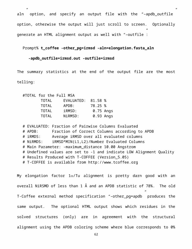
Figure Legends
Figure 1. A phylogram of the HPV type assemblage most closely related to Type16 based on
the L1 major capsid protein, and the corresponding GCG PlotSimlarity traces. The ellipses
denote potential areas in which to localize PCR primers within the gene that would differentiate
the Type 16 clade from it’s closest relatives. These are areas of high L1 conservation in the
Type 16 clade (the red, dashed line) that correspond to areas of much weaker conservation in
the other clades (the blue, solid line).
Figure 2. A GCG SeqLab PostScript graphic of the most conserved portion of the HMG-box
DNA binding domain from a collection of paralogous human HMG-box protein sequences.
Figure 3. A RasMac representation of the Swiss-Model Giardia EF-1 structure superimposed
over the eight most similar solved structures.
62