Embed Size (px)
Citation preview
Biogenesis of the pore architecture ofa voltage-gated potassium channelChristine Gajewskia, Alper Dagcanb, Benoit Rouxb, and Carol Deutscha,1
aDepartment of Physiology, University of Pennsylvania, 3700 Hamilton Walk, Philadelphia, PA 19104-6085; and bDepartment of Biochemistry andMolecular Biology, University of Chicago, Chicago, IL 60637
Edited by Richard W. Aldrich, University of Texas at Austin, Austin, TX, and approved January 3, 2011 (received for review November 15, 2010)
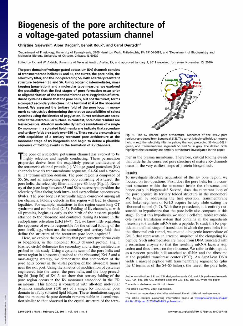
The pore domain of voltage-gated potassium (Kv) channels consistsof transmembrane helices S5 and S6, the turret, the pore helix, theselectivity filter, and the looppreceding S6,with a tertiary reentrantstructure between S5 and S6. Using biogenic intermediates, masstagging (pegylation), and a molecular tape measure, we exploredthe possibility that the first stages of pore formation occur priorto oligomerization of the transmembrane core. Pegylation of intro-duced cysteines shows that the pore helix, but not the turret, formsa compact secondary structure in the terminal 20 Å of the ribosomaltunnel. We assessed the tertiary fold of the pore loop in mono-meric constructs by determining the relative accessibilities of selectcysteines using the kinetics of pegylation. Turret residues are acces-sible at the extracellular surface. In contrast, pore helix residues areless accessible. All-atommolecular dynamics simulations of a singleKv monomer in a solvated lipid membrane indicate that secondaryand tertiary folds are stable over 650 ns. These results are consistentwith acquisition of a tertiary reentrant pore architecture at themonomer stage of Kv biogenesis and begin to define a plausiblesequence of folding events in the formation of Kv channels.
The pore of a selective potassium channel has evolved to behighly selective and rapidly conducting. These permeation
properties derive from the exquisitely precise architecture ofthe tetrameric channel protein (1). Voltage-gated potassium (Kv)channels have six transmembrane segments, S1–S6 and a cytoso-lic T1 tetramerization domain. The pore region is composed ofS5, S6, and an intervening pore loop consisting of a turret, thepore helix, the selectivity filter, and a pre-S6 loop (Fig. 1). Reen-try of the pore loop between S5 and S6 is necessary to position theselectivity filter facing both intra- and extracellular aqueous ves-tibules. The pore loop is structurally highly conserved among allion channels. Folding defects in this region will lead to channe-lopathies. For example, mutations in this region cause long QTsyndrome and can be fatal (2, 3). Folding of Kv channels, as withall proteins, begins as early as the birth of the nascent peptideattached to the ribosome and continues during its tenure in theendoplasmic reticulum (ER) (4–7). Yet, we know little regardingthe sequence of events responsible for the critical folding of thepore itself, e.g., when are the secondary and tertiary folds thatdefine the structure of the reentrant pore loop acquired?
Here, we explore the possibility that pore structure forms earlyin biogenesis, in the monomer Kv1.3 channel protein. Fig. 1(dashed circle) delineates the secondary and tertiary architectureprobed in this study. Using a cysteine scan of the pore helix andturret region in a nascent (attached to the ribosome) Kv1.3 and amass-tagging strategy, we demonstrate that compaction of thepore helix occurs in the distal portion of the ribosomal tunnelnear the exit port. Using the kinetics of modification of cysteinesengineered into the turret, the pore helix, and the loop preced-ing S6 (loop-S6) of Kv1.3, we show that tertiary folding of thepore region occurs in the Kv monomer embedded in the ERmembrane. This finding is consistent with all-atom moleculardynamics simulations (650 ns) of a single Kv monomer poredomain in a fully solvated lipid bilayer. These simulations suggestthat the monomeric pore domain remains stable in a conforma-tion similar to that observed in the crystal structure of the tetra-
mer in the plasma membrane. Therefore, critical folding eventsthat underlie the conserved pore structure of mature Kv channelsoccur in the very earliest steps of protein biosynthesis.
ResultsTo investigate structure acquisition of the Kv pore region, wefocused on two questions. First, does the pore helix form a com-pact structure within the monomer inside the ribosome, andhence early in biogenesis? Second, does the reentrant loop ofthe pore acquire its tertiary folded structure in the monomer?We began by addressing the first question. Transmembraneand linker segments of Kv1.3 acquire helicity while exiting theribosomal tunnel (5, 7). With these precedents, it is entirely rea-sonable to hypothesize that the pore helix also compacts at thisstage. To test this hypothesis, we used a cell-free rabbit reticulo-cyte lysate translation system that contains all the ingredientsnecessary to translate mRNA into a peptide. To study the Kv pep-tide at a defined stage of translation in which the pore helix is inthe ribosomal exit tunnel, we created a biogenic intermediate ofKv1.3 that represents an arrested snapshot of the elongating Kvpeptide. Such intermediates are made from DNA truncated witha restriction enzyme so that the resulting mRNA lacks a stopcodon and thus arrests on the ribosome. We refer to this peptideas a nascent peptide, still attached to tRNA and the ribosomeat the peptidyl transferase center (PTC). An SgrAI-cut DNAyields a nascent peptide with transmembrane segment S5 (plusthe C terminus of the S4–S5 linker), the turret, the pore helix,
Fig. 1. The Kv channel pore architecture. Monomer of the Kv1.2 poreregion, reproduced from Long et al. (13). The turret is depicted in blue, the porehelix in red, the selectivity filter in yellow, the loop preceding S6 (loop-S6) ingreen, and transmembrane segments S5 and S6 in gray. The dashed circlehighlights the secondary and tertiary architecture investigated in this paper.
Author contributions: B.R. and C.D. designed research; C.G. and A.D. performed research;C.G., A.D., B.R., and C.D. analyzed data; and C.G., B.R., and C.D. wrote the paper.
The authors declare no conflict of interest.
This article is a PNAS Direct Submission.1To whom correspondence should be addressed. E-mail: [email protected].
This article contains supporting information online at www.pnas.org/lookup/suppl/doi:10.1073/pnas.1017097108/-/DCSupplemental.
3240–3245 ∣ PNAS ∣ February 22, 2011 ∣ vol. 108 ∣ no. 8 www.pnas.org/cgi/doi/10.1073/pnas.1017097108
Dow
nloa
ded
by g
uest
on
July
29,
202
0

the selectivity filter, loop-S6, and the N-terminal half of trans-membrane segment S6 (Fig. 2A, right cartoon).
We engineered cysteines, one at a time, in this construct anddetermined the extent to which each cysteine could be covalentlymodified by PEG maleimide (PEG-MAL, 5 kD). The adduct isdetected as a shift in mobility of the peptide on a protein gel(5, 8). We refer to this mass-tagging method as pegylation. Theresults are calibrated against a cysteine scan of an all-extendedmolecular tape measure (Fig. 2A, left cartoon), for whichthe extent of cysteine labeling depends on the distance fromthe PTC to each individual cysteine (5). The tape measure isa 95-residue nascent chain composed of the N terminus ofKv1.3. This N terminus includes a portion of the T1 sequencethat is known to be all-extended and has been engineered toreside inside the ribosomal exit tunnel. Tape-measure cysteinesthat are 26 amino acids from the PTC are relatively inaccessibleto PEG-MAL. As the target cysteine is moved increasinglyfurther than 26 residues from the PTC, labeling increases mono-tonically. Those residues that are ≥33 amino acids from the PTCare ≥80% labeled with PEG-MAL (rightmost gel, Fig. 2B; blacksymbols, Fig. 2C). For an all-extended peptide, 33 residuescorresponds to a distance of approximately 99–112 Å (using3–3.4 Å∕amino acid) between the PTC and the exit port, inexcellent agreement with anatomical dimensions of the tunnelderived from structural studies (9).
A cysteine scan of selected pore helix residues positioned inthe lower portion of the exit tunnel, e.g., residues 389, 387,and 383, shows relatively little pegylation even for a residue thatis 30 amino acids from the PTC (Fig. 2B, three leftmost lanes:band 1 vs band 0). In contrast, turret residues, 379, 377, and373 (center three lanes), show relatively more pegylation, e.g.,66% for residue 373 near the top of the turret. A comparisonof the fraction pegylated (Fpeg) for the pore helix (red symbols,Fig. 2C) with the all-extended tape measure reveals a shallowerslope and right-shifted curve, whereas the turret (blue symbols)has a steeper slope compared to the pore helix. However, it is
somewhat shallower than the tape measure, which is likely dueto some contamination of the region probed with the N terminusof the pore helix. Nonetheless, the pore helix is relatively morecompact than the turret, as expected from the mature structure.
Tertiary Structure of the Pore. Having determined that the porehelix may already be compact as it exits the ribosomal tunnel, weconsidered two extreme scenarios for tertiary folding of the pore(Fig. 3A). The cartoon on the left depicts the S5 and S6 segments(gray), which are normally anchored in the membrane, and theintervening pore loop as unstructured and accessible in the aqueouscompartment of the ER lumen (topologically equivalent to theextracellular compartment). The second scenario (Fig. 3A, Right)is derived from the crystal structure of the tetrameric Kv1.2/2.1chimera (10) and manifests a reentrant pore. The two scenariosmake different predictions regarding the accessibility of engineeredcysteines. In the first scenario, all the indicated cysteines would beaccessible to the aqueous compartment; in the second scenario, onlythose in the exposed turret and loop-S6 would be accessible. Thecysteine at the bottom of the pore helix would be buried at proteinor lipid interfaces. We used the kinetics of pegylation to explorethese predictions. An accessible residue will have a relatively fastmodification rate, whereas a buried residue will have a relativelyslow modification rate. For these experiments, we chose two Kv1.3constructs, both shown to bemonomeric in theERmembrane (8): aT1-deletion [T1(-)] and an S5-P-S6C-terminus fragment (FRAG;Fig. 3B). The T1(-) peptide is monomeric under the conditionsof our experiments, but can produce tetramers under high-RNAconditions and long times (8, 11). In contrast, FRAG never formstetramers (8) but inserts into the ERmembrane with correct orien-tation (12). A further advantage of FRAG is that the foldingphenotype we discover will reflect folding of the pore region itselfin the monomer independent of a voltage sensor domain (S1–S4)and therefore be relevant for folding of two-transmembrane-segment Kþ channels, e.g., KcsA and Kir.
Fig. 2. Probing secondary structure formation in the ribosomal exit tunnel. (A) Cartoons of two different nascent peptide–ribosome complexes. The graycircles represent the two subunits of the ribosome. The PTC, at the cleft between the two subunits, is approximately 100 Å from the exit port of the tunnel. Anall-extended peptide (molecular tape measure) is represented by a solid black line (Left) and a nascent fragment of Kv1.3 containing the pore helix (red) andturret (blue) is depicted schematically as a solid line (Right). The native Kv1.3 peptide is attached to the PTC at residue A415 (SgrAI restriction enzyme). Thecomplex is not drawn to scale. (B) Pegylation of the pore helix and turret. Nascent peptides were translated from mRNA engineered from a DNA that lacked astop codon, pegylated (1 mM PEG-MAL for 4–6 h), and fractionated using SDS-PAGE as described in the SI Appendix. The number directly under each laneindicates the residue in Kv1.3 mutated to cysteine (pore helix residues in red, turret residues in blue); the number with a Δ indicates the number of amino acidsfrom the PTC to (and including) the cysteine; the numbers to the left of the gel correspond to molecular weight standards; numbers to the right of the gelindicate unpegylated (0) and singly pegylated (1) protein; the number directly above each lane indicates the calculated fraction of nascent peptide pegylated(Fpeg). The rightmost lane is the all-extended control (Extended CTL). (C) Cysteine scan of the pore helix and turret of nascent Kv1.3 peptide. The x axis is thenumber of amino acids from the PTC to (and including) the labeled cysteine. Pegylation of a known all-extended nascent peptide, is shown by the black circlesand represents data taken from Lu and Deutsch (5). The final extent of pegylation of individual residues in the pore helix and turret is represented by the redand blue circles, respectively. Symbols are mean� SEM (n ≥ 3).
Gajewski et al. PNAS ∣ February 22, 2011 ∣ vol. 108 ∣ no. 8 ∣ 3241
BIOPH
YSICSAND
COMPU
TATIONALBIOLO
GY
Dow
nloa
ded
by g
uest
on
July
29,
202
0

We began with the T1(-) construct, translating it in the pre-sence of microsomal membranes. We solubilized the isolatedmembranes in dodecylmaltoside (C12M, 0.1%), typically a non-denaturing detergent (SI Appendix), and assessed the accessibilityof specific cysteines in the turret, pore helix, and loop-S6. Thekinetics of pegylation of residues 371, 389, and 398 in the turret(blue), pore helix (red), and loop-S6 (green), respectively, areshown in Fig. 4B. Note that the concentration of PEG-MAL usedin these modification reactions was 1, 2, and 0.3 mM, respectively,in order to capture well-resolved time courses despite the differ-ence in modification reaction rates. For each gel, the lower doub-let represents the unpegylated peptide (“0” band), comprised ofboth unglycosylated (lower band of the doublet) and core (ER)glycosylated (upper band of the doublet) peptide. The upperband(s) represents the pegylated peptide (band “1”). For eachindicated time, Fpeg was calculated, normalized to the final datapoint, and plotted in Fig. 4B below each series of gels. The datashow monotonically increasing values that saturate and could befit with a single exponential to give modification rate constants of20.2, 1.3, and 6.3 M−1 s−1, respectively. The turret residue, 371,and the loop-S6 residue, 398, are each modified much faster thanthe pore helix residue, 389. The latter is less accessible than theformer two, consistent with a reentrant architecture of the pore.Replicate experiments for each of these cysteines gave meanrate constants of 15.5� 2.4 (n ¼ 3, �SEM), 1.0� 0.3 (n ¼ 2,�average deviation), and 6.6� 0.4 (n ¼ 3, �SEM) M−1 s−1, for371, 389, and 398, respectively. The rate constant in the pore helixis significantly lower than that in either the turret (P ¼ 0.02) orthe loop-S6 (P ¼ 0.002). Residue 390, also at the bottom of thepore helix, gives a mean rate constant of 0.4� 0.1 (n ¼ 3,�SEM)M−1 s−1, which is significantly different from the turret and loop-S6 (both P ≤ 0.003). These results (also see SI Appendix) suggestthat residues in the C-terminal pore helix are more hindered andthe pore region is folded in the T1(-) Kv1.3 monomer.
If the folded monomer is stable, then those residues ultimatelyfated to be at an intersubunit interface in the mature Kv channelwill no longer bemasked by protein in the ERmembrane, whereasthose residues fated to be at an intrasubunit or lipid interface willremain masked by the lipid or protein. For example, residue 398,equivalent to residue L81 in KcsA and M380 in Kv1.2, is locatedat an intersubunit interface according to the models derived fromthe respective crystal structures (1, 13). Additionally, this residuecan be crosslinked to produce dimers and the equivalent mutationin Shaker (M448C) also can be crosslinked to give dimers (14).
This residue should therefore be relatively accessible in theT1(-) Kv1.3 monomer, which is indeed the case (see also Fig. 4C).
To probe these regions more completely, we carried out a moreextensive cysteine scan of the three regions, using a fixed concen-tration of PEG-MAL (2 mM). The range of normalized Fpeg at1 min is approximately 0.4–0.7 for turret residues, approximately0.1 for the bottom of the pore helix, and approximately 0.4–0.8for loop-S6 residues (Fig. 4C). A pattern emerges that indicatesrelatively faster modification rates for the turret and loop-S6 thanfor the pore helix, consistent with a reentrant pore architecture inthe T1(-) monomer. To support this conclusion, we probedFRAG for the same three residues (371, 389, and 398) shown inFig. 4B. Fig. 4D recapitulates the results obtained for the T1(-)monomer. The modification rate for the 389 residue is muchslower, approximately 0.4 M−1 s−1 compared with that for the 371and 398 residues. We suggest that the pore is reentrant in themonomer and does not require a tetramer to achieve a pore-likearchitecture. Furthermore, we do not think that the pore helixpredominantly lies parallel to the membrane surface, but ratherthat it adopts a reentrant position. This conclusion derives fromthree arguments. First, the pore helix itself is not amphipathicand has a mean relative hydrophobic moment of 0.12, whichrepresents its hydrophobic moment relative to a perfectly amphi-pathic peptide (Eisenberg scale, HydroMCalc; http://www.bbcm.univ.trieste.it/~tossi/HydroCalc/HydroMCalc.html). Second, thereare many strong interactions between the pore helix and S5 andS6 within the monomer proper (see calculated van der Waals inter-actionenergies, Fig. 5C), so that theporehelix is stableas a reentrantloop. Third, three adjacent residues, 389, 390, and 391, at the C ter-minus of the pore helix, each exhibit slow modification rate con-stants. It is unlikely that all three residues would be slow if the porehelix were lying at the membrane–water interface parallel to themembrane. The most parsimonious explanation for the observedpattern of rate constants is a reentrant loop in the monomer.
If the monomer is folded, then unfolding the pore should in-crease the relative accessibility of a residue at the bottom of thepore helix. We undertook two approaches to explore this hypo-thesis. First, we solubilized the ER membrane-integrated T1(-)peptides in lithium dodecyl sulfate, a denaturing detergent,pegylated and determined the Fpeg. Under these conditions, re-sidues 371, 389, and 398 gave normalized 1-min Fpeg values of0.97, 0.62, and 0.99, respectively, a marked increase over thoseshown in Fig. 4C for the folded monomer. Second, we mutatedresidues in the pore helix that might contribute to folding freeenergy. We chose the two adjacent tryptophan residues (W384and W385) in the middle of the pore helix of Kv1.3 because tryp-tophans have a preference for membrane interfaces (15–17).W384C and W385C each exhibit increased pegylation at 1 min(Fig. 4E, left bar graph), compared to other pore helix residues(dashed red line, derived Fig. 4C). If these results are a conse-quence of unfolding of the pore tertiary structure due to removalof aromatic residues, then a 390C at the bottom of the pore helixshould be more accessible in a construct in which the W384 andW385 have been mutated to nonaromatic residues. The triplemutant, W384A/W385A/M390C, is pegylated approximately five-fold faster than the single M390C mutant containing the WWmotif (Fig. 4E, right bar graph), consistent with increased acces-sibility of 390C. Mutation of either or both tryptophans fails toproduce current upon transfection in Xenopus oocytes, consistentwith failure of the channel to form a proper pore architecture.
Molecular Dynamics Simulations of a Kv Monomer. To examine theconclusion that the reentrant pore loop can exist in the Kv mono-mer, we performed all-atom molecular dynamics simulations.The simulation system was constructed from the crystal structureof the Kv1.2 [Protein Data Bank (PDB) ID 2A79] pore domain(99 residues from the beginning of S5, Ala323, to the end ofS6, Thr421) embedded in palmitoyloleoylphosphatidylcholine
Fig. 3. Strategy for assessing tertiary folding. (A) Two scenarios. Depictedare predictions for relative pegylation kinetics for individually engineeredcysteines in three component segments of the pore loop between S5 andS6 for an unfolded tertiary pore loop (Left) and a reentrant pore loop (Right).(B) Monomeric constructs for the accessibility assay include Kv1.3 T1(-)(Upper), which lacks only the N-terminal residues 1–141, and a Kv1.3S5-P-S6C-terminus fragment (FRAG; residues 336–523) (Lower).
3242 ∣ www.pnas.org/cgi/doi/10.1073/pnas.1017097108 Gajewski et al.
Dow
nloa
ded
by g
uest
on
July
29,
202
0

(POPC) and surrounded by an aqueous solution of 150-mM KCl.After a 50-ns equilibration, the structure was simulated for anadditional 650 ns. Fig. 5A shows a backbone structure at
650 ns (blue) superimposed on the backbone of the crystal struc-ture (gray). There are some local structural deviations (rmsd is3.9 Å for all atoms and 3.0 Å for α-carbon atoms for this snapshot
Fig. 4. Relative pegylation kinetics of select cysteines in Kv1.3T1(-) and FRAG. Cysteines were engineered, one at a time, in each of three segments: the turret,the pore helix, and loop-S6, and pegylated. (A) The primary sequence of the pore region. (B) Pegylation kinetics of D371C (turret, blue), T389C (pore helix, red),and M398C (loop-S6, green) were determined at 1, 2, and 0.3 mM PEG-MAL, respectively. Each lane represents a sample quenched at the indicated time (beloweach lane, in minutes) and the number directly above each lane indicates the calculated Fpeg. Right and left numbers as in Fig. 2B. For each residue, a timecourse of normalized Fpeg is plotted below each set of gels and fit with a single exponential. (C) Normalized Fpeg at one minute in T1(-) for residues in theturret (blue), pore helix (red), and loop-S6 (green). The final PEG-MAL concentration was 2 mM in all reactions shown here. Identical pegylation reactions werequenched at 1 min and at >3 h, fractionated using SDS-PAGE, and quantified as described in the SI Appendix. Pegylation at 1 min was normalized to themaximum fraction pegylated at >3 h (maximum Fpeg, dot plot) and shown as Fpeg at 1min (bar graph). Symbols are mean� SEM (n ≥ 3) for all residues except377 and 397, which are mean� average deviation for duplicate samples. (D) Time course of cysteine modification in FRAG. Cysteines engineered in the turret(371), the pore helix (389), and the loop-S6 (398) were engineered in the background of FRAG and pegylated (2 mM PEG-MAL), as described in Fig. 4B. Resultswere fit with a single exponential. (E) Cysteines were engineered in the pore helix at positions 384 and 385, each a tryptophan in the native sequence. (Left)W384C and W385C, separately, were pegylated (2 mM PEG-MAL) as described in Fig. 4B. Data are shown as Fpeg at 1 min normalized to the maximum Fpeg at4 h. Values are mean� SEM for n ¼ 3. The red dashed line (derived from Fig. 4C) indicates a minimum Fpeg at 1 min for pore helix residues and the blue andgreen dashed lines (derived from Fig. 4C) indicate a maximum Fpeg value at 1 min for the turret and loop-S6, respectively. (Right) W384C and W385C weresimultaneously mutated in the background of M390C and pegylated as described in Fig. 4B. The normalized Fpeg at 1 min for this triple mutant, W384A/W385A/M390C, is compared with M390C alone, and shown as mean� SEM, n ¼ 3, for M390C, and mean� average deviation, n ¼ 2, for the triple mutant.Dashed lines as described above for Fig. 4E, Left.
Gajewski et al. PNAS ∣ February 22, 2011 ∣ vol. 108 ∣ no. 8 ∣ 3243
BIOPH
YSICSAND
COMPU
TATIONALBIOLO
GY
Dow
nloa
ded
by g
uest
on
July
29,
202
0

at 650 ns) along the pore loop and transmembrane segments, andthe pore helix is shifted about a half turn toward the extracellularside in the 650-ns snapshot. Nevertheless, the tertiary reentrantfold of the pore loop is manifest in both structures. The time-dependence of the rms deviations of the monomer relative toeither the crystal structure or the final snapshot of the trajectory(650 ns) indicates that the monomer stabilizes in its final confor-mation after 350 ns.
In Fig. 5B, a detailed snapshot of the simulated structure at650 ns is shown depicting side chains and water molecules locatedwithin 4 Å of the protein. The inset (box) highlights the presenceof a mini-water-droplet, five water molecules, lodged between thepore helix and the selectivity filter, which likely contributes to sta-bilizing the tertiary fold. The surrounding water molecules, alongwith surrounding lipid carbons are represented quantitatively inFig. 5C, Upper. Most of transmembrane segments S5 and S6,especially the middle stretch of residues, is in contact with lipidchains, whereas the turret and loop-S6 are in contact with watermolecules. In contrast, for most of the pore helix and selectivityfilter, residues are in contact with some lipid and some water(see SI Appendix). The pore helix does not need to reside atthe lipid–water interface to be hydrated. This hydration of thereentrant helix may be sufficient for cysteine modification tooccur. It is striking that the profile for the water distribution isvery similar to that for distribution of normalized Fpeg (Fig. 4C).Because cysteine modification rates are a measure of water ac-cessible volume surrounding the cysteine side chain, the excellentcorrelation of the two plots is strong evidence in support of theconclusion that the pore domain of a monomer contains a reen-trant pore loop.
To better understand the energetics underlying the stability ofthe monomer reentrant architecture, we calculated the van derWaals interaction energies from the Kv1.2 crystal structure. The
van der Waals energy directly reflects the extent of atomic contactbetween the side chains, which are predominantly nonpolar. Intra-monomer energies correspond to the interactions of the side chainof residue i in monomer A with the side chains of all residues ofmonomer A (excluding the nearest neighbors along the sequencei − 1 and iþ 1). Similarly, intermonomer energies correspond tothe interactions of the side chain of residue i in monomer A withthe side chains of all residues of monomers B, C, and D in thetetramer. As shown in Fig. 5C, Lower, the major contribution(except for residues 366 and 376) is always intramonomer.
DiscussionThe pore of a potassium channel was selected by evolution for itshigh conductance and exquisite selectivity for potassium ions.These properties derive from the precise, and highly conserved,architecture of the tetrameric channel protein, specifically that ofa reentrant pore loop. Although the primary sequence of the poreis synthesized vectorially in the ribosome, the stage in Kv biogen-esis when the loop between S5 and S6 folds back into the mem-brane is not known. The pore helix is compact in the more distalregions of the tunnel, near the exit port, whereas the turret ismore extended. Because secondary structure acquired in theribosomal tunnel is retained upon migration through the translo-con (7), it is likely that the compact structure of the pore helixsequence is retained and available for tertiary folding when thissegment of Kv1.3 exits the translocon. Cooperativity among sec-ondary, tertiary, and quaternary folding events may be pivotal inacquisition of correctly folded mature Kv structure.
Tertiary Structure Formation of the Pore.Two arguments support thehypothesis that a reentrant pore architecture may be establishedearly in biogenesis and does not require tetramerization of Kv sub-units. First, there is precedent for a structurally homologous helix/
Fig. 5. Stability of the reentrant pore loop. (A) Overlay of the monomer pore domain from the crystal structure of Kv1.2 (gray) and the pore domain at the650th nanosecond of the molecular dynamics simulation (blue). (B) Snapshot of Kv1.2 pore domain at 650 ns. The pore helix and the selectivity filter backboneatoms (except O) and side chains, including Met372 (corresponds to Kv1.3 Met390), are shown. Waters within 4 Å of protein at 650 ns are shown. Pictures wereprepared in the molecular viewing program VMD. (Inset) Same structure as in B, highlighting five water molecules between the pore helix and selectivity filteras space filling Corey–Pauling–Koltun atoms. (C, Upper) Average number of water and lipid heavy atoms around each residue. Number of water (blue bar) orlipid (black bar) heavy atoms (all atoms except for hydrogen) calculated within 4 Å of each heavy atom in each side chain and averaged throughout the 650-nssimulation. The bar along the top of the plot is color coded as described in Fig. 1 to indicate Kv segments. The numbers along the x axis are residue numbers ofnative Kv1.2. Tcl command interface of the VMD program were used to perform the analysis. Residues 346, 364, and 368 are probably surrounded only byprotein (i.e., neither lipid nor water). The corresponding Kv1.3 residues 362 (S5, gray), 371 (turret, blue), 390 (pore helix, red), and 398 (loop-S6, green) areindicated by arrows, as is the GYG sequence (yellow). (Lower) The van der Waals interaction energies within a Kv1.2 monomer (green bars) and between onemonomer and its three partner subunits in the tetramer Kv1.2 (red bars), both calculated from the Kv1.2 crystal structure.
3244 ∣ www.pnas.org/cgi/doi/10.1073/pnas.1017097108 Gajewski et al.
Dow
nloa
ded
by g
uest
on
July
29,
202
0

fold/helix motif among soluble proteins. Using DaliLite version 3(http://ekhidna.biocenter.helsinki.fi/dali_server) to search a pro-tein structure database, several examples of monomeric solubleproteins were identified that are similar (rmsd values of ≤3 Å;see SI Appendix) to the Kv pore segment (residues 333–398). Sec-ond, we previously showed that S5, the pore loop, and S6C-termi-nus likely exist as a cotranslationally folded biogenic unit (12).
Although pore architecture can arise early in biogenesis of themonomer and does not require formation of a Kv tetramer, thetertiary fold of the monomer likely differs from that in the maturetetramer because additional folding may accompany tetrameriza-tion of the pore domain. Such fine tuning would serve to optimizeion conductance and selectivity of the functional tetrameric chan-nel. Moreover, formation of a reentrant loop might facilitate tet-ramerization of the transmembrane segments due to additionalintersubunit interactions (1). Although the monomer—by itself—has enough van der Waals stabilization energy to maintain theconformation observed in the tetramer, tetramerization may benecessary to help catalyze the folding process, the subunits actingcollectively as chaperones for one another. Regardless, the pegy-lation results suggest that the monomer can fold on its own.
What Drives Reentry of the Loop in theMonomer?Our results suggestthat van der Waals interactions within the monomer are sufficientto stabilize a reentrant configuration of the pore loop. Moreover,the crystal structure of the Kv1.2/Kv2.1 chimera reveals a networkof interactions within 3.8 Å, consistent with extensive intrasubunithydrogen bond formation (see SI Appendix for examples), albeitthese interactions exist in the monomer within the context of themature tetramer andmay not be as extensive at an earlier biogenicstage. Mutation of some analogous hydrogen-bonding residues inKv1.3 prevents functional expression in Xenopus oocytes. In addi-tion, the molecular dynamics simulations reveal a droplet of fivewater molecules that simultaneously bridges the pore helix andselectivity filter in the tertiary folded conformation and may con-tribute to stabilizing the reentrant topology of the monomer.
An additional consideration is the role of aromatic pore resi-dues in promoting reentry. Mutation of W384 and W385 to ala-nine produced a more accessible T1(-) M390C. The pair oftryptophans in the pore helix is highly conserved across Kþ chan-nels. Tryptophans preferentially reside at membrane interfaces(17–19). The complex properties known as aromaticity underliethis preference (17). In addition, aromatic side chains are keydeterminants for insertion of transmembrane segments into thelipid bilayer and topogenic events (18). We therefore suggest thatthis preference of aromatic residues for the lipid–water interfacemay also contribute to reentry of the pore sequence into themembrane. In Kv1.3, mutation of W384 or W385 to A, E, L,or P, prevents functional expression in Xenopus oocytes. In KcsA,mutations W67A or W68A in the pore helix yield a peptide that
fails to assemble correctly (20). These scenarios implicate a rolefor aromatic pore helix residues in tertiary folding.
The sequential coordination of folding events is undoubtedlycritical for acquisition of a mature structure, and our results sup-port the plausibility that some aspects of tertiary folding, notablythe formation of a reentrant pore loop, precede oligomerization.Folding of the monomer might help create an appropriate inter-action interface that energetically favors tetramer formation. Analternative possibility is that tetramer formation provides a scaf-fold that favors the entry of an extracellular pore loop into itsmature residence within the transmembrane domain. Our resultsindicate that this prerequisite is not necessary. Defining thesequence of folding events is key to understanding not only Kvbiogenesis and its efficiency, but also subsequent traffickingmechanisms, because retention, export, and degradation signalsare modulated by how the protein is folded and packed.
MethodsConstructs, in Vitro Translation, Pegylation. All constructs were created in apSP64 plasmid. The pSP64/full-length Kv1.3, pSP64/T1(-)Kv1.3, and pSP64/S5-P-S6C-terminus Kv1.3 (FRAG) were constructed as described previouslyand are cysteine free (8). Engineered cysteines and restriction enzyme siteswere introduced using Stratagene™QuikChange Kit. For details of molecularbiology methods and in vitro translation see SI Appendix and ref. 8.
Isolated ribosome-nascent peptide-membrane pellets were resuspendedon ice with PBS* (CaCl2- and MgCl2-free Dulbecco’s PBS, pH 7.4, supplemen-ted with 4 mM MgCl2) containing 50 μM DTT. An equal volume of methylPEG-MAL (Mr 5000, >99%, SunBio, Inc.; prepared in PBS*) was added (finalPEG-MAL concentration of 1–2 mM) and incubated on ice from 0 to 6 h. Thepegylation reaction was quenched with 100-fold excess DTT at ambient tem-perature for 10 min and then 0 °C for 10 min and fractionated as described inthe SI Appendix.
Pegylation Measurements for Tertiary Folding. Pellets containing membrane-embedded peptides were resuspended on ice with 50–100 μL of 0.1% Ana-trace™ n-dodecyl-β-D-maltopyranoside, Anagrade® (C12M, prepared in PBS*supplemented with 50–500 μM DTT) and incubated 1 h on ice to dissolvemembranes. Samples were centrifuged to pellet undissolved membraneand the supernatant transferred to a fresh tube and an equal volumeof PBS* solution containing PEG-MAL (final PEG-MAL concentration was1–2 mM), incubated on ice from 0 to 3 h, quenched as described above,and analyzed as described in the SI Appendix.
Molecular Dynamics. The simulation system was constructed from the crystalstructure of the Kv1.2 (PDB ID 2A79) pore domain embedded in POPCsurrounded by an aqueous solution of 150 mM KCl. See the SI Appendixfor details.
ACKNOWLEDGMENTS. We thank Dr. Richard Horn for careful reading of themanuscript and helpful discussion, Timothy Pian for technical assistance,and Dr. LiWei Tu for constant guidance and helpful discussion. Supported byNational Institutes ofHealthGrantGM52302 (toC.D.) andGM062342 to (B.R.).
1. Doyle DA, et al. (1998) The structure of the potassium channel: Molecular basis ofKþ conduction and selectivity. Science 280:69–76.
2. Benson DW, et al. (1996) Missense mutation in the pore region of HERG causes familiallong QT syndrome. Circulation 93:1791–1795.
3. Huang FD, Chen J, Lin M, Keating MT, Sanguinetti MC (2001) Long-QT syndrome-associated missense mutations in the pore helix of the HERG potassium channel.Circulation 104:1071–1075.
4. Kosolapov A, Tu L, Wang J, Deutsch C (2004) Structure acquisition of the T1 domain ofKv1.3 during biogenesis. Neuron 44:295–307.
5. Lu J, Deutsch C (2005) Secondary structure formation of a transmembrane segment inKv channels. Biochemistry 44:8230–8243.
6. Tu L, Wang J, Deutsch C (2007) Biogenesis of the T1-S1 linker of voltage-gatedKþ channels. Biochemistry 46:8075–8084.
7. Tu LW, Deutsch C (2010) A folding zone in the ribosomal exit tunnel for Kv1.3 helixformation. J Mol Biol 396:1346–1360.
8. Lu J, Deutsch C (2001) Pegylation: A method for assessing topological accessibilities inKv1.3. Biochemistry 40:13288–13301.
9. Ban N, Nissen P, Hansen J, Moore PB, Steitz TA (2000) The complete atomic structureof the large ribosomal subunit at 2.4 A resolution. Science 289:905–920.
10. Long SB, Tao X, Campbell EB, MacKinnon R (2007) Atomic structure of a voltage-dependent Kþ channel in a lipid membrane-like environment. Nature 450:376–382.
11. Tu L, et al. (1996) Voltage-gated Kþ channels contain multiple intersubunit associationsites. J Biol Chem 271:18904–18911.
12. Tu L, Wang J, Helm A, Skach WR, Deutsch C (2000) Transmembrane biogenesis ofKv1.3. Biochemistry 39:824–836.
13. Long SB, Campbell EB, MacKinnon R (2005) Crystal structure of a mammalian voltage-dependent Shaker family Kþ channel. Science 309:897–903.
14. Liu Y, Jurman ME, Yellen G (1996) Dynamic rearrangement of the outer mouth of aKþ channel during gating. Neuron 16:859–867.
15. Wimley WC, White SH (1993) Membrane partitioning: Distinguishing bilayer effectsfrom the hydrophobic effect. Biochemistry 32:6307–6312.
16. Wimley WC, White SH (1996) Experimentally determined hydrophobicity scale forproteins at membrane interfaces. Nat Struct Biol 3:842–848.
17. Yau WM, Wimley WC, Gawrisch K, White SH (1998) The preference of tryptophan formembrane interfaces. Biochemistry 37:14713–14718.
18. HigyM,Gander S, SpiessM (2005) Probing the environment of signal-anchor sequencesduring topogenesis in the endoplasmic reticulum. Biochemistry 44:2039–2047.
19. Braun P, von Heijne G (1999) The aromatic residues Trp and Phe have different effectson the positioning of a transmembrane helix in the microsomal membrane. Biochem-istry 38:9778–9782.
20. Cordero-Morales JF, et al. (2006) Molecular determinants of gating at the potassium-channel selectivity filter. Nat Struct Mol Biol 13:311–318.
Gajewski et al. PNAS ∣ February 22, 2011 ∣ vol. 108 ∣ no. 8 ∣ 3245
BIOPH
YSICSAND
COMPU
TATIONALBIOLO
GY
Dow
nloa
ded
by g
uest
on
July
29,
202
0