Embed Size (px)
Citation preview
Enzymatic Digestion of Polysaccharides
Part I: Monitoring Polymer Digestion and Glucose Production in Microplates
A p p l i c a t i o n N o t e
The production of ethanol for use as a biofuel from grains or cellulosic material requires the hydrolysis of carbohydrate polymers such as cellulose, xylan and starch to monomeric sugars that are fermentable by cellular organisms such as bacteria and yeast. Here we describe fluorescence detection analysis in microplates of some of the enzymes that catalyze the digestive hydrolysis of polysaccharide polymers into monomeric constituents.
Introduction
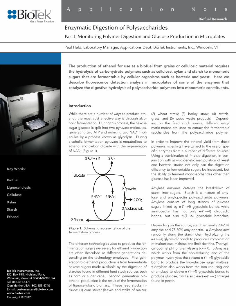
While there are a number of ways to produce eth-anol, the most cost effective way is through alco-holic fermentation. During this process, the hexose sugar glucose is split into two pyruvate molecules, generating two ATP and reducing two NAD+ mol-ecules by a process known as glycolysis. During alcoholic fermentation pyruvate is metabolized to ethanol and carbon dioxide with the regeneration of NAD+ (Figure 1).
The different technologies used to produce the fer-mentation sugars necessary for ethanol production are often described as different generations de-pending on the technology employed. First gen-eration bio-ethanol production is from fermentable hexose sugars made available by the digestion of starches found in different feed stock sources such as corn or sugar cane. Second generation bio-ethanol production is the result of the fermentation of lignocellulosic biomass. These feed stocks in-clude: (1) corn stover (leaves and stalks of maize);
(2) wheat straw; (3) barley straw; (4) switch-grass; and (5) wood waste products. Depend-ing on the feed stock source, different enzy-matic means are used to extract the fermentable saccharides from the polysaccharide polymer. In order to improve the ethanol yield from these polymers, scientists have turned to the use of spe-cific enzymes from a number of different sources. Using a combination of in vitro digestion, in con-junction with in vivo genetic manipulation of yeast and bacteria strains not only can the digestion efficiency to fermentable sugars be increased, but the ability to ferment monosaccharides other than glucose has been improved.
Amylase enzymes catalyze the breakdown of starch into sugars. Starch is a mixture of amy-lose and amylopectin polysaccharide polymers. Amylose consists of long strands of glucose sugars linked by α-(1→4) glycosidic bonds, while amylopectin has not only α-(1→4) glycosidic bonds, but also α-(1→6) glycosidic branches.
Depending on the source, starch is usually 20-25% amylase and 75-80% amylopectin. α-Amylase acts randomly along the starch chain hydrolyzing the α-(1→4) glycosidic bonds to produce a combination of maltotriose, maltose and limit dextrins. The typi-cal optimal pH for α-amylase is 6.7-7.0. β-Amylase, which works from the non-reducing end of the polymer, hydrolyzes the second α-(1→4) glycosidic bond to produce the two-glucose sugar maltose. γ-Amylase also works from the non reducing end of amylase to cleave α-(1→4) glycosidic bonds to produce glucose, it will also cleave α-(1→6) linkages found in pectin.
Figure 1. Schematic representation of the fermentation process.
BioTek Instruments, Inc.P.O. Box 998, Highland Park, Winooski, Vermont 05404-0998 USATel: 888-451-5171 Outside the USA: 802-655-4740 E-mail: [email protected] www.biotek.comCopyright © 2012
Paul Held, Laboratory Manager, Applications Dept, BioTek Instruments, Inc., Winooski, VT
Key Words:
Biofuel
Lignocellulosic
Cellulose
Xylan
Starch
Ethanol
Biofuel Research
from the stock solution immediately prior to use by diluting it 1:5 with 1X reaction buffer.
Typical assay buffer was 50 mM MOPS pH 6.9. Enzyme dilutions were made using 1X reaction buffer as the diluent. Aliquots (50 µL) of each enzyme dilution were added to wells of a ½ area solid black 96-well microplate. Reactions were initiated by the addition of 50 µL of working substrate solution. Fluorescence for all experiments was measured kinetically for 30 minutes with a Synergy™ H4 Multi-Mode Microplate Reader. Excitation was set to 485 nm and emission wavelength at 528 nm.
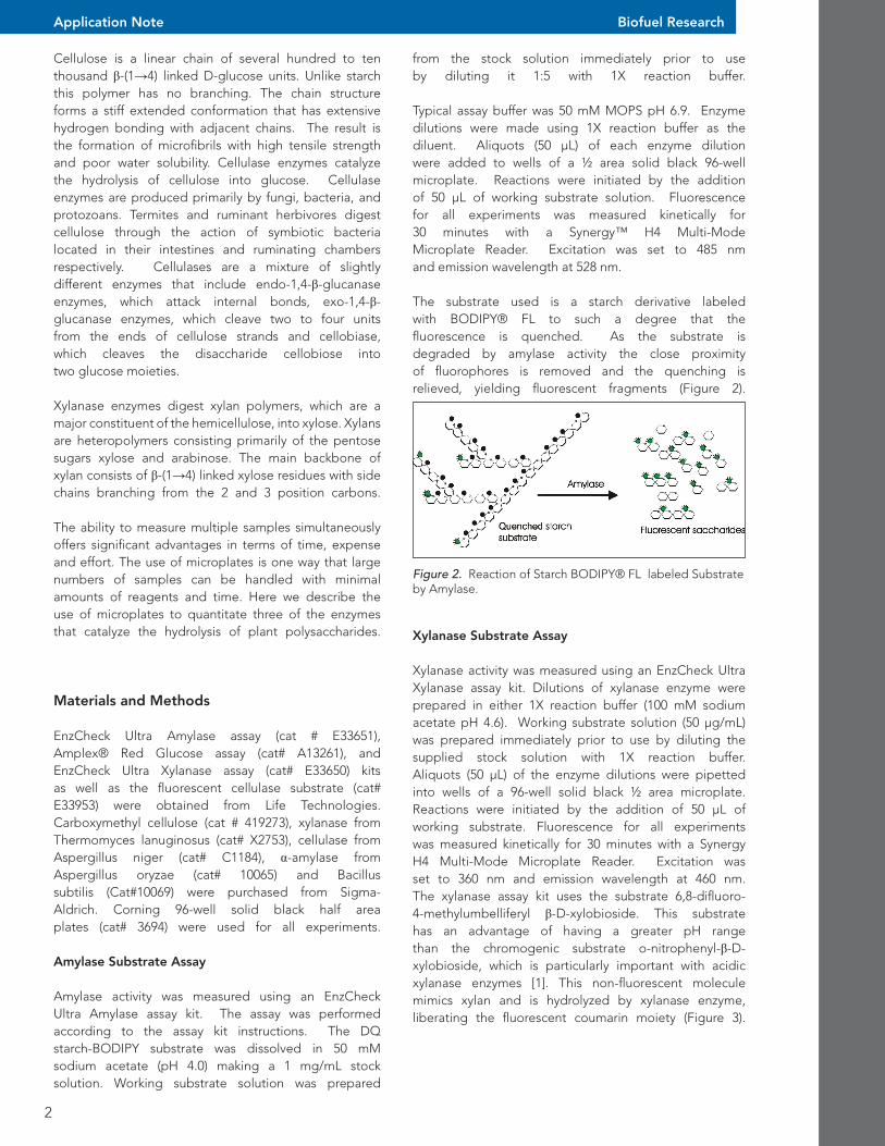
The substrate used is a starch derivative labeled with BODIPY® FL to such a degree that the fluorescence is quenched. As the substrate is degraded by amylase activity the close proximity of fluorophores is removed and the quenching is relieved, yielding fluorescent fragments (Figure 2).
2
Application Note
Cellulose is a linear chain of several hundred to ten thousand β-(1→4) linked D-glucose units. Unlike starch this polymer has no branching. The chain structure forms a stiff extended conformation that has extensive hydrogen bonding with adjacent chains. The result is the formation of microfibrils with high tensile strength and poor water solubility. Cellulase enzymes catalyze the hydrolysis of cellulose into glucose. Cellulase enzymes are produced primarily by fungi, bacteria, and protozoans. Termites and ruminant herbivores digest cellulose through the action of symbiotic bacteria located in their intestines and ruminating chambers respectively. Cellulases are a mixture of slightly different enzymes that include endo-1,4-β-glucanase enzymes, which attack internal bonds, exo-1,4-β-glucanase enzymes, which cleave two to four units from the ends of cellulose strands and cellobiase, which cleaves the disaccharide cellobiose intotwo glucose moieties.
Xylanase enzymes digest xylan polymers, which are a major constituent of the hemicellulose, into xylose. Xylans are heteropolymers consisting primarily of the pentose sugars xylose and arabinose. The main backbone of xylan consists of β-(1→4) linked xylose residues with side chains branching from the 2 and 3 position carbons.
The ability to measure multiple samples simultaneously offers significant advantages in terms of time, expense and effort. The use of microplates is one way that large numbers of samples can be handled with minimal amounts of reagents and time. Here we describe the use of microplates to quantitate three of the enzymes that catalyze the hydrolysis of plant polysaccharides.
Materials and Methods
EnzCheck Ultra Amylase assay (cat # E33651), Amplex® Red Glucose assay (cat# A13261), and EnzCheck Ultra Xylanase assay (cat# E33650) kits as well as the fluorescent cellulase substrate (cat# E33953) were obtained from Life Technologies. Carboxymethyl cellulose (cat # 419273), xylanase from Thermomyces lanuginosus (cat# X2753), cellulase from Aspergillus niger (cat# C1184), α-amylase from Aspergillus oryzae (cat# 10065) and Bacillus subtilis (Cat#10069) were purchased from Sigma-Aldrich. Corning 96-well solid black half area plates (cat# 3694) were used for all experiments.
Amylase Substrate Assay
Amylase activity was measured using an EnzCheck Ultra Amylase assay kit. The assay was performed according to the assay kit instructions. The DQ starch-BODIPY substrate was dissolved in 50 mM sodium acetate (pH 4.0) making a 1 mg/mL stock solution. Working substrate solution was prepared
Biofuel Research
Figure 2. Reaction of Starch BODIPY® FL labeled Substrate by Amylase.
Xylanase Substrate Assay
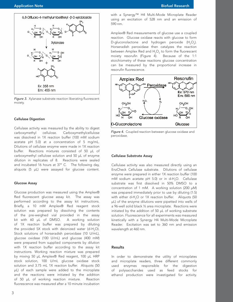
Xylanase activity was measured using an EnzCheck Ultra Xylanase assay kit. Dilutions of xylanase enzyme were prepared in either 1X reaction buffer (100 mM sodium acetate pH 4.6). Working substrate solution (50 µg/mL) was prepared immediately prior to use by diluting the supplied stock solution with 1X reaction buffer. Aliquots (50 µL) of the enzyme dilutions were pipetted into wells of a 96-well solid black ½ area microplate. Reactions were initiated by the addition of 50 µL of working substrate. Fluorescence for all experiments was measured kinetically for 30 minutes with a Synergy H4 Multi-Mode Microplate Reader. Excitation was set to 360 nm and emission wavelength at 460 nm. The xylanase assay kit uses the substrate 6,8-difluoro-4-methylumbelliferyl β-D-xylobioside. This substrate has an advantage of having a greater pH range than the chromogenic substrate o-nitrophenyl-β-D-xylobioside, which is particularly important with acidic xylanase enzymes [1]. This non-fluorescent molecule mimics xylan and is hydrolyzed by xylanase enzyme, liberating the fluorescent coumarin moiety (Figure 3).
3
Application Note Biofuel Research
with a Synergy™ H4 Multi-Mode Microplate Reader using an excitation of 528 nm and an emission of 590 nm.
Amplex® Red measurements of glucose use a coupled reaction. Glucose oxidase reacts with glucose to form D-gluconolactone and hydrogen peroxide (H2O2). Horseradish peroxidase then catalyzes the reaction between Amplex Red and H2O2 to form the fluorescent moiety resorufin (Figure 4). Because of the 1:1 stoichiometry of these reactions glucose concentration can be measured by the proportional increase in resorufin fluorescence.
Figure 4. Coupled reaction between glucose oxidase and peroxidase.
Figure 3. Xylanase substrate reaction liberating fluorescent moiety.
Cellulase Digestion
Cellulase activity was measured by the ability to digest carboxymethyl cellulose. Carboxymethylcellulose was dissolved in 1X reaction buffer (100 mM sodium acetate pH 5.0) at a concentration of 5 mg/mL. Dilutions of cellulase enzyme were made in 1X reaction buffer. Reactions mixtures consisted of 50 µL of carboxymethyl cellulose solution and 50 µL of enzyme dilution in replicates of 8. Reactions were sealed and incubated 16 hours at 37° C. The following day, aliquots (5 µL) were assayed for glucose content.
Glucose Assay
Glucose production was measured using the Amplex® Red fluorescent glucose assay kit. The assay was performed according to the assay kit instructions. Briefly, a 10 mM Amplex® Red reagent stock solution was prepared by dissolving the contents of the pre-weighed vial provided in the assay kit with 60 µL of DMSO. A working solution of 1X reaction buffer was prepared by diluting the provided 5X stock with deionized water (d-H2O). Stock solutions of horseradish peroxidase (10 U/mL), glucose oxidase (100 U/mL) and glucose (400 mM) were prepared from supplied components by dilution with 1X reaction buffer according to the assay kit instructions. Working reaction mixture was prepared by mixing 50 µL Amplex® Red reagent, 100 µL HRP stock solution, 100 U/mL glucose oxidase stock solution and 3.75 mL 1X reaction buffer. Aliquots (50 µL) of each sample were added to the microplate and the reactions were initiated by the addition of 50 µL of working reaction mixture. Reaction fluorescence was measured after a 10 minute incubation
Cellulase Substrate Assay
Cellulase activity was also measured directly using an EnzCheck Cellulase substrate. Dilutions of cellulase enzyme were prepared in either 1X reaction buffer (100 mM sodium acetate pH 5.0) or in d-H2O. Cellulase substrate was first dissolved in 50% DMSO to a concentration of 1 mM. A working solution (200 µM) was prepared immediately prior to use by diluting (1:5) with either d-H2O or 1X reaction buffer. Aliquots (50 µL) of the enzyme dilutions were pipetted into wells of a 96-well solid black ½ area microplate. Reactions were initiated by the addition of 50 µL of working substrate solution. Fluorescence for all experiments was measured kinetically with a Synergy H4 Multi-Mode Microplate Reader. Excitation was set to 360 nm and emission wavelength at 460 nm.
Results
In order to demonstrate the utility of microplates and microplate readers, three different commonly used enzymes responsible for the digestion of polysaccharides used as feed stocks for ethanol production were investigated for activity.
Cellulase Cellulase activity was determined using either a dedicated substrate or by the production of glucose. As demonstrated in Figure 7, there is a linear relationship between reacted Amplex red fluorescence and glucose concentration. Using a glucose calibration curve, one can then determine glucose production as a function of cellulase enzyme concentration.
4
Application Note
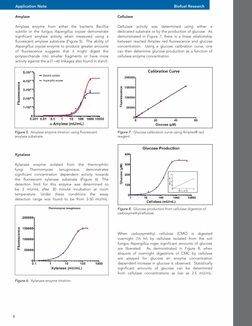
Amylase
Amylase enzyme from either the bacteria Bacillus subtilis or the fungus Aspergillus oryzae demonstrate significant amylase activity when measured using a fluorescent amylase substrate (Figure 5). The ability of Aspergillus oryzae enzyme to produce greater amounts of fluorescence suggests that it might digest the polysaccharide into smaller fragments or have more activity against the α-(1→6) linkages also found in starch.
Biofuel Research
Figure 5. Amylase enzyme titration using fluorescent amylase substrate.
Xynalase
Xylanase enzyme isolated from the thermophilic fungi Thermomyces lanuginosus, demonstrates significant concentration dependent activity towards the fluorescent xylanase substrate (Figure 6). The detection limit for this enzyme was determined to be 3 mU/mL after 30 minute incubation at room temperature. Under these conditions the assay detection range was found to be from 3-50 mU/mL.
Figure 6. Xylanase enzyme titration.
Figure 7. Glucose calibration curve using Amplex® red reagent.
Figure 8. Glucose production from cellulase digestion of carboxymethylcellulose.
When carboxymethyl cellulose (CMC) is digested overnight (16 hr) by cellulase isolated from the soil fungus Aspergillus niger significant amounts of glucose are liberated. As demonstrated in Figure 8, when aliquots of overnight digestions of CMC by cellulase are assayed for glucose an enzyme concentration dependent increase in glucose is observed. Statistically significant amounts of glucose can be determined from cellulase concentrations as low as 2.5 mU/mL.
Biofuel research has employed several different means to improve enzymatic digestion of polysaccharides. Screening of newly isolated wild microorganisms can often identify strains with unique or superior characteristics. The development and selection of genetically modified strains, where transport genes and metabolic pathways have been introduced can identify new strains that express greater amounts of enzyme activity or have superior fermentation abilities.Towards that end, researchers investigating digestive enzymes necessary for production of fermentable sugars used in the production of biofuels (e.g. ethanol, butanol, and propanol) require the means to quantitate enzyme activity. The fluorescent assays described provide the ability to assess lysates from microorganisms for enzyme activity.
References
1. Ge, Y. Antoulinakis, E.G., Gee, K.R. Johnson, I (2007) An Ultrasensitive, Continous Assay for Xylanase using the Fluorogenic substrate 6,8-difluoro- 4-methylumbelliferyl beta-D-xylobioside. Anal. Biochem.362(1):63-68.
5
Application Note
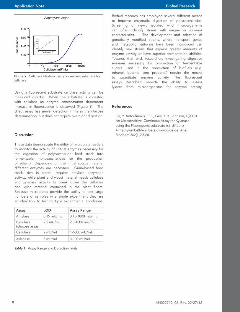
Using a fluorescent substrate cellulase activity can be measured directly. When the substrate is digested with cellulase an enzyme concentration dependent increase in fluorescence is observed (Figure 9). The direct assay has similar detection limits as the glucose determination, but does not require overnight digestion.
Discussion
These data demonstrate the utility of microplate readers to monitor the activity of critical enzymes necessary for the digestion of polysaccharide feed stock into fermentable monosaccharides for the production of ethanol. Depending on the initial source material different enzymes are necessary. Grain-based feed stock, rich in starch, requires amylase enzymatic activity, while plant and wood material needs cellulase and xylanase activity to break down the cellulose and xylan material contained in the plant fibers. Because microplates provide the ability to test large numbers of samples in a single experiment they are an ideal tool to test multiple experimental conditions.
Biofuel Research
Figure 9. Cellulase titration using fluorescent substrate for cellulase.
Table 1. Assay Range and Detection limits.
Assay LOD Assay Range
Amylase 0.15 mU/mL 0.15-1000 mU/mL
Cellulase (glucose assay)
2.5 mU/mL 2.5-1000 mU/mL
Cellulase 2 mU/mL 1-5000 mU/mL
Xylanase 3 mU/ml 3-100 mU/mL
AN022712_06, Rev. 02/27/12