Embed Size (px)
Citation preview
Chapter 3
Biofilms: A Challenge toMedical Fraternity in Infection Control
Silpi Basak, Monali N. Rajurkar, Ruchita O. Attal andSanjay Kumar Mallick
Additional information is available at the end of the chapter
http://dx.doi.org/10.5772/55649
1. Introduction
Microbes have been characterized as planktonic, free-floating single cells. The morphologicaland physiological properties of microbes have been described as they grow in nutritionallyrich culture media. Earlier very little thought have been given how microbes survive in theenvironment. But, the fact is, in natural environment, microbes are commonly found to beattached to surfaces as biofilms. Hence, the formation of surface attached microbial cells knownas biofilms open a new horizon to study the micro-organisms.
Automatically, the question arises, “What is biofilm?” According to the recent definition,Biofilms can be defined as sessile communities of microbial cells irreversibly attached to asurface or interface or to each other which are embedded in a self produced matrix of extrac‐ellular polymeric biomolecules and are physiologically different from planktonic cells withrespect to growth rate and gene transcription [1]. While studying Pseudomonas aeruginosaDavis and Geesay have shown that gene algC controlling phosphomannomutase involved inalginate (exopolysacharide) synthesis is upregulated within 15 minutes of adhesion to a solidsurface [2].
Biofilms are ubiquitous. They can be present on any surface – biotic or abiotic. Biofilms can befound on ship hulls, dairy and petroleum pipeline and rocks or pebbles at the bottom of streamsor rivers. They can grow in hot acidic pools in Yellowstone National Park (USA) and on glaciersin Antarctica. Biofilms can form anywhere with easy access to water e.g. on tiles of floor,kitchen platform or clogged sink etc. They are also found on plants and can remain symbiot‐ically or cause crop diseases like citrus canker, Pierce’s disease of grapes etc [3]. Fossilisedbioilms with 3.5 billion years are among the oldest records of life on earth [4]. Biofilms are also
© 2013 Basak et al.; licensee InTech. This is an open access article distributed under the terms of the CreativeCommons Attribution License (http://creativecommons.org/licenses/by/3.0), which permits unrestricted use,distribution, and reproduction in any medium, provided the original work is properly cited.
associated with biocorrosion of metals(microbiologically influenced corrosion.i.e.MIC) whichaffect kinetics of cathodic and or anodic reactions [5]. Biofilms can also grow in contact lenses,biomedical implants and transcutaneous devices.
Nearly every species of microorganisms e.g. bacteria, fungi, algae and protozoa have mecha‐nisms to adhere to surfaces and to each other. It has been found that over 90% of all bacterialive in biofilms. Biofilms can be formed by single species of microorganism or by multiplespecies of bacteria, fungi, protozoa etc. Mixed species biofilms predominate in environment.Single species biofilm usually exist in a variety of infections and on medical implants and arethe focus of current research [6].
Study of biofilm began when it was discovered that in natural aquatic system bacteriapredominantly remain attached to surfaces [7]. The first recorded observation of biofilm waspresented by Henrici in 1933 as ‘it is quite evident that for the most part water bacteria are notfree floating organisms, but grow upon submerged surfaces’ [8]. The fouling of ship hulls bymicrobes in marine environment was already known to mankind. Hence, the study of biofilmhas been started with marine bacteria, followed by fresh water microbial ecosystem andformation of biofilm on surface of eukaryotic tissue.
In early part of 20th Century it was difficult to observe biofilm as electron microscopy requiredcomplete dehydration of highly hydrated bioilm matrices and light microscopy was badlydistorted by out-of-focus effects [1]. Though Confocal Laser Scanning Microscope (CLSM) wasinvented in 1950s it was never used to study bacteria. CLSM produces optical slices of complexstructures, so out of focus effects are removed and it requires no sample preparations, so livingmicroorganisms can be observed if fluorescent dye is introduced to observe the cells [1]. Hence,the modern biofilm era began with the use of Confocal Laser Scanning Microscope (CLSM)which showed the image of biofilm as sessile microbial cells embedded in matrix interspersedbetween open water channels [9].
The development of biofilm is a 5 stage process – 1) reversible attachment 2) irreversibleattachment 3) early development 4) maturation 5) detachment or dispersal of cells. When themicrobial cell reaches very closer to a surface (<1nm), the initial attachment depends upon thetotal attractive or repulsive forces between two surfaces. These forces include electrostatic andhydrophobic interactions, steric hindrance, van der Waals forces etc. Probably hydrophobicinteractions play important role in primary adhesion [10]. The second stage of irreversibleattachment employs molecular binding between specific adhesins and the surfaces [11].
The factors controlling biofilm formation are: i) recognition of attachment sites on a surface ii)nutritional cues iii) change of pH and temperature iv) exposure to antibiotics, chemicalbiocides, and host defense mechanisms e.g. complement system etc.
The gene expression in biofilm cells differ from planktonic cells and by 2D gel electrophoresisit had been found that in mature biofilm of Pseudomonas aeruginosa >300 proteins weredetectable that were undetectable in planktonic cells [12].
During colonisation, microbial cells communicate via quorum sensing. In mature biofilmquorum sensing regulates formation of channels and pillar like structure for nutrient delivery.
Infection Control58
Microbial cells in biofilms undergo cell density-dependent gene regulation i.e. quorum sensingand thus coordinate through signalling molecules called autoinducers. Autoinducers increasein concentration as a function of cell density [13]. Usually Gram positive bacteria use processedoligopeptides to communicate, where as Gram negative bacteria use N- acyl homoserinelactones (AHLs) as autoinducers [14]. The widespread AI-2 quorum-sensing system is foundin several, Gram positive and Gram negative bacteria also [15]. For acyl-HSL quorum-sensing,an enzyme belong to Lux I family is required for synthesis of signal from cellular metabolites[16]. For AI-2 quorum-sensing system which has been implicated in interspecies communica‐tion, the synthesis of signalling molecule is directed by the Lux S gene product [17]. The ahyR/I acyl- HSL quorum sensing system of Aeromonas hydrophila has been shown to be requiredfor biofilm maturation [12]. Similarly the Lux S type quorum sensing system in Streptococcusmutants is also involved in biofilm development. Lux S system of Salmonella enterica serovarTyphimurium is required for biofilm formation on human gallstones [18].
Duenne described biofilm architecture as underwater coral reef with pyramid or mushroomshaped projections from the surface and channels and caverns running through out [19]. UsingCLSM, Lawrence et al has shown that Pseudomonas biofilms were more tightly packed at thesurface and less dense near the periphery whereas Vibrio parahaemolyticus biofilms showgreatest cell density near the periphery [20].
The adherent cells in a biofilm are embedded with a self produced matrix of extracellularpolymeric biomolecules. 97% of a biofilm matrix is water. A complex of secreted polymers,absorbed nutrients and metabolites, cell lysis product and even particulate materials from thesurrounding environment can form matrix. Actually the matrix surrounds, anchors andprotects surface-bound microbes. The matrix actually prevent the access of antimicrobials anddisinfectants and confer protection against environmental stresses such as UV radiation, pHshifts, osmotic shock and dessication [21].
Besides microbial cells all major classes of macromolecules i.e. proteins, polysaccharides,nucleic acids can be observed within a biofilm. Even transformation, transduction andconjugation result in gene transfer amongst the cells in biofilm.
Biofilms are formed by many bacterial species of medical importance e.g. Staphylococcusepidermidis, Staphylococcus aureus, Enterococci, Streptococcus mutans, Pseudomonas aeruginosa,E.coli O157:H7, Neisseria gonorrhoeae, Vibrio cholerae, Nontuberculous mycobacteria (NTM) etc [6].Amongst fungi - Candida albicans can usually form biofilm [22]. The two most intenselystudied biofilms are produced by: Staphylococcus epidermidis and Pseudomonas aeruginosa.
1.1. Biofilms and human disease
The microbial biofilm has received much attention recently because biofilm mode of growthmay be the key factor in persistent or chronic infections. The biofilms can act as nidus of acuteinfections and the microbial cells from biofilm are released at any one time during chronicinfection [23]. Clinicians are very much concerned about the fact that it is really difficult toeradicate biofilm bacteria with antibiotics. Even in immunocompetent host the biofilm growthare rarely resolved by host’s immune system as antigen may be hidden and key ligands may
Biofilms: A Challenge to Medical Fraternity in Infection Controlhttp://dx.doi.org/10.5772/55649
59
be repressed [24]. Biofilms are associated with kidney stones of infective origin, formation ofdental plaques, infections in cystic fibrosis, infections of permanent indwelling devices suchas joint prosthesis & heart valves, intrauterine devices (IUDs) and urinary catheters etc [25].
However in many chronic infections both the biofilm and planktonic growth may coexist.Parsek and Singh in 2003 have proposed few criteria to define the role of biofilms in humandiseases [12]: a) the causative bacteria are surface associated b) examination of infected tissueshows bacteria living in microcolonies and embedded in extracellular matrix c) infection isusually confined to a particular site and dissemination occurs as a secondary phenomenon d)the infection is difficult to eradicate with antibiotics though the causative bacteria are suscep‐tible to that antibiotics in planktonic state.
1.1.1. Infection-related kidney stones
15-20% of kidney stones occur in the setting of urinary tract infections. Infact, infection stonesare produced by interplay between infecting bacteria and mineral substrates derived fromurine resulting in formation of a complex biofilm. Microscopic analysis of stone has revealedthat bacteria are organized in microcolonies and surrounded by an anionic matrix composedof both polysaccharides and crystallized minerals [26]. It requires an alkaline environment todecrease solubility of phosphate, increased concentration of NH4
+ for struvite and CO3- for
carbon apatite formation as these are major constituents of this type of stone. The normal urineis not saturated with struvite and carbon apatite. The alkaline pH of urine occurs in infectionwith urease producing organisms like Proteus, Providencia, Klebsiella and Pseudomonas species.It is hypothesized that biofilms provide localized and concentrated urease activity to formstones [26].
1.1.2. Bacterial endocarditis
The primary lesion in endocarditis is due to vegetation (valve biofilm), which is composedmainly of bacteria and their products, platelets and fibrin derived from circulation with thedamaged endothelial surface as substratum. Durack in1975, developed nonbacterial throm‐botic endocarditis by leaving a polyethylene catheter in contact with aortic valve of a rabbitand showed how bacterial microcolonies were formed within 24 hours [27].
1.1.3. Airway infections in cystic fibrosis
Cystic fibrosis (CF), a common inherited disease of lower respiratory tract is caused bymutation in the gene which encodes Cystic fibrosis transmembrane regulator protein(CFTR). CFTR functions as a chloride ion channel protein [1]. Chloride ion transport isseverely impaired when CFTR is defective in CF patients, resulting in hyperviscous mucus.Initially CF patients suffer from intermittent respiratory infections but in late stagepermanent infection with P. aeruginosa occurs. It has been found that even with higherantibiotics given parenterally P. aeruginosa could not be eradicated from sputum of CFpatients in the late stage and it may persist for the rest of the patient’s life. In permanentinfection phase of CF patients, P. aeruginosa biofilm may be found in airways. Another
Infection Control60
interesting finding is emergence of P. aeruginosa with mucoid phenotype in late stage CFpatients [28]. This mucoid material is a polysaccharide i.e. alginate which probably preventantibody coating and opsonic phagocytosis. In fact, biofilm protects P. aeruginosa fromantimicrobials and host defenses. Genetic fingerprinting studies show same strain of P.aeruginosa can persist in CF patients for decades leading to chronic inflammation and declinein lung function and ultimately respiratory failure [29].
1.1.4. Endodontics
Biofilms also play a major role in causing dental caries, gingivitis, periodontitis, apicalperiodontitis etc [30].The anatomical complexities in root canal system provide favourablecondition for biofilm formation, which is actually initiated by invasion of pulp chamber byoral flora after tissue breakdown. Facultative or strict anaerobes are more frequently associatedthan aerobic microorganisms. Porphyromonas gingivalis is the primary agent responsible forperiodontitis [31]. Endodontic biofilm can be—i) intracanal, ii) extraradicular, iii) periapicaland iv) foreign body centered. Foreign body centered biofilm is a major complication associ‐ated with prosthesis and implant supported prosthesis [32].
1.1.5. other conditions
Similarly during acute phase of osteomyelitis, microscopical examination have shown biofilmformation on infected bone surfaces [33]. In chronic prostatitis, adherent bacterial colonies onthe surface of prostatic duct have been observed on microscopical studies, even in culturenegative cases [34].
1.1.6. Indwelling medical devices
Biofilms can develop on indwelling medical devices like prosthetic heart valve, pacemakers,central venous catheter, urinary catheter, contact lenses, intrauterine devices etc. and can causepersistent infections which are usually lethal. Scanning electron microscopy clearly showsbiofilm formation at the tip of urinary catheter kept for 7 days. On medical devices, biofilmsare most commonly formed by coagulase negative Staphylococci (CoNS) especially S. epider‐midis followed by S. aureus, Enterococci, Pseudomonas aeruginosa etc.
Biofilms can develop on both types of contact lenses i.e. soft and hard and also on contact lensstorage cases. Pseudomonas aeruginosa, Staphylococcus aureus, Staphylococcus epidermidis, E.coli,Candida species can adhere to contact lenses [35]. Evidence of biofilm on contact lenses andit’s storage cases have been reported from patients with microbial kerattis [36]. The rate ofprosthetic valve endocarditis (PVE) range from 0.5% to 4% [37]. Coagulase negative Staphy‐lococci are the commonest early colonizers after surgical implantation of prosthetic valvewhereas Streptococcus viridans most commonly colonize during late PVE (i.e. 12 monthsfollowing valve replacement) [38]. Though S. aureus, Gram negative coccobacilli or fungi mayalso be responsible for PVE.
Infection with central venous catheter is a quite common device related infection. Biofilmshave been shown by CLSM to be present outside the catheter or inner lumen [34].
Biofilms: A Challenge to Medical Fraternity in Infection Controlhttp://dx.doi.org/10.5772/55649
61
In S. epidermidis biofilm initial adherence is by polysaccharide adhesin (PSA) and accumulationof cells is due to production of polysaccharide intercellular adhesin (PIA).) PIA is encoded byica (intercellular adhesin) operon ica ADBC [39]. The icaR gene regulates ica operon. Productionof PIA is also subject to ON - OFF switching (phase variation). Majority of clinical isolates ofS. aureus also possess ica structural genes [40].
1.1.7. Health care Associated Infections (HAI) and biofilm
Catheter Associated Urinary Tract Infection( CA-UTI) is the commonest (>40%) HAI [41].Nosocomial bacteriuria or candiduria develops in 25% of patients having urinary catheter for>7 days with a daily risk of 5% [42]. Most infected urinary catheters are covered by a thickbiofilm containing infecting microorganisms. A biofilm forms intraluminally or extraluminallyor both ways.
With the increasing use of vascular access devices, catheter related bloodstream infection (CR-BSI), septic thrombophlebitis, endocarditis and other metastatic infections e.g.lung abscessosteomyelitis and endophthalmitis etc. are also increasing. In the United States out of 5 millionCentral Venous cathetes used each year, 3-8% lead to BSI [43]. The initiation of cathetercolonization occurs with the formation of a biofilm in the catheter lumen. Moreove theresistance levels of biofilm associated organisms may be much higher than those of planktonicorganisms [44]. After stoppage of antimicrobial therapy, the biofilm associated organismsresurge and cause another clinical infectios. A recent approach to reduce CR-BSI is bundles ofpreventive measures, which means a group of preventive measures, when executed together,result in better outcomes than when implemented alone [45]. This included handwashing,using full barrier precautions during insertion of central venous line, cleaning the skin withchlorhexidine. The femoral site should be avoided if possible and catheters should be removedas early as possible.
Hospital acquired pnumoniais are the second most common cause of HAI and has the highestmorbidity and mortality of all HAIs [46]. The initial step in pathogenesis of HAP is colonizationof patient’s oropharynx with resistant hospital pathogen. The endotracheal tube lumen is anidus for the growth of bacteria within the biofilm. Hand washing and Personal protectiveequipment (PPE) must be used to reduce the incidence of HAP/ Ventilator assaociatedpneumonia (VAP).
1.1.8. Resistance of biofilm to antimicrobials and disinfectants
It has been observed that biofilms are not easily eradicated even by cidal antimicrobials,quarternary ammonium compounds, halogens and halogen release agents. The crux of theproblem is the presence of persisters within the biofilms that can rebound when antibioticconcentration falls. The causes are multifactorial – i) restricted penetration of antimicrobialswithin the biofilm architecture, ii) decreased growth rate of bacterial cells forming the biofilm,iii) expression of resistance gene by the bacterial cells within the biofilm etc [47]. Restrictedpenetration of antimicrobials may occur as negatively charged exopolysaccharide restrictpermeation of positively charged antibiotics e.g. Aminoglycoside and exopolymer matrix also
Infection Control62
restrict diffusion of antimicrobial within the biofilm. Synergy between retarded diffusion anddegradation by enzymes (e.g. β-lactamase) also provide effective resistance to antimicrobials.Fluoroquinolones are very effective in stopping the growth of a biofilm but restricted diffusioncan protect the microbial cells within the biofilm [48]. All antimicrobials are more effective inkilling rapidly growing cells. Penicillin & ampicillin do not kill non-growing cells as rate ofkilling is directly proportional to rate of growth for these two antibiotics. Even cephalosporins,aminoglycosides & fluroquinolones can kill rapidly dividing cells more effectively. Multipledrug resistance (MDR) pumps may play a role in biofilm resistance at low antibiotic concen‐tration. Sometimes unknown MDR pumps might be over expressed in biofilm e.g. for chlor‐amphenicol in E. coli biofilm. Moreover, the biofilms increase the opportunity of gene transferbeteen the microorganisms and can convert a previously avirulent commensal organism to ahighly virulent pathogen. The enhanced efficiency of gene transfer in biofilms also fascilitatesthe spread of antibiotic resistance and virulence factors [49]. Though most of the researchworks deals with single species biofilms, multispecies biofilm amongst different bacteria andinterkingdom biofilms between fungus Candida albicans and various bacterial species are alsogaining importance in causing different diseases [50]. Biofilm formation is a major virulencefactor for Candida albicans and Candida biofilms are difficult to eradicate due to their highresistance to antifungals. A recent study has reported that within the biofilm Staphylococcusaureus was attached uniquely with the pseudohyphae of Candida albicans. This synergisticinteraction resulted in differential protein expressions which are actually virulence factors forStaphylcoccus aureus. This indicate C. albicans may enhance S.aureus pathogenesis [51].Recently it has been reported that co-existence of S.aureus and C.albicans in a biofilm resultedin increased Vancomycin resistance in S.aureus [52] However antagonistic interaction has beenreported between Pseudomonas aeruginosa and Candida albicans [53].
It is not possible to detect the antimicrobial resistance of biofilms by conventional methods ofdisc diffusion and broth microdilution as per CLSI guideline because these methods are onlymeant for planktonic cells.
1.1.9. Biofilms and altruism
Biofilms are like small cities and encourage altruism. Microbial cells within biofilm oftensacrifice their maximum growth rate to use the available community resources more efficient‐ly. In a biofilm atleast some of the microbial cells experience nutrient limitation and exist in aslow growing state [54]. In this process while individual cells are disadvantaged, the microbialcommunity as a whole is benefited. Hence, it is said that biofilms are the colonial way of lifeof microorganisms.
Detection of biofilms can be done by both phenotypic methods and genotypic methods. Inphenotypic methods biofilms are detected by Congo red agar method(CRA),Plastic tubemethod(TM), Tissue culture plate method(TCP) and Confocal Laser Scanning Microscopy(CLSM). In genotypic method, usually Polymerase chain reaction (PCR) for amplification ofmicrobial DNA, coding for biofilm formation is done. The phenotypic methods are easy andcheap compared to genotypic method.
Biofilms: A Challenge to Medical Fraternity in Infection Controlhttp://dx.doi.org/10.5772/55649
63
Hence, the present study was undertaken to detect the biofilm producing organisms, isolatedfrom different clinical specimens in our laboratory.
2. Material and methods
The present study was conducted from 2009 to 2012. A total number of 350 bacterial and 50Candida strains were studied. The microbial strains were isolated from different clinicalspecimens like urine, blood, pus and wound swab, endotracheal aspirate, urinary catheter tip,central venous catheter tip etc. All the microbial strains were identified by conventionalmethods [55]. We used microtitre plate biofilm assay to detect microbial attachment to anabiotic surface [56].
Steps:
1. The microbial cells were grown in Brain heart infusion broth overnight.
2. On next day, the cultures were diluted 1:100 using the brain heart infusion broth.
3. 100µl of each diluted culture was inoculated into each of three wells in a microtiter platewhich has not been tissue culture treated. The plates were covered by the lid and wasincubated at optimal growth temperature [56] for 48 hours
4. Then the wells were washed twice to remove planktonic cells.
5. Microbial cells which were adhered to the wells were subsequently stained with crystalviolet solution that allowed visulisation of the attachment pattern. 125 µl of 0.1% crystalviolet solution was added to each well and stained for 10 minutes at room temperature.
6. The microtiter plates were shaken and the crystal violet solution was removed.
7. The plates were washed successively twice with distilled water. Any crystal violet that isnot specifically staining the adherent microbial cells were removed by this washing step.
8. The microtiter plates were then inverted and tapped vigorously on tissue paper to removeany excess liquid.
9. The microtiter plates were then air dried. The dried microtiter plates may be stored atroom temperature for several weeks.
10. This surface associated dye was solubilized by adding ethanol or any other solvent forsemiquantitative assessment of biofim formed. 200µl of 95% ethanol or other appropriatesolvent [56] was then added to each stained well and was kept for 10 to 15 minutes
11. The contents of each well were mixed by pipetting and then 125µl of the crystal violet /ethanol solution from each well was transferred to a separate well of another 96 wellmicrotiter plate maintaining the same sequence.
Infection Control64
12. Then the optical density of each well containing 125µl solution was measured at awavelength of 545nm in an ELISA reader. As each strain was put in triplicate the averageof the three readings were taken.
The biofilm formation of different strains were classified in three groups according to the cutoff OD. The cut off OD (ODc) for the microtiter plate test was defined as three standarddeviations above the mean OD of the negative control. Isolates were classified into four groupsas nonadheremt, weakly adherent, moderately adherent and strongly adherent according toStepanovi et al [57].
3. Observation and results
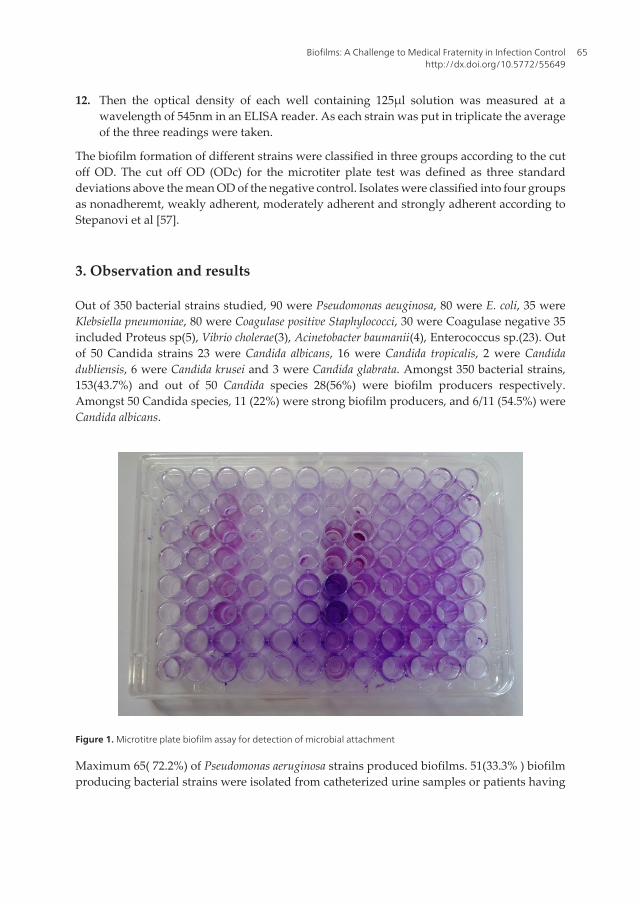
Out of 350 bacterial strains studied, 90 were Pseudomonas aeuginosa, 80 were E. coli, 35 wereKlebsiella pneumoniae, 80 were Coagulase positive Staphylococci, 30 were Coagulase negative 35included Proteus sp(5), Vibrio cholerae(3), Acinetobacter baumanii(4), Enterococcus sp.(23). Outof 50 Candida strains 23 were Candida albicans, 16 were Candida tropicalis, 2 were Candidadubliensis, 6 were Candida krusei and 3 were Candida glabrata. Amongst 350 bacterial strains,153(43.7%) and out of 50 Candida species 28(56%) were biofilm producers respectively.Amongst 50 Candida species, 11 (22%) were strong biofilm producers, and 6/11 (54.5%) wereCandida albicans.
Figure 1. Microtitre plate biofilm assay for detection of microbial attachment
Maximum 65( 72.2%) of Pseudomonas aeruginosa strains produced biofilms. 51(33.3% ) biofilmproducing bacterial strains were isolated from catheterized urine samples or patients having
Biofilms: A Challenge to Medical Fraternity in Infection Controlhttp://dx.doi.org/10.5772/55649
65
other medical devices. 108(70.6% ) bacterial strains producing biofilms were isolated frompatients having chronic infections eg persistent or recurrent UTI, Chronic obstructive airwaydisease, cystic fibrosis etc.
In our study the cut off OD(ODc) was 0.003. The biofilm forming organisms are groupedinto weak group (OD ≥ 0.003 to 0.006), moderate group (OD ≥ 0.006 to 0.012) and stronggroup (OD > 0.012).
Newer β – lactamases producers Non β –
lactamase
producersOrganismsESBL
Only
AmpC
Only
MBL
Only
ESBL +
AmpC
ESBL +
MBL
AmpC
+ MBL
ESBL + AmpC
+MBL
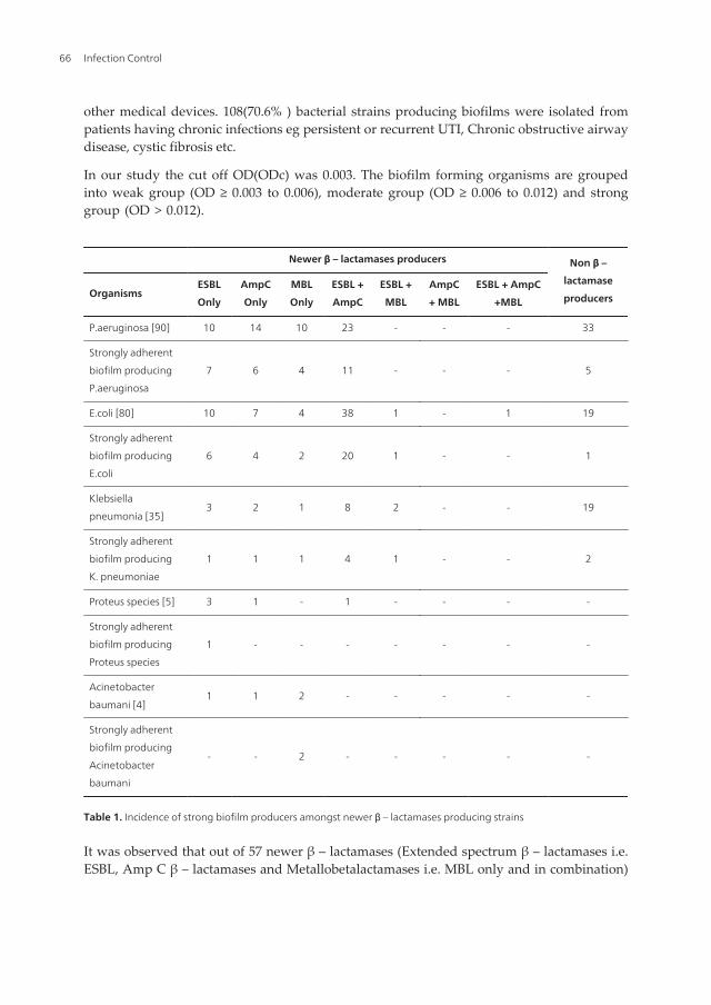
P.aeruginosa [90] 10 14 10 23 - - - 33
Strongly adherent
biofilm producing
P.aeruginosa
7 6 4 11 - - - 5
E.coli [80] 10 7 4 38 1 - 1 19
Strongly adherent
biofilm producing
E.coli
6 4 2 20 1 - - 1
Klebsiella
pneumonia [35]3 2 1 8 2 - - 19
Strongly adherent
biofilm producing
K. pneumoniae
1 1 1 4 1 - - 2
Proteus species [5] 3 1 - 1 - - - -
Strongly adherent
biofilm producing
Proteus species
1 - - - - - - -
Acinetobacter
baumani [4]1 1 2 - - - - -
Strongly adherent
biofilm producing
Acinetobacter
baumani
- - 2 - - - - -
Table 1. Incidence of strong biofilm producers amongst newer β – lactamases producing strains
It was observed that out of 57 newer β – lactamases (Extended spectrum β – lactamases i.e.ESBL, Amp C β – lactamases and Metallobetalactamases i.e. MBL only and in combination)
Infection Control66
producing Pseudomonas aeruginosa 28 (49.1%) were strongly adherent biofilm producers,compared to only 5/33 (15.1%) non β – lactamase producers. Amongst the 120 Enterobacter‐iaceae strains studied. 82 (68.3%) were newer β – lactamases producers, whereas 48/82 (58.5%)were strong biofilm producers and only 3/38 (7.9%) non β – lactamase producing strains werestrong biofilm producers.
Organisms Methicillin resistant Methicillin sensitive
Coagulase positive Staphylococcus [80] 34 46
Strongly adherent biofilm producing
Coagulase positive Staphylococcus14 2
Coagulase negative Staphylococci
(CONS) [30]11 19
Strongly adherent biofilm producing
Coagulase negative Staphylococci
(CONS)
4 2
Table 2. Incidence of strong biofilm producing Methicillin resistant Staphylococcus strains.
Table 2 shows amongst the Methicillin Resistant Staphylococcus aureus (MRSA) strains, 14/34(41.2%) and Methicillin Resistant Coagulase negative Staphylococci (MR – CONS) 4/11 (36.4%)were strong biofilm producers compared to 2/46 (4.3%) Methicillin sensitive Staphylococcusaureus (MSSA) and 2/19 (10.5%) Methicillin sensitive CONS.
Out of 23 Enterococcus species 13/23 (56.5%)were High level Aminoglycoside Resistant(HLAR) strains and it was also found that 8/13 (61.5%) HLAR strains were strong biofilmproducers compared to only 2/10 (20%) of non HLAR strains.
4. Discussion
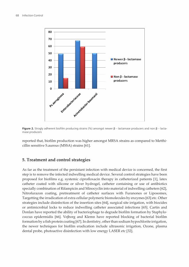
Our Hospital is a tertiary care centre in a rural setup. Though CLSM is the best phenotypicmethod, it could not be used as it is very costly. We did a pilot study with Staphylococci in2008 and found 33% of Staphylococcus aureus and 44.7% of Coagulase Negative Staphylococci(CONS) were biofilm producers and amongst the 3 phenotypic methods tissue culture platemethod gave the best results [58]. The present study correlated well with reports of otherauthors that Extended Spectrum β-Lactamase (ESBL) producing strains, Methicillin ResistantStaphylococcs aureus(MRSA) were more adherent to microtitre plate than Non ESBL and NonMRSA strains (Figure 2).
Lee et al in 2008 have also reported a positive correlation between biofilm formation and ESBLproducing Acinetobacter baumanii [59]. Norouzi et al in 2010 have reported that in their study14% ESBL producing Pseudomonas aeruginosa has formed strongly adherent biofilm com‐pared to only 4% of non-ESBL producing Pseudomonas aeruginosa [60]. It has also been
Biofilms: A Challenge to Medical Fraternity in Infection Controlhttp://dx.doi.org/10.5772/55649
67
reported that, biofilm production was higher amongst MRSA strains as compared to Metthi‐cillin sensitive S.aureus (MSSA) strains [61].
5. Treatment and control strategies
As far as the treatment of the persistant infection with medical device is concerned, the firststep is to remove the infected indwelling medical device. Several control strategies have beenproposed for biofilms e.g. systemic ciprofloxacin therapy in catheterized patients [1], latexcatheter coated with silicone or silver hydrogel, catheter containing or use of antibioticsspecially combination of Rifampicin and Minocyclin into material of indwelling catheters [62],Nitrofurazon coating, pretreatment of catheter surfaces with Furanones or Liposomes,Targetting the irradication of extra cellular polymeric biomolecules by enzymes [63] etc. Otherstrategies include disinfection of the insertion sites [64], surgical site irrigation, with biocidesor antimicrobial locks to reduce indwelling catheter associated infections [65]. Cartin andDonlan have reported the ability of bacteriophage to degrade biofilm formation by Staphylo‐coccus epidermidis [66]. Vejborg and Klemn have reported blocking of bacterial biofilmformation by a fish protein coating [67]. In dentistry, other than sodium hypochlorite irrigation,the newer techniques for biofilm eradication include ultrasonic irrigation, Ozone, plasmadental probe, photoactive disinfection with low energy LASER etc [32].
Figure 2. Strogly adherent biofilm producing strains (%) amongst newer β – lactamase producers and non β – lacta‐mase producers
Infection Control68
To prevent biofilm formation, the physical approaches like the use of low strength electricalfield [68], electromagnetic field or ultrasound along with antibiotic therapy [69] are also verypromising. A novel treatment based on disruption of quorum sensing system to inhibit biofilmformation has also been suggested by many workers [70]. Even the workers have suggestedthe inhibition of transcription of genes that are activated or repressed during initial biofilmformation will also help to prevent persistent infection due to biofilms. All these controlstrategies are on experimental basis and are not applicable for medical devices and have theirown limitations to be used cuurently in patients.
To conclude, we must say biofilm develops slowly but has a major impact both clinically andeconomically on overall outcome of the patients treatment. The authors feel that, EARLYDETECTION AND NEWER TREATMENT OPTIONS FOR BIOFILM ASSOCIATED INFEC‐TIONS ARE NEED OF THE HOUR.
Author details
Silpi Basak*, Monali N. Rajurkar, Ruchita O. Attal and Sanjay Kumar Mallick
Department of Microbiology, Jawaharlal Nehru Medical College, Wardha (M.S.), India
References
[1] Doulam RM, Costerton JW. Biofilms: Survival mechanisms of clinically relevant mi‐cro-organisms. Clin Microbiol Rev. 2002; 15(2): 167-193.
[2] Davis DG, Geesy GG. Regulation of the alginate biosynthesis gene algC in Pseudo‐monas aeruginosa during biofilm development in continuous culture. Appl EnvironMicrobiol. 1995; 61: 860-7.
[3] Introduction to Biofilms: Negative and positive impacts of biofilm. Available from:http://www.cs.montana.edu/ross/personal/intro-biofilms-s3.html.
[4] Schopf JW, Hayes JM, Walter MR. Evolution on earth’s earliest ecosystems: recentprogress and unsolved problems. In: Schopf JW Ed, Earth’s earliest biosphere.Princeton University Press, New Jersy, 1983; 15:143-147.
[5] Jones DA, Amy PS. A thermodynamic interpretation of microbiologically influencedcorrosion. Corros2002;58:638-645.
[6] Toole GO, Kaplan HB, kolter R. Biofilm formation as microbial development. AnnuRev Microbiol.2000; 54:49-79.
Biofilms: A Challenge to Medical Fraternity in Infection Controlhttp://dx.doi.org/10.5772/55649
69
[7] Geesy GG, Richardson WT, Yeomans HG, Irvin RT, Costeton JW. Microscopic exami‐nation of natural sessile bacterial populations from an alpine stream. Can.J.Microbiol.1977;23(12): 1733-1736.
[8] Henrici AT. Studies of fresh water bacteria. Adirect microscopic technique. J.Bacter‐iol. 1933;25: 277-287.
[9] Stoodley P, Sauer K, Davies D G, Costerton J W. Biofilms as complex differentiatedcommunities. Annu Rev Microbiol. 2002; 56: 187: 209.
[10] Carpentier B, Cerf O. Biofilms and their consequences, with particular reference tohygiene in food industry. J Appl Bacteriol. 1993; 75: 499-511.
[11] An. YH, Dickinson RB and Doyle RJ. Mechanism of bacterial adhesion and pathogen‐esis of implant and tissue infections, In: Anand YH and Fridman RJ editors. Hand‐book of bacterial adhesions: Principles, methods and applications. Humana press;Totowa. NJ: 2000. P: 1-27.
[12] Sauer K, Camper AK, Ehrlich GD, Costerton JW, Davies DG. Pseudomonas aerugi‐nosa displays multiple phenotypes during development as a biofilm. J Bacteriol 2002;184: 1140-54.
[13] Miller MB, Bassler BL. Quorum sensing in bacteria. Ann. Rev. Microbiol.2001;55:165-199.
[14] Miller MB, Bassler BL. Quorum sensing in bacteria. Annu Rev Microbiol 2001; 55:165-99.
[15] Bassler B L, Small talk. Cell-to-cell communication in bacteria. Cell 2002; 109: 421-4.
[16] Schafer AL. Generation of Cell-to-cell signals in quorum sensing: acyl homo serinelactone synthase activity of a purified Vibrio fischeri Lux I protein. Proc Natl AcadSci. USA 1996; 93: 9505-9.
[17] Schauder S, Shokat K, Surette MG, Bassler BL. The Lux S family of bacterial autoin‐ducers: biosynthesis of a novel quorum-sensing signal molecule. Mol Microbiol 2001;41: 463-76.
[18] Prouty AM. Biofilm formation and interaction with the surfaces of gallstones by Sal‐monella spp. Infect Immun 2002; 70: 2640-9.
[19] Dunne WH Jr. Bacterial adhesion: seen any good biofilm lately. Clin Microbiol Rev.2002 Apr: 155-166.
[20] Lawrence JR, Korber DR, Hoyle BD, Costerton JW, Caldwell DE. Optical sectioningof microbial biofilms. J Bacteriol. 1991: 173:6558-67.
[21] Flemming HC. Bifilms and environmental protection. Water Sci Technol. 1993;27:1-10.
Infection Control70
[22] Baillie GS, Douglas LJ. Role of dimorphism in the development of Candida albicansbiofilms. J Med Microbiol 1991; 48: 671-79.
[23] Costerton JW, Stewart PS, Greenberg EP. Bacterial biofilms: a common cause of per‐sistent infections. Science1999; 284:1318-22.
[24] Mahenthiralingam E, Campbell ME, Speert DP. Non motility and phagocytic resist‐ance of Pseudomonas aeruginosa isolates from chronically colonized patients withcystic fibrosis. Infect Immun 1994; 62: 596-605.
[25] Parsek MR, Singh PK. Bacterial biofilm: An emerging link to disease pathogenesis.Ann Rev Microbiol. 2003; 57: 677-701.
[26] Nickel JC, Olson M, McLean RJ, Grant SK, Costerton JW. An ecological study of in‐fected urinary stone genesis in animal model. Br J Urol. 1987; 59: 21-30.
[27] Durack DT. Experimental bacterial endocarditis. IV. Structure and evolution of veryearly lesion. J Pathol. 1975; 115: 81-9.
[28] Koch C, Hoiby N. Pathogenesis of cystic fibrosis. Lancet 1993; 341: 1065-9.
[29] Govan JR, Deretic V. Microbial pathogenesis in cystic fibrosis: mucoid Pseudomonasaeruginosa and Burkolderia Cepacia. Microbiol Rev. 1996; 60 (3): 539-74.
[30] Marsh D. Microbiological ecology of dental plaque and its significance in health anddisease. Adv Dent Res 1994;8: 263-271.
[31] Lamont RJ, Jenkinson HF. Life below gun line: pathogenic mechanism of Porphyro‐monas gingivalis. Microbiol Mol Biol Rev. 1998; 62: 1244-63.
[32] Usha HL, Kaiwar A, Mehta D. Biofilm in endodontics: New understanding to an oldProblem. Int. journal of Contemporary Dentistry. 2010; 1(3): 44-51.
[33] Marrie TJ, Costerton JW. Mode of growth of bacterial pathogens in chronic polymi‐crobial human osteomyelitis. J Clin Microbiol. 1985; 22: 924-33.
[34] Nickel JC, Costerton JW. Bacterial localization in antibiotic-refractory chronic bacteri‐al prostatis. Prostate 1993; 23: 107-114.
[35] Dart JKG. Contact lens and prosthesis infections. In Tasman W and Jager EA editors.Duane’s foundation of Clinical ophthalmology. Lippincott-Raven, Pa: Philadelphia:1996; p: 1-30.
[36] McLaughlin-Borlace L, Stapleton F, Matheson M, Dart JKG. Bacterial biofilm on con‐tact lenses and lens storage cases in wearers with microbial keratitis. J Appl Micro‐biol. 1998;84: 827-38.
[37] Hancock EW. Artificial valve disease. In: Schtant RC, Alexander RW, Rourke RAORoberts R and Sonnenblick EH, editors. The heart artiers and veins, 8th ed, vol. 2.New York: McGraw-Hill; NY. 1994. p:1539-45.
Biofilms: A Challenge to Medical Fraternity in Infection Controlhttp://dx.doi.org/10.5772/55649
71
[38] Douglas JK, Cobbs CG. Prosthetic valve endocarditis In: Kaye D, editor. Infective en‐docarditis, 2nd ed. New York: Raven Press Ltd; N.Y. 1992. p: 375-396.
[39] Cramton SE, Gerke C, Schll NF, Nicholas WW, Gotz F. The intercellular adhesion (icalocus) is present in Staphylococcus aureus and is required for biofilm formation. In‐fect Immun. 1999; 67:5427-33.
[40] Heilmann C, Schweitzer O, Gerke C, Vanittanakom N, Mack D, Gotz F. Molecularbasis of intercellular adhesions in the biofilm forming Staphylococcus epidermidis.Mol Microbiol. 1996; 20: 1083-91.
[41] National Nosocomial Infections Surveillance (NNIS) System Report. Data summaryfrom January 1992 through June2004. Am Jinfect Control 2004; 32; 470-485.
[42] Warren JW. The catheter and urinary tract infection. Med Clin North Am1991; 75:481- 493.
[43] Darouiche R, Device associated infections : A macroproblem that starts with micro‐adherence. Clin. Infect Dis. 2001; 33: 1567-1572.
[44] Traunter BW and Darouiche RO. Catheter –associated infections: Pathogenesis af‐fects Prevention. Arch Intern Med, 2004; 164: 842-850 .
[45] Lachman P and Yuen S. Using care bundles to prevent infection in neonatal andpaediatric .ICUs. Curr. Opin infect Dis,2009; 22: 224-228.
[46] Fiel S. Guidelines and critical pathways for severe hospital- acquired pneumonia,Chest. 2011;119:412-418.
[47] Levis K. riddle of biofilm resistance. Antimicrob Agents Chemother 2001;45(4) : 999-1007.
[48] Brooun a, Liu S, Lewis K. A dose- response study of antibiotic resistance in Pseudo‐monas aeruginosa biofilms. Antimicrob Agents Chemother. 2000; 44; 640-646.
[49] Molin S, Tolker-Nielson T. Gene transfer occurs with enhanced efficiency in biofilmsand induces enhanced stabilization of the biofilm structure. Curr Opin Biotechnol.2003;14(3): 255-256.
[50] Shirtliff ME, Peters BM, Jabra-Rizk MA. Cross-kingdom intractions: Candida alicansand bacteria. FEMS Microbiol Lett. 2009; June:1-8.
[51] Peters BM, Jabra-Rizk MA, Scheper MA et al. Microbial interactions and differentialprotein expression in Staphylococcus aureus and Candida albicans dual-species bio‐films. FEMS Imm Med Microbiol 2010; 59: 493-503.
[52] Harriott MM, Noverr MC. Candida albicans and Staphylococcus aureus form poly‐microbial biofilms: effects on antimicrobial resistance. Antimicrob Agents Chemo‐ther. 2009; 53(9): 3914-3922.
Infection Control72
[53] Hogan DA, Kolter R. Pseudomonas-Candida interactions: an ecological role for viru‐lence factors. Science. 2002; 296(5576): 2229-2232.
[54] Costerton JW, Stewart PS, Greenberg EP. Bacterial biofilms: a common cause of per‐sistentinfections. Science1999; 284:1318-1322.
[55] Washington CW Jr, Stephen DA, William MJ, Elmer WK, Gray WP, Paul CS, GailLW. In Koneman’s Colour Atlas and Textbook Of Diagnostic Microbiology, 6th ed,Lippincott Williams &Wilkins, Philadelphia PA, USA, 2006.
[56] Merritt JN, Kadouri DE, Toole G0. Grouping and analysing static biofilms. Ch BasicProtocol I,In current protocols in Microbiology, 2005; unit 1B. 1.1-1B.1.7
[57] Stepanovi s, Vukovi D, Daki I, Savib, Svabi-Vlahovi (M). A modified micotiter-platetest for quantitation of staphylococcal biofilm formation. JMicrobiol.Methods. 2000;40:175- 179.
[58] Bose S, Khodke M, Basak S, Mallick S K. Detection of biofilm producing Staphylococ‐ci: Need of the hour. J. Clin and Diagnostic Res [serial online] 2009 December [cited:2009 December 7]; 3:1915-1920. Available from http://www.jcdr.net/back asp?issn=0973-709x&year=2009&month= December &volume=3&is‐sue=6&page=1915-1920 &id=469.
[59] Lee HW, Koh YM, Kim J et al Capacity of multidrug resistant clinical isolates of Aci‐netobacter baumanii to form biofilm and adhere to epithelial cell surfaces Clin. Mi‐crobiol Infect. 2008;14:49-54.
[60] norouzi F, Mansouri S, Moradi M, Razavi M Comparison of cell surface hydropho‐bicity and biofilm formation among ESBL and non ESBL producing Pseudomonasaeruginosa from clinical isolates. African Journal of Microbiology Research2010;4(11):1143-1147.
[61] KhanF, Shukla I, Rizvi M, Mansoor T, Sharma SC Detection of biofilm formation inStaphylococcus aureus. Does it have a role in treatment of MRSA infections? Trendsin Medical Research 2011;6:116-123.
[62] Spencer RC. Novel methods for prevention of infection of intravascular devices. JHosp Infect. 1999; 43suppl: S127- 35.
[63] Johansen C, Falholt P, Gram L. Engymatic removal and disinfection of bacterial bio‐film. Appl Enviorn Mirobial. 1997; 63: 3724-8.
[64] Maki DG, Ringer M, Alvarado CJ Prospective randomized trial of povidone iodine,alcohol and chlorhexidine for prevention of infection associated with central venousand arterial catheters. Lancet 1991;338:339-343.
[65] Curtin J, Cormican M, Fleming G, Keeiehan J, Colleran E Linezolid compared witheperezolid, vancomycin and gentamicin in an in vitro model of antimicrobial lock
Biofilms: A Challenge to Medical Fraternity in Infection Controlhttp://dx.doi.org/10.5772/55649
73
therapy for Staphylococcus epidermidis central venous catheter related biofilm infec‐tions. Antimicrobial. Agents Chemother. 2003;47:3145-3148.
[66] Curtin JJ and Donlan RM Using bacteriophages to reduce formation of catheter asso‐ciated biofilms by Staphylococcus epidermidis. Antimicrobial. Agents Chemother.2006;50(4):1268-1275.
[67] Vejborg RM and Klemm P Blocking of bacterial biofilm formation by a fish proteincoating Appl.Environ.Microbiol. 2008.
[68] Blenkinsopp SA, Khoury AE, Costerion JW Electrical enhancement of biocide effica‐cy against Pseudomonas aeruginosa biofilms. Appl. Envioron.Microbiol.1992;58:3770-3773.
[69] Huang CT, James G, Pitt WG, Stewart PS Effects of ultrasonic treatment on the effica‐cy of gentamicin against established Pseudomonas aeruginosa biofilms. Colloids Sur‐faces B Bioineterfaces 1996;6:235-242.
[70] Hartman G, Wise R. Quonum sensing. Potential means of treating Gram negative in‐fection? Lancet 1998; 351: 848-9.
Infection Control74