Embed Size (px)
Citation preview

1
Biodistribution of Busulphan Loaded Biodegradable Nano-carrier Designed
for Multimodal Imaging
Heba Asem1, 2, Ying Zhao2, 3, Fei Ye2, Åsa Barrefelt2, Manuchehr Abedi-Valugerdi2, Ramy
El-Sayed2, Ibrahim El-Serafi2, Svetlana Pavlova2, Khalid M. Abu-Salah4, Salman A.
Alrokayan5, Jörg Hamm6, Mamoun Muhammed1, Moustapha Hassan2,3
1 Division of Functional Materials (FNM), Department of Materials and Nanophysics, Royal Institute
of Technology (KTH), SE-164 40 Stockholm, Sweden.
2 Division of Experimental Cancer Medicine (ECM), Department of Laboratory Medicine
(LABMED), Karolinska Institutet (KI), SE-141 86 Stockholm, Sweden.
3 Clinical Research Center (KFC), Karolinska University Hospital Huddinge, SE-141 86, Stockholm,
Sweden.
4 King Abdullah International Medical Research Center, King Abdulaziz Medical City, PO Box 22490,
Riyadh 11426, Saudi Arabia
5 King Abdullah Institute for Nanotechnology and Department of Biochemistry, King Saud University,
PO Box 2455, Riyadh 11451, Saudi Arabia
6 PerkinElmer, 68 Elm St., Hopkinton, MA 01748, USA
Correspondence:
Moustapha Hassan, Clinical Research Center (KFC), Karolinska University Hospital
Huddinge, SE-141 86 Stockholm, Sweden. Telephone: +46-8-58583862.
E-mail: [email protected]
Keywords
Biodegradable polymer, drug delivery, magnetic resonance imaging, cellular uptake,
cytotoxicity, biodistribution, in vivo fluorescence imaging, fluorescence/CT imaging co-
registration

2
Graphical abstract
Abstract
Multifunctional nanocarriers for pathological site imaging and regulated drug delivery are
increasingly promising for disease diagnosis and treatment. We developed a multifunctional
theranostic nanocarrier system for anticancer drug delivery and molecular imaging.
Superparamagnetic iron oxide nanoparticles (SPIONs) as an MRI contrast agent and busulphan
as an antineoplastic agent were encapsulated into poly (ethylene glycol)-co-poly (caprolactone)
(PEG-PCL) nanoparticles (NPs) via the emulsion-evaporation method. Busulphan entrapment
efficiency was 83% and the drug release showed a sustained pattern over 10 hours. SPION
loaded-PEG-PCL NPs showed contrast enhancement in T2*-weighted MRI with high r2*
relaxivity. In vitro time-dependent cellular PEG-PCL NP uptake was observed in macrophage
cells (J774A). PEG-PCL NPs were further functionalized with VivoTag 680XL Fluorochrome
for in vivo fluorescence imaging for study of their biodistribution in Balb/c mice over 48 h. The
results of real-time imaging were then confirmed by ex vivo organ imaging and histological
examination. Generally, PEG-PCL NPs were highly distributed in the lungs until 4 h post
intravenous administration, then redistributed and accumulated in liver and spleen until 48 h.
No pathological impairment was found in the studied tissues. Thus, PEG-PCL NPs as
biodegradable and biocompatible nanocarriers are an efficient multimodal imaging agent, offer
high drug loading capacity, and provide the possibility of disease treatment.

3
1. Introduction
Nanoparticles (NPs) offer great potential for various biomedical applications such as drug
delivery,1-3 diagnostics4,5 and bioimaging.6-7 With rapid development it has been possible to
create NP-based advanced multifunctional drug delivery carriers. These all-in-one carriers offer
the possibility of fulfilling several therapeutic needs simultaneously, such as delivery of
therapeutic cargo, real-time imaging, targetability and controlled release.8-10 Biodegradable
polymers are one of the most attractive biomaterials utilized for nanomedicine.11-14 Controlled
and sustained drug delivery, improved drug pharmacokinetics, reduced side effects and
biodegradability make these materials ideal carrier systems for a variety of active agents.15 For
amphiphilic biodegradable polymers, they can self-assemble and form micellar architecture in
aqueous media.16 Therefore, an important benefit of these polymeric micelles in medical
imaging as well as in drug delivery17 is that they have the ability to load and deliver the contrast
agents or drugs to pathological areas for imaging and therapy.18
Imaging moieties are often incorporated in drug delivery carriers for tracking and evaluation
purposes. Among others, the inorganic contrast agent superparamagnetic iron oxide
nanoparticles (SPIONs) are widely used in magnetic resonance imaging (MRI)14,19 owing to
their ability of shortening the spin-spin relaxation time and significantly increasing the imaging
contrast.20 However, un-functionalized SPIONs tend to aggregate and form clusters after
intravenous injection due to van der Waals interactions between the particles14 and hence, the
aggregate particles are rapidly eliminated by macrophages in mononuclear phagocyte system
(MPS).21 This undesired behavior can be avoided by using amphiphilic block copolymers as
carriers to protect SPIONs from the surrounding environment. The amphiphilic block
copolymers can form micellar NPs with a hydrophobic core and a hydrophilic shell. The
hydrophobic core allows entrapment of agents with low aqueous dispersion, such as
hydrophobic SPIONs and lipophilic drugs, while hydrophilic segments can render aqueous
dispersion and enhance colloidal stability of the polymeric NPs.
In vivo optical imaging is based on non-invasive detection of fluorescence or luminescence
from small animals, enabling time course data acquisition and minimizing inter-individual
variation. By adding a fluorescent tag to existing contrast agents used in other imaging modals,
dual or multimodal imaging can be realized, such as optical imaging/MRI, optical imaging/CT
and optical imaging/ ultrasound. In our previous study, air-filled polyvinyl alcohol
microbubbles (PVA MBs) were labeled with a near infrared (NIR) fluorophore, VivoTag

4
680XL, and therefore proved to be a useful contrast agent for both ultrasound imaging and
fluorescence imaging. In vivo optical imaging of fluorescence probe labeled contrast agents in
small animals facilitates pre-clinical biodistribution and kinetic studies without the need of
radio isotopes, and the in vivo behavior can be ultimately verified by fluorescence microscopy.
Busulphan as an alkylating agent has a powerful effect on the treatment of hematological
malignancies22 and non-malignant disorders such as immune deficiencies.23 It interacts
chemically with the DNA to render its replication and induce DNA damage. However, a high-
dose regimen of busulphan may induce veno-occlusive disease (VOD).24 Furthermore, the poor
water solubility of busulphan has limited its dosing variation for optimal delivery. These
limitations of busulphan could be overcome if encapsulated in polymeric nanocarriers to
achieve its therapeutic potential.
In the current work, poly (ethylene glycol)-co-poly (caprolactone) (PEG-PCL) NPs have
been prepared as a multifunctional carrier for encapsulating busulphan for anticancer drug
delivery, SPIONs for MR imaging and fluorescence probe VivoTag 680XL for optical
fluorescence imaging. The copolymer was synthesized by the ring opening polymerization
technique using tin (II) 2-ethylhexanoate as a catalyst. The prepared copolymer was
characterized by Fourier transform infrared spectroscopy (FTIR), thermogravimetric analysis
(TGA), differential scanning calorimetry (DSC), and proton nuclear magnetic resonance (1H-
NMR). The multifunctional NPs were prepared by the single oil-in-water (O/W) emulsion-
solvent evaporation method which incorporated theranostic agents, i.e., SPIONs and busulphan.
This method is particularly suitable for the microencapsulation of lipophilic drugs than can
either dispersed or dissolved in a volatile solvent. In this process, dichloromethane and
chloroform are the most common used water-immiscible solvents as dispersed phase for water-
insoluble polymer and drug, whereas PVA and other surfactants are widely used as emulsifiers
to obtain emulsion. The phantoms of SPION-loaded PEG-PCL NPs show a high r2* relaxivity
under T2*-weighted MRI. The PEG-PCL nanocarriers with a payload of busulphan as well as
SPIONs exhibit high drug entrapment efficiency and slow drug release in PBS solution at 37°C
and pH 7.4. The NPs labeled with a fluorescence dye such as fluorescein were found to be
efficiently taken up by murine macrophage cells (J774A) and the maximal fluorescence
imaging intensity of the cells was observed after 24 hours of incubation. The cytotoxicity of the
PEG-PCL NPs has been evaluated in the HL60 cell line. Furthermore, biodistribution of
VivoTag 680XL labeled-PEG-PCL NPs has been studied by in vivo fluorescence imaging. The
distribution of NPs in different organs was examined by histological analysis. The preclinical

5
biodistribution studies of these NPs on mice build a basis for potential application of these drug
carriers for disease treatment.
2. Materials and methods
2.1. Reagents and chemicals
ε-Caprolactone monomer (ε-CL, 99%), tin (II) 2-ethylhexanoate, polyvinyl alcohol (PVA), N-
hydroxysuccinimide (NHS), 1-ethyl-3-(3-dimethylaminopropyl) carbodiimide (EDC), 5-
carboxyfluorescein, (3-aminopropyl) trimethoxysilane (APTMS), MTT reagent (3-(4, 5-
dimethylthiazol-2-yl)-2, 5-diphenyltetrazolium bromide), poly (ethylene glycol) monomethyl
ether (PEG) (molecular weight approximately 5 kDa) were purchased from Sigma-Aldrich
Chemical Co., Munich, Germany. PEG was dried under vacuum at 60 °C for 48 h before use.
VivoTag® 680XL (λex= 665 ± 5 nm, λem= 688 ± 5 nm) was purchased from PerkinElmer Co.,
Boston, USA. Dichloromethane (DCM) and all solvents were provided by Sigma-Aldrich
Chemical Co., Munich, Germany.
2.2. Synthesis and characterization of PEG-PCL copolymer
Amphiphilic PEG-PCL copolymer was synthesized by ring opening polymerization of ε-CL
monomer using macroinitiator of PEG and catalyst of tin (II) 2-ethylhexanoate as reported
previously.25, 26 The prepared polymer was characterized by a Bruker AM 400 proton nuclear
magnetic resonance (1H-NMR) (Bruker Co., Billerica, USA) at 400 MHz using deuterated
chloroform (CDCl3) as solvent. The solvent signal was used as an internal standard. Fourier
transform infrared (FTIR) spectroscopy was performed using a Thermo Scientific Nicolet iS10
spectrometer (Thermo Fisher Scientific Co., Kungens Kurva, Sweden) in the attenuated total
reflection (ATR) mode with a ZnSe crystal. Gel permeation chromatography (GPC) with
dimethylformamide (DMF) as mobile phase was used to determine the molecular weight and
polydispersity index (PdI) of polymers. The analyses were performed on EcoSEC HLC-8320
GPC system (TOSOH Co., Tokyo, Japan) equipped with an EcoSEC RI detector and three
columns (PSS PFG 5µm; Microguard, 100 Å, and 300 Å) (PSS GmbH, Frankfurt, Germany).
The calibration curve was established using mono-dispersed poly (methyl methacrylate)
standards. The thermal behavior including melting temperature of the PEG-PCL copolymer was
measured using differential scanning calorimetry DSCQ2000 (TA Instruments, MA, USA) at a
constant heating rate (10°C/min) ranging from 25 to 100°C. Thermogravimetric analysis was
performed on a TGA-Q500 (TA Instruments, MA, USA) to detect the changes of polymer

6
sample weight with regard to temperature increase to 700°C at constant heating rate of
10°C/min under nitrogen as the purging gas.
2.3. Preparation and characterization of SPION-loaded-PEG-PCL NPs
Monodispersed SPIONs were synthesized using the thermal decomposition method according
to our previous work.27 Briefly, an iron-oleate complex synthesized from the reaction of sodium
oleate and FeCl3·6H2O was decomposed into SPIONs in a solvent of octyl ether at
approximately 297°C. SPION-PEG-PCL NPs were prepared by the O/W emulsion solvent
evaporation technique. Briefly, an appropriate amount of PEG-PCL polymer and SPIONs were
dissolved in DCM. The organic solution was mixed with aqueous poly (vinyl alcohol) (PVA)
solution (1:10 oil to water ratio) under probe type sonication for 15 min to form emulsion. The
resulting brownish emulsion was stirred overnight to evaporate organic solvent at room
temperature. The obtained SPION-PEG-PCL NPs were then washed three times against de-
ionized (DI) water (15 MΩ·cm at 25 °C).
The morphologies of SPION-PEG-PCL NPs were examined by field emission transmission
electron microscopy (FE-TEM) JEM-2100 (JEOL Ltd., Tokyo, Japan) operating at an
accelerating voltage of 200 KV. Several drops of the suspended NPs were placed on a carbon
film copper grid and positively stained using a 2% aqueous solution of phosphotungstic acid
(H3PW12O40). The hydrodynamic diameter of the SPION-PEG-PCL NPs was measured using
dynamic light scattering (DLS) Delsa™Nano particle size analyzer (Beckman Coulter, Brea,
CA, USA). The concentration of iron was measured by Thermo Scientific iCAP 6500
inductively coupled plasma atomic emission spectroscopy (ICP-AES) (Thermo Fisher
Scientific Co., Kungens Kurva, Sweden). The optical absorbance and fluorescence intensity of
fluorescein-PEG-PCL NPs were measured by Lambda 900 UV–Vis–NIR spectrometer (Perkin
Elmer, Waltham, MA, USA) and LS 55 Fluorescence spectrometer (Perkin Elmer, Waltham,
MA, USA), respectively.
2.4. In vitro drug release
To study the busulphan release from SPION-PEG-PCL NPs, 30 mg busulphan and 50 mg PEG-
PCL were dissolved in DCM with SPION solution to form an organic phase. The organic phase
was then emulsified with aqueous PVA solution under probe type sonication. After evaporation
of the organic solvent, drug-loaded NPs were recovered by centrifugation at 7800 rpm for 20
min and washed against DI water (15 MΩ.cm) for several times to remove physical absorbed

7
or unloaded drug. The washed busulphan-loaded SPION-PEG-PCL NPs were redispersed in 3
ml PBS and placed in a dialysis tube with a molecular weight cutoff (MWCO) of 10 kDa to
dialyze against PBS (pH 7.4) solution at 37 ± 0.4°C under continuous shaking at 80 rpm
(Multitron shaker, INFORS HT, Bottmingen, Switzerland). By this way the drug will released
through porous polymer surface and permeated into dialysis media through pores on dialysis
membrane due to the concentration difference. At predetermined intervals, 5 ml aliquots were
withdrawn and replaced with fresh medium adjusted to 37°C. The concentration of released
busulphan as well as the drug retained in the dialysis bag after release period was measured by
gas chromatography (SCION 436-GC; Bruker, Billerica, MA, USA) with electron capture
detector (ECD) according to a method reported previously by Hassan et al.28 Entrapment
efficiency of busulphan in SPION-PEG-PCL NPs was calculated as [(amount of released drug
from the NPs + residual drug in the dialysis membrane)/ total amount of drug added initially)
× 100 %.
2.5. In vitro magnetic resonance imaging
MRI phantoms were made of SPION-PEG-PCL NPs (10 ml) with iron concentrations of 0.1,
0.3, 0.5 and 1mM. Phantoms were prepared by mixing the NP suspension with agarose (Sigma-
Aldrich Chemical Co., Munich, Germany) aqueous solution (3%) which was heated and then
cooled down in falcon tubes overnight to form a gel. The phantoms were placed in the extremity
coil of a clinical 3T MR scanner (Siemens, Erlangen, Germany) at room temperature and a
gradient echo T2* sequence was applied. The repetition time was 2000 ms and six stepwise
increasing echo times (TEs) of 2-17.2 ms was used to obtain the T2*-weighted images of the
phantoms. Regions of interest (ROI) were manually placed on the images. The relaxation time,
T2*, was then calculated as the slope of a semi-log plot of the signal intensity in the ROI versus
the TEs. The relaxivity R2* was calculated as 1/T2*. All R2* values for the phantoms were
subtracted by R2* value for the control sample (plain agarose gel). A standard curve was plotted
with R2* (s-1, y-axis) versus iron concentration (mM, x-axis).
2.6. In vitro cellular uptake
The fluorescein-labeled PEG-PCL was prepared by a one-step conjugation reaction. In brief,
the amino terminated PEG-PCL copolymer was synthesized by adding 2 mmol APTMS mixed
with 0.3 mmol PEG-PCL copolymer solution in tetrahydrofuran, and reflux under N2 overnight.
The copolymer was collected by precipitation in diethyl ether, filtered and dried. The carboxy
fluorescein was then conjugated with amino-terminated copolymer via carbodiimide

8
crosslinking using EDC and NHS. The NPs were prepared from fluorescein-PEG-PCL in DCM
and emulsified in PVA aqueous solution.
For the cell uptake study, the murine macrophage cell line (J774A, the European Type Tissue
Culture Collection) (CAMR, Salisbury, UK) was a kind gift from Professor Carmen Fernandez
(Department of Immunology, Wenner-Gren Institute, Stockholm University, Stockholm,
Sweden). J774A cells were cultured in Dulbecco’s modified Eagle medium (DMEM)
supplemented with 10% heat-inactivated fetal bovine serum (Invitrogen), penicillin (100 µg/ml)
(Invitrogen), and streptomycin (100 µg/ml) (Invitrogen) in 50 cm2 tissue culture flasks (Costar,
Corning, NY, USA). The cultures were maintained at 37°C in a humidified atmosphere
containing 5% carbon dioxide. J774A cells were cultured in 8-chamber polystyrene vessel
tissue culture treated glasses at a density of 5 × 10⁵ cells/chamber at 37°C for 12 hours in an
atmosphere containing 5% carbon dioxide (to allow cell attachment). Thereafter, the cell culture
medium was aspirated from each chamber and substituted with the medium alone (control) or
same medium containing fluorescein-PEG-PCL NPs at concentrations of 1000, 100, 50, 25,
12.5, 6.25 and/or 3 μg/ml. Chambers were then incubated at 37°C for 2, 4 and 24 hours in an
atmosphere containing 5% carbon dioxide. The cell uptake experiment was terminated at each
time point by aspirating the test samples, removing the chamber and washing the cell
monolayers with ice-cold PBS three times. Each slide was then fixed with methanol-acetone
(1:1 v/v), followed by examination under fluorescence microscopy. The uptake of fluorescein-
PEG-PCL NPs could be visualized by virtue of the intrinsic green fluorescence of fluorescein
dye by employing fluorescent microscope Eclipse i80 (Nikon, Tokyo, Japan) at a wavelength
of 520 nm.
2.7. In vitro MTT assay
HL60 cells were seeded at a density of 10 000 cells per well in 96-well plate and maintained in
Roswell Park memorial institute medium (RPMI 1640) supplemented with 10% heat-
inactivated fetal bovine serum, penicillin (100 µl) and streptomycin (100 µl). After incubation
with different concentrations of PEG-PCL NPs for 48 h in a humidified incubator (5% carbon
dioxide) at 37oC, MTT reagent was added to each well. The cells were further incubated for 4
hours at 37°C. The solubilizing agent was added to each well and crystals were solubilized by
pipetting up and down. The absorbance was measured at 570 nm, and absorbance at 690 nm
was used as reference. PBS was used as blank and cells in medium as control. The cell viability
percentage was calculated as (cells treated with the NPs/non-treated control cells) ×100%.

9
2.8. In vivo fluorescence imaging/computed tomography (CT)
Animal studies were approved by the Stockholm Southern Ethical Committee and performed
in accordance with Swedish Animal Welfare law. The distribution of fluorescence was
observed by an IVIS® Spectrum (PerkinElmer, Waltham, MA, USA). Quantum FX (Perkin
Elmer, Waltham, MA, USA) was also used to co-register functional optical signals with
anatomical μCT. Balb/C mice (22 ± 2 g) were purchased from Charles River (Charles River
Laboratories, Sulzfeld, Germany) and kept for one week in the animal facility to acclimatize
before the experiments. The animals had free access to food and water, ad libitum, and were
kept in a 12 h light/dark cycle under controlled humidity (55% ± 5%) and temperature (21°C ±
2°C). Prior to all experiments, mice were fed for three weeks on a synthetic diet free of
unrefined chlorophyll-containing ingredients (alfalfa free, Research diets, Inc., USA) to
minimize the fluorescence noise signal from the gastrointestinal tract.
The VivoTag 680XL-labeled PEG-PCL was prepared as follows: a solution of VivoTag
680XL in dimethyl sulfoxide (0.37 mg/ml) was mixed with amino-terminated PEG-PCL
copolymer solution under stirring for 1h. The labeled copolymer was collected by precipitation
in cold diethyl ether, and dried at room temperature. The non-conjugated VivoTag 680XL was
removed by dialysis (MWCO 10 kDa) against PBS at room temperature. The VivoTag 680XL-
labeled PEG-PCL NPs were prepared by the formerly described emulsion solvent evaporation
method. A suspension of VivoTag 680XL-labeled PEG-PCL NPs (0.2 ml equivalent to 4
mg/mouse) was intravenously injected into the lateral tail vein of the mice. The mice (n= 2 per
time point) were anaesthetized using 2-3% isoflurane (Baxter Medical AB, Kista, Sweden)
during the whole imaging procedure. The 2D/3D fluorescence imaging and µCT scans were
performed at 1, 4, 24 and 48 h post injection. The Mouse Imaging Shuttle (MIS, 25 mm high,
PerkinElmer) was used to transfer the mice from the IVIS Spectrum to the Quantum FX-µCT
while maintaining their positions. Mice were firstly imaged by 2D epi-illumination fluorescent
imaging in a ventral position. Subsequently, the mice were imaged in the MIS using the 3D
Fluorescent Imaging Tomography (FLIT) with trans-illumination in a dorsal position. The 3D
FLIT imaging sequence was set up and images were acquired at excitation 675 nm and emission
720 nm. The mouse in the MIS was then transferred to the Quantum FX-µCT and subjected to
a fast, low dose CT scan with a field of view (FOV) at 60 mm and 17 second scan-time. All
images were generated using the Living Image® 4.3.1 software (PerkinElmer, Waltham, MA,
USA).

10
2.9. Necropsy and histology
The mice were sacrificed (n=3) at pre-determined time points immediately after the imaging
procedure, and histological analysis of lungs, liver, spleen and kidneys was performed using
phase contrast and fluorescence microscopy. To verify the observations from the in vivo live
imaging, the organs were removed from the mice, fixated in paraformaldehyde (4%) for 24 h,
then transferred to ethanol (70%), routinely processed and embedded in paraffin. Later, tissue
sections (4 µm) were mounted on super frost glass slides. Slides were routinely stained with
4',6-diamidino-2-phenylindole (DAPI, 300 nM) to produce nuclear counter stain for
fluorescence microscopic evaluation. Immunofluorescence was performed on deparaffinized
tissue sections. Nonspecific binding sites and endogenous biotin were blocked with goat serum
(1:10, Dako-X0907) and Avidin/Biotin Blocking Kit (Vector Laboratories- SP-2001),
respectively. Primary anti-IBA1 antibody (WAKO) was used AT 1:200 dilution, overnight at
4°C, and subsequently with a biotinylated secondary antibody (goat-anti-rabbit
immunoglobulins, 1:200 Dako-E0432) for 1 hour at room temperature. Thereafter, the sections
were incubated with DyLight488-conjugated streptavidin (1 μg/ml, Jackson ImmunoResearch)
for 30 min at room temperature. Nuclei were stained with DAPI. H&E staining was performed
according to manufacture instruction.
3. Results
3.1. Synthesis and characterization of PEG-PCL copolymer
The biodegradable polymer, poly (ε-caprolactone) (PCL), was polymerized in presence of PEG
using the ring opening polymerization method. The prepared PEG-PCL copolymer shows a
narrow molecular weight distribution with an average molecular weight of 30.63 kDa and
polydispersity index of 1.4. The structural analysis of the prepared PEG-PCL copolymer is
shown in Fig. 1. The ¹H-NMR spectrum of PEG-PCL copolymer (Fig. 1A) exhibits a sharp
singlet peak at 3.60 ppm which is attributed to the methylene protons of the PEG blocks unit in
PEG-PCL copolymer. It is clearly seen from the spectrum that there are two equally intense
triplet peaks at 2.26 and 4.01 ppm, assigned to the methylene protons in the PCL chain.
Additionally, successful synthesis of a copolymer was confirmed by thermogravimetric analysis;
as shown in Fig. 1B, PEG-PCL was thermally decomposed at two weight loss events. These two
stages of copolymer weight loss are corresponding to the hydrophilic and hydrophobic polymer
chains in PEG-PCL copolymer. The FTIR spectra of PEG-PCL diblock copolymer has been
illustrated in supplementary data (Fig. S1A). A strong sharp absorption band appearing at 1721

11
cm-1 is characteristic of the stretching vibration of the ester carbonyl group (C=O) of PCL. The
(C-H) stretching bands in PEG and PCL are vibrating at 2863 and 2942 cm-1, respectively. The
melting temperature of PEG-PCL diblock copolymer was measured by DSC (Fig. S1B), and a
bimodal melting peak was observed at 54.8°C. All these chemical shifts and peaks confirm the
chemical structure of the prepared diblock copolymer which is similar to the results of previous
studies on synthesized PEG-PCL copolymer.29-31
3.2. Synthesis and characterization of SPION-loaded PEG-PCL NPs
The NPs were synthesized from the prepared PEG-PCL copolymer by the O/W emulsion solvent
evaporation technique. The oil phase containing copolymer was emulsified into an aqueous
phase containing PVA as a stabilizing agent. The morphology of SPIONs, PEG-PCL NPs and
SPION-loaded polymer NPs was investigated by TEM. Fig. 2A shows the uniform size
distribution of the SPIONs with average diameter of 10.7 nm (standard deviation σ ~ 8%) as
reported in our previous results.27 The structure of synthesized iron oxide has been characterized
by powder X-ray diffraction (XRD) (Figure S2 in Supporting Information) and it indicates the
composition as Fe3O4 after comparing the pattern with JCPDS cards #75-0033. Furthermore,
field-dependent magnetization measurement of these Fe3O4 NPs indicates their
superparamagnetic property. A high resolution TEM image (Fig. 2B) shows the single crystal
structure of SPION. According to the TEM micrograph of unloaded PEG-PCL NPs (Fig. 2C),
the average diameter is 212 nm (σ ~ 30%). The loading of SPION into PEG-PCL nanocarriers
is clearly seen in Fig. 2d. The distribution of hydrodynamic size of SPION-PEG-PCL NPs is
Fig. 1. Characterization of PEG-PCL copolymer, (A) proton nuclear magnetic
resonance, (B) thermogravimetric analysis of PEG-PCL copolymer spectra showing
the diblock structure of copolymer.

12
shown in Fig. S3. The surface charge of SPION-PEG-PCL NPs was measured with a negative
zeta potential of approximately -2.8 mV at neutral pH.
3.3. In vitro drug release
Busulphan, a hydrophobic anticancer drug, was entrapped in the hydrophobic core of the PEG-
PCL NPs using the O/W emulsion solvent evaporation technique. The entrapment efficiency of
busulphan in the PEG-PCL NPs was calculated as 83 ± 2%. The release behavior of busulphan
from SPION-PEG-PCL NPs was studied in a PBS dissolution medium at pH 7.4 and 37 ± 0.4°C.
The drug release profile against time is illustrated in Fig. 3. The release is expressed as real
concentration of busulphan in dialysis media. Sustained drug release pattern was observed
during 10 hours and around 98% of the drug was released. The percentage of busulphan released
was calculated by dividing the amount of drug diffused from the dialysis membrane to the
release media by the total drug amount loaded into the PEG-PCL nanocarrier.
Fig. 2. Morphological images of SPIONs, PEG-PCL NPs and SPION-loaded polymer
NPs using field emission transmission electron microscope (FE-TEM). (A) SPIONs,
(B) high resolution image of a single SPION, (C) positively stained PEG-PCL NPs,
(D) positively stained SPION-PEG-PCL NPs.

13
3.4. In vitro magnetic resonance imaging
The T2*-weighted MRI was measured on phantoms containing SPION-PEG-PCL NPs with
different iron concentrations at different echo times (TEs). The T2*-weighted MRI of phantoms
is shown in Fig. 4a. We found that by increasing the concentration of iron oxide from 0.1 mM
to 1.0 mM in the phantoms of SPION-PEG-PCL NPs, the signal intensity of MRI was decreased.
The relaxivity r2* for the SPION-PEG-PCL NPs was calculated from the slope of the linear
plots of R2* relaxation rates versus Fe concentration (Fig. 4B), which is 103.3 Fe mM-1s-1.
Fig.3. Profile of busulphan release from SPION-PEG-PCL NPs in PBS solution at
37± 0.4°C. Data points represent average values (n=3) ± SD.
Fig.4. Magnetic resonance imaging (MRI) of SPION-PEG-PCL NPs phantom. (A) T2*-
weighted MR phantom images of SPION-PEG-PCL at different TEs (TR =1200 ms; TE =
2, 5.8, 11.5 and 17.2 ms), (B) Proton transverse relaxation rate (R2* =1/T2
*) of phantom
samples versus iron concentration in mM.
14
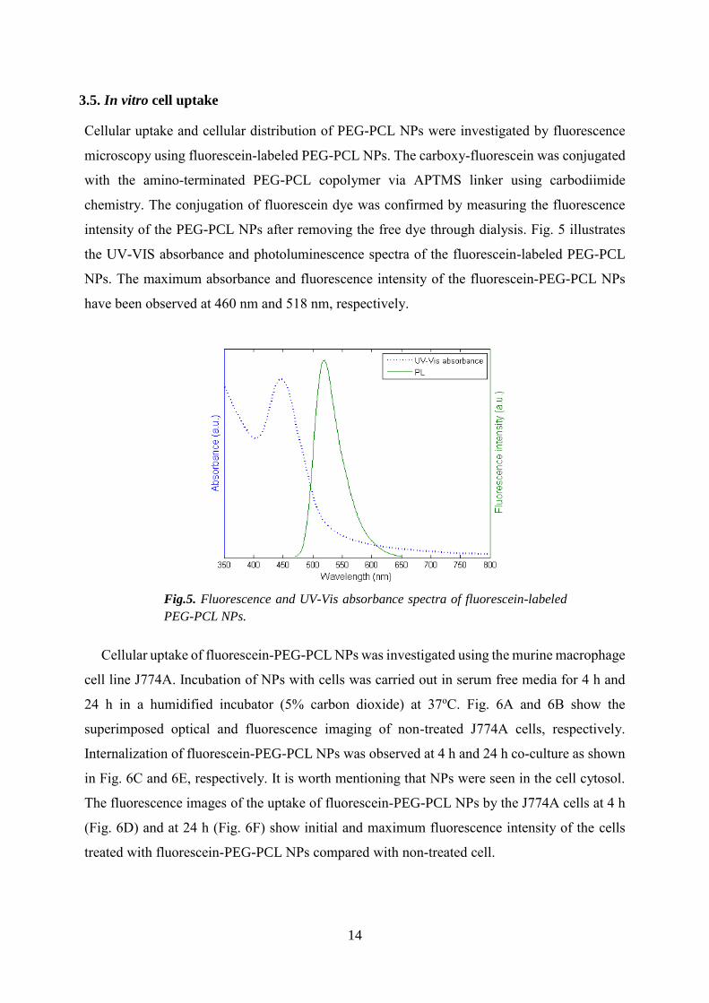
3.5. In vitro cell uptake
Cellular uptake and cellular distribution of PEG-PCL NPs were investigated by fluorescence
microscopy using fluorescein-labeled PEG-PCL NPs. The carboxy-fluorescein was conjugated
with the amino-terminated PEG-PCL copolymer via APTMS linker using carbodiimide
chemistry. The conjugation of fluorescein dye was confirmed by measuring the fluorescence
intensity of the PEG-PCL NPs after removing the free dye through dialysis. Fig. 5 illustrates
the UV-VIS absorbance and photoluminescence spectra of the fluorescein-labeled PEG-PCL
NPs. The maximum absorbance and fluorescence intensity of the fluorescein-PEG-PCL NPs
have been observed at 460 nm and 518 nm, respectively.
Cellular uptake of fluorescein-PEG-PCL NPs was investigated using the murine macrophage
cell line J774A. Incubation of NPs with cells was carried out in serum free media for 4 h and
24 h in a humidified incubator (5% carbon dioxide) at 37oC. Fig. 6A and 6B show the
superimposed optical and fluorescence imaging of non-treated J774A cells, respectively.
Internalization of fluorescein-PEG-PCL NPs was observed at 4 h and 24 h co-culture as shown
in Fig. 6C and 6E, respectively. It is worth mentioning that NPs were seen in the cell cytosol.
The fluorescence images of the uptake of fluorescein-PEG-PCL NPs by the J774A cells at 4 h
(Fig. 6D) and at 24 h (Fig. 6F) show initial and maximum fluorescence intensity of the cells
treated with fluorescein-PEG-PCL NPs compared with non-treated cell.
Fig.5. Fluorescence and UV-Vis absorbance spectra of fluorescein-labeled
PEG-PCL NPs.

15
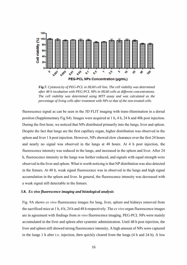
3.6. In vitro cytotoxicity assay
The cytotoxicity of the busulphan free PEG-PCL NPs was evaluated in the HL60 cell line. The
cell viability was determined by MTT assay after incubation with PEG-PCL NPs for 48 h and
the results are plotted in Fig. 7. Cell viability was calculated as the percentage of living cells
treated with NPs to that of the non-treated cells. PEG-PCL NPs exhibit 100% cell viability and
do not induce major cytotoxicity up to 25 µg/ml. Cell viability was observed to be 80% at PEG-
PCL NP concentrations of 50 and 100 µg/ml.
3.7. In vivo fluorescence imaging/computed tomography (CT)
The PEG-PCL NPs were further functionalized for in vivo fluorescence imaging using a near
infrared (NIR) fluorescence probe, VivoTag 680XL. VivoTag 680XL is suitable for in vivo
fluorescence imaging due to its high light penetration property and low autofluorescence at
activation wavelength (Ex/Em 675/720 nm). After IV administration of PEG-PCL NPs, the
biodistribution was followed up to 48 h (Fig. 8). Fig. 8a shows whole body 2D fluorescence
imaging prior to injection and at 1, 4, 24 and 48 h post injection, while Fig 8b shows whole
body 3D fluorescence imaging co-registered with 3D µCT imaging. 3D µCT images provide
the anatomical referencing for 3D fluorescence imaging to accurately predict the location of
Fig.6. Superimposed fluorescence and light microscopy images of J774A
incubated with fluorescein-labeled PEG-PCL NPs. (A, B) Non-treated control
J774A cells, overlay image and fluorescence image, respectively. (C, E)
Overlay images of J774A incubated with NPs for 4 h and 24 h, respectively. (D,
F) Fluorescence images of J774A incubated with NPs for 4 h and 24 h,
respectively.
16
fluorescence signal as can be seen in the 3D FLIT imaging with trans-illumination in a dorsal
position (Supplementary Fig S4). Images were acquired at 1 h, 4 h, 24 h and 48h post injection.
During the first hour, we noticed that NPs distributed primarily into the lungs, liver and spleen.
Despite the fact that lungs are the first capillary organ, higher distribution was observed in the
spleen and liver 1 h post injection. However, NPs showed slow clearance over the first 24 hours
and nearly no signal was observed in the lungs at 48 hours. At 4 h post injection, the
fluorescence intensity was reduced in the lungs, and increased in the spleen and liver. After 24
h, fluorescence intensity in the lungs was further reduced, and signals with equal strength were
observed in the liver and spleen. What is worth noticing is that NP distribution was also detected
in the femurs. At 48 h, weak signal fluorescence was in observed in the lungs and high signal
accumulation in the spleen and liver. In general, the fluorescence intensity was decreased with
a weak signal still detectable in the femurs.
3.8. Ex vivo fluorescence imaging and histological analysis
Fig. 9A shows ex vivo fluorescence images for lung, liver, spleen and kidneys removed from
the sacrificed mice at 1 h, 4 h, 24 h and 48 h respectively. The ex vivo organ fluorescence images
are in agreement with findings from in vivo fluorescence imaging. PEG-PCL NPs were mainly
accumulated in the liver and spleen after systemic administration. Until 48 h post injection, the
liver and spleen still showed strong fluorescence intensity. A high amount of NPs were captured
in the lungs 1 h after i.v. injection, then quickly cleared from the lungs (4 h and 24 h). A low
Fig.7. Cytotoxicity of PEG-PCL in HL60 cell line. The cell viability was determined
after 48 h incubation with PEG-PCL NPs in HL60 cells at different concentrations.
The cell viability was determined using MTT assay and was calculated as the
percentage of living cells after treatment with NPs to that of the non-treated cells.

17
fluorescence signal was generally observed in the kidneys throughout the experiment.
Microscopic analysis showed the presence of single NPs and agglomerates of PEG-PCL NPs
in the lungs, liver, spleen and kidneys as shown in Fig. 9B and supplementary Fig 4.
Fluorescence microscopy with phase contrast showed tissue structure in addition to a red signal
for VivoTag 680XL-labeled PEG-PCL NPs and a blue signal from DAPI stained nuclei. At
early investigation time points (1 h and 4 h), NPs are clearly present in the lungs as shown in
Fig. 9B. Moreover, single NPs are found in septal capillaries and as small endocytosed clusters
in alveolar/interstitial macrophages. Notably, at 24 h post injection, relatively larger aggregates
of NPs appear in larger arterioles without any sign of emboli/fibrillary material (eosinophilic
intravascular occlusive material causing fibrin thrombo-emboli). At 48 h post injection, very
few NPs were observed in the lungs. There was no sign of increased cellularity in the pulmonary
septa and no macrophage accumulation in any of the mice, nor any sign of inflammation.
To a higher extent, we noticed NP presence in the liver. Histological investigation shows
NPs association with sinusoidal Kupffer cells. There is no remarkable change in the amount of
NPs in liver over the time. The spleen seems to be another major target organ, which can be
seen in histological examinations as well as in the fluorescence imaging. The NPs were
observed mainly in association with macrophages, most commonly in the marginal zone. Slight
signs of clearance of NPs in the spleen and liver were observed at 48 h. Very few NPs were
observed in the kidneys (Supplementary Fig. S5). Neither glomerular hyperemia nor glomerular
hemorrhage was observed in the kidneys during the time the mice were studied.

18
Lung, liver, spleen and kidney were removed at 1, 4, 24, 48 h post injection. After
Hematoxylin-eosin (H&E) staining (Fig. 9 C), histopathological examination was performed
on the target organs to determine the organ toxicity of NPs. No evidence of toxic effect on either
ion tubules or glomeruli was observed in kidney during the 48h. In lung, the alveolar areas
retain normal structure at all post treatment time points (TB = terminal bronchiole). Further in
spleen, the white pulp (WP) areas showed normal structure and organization, while the
hematopoietic red pulp (RP) areas display a relatively active extra medullary hematopoiesis as
expected in young animals. Moreover, the livers retain a normal structure with a homogenous
hepatocyte nuclear size. Relationship between central veins (CV) and portal areas (P) is normal.
Similar observations in all organs at different time points were found (data not shown). In Fig.
Fig.8. In vivo fluorescence imaging of Balb/c mice after intravenous administration of
PEG-PCL NPs labelled with VivoTag 680XL. (A) 2D fluorescence imaging of Balb/c
mice at 1h, 4h, 24 h and 48 h after IV administration, ventral side. Fluorescence images
are overlaid with light photography. Control is a mouse without VivoTag 680XL PEG-
PCL NPs injection. (B) 3D fluorescence imaging-CT imaging co-registration of Balb/c
mice at 1h, 4h, 24 h and 48 h after IV administration, dorsal side and left side as shown.
Heat map represents the area with fluorescence signal and color represents the intensity.

19
9D, macrophage cells were stained with anti-IBA1 antibody and the localization of NPs was
seen with macrophage cells in liver and spleen.

20
Fig.9. Ex vivo organ analysis post VivoTag 680XL PEG-PCL NP administration. (A) Ex vivo organ
fluorescence imaging of lungs, liver, spleen. Images were taken at1 h, 4 h and 24 h and 48 h. The
signal decreases in lungs from 1 h until 48 h, and increases in liver and spleen within the same time.
A slight decrease in signal intensity was observed at 48 h in liver and spleen. (B) Fluorescence
microscopy overlaid phase contrast images of lung, liver and spleen harvested at 1, 4, 24 and 48 h.
Fluorescence images of VivoTag 680XL PEG-PCL NPs are shown in red (CY5) while nuclei are
stained with (DAPI) using 4',6-diamidino-2-phenylindole (scale bar = 100 µm). (C). H&E staining
of lung, liver, spleen and kidney at 48 hour post injection. TB: terminal bronchiole; WP: white pulp;
RP: red pulp; P: portal areas. Scale bar: 100 μm. (D). Immunofluorescence staining of IBA1 in liver
and spleen at 1 h post injection. Red: PEG-PCL NPs. Green: IBA 1. Blue: DAPI. Scale bar: 50 μm.

21
4. Discussion
A multifunctional nanocarrier system with well-defined physico-chemical properties for
anticancer drug delivery and multimodal imaging purposes was developed. In the current
investigation we studied the loading capacity of busulphan as a therapeutic anticancer drug into
PEG-PCL NPs, the encapsulation of SPION as contrast agent to enhance MR imaging and the
chemical conjugation of an NIR fluorescent molecule for multi-model imaging capacities.
Moreover, we have investigated the cellular toxicity and organ toxicity in vivo.
The classical O/W solvent evaporation emulsion method was used to prepare PEG-PCL NPs
in the presence of PVA as a stabilizer. The evaporation of the organic solvent leads to
solidification of NPs32,33 resulting in spherical shape and no aggregations (Fig. 2 and DLS in
Fig. S3). They have an average particle size of 212 nm, but with a broad size distribution (σ ~
30%). The broad size distribution is resulted from the formation of micelle of amphiphilic
copolymer (PEG-PCL), where the clustering of the hydrophobic tails is driven by
thermodynamic equilibrium and great freedom is allowed in the possible morphology formation.
Therefore, the fast formation of polymer shell around the oil droplets (DCM in this work)
facilitates the encapsulation of water-insoluble molecules; however the control on size
distribution is weak and hence results in a broad size range. A negative zeta potential of PEG-
PCL has been found, which is attributed to the negative charged state of the PCL block in the
copolymer. The high encapsulation efficiency implies that PEG-PCL NPs are suitable for
busulphan delivery, which is probably due to the good affinity between busulphan and the
hydrophobic core of PEG-PCL copolymer. In this drug delivery system, we have shown a
sustained release of busulphan from SPION-PEG-PCL NPs in PBS buffer solution over a period
of 10 hours with a burst release of approximately 22% in the first hour release pattern (Fig. 3).
The burst is attributed to diffusion of the drug located at the interface between the NP core and
the surface into the dissolution medium, and to the hydrophobicity of the drug carrier.34 In the
present study, the burst effect was controlled by increasing the initial concentration of the
monomer (105 mM of ε-caprolactone) during the polymerization reaction to produce a long
hydrophobic PCL chain, which reduces the initial penetration of water into the copolymer, and
hence suppresses the burst release.
The properties of SPION-PEG-PCL NPs as contrast agent were evaluated by examining the
phantom of these NPs under MRI. The T2*-weighted MRI images of phantoms (Fig. 4A)

22
showed a decrease in the MR signal intensity at different TE compared with control. Since
SPIONs can produce spin-spin relaxation effects and cause local field inhomogeneity, MRI
appears predominantly dark due to shortening of the relaxation time by iron oxide. Furthermore,
when concentration of iron oxide increases, the extent of signal decrease is enhanced; i.e., much
darker for a higher concentration than lower one. The relaxivity r2* measures the change in
spin-spin relaxation rate (1/T2*) per unit concentration of iron oxide (mM-1s-1). SPION-PEG-
PCL NPs show a relatively high relaxation rate. As a result, the r2* for the SPION-PEG-PCL
NPs is significantly larger than those obtained from SPION-dextran particles (30-50 Fe mM-1s-
1).35 These results illustrate that the formulation of SPION-PEG-PCL NPs has potential to be
used as contrast agent for MRI applications. We demonstrated that cells are capable of
internalizing PEG-PCL NPs as shown in Fig.6. The cellular interaction with the NPs is
influenced by particle size, shape, and surface charge.36-38 Cell internalization is expected to be
through phagocytosis, endocytosis and receptor mediated endocytosis as the size is below 200
nm. We also noticed that PEG-PCL NPs showed non-cytotoxicity at different concentrations in
HL60 cells (Fig.7), which implied that the PEG-PCL NPs are safe for biomedical
applications. VivoTag 680XL is an NIR fluorescence dye with high quantum yield and high
photo-stability, which allows detection of low-abundance biological structures in deep tissues
with good sensitivity. VivoTag 680XL dye molecules can be attached to proteins/NPs and
micro-vehicles at high molar ratios without significant self-quenching, resulting in brighter
conjugates and low detection limits. The long emission wavelength of VivoTag 680XL makes
it suitable for in vivo biodistribution study since there is little interference from absorption by
hemoglobin (absorption peak at 550 to 600 nm).
In vivo biodistribution investigation revealed that after intravenous administration, the
majority of PEG-PCL NPs distributed to the liver and spleen. A high amount of NPs were
captured in the lungs immediately after injection followed by rapid clearance. At 1 h, the rapid
distribution of PEG-PCL NPs into the lungs, liver and spleen was most probably due to the
sequestration effect by the mononuclear phagocyte system (MPS). The MPS is mainly
composed of resident macrophages and plays an important role in maintaining homeostasis.39
The macrophages are found in all the connective tissues including the liver (Kupffer cells),
spleen, lymph nodes, lungs and central nervous system (microglia). The uptake of PEG-PCL
NPs by the resident macrophages in the lungs, liver and spleen was confirmed by histological
analysis; at 1 h, NPs were found in the lungs as single particles in septal capillaries or as small
endocytosed clusters in alveolar/interstitial macrophages. In the liver, NPs were associated with

23
sinusoidal Kupffer cells, while in the spleen NPs were also observed to be affiliated with
macrophages, mostly in the marginal zone. In our previous investigation of the biodistribution
of carbon 11 labelled busulphan in the monkey study, we have shown that free busulphan
rapidly distributed into liver and lungs within 1 h after i.v. injection. The uptake of busulphan
in the lungs was 25 to 45% of that found in the liver.40 At later time points (4 and 24 h), PEG-
PCL NPs which were resident in the lungs were cleared by macrophages and then further
accumulated in the spleen and liver. NP size is the major factor for NP circulation, extravasation
through vasculature leakage, macrophage uptake and renal clearance upon intravenous
administration.41 In the liver, the vascular fenestrations in non-continuous endothelia are
between 50 to 100 nm, which leads to liver accumulation of NPs larger than 50 nm.41,42 Splenic
filtration causes the retention of NPs larger than 200 nm, due to the slits between inter-
endothelial cells being 200-500 nm in size.41,43 According to the TEM micrograph, the average
diameter of our PEG-PCL NPs is 212 nm (σ ~ 30%). Therefore, the blood circulating PEG-
PCL NPs were accumulated in the liver and spleen based on the endothelial filtration cut-off
size range in those organs. At 48 h, there was a slight sign of clearance of NPs in the spleen and
liver, which indicates that a part of the PEG-PCL NPs was degraded.
NPs have emerged as suitable delivery vehicles for anti-cancer drugs and tumor imaging
contrast agents due to the enhanced permeability and retention (EPR) effect, in which fenestrated
blood vessels in solid tumors lead to a high extent-accumulation of long-circulating NPs.44
However, leukemia is a hematological disease with leukemic cells in blood and bone marrow
and lacks the EPR effect. To target leukemia, the strategy is to develop long-circulating NPs
with active ligands targeting leukemic cell populations.41 Once NPs are injected intravenously,
protein coronas are formed on the surface of the circulating NPs facilitating the binding to
receptors on the surface of phagocytes, which is followed by internalizing and fusing with
lysosomes.45 Ethylene glycol units on PEG-PCL NPs together with plasma water form a
hydrating layer on the surface of the NPs, which prohibit protein adsorption and clearance by
MPS.41,46 The surface charge of NPs also plays an important role in protein adsorption and
therefore affects the biodistribution of NPs. Highly positively charged NPs are rapidly cleared
from circulation while neutral and lightly negatively charged NPs have reported significantly
prolonged blood circulation.47 Zeta potential measurement showed that the PEG-PCL NPs used
in this study have a negative charge of approximately -2.8 mV at neutral and/or physiological
pH. Hence, the PEG unit on the PEG-PCL copolymer together with the negatively-charged
surface of the PEG-PCL NPs has positive effect on particle circulation. Circulating PEG-PCL
NPs existed until 24 h post i.v. injection, since there was no further fluorescence intensity

24
increase in the liver and spleen at 48 h. A fluorescence signal was detectable in the femurs at 4,
24 and 48 h, suggesting that PEG-PCL NPs might also distribute to bone marrow. At the present,
we are planning a new investigation for hematological malignancies, namely leukemia, in a
mouse model to confirm the present findings. It will also be of interest to tag specific
antibodies/peptides against leukemia cells, such as anti-CD33 ligand and/or CD20, onto the
surface of PEG-PCL NPs to further improve their efficacy for drug delivery and in vivo imaging.
5. Conclusions
We developed a multifunctional nanoparticulate system based on biodegradable amphiphilic
polymer containing an antineoplastic agent, busulphan, with high entrapment efficacy. We also
succeeded in encapsulating SPION into the PEG-PCL nanocarrier, and showed its suitability as
contrast agent via enhancing T2*-weighted MR imaging. Together with high contrast
enhancement of SPION and sustained release of busulphan, fluorescence-labeled PEG-PCL
nanocarriers allowed for real-time observation of in vivo biodistribution and uptake by different
tissues in a 48 h period with no tissue impairment or toxicity observed. We believe that the
SPION-PEG-PCL nanocarrier is a promising candidate for biomedical applications, where in
vivo imaging and controlled drug delivery for therapy are necessary.
Supporting Information. Characterization of PEG-PCL diblock copolymer, SPIONs and
SPION-PEG-PCL NPs, co-registration of FLIT/CT imaging, and fluorescence microscopy
images of kidney.
Acknowledgements
The authors thank Jennifer Usterud for language editing and proof reading. The authors
acknowledge The Swedish Cancer foundation for grant support (CAN2014/759). The work
was supported partially by the National Science, Technology and Innovation Program of King
Abdulaziz City for Science and Technology (Project No: 11- NAN1462-02). YZ was supported
by the Strategic Research Program (StratCan) from the Swedish government. FY thanks
Stockholms läns landsting (SLL) for financial support of HMT grant.
Conflict of interest
The authors have no conflict of interest. Jörg Hamm is employed by Perkin Elmer; however,
his input in the manuscript was pure scientific advice.
References
25
(1) Pehlivan, S. B. Nanotechnology-based drug delivery systems for targeting, imaging and
diagnosis of neurodegenerative diseases. Pharm. Res. 2013, 30, 2499-2511.
(2) El-Sayed, R.; Eita, M.; Barrefelt, Å.; Ye, F.; Jain, H.; Fares, M.; Lundin, A.; Crona, M.;
Abu-Salah, K.; Muhammed, M.; Hassan, M. Thermostable luciferase from Luciola
cruciate for imaging of carbon nanotubes and carbon nanotubes carrying doxorubicin
using in vivo imaging system. Nano Lett. 2013, 13, 1393-1398.
(3) Cho, K.; Wang, X.; Nie, S.; Chen, Z.; Shin, D. M. Therapeutic nanoparticles for drug
delivery in cancer. Clin. Cancer Res. 2008, 14, 1310-1316.
(4) Janib, S. M.; Moses, A. S.; MacKay, J. A. Imaging and drug delivery using theranostic
nanoparticles. Adv. Drug Delivery Rev. 2010, 62, 1052-1063.
(5) Lin, M.; Pei, H.; Yang, F.; Fan, C.; Zuo, X. Applications of gold nanoparticles in the
detection and identification of infectious diseases and biothreats. Adv. Mater. 2013, 25,
3490-3496.
(6) Barreto, J. A.; O’Malley, W.; Kubeil, M.; Graham, B.; Stephan, H.; Spiccia, L.
Nanomaterials: Applications in cancer imaging and therapy. Adv. Mater. 2011, 23, H18-
H40.
(7) Sharma, P.; Brown, S.; Walter, G.; Santra, S.; Moudgil, B. Nanoparticles for bioimaging.
Adv. Colloid Interface Sci. 2006, 123–126, 471-485.
(8) Torchilin, V. P. Multifunctional nanocarriers. Adv. Drug Delivery Rev. 2006, 58, 1532-
1555.
(9) Cheng, Y.; Morshed, R. A.; Auffinger, B.; Tobias, A. L.; Lesniak, M. S. Multifunctional
nanoparticles for brain tumor imaging and therapy. Adv. Drug Delivery Rev. 2014, 66,
42-57.
(10) Soppimath, K. S.; Liu, L. H.; Seow, W. Y.; Liu, S. Q.; Powell, R.; Chan, P.; Yang, Y. Y.
Multifunctional core/shell nanoparticles self-assembled from pH-induced
thermosensitive polymers for targeted intracellular anticancer drug delivery. Adv. Funct.
Mater. 2007, 17, 355-362.
(11) Liang, R.; Wang, J.; Wu, X.; Dong, L.; Deng, R.; Wang, K.; Sullivan, M.; Liu, S.; Wu,
M.; Tao, J.; Yang, X.; Zhu, J. Multifunctional biodegradable polymer nanoparticles with
uniform sizes: Generation and in vitro anti-melanoma activity. Nanotechnology 2013, 24,
455302.
(12) Park, J. H.; Lee, S.; Kim, J.-H.; Park, K.; Kim, K.; Kwon, I. C. Polymeric nanomedicine
for cancer therapy. Prog. Polym. Sci. 2008, 33, 113-137.
(13) Tong, R.; Cheng, J. Anticancer polymeric nanomedicines. Polym. Rev. 2007, 47, 345-381.
(14) Talelli, M.; Rijcken, C. J. F.; Lammers, T.; Seevinck, P. R.; Storm, G.; van Nostrum, C.
F.; Hennink, W. E. Superparamagnetic iron oxide nanoparticles encapsulated in
biodegradable thermosensitive polymeric micelles: Toward a targeted nanomedicine
suitable for image-guided drug delivery. Langmuir 2009, 25, 2060-2067.
(15) Nair, L. S.; Laurencin, C. T. Biodegradable polymers as biomaterials. Prog. Polym. Sci.
2007, 32, 762-798.
(16) Xiong, X.-B.; Binkhathlan, Z.; Molavi, O.; Lavasanifar, A. Amphiphilic block co-
polymers: Preparation and application in nanodrug and gene delivery. Acta Biomater.
2012, 8, 2017-2033.
(17) Mieszawska, A. J.; Kim, Y.; Gianella, A.; van Rooy, I.; Priem, B.; Labarre, M. P.; Ozcan,
C.; Cormode, D. P.; Petrov, A.; Langer, R.; Farokhzad, O. C.; Fayad, Z. A.; Mulder, W.
J. M. Synthesis of polymer–lipid nanoparticles for image-guided delivery of dual
modality therapy. Bioconjugate Chem. 2013, 24, 1429-1434.
(18) Ling, Y.; Wei, K.; Luo, Y.; Gao, X.; Zhong, S. Dual docetaxel/superparamagnetic iron
oxide loaded nanoparticles for both targeting magnetic resonance imaging and cancer
therapy. Biomaterials 2011, 32, 7139-7150.

26
(19) Sun, C.; Lee, J. S. H.; Zhang, M. Magnetic nanoparticles in MR imaging and drug
delivery. Adv. Drug Delivery Rev. 2008, 60, 1252-1265.
(20) Shubayev, V. I.; Pisanic Ii, T. R.; Jin, S. Magnetic nanoparticles for theragnostics. Adv.
Drug Delivery Rev. 2009, 61, 467-477.
(21) Jain, T. K.; Reddy, M. K.; Morales, M. A.; Leslie-Pelecky, D. L.; Labhasetwar, V.
Biodistribution, clearance, and biocompatibility of iron oxide magnetic nanoparticles in
rats. Mol. Pharmaceutics 2008, 5, 316-327.
(22) Galton, D. A. G. Myleran in chronic myeloid leukemia results of treatment. The Lancet
1953, 261, 208-213.
(23) Blazar, B. R.; Ramsay, N. K.; Kersey, J. H.; Krivit, W.; Arthur, D. C.; Filipovich, A. H.
Pretransplant conditioning with busulfan (Myleran) and cyclophosphamide for
nonmalignant diseases. Transplantation 1985, 39, 597–603.
(24) Iwamoto, T.; Hiraku, Y.; Oikawa, S.; Mizutani, H.; Kojima, M.; Kawanishi, S. DNA
intrastrand cross-link at the 5′-GA-3′ sequence formed by busulfan and its role in the
cytotoxic effect. Cancer Sci. 2004, 95, 454-458.
(25) Chang, K.-Y.; Lee, Y.-D. Ring-opening polymerization of ε-caprolactone initiated by the
antitumor agent doxifluridine. Acta Biomaterialia 2009, 5, 1075-1081.
(26) Nafee, N.; Youssef, A.; El-Gowelli, H.; Asem, H.; Kandil, S. Antibiotic-free
nanotherapeutics: Hypericin nanoparticles thereof for improved in vitro and in vivo
antimicrobial photodynamic therapy and wound healing. Int. J. Pharm. 2013, 454, 249-
258.
(27) Ye, F.; Barrefelt, Å.; Asem, H.; Abedi-Valugerdi, M.; El-Serafi, I.; Saghafian, M.; Abu-
Salah, K.; Alrokayan, S.; Muhammed, M.; Hassan, M. Biodegradable polymeric vesicles
containing magnetic nanoparticles, quantum dots and anticancer drugs for drug delivery
and imaging. Biomaterials 2014, 35, 3885-3894.
(28) Hassan, M.; Ehrsson, H. Gas chromatographic determination of busulfan in plasma with
electron-capture detection. J. Chromatogr. B 1983, 277, 374-380.
(29) Shin, I. G.; Kim, S. Y.; Lee, Y. M.; Cho, C. S.; Sung, Y. K. Methoxy poly(ethylene
glycol)/ε-caprolactone amphiphilic block copolymeric micelle containing indomethacin.:
I. Preparation and characterization. J. Controlled Release 1998, 51, 1-11.
(30) Bogdanov, B.; Vidts, A.; Van Den Buicke, A.; Verbeeck, R.; Schacht, E. Synthesis and
thermal properties of poly(ethylene glycol)-poly(ε-caprolactone) copolymers. Polymer
1998, 39, 1631-1636.
(31) Choi, C.; Chae, S. Y.; Kim, T.-H.; Kweon, J. K.; Cho, C. S.; Jang, M.-K.; Nah, J.-W.
Synthesis and physicochemical characterization of amphiphilic block copolymer self-
aggregates formed by poly(ethylene glycol)-block-poly(ε-caprolactone). J. Appl. Polym.
Sci. 2006, 99, 3520-3527.
(32) Dash, T. K.; Konkimalla, V. B. Poly-ε-caprolactone based formulations for drug delivery
and tissue engineering: A review. J. Controlled Release 2012, 158, 15-33.
(33) Pinto Reis, C.; Neufeld, R. J.; Ribeiro, A. J.; Veiga, F. Nanoencapsulation I. Methods for
preparation of drug-loaded polymeric nanoparticles. Nanomedicine: NBM 2006, 2, 8-21.
(34) Ostacolo, L.; Marra, M.; Ungaro, F.; Zappavigna, S.; Maglio, G.; Quaglia, F.; Abbruzzese,
A.; Caraglia, M. In vitro anticancer activity of docetaxel-loaded micelles based on
poly(ethylene oxide)-poly(epsilon-caprolactone) block copolymers: Do nanocarrier
properties have a role? J. Controlled Release 2010, 148, 255-263.
(35) Wang, Y.-X.; Hussain, S.; Krestin, G. Superparamagnetic iron oxide contrast agents:
physicochemical characteristics and applications in MR imaging. Eur. Radiol. 2001, 11,
2319-2331.
27
(36) Geng, Y. A. N.; Dalhaimer, P.; Cai, S.; Tsai, R.; Tewari, M.; Minko, T.; Discher, D. E.
Shape effects of filaments versus spherical particles in flow and drug delivery. Nat.
Nanotechnol. 2007, 2, 249-255.
(37) Miller, C. R.; Bondurant, B.; McLean, S. D.; McGovern, K. A.; O'Brien, D. F.
Liposome−cell interactions in vitro: Effect of liposome surface charge on the binding and
endocytosis of conventional and sterically stabilized liposomes. Biochemistry 1998, 37,
12875-12883.
(38) Gratton, S. E. A.; Ropp, P. A.; Pohlhaus, P. D.; Luft, J. C.; Madden, V. J.; Napier, M. E.;
DeSimone, J. M. The effect of particle design on cellular internalization pathways. Proc.
Natl. Acad. Sci. U.S.A. 2008, 105, 11613-11618.
(39) Chow, A.; Brown, B. D.; Merad, M. Studying the mononuclear phagocyte system in the
molecular age. Nat. Rev. Immunol. 2011, 11, 788-798.
(40) Hassan, M.; Oberg, G.; Ericson, K.; Ehrsson, H.; Eriksson, L.; Ingvar, M.; Stone-Elander,
S.; Thorell, J. O.; Smedmyr, B.; Warne, N.; Widén, L. In vivo distribution of [11C]-
busulfan in cynomolgus monkey and in the brain of a human patient. Cancer Chemother.
Pharmacol. 1992, 30, 81-85.
(41) Blanco, E.; Shen, H.; Ferrari, M. Principles of nanoparticle design for overcoming
biological barriers to drug delivery. Nat. Biotechnol. 2015, 33, 941-951.
(42) Braet, F.; Wisse, E.; Bomans, P.; Frederik, P.; Geerts, W.; Koster, A.; Soon, L.; Ringer,
S. Contribution of high-resolution correlative imaging techniques in the study of the liver
sieve in three-dimensions. Microsc. Res. Techniq. 2007, 70, 230-242.
(43) Chen, L. T.; Weiss, L. The role of the sinus wall in the passage of erythrocytes through
the spleen. Blood 1973, 41, 529-537.
(44) Matsumura, Y.; Maeda, H. A new concept for macromolecular therapeutics in cancer
chemotherapy: mechanism of tumoritropic accumulation of proteins and the antitumor
agent smancs. Cancer Res. 1986, 46, 6387-6392.
(45) Sahay, G.; Alakhova, D. Y.; Kabanov, A. V. Endocytosis of nanomedicines. J. Controlled
Release 2010, 145, 182-195.
(46) Harris, J. M.; Chess, R. B. Effect of pegylation on pharmaceuticals. Nature Rev. Drug
Discov. 2003, 2, 214-221.
(47) Arvizo, R. R.; Miranda, O. R.; Moyano, D. F.; Walden, C. A.; Giri, K.; Bhattacharya, R.;
Robertson, J. D.; Rotello, V. M.; Reid, J. M.; Mukherjee, P. Modulating pharmacokinetics,
tumor uptake and biodistribution by engineered nanoparticles. PloS ONE 2011, 6, e24374.

28
Supporting Information
Biodistribution of Busulphan Loaded Biodegradable Nano-carrier Designed
for Multimodal Imaging
Heba Asem1, 2, Ying Zhao2, 3, Fei Ye2, Åsa Barrefelt2, Manuchehr Abedi-Valugerdi2, Ramy
El-Sayed2, Ibrahim El-Serafi2, Svetlana Pavlova2, Khalid M. Abu-Salah4, Salman A.
Alrokayan5, Jörg Hamm6, Mamoun Muhammed1, Moustapha Hassan2,3
Fig.S1. Characterization of PEG-PCL diblock copolymer. (A) The FTIR spectra of PEG-PCL
copolymer, where the observed strong and sharp absorption band appearing at 1721 cm-1 is
characteristic of the stretching vibration of the ester carbonyl group (C=O) of PCL. The (C-H)
stretching bands in PEG and PCL are vibrating at 2863 and 2942 cm-1, respectively; (B) Thermal
property of PEG-PCL diblock copolymer. The melting temperature of the copolymer was measured by
differential scanning calorimetry (DSC) and a bimodal melting peak was observed at 54.8°C.

29
Fig.S2. Characterization of SPIONs. (A) Powder X-ray diffraction (XRD) pattern and (B) field-
dependent magnetization measurement of Fe3O4 NPs.
Fig. S3. Dynamic light scattering (DLS) of SPION-PEG-PCL nanoparticles dispersed in DI water.
The hydrodynamic diameter is represented by differential volume, intensity, or number, respectively.
The surface charge of SPION-PEG-PCL NPs is also measured and it showed a negative zeta potential
of ca.-2.8 mV at neutral pH.

30
Fig.S4. 3D Fluorescence imaging tomography-CT imaging co-registration
The 3D FLIT imaging with trans-illumination in a dorsal position sequence was set up and images were
acquired at excitation 675 nm and emission 720 nm. The mouse in the MIS was then transferred to the
Quantum FX-µCT and subjected to a fast, low dose CT scan with a field of view (FOV) at 60 mm and
17 second scan-time. The slice views show the images of coronal, sagittal and transversal plane of balb/c
mice at 1, 4, 24 and 48 h post i.v. administration. At 1 h after administration signal was observed in
lungs, spleen and liver. During the time of the study, the signal was redistributed mainly, to liver and
spleen.

31
Fig.S5. Fluorescence microscopy overlaid phase contrast images of kidney.
Fluorescence images of VivoTag 680XL tagged PEG-PCL NPs are shown in red while nuclei are stained
with (DAPI) using 4',6-diamidino-2-phenylindole (scale bar=100 µm). Very few NPs were observed in
the kidneys. Neither glomerular hyperemia nor glomerular hemorrhage was observed in the kidneys
during the study time.


![Radiolabeling of Biodegradable Polymeric Microspheres with [ … · 2010-03-07 · Radiolabeling of Biodegradable Polymeric Microspheres with [99mTc(CO)3] and in ViWo Biodistribution](https://img.pdfslide.us/doc/110x75/5e6a12bcc5fb5446bd082abb/radiolabeling-of-biodegradable-polymeric-microspheres-with-2010-03-07-radiolabeling.jpg)