Embed Size (px)
Citation preview
BIODEGRADATION OF AROMATIC COMPOUNDS BY
HIGH LATITUDE PHYTOPLANKTON
by
C. Van Baalen
University of Texas Marine Science InstitutePort Aransas Marine Laboratory
Port Aransas, Texas 78373
and
David T. Gibson
Department of MicrobiologyUniversity of Texas at Austin
Austin, Texas 78712
Final ReportOuter Continental Shelf Environmental Assessment Program
Research Unit 607
June 15, 1982
127

TABLE OF CONTENTS
SUMMARY . . . . . . . . . . . . . .
INTRODUCTION. . . . . . . . . . . .
OXIDATION OF NAPHTHALENE BY DIATOMSTHE KACHEMAK BAY REGION OF ALASKA
Materials and Methods .
Results and Discussion.
Acknowledgements. . . .
References. . . . . . .
.
.
●
✎
●
✎
✎
●
.
●
●
●
,
.
.
.
.
. ..0.. .
. . ...0 ●
ISOLATED FROM.
.
.
.
.
.
●
✎
✎
✎
●
●
✎
✎
✎
●
●
✎
✎
✎
BERING SEA DIATOMS: GROWTH CHARACTERISTICS,
.
.
.
●
✎
.
.
.
●
✎
●
✎
✎
✎
●
.
●
✎
●
✎
✎
●
●
●
✎
✎
✎
✎
✎
OF NAPHTHALENE, AND SENSITIVITY TO CRUDE OIL. . . . .
References and Notes. . . . . . . . . . . . . . .
RATE STUDIES OF 1-NAPHTHOL FORMATION IN MESOPHILICALGAE. . . . . . . . . . . . . . . . . . . . . . . .
.
.
●
●
●
●
✎
✎
✎
●
Reference. . . . . . . . . . . . . . . . . . . . .
*
.
●
✎
●
●
●
●
●
●
✎
●
✎
✎
✎
✎
●
●
●
✎
●
✎
.
.
.
.
.
.
.
●
✎
●
✌
.
.
●
✎
✎
●
✎
●
●
✎
●
●
✎
✎
✎
✎
✎
✎
✎
✎
●
✎
.
.
●
●
✎
✎
●
✎
●
✎
✎
Page
131
133
133
134
137
140
141
143
154
156
158
129
SUMMARY
It was the purpose of the work undertaken to bring into pure
culture representative diatoms from the Cook Inlet and the ice-edge in the
Bering Sea and to examine their capacity for the oxidation of aromatic
compounds using naphthalene as a model substrate. Three diatoms from the
Cook Inlet (Kasitsna Bay) were shown to metabolize naphthalene at 6 or 12°C
to l-naphthol and other unidentified ethyl acetate and water-soluble
products. Likewise, three diatoms isolated from samples collected at the
ice-edge in the Bering Sea also formed small amounts of l-naphthol from
naphthalene when incubated in the light at O or 10°C.
We have not been able to rigorously prove that any algal cell,
be it a blue-green alga, a green alga, or a diatom can metabolize (1-14C)
naphthalene far enough to produce 14C02. However, if we assume a
stoichiometery of one l-naphthol in the algae equivalent to one C02 in
bacteria, then for mesophilic algae, the rate of l-naphthol production is
roughly estimated as 10% of the in situ marine potential, and perhaps——
higher if only the photic zone is considered. We have as yet, no
corresponding values for rate of l-naphthol formation from naphthalene by
cold-adapted or psychrophilic diatom cultures, however, it seems reasonable
to suggest that algal aromatic transformations may also be a significant
fraction of bacterial activity in cold environments. In addition to
studies on the oxidation of naphthalene we have also examined the
sensitivity of the Bering Sea psychrophilic diatoms to crude oil samples
from Cook Inlet and Prudhoe Bay. The results with pure cultures indicate
that the toxicity of crude oil was enhanced in psychrophilic diatoms grow-
ing at O°C or 10°C as compared to previous studies with mesophilic forms.
131

There are several important consequences of the results for
Alaskan OCS oil and gas development. It is now clear that pure cultures of
diatoms isolated from either the lower Cook Inlet or from the ice-edge in
the Bering Sea can oxidize aromatic compounds such as naphthalene. Whether
the metabolizes persist through the food chain and will be more or less
toxic than naphthalene itself is not known. The results with naphthalene
also imply that the photic zone can be an important sink for aromatic
hydrocarbon transformations. There are certainly differences among
microalgae in the capacity to oxidize naphthalene. It
therefore, to insure, via monitoring, that accidental
matic compounds in Alaskan waters does not cause a
effect on existing phytoplankton populations.
seems prudent,
introduction of aro-
selective or enrichment
A second area of environmental concern is the suggestion of an
enhanced crude oil toxicity in slower growing psychrophilic diatoms as
compared to their mesophilic cousins. Crude oil spills near or under the
sea ice may severely impact primary productivity, and thereby higher
tropic level.
132

INTRODUCTION
The results are presented in two sections, one dealing with the
Cook Inlet isolates and the other with the isolates from the ice-edge in
the Bering Sea. Each section has an introduction, description of materials
and methods, results and discussion section, and references.
THE OXIDATION OF NAPHTHALENE BY DIATOMS ISOLATED FROM THE
Aromatic
in open ocean waters
and/or their metabolites
KACHEMAK BAY REGION OF ALASKA
hydrocarbons have been found to be widely distributed
(Brown and tiuffman, 1976). Many of these compounds
have toxic properties which include initiation of
tumor formation and cancer (Miller and Miller, 1976). In studies of the
fate of hydrocarbons in aquatic ecosystems, a considerable amount of
information is available on the bacterial and fungal degradation of these
compounds and their derivatives (Atlas, 1981; Cerniglia, 1981). In view
of the fact that cyanobacteria and microalgae are widely distributed in
many aquatic environments and may be important in the catabolism of hydro-
carbons, we initiated a research program on the algal oxidation of aromatic
hydrocarbons (Cernigl ia et al. 1979, 1980 a,b,c).——
Most of the studies on the microbial oxidation of hydrocarbons
have been conducted at temperatures between 20”C to 30*C. Since there has
been increased activities of oil exploration and transport of petroleum in
Alaskan waters, there has been recent interest in the microbia degradation
of crude oil at low water temperatures (Atlas, 1981).
In this investigation, we report on three diatoms isolated from
the Kachemak Bay region of Alaska which have the ability to metabolize the
aromatic hydrocarbon, naphthalene at low temperatures.
133
MATERIALS AND METHODS
Organisms and Growth Conditions. The diatoms KIA (Navicula ~.), K8A——
(Nitzschia ~.) and 40 (Synedra sp.) were isolated via enrichment culture
at 6 to 10”C from oblique net (20pm Nitex nylon) tows made during August
1979 and April, 1980 in the Kachemak Bay region, south of Homer, Alaska.
The enrichment medium was local sea water plus 5, 20, or 50% ASP-2 medium
(Van Baalen, 1962). Pure cultures were obtained by repeated streaking or
by several minutes treatment with ultraviolet radiation (254 nm, 15 W
germicidal lamp) and subsequent pour plates. Organism N-1 (Cylindrotheca
~.) was isolated from a water sample taken from the Pass adjacent to the
Port Aransas Marine Laboratory (Estep et al., 1978). The organisms were
grown on ASP-2 medium containing 125 mg 1-1 Na2 Si03.9H20, 4pg 1-1 vitamin
B12 and 250 pg 1-1 thiamine in 22 x 175 mm Pyrex test tubes at 12”C. The
growth tubes were illuminated with fluorescent lamps F20T12-WWX two on each
side of the water bath, 8 cm from the front edge of the lamp to the tube
center. The cultures were continuously aerated with 1~0.1% C02 enriched
air. The generation times under these conditions for the four organisms
were about 24 hours.
Naphthalene Biotransformation Experiments. [1-14C]-Naphthalene experiments
were conducted in order to determine the amount of naphthalene oxidized by
each organism. Cells (0.5 to 0.8 mg) were pooled from several growth tubes
and placed in 22 x 175 mm screw cap tubes, final volume 10 ml.
[14 C]-Naphthalene (1 PCi in 20 P1 ethanol, 6.9 mg/liter) was added just
before closing the tube with a plastic top lined with a chromatography
septum, aluminum foil and 1 ml Teflon film. Carbon dioxide was added
134

through a small hole in the plastic top with a gas tight syringe to an
initial concentration of 1%. The screw cap tubes were clamped to a glass
rod and rotated slowly in the same illuminated water bath used for growing
the cultures. The tubes were incubated at either 6°C or 12°C.
After 22 hr incubation, cells were removed by centrifugation and
each supernatant extracted with five thirty ml volumes of ethyl acetate.
The organic extracts were dried over anhydrous sodium sulfate and the
solvent was removed in vacuo at 42*C. Each residue was redissolved in——
methanol and analyzed by high pressure liquid chromatography. The ratio of
ethyl acetate soluble metabolizes to water soluble metabolizes was
determined by taking each organic soluble extract and redissolving in 50 UI
of acetone and 10 U1 aliquots was added to vials containing 10.0 ml of
scintillation fluid. The radioactivity present was determined in a liquid
scintillation counter. Corrections were made for machine efficiency and
quenching.
An experiment with unlabeled naphthalene was conducted with
organism K8A in order to obtain sufficient material for the isolation and
structure elucidation of the naphthalene biotransformation products. Four
10 ml samples of organism K8A were incubated in crew cap tubes as described
above with 6.9 mg/liter naphthalene at 12*C. After 22 hr the cells were
centrifuged and the supernatant was extracted and concentrated as described
above. The residue was redissolved in methanol and analyzed by gas-chroma-
tography and mass spectrometry.
Analysis of Metabolic Products. High pressure liquid chromatography (hplc). ——
was used for the separation of metabolizes. All hplc analysis were
135

performed on a Beckman Model 332 hplc and Model 155-10 variable wavelength
absorbance detector (Beckman Instruments, Inc., Berkeley, CA, USA) operated
at 254 nm. An Altex Ultrasphere-ODS Column (25 cm x 4.6 mm id) [Altex
Scientific, Inc., Berkeley, CA, USA] was used for the separation of
naphthalene metabolizes, which was achieved with a programmed
methanol/water gradient (50 to 95%, v/v, 30 min.) with a flow rate of 1
ml/min. In experiments with [14C]-naphthalene, 0.5 ml fractions were
collected at 0.5 min. intervals in scintillation vials and 5.0 ml of
Aquasol-2 (New England Nuclear Corp., Boston, MA, USA) was added to each
vial. The radioactivity present in ach fraction was determined in a
Beckman LS-250 liquid scintillation counter.
Gas chromatographic and mass spectral analysis (GC-MS) of
naphthalene metabolizes was performed on a Finnigan Model 3100 mass
spectrometer coupled to a gas chromatography equipped with a glass column (2
m x 1.5 mm id) packed with 3% OV-1 on Chromosorb Q. The injection
temperature was 50°C with a temperature program of 1OO-25O”C at 8°C/min.
The carrier gas was helium, with a flow rate of 30 ml/min. The following
conditions were used for mass spectrometry: molecular separator temperature
350”C; ion source temperature 100”C ionization beam 70 ev; and ionization
current 200 uA.
Chemicals: Naphthalene (99.9%) was from Aldrich Chemical Co., Milwaukee,
Wis., USA. [1(4,5,8)-14C]-Naphthalene [5 mCi/mmol] was from Amersham
Searle, Arlington Heights, Il., USA. All naphthalene derivatives were
purified as described previously (Cerniglia and Gibson, 1977). Solvents
for hplc were purchased from Burdick and Jackson Laboratories, Muskegon,
Mich., IJSA.

RESULTS AND DISCUSSION
Three pure cultures of diatoms isolated from Alaskan waters
(strains K8A, 4D and KIA) were incubated with [14C]-naphthalene at either
6°C or 12°C. The hplc elution profile of the ethyl-acetate soluble
naphthalene metabolizes formed by each diatom is shown in Fig. 1. For
comparative purposes the chromatographic properties of synthetic
naphthalene derivatives is shown in Fig. 1A. All of the organisms oxidized
naphthalene to a compound which co-chromatographed with l-naphthol. These
results are similar to our earlier studies on the oxidation of naphthalene
by cyanobacteria and microalgae (Cerniglia et al., 1980b)..—
In order to confirm that l-naphthol was the major metabolize in
the oxidation of naphthalene, cells of Nitzschia sp. strain K8A were—
incubated for 22 hr. in the presence of naphthalene and the ethyl acetate
soluble extract analyzed by GC-MS. The GC-MS analysis of the ethyl acetate
extract of the metabolism of naphthalene by Nitzschia ~. strain K8A showed
a compound that had a similar retention time (9.5 min.) and mass spectrum
(m/e 144) to that of authentic l-naphthol.
Table 1 shows that these diatoms oxidized naphthalene to both
organic soluble and water soluble derivatives. The amount of naphthalene
oxidized ranged from 0.7 to 1.2%. It is also interesting to note that
Cylindrotheca ~. strain N-1 when grown at 12°C, wherein it had a similar
rate as oranism 40 gave less total naphthalene oxidation (Table 1). This
data suggests that cold-water adapted microalgae may prove to be more
metabolically active than is implied by their slow growth rates.
In an earlier study we showed that the cyanobacterium
Oscillatoria ~. strain JCM oxidized 4.8% of the added naphthalene. The
137

Anaphd
noph~holel,2ne diol
JJ%JL20 245 3’0;me (;n.)
l-nephthol
l-nophth.d
‘i j ---’L-kA rl,.1J5 10 15 20 25 30 35 40 45 50 55 60
Fraction number
B
Fraction number,- D7-
E & I-moph?holzx 5-Cy0 4-- 3-=wL 2-
1-
5 10 15 2’0 2’5 3’0 3’5 40 43 St) 53 60Fraction number
Figure 1. Hplc elution profile of metabolizes formed from [14C]-naptha-lene by different diatoms. A, resolution of a mixture of synthetic naph-thalene derivatives. B, Nitzschia~. strain K8A; C, Synedra sp. strain4D; D, Navicula~. strain KIA.
Hplc conditions were as described in Methods.

Table 1. Distribution of Radioactivity in the Ethyl Acetate Soluble and Water-Soluble
Metabolizes Formed from [14C]-Naphthalene by Diatoms.
d.p.m. mg dry wt-lPercentage
Total MetabolismOrganism Organic-Soluble Water-Soluble Radioactivity of Naphthalene
Nitzschia sp. strain K8A 8,965 (49)* 9,311 (51) 18,276 0.7
Synedra sp. strain 4D 18,044 (58) 13,332 (42) 31,376 1.2
Navicula sp. strain KIA 9,658 (55) 7,987 (45) 17,645 0.7
Cylindrotheca sp. strain N-1 6,550 (43) 8,784 (57) 15,334 0.6
* percent of total metabolizes

ratio of ethyl acetate soluble metabolizes to water-soluble metabolizes was
41:59. Table 1 shows that all of the diatoms formed water-soluble
products. The identification of these products remains to be determined
but these results suggest that diatoms may have the ability to oxidize
naphthalene to ring cleavage or conjugated products.
The results herein extend the original observations on the
oxidation of naphthalene by temperate forms (Cerniglia et al., 1980b) to——
cold-adapted diatoms and reinforce the view that the capacity for oxidation
of aromatic compounds is a general metabolic feature in the microalgae.
Algal rates of aromatic oxidation versus rates for the aerobic
heterotrophic microbial populations in the photic zone are unknown.
However, the photic zone in the sea may prove to be a major sink for
transformations of aromatic compounds in nature. Whether this will
increase or decrease their toxicity for zooplankton and higher trophic
levels is unknown.
ACKNOWLEDGEMENTS
We are very indebted to Dr. Robert P. Griffiths for his
invaluable help with the phytoplankton tows. We are grateful to
Drs. Rita Homer and Robert A. Gibson for their help with identification of
the cultures.
140

REFERENCES
Atlas, R.M. (1981). Microbial Degradation of Petroleum Hydrocarbons: An
Environmental Perspective. Microbiological Reviews, ~,
1813-209.
Brown, R.A. and Huffman, H.L., Jr. (1976). Hydrocarbons in Open Ocean
Cerniglia,
Cerniglia,
Cerniglia,
Cerniglia,
Cerniglia,
Naters. Science, 191, 847-849.
C.E. (1981). Aromatic Hydrocarbons: Metabolism by Bacteria,
Fungi and Algae. In Reviews in Biochemical Toxicology, 3_,— —
pp. 321-361. Edited by E. Hodgson, J.R. Bend and R.M. Philpot.
Elsevier/North Holland, New York, U.S.A.
C.E., Gibson, D.T. and Van Baalen, C. (1979). Algal Oxidation
of Aromatic Hydrocarbons: Formation of l-Naphthol from
Naphthalene by Agmenellum quadruplicatum, Strain PR-6. Bio-
chemical and Biophysical Research Communications, 88, 50-58.
C.E., Gibson, D.T. and Van Baalen, C. (1980a). Oxidation of
Naphlhalene by the Cyanobacterium Oscillatoria ~. Strain JCM.
Journal of General Microbiology, 116, 485-494.—
C.E., Gibson, D.T. and Van Baalen,. C. (1980b). Oxidation of
Naphthalene by Cyanobacteria and Microalgae. Journal of General—
Microbiology , 116, 495-500.
C.E., Van Baalen, C., and Gibson, D.T. (1980c). Oxidation of
Biphenyl by the Cyanobacterium Oscillatoria~. Strain JCM.
Archives of Microbiolgoy, 125, 203-209.—
Estep, M.F., Tabita, F.R. and Van Baalen, C. (1978). Purification of
Ribulose l,5-Bisphosphate Carboxylase and Carbon Isotope
Fractionation by Whole Cells and Carboxylase from Cylindrotheca
~. (Bacillariophyceae). Journal of Phycology, 14, 183-188.— —
141
Miller, E.C. and Miller, J.A. (1976). The Metabolism of Chemical
Carcinogens to Reactive Electrophiles and Their Possible
Mechanisms of Action in Carcinogenesis. In Chemical
Carcinogens, pp 737-762. Edited by C.E. Searle. ACS Monograph
173, Washington, D.C.: American Chemical Society.
Van Baalen, C. (1962). Studies on Marine Blue-Green Algae. Botanica
Marina, 4, 129-139.— .
142
BERING SEA DIATOMS: GROWTH CHARACTERISTICS, OXIDATION OF NAPHTHALENE,
AND SENSITIVITY TO CRUDE OIL
It has been known for many years
microalgae, primarily diatoms and small
underside of the sea ice, the so-called
that a rich assemblage of
flagellates, is associated with the
ice algae (l). These ice forms are
believed to contribute importantly to the primary production of the polar
regions (2,3). In the Bering Sea the ice algae comprised the first spring
bloom well preceding blooms that occurred in the open water further south
(3). We herein describe the photoautotrophic growth characteristics of
pure cultures of psychrophilic diatoms isolated from water and ice samples
taken at the ice edge in
addition, because of the
production in the Arctic
measured growth rates in
the Bering Sea, February-March, 1981 (4). In
very real impact that petroleum exploration and
may have on the ice microflora (5) we have
the presence of two representative crude oils
from the Cook Inlet and Prudhoe Bay.
Samples were collected from water pumped from the bow intake system
of the R/V Surveyor, from melted ice cores, or from small pieces of
floating, brown colored ice (Table 2). The samples were submitted to
enrichment culture within one hour of collection. The medium for the
enrichment cultures was composed of filtered (0.4~m) 1/2 local sea water
plus 1~ of a synthetic algal medium, KASP-2 (6). Medium KASP-2 contained
per liter of glass distilled water: 18 g NaCl; 5 g MgS04.7H20; 0.60 g KC1;
0.37 g CaC12.2H20; 1 g NaN03; 0.05 g KH2P04; 1 g tris(hydroxymethyl)-
aminomethane; 0.03 g ethyl enediaminetetraacetic acid, disodium salt,
143
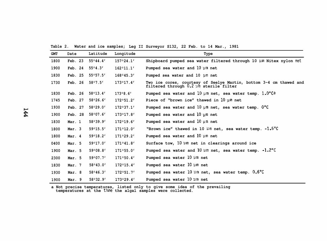
Table 2. Water and ice samples; Leg II Surveyor S132, 22 Feb. to 14 Mar., 1981
GMT Date Latitude Longitude Type
1800
1900
1830
1730
18301745
1930
1900
1830
1800
1800
0400
1900
2300
1830
1930
1900
Feb. 23
Feb. 24
Feb. 25
Feb. 26
Feb. 26Feb. 27
Feb. 27
Feb. 28
Mar. 1
Mar. 3
Mar. 4
Mar. 5
Mar. 5
Mar. 5
Mar. 7
Mar. 8
Mar. 9
55°44.4’
55°4.3’
55°57.5’
58°7.5’
58°13.4’58°26.6’
58°29.0’
58°07.6’
58°39.9’
59°15.5’
59°18.2’
59°17.0’
59°08.8’
59°07.7’
58°43.0’
58°46.3’
58°32.9’
157°24.1’
162°11.1’
168°45.3’
173°17.4’
173°8.6’172°51.2’
172°37.1’
173°17.8’
172°19.6’
171°12.0’
171°29.2’
171°41.8’
171°55.0’
171°50.4’
172°15.4’
172°51.7’
173°29.4’
Shipboard pumped sea water filtered through 10 Vm Nitex nylon net
Pumped sea water and 10~m net
Pumped sea water and 10 pm net
Two ice cores, courtesy of Seelye Martin, bottom 3-4 cm thawed andfiltered through 0.2um sterile filter
Pumped sea water and 10pm net, sea water temp. l.O°CaPiece of “brown ice” thawed in 10~m net
Pumped sea water and 10pm net, sea water temp. O“C
Pumped sea water and 10~m net
Pumped sea water and 10Um net
“Brown ice” thawed in 10 urn net, sea water temp. -1.5°C
Pumped sea water and 10Um net
Surface tow, 10um net in clearings around ice
Pumped sea water and 10um net, sea water temp. -1.2°C
Pumped sea water 10vm net
Pumped sea water 10pm net
Pumped sea water 10vm net, sea water temp. 0.6°C
Pumped sea water 10um net
a Not precise temperatures, listed only to give some idea of the prevailingtemperatures at the time the algal samples were collected.

dihydrate; 0.004 g FeC13.6H20; 0.034 g H3B03; 0.004 g MnC12.4H20; 670 g
ZnSo4.7H20; 38 g Na2Mo04.2H20; 12 g COC12.6H20; 0.3 g CUS04.5H20
and supplemented with 0.125 g Na2Si03.9H20, 300 g thiamine; 8 g vitamin
B12; 30 g biotin. The enrichment cultures were incubated in continuous
light at -1 to O“C in the shipboard flowing water system (one overhead
fluorescent fixture) or at 5 to 7°C in a refrigerator (one 40W tungsten
lamp). The cultures were frequently examined microscopically and
transferred to fresh medium as appropriate. Unialgal cultures were
purified by repeated streaking on petri dishes containing agarized (1% low
gelling temperature agar, No. A4018, Sigma Chemical Co., St. Louis, MO)
medium composed of Ifi offshore Gulf of Mexico sea water plus 1/2 medium
KASP-2 . The dishes were incubated in continuous fluorescent or tungsten
light in sealed plastic containers in an atmosphere of 0.5 to 1% C02-in-air
at 5 or 10”C. After 5 to 15 days suitable micro-colonies were excised,
transferred to agar slants, and examined for purity microscopically and in
the basal medium supplemented with complex organic materials; 0.1% each of
yeast extract, trypticase, and soytone (all products of Difco Laboratories,
Detroit, MI). Stock cultures were routinely maintained as slants in a
refrigerator at 5-10° with illumination provided by one 40W tungsten lamp
25 to 40 cm from the cultures.
The cultures were kindly identified by Professor Qi Yu-zao of the
Department of Biology, Jinan University, Guangzhou, P.R.C. as;
Thalassiosira sp. (our notation D1-2), Navicula sp. (J-4), Nitzschia
sp. (K3-3), Chaetocerous sp. (K3-1O) and KD-50). Organisms K3-10 and KD-50
isolated from two different samples may be the same species, tentatively
145

C. laciniosus Schutt$ but they were sufficiently different in physiology to—
warrant experimentally being considered two different organisms. It should
be noted that these diatoms isolated from the enrichment cultures, while
certainly not all the organisms present, were common in numerous fresh
samples examined on shipboard.
The light-temperature gradient plate (7) was used to survey the
general growth characteristics of the isolates from6 to 22°C (Fig. 2).
All the cultures were clearly cold-adapted. Only one strain, the
Chaetocerous sp. (K3-1O) , grew well at 18°C. The optimum temperatures were
from 10 to 14°C. It was not practical to operate the light-temperature
gradient plate below 6°C nor was the plate useful for measuring growth
rates. Growth rates were therefore measured in liquid cultures at O or
10°C (Table 3). Four of the isolates, KD-50, K3-10, K3-3, and J-4
maintained reproducible generation times at O°C of from 5 to 7 days. At
10”C the growth rates were 1 to 21/2 days. The Thallassiosira sp. (111-2)
grew at such a slow rate even at 10*C as to preclude useful experimental
work. Organism K3-3 was found to require vitamin B12, organism J-4 was
stimulated by vitamin B12. The other cultures grew without added vitamins.
Of particular interest were the exceedingly slow growth rates, especially
at O°C. We have looked for chemical or physical factors having significant
effect on the growth rate. Light and dark cycles (18L:6D) or addition of
reduced nitrogen, NH4C1 or organic nitrogen in the form of casamino acids,
had little effect. The choice of lamps, deluxe
fluorescent lamps shielded by one screen to cut
basis of extensive early screening of different
warm-white phosphor
intensity, was made on the
combinations of phosphors

100 LOW LIGHT1
TEMPERATURE (“C)
Fig. 2. Relative growth of ice edge diatoms as a function of temperatureand light intensity. The aluminum light-temperature gradient plate was46x63.5x1.27 cm. It was illuminated by two rows (2 lamps per row placedend to end) of F20T12-WWX lamps placed 34 cm above the front edge. Thelight intensity over the front edge of the plate was 420vw/cm2 (Model 65Radiometer, YSI Co., Yellow Springs, OH). Pyrex petri dishes, 60x15 mm,containing 10 ml of medium (1/2 KASP-2 plus 1/2 sea water) plus inoculum,were placed at desired locations on the plate. Growth was judged visuallyor optically, if dense enough. For each organism the data were recordedrelative to the position on the plate which gave the best growth. Theexperiments were purposely terminated after 9-12 days at relatively lowcell densities to avoid severe C02 or light limitations on growth. Thenotation NG means no growth.
147

Table 3. Growth rates, as generation times in hours, of diatoms isolated
from the ice edge in the Bering Sea and inhibition of their growth by two
crude oils.
Temperature (°C)o 10 0 10
Strain No. Cook Inlet Crude (PP ) Prudhoe Bay Crude (PP )o 50 500 0 5: 500 50 500 50 50:
KD-50 144 144 0 30 33 36 0 0 42 72
K3-10 120 120 0 30 30 39 120 0 36 55
K3-3 144 0 0 48 48 NG* 0 0 45 NG
J-4 170 ND+ ND 60 60 60 ND ND 72 103
*NG means no growth. t ND means not determined. Continuous illumination
was provided by two F20T12/WWX fluorescent lamps 10 cm from the lamp center
to the growth tube center. Lamp output was cut to approximately 60% by one
copper screen inserted between the lamps and the growth bath. Temperatures
were held to + 0.2 at l°C and~O.5 at 10”C. The growth tubes were
continuously bubbled with 1 ~ 0.1% C02 in air, cell concentration was
measured turbidimetrically or by collecting cells on a 0.4 Urn filter and
drying at 45°C in a vacuum oven over P205. The crude oils were sterilized
by filtration with pressure (N2) through 0.45 pm silver membranes (Selas
Corp., Dresher, PA). The crude oil was absorbed onto washed 12.7 mm filter
paper discs and the discs placed directly into the culture tubes. Crude
oils presented in this manner remain absorbed on the discs and in contact
with the algae (15). The generation times shown are conservatively good to
+ 15%.
148

and intensities. Moreover short-time photosynthesis measurements (14C02
fixation) carried out under these same lighting conditions gave linear and
saturated rates of C02 uptake over several hours. By several fundamental
criteria of algal culture, cell density and elementary analysis, these
cultures are behaving as expected. Cell yields of 0.5mg dry weight
ml-l were routinely achieved. The elemental analysis of organism KD-50
grown at 0° or 10”C was: %C, 32.29 and 32.91; %H, 4.99 and 4.98; %N, 5.16
and 5.42, %residue, 34.9 and 30.9. On an ash-free basis these values
compare very favorably with a variety of algal cells (8).
There are, then, two very interesting features which emerge from the
characterization of growth in these ice edge diatoms. First, these
organisms fit the textbook definition of obligate psychrophiles, micro-
organisms that can grow well at O°C and that do so optimally below 20°C
(9). In other words these are not just mesophilic forms capable of growth
at O“C but with optimum temperatures above 20”C, but rather strains
restricted to temperatures below 18°C (Fig. 2). Their second significant
characteristic was their exceedingly slow measured generation times, 5 to 7
days at O“C. Such very slow generation times are not anticipated from the
existing large body of information primarily on mesophilic microalgae (10).
Indeed a theoretical treatment of algal growth rates versus temperature
predicted generation times approaching 1 day at O“C (11). In work with
unialgal (bacterized) cultures of four Arctic ice diatoms at 5°C generation
times of 1 to 2 days were found (12). A unialgal strain of Skeletonema
costatum, a typical mesophilic form, had an estimated generation time of
approximately 2 days at O“C (13).

The generation times measured herein at O°C with pure cultures of
cold-adapted diatoms appear to be the first of their kind. The very slow
growth rates at O°C may perhaps be a reflection of one or several enzymes
with unavoidably low turnover times at O°C. However, the very marked
increase in the volubility of oxygen at low temperatures may cause special
problems for a photosynthetic cell, for example, with the oxygenase
reaction catalyzed by ribulose l,5-bisphosphate carboxylase (14). If
generation times approaching one week are typical under supposedly optimum
conditions in the lab for ice edge algae then their turnover times in situ——
may be much lower. These unique Arctic (probably Antarctic as well) ice
phytoplankton and hence these ecosystems may truly merit the appellation of
fragile.
Notwithstanding the slow growth rates of these psychrophilic
diatoms, we have been able to grow enough cells to examine their capacity
for oxidation of aromatic hydrocarbons using naphthalene as a model
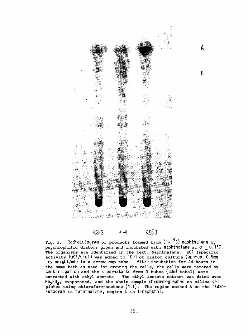
substrate. Figures 3 and 4 demonstrate that l-naphthol was formed from
(1-14C) naphthelene at O or 10”C. The amounts were very smal’
and suggest that cold-adapted microalgae can oxidize aromatic
as is now well-described in mesophilic forms (see page 1).
but are rea
hydrocarbons
The observations on the toxicity of crude oils (Table 3) also
suggest that cold-adapted diatoms will generally prove more sensitive to
any accidental crude oil spills in or around the ice edge in the Bering
Sea. Lethality was evident in two of the diatoms, KD-50 and K3-3 at 50 ppm
at O*C, while 500 ppm was lethal to all four organisms. At 10”C toxicity
was lessened. For comparison the same Prudhoe Bay crude had no effect at
150
. .-..
.“
. .
B
K3-3 J -~ K!150Fig. 3. Radioautogram of products formed from (1-’4C) naphthalene bypsychrophilic diatoms grown and incubated with naphthalene at O ~O.l°C.The organisms are identified in the text. Naphthalene, lpCi (specificactivity lpCi/umol) was added to 10ml of diatom culture (approx. 0.5mgdryweight/ml) in a screw cap tube. After incubation for 24 hours inthe same bath as used for growing the cells, the cells were removed bycentrifugation and the supernatants from 3 tubes (30ml total) wereextracted with ethyl acetate. The ethyl acetate extract was dried overNa2S04, evaporated, and the whole sample chromatographed on silica gelplates using chloroform-acetone (4:1). The region marked A on the radio-autogram is naphthalene, region E? is l-naphthol.
151

A
B
Figure 4. Radioautogram of products formed from (1-14C) naphthalene bypsychrophilic diatoms grown and incubated with naphthalene at 10 ~0.5°C.Experimental details were the same as in Figure 3.
152

500 ppm and caused only slight lags “
30°C against three mesophilic algae,
d i a t o m ( 1 5 ) .In work with four unia’
n growth at 1500 ppm when tested at
a blue-green alga, a green alga, or a
gal cultures isolated from the
southern Beaufort Sea growth of diatoms and a green flagellate was markedly
inhibited by crude oil concentrations higher than 100 ppm but diatoms
seemed more sensitive than the green
work greater inhibition was observed
between 5 to 10”C than at O°C.
The capacity for oxidation of
flagellate (16). Curiously, in this
with longer exposure at temperatures
aromatic hydrocarbons and enhanced
toxicity of crude oil in psychrophilic diatoms may, in the case of an oil
spill, be important to maintenance of primary production levels and
therefore to higher trophic levels in the Bering Sea. These observations
need broader confirmation both in laboratory and field studies.
With the enrichment and isolation in pure culture of these
psychrophilic Arctic diatoms, especially with the easily cultivated
Nitzschia sp. (K3-3’) and the Chaetocerous sp. (K3-1O, KD-50) as
experimental tools, we should now gain further understanding of regulation
of photosynthetic and biosynthetic pathways in cold-adapted microalgae.

REFERENCES AND NOTES
1. J.S. Bunt and E.J.F. Wood, Nature 199, 1254 (1963);— —
M.J. Dunbar, J. Fish. Res. Board Can. 32, 2276 (1975);— — — — — .
R.S. Homer, Oceanogr. Mar. Biol. Ann. Rev. 14, 167 (1976);— — — — .
K. Saito and A. Taniguchi, Asarte 11, 27 (1978).— —
2. J.S. Bunt, Nature 199, 1255 (1963);— —
3. C.P. McRoy and J.J. Goering, in Oceanography of the Bering Sea, D.W.— — — —
Hood and E.J. Kelley, Eds. (Institute of Marine Science, Fairbanks,
1974), p. 403.
4. C.H. Pease and R.D. Muench, Coastal Oceangr. Climatology News 3, 43— -
(1981).
5. W.J. Campbell and S. Martin, Science 181, 56 (1973);
L.S. Wolfe andD.P. Hoult, J. Glaciol. ~, 473 (1974);—
D.U. Hood and J.A. Calder, in The Eastern Bering Sea Shelf:——
Oceanography and Resources, D.W. Hood and J.A. Calder Eds. (Office of
Marine Pollution Assessment, NOAA) p. 1299 (1981).
6. C. Van Baalen, Botanica Mar. 4, 129, (1962). Medium KASP-2 derived—-
from medium ASP-2 described in this paper.
7. C. Van Baalen and P. Edwards, in Handbook of Phycological Methods,—
Culture Methods and Growth Measurements Methods, J.R. Stein, Ed..—
(University Press, Cambridge, 1973), p. 267.
154

8. B. Kok, Acts Bet. Neerl. 1,445 (1952).—— — -
C. Van Baalen and J.E. Marler, J. Gen. Microbial. ~, 457 (1962).——
C. Van Baalen, D.S. Hoare, E. Brandt, J. Bacteriol. 105, 685 (1971).—
P.J. Bottomley and C. Van Baalen, J. Gen. Microbiol. 107, 309 (1978)..—
The microanalysis were done by Galbraith Laboratories, Inc., Knoxville,
TN.
9. J.L. Ingraham and J.L. Stokes, Bact. Revs. ~, 97 (1959);——
W.E. Inniss, Ann. Rev. Microbiol. 29, 445 (1975);—— —
R.Y. Morita, Bact. Revs. 3&, 144 (1975).— —
10. C. Van Baalen, in Handbook of Microbiology, A.I. Laskin and H.A.—
Lechevalier Eds. (CRC Press, Cleveland, OH, 1974), vol IV, p. 21.
11. R.W. Eppley, Fishery Bull.~, 1063 (1972).
12. W.S. Grant andR.A. Homer, J. Phycol. 12 180 (1976).— — —
13. J.A. Yoder, J. Phycol. ~, 362 (1979).—
14. G.H. Lorimer, Ann. Rev. Plant Physiol. ~, 349 (1981).. — —
15. J.C. Batterton, K. Winters, C. Van Baalen, Mar. Environ. Res. ~, 31
(1978).
16. S.I.C. Hsiao, Environ. Pollut. ~, 93 (1978).
17. We are grateful to Captain Bruce Williams and the crew of the R/VSURVEYOR for their invaluable help in collecting the samples. Wethank Seelye Martin for providing several ice cores. The sample ofCook Inlet crude oil was kindly provided by James R. Payne and thePrudhoe Bay sample by C.P. Falls.
155

RATE STUDIES OF 1-NAPHTHOL FORMATION IN MESOPHILIC ALGAE
To determine if microalgae can degrade naphthalene to C02 we have
incubated a blue-green alga, a green alga, and a diatom in closed flasks at
30°C with (1-14C) naphthalene and recovered C02 from the gas phase by
precipitation as BaC03. The BaC03 was carefully washed with water, ethanol
and again with water, then acidified and any radioactivity trapped in 5 ml
of O.lN NaOH. Part of the NaOH solution was added to scintillation
cocktail and counted. The above procedure completely eliminated any carry
over of naphthalene. Recoveries using NaH14C03 carried through the
precipitation , washing, acidification and trapping in NaOtl steps were 90%
or better. We have not found any evidence that the above cultures can
metabolize naphthalene to 14C02. we have examined the time course of
l-naphthol formation in the blue-green alga, Oscillatoria sp. our strain
JCM (Fig. 5). We estimate from such data that strain JCM can form
20 nmol of l-naphthol per mg dry weight of cells in 24 hours. If we assume
that the experimentally measured algal rate formation of l-naphthol can be
equated with bacterial hydrocarbon biodegradation rates (Bartha and Atlas,
1977) then at the reasonable level of 1 pg chlorophy’
system algal hydrocarbon oxidation can amount to 10%
marine potential.
1 ~/liter in a natura
of the “in situ”——
156

1oo-1- Naphthol
75-
50- 4 ‘Hydroxy - I ‘Tetralone
25-
0///0 I I I I I 1
0 4 8 12 16 20 24TIME (HOURS)
Figure 5. Time course of formation of l-naphthol and4-hydroxy-l-tetralone by the blue-green alqa,Oscillatoria sp. strain JChl.

REFERENCE
Bartha, R. and Atlas, R.M. (1977). The Microbiology of Aquatic Oil Spills.
Adv. Appl. Microbial. ~, 225-266.
158