Embed Size (px)
Citation preview
REVIEW
Biodegradable magnesium implants for orthopedic applications
Hazibullah Waizy • Jan-Marten Seitz • Janin Reifenrath •
Andreas Weizbauer • Friedrich-Wilhelm Bach • Andrea Meyer-Lindenberg •
Berend Denkena • Henning Windhagen
Received: 9 March 2012 / Accepted: 9 May 2012
� Springer Science+Business Media, LLC 2012
Abstract The clinical application of degradable ortho-
pedic magnesium implants is a tangible vision in medical
science. This interdisciplinary review discusses many dif-
ferent aspects of magnesium alloys comprising the manu-
facturing process and the latest research. We present the
challenges of the manufacturing process of magnesium
implants with the risk of contamination with impurities and
its effect on corrosion. Furthermore, this paper provides a
summary of the current examination methods used in in
vitro and in vivo research of magnesium alloys. The
influence of various parameters (most importantly the
effect of the corrosive media) in in vitro studies and an
overview about the current in vivo research is given.
Introduction
The quality and speed of bone healing depends on the size
of the fracture gap and the achieved stability [1]. Therefore,
an adjusted stability is essential for fracture healing. Early
recovery after bony healing is necessary to prevent long-
term disabilities and hospitalization periods, as well as the
risk of malunion, nonunion, and infection. Intramedullary
nailing and the usage of pins, screws, or plates are accepted
approaches for fracture stabilization. Stainless steel and
titanium are the most frequently used materials in medical
applications in the last few decades. These currently
applied permanent metallic internal fixation devices have
several negative aspects such as stress shielding [2, 3], an
inflammatory osteolysis caused by released toxic titanium
particles [4], interference in radiological studies [5], and
the need of a second surgery for implant removal.
In recent years, biodegradable biomaterials for medical
use have gained interest and are intensively investigated;
promising candidates are magnesium alloys. These bio-
materials have to comply several requirements: (i) good
biocompatibility and non-toxicity of degradation products,
(ii) appropriate mechanical properties, and (iii) a moderate
degradation rate adapted to the fracture healing process.
Mg2? is an essential component of the human body and it
is mainly stored in bones. Many enzymes require magne-
sium as a co-factor to provoke a chemical reaction for
example DNA replication [6]. The recommended daily
intake of magnesium is approximately 300 mg for adults
[6]. Human adult blood plasma obtains a total Mg con-
centration of 0.65–1.05 mmol/l [7].
In addition, magnesium possesses desirable mechanical
properties (density: 1.74 g/cm3; elastic modulus 45 GPa,
and compressive yield strength 65–100 MPa) closer to
those of natural bone than currently used titanium alloys
H. Waizy (&) � A. Weizbauer � H. Windhagen
Department of Orthopaedic Surgery, Hannover Medical School,
Anna-von-Borries-Str.1-7, 30625 Hannover, Germany
e-mail: [email protected]
J.-M. Seitz � F.-W. Bach
Institute of Materials Science, Leibniz University of Hannover,
An der Universitat 2, 30823 Garbsen, Germany
J. Reifenrath
Small Animal Clinic, University of Veterinary Medicine
Hannover, Bunteweg 9, 30559 Hannover, Germany
A. Meyer-Lindenberg
Clinic for Small Animal Surgery and Reproduction, Centre of
Clinical Veterinary Medicine, Faculty of Veterinary Medicine
Ludwig-Maximilians-Universitat Munchen, Veterinarstr. 13,
80539 Munich, Germany
B. Denkena
Institute of Production Engineering and Machine Tools (IFW),
Leibnitz University of Hannover, An der Universitat 2, 30823
Garbsen, Germany
123
J Mater Sci
DOI 10.1007/s10853-012-6572-2

[4, 8]. These mechanical properties of magnesium mini-
mize the disturbance of bone growth and remodeling by
reduced stimulation (‘‘stress-shielding’’) [2]. An approach
of enhancing the mechanical properties for clinical appli-
cations is alloying [9]. Most commonly used for alloying
are elements like calcium, lithium, zinc, zirconium, rare
earth metals, or aluminum [10]. Some of these elements,
most notably aluminum and rare earth metals, are sus-
pected to cause adverse effects in organism [11, 12].
In physiological fluids, magnesium and its alloys
degrade according to the following reactions [13]:
Anodic reaction : Mg ! Mg2þ þ 2e�
Cathodic reaction : 2 H2O þ 2e� ! H2 " þ 2 OH�
Mg2þ þ 2 OH� ! Mg OHð Þ2 sð Þ
The corrosion of magnesium and its alloys are accompa-
nied by hydrogen-gas development. The production of high
amounts of gas in a short period of time is not desired for
clinical application. The gas development is dependent on
the corrosion rate. One of the first clinical applications of
pure magnesium implants was in 1906 by Lambotte [14].
The results of Lambotte at this early stage of magnesium
research were not satisfactory and the pure magnesium
plates were removed due to extensive gas cavities, local
swelling, and heavy pain [14].
In this review, we provide a survey about many different
aspects of recent magnesium alloys research and the dif-
ficulties in the magnesium manufacturing process by con-
tamination primarily with iron, nickel, and copper and its
effect on the corrosion rate. A series of different in vitro
studies investigated the degradation behavior of magne-
sium alloys. The influence of various parameters (most
importantly the choice of the corrosive media) is presented
in that paragraph. In vivo evaluation of magnesium alloy
samples plays an important role and the current state of
research is given.
Challenges in magnesium alloying: the risk
of contamination with impurities
Although pure magnesium demonstrates generally suitable
corrosion properties as an implant material for resorbable
applications, it frequently possesses insufficient mechani-
cal properties. One possibility of enhancing its mechanical
properties is represented by the use of magnesium alloys.
By means of alloying with suitable alloying elements, it is,
for instance, possible to eliminate the mechanical defi-
ciencies of pure magnesium. Here, however, it must be
taken into consideration that such elements simultaneously
modify the material’s corrosion behavior (rate and type of
corrosion) [15].
As a rule, the effects which the alloying elements pro-
duce in this respect are deliberate and intentionally
obtained. However, such effects can also be unintentionally
generated by means of contaminants during the implants
manufacture. Elements such as iron, nickel, and copper,
which are recurrently found in magnesium alloys are fre-
quently unintentional and due to the manufacturing process
[16]. In this respect, iron in particular plays a significant
role owing to its frequently inevitable contact with mag-
nesium alloys during their manufacture. During the melting
and stirring of the melt in the steel crucible, which occa-
sionally contains nickel, the iron and nickel contents can
precipitate out of the crucible’s wall and thus end up in the
magnesium’s melt in which it represents just one constit-
uent of the alloy [17, 18]. Contaminants containing copper
mainly arise by employing ‘‘impure’’ aluminum alloying
elements [19]. The solubility characteristics of the con-
taminating elements in magnesium are as follows:
The equilibrium diagram (Fig. 1) for magnesium and
iron shows a eutectic at 650 �C which lies close to the
melting point of magnesium. The solubility of iron in
magnesium is limited to 0.008 at.% within this eutectic.
Above this, the element exists as alpha-iron in the mag-
nesium matrix [20]. Typical melting or, as the case may be,
casting temperatures for alloying magnesium lie in the
range of 650 and 700 �C depending on the added alloying
elements. Table 1 depicts the fraction of dissolved iron
from various sources at precisely these temperatures. The
fractions range from 0.0044 to 0.0218 at.%. The solubility
of nickel in magnesium is reported to be very small and lies
significantly below 0.04 at.% at a temperature of 500 �C
[21, 22]. Likewise, the solubility of copper in magnesium is
presented as extremely small. Here, the solubility is
between a range of 0.153 and 0.191 at.% at a temperature
of 485 �C [23].
Fig. 1 Curve to illustrate the tolerance limit for a contamination of
magnesium with the elements Fe, Ni, Cu, according to [15]
J Mater Sci
123
Even in small amounts, iron, copper, and nickel as
constituents in pure magnesium lead to an increase in the
corrosion rate. It was possible for Ref. [15, 24, 25] to
demonstrate this in comparison with other elements (Ag,
Ca, Zn, Cd, Sn, Pb, Al,…). The corrosion rates of binary
alloys were examined in (3 % NaCl) saline solution. In
doing this, concentrations of iron, copper, and nickel,
smaller than 0.2 %, lead to a significant increase in the rate
of corrosion [15, 26]. This increase of the corrosion rate is
attributed to the elements’ low solubility in magnesium as
well as their distinctly more noble position in the electro-
chemical series [15, 27, 28]. A particularly large impact on
the corrosion rate of magnesium and its alloys is accepted
as a contamination by iron, if nothing else, because of its
frequent occurrence. Here, a galvanic couple is formed in
the magnesium’s microstructure in which the undissolved
iron particles within the magnesium matrix represents the
cathodic pole and reduces the neighboring magnesium
phases [29, 30]. Owing to the low solubility of nickel in
magnesium, the effect on the corrosion rate is described in
even more drastic terms since elementary, galvanic cells
are already formed early without the ability of compen-
sating for these cells in the form of phases [31, 32].
Although copper contaminants also demonstrate a signifi-
cant influence on accelerating corrosion, it must, however,
exist in higher doses owing to its good solubility in mag-
nesium [33].
To be able to define tolerable amounts of contaminating
elements in magnesium, the concept of tolerance limits is
introduced. According to this, the tolerance limit is given at
a location at which the contaminant’s concentration leads
to a significant acceleration of corrosion (Fig. 1). Before
reaching the tolerance limit due to contaminating elements,
the corrosion rate appears low and thereby tolerable [15].
In purely binary alloys of as-cast magnesium and iron,
nickel, or copper, the tolerance limits result as depicted in
Table 2. By alloying with a third element, the measured
tolerances can, in part, be considerably shifted. As an
example with respect to this, on combining magnesium,
aluminum, and iron, a Fe–Al phase is formed which
reduces the mitigation of tolerable amounts of iron in the
magnesium alloy to 5 ppm (for 7 % aluminum). The rea-
son for this is the presence of the FeAl3 phase which is
more electrochemically unfavorable for magnesium alloys
and, in comparison with pure iron particles, acts more
nobly and therefore more corrosion intensively in magne-
sium [34, 35].
The corrosion promoting properties of iron, nickel, and
copper contaminants within magnesium alloys require
counteractive measures to enable the material’s properties
to be consistent and reproducible. In order to prevent the
introduction of iron and nickel quantities during manu-
facturing by casting technology, one can dispense with
materials for the crucible, stirrer, and dies which contain
such elements. Here, materials based on titanium represent
an already real but expensive alternative. On the other
hand, it is also possible to exploit the properties of addi-
tional alloying elements. One such alloying element is
manganese which can effectively reduce the effect of the
iron content on the corrosion of magnesium materials [16].
At a higher temperature than that at which casting is per-
formed, quantities of manganese are added to the melt.
During the cooling phase down to the casting temperature,
intermetallic phases now form from the added manganese.
The iron contaminants which, owing to their density, are
deposited onto the crucible’s base and the iron concentra-
tion in the melt are lowered [36, 37]. Moreover, it is
assumed that the added manganese envelops the iron par-
ticles and thus deprives them of direct contact with mag-
nesium. In this way, only galvanic cells arise between the
manganese and the magnesium which, owing to the dif-
ference of the chemical standard potential, are less corro-
sion intensive as cells consisting of magnesium and iron
[18, 37, 38]. Using the same method, constituents of nickel
can also be bound and eliminated from the cast. However,
this is not considered as an effective procedure for manu-
facturing a highly pure magnesium alloy. In a series of
investigations using AZ alloys, even the smallest amounts
(\1 %) of manganese could lead to a significant
improvement in corrosion behavior by which they raised
the tolerance limits for both iron (20 wt.ppm) and nickel
contaminants [18, 31, 38, 39]. It was possible to relate the
corrosion behavior of magnesium alloys and, in particular,
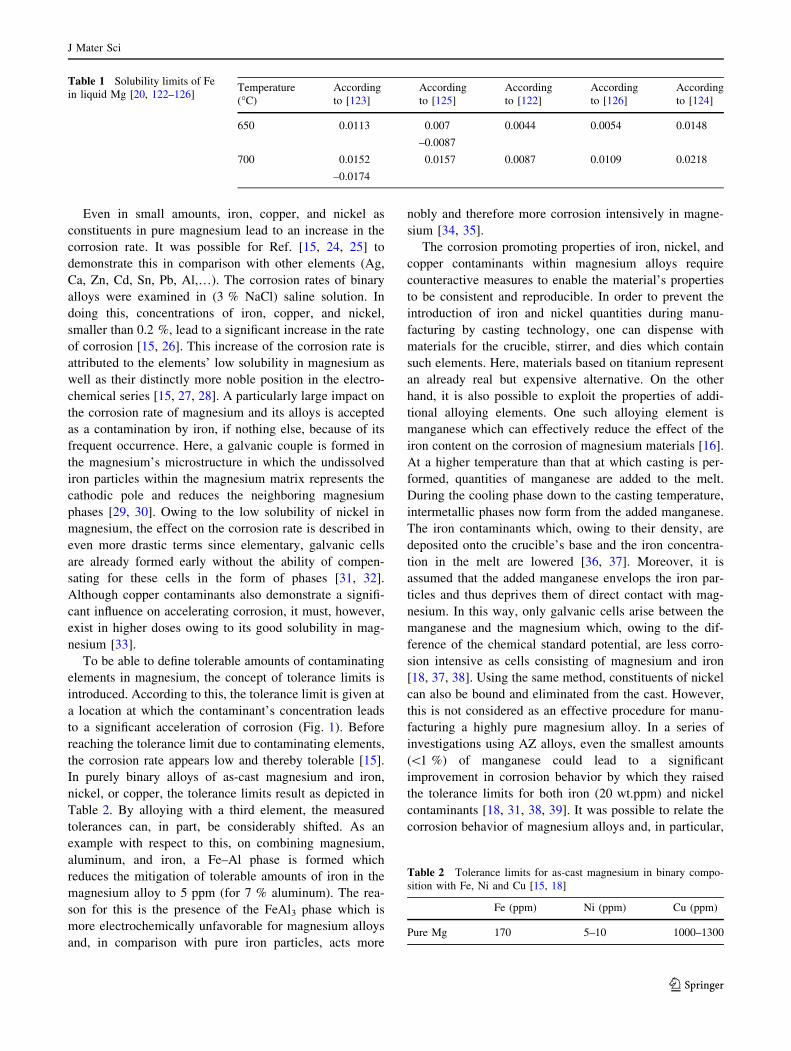
Table 1 Solubility limits of Fe
in liquid Mg [20, 122–126]Temperature
(�C)
According
to [123]
According
to [125]
According
to [122]
According
to [126]
According
to [124]
650 0.0113 0.007 0.0044 0.0054 0.0148
–0.0087
700 0.0152 0.0157 0.0087 0.0109 0.0218
–0.0174
Table 2 Tolerance limits for as-cast magnesium in binary compo-
sition with Fe, Ni and Cu [15, 18]
Fe (ppm) Ni (ppm) Cu (ppm)
Pure Mg 170 5–10 1000–1300
J Mater Sci
123
the Fe/Mn ratio. In general, for large values of Fe/Mn
ratios, a high corrosion potential could be verified,
whereas, for small values, a slow corrosion behavior
appeared uniformly over the surface [30, 40]. It was pos-
sible to identify the relationship between the Fe/Mn ratio
and the tolerance limit for iron by means of an AZ91 alloy
[40]. Following the evaluation of the salt spray test, a linear
relationship was clearly demonstrated between the iron/
manganese ratios and the resulting tolerance limit for iron.
However, the most effective method to be able to control
the unfavorable influence of contaminants of iron and
particularly of nickel and copper currently consists of
resorting to highly pure alloying additives [16].
Owing to its numerous alloying elements and their com-
paratively high quantities, the production of the LAE442
alloy harbors a huge risk of becoming contaminated by
nickel, copper, and iron. As described above, it is possible
that additional amounts of copper are introduced into the
alloy via the alloying element aluminum [19]. Moreover, the
alloying element, aluminum, lowers the tolerance limit of
magnesium alloys for iron to about 20 wt% with which it
forms a strongly cathodic acting Al–Fe compound [29, 31].
The largest risk of contaminating with iron results from the
casting process since steel crucibles, steel stirrers, and steel
dies are conventionally employed for producing the non-
commercially available LAE442 alloy.
Surface treatments and subsequent processes like hot
extruding and rolling in general show improving impacts
on the corrosion performances of Mg and Mg alloys [41–
44]. Surface treatments like protective films and coatings
(e.g., resorbable polymers, resorbable hydroxyapatites, and
epoxy resins) prevent the substrate magnesium from direct
exposition to surrounding fluids (electrolytes) [15, 42, 43,
45–47]. The protective surface films improve the electro-
chemical behavior by surface passivation which should,
even in case the impurity tolerance limits of the substrate
material are exceeded, delay corrosion. Corrosion mecha-
nisms which are dependant on an electrolyte should be
permanently or temporarily (depending on the coating)
inhibited, while contact corrosion which exists due to the
different phases still remains [15].
Another alternative to optimize the corrosion properties
of Mg and its alloys is to use altered casting processes [48]
and/or subsequent processes like extruding [41] or rolling
[44]. Here, grain refinement occurs having a significant
impact on the metal’s corrosion properties [41, 44, 48]. In
general, grain boundaries act as physical corrosion barriers;
a smaller grain size increases the amount of grain bound-
aries which furthermore decreases the rate of corrosion
[49]. However, it remains unclear if finer grains positively
affect corrosion which occurs due to impurities. In this
case, phases of impurity contact a higher number of adja-
cent Mg grains inflicting contact corrosion on them.
In vitro evaluation of magnesium alloys
In vitro experiments provide the opportunity to examine
newly developed magnesium alloys under standardized
conditions before testing in animals. These studies are
mainly carried out (i) to test cytotoxicity or (ii) to inves-
tigate the corrosion behavior.
In vitro cytotoxicity evaluations are useful for assessment
of feasible destructive effects of magnesium alloys and its
degradation products on cell viability and proliferation.
Surveys of the different tests are given in Table 3. MTT and
XTT assays of corrosion extracts are most often performed
to quantify cell viability using metabolic markers though it is
reported that the interference between test reagent and cor-
rosion of some alloys restrict this method [10, 50]. These
assays are in accordance with EN ISO 10993-5 which
describes tests for in vitro cytotoxicity for the evaluation of
medical devices. In few studies, the cytotoxicity is evaluated
by the determination of live cells via trypan blue exclusion
method or via cell adhesion by DAPI staining [51–53]. SEM
is often applied to analyze the cell morphology on the surface
[52, 54]. The assessment of certain cell proteins and DNA
content is sometimes applied such as the expression of the
osteogenetic-specific m-RNA (collagen 1a1, alkaline phos-
phatase ALP, and osteocalcin OC) [54, 55].
Different cell lines are used to evaluate the cytotoxicity of
magnesium alloys via in vitro experiments (Table 3). The
choice of a specific cell line is important for the performance
of cytotoxicity tests [56]. A variety of cell lines are used with
different origin (animal or human). Most of the cells that are
used for the evaluation of orthopedic magnesium implants
are osteoblasts and fibroblasts. Fischer et al. [57] compared
cytotoxicity tests with cells with cancerous origin (human
osteosarcoma cell line MG63) to an isolated primary cell line
of human osteoblasts. The results showed that the tolerance
toward magnesium extracts was higher for osteoblasts in
comparison to MG63 [57]. Feyerabend et al. [56] reported
similar observations investigated in rare earth elements
(REE) on primary mesenchymal stem cells, MG63 and a
RAW 264.7 tumor-derived mouse cell line. The higher tol-
erance of the primary mesenchymal cell line toward REE-
salts was attributed to the direct impact on cancerous cell
lines [56]. It is considered to perform cytotoxicity tests with
human bone-derived cells or mesenchymal stem cells
because of their high osteogenic differentiation potential [56,
57]. These findings are in contrast to cell lines recommended
by EN ISO 10993-5.
Many in vitro studies were performed to determine the
corrosion behavior of the investigated magnesium alloys.
Electrochemical and gravimetric measurements are fre-
quently applied to study the degradation behavior.
Immersion tests are often performed to determine the
hydrogen evolution rate [10]. Song reported that the
J Mater Sci
123
evolution of 1 ml H2 corresponds to the dissolution of 1 mg
magnesium [58]. In few studies, the degradation rate was
calculated according to the weight loss method [51, 52,
59]. Sometimes the Mg2? release from the alloy was
measured by colorimetric method using xylidyl blue-I or
via inductively coupled plasma atomic emission spectros-
copy (ICP-AES) [9, 55, 60, 61]. These in vitro methods are
useful tools to determine the corrosion rate under stan-
dardized physiological conditions. A critical discussion of
the limitations of these methods and their benefit was
compiled by Kirkland et al. [62]. Many previous studies
have shown that the biocorrosion rates displayed by in vitro
and in vivo tests are difficult to correlate with faster cor-
rosion rates in vitro [63, 64]. Recent researches have shown
that this challenge in spite of optimizing in vitro experi-
ments still exists [65]. Although in vitro studies are
insufficient to reliably predict in vivo corrosion rates, these
studies are suitable for the comparison of different alloys
under standardized conditions. A survey of a variety of
commercial and experimental alloys in minimum essential
medium (MEM) at 37 �C was given by Ref. [66].
Microstructural characterizations of the surfaces are
most commonly determined by scanning electron micros-
copy (SEM) and energy-dispersive X-ray spectroscopy
(EDS) [10]. In some studies, the grain size of the alloy is
determined according to the linear intercept method
described in ASTM E112-96 [55, 61].
The influence of the corrosion medium
Many factors have an influence on the corrosion behavior,
most importantly the choice of the corrosive medium.
Previous in vitro studies were performed with NaCl solu-
tion, phosphate buffer solution (PBS), simulated body fluid
(SBF), Hank’s solution, Dulbecco’s modified eagle med-
ium (DMEM), Ringer’s solution, or MEM. The indigents
of five solutions are given in Ref. [50]. A summary of the
corrosion media used in previous works is given in
Table 4. The composition of the various corrosive media is
crucial and varies in the concentration of aggressive ions
(e.g., Cl-), buffering agents, and additives like proteins or
organic acids. Some studies investigated the influence of
the addition of proteins on the corrosion rate. Albumin is
the most abundant protein in the human blood with a
molecular weight of 66 kDa [67, 68]. The adsorption of
proteins on the surface is complex including van der Waals
forces, hydrophobic and electrostatic interactions, and
hydrogen bonding [69]. Some authors reported a decreased
corrosion rate of MgCa alloy with the addition of proteins
[51, 70, 71]. Yamamoto et al. [60] observed a decreased
Mg2? release of pure magnesium in the beginning of
immersion and a lower total release after 14 days com-
pared to solutions without protein. In agreement, this initial
corrosion protective effect of an albumin layer in SBF was
observed in other studies as well [72, 73]. The hydrogen
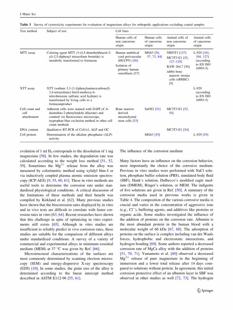
Table 3 Survey of cytotoxicity experiments for evaluation of magnesium alloys for orthopedic applications excluding coated samples
Test method Subject of test Cell lines
Human cells of
non cancerous
origin
Human cells
of cancerous
origin
Animal cells of
non cancerous
origin
Animal cells
of cancerous
origin
MTT assay Coloring agent MTT (3-(4,5-dimethylthiazol-2-
yl)-2,5-diphenyl tetrazolium bromide) is
metabolic transformed to formazan
Human umbilical
cord perivascular
(HUCPV) [56]
Isolation of
primary human
osteoblasts [57]
MG63 [56,
57, 73, 84]
NIH3T3 [127]
MC3T3-E1 [52,
127–129]
RAW 264.7 [56]
rabbit bone
marrow stroma
cells (rBMSC)
[9]
L-929 [101,
104, 127]
(according
to EN ISO
10993-5)
XTT assay XTT (sodium 3,3-{1-[(phenylamino)carbonyl]-
3,4-tetrazolium}-bis(4-methoxy-6-
nitro)benzene sulfonic acid hydrate) is
transformed by living cells to a
formazanproduct
L-929
(according
to EN ISO
10993-5)
Cell count and
cell
attachement
Adherent cells were stained with DAPI (4’,6-
diamidino-2-phenylindole dilactate) and
counted via fluorescence microscope,
tryptophan blue exclusion method or other cell
count methods
Bone marrow
derived
mesenchymal
stem cells [53]
SaOS2 [51] MC3T3-E1 [52,
54]
DNA content Qualitative RT-PCR of Col1a1, ALP and OC MC3T3-E1 [54]
Cell protein Determination of the alkaline phosphatse (ALP)
activity
MG63 [55] L-929 [55]
J Mater Sci
123
evolution rate of the M1A alloy was suppressed in the first
4 h; however, the long-term degradation rate was higher
with albumin [73]. Similar results were reported in the first
hours of exposure to the albumin containing medium with
WE43. The similar electrochemical behavior after 9 h in
SBF with and without protein indicated that the protein
layer is either destroyed or no longer dominant [72]. The
influence of different amount of proteins in the corrosive
solution was investigated by various studies [67, 71, 74].
Liu et al. [71] reported an accelerated corrosion inhibition
effect at higher albumin concentrations (10 g/l). In addi-
tion, Feyerabend et al. [75] reported that the Mg2? release
from the metal was increased with the addition of proteins.
Many corrosive media are buffered to avoid change of
the pH value. The choice and amount of buffer consider-
ably affects the corrosion rate. HEPES, TRIS, phosphate
buffers, and HCO3-/CO2 are widely used [68]. A study
with NaCl and PBS solution showed that PBS decreases the
corrosion rate after long term observation because of the
precipitation of phosphate containing salts on the surface.
This precipitation layer contributes to higher corrosion
resistance than magnesium hydroxide in NaCl solution
[76]. The study of Yamamoto et al. [60] investigated the
effect of the buffering reagents HEPES (N-2-hydro-
xyethylpiperazine-N0-2-ethane sulfonic acid) or NaHCO3
(sodium bicarbonate) added to NaCl on degradation
behavior. It was observed that the Mg2? release steeply
decreased in the first 2 days in NaCl ? NaHCO3 in con-
trast to NaCl and NaCl ? HEPES, which indicates the
precipitation of insoluble carbonates on the surface of
magnesium in NaCl ? NaCO3. The total amount of
released Mg2? ions after 14 days immersion was higher in
NaCl solution with HEPES than with NaHCO3. Similar
results were found in a study with different concentrations
of the buffer agent HCO3-. This study showed decreased
corrosion rate of pure magnesium with higher concentra-
tions of HCO3- in SBF. The initial degradation rate was
lower with higher buffering reagents content [77]. Contrary
results were presented in a study by Xin et al. [78] with
AZ91. The initial hydrogen evolution rate of the two
solutions with HCO3- was higher which indicated an
accelerated consumption of OH- by HCO3- which pro-
motes the dissolution reaction of magnesium. After 96 h,
the corrosion rate was more decreased in solutions with
HCO3- which was explained by protection effects of
phosphate and carbonate on the surface.
In vitro studies under cell culture conditions
and with CO2 gas application
Some studies were performed under cell culture conditions
(95 % humidity, 37 �C, 5 % CO2) [51, 53, 65, 66, 75, 79,
80]. This approach is intended to more preferably simulate
the environment in blood of in vivo studies. The main
buffering system in blood is HCO3- and dissolved CO2
[75]. Zainal Abidin et al. [81] maintained the pH value due
to constant CO2 gas application through the solution in
bubbles. In vitro studies have shown that the presence of
CO2 has a significant effect on the composition of the
corrosion product layer [75, 79]. The formation of MgCO3
and the precipitation of a crystalline structure were
observed [75, 79]. MgCO3 is suspected to possess corrosion
inhibition properties after the formation of a layer [75].
Influence of additional parameters: temperature, pH,
microstructure and flow velocity
Most of the studies were performed with a temperature in
the range of the human physiological temperature
(35.8–37.2 �C). Kannan et al. [82] reported a decreased
corrosion resistance of the AZ91 alloy at body temperature
compared to room temperature (20 ± 2 �C). Commonly,
the corrosion rate increases when the temperature is ele-
vated [15]. Magnesium and its alloy lead to an alkalization
of the corrosion solution due to the production of OH-
which causes an increase of pH value [83]. The corrosion
rate decelerated at pH values above 10.5 because of the
greater tendency for film formation [15].
Furthermore, the microstructure of the alloy caused by
the production process is important. It is reported that grain
refinement decreased the corrosion rate [55, 76, 84, 85].
Equal-channel angular pressing (ECAP) is previously used
to produce homogenous ultrafine grains [76, 84]. The
average grain size of the as-casted Mg–Zn–Zr was
76 ± 5 lm, the as-extruded alloy exhibit a grain size of
2.9 ± 5 lm [55]. In addition, calcium as an alloying ele-
ment also contributes to grain refinement [10]. In a previ-
ous study, a reduction of grain size from 175 ± 15 to
51 ± 5 lm was reported with increasing the Ca content in
Mg–Zn–Mn–Ca-alloy from 0.3 to 1.0 wt% [61].
The velocity of the corrosive media has a considerable
impact on the corrosion rate of magnesium alloys due to
destruction or prevention of a protective film [10, 15].
Waizy et al. [86] developed a corrosion system with a
constant flow rate of Hank’s solution corresponding to the
blood velocity in natural bone. In this in vitro model,
MgCa0.8 screws were tested in synthetic bone via
mechanical pull-out test. The results showed that the pull-
out force decreased of 30 % after 96 h in corrosive med-
ium compared to the non-corrosion group. A maximum
load capacity of 28 ± 7.6 N/h was determined. The bio-
mechanical data suggested that biodegradable screws pro-
vide a promising bone-screw fixation and have great
potential for medical application. A further study by Waizy
et al. [87] was also performed with a constant flow rate. In
this study, orthopedic ZEK100 plates were investigated
J Mater Sci
123

(Fig. 2). After the immersion test, high amounts of O, Mg,
Ca, P, and C were detected on the surface via EDX analysis
which was associated with the precipitation of carbonated
calcium phosphates (Mg,Ca)x(PO4)y(CO3)z(OH)i. The
samples were biomechanically tested via four-point-bend-
ing test [87]. These in vitro studies of orthopedic screws
and plates made of magnesium alloys should be supple-
mented by a forthcoming investigation of intramedullary
nails consisting of LAE442 (Fig. 2).
In conclusion, in vitro studies are useful to investigate
the degradation behavior of new biomedical magnesium
alloys under standardized conditions. The comparison of
the corrosion rate between different alloys is able to pro-
vide valuable information about its corrosion resistance
and behavior. Although in vitro studies attempt to simulate
the biological environment in vivo very closely, the testing
of the degradation in an animal study cannot be omitted
because of the various biological interactions with the
alloy.
In vivo examination of new implants
For the medical or veterinary application of new bioma-
terials, experimental animal testing is indispensable.
Adjacent to the biocompatibility of the newly developed
biodegradable material, degradation rate and mechanical
parameters are of special interest. Different animal models
are available for these questions [88, 89]. The mouse is
commonly used for studies of osteogenesis in bone or soft
tissue [88] and the rat and the guinea pig for simple implant
geometries [8, 90] or fracture models [88]. An alternative
animal model, which combines the easy handling as well as
the possibility to examine orthopedic implant geometries,
is the rabbit [91, 92]. Evaluation of the degradation
behavior of the implant can be performed as well as sur-
rounding tissue reactions [93–95]. In addition, adjacent to
the clinical examination, radiographic and in vivo lCT
examinations can be used to quantitatively assess the
implant degradation as well as changes in bone volume,
bone density, and bone porosity in the direct implant sur-
rounding [96]. After different postoperative observation
periods, histological examinations of the implant interface,
the bone, and the soft tissue around the implant can be
performed. Examination of degradation properties, with
higher resolution lCT and SEM are established methods to
verify the results of the radiographic and in vivo lCT
imaging [96]. For biomechanical analysis of the implant
material, three point bending tests [97–99] and pull-out
tests are available [94]. In addition, examinations of
regional lymph nodes [100] and inner organs [101] can
give evidence of systemic reactions due to the degradation
products and their elimination.
In vivo examination of different magnesium alloys
Whereas specific magnesium-based alloys are already used
in cardiology [102, 103], implant materials for orthopedic
applications in clinical use are not applicable yet. Different
alloys are examined as potential orthopedic implant
material in vivo. As single alloying elements adjacent to
magnesium, calcium [99, 104, 105], and zinc [101] were
tested so far. More than one alloying element is used in
implants with aluminum in combination with zinc [8, 95,
106, 107], calcium [96, 108], or together with lithium and
rare earths [8, 97–99, 109, 110] as in the examined mag-
nesium alloys WE43 [8, 90, 97] and MgZnxMny [101, 111],
ZX50, WZ21 [107], and MgBiCa [112]. Most authors
attest magnesium a good biocompatibility [8, 90, 98, 99,
101, 105, 107] with enhanced bone bonding to implant
surfaces in comparison to conventional used materials [90,
107, 113, 114] and a low inflammatory and immunogenic
potential [90, 93, 100, 101, 107, 111]. So far, only
LACer442 is not recommended for in vivo application due
to a too fast degradation rate in vivo [109]. In comparison
to other degradable materials like polymers, magnesium
alloys exceed in the biomechanical stability [115]. A
negative effect which is described in different studies is the
formation of gas cavities during the degradation process in
vivo. However, no clinical relevance could be observed
Fig. 2 Picture of different orthopedic implant geometries made of magnesium alloys. a MgCa0.8 screw b ZEK100-plate c intramedullary
LAE442-nail
J Mater Sci
123

[96, 116]. Until now, only a single in vivo study exists in
the available literature which describes full degradation of
magnesium-based implants after fixation bone. In this
study, Kraus et al. [117] observed complete bone recovery
even after severe bone affection due to massive gas for-
mation in a juvenile animal model. However, self-healing
properties are less in adults. Therefore, severe bone
affections as described there do not seem to be acceptable
for general orthopedic use. With exception to Kraus et al.
[117] and Castellani et al. [90] who used juvenile rats, all
other studies were performed with adult animal models [96,
99, 101, 104, 118, 119].
Own in vivo research results
In our research group, the magnesium alloys LAE442,
WE43, ZEK100, AX30, MgCa0.8 [96–99, 119], LACer442
[109], AL33 [120], and LANd442 [118] are examined yet.
Therefore, pins of each material (2.5 mm diameter, 25 mm
lengths) were implanted in rabbit tibiae and observation
periods of 3 and 6 months as well as 12 months for
MgCa0.8 and LAE442 were examined with clinical,
radiographical, and histological examinations. In some
alloys (AL33, ZEK100, AX30, and LANd442), in vivo
l-computed tomography with evaluation of changes in
bone volume, density, and porosity in the surrounding of
the implant material was performed (Fig. 3) [96, 118].
Implant degradation was characterized with ex vivo lCT,
volume and/or weight loss after explantation and in some
cases SEM [96, 97, 99, 119].
In addition, regional lymph nodes as well as inner
organs (liver, spleen, and kidney) were examined histo-
logically after implantation of LAE442 and MgCa0.8
implants in comparison to PLA as resorbable and titanium
as permanent implant material [100].
LACer442 and AL33 were excluded for further testing
due to their high degradation rate and their insufficient
biocompatibility [109, 120]. After six months implantation
time, with the exception of ZEK100, all other implant
materials still showed their cylindrical shape. ZEK100 and
LANd442 showed cleft surfaces and losses of implant
material in greater extent than in the other groups [96, 118,
121]. Pitting corrosion was seen as predominant type of
corrosion in MgCa0.8, AX30, LANd 442, and ZEK100
(Fig. 4) [96, 98, 118, 121] in contrast to WE43, which
showed soil-like ablations on the surface and LAE442
which was characterized by homogeneous fissured corro-
sion [97]. All the tested magnesium alloys caused a peri-
osteal increase in the mineral apposition rate, which was
calculated after intravital fluorescent labeling [98, 118]. An
increase in osteoclast activity was seen as well, which was
moderate in the slow degrading alloy LAE442 and higher
in the faster degrading alloys AX30, ZEK100 [98]
(Huhnerschulte, unpublished data). LANd442 caused high
Fig. 3 Trabecular bone formation at the implant interface (redarrow); l-computed tomographic picture
Fig. 4 Pitting corrosion at the surface of an explanted magnesium alloy (stereo-microscopy)
J Mater Sci
123
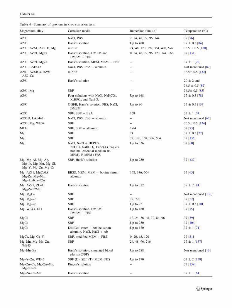
Table 4 Summary of previous in vitro corrosion tests
Magnesium alloy Corrosive media Immersion time (h) Temperature (�C)
AZ31 NaCl, PBS 2, 24, 48, 72, 96, 144 37 [76]
AZ31 Hank’s solution Up to 480 37 ± 0.5 [84]
AZ31, AZ61, AZ91D, Mg m-SBF 24, 48, 120, 192, 384, 480, 576 36.5 ± 0.5 [130]
AZ31, AZ91, MgCa Hank’s solution, DMEM and
DMEM ? FBS
0, 24, 48, 72, 96, 120, 144, 168 37 [131]
AZ31, AZ91, MgCa Hank’s solution, MEM, MEM ? FBS – 37 ± 1 [70]
AZ31, LAE442 NaCl, PBS, PBS ? albumin – Not mentioned [67]
AZ61, AZ61Ca, AZ91,
AZ91Ca
m-SBF – 36.5± 0.5 [132]
AZ91 Hank’s solution – 20 ± 2 and
36.5 ± 0.5 [82]
AZ91, Mg SBF – 36.5± 0.5 [85]
AZ91 Four solutions with NaCl, NaHCO3,
K2HPO4 and Na2SO4
Up to 168 37 ± 0.5 [78]
AZ91 C-SFB, Hank’s solution, PBS, NaCl,
DMEM
Up to 96 37 ± 0.5 [133]
AZ91 SBF, SBF ? BSA 168 37 ± 1 [74]
AZ91D, LAE442 NaCl, PBS, PBS ? albumin – Not mentioned [67]
AZ91, Mg, WE54 SBF – 36.5± 0.5 [134]
M1A SBF, SBF ? albumin 1-24 37 [73]
Mg SBF 24 37 ± 0.5 [77]
Mg SBF 72, 120, 168, 336, 504 37 [135]
Mg NaCl, NaCl ? HEPES,
NaCl ? NaHCO3, Earle(?), eagle’s
minimal essential medium (E-
MEM), E-MEM?FBS
Up to 336 37 [60]
Mg, Mg–Al, Mg–Ag,
Mg–In, Mg–Mn, Mg–Si,
Mg–Y, Mg–Zn, Mg–Zr
SBF, Hank’s solution Up to 250 37 [127]
Mg, AZ31, MgCa0.8,
Mg-Zn, Mg–Mn,
Mg–1.34Ca–3Zn
EBSS, MEM, MEM ? bovine serum
albumin
168, 336, 504 37 [65]
Mg, AZ91, ZE41,
Mg2Zn0.2Mn
Hank’s solution Up to 312 37 ± 2 [81]
Mg, MgCa SBF – Not mentioned [136]
Mg, Mg–Zn SBF 72, 720 37 [52]
Mg, Mg–Zn SBF Up to 72 37 ± 0.5 [101]
Mg, WE43, E11 Hank’s solution, DMEM,
DMEM ? FBS
Up to 180 37 [75]
MgCa SBF 12, 24, 36, 48, 72, 84, 96 37 [59]
MgCa SBF Up to 250 37 [104]
MgCa Distilled water ? bovine serum
albumin, NaCl, NaCl ? Ab
Up to 120 37 ± 1 [71]
MgCa, Mg–Ca–Y SBF, modified-MEM ? FBS 0, 20, 65, 120 37 [51]
Mg–Mn, Mg–Mn–Zn,
WE43
SBF 24, 48, 96, 216 37 ± 1 [137]
Mg–Mn–Zn Hank’s solution, simulated blood
plasma (SBP)
Up to 288 Not mentioned [13]
Mg–Y–Zn, WE43 SBF (H), SBF (T), MEM, PBS Up to 170 37 ± 2 [138]
Mg–Zn–Ca, Mg–Zn–Mn,
Mg–Zn–Si
Ringer’s solution – 37 [139]
Mg–Zn–Ca–Mn Hank’s solution – 37 ± 1 [61]
J Mater Sci
123
osteoclastic activity although the degradation rate was
moderate in comparison to the other alloys [118, 121].
Owing to profuse bone reactions caused by the implant
material with bone resorption and periosteal bone prolif-
eration, ZEK100, AX30, and LANd442 could not be rec-
ommended for orthopedic application in weight bearing
bones [96, 118]. Promising implant materials after
6 months implantation time were MgCa0.8 and LAE442
[97, 109]. Therefore, longer implantation periods were
performed [99]. After 12 months, MgCa0.8 implants could
only be taken out in sections with exception of one pin with
deep pits of corrosion in contrast to LAE442 which
remained the cylindrical shape to a large extend and
showed fissured corrosion [99]. Bone reactions as sign of
biocompatibility were less in the groups with the slower
and more uniform degrading implant materials, particularly
in the LAE442 group [98, 99].
Systemic reactions could not be observed in any of the
examined degrading magnesium alloys. Even in LANd442,
which is not recommended due to insufficient biocompati-
bility, an increase of IL6 as parameter for systemic inflam-
matory reactions could not be observed [118]. Examined
inner organs (liver, spleen, and kidney) did not show any
histological changes. In regional lymph nodes of animals
with MgCa0.8 and LAE442 implants, non-specific immu-
nological reactions could be found as predominantly foreign
body reactions and were less than in regional lymph nodes of
the clinical accepted biomaterials titanium and PLA [100].
For the use in weight bearing bones, the good biome-
chanical properties are essential as well. Pull-out tests in
the rabbit tibia, with MgCa0.8 as screw material in com-
parison to surgical steel, were performed. MgCa0.8 screws
showed stable pull-out forces during an implantation per-
iod of 4 weeks, followed by a decrease from week six,
which was significant after week eight [94]. It is ques-
tionable if this fast reduction in pull-out forces is sufficient
for the clinical use but MgCa0.8 might be an appropriate
material for screws with less weight bearing. Indeed, the
slower degrading material LAE442 might have a slower
decrease in pull-out forces, and therewith might be an
appropriate implant material even for screws and plates
which has to be examined.
For intramedullary nailing, especially, a high initial
strength is necessary, which should decrease during the
implantation period to avoid stress-shielding effects.
Among the tested materials, LAE442 showed the highest
initial maximum forces in the three point bending test
followed by ZEK100 and WE43 [96, 98].
Summarizing, the alloy LAE442 is the most promising
implant material in our in vivo studies. Following steps
will be to examine orthopedic systems like bolted intra-
medullary nails or screw-plate systems to evaluate the
degradation properties at the interfaces of the material as
well as the bone reactions to higher amounts of degrading
implant material.
It will be the aim of further studies to examine complex
implants made of LAE442 in the sheep as large animal
model. Another approach will be to substitute the rare earth
composition metal by exact defined REE to improve the
reproducibility of the material, and therewith improve the
prediction of degradation rate, mechanical stability, and
biocompatibility.
Acknowledgements The authors gratefully acknowledge the
financial support given by German research society (DFG) within the
collaborative research project (SFB 599). We thank Christopher
Muller for the design of Fig. 2.
Reference
1. Chao EY, Aro HT, Lewallen DG, Kelly PJ (1989) Clin Orthop
Relat Res 241:24
2. Nagels J, Stokdijk M, Rozing PM (2003) J Shoulder Elbow Surg
12:35
3. Uhthoff HK, Finnegan M (1983) J Bone Joint Surg Br 65:66
4. Staiger MP, Pietak AM, Huadmai J, Dias G (2006) Biomaterials
27:1728
5. Sullivan PK, Smith JF, Rozzelle AA (1994) Plast Reconstr Surg
94:589
6. Hartwig A (2001) Mutat Res 475:113
7. Saris N-EL, Mervaala E, Karppanen H, Khawaja JA, Lewen-
stam A (2000) Clin Chim Acta 294:1
8. Witte F, Kaese V, Haferkamp H, Switzer E, Meyer-Lindenberg
A, Wirth CJ, Windhagen H (2005) Biomaterials 26:3557
9. Huan ZG, Leeflang MA, Zhou J, Fratila-Apachitei LE, Dus-
zczyk J (2010) J Mater Sci Mater Med 21:2623
Table 4 continued
Magnesium alloy Corrosive media Immersion time (h) Temperature (�C)
Mg–Zn–Zr Hank’s solution, DMEM,
DMEM ? FBS
Up to 480 37 ± 0.5 [55]
Mg–Zn–Zr, WE43 Hank’s solution Up to 3528 (21 weeks) 37 [9]
WE43 SBF Up to 654 37 ± 2 [140]
WE43 NaCl, NaCl ? CaCl2,
NaCl ? K2HPO4, m-SFB,
m-SFB ? albumin
Up to 120 37 [72, 141]
J Mater Sci
123
10. Witte F, Hort N, Vogt C, Cohen S, Kainer KU, Willumeit R,
Feyerabend F (2008) Curr Opin Solid State Mater Sci 12:63
11. El-Rahman SSA (2003) Pharmacol Res 47:189
12. Yumiko N, Yukari T, Yasuhide T, Tadashi S, Yoshio I (1997)
Fund Appl Toxicol 37:106
13. Yang L, Zhang E (2009) Mater Sci Eng C 29:1691
14. Witte F (2010) Acta Biomater 6:1680
15. Song GL, Atrens A (1999) Adv Eng Mater 1:11
16. Westenge H, Aune TK (2005) Magnesium technology: metal-
lurgy, design data, applications, 1st edn. Springer, Berlin
17. Lunder O, Nisancioglu K, Hansen RS (1993) SAE Special Publ
962:117
18. Makar GL, Kruger L (1993) Int Mater Rev 38:138
19. Buhler K (1990) Metallurgy 44:748
20. Nayeb-Hashemi AA, Clark JB (1985) Bull Alloy Phase Diagr
6:235
21. Haughton JL, Payne RI (1943) J Inst Met 54:275
22. Nayeb-Hashemi AA, Clark JB (1985) Bull Alloy Phase Diagr
6:238
23. Hansen M (1927) J Inst Metal 37:93
24. Hanawalt JD, Nelson CE, Peloubet JA (1942) Trans AIME
147:273
25. Hillis JE, Reichek KN (1986) SAE technical paper series
#860288
26. Hillis JE, Murray RW (1987) SDCE 14th International die cast
congress and exposition paper no. G-T87-003
27. Froats A, Aune TK, Hawke D, Unsworth W, Hillis JE (1987)
Metals handbook, 1st edn. ASM Int Material Park, Ohio
28. Olsen AL (1991) Translation of paper presented at the Baut-
eil’91. DVM, Berlin
29. Lunder O, Lein JE, Aune TK, Nisancioglu K (1989) Corrosion
45:741
30. Nisancioglu K, Lunder O, Aune TK (1990) Proceedings of the
47th World Magnesium Association, McLean
31. Emley EF (1966) Principles of magnesium technology, 1st edn.
Pergamon Press, New York
32. Hawke D (1975) SYCE 8th International Die Casting Exposition
and Congress Paper No. G-T75-114
33. Tawil DS (1987) Magnesium technology. In: Proceedings of the
Conference of the institute of metals. The Institute of Metals,
London
34. Hake D (1975) SYCE 8th international die casting exposition
and congress paper no. G-T75-114
35. Loose WS (1946) Corrosion and protection of magnesium, 1st
edn. ASM International, Ohio
36. Nelson CE (1944) Trans AIME 159:392
37. Robinson H-A, George PF (1954) Corrosion 10:182
38. Polmaer IJ (1992) Physical metallurgy of magnesium alloys.
DGM Informationsgesellschaft, Oberursel
39. Hillis JE (1983) SAE technical paper #830523
40. Reichek KN, Clark KL, Hillis JE (1985) SAE technical paper
series #850417
41. Ben Hamu G, Eliezer D, Wagner L (2009) J Alloys Compd
468:222
42. Chiu KY, Wong MH, Cheng FT, Man HC (2007) Surf Coat
Technol 202:590
43. Hiromoto S, Yamamoto A (2009) Electrochim Acta 54:7085
44. Wang H, Estrin Y, Fu G, Song G, Zuberova Z (2007) Adv Eng
Mater 9:967
45. Gray JE, Luan B (2002) J Alloys Compd 336:88
46. Song G (2005) Adv Eng Mater 7:563
47. Tamar Y, Mendler D (2008) Electrochim Acta 53:5118
48. Ambat R, Aung NN, Zhou W (2000) Corros Sci 42:1433
49. Aung NN, Zhou W (2010) Corros Sci 52:589
50. Xin Y, Hu T, Chu PK (2011) Acta Biomater 7:1452
51. Li Y, Hodgson PD, Wen C (2011) J Mater Sci 46:365. doi:
10.1007/s10853-010-4843-3
52. Zhang S, Li J, Song Y, Zhao C, Zhang X, Xie C, Zhang Y,
Tao H, He Y, Jiang Y, Bian Y (2009) Mater Sci Eng C 29:
1907
53. Johnson I, Perchy D, Liu H (2011) J Biomed Mater Res A
100A:477
54. Chen D, He Y, Tao H, Zhang Y, Jiang Y, Zhang X, Zhang S
(2011) Int J Mol Med 28:343
55. Gu XN, Li N, Zheng YF, Ruan L (2011) Mater Sci Eng B
176:1778
56. Feyerabend F, Fischer J, Holtz J, Witte F, Willumeit R, Drucker
H, Vogt C, Hort N (2010) Acta Biomater 6:1834
57. Fischer J, Profrock D, Hort N, Willumeit R, Feyerabend F
(2010) Mater Sci Eng B 176:830
58. Song G (2007) Corros Sci 49:1696
59. Harandi SE, Idris MH, Jafari H (2011) Mater Des 32:2596
60. Yamamoto A, Hiromoto S (2009) Mater Sci Eng C 29:1559
61. Zhang E, Yang L (2008) Mater Sci Eng A 497:111
62. Kirkland NT, Birbilis N, Staiger MP (2012) Acta Biomater
8:925
63. Mueller W-D, Nascimento ML, de Mele MF (2010) Acta Bio-
mater 6:1749
64. Witte F, Fischer J, Nellesen J, Crostack HA, Kaese V, Pisch A,
Beckmann F, Windhagen H (2006) Biomaterials 27:1013
65. Walker J, Shadanbaz S, Kirkland NT, Stace E, Woodfield T,
Staiger MP, Dias GJ (2012) J Biomed Mater Res B 100:1134
66. Kirkland NT, Lespagnol J, Birbilis N, Staiger MP (2010) Corros
Sci 52:287
67. Mueller W-D, de Mele MF, Nascimento ML, Zeddis M (2008) J
Biomed Mater Res A 90A:487
68. Virtanen S (2011) Mater Sci Eng B 176:1600
69. Roach P, Farrar D, Perry CC (2005) J Am Chem Soc 127:8168
70. Kirkland NT, Birbilis N, Walker J, Woodfield T, Dias GJ,
Staiger MP (2010) J Biomed Mater Res B 95:91
71. Liu CL, Wang YJ, Zeng RC, Zhang XM, Huang WJ, Chu PK
(2010) Corros Sci 52:3341
72. Rettig R, Virtanen S (2008) J Biomed Mater Res A 85:16773. Wang Y, Lim CS, Lim CV, Yong MS, Teo EK, Moh LN (2010)
Mater Sci Eng C 31:579
74. Liu CL, Xin Y, Tian X, Chu PK (2007) J Mater Res 22:1806
75. Feyerabend F, Drucker H, Laipple D, Vogt C, Stekker M, Hort
N, Willumeit R (2012) J Mater Sci Mater Med 23:9
76. Alvarez-Lopez M, Pereda MD, del Valle JA, Fernandez-Lore-
nzo M, Garcia-Alonso MC, Ruano OA, Escudero ML (2010)
Acta Biomater 6:1763
77. Xin Y, Hu T, Chu PK (2011) Corros Sci 53:1522
78. Xin Y, Huo K, Tao H, Tang G, Chu PK (2008) Acta Biomater
4:2008
79. Willumeit R, Fischer J, Feyerabend F, Hort N, Bismayer U,
Heidrich S, Mihailova B (2011) Acta Biomater 7:2704
80. Kirkland NT, Waterman J, Birbilis N, Dias G, Woodfield TB,
Hartshorn RM, Staiger MP (2012) J Mater Sci Mater Med
23:283
81. Zainal Abidin NI, Atrens AD, Martin D, Atrens A (2011) Corros
Sci 53:862
82. Kannan MB, Raman RK (2009) J Biomed Mater Res A
93A:1050
83. Song G, Song S (2007) Adv Eng Mater 9:298
84. Gu XN, Li N, Zheng YF, Kang F, Wang JT, Ruan L (2011)
Mater Sci Eng B 176:1802
85. Kannan MB (2010) Mater Lett 64:739
86. Waizy H, Weizbauer A, Maibaum M, Witte F, Windhagen H,
Lucas A, Denkena B, Meyer-Lindenberg A, Thorey F (2011) J
Mater Sci Mater Med 23:649
J Mater Sci
123
87. Waizy H, Weizbauer A, Modrejewski C, Witte F, Windhagen H,
Lucas A, Kieke M, Denkena B, Behrens P, Meyer-Lindenberg
A, Bach F-W, Thorey F (2012) Biomed Eng Online 11:12
88. An YH, Friedman RJ (1998) Animal models of orthopedic
research, 1st edn. CRC Press, Boca Raton
89. Pearce AI, Richards RG, Milz S, Schneider E, Pearce SG (2007)
Eur Cell Mater 13:1
90. Castellani C, Lindtner RA, Hausbrandt P, Tschegg E, Stanzl-
Tschegg SE, Zanoni G, Beck S, Weinberg AM (2011) Acta
Biomater 7:432
91. Bostman O, Paivarinta U, Partio E, Vasenius J, Manninen M,
Rokkanen P (1992) J Bone Joint Surg Am 74:1021
92. Chen YQ, Dai KR, Qiu SJ, Zhu ZA (1994) Chin Med J (Engl.)
107:766
93. Erdmann N, Bondarenko A, Hewicker-Trautwein M, Angrisani
N, Reifenrath J, Lucas A, Meyer-Lindenberg A (2010) Biomed
Eng Online 9:63
94. Erdmann N, Angrisani N, Reifenrath J, Lucas A, Thorey F,
Bormann D, Meyer-Lindenberg A (2011) Acta Biomater 7:1421
95. Huang J, Ren Y, Jiang Y, Zhang B, Yang K (2007) Front Mater
Sci Chin 1:405
96. Huehnerschulte TA, Angrisani N, Rittershaus D, Bormann D,
Windhagen H, Meyer-Lindenberg A (2011) Materials 4:1144
97. Krause A, von der Hoh N, Bormann N, Krause C, Bach F-W,
Windhagen H, Meyer-Lindenberg A (2010) J Mater Sci 45:624.
doi:10.1007/s10853-009-3936-3
98. Reifenrath J, Bormann D, Meyer-Lindenberg A (2011) Mag-
nesium alloys—corrosion and surface treatments, 1st edn. In-
tech, Rijek
99. Thomann M, Krause C, Bormann D, von der Hoh N, Windhagen
H, Meyer-Lindenberg A (2009) Mat-wiss Werkst 40:82
100. Bondarenko A, Hewicker-Trautwein M, Erdmann N, Angrisani
N, Reifenrath J, Meyer-Lindenberg A (2011) Biomed Eng
Online 10:32
101. Zhang S, Zhang X, Zhao C, Li J, Song Y, Xie C, Tao H, Zhang
Y, He Y, Jiang Y, Bian Y (2010) Acta Biomater 6:626
102. Erbel R, Di MC, Bartunek J, Bonnier J, de BB, Eberli FR, Erne
P, Haude M, Heublein B, Horrigan M, Ilsley C, Bose D, Koolen
J, Luscher TF, Weissman N, Waksman R (2007) Lancet
369:1869
103. Waksman R, Erbel R, Di MC, Bartunek J, de BB, Eberli FR,
Erne P, Haude M, Horrigan M, Ilsley C, Bose D, Bonnier H,
Koolen J, Luscher TF, Weissman NJ (2009) JACC Cardiovasc
Interv 2:312
104. Li Z, Gu X, Lou S, Zheng Y (2008) Biomaterials 29:1329
105. von der Hoh N, Bormann D, Lucas A, Denkena B, Hacken-
broich C, Meyer-Lindenberg A (2009) Adv Eng Mater 40:88
106. Witte F, Reifenrath J, Muller PP, Crostack HA, Nellesen J, Bach
F-W, Bormann D, Rudert M (2006) Mat-wiss Werkst 37:504
107. Witte F, Ulrich H, Palm C, Willbold E (2007) J Biomed Mater
Res A 81:757
108. Lalk M, Reifenrath J, Rittershaus D, Bormann D, Meyer-Lin-
denberg A (2010) Mat-wiss Werkst 41:1025
109. Reifenrath J, Krause A, Bormann D, von Rechenberg B,
Windhagen H, Meyer-Lindenberg A (2010) Mat-wiss Werkst
41:1054
110. Witte F, Fischer J, Nellesen J, Vogt C, Vogt J, Donath T,
Beckmann F (2010) Acta Biomater 6:1792
111. Xu L, Yu G, Zhang E, Pan F, Yang K (2007) J Biomed Mater
Res A 83:703
112. Remennik S, Bartsch I, Willbold E, Witte F, Shechtman D
(2011) Mater Sci Eng B 176:1653
113. Revell PA, Damien E, Zhang XS, Evans P, Howlett CR (2004)
Key Eng Mater 254–256:447
114. Zreiqat H, Howlett CR, Zannettino A, Evans P, Schulze-Tanzil
G, Knabe C, Shakibaei M (2002) J Biomed Mater Res 62:175
115. Shikinami Y, Okuno M (1999) Biomaterials 20:859
116. Witte F, Ulrich H, Rudert M, Willbold E (2007) J Biomed Mater
Res A 81:748
117. Kraus T, Fischerauer SF, Hanzi AC, Uggowitzer PJ, Loffler JF,
Weinberg AM (2012) Acta Biomater 8:1230
118. Hampp C, Ullmann B, Reifenrath J, Angrisani N, Dziuba D,
Bormann D, Seitz J-M, Meyer-Lindenberg A (2011) Adv Eng
Mater 14:B28
119. Thomann M, Krause C, Angrisani N, Bormann D, Hassel T,
Windhagen H, Meyer-Lindenberg A (2010) J Biomed Mater Res
A 93:1609
120. Rittershaus D, Ullmann B, Bormann D, Meyer-Lindenberg A
(2011) EuroBioMat E 51
121. Ullmann B, Reifenrath J, Dziuba D, Seitz J-M, Bormann D,
Meyer-Lindenberg A (2011) Materials 4:2197
122. Beerwald A (1944) Metallwirtschaft 23:404
123. Fahrenhorst E, Bulian W (1941) Z Metallkd 33:31
124. Mitchell DW (1948) Trans AIME 175:570
125. Siebel G (1948) Z Metallkd 39:22
126. Yensen TD, Ziegler NA (1931) Trans AIME 95:313
127. Gu X, Zheng Y, Cheng Y, Zhong S, Xi T (2009) Biomaterials
30:484
128. Datta M, Chou D-T, Hong D, Saha P, Chung SJ, Bouen L,
Sirinterikci A, Ramanathan M, Roy A, Kumta PN (2011) Mater
Sci Eng B 176:1637
129. Park RS, Kim YK, Sook JL, Jiang Y, Park IS, Yun YH, Bae TS,
Lee MH (2012) J Biomed Mater Res B 100B:911
130. Wen Z, Wu C, Dai C, Yang F (2009) J Alloys Compd 488:392
131. Gu XN, Zheng YF, Chen LJ (2009) Biomed Mater 4:0650111
132. Kannan MB, Raman RK (2008) Biomaterials 29:2306
133. Xin Y, Hu T, Chu PK (2010) J Electrochem Soc 157:C238
134. Walter R, Kannan MB (2010) Mater Lett 65:748
135. Wang Y, Wei M, Gao J, Hu J, Zhang Y (2008) Mater Lett
62:2181
136. Wan Y, Xiong G, Luo H, He F, Huang Y, Zhou X (2008) Mater
Des 29:2034
137. Xu L, Zhang E, Yin D, Zeng S, Yang K (2008) J Mater Sci
Mater Med 19:1017
138. Hanzi AC, Gerber I, Schinhammer M, Loffler JF, Uggowitzer PJ
(2010) Acta Biomater 6:1824
139. Rosalbino F, De NS, Saccone A, Angelini E, Delfino S (2010) J
Mater Sci Mater Med 21:1091
140. Hanzi AC, Gunde P, Schinhammer M, Uggowitzer PJ (2009)
Acta Biomater 5:162
141. Rettig R, Virtanen S (2009) J Biomed Mater Res A 88:359
J Mater Sci
123