Embed Size (px)
Citation preview
This article was downloaded by: [University of Cambridge]On: 15 October 2014, At: 11:42Publisher: Taylor & FrancisInforma Ltd Registered in England and Wales Registered Number: 1072954 Registered office: Mortimer House,37-41 Mortimer Street, London W1T 3JH, UK
HFSP JournalPublication details, including instructions for authors and subscription information:http://www.tandfonline.com/loi/tfls19
Biocrystallography: Past, present, futureRichard Giegé a & Claude Sauter ba Architecture et Réactivité de l'ARN , Université de Strasbourg , CNRS, IBMC, 15 rue RenéDescartes, Strasbourg, 67084, France E-mail:b Architecture et Réactivité de l'ARN , Université de Strasbourg , CNRS, IBMC, 15 rue RenéDescartes, Strasbourg, 67084, France E-mail:Published online: 07 Sep 2010.
To cite this article: Richard Giegé & Claude Sauter (2010) Biocrystallography: Past, present, future, HFSP Journal, 4:3-4,109-121
To link to this article: http://dx.doi.org/10.2976/1.3369281
PLEASE SCROLL DOWN FOR ARTICLE
Taylor & Francis makes every effort to ensure the accuracy of all the information (the “Content”) containedin the publications on our platform. However, Taylor & Francis, our agents, and our licensors make norepresentations or warranties whatsoever as to the accuracy, completeness, or suitability for any purpose of theContent. Any opinions and views expressed in this publication are the opinions and views of the authors, andare not the views of or endorsed by Taylor & Francis. The accuracy of the Content should not be relied upon andshould be independently verified with primary sources of information. Taylor and Francis shall not be liable forany losses, actions, claims, proceedings, demands, costs, expenses, damages, and other liabilities whatsoeveror howsoever caused arising directly or indirectly in connection with, in relation to or arising out of the use ofthe Content.
This article may be used for research, teaching, and private study purposes. Any substantial or systematicreproduction, redistribution, reselling, loan, sub-licensing, systematic supply, or distribution in anyform to anyone is expressly forbidden. Terms & Conditions of access and use can be found at http://www.tandfonline.com/page/terms-and-conditions
Biocrystallography: past, present, future
Richard Giegé1 and Claude Sauter1
1Architecture et Réactivité de l’ARN, Université de Strasbourg, CNRS, IBMC, 15 rue René Descartes,67084 Strasbourg, France
�Received 17 December 2009; accepted 2 March 2010; published online 22 April 2010)
The evolution of biocrystallography from the pioneers’ time to the present eraof global biology is presented in relation to the development of methodologicaland instrumental advances for molecular sample preparation and structureelucidation over the last 6 decades. The interdisciplinarity of the field thatgenerated cross-fertilization between physics- and biology-focused themes isemphasized. In particular, strategies to circumvent the main bottlenecks ofbiocrystallography are discussed. They concern „i… the way macromoleculartargets are selected, designed, and characterized, „ii… crystallogenesis andhow to deal with physical and biological parameters that impact crystallizationfor growing and optimizing crystals, and „iii… the methods for crystal analysisand 3D structure determination. Milestones that have marked the history ofbiocrystallography illustrate the discussion. Finally, the future of the field isenvisaged. Wide gaps of the structural space need to be filed and membraneproteins as well as intrinsically unstructured proteins still constitute challengingtargets. Solving supramolecular assemblies of increasing complexity, developinga “4D biology” for decrypting the kinematic changes in macromolecularstructures in action, integrating these structural data in the whole cellorganization, and deciphering biomedical implications will represent the newfrontiers. [DOI: 10.2976/1.3369281]
CORRESPONDENCE
Richard Giegé:
Claude Sauter:
Contemporary biocrystallographyis interdisciplinary, by essence, in com-bining biology with physics, chemistry,and engineering. The field has alwaysbeen knowledge driven by the need tovisualize and to comprehend the mol-ecules that underlie the basic life pro-cesses. This explains why its historyparalleled the highlights of biologicalresearch and has regularly been distin-guished by the Nobel Committee. Thus,in 2009 Venki Ramashrisknan, ThomasSteitz, and Ada Yonath shared theChemistry Nobel Prize for their con-tribution to the determination of thecrystal structure of the ribosome, themacromolecular machine that fabri-cates proteins (see comments by Carter,2009; Nierhaus, 2009). Because ofthis tribute, this essay will highlightdata on ribosomes and partners ofthe protein synthesis machinery thatcontributed to the development of mod-ern crystallography.
The field was also methodology andtechnology driven. The first examplesfrom the early ages concern the imple-mentation of appropriate methods tosolve structures (Arnold et al., 2010).Continuous developments have laterbeen focused on the improvement ofprotein expression and purification(Christendat et al., 2000; Koehn andHunt, 2009) as well as of crystallization(Sauter et al., 2010) and diffractiondata collection and processing (Arnoldet al., 2010). In this respect, the novelgenerations of synchrotron sources,of 3D graphics, and computing facili-ties for solving, building, and refiningstructures were essential. The fieldbenefited also from protein and nucleicacid sequencing and synthesis tech-nologies that provided the materialto be crystallized and the chemicalinformation to be fitted to the electrondensity maps.
HFSP Journal P E R S P E C T I V E
HFSP Journal © HFSP Publishing $25.00 109Vol. 4, Nos. 3-4, June-August 2010, 109–121 http://hfspj.aip.org
Dow
nloa
ded
by [
Uni
vers
ity o
f C
ambr
idge
] at
11:
42 1
5 O
ctob
er 2
014
Over the years with the increasing number of solved crys-tal structures, biocrystallography reached the mature age andtransformed into structural biology. Stimulated by the wealthof data originating from genomic programs, a new branchcalled structural genomics or structural proteomics emergedin the mid-1990s. It was based on a systematic high-throughput approach aimed to rapidly determine the en-semble of structures coded by selected genomes or belong-ing to specific biological functions or pathways (Terwilligeret al., 2009). In parallel, the interest in understanding the ar-chitecture, functioning, and dynamics of large supramolecu-lar assemblies, as well as ultrahigh-resolution of essentialstructures increased. As a result, new bottlenecks and chal-lenges appeared while the questions addressed in biocrystal-lography gained in complexity.
Figure 1 outlines the five steps that have to be mastered inorder to determine a 3D structure. They first concern thechoice of the most appropriate target macromolecule, itscloning, expression, purification, and assessment of purityin terms of chemical and conformational homogeneity.Although mainly dependent on biology methodologies,this step also requires bioinformatics and structure analysistools to select a native target or to design variants. Likewise,the next three steps dealing with crystallization and crystalcharacterization definitely make an extensive use of inter-disciplinary approaches (Sauter et al., 2010). They cover (i)the search of initial crystallization conditions by trial-and-error strategies using sparse matrix screening or rational-guided diagnostics, (ii) the optimization of crystal qualityby seeding, phase diagram exploration, or more advancedapproaches such as growth in diffusive media or in the pres-
ence of additives among them natural ligands or inhibitors,and (iii) the assessment of the diffraction properties of crys-tals such as resolution, mosaicity, and isotropy. Note thatat this stage, crystals also constitute interesting objects toinvestigate physics related issues—crystal perfection studiesby X-ray topography, rheology and other mechanical aspects,impurity inclusion, crystal surface poisoning, and crystalengineering—as well as in crystallo enzymology. These fourinitial steps are the main scope of biocrystallogenesis; thefield that has been developed since the late 1980s to rational-ize the preparation of well-diffracting crystals (McPhersonand Giegé, 2007). The ultimate step, that is the determinationand the analysis of the 3D structure, benefited as wellfrom constant methodological and instrumental innovations(Arnold et al., 2010). However, despite all the gained ex-pertise, a biocrystallographic project can be stuck at eachof these steps and overcoming the bottlenecks often requiresinventiveness and efforts. This essay will discuss thesedifferent aspects from the viewpoints of past, present, andfuture.
HISTORICAL BACKGROUND—FROM SMALLTO LARGEBiocrystallography started in the mid-1930s when it was re-alized that X-ray diffraction patterns recorded from macro-molecular crystals (Bernal and Crowfoot, 1934) containstructural information that can be translated in atomic mod-els of the crystalline macromolecules (Kendrew et al., 1958).The first bottleneck was the lack of suitable methods forstructure solving, in particular to overcome the phase prob-lem. As soon as these methods were developed and the first
Figure 1. Biocrystallography, the multidisciplinary route to the 3D vision of biological processes. �Left� main steps to a 3D crystalstructure. �Right� contribution of biocrystallography to the PDB. The plot illustrates the growth of the PDB content since 1975 �blue curve�and shows that the biocrystallography community is by far the strongest contributor �green curve� with X-ray structures solved by struc-tural genomics consortia already reaching 10% of the total �yellow curve�. These data were extracted from the PDB http://www.rcsb.org/pdb/
HFSP Journal
110 Biocrystallography: past, present, future | R. Giegé and C. Sauter
Dow
nloa
ded
by [
Uni
vers
ity o
f C
ambr
idge
] at
11:
42 1
5 O
ctob
er 2
014
structures solved, a dozen of structure determinations fol-lowed, comprising small proteins and enzymes as well asnucleic acid fragments (Fig. 2). They wonderfully confirmedthe models of �-helices and �-sheets in proteins (Paulingand Corey, 1951) and of the DNA double-helix (Watson andCrick, 1953). Over the years, biocrystallography targetsgained in size and complexity, covering soluble proteinsof increasing size, small RNAs, pieces of DNA and theircomplexes with proteins, spherical viruses with high intrin-sic symmetry, membrane proteins, and nucleoprotein com-plexes to culminate nowadays with assemblies as intricate asthe bacterial ribosome, a �2.3 MDa particle comprisingthree RNAs and �50 proteins, which structure was solved invarious forms without or with combinations of bound tRNA,mRNA, and antibiotics substrates. This diversity is depicted inFig. 2 by a series of emblematic milestone structures (see alsoSupplementary Material Table S1).
In the early time, a second bottleneck appeared rapidly,namely, how to grow “good” crystals of “biologically hot”macromolecules. In this respect, two methodological break-throughs were essential. The first one that paralleled the de-
velopment of X-ray methods occurred in the late 1960s andwas the implementation of micromethods allowing crystalli-zation trials in assays of 10–50 µl. This allowed solving struc-tures with sample quantities decreasing from more than 100 mgdown to less than 1 mg, nowadays, in most favorable cases(Supplementary Material Table S2). The second breakthroughcame in the early 1990s with the development of screening kitsto rapidly explore crystallization parameter-spaces (Jancarikand Kim, 1991) together with the generalized use of biotechno-logical tools for sample preparation and the availability of novelcomputing and synchrotron facilities.
During its rather short history, biocrystallography had atremendous impact on biology. The study of transfer RNAs(tRNAs) in the context of protein synthesis (Fig. 3) illustrateswell how the field has evolved and transformed the structuralview biologists had on major macromolecular actors of life.The tRNA story started in the 1960s when small angle X-rayscattering (SAXS) studies on bulk E. coli tRNA revealed anoverall L-shaped envelope for these molecules. During thenext decade, several crystal structures of free tRNAs re-vealed their internal atomic anatomy and another 10-year pe-
Figure 2. Milestone structures that recapitulate the history of biocrystallography. The figure illustrates the diversity of 3D structuressolved and the evolution of their complexity since the birth of the field in the late 1950s. A star �*� indicates crystal structures linked to a NobelPrize award. The proposed selection is displayed in chronological rank and includes: sperm whale myoglobin �PDB identifier: 1mbn*�, horsehemoglobin �2mhb*�, hen egg white lysozyme �1lyz�, Saccharomyces cerevisiae tRNAPhe �1tn2�, icosahedral Tomato Bushy Stunt Virus �2tbv�,Rhodopseudomonas viridis photosynthetic reaction center �1prc*�, Rhodobacter capsulatus porin �2por�, human TATA binding protein incomplex with TATA box DNA �1tgh�, S. cerevisiae GCN4 leucine zipper �1ysa�, Drosophila melanogaster Tramtrack zinc finger domaincomplexed with its DNA target �2drp�, bovine ATP synthase �1qo1*�, synthetic construct of a hammerhead ribozyme �1mme�, Xenopus laevisnucleosome with synthetic DNA construct �1aoi�, Halobacterium salinarum bacteriorhodopsin �1ap9�, Streptomyces lividans K+ channel�1bl8*�, S. cerevisiae RNA polymerase II �1i6h*�, Thermus thermophilus 30S ribosomal subunit �1fjg*�, and a human G Protein CoupledReceptor or GPCR �2rh1�. Ligands, cofactors, and additives are shown in CPK form. For details and other milestones, see SupplementaryMaterial Table S1. All structures are displayed at the same scale using PyMol �Delano Scientific—http://www.pymol.org�. Membrane proteinsare shown with their trans membrane region emphasized in a schematized membrane �notice the lysozyme module fused to the intracellularpart of the GPCR structure, see text for details�.
P E R S P E C T I V E
HFSP Journal Vol. 4, June-August 2010 111
Dow
nloa
ded
by [
Uni
vers
ity o
f C
ambr
idge
] at
11:
42 1
5 O
ctob
er 2
014
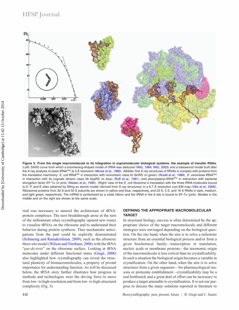
riod was necessary to unravel the architecture of tRNA/protein complexes. The next breakthrough arose at the turnof the millennium when crystallography opened new routesto visualize tRNAs on the ribosome and to understand theirbehavior during protein synthesis. Thus mechanistic antici-pations from the past could be explicitly demonstrated(Schmeing and Ramakrishnan, 2009), such as the allostericthree-site model (Wilson and Nierhaus, 2006) with the tRNA“pas-de-trois” on the ribosome surface. Looking at tRNAmolecules under different functional states (Giegé, 2008)also highlighted how crystallography can reveal the struc-tural plasticity of biomacromolecules, a property of pivotalimportance for understanding function. As will be discussedbelow, the tRNA story further illustrates how progress inmethods and technologies were the driving force to movefrom low- to high-resolution and from low- to high-structuralcomplexity (Fig. 3).
DEFINING THE APPROPRIATE MACROMOLECULARTARGETIn structural biology, success is often determined by the ap-propriate choice of the target macromolecule and differentstrategies were envisaged depending on the biological ques-tion. On the one hand, when the aim is to solve a milestonestructure from an essential biological process and/or from agiven biochemical family—transcription or translation,nucleic acids or membrane proteins—the taxonomic originof the macromolecule is less critical than its crystallizability.In such a situation the biological origin becomes a variable incrystallization. On the other hand, when the aim is to solvestructures from a given organism—for pharmacological rea-sons or proteome establishment—crystallizability may be areal bottleneck and a great deal of effort can be necessary toproduce a target amenable to crystallization. It is not our pur-pose to discuss the many solutions reported in literature to
Figure 3. From the single macromolecule to its integration in supramolecular biological systems: the example of transfer RNAs.�Left� SAXS curve from which a boomerang-shaped model of tRNA was deduced �Witz, 1964; Witz, 2003� and a balsawood model built afterthe X-ray analysis of yeast tRNAAsp at 3 Å resolution �Moras et al., 1980�. �Middle� first X-ray structures of tRNAs in complex with proteins fromthe translation machinery: E. coli tRNAGln in interaction with monomeric class Ib GlnRS �in green� �Rould et al., 1989�, S. cerevisiae tRNAAsp
in interaction with its cognate dimeric class IIb AspRS �in blue� �Ruff et al., 1991�, and phenylalanyl-tRNAPhe in interaction with bacterialelongation factor EF-Tu �in pink� �Nissen et al., 1995�. �Right� view of the E. coli ribosome in translation with the three tRNA molecules boundto A, P, and E sites obtained by fitting an atomic model �derived from X-ray structures� in a 6.7 Å resolution cryo-EM map �Villa et al., 2009�.Ribosomal proteins from 30 S and 50 S subunits are shown in yellow and blue, respectively, and 23 S, 5 S, and 16 S RNAs in dark, medium,and light green, respectively. The mRNA is symbolized by a violet ribbon and the tRNA in the A site is bound to EF-Tu �pink�. Models in themiddle and on the right are shown at the same scale.
HFSP Journal
112 Biocrystallography: past, present, future | R. Giegé and C. Sauter
Dow
nloa
ded
by [
Uni
vers
ity o
f C
ambr
idge
] at
11:
42 1
5 O
ctob
er 2
014
circumvent these concerns. Instead we will illustrate the sub-ject by examples relevant to structural investigations on thetranslation machinery and on membrane proteins that havebeen leading in many respects to major developments inbiocrystallogenesis.
Regarding the choice of the taxonomic origin of the tar-get one has to remember that many organisms are adapted toextreme life conditions, notably with a temperature of up to110 °C, pressures of up to 100 MPa, and high radiation levelsor salt concentrations. To do so, they have evolved macromol-ecules, which are stable under such conditions.The pivotal find-ing that triggered the rush toward extremophiles was the goodcrystallizability of the tyrosyl-tRNA synthetase (TyrRS), amember of the aminoacyl-tRNA synthetase (aaRS) family, iso-lated from heat-loving Bacillus stearothermophilus (Reid et al.,1973). The many structures in the protein data bank (PDB)from extremophiles confirm the idea of the relative ease tocrystallize their macromolecular components (Liebl, 2004).The concept is particularly true for the aaRS family, where�60% of the 3D structures stem from extremophiles (Giegéet al., 2008). Likewise, the known ribosome structures stemfrom three different extremophiles—T. thermophilus (Cateet al., 1999; Clemons et al., 1999; Tocilj et al., 1999),Haloarcula marismortui (Ban et al., 1999; Gluehmann et al.,2001), and Deinococcus radiodurans (Davidovich et al.,2007)—and were solved as the result of years of intensiveand innovative worldwide research efforts (e.g., Mooreand Steitz, 2003; Noller, 1991; Wilson and Nierhaus, 2006;Yonath et al., 1998; Yusupov et al., 1991). Note that theopportunity to obtain structures of the same biological entityoriginating from different taxa changes the traditional wayto approach molecular evolution that can now be addressed
by 3D structure instead of by 1D sequence analysis. Impor-tant applications have already emerged from studies onaminoacyl-tRNA synthetases (O’Donoghue and Luthey-Schulten, 2003) and it can be anticipated that evolutionarybiology will be deeply impacted by the 3D vision of proteinstructures (Abad-Zapatero, 2009).
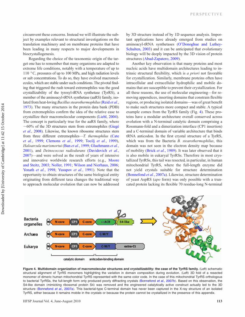
Another key observation is that many proteins and mostnucleic acids have multidomain architectures leading to in-trinsic structural flexibility, which is a priori not favorablefor crystallization. Similarly, membrane proteins often haveintracellular and extracellular hydrophilic and mobile do-mains that are susceptible to prevent their crystallization. Forall these reasons, the use of molecular engineering—for re-moving appendices, inserting domains that constrain flexibleregions, or producing isolated domains—was of great benefitto make such structures more compact and stable. A typicalexample comes from the TyrRS family (Fig. 4). These pro-teins have a modular architecture overall conserved acrossevolution with a N-terminal catalytic domain comprising aRossmann-fold and a dimerization interface (CP1 insertion)and a C-terminal domain of variable architecture that bindstRNA anticodon. In the first crystal structure of a TyrRS,which was from the Bacteria B. stearothermophilus, thisdomain was not seen in the electron density map becauseof mobility (Brick et al., 1989). It was later observed that itis also mobile in eukaryal TyrRSs. Therefore in most crys-tallized TyrRSs, this tail was resected, in particular, in humanmitochondrial TyrRS, where the full-length enzyme didnot yield crystals suitable for structure determination(Bonnefond et al., 2007a). Likewise, structure determinationof yeast AspRS (apo form) was only possible with a trun-cated protein lacking its flexible 70 residue-long N-terminal
Figure 4. Multidomain organization of macromolecular structures and crystallizability: the case of the TyrRS family. �Left� schematicstructural alignment of TyrRS monomers highlighting the variation in domain composition during evolution. �Left� 3D fold of a resectedmonomer of dimeric human mitochondrial TyrRS represented with the same color code. In the case of this mitochondrial TyrRS orthologousto bacterial TyrRSs, the full-length form only produced poorly diffracting crystals �Bonnefond et al., 2007b�. Based on this observation, theS4-like domain �mimicking ribosomal protein S4� was removed and the engineered catalytically active construct actually led to the 3Dstructure �Bonnefond et al., 2007a�. This bacterial-type C-terminal domain has never been captured in the X-ray structure of an isolatedTyrRS, either because it remains mobile in the crystals or because the protein cannot be crystallized in the presence of this appendix.
P E R S P E C T I V E
HFSP Journal Vol. 4, June-August 2010 113
Dow
nloa
ded
by [
Uni
vers
ity o
f C
ambr
idge
] at
11:
42 1
5 O
ctob
er 2
014
extension (Sauter et al., 2000). Other examples concernmembrane proteins (Supplementary Material Table S1), no-tably the K+ channel from Streptomyces lividans and the hu-man �2-adrenergetic G protein coupled receptor (GPCR)protein (Fig. 2). The first one needed two types of engineer-ing for structure determination, namely, the resection of itsC-terminal extension and the growth of crystals of a mutantwith a single amino acid change that diffracted better thancrystals grown from the wild-type protein (Doyle et al.,1998). As to the second example, a mobile intracellular do-main prevented crystallization of the receptor. Here, the dif-ficulty was circumvented by stabilizing the GPCR structureby the insertion of a T4 lysozyme molecule in the flexibleregion (Rosenbaum et al., 2007).
Structural plasticity of biomacromolecules, although det-rimental for crystallization, is essential for function. Thusaddition of ligands or of any type of small molecules able torestrain the conformational space of the macromolecule canhelp its crystallization. This has proven particularly usefulfor proteins such as aaRSs, where addition of small substratederivatives or of tRNA allowed crystallization or led to crys-tals of improved diffraction properties (Giegé et al., 2008).On the other hand, crystallizing complexes containing mac-romolecular ligands is a way to explore the conformationalspace of these ligands. Again, this is well illustrated with tR-NAs that show a large repertoire of conformations when in-teracting, for example, with maturation enzymes, aaRSs,elongation factor, or the ribosome (Giegé, 2008).
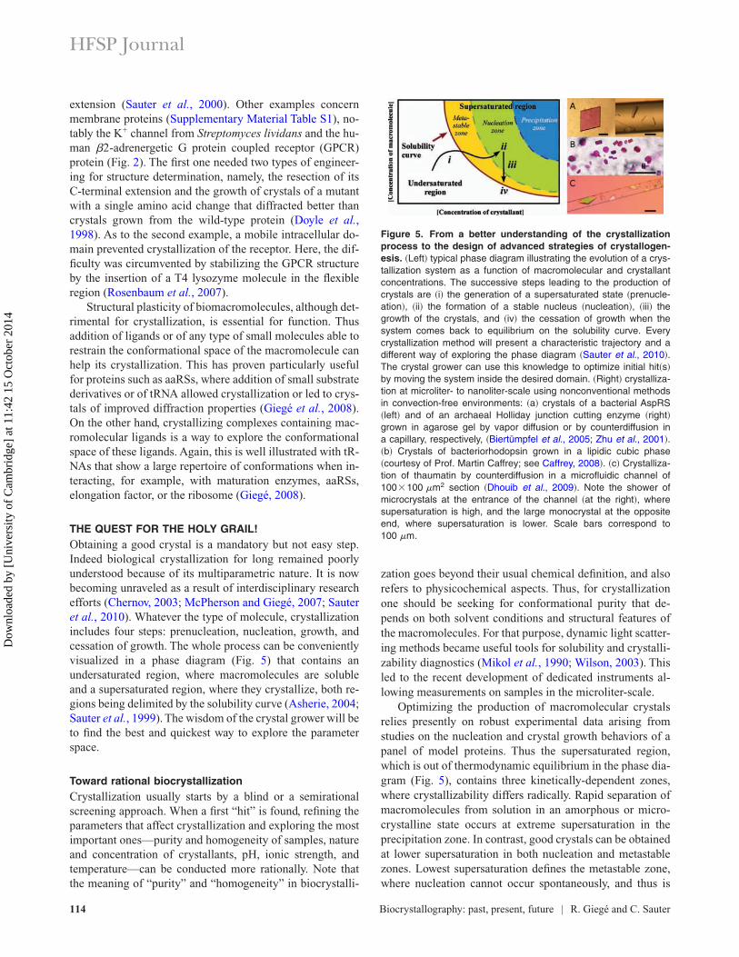
THE QUEST FOR THE HOLY GRAIL!Obtaining a good crystal is a mandatory but not easy step.Indeed biological crystallization for long remained poorlyunderstood because of its multiparametric nature. It is nowbecoming unraveled as a result of interdisciplinary researchefforts (Chernov, 2003; McPherson and Giegé, 2007; Sauteret al., 2010). Whatever the type of molecule, crystallizationincludes four steps: prenucleation, nucleation, growth, andcessation of growth. The whole process can be convenientlyvisualized in a phase diagram (Fig. 5) that contains anundersaturated region, where macromolecules are solubleand a supersaturated region, where they crystallize, both re-gions being delimited by the solubility curve (Asherie, 2004;Sauter et al., 1999). The wisdom of the crystal grower will beto find the best and quickest way to explore the parameterspace.
Toward rational biocrystallizationCrystallization usually starts by a blind or a semirationalscreening approach. When a first “hit” is found, refining theparameters that affect crystallization and exploring the mostimportant ones—purity and homogeneity of samples, natureand concentration of crystallants, pH, ionic strength, andtemperature—can be conducted more rationally. Note thatthe meaning of “purity” and “homogeneity” in biocrystalli-
zation goes beyond their usual chemical definition, and alsorefers to physicochemical aspects. Thus, for crystallizationone should be seeking for conformational purity that de-pends on both solvent conditions and structural features ofthe macromolecules. For that purpose, dynamic light scatter-ing methods became useful tools for solubility and crystalli-zability diagnostics (Mikol et al., 1990; Wilson, 2003). Thisled to the recent development of dedicated instruments al-lowing measurements on samples in the microliter-scale.
Optimizing the production of macromolecular crystalsrelies presently on robust experimental data arising fromstudies on the nucleation and crystal growth behaviors of apanel of model proteins. Thus the supersaturated region,which is out of thermodynamic equilibrium in the phase dia-gram (Fig. 5), contains three kinetically-dependent zones,where crystallizability differs radically. Rapid separation ofmacromolecules from solution in an amorphous or micro-crystalline state occurs at extreme supersaturation in theprecipitation zone. In contrast, good crystals can be obtainedat lower supersaturation in both nucleation and metastablezones. Lowest supersaturation defines the metastable zone,where nucleation cannot occur spontaneously, and thus is
Figure 5. From a better understanding of the crystallizationprocess to the design of advanced strategies of crystallogen-esis. �Left� typical phase diagram illustrating the evolution of a crys-tallization system as a function of macromolecular and crystallantconcentrations. The successive steps leading to the production ofcrystals are �i� the generation of a supersaturated state �prenucle-ation�, �ii� the formation of a stable nucleus �nucleation�, �iii� thegrowth of the crystals, and �iv� the cessation of growth when thesystem comes back to equilibrium on the solubility curve. Everycrystallization method will present a characteristic trajectory and adifferent way of exploring the phase diagram �Sauter et al., 2010�.The crystal grower can use this knowledge to optimize initial hit�s�by moving the system inside the desired domain. �Right� crystalliza-tion at microliter- to nanoliter-scale using nonconventional methodsin convection-free environments: �a� crystals of a bacterial AspRS�left� and of an archaeal Holliday junction cutting enzyme �right�grown in agarose gel by vapor diffusion or by counterdiffusion ina capillary, respectively, �Biertümpfel et al., 2005; Zhu et al., 2001�.�b� Crystals of bacteriorhodopsin grown in a lipidic cubic phase�courtesy of Prof. Martin Caffrey; see Caffrey, 2008�. �c� Crystalliza-tion of thaumatin by counterdiffusion in a microfluidic channel of100�100 �m2 section �Dhouib et al., 2009�. Note the shower ofmicrocrystals at the entrance of the channel �at the right�, wheresupersaturation is high, and the large monocrystal at the oppositeend, where supersaturation is lower. Scale bars correspond to100 �m.
HFSP Journal
114 Biocrystallography: past, present, future | R. Giegé and C. Sauter
Dow
nloa
ded
by [
Uni
vers
ity o
f C
ambr
idge
] at
11:
42 1
5 O
ctob
er 2
014
suitable for seeding (D’Arcy et al., 2007). In the nucleationzone, where nucleation occurs spontaneously, number ofnuclei, growth rate, and growth mechanisms depend on pre-cise solution conditions that should be tuned appropriately.
Nucleation can be homogeneous in the bulk of the solu-tion, but in most of the cases, it is heterogeneous and occurson solid surfaces such as the walls of the crystallizationchamber or dust particles and other impurities. The meansthat minimize heterogeneous nucleation and thus favor re-producibility of experiments have been found (Sauter et al.,2010).
Growth of macromolecular crystals can occur by twomechanisms that highly depend on supersaturation. Whilecrystals grow by screw dislocation—by a helical path propa-gating around a lattice defect—at low supersaturation, theypredominantly grow by 2D island formation—from 2Dclusters/nuclei that form randomly on flat crystal faces—athigher supersaturation. High-quality crystals are obtained atlowest supersaturation and under constant growth regime butthese are not easy to obtain in practice since crystal growth isaccompanied by a decrease in supersaturation in the motherliquor that could trigger a modification of the growth regime.Atomic force microscopy revealed this effect duringtRNAPhe crystallization (Ng et al., 1997). Perturbation ofgrowth regime, likely accounts for nonreproducibility of dif-fraction properties, can also result from impurity incorporationin growing crystals. Such poisoning is favored when the impu-rity has resemblance with the crystallizing macromolecule.Therefore, the macromolecule itself can be the worst contami-nant due to conformational heterogeneity or partially frag-mented isoforms.
Crystallization improvements in current practiceAll methods used in biocrystallization aim to bring the mac-romolecule to an appropriate state of supersaturation (Sauteret al., 2010). Although structural biologists favor vaporphase equilibrium techniques, batch, dialysis, and free-interface diffusion methods are alternatives. One shall recallthat besides physical and chemical variables, the crystalliza-tion method itself and the geometry of the setup also affectcrystallization. As mentioned above, crystal growth seldomoccurs at constant protein concentration, thus introducingchanges in supersaturation and, hence, possible changes inthe growth regime. Crystallization at constant macromol-ecule concentration could be achieved in liquid circulationcells but is not obvious to implement in practice.
Batch crystallization was the method of choice in the pio-neers’ time and remains the simplest since it just requiresmixing macromolecules and crystallants until supersatura-tion is reached. It is the first crystallization method that wasautomated in a microdroplet version under oil (Chayen et al.,1990) and more recently was further miniaturized (D’Arcyet al., 2003). Dialysis permits easy variation in parametersbut is less adapted for small sample volumes and screening
procedures. In contrast, crystallization by vapor diffusion,which was invented for the production of tRNA crystals(Hampel et al., 1968), is very handy and has rapidly becomethe favored method in most laboratories. It is practiced in avariety of forms, mainly in microliter-size sitting drops. Infree-interface and counterdiffusion methods, equilibrationoccurs by direct diffusion of the crystallant into the macro-molecule solution (García-Ruiz and Moreno, 1994). Bothmethods require minimal convection and, therefore, experi-ments are conducted in capillaries. The advantage of coun-terdiffusion is that a wide range of supersaturation condi-tions can be tested in a single experiment and that all stepsfrom crystallization to structure determination can be per-formed in situ without any crystal handling (Gavira et al.,2002).
Advanced crystallization strategiesOver the past decade, new strategies have been developedeither to screen physical variables or to give emphasis to pe-culiar growth media and to take advantage of novel biotech-nological tools for stabilizing macromolecular conforma-tions by chaperone macromolecules. These methods wereshown to be efficient for both ab initio search of crystalliza-tion conditions and optimization procedures for improvingcrystal quality. As an illustration, Fig. 5 displays crystalsgrown by three unconventional methods taking advantage ofgelled media, cubic mesophases, or microfluidic counterdif-fusion channels. On the other hand and in view of high-throughput structural genomics projects, automated instru-ments and entirely integrated systems have been developedto accelerate crystallization and optimization procedures(Newman et al., 2008).
The ways physical variables affect crystallization aremanifold (Sauter et al., 2010). Thus gravity influences fluidproperties and movement of molecules, pressure and tem-perature alter conformation of macromolecules, magneticfields orient crystals, and electric fields can reduce nucle-ation rates. Likewise gelled and microfluidic environmentsreduce convection and thus favor crystallization while cubicgel-like mesophases provide conditions to crystallize hydro-phobic proteins. Convection and sedimentation always takeplace in current procedures and severely influence crystal-lization. In the absence of gravity, theory predicts regularcrystal growth under diffusive regime that should enhancecrystal quality. Such considerations have justified space-crystallization programs and have contributed to a deeperunderstanding of protein crystallization (Kundrot et al.,2001). However, due to experimental limitation, crystalliza-tion in weightlessness is not a panacea and ways to mimic itsbeneficial effects on earth were searched. Because convec-tion depends on viscosity, gels represent a convection-freeenvironment and thus a good media to improve crystal qual-ity (Lorber et al., 2009). As was anticipated, crystals grownin gels are often of superior quality than controls grown from
P E R S P E C T I V E
HFSP Journal Vol. 4, June-August 2010 115
Dow
nloa
ded
by [
Uni
vers
ity o
f C
ambr
idge
] at
11:
42 1
5 O
ctob
er 2
014
solutions. They can easily be removed from their soft envi-ronment and set up for X-ray analysis. Microfluidic devicesalso provide a diffusive environment due to their small size.The first microfluidic applications in biocrystallization werea free-interface system (Hansen et al., 2002) followed by a“batch in nanodroplets” chip (Zheng et al., 2003) suitable forhigh-throughput screening. The absence of convection in mi-crofluidic channels makes microsystems very appealing forcounterdiffusion experiments. When made of appropriatepolymer material, counterdiffusion chips allow a direct on-chip characterization of the crystals by X-ray diffractionwithout any further sample handling (Dhouib et al., 2009;Ng et al., 2008). Finally, it was conjectured that suppressionof convection could be achieved under hypergravity or whenmagnetic or electric fields are applied. Although macromol-ecule crystal growth under such conditions is not widespreadand the underlying physics not completely validated, theseapproaches can be useful in special cases, for instance, toreduce the number of nucleation sites (Sauter et al., 2010).
Temperature and pressure are two thermodynamic pa-rameters that can trigger nucleation and sustain protein crys-tal growth (Rosenberger et al., 1993; Suzuki et al., 2002)but were hardly exploited although temperature-inducedcrystallization often occurs consequently of accidental tem-perature variation in the laboratory. Dedicated crystallizationsystems have been designed for temperature-induced crys-tallization that find application, e.g., in the growth of largecrystals for neutron crystallography (Budayova-Spano et al.,2007). Pressure-induced crystallization is trickier and re-quires other specialized equipments (Suzuki et al., 2002). In-terestingly, cowpea mosaic virus crystals compressed at 330MPa in a diamond anvil cell demonstrated pressure-inducedordering of the crystals, lower ADPs, and a larger number ofordered water molecules (Girard et al., 2005).
In a more biological perspective, use of “crystallizationhelper” chaperones is becoming useful to crystallize recalci-trant proteins (Koide, 2009) or RNA fragments (Ye et al.,2008). A typical example of the chaperone strategy is thestructure determination of Escherichia coli tRNACys fromcrystals, where the tRNA was sequestered by elongation factor(Nissen et al., 1999). First tested with antibody fragment chap-erones, it was rejuvenated with the DARPin technology basedon the natural ankyrin repeat protein fold with randomized sur-face residue positions allowing specific binding to virtually anytarget protein (Sennhauser and Grütter, 2008).
BETTER AND FASTER METHODS FOR STRUCTUREDETERMINATIONWhen the pioneers of biocrystallography showed that bio-logical samples could potentially produce high-resolutiondiffraction patterns (Bernal and Crowfoot, 1934; Perutz,1985), structure determination was still very empirical andtedious. The first breakthrough came with the developmentof a robust phasing method based on the introduction of
heavy atoms in the crystals, a procedure called multiple iso-morphous replacement (MIR). This approach led to theatomic models of myoglobin and hemoglobin in the early1960s (Kendrew et al., 1960; Perutz et al., 1968). It is still inuse in various forms nowadays and was the key for phasingdiffraction data from ribosome crystals soaked with largeheavy-atom clusters (Ban et al., 1998; Yonath et al., 1998).Of course, the concomitant boom of computing systems wasalso pivotal. Following the first success stories and the in-crease in the number of biological systems investigated, themajor difficulty became soon, and still remains, the availabil-ity of the biomolecule and the difficulty to produce crystalsof adequate quality.
The 1990s brought a radical change in the practice ofbiocrystallography. The first major development was crystalcryocooling at around 100 K in a stream of nitrogen gas, amethod known for long in chemistry and introduced in biol-ogy with ribosome crystals (Yonath et al., 1998). It is pres-ently systematized in biocrystallography to slow down radia-tion damages and to increase the lifetime of samples duringX-ray analysis (Garman, 2003). Further, post-crystallizationmethods to enhance crystal quality—dehydration, annealing,soaking, and other treatments—became popular and curedmany “poor” crystals (Heras and Martin, 2005). Here aswell, observations on crystals of proteins from the transla-tion machinery—a GlnRS and EF-Tu—where among thefirst that opened the field (Rould et al., 1991; Schick andJurnak, 1994). Second, the access to strong synchrotronX-ray light sources increased sharply with the building ofnew third generation facilities worldwide. The availability ofintense and tunable radiations facilitated the development anew phasing method, the multiwavelength anomalous dis-persion (MAD) (Hendrickson, 1991), which simplifies theoriginal MIR approach. It eliminates the necessity of prepar-ing several crystal derivatives and associated isomorphismdistortion since the entire structure determination can be per-formed on a single cryocooled crystal including an anoma-lous scatterer. The combination of all these methods was in-strumental to the explosion of 3D data in the PDB thatoccurred in the mid-1990s (Fig. 1) and here again the ribo-some adventure is a striking illustration of their impact instructural biology (Abrahams and Ban, 2003; Gluehmannet al., 2001; Yonath et al., 1998).
At the end of the 1990s, the structural biology commu-nity invented the concept of structural genomics and jumpedinto the post-genomics era. The effective implementation ofstructural genomics implied to deal with hundreds of targetsin a massively parallel manner in order to achieve high-throughput at each stage of a project (Terwilliger et al., 2009)and these developments found also applications at smallscale in academic laboratories. They include the productionand fast purification of tagged molecules, the use of auto-mated data collection protocols on cryocooled samples (Arztet al., 2005), and of automated pipelines for X-ray structure
HFSP Journal
116 Biocrystallography: past, present, future | R. Giegé and C. Sauter
Dow
nloa
ded
by [
Uni
vers
ity o
f C
ambr
idge
] at
11:
42 1
5 O
ctob
er 2
014
solution and refinement (Adams et al., 2009) exploiting theincorporation of selenomethionine in proteins for MAD orsingle autonomous dispersion (SAD) phasing (Joachimiak,2009). On the other hand, the increase in the 3D repertoirewith �50% of new folds in the PDB provided by structural ge-nomics consortia, is rejuvenating the effectiveness of molecularreplacement methods as alternate phasing and refinement tools.
Other breakthroughs stem from methodological ad-vances in single particle cryoelectron microscopy (cryo-EM)and were applied in the ribosome field for imaging variousribosomes (Becker et al., 2009; Frank, 2009; Spahn andPenczek, 2009), including minimalist mitochondrial ribo-somes (Sharma et al., 2009). When cryo-EM was combinedwith X-ray crystalloghraphy, molecular dynamics andmodeling, new biological questions could be addressed suchas uncovering high-resolution snapshots of functional ribo-somes during initiation (Simonetti et al., 2009) or elongation(Villa et al., 2009; Fig. 3) of protein synthesis. They werealso essential for structure determination of other largeassemblies such as viruses with asymmetric properties(Mueller et al., 2007; Rossmann et al., 2007; Steven andBaumeister, 2008).
The recent progress in terms of synchrotron and datacollection facilities will certainly help to tackle new appeal-ing biological systems. Of particular interest are the micro-focused beams that allow analysis of crystals with a sizedown to a few microns (Moukhametzianov et al., 2008;Schneider, 2008). Thus, a 2 Å crystal structure of both re-combinant and infectious silkworm cypovirus polyhedracould be determined using crystals of 5–12 µm, the smallestcrystals yet used for de novo X-ray protein structure determi-nation (Coulibaly et al., 2007). Likewise, the development ofa new generation of ultrafast and sensitive X-ray detectors(Kraft et al., 2009) enables the exploitation of radiation-sensitive or weakly diffracting samples, and gives the possi-bility to analyze crystals in their growth environment—either in microplates or microfluidic devices (Dhouib et al.,2009; Jacquamet et al., 2004)—avoiding potentially detri-mental handling.
Today, structure determination of a new target can gener-ally be carried out in a few months, where it may have takenyears if not decades, in the past. The situation is well de-picted by the metaphor of the “flying crystallographer,” rush-ing from one synchrotron facility to the next, solving andrefining his new structures on the way back to the laboratory,if not directly on the beamline while collecting the data.However, this apparent ease, which might reflect a majorityof cases, should not mask that every single project is uniqueand that the starting point will always remain a good, well-diffracting crystal. Further, it should not be forgotten that en-tire regions/areas of the “3D-space” underlying the tree oflife remain essentially unexplored such as that of nativemetazoan proteins with post-translational modifications andwithout resected motifs.
UNSOLVED ISSUES, HIGHER COMPLEXITY,AND 4D BIOLOGY
The questions addressed to biocrystallography have dramati-cally evolved since the first protein structures were solved.The fact that high quality X-ray diffraction data can be ob-tained from a single crystal of dimensions in the range of20–50 µm has changed the objectives considerably. One shallrecall that 40 years ago a structure analysis required many crys-tals in the mm size range. Since fewer and smaller crystals arenow the rule—except for neutron diffraction (Budayova-Spanoet al., 2007)—it is easier today to envisage more challengingprojects dealing with membrane proteins, lipoproteins, intrinsi-cally unstructured proteins, large RNAs, or nucleoproteincomplexes or assemblies. Intrinsically unstructured proteins orproteins with disordered regions represent a real challenge.Such proteins are especially abundant in eukarya and remainpoorly understood but may fold and play important roles uponbinding to their cellular partners (Dyson and Wright, 2005;Fukuchi et al., 2009). Besides understanding their biology, it isanticipated that their study will provide clues to comprehendprotein folding and protein dynamics. Membrane proteins con-stitute another tricky category due to their lipophylic nature andthe difficulty to make them stable in solution and thus amenableto crystallization (Caffrey, 2008). The great deal of effort al-ready invested to enlarge the repertoire of their 3D structures,certainly will be pursued, notably for applications since theseproteins represent almost 50% of the promising pharmaceuticaltargets. In a wider perspective of applications, biocrystallogra-phy provides a powerful platform for the conception of newdrugs with the possibility to screen for ligand binding in crys-tallo (Blundell et al., 2006) and can deliver valuable 3D datato fight against new threats such as emerging pathogenic vi-ruses (Anand et al., 2003; Bollati et al., 2009). Crystallogra-phy alone, however, is not sufficient for applications such asligand screening or drug design that may require comple-mentary biophysical and computational techniques (Renaudand Delsuc, 2009).
Although crystallography gives access to static atomicsnapshots of objects frozen in a crystal lattice, solving struc-tures with and without ligands or exploiting different crystalforms generated during crystallization screening are power-ful means to capture alternate functional states of biomacro-molecules. Interestingly, packing plasticity does even exist incrystals diffracting to high-resolution that can sustain a highdegree of disorder—up to 30–35%—in their packing (Touzéet al., 2007; Troffer-Charlier et al., 2007). This brings tothe question of decrypting the kinematic changes in macro-molecular structures in action, in other words the potential ofa time-resolved crystallography with perspective of “4D bi-ology,” where the fourth dimension would be the temporalcomponent. Along these lines, the example of crystallo-graphic snapshots obtained during the maturation of a tRNA,provided a “movie” of this enzymatic reaction and gave a
P E R S P E C T I V E
HFSP Journal Vol. 4, June-August 2010 117
Dow
nloa
ded
by [
Uni
vers
ity o
f C
ambr
idge
] at
11:
42 1
5 O
ctob
er 2
014
robust support to the approach (Tomita et al., 2006). Themore direct approach would be to capture transient 3D infor-mation in crystallo. This dream of crystallographers was al-ready experimentally assayed in the late 1980s and gave elec-tron density maps from millisecond diffraction datacollected on Laue photographs (Hajdu et al., 1988). Recentlysophisticated methodologies for time-resolved crystallogra-phy have been successfully validated with several model en-zymes (Bourgeois and Royant, 2005). With the next genera-tion of X-ray light sources, the coming era of single-molecule X-ray diffraction (Helliwell, 2004) and thedevelopment of new tools for analysis intensity changes inLaue diffraction experiments (Coppens et al., 2009), one cananticipate a flourishing future for time-resolved crystallogra-phy not only for enzymology but also to get a kinematic in-sight of macromolecular recognition processes.
Hybrid approaches combining X-ray diffraction withEM, NMR spectroscopy, biophysical, and computationalmethods will continue to improve and their importance instructural biology will undoubtedly increase (Steven andBaumeister, 2008). Thus, the association of correlative lightmicroscopy and EM, electron or X-ray tomography withcrystallography makes possible to apprehend a living cell atdifferent scales, starting from its global organization andzooming down to capture macromolecular events at atomicresolution (Hoenger and McIntosh, 2009; McDermott et al.,2009; Plitzko et al., 2009). This old dream of biologists isnow becoming a reality and promising results such as the vi-sualization of the cadherin network bridging the extracellularspace in the epidermal desmosome have already been ob-tained (Al-Amoudi et al., 2007). Interestingly, recent crystal-lographic work combined with cell biology and modelingalso links cadherin biology with the angiostatic activity of ahuman aaRS, namely, TrpRS (Zhou et al., 2010), a step to-ward the integrative biology of these two classes of proteins.The perspectives are wide and it can be anticipated thatstructural biology in its perpetual evolution will become anintegral component of integrative biology in a near future.
CONCLUSIONSince the first picture of myoglobin at 5 Å resolution(Kendrew et al., 1958) and the first use of a synchro-tron to collect diffraction photographs on a virus crystal(Rosenbaum et al., 1971), immense progresses have beenmade in the precision of the structural data delivered byX-ray crystallography. The highest resolution for largesoluble proteins has recently reached 0.66 Å for human aldo-lase reductase, a protein of 36 kDa (Podjarny et al., 2004),and 1.15 Å for a membrane protein, namely, a yeast aqua-porin (Fischer et al., 2009). Neutron crystallography has pro-gressed as well and provided a structure of bovine pancreaticRNase A at 1.7 Å resolution (Yagi et al., 2009). All along itshistory the field has been built on a strong interdisciplinaryspirit that contributed to solve successive bottlenecks and
helped to tackle biological questions of increasing complex-ity. As a result biocrystallography has become a must andcontinues to be at the frontiers of biological research. Whilestructural knowledge remains sparse in many respects re-garding membrane proteins or eukaryal proteomes, includ-ing the human proteome and related biomedical issues, su-pramolecular crystallography of large and even giantassemblies is just at the verge of a golden age.
As a concluding remark, let us note that biocrystallogra-phy transformed from a multi- to an interdisciplinary disci-pline with scientific fields a priori disconnected that progres-sively became tightly interconnected. This is well illustratedby the semantic and operational changes in the name of thediscipline that transformed to structural biology with its ex-perts coming from physics and chemistry progressively inte-grating biochemistry and molecular biology in their researchpractice. At the opposite an increasing number of groupswith biochemistry and molecular biology background haveadopted crystallography as a major investigation tool. We an-ticipate that the present structural biology will undoubtedlyplay a key role in the coming “mutations” toward integratedand global biology and will completely merge with thesenovel biodisciplines.
ACKNOWLEDGMENTSWe dedicate this essay to the memory of Dr. Warren L.Delano, creator of PYMOL, the powerful molecular visualiza-tion software used to prepare the illustrations. We thank Dr.Marat Yusupov for sharing with us his experience on thestructural study of the ribosome. This work received supportfrom the Centre National de la Recherche Scientifique(CNRS), the Université de Strasbourg, and the Agence Na-tionale de la Recherche (ANR) under Grant Nos. ANR-07-NANO-060 and ANR-09-BLAN-009-01.
REFERENCESAbad-Zapatero, C (2009). “A note of a protein crystallographer: the
molecular structure of evolutionary theory.” Acta Crystallogr.,Sect. D: Biol. Crystallogr. 65, 1341–1349.
Abrahams, JP, and Ban, N (2003). “X-ray crystallographic structuredetermination of large asymmetric macromolecular assemblies.”Methods Enzymol. 374, 163–188.
Adams, PD, Afonine, PV, Grosse-Kunstleve, RW, Read, RJ, Richardson,JS, Richardson, DC, and Terwilliger, TC (2009). “Recentdevelopments in phasing and structure refinement for macromolecularcrystallography.” Curr. Opin. Struct. Biol. 19, 566–572.
Al-Amoudi, A, Diez, DC, Betts, MJ, and Frangakis, AS (2007). “Themolecular architecture of cadherins in native epidermaldesmosomes.” Nature (London), 450, 832–837.
Anand, K, Ziebuhr, J, Wadhwani, P, Mesters, JR, and Hilgenfeld, R(2003). “Coronavirus main proteinase (3CLpro) structure:basis for design of anti-SARS drugs.” Science 300, 1763–1767.
Arnold, E, Himmel, DM, and Rossmann, M, eds. (2010). Crystallographyof Biological Macromolecules, 2nd Ed., Wiley, Chichester.
Arzt, S, et al. (2005). “Automation of macromolecular crystallographybeamlines.” Prog. Biophys. Mol. Biol. 89, 124–152.
Asherie, N (2004). “Protein crystallization and phase diagrams.” Methods34, 266–272.
Ban, N, Freeborn, B, Nissen, P, Penczek, P, Grassucci, RA, Sweet, R,
HFSP Journal
118 Biocrystallography: past, present, future | R. Giegé and C. Sauter
Dow
nloa
ded
by [
Uni
vers
ity o
f C
ambr
idge
] at
11:
42 1
5 O
ctob
er 2
014
Frank, J, Moore, PB, and Steitz, TA (1998). “A 9 Å resolution x-raycrystallographic map of the large ribosomal subunit.” Cell 93,1105–1115.
Ban, N, Nissen, P, Hansen, J, Capel, M, Moore, PB, and Steitz, TA(1999). “Placement of protein and RNA structures into a 5 Å-resolution map of the 50S ribosomal subunit.” Nature (London) 400,841–847.
Becker, T, et al. (2009). “Structure of monomeric yeast and mammalianSec61 complexes interacting with the translating ribosome.”Science 326, 1369–1373.
Bernal, JD, and Crowfoot, D (1934). “X-ray photographs of crystallinepepsin.” Nature (London) 133, 794–795.
Biertümpfel, C, Basquin, J, Birkenbihl, RP, Suck, D, and Sauter, C (2005).“Characterization of crystals of the Hjc resolvase from Archaeoglobusfulgidus grown in gel by counter-diffusion.” Acta Crystallogr.Sect. F: Struct. Biol. Cryst. Commun. 61, 684–687.
Blundell, TL, Sibanda, BL, Montalvao, RW, Brewerton, S, Chelliah, V,Worth, CL, Harmer, NJ, Davies, O, and Burke, D (2006).“Structural biology and bioinformatics in drug design: opportunitiesand challenges for target identification and lead discovery.”Philos. Trans. R. Soc. London, Ser. B 361, 413–423.
Bollati, M, et al. (2009). “Structure and functionality in flavivirus ns-proteins: perspectives for drug design.” Antiviral Res. (in press).
Bonnefond, L, Frugier, M, Touzé, E, Lorber, B, Florentz, C, Giegé, R,Rudinger-Thirion, J, and Sauter, C (2007b). “Tyrosyl-tRNAsynthetase: the first crystallization of a human mitochondrialaminoacyl-tRNA synthetase.” Acta Crystallogr. Sect. F: Struct. Biol.Cryst. Commun. 63, 338–341.
Bonnefond, L, Frugier, M, Touzé, E, Lorber, B, Florentz, C, Giegé, R,Sauter, C, and Rudinger-Thirion, J (2007a). “Crystal structure ofhuman mitochondrial tyrosyl-tRNA synthetase reveals common andidiosyncratic features.” Structure (London) 15, 1505–1516.
Bourgeois, D, and Royant, A (2005). “Advances in kinetic proteincrystallography.” Curr. Opin. Struct. Biol. 15, 538–547.
Brick, P, Bhat, TN, and Blow, DM (1989). “Structure of tyrosyl-tRNAsynthetase refined at 2.3 Å resolution. Interaction of the enzymewith the tyrosyl adenylate intermediate.” J. Mol. Biol. 208, 83–98.
Budayova-Spano, M, Dauvergne, F, Audiffren, M, Bactivelane, T, andCusack, S (2007). “A methodology and an instrument for thetemperature-controlled optimization of crystal growth.” ActaCrystallogr., Sect. D: Biol. Crystallogr. 63, 339–347.
Caffrey, M (2008). “On the mechanism of membrane proteincrystallization in lipidic mesophases.” Cryst. Growth Des. 8,4244–4254.
Carter, CW, Jr. (2009). “E pluribus tres: the Nobel Prize in chemistry.”Structure (London) 17(12), 1588–1561.
Cate, JH, Yusupov, MM, Yusupova, GZ, Earnest, TN, and Noller, HF(1999). “X-ray crystal structures of 70S ribosome functionalcomplexes.” Science 285, 2095–2104.
Chayen, NE, Shaw Stewart, PD, Maeder, DL, and Blow, DM (1990). “Anautomated system for microbatch protein crystallisation andscreening.” J. Appl. Crystallogr. 23, 297–302.
Chernov, AA (2003). “Protein crystals and their growth.” J. Struct. Biol.142, 3–21.
Christendat, D, Yee, A, Dharamsi, A, Kluger, Y, Gerstein, M, Arrowsmith,CH, and Edwards, AM (2000). “Structural proteomics: prospects forhigh throughput sample preparation.” Prog. Biophys. Mol. Biol.73, 339–345.
Clemons, WM, Jr., May, JLC, Wimberly, BT, McCutcheon, JP, Capel,MS, and Ramakrishnan, V (1999). “Structure of a bacterial 30Sribosomal subunit at 5.5 Å resolution.” Nature (London) 400, 833–840.
Coppens, P, et al. (2009). “The RATIO method for time-resolvedLaue crystallography.” J. Synchrotron Radiat. 16, 226–230.
Coulibaly, F, Chiu, E, Ikeda, K, Gutmann, S, Haebel, PW, Schulze-Briese,C, Mori, H, and Metcalf, P (2007). “The molecular organization ofcypovirus polyhedra.” Nature (London) 446, 97–101.
D’Arcy, A, Mac Sweeney, A, Stihle, M, and Haber, A (2003). “Theadvantages of using a modified microbatch method for rapidscreening of protein crystallization conditions.” Acta Crystallogr., Sect.D: Biol. Crystallogr. 59, 396–399.
D’Arcy, A, Villard, F, and Marsh, M (2007). “An automated microseedmatrix-screening method for protein crystallization.” ActaCrystallogr., Sect. D: Biol. Crystallogr. 63, 550–554.
Davidovich, C, Bashan, A, Auerbach-Nevo, T, Yaggie, RD, Gontarek, RR,and Yonath, A (2007). “Induced-fit tightens pleuromutilins binding toribosomes and remote interactions enable their selectivity.”Proc. Natl. Acad. Sci. U.S.A. 104, 4291–4296.
Dhouib, K, et al. (2009). “Microfluidic chips for the crystallization ofbiomacromolecules by counter-diffusion and on-chip crystal x-ray analysis.” Lab Chip 9, 1412–1421.
Doyle, DA, Morais Cabral, J, Pfuetzner, RA, Kuo, A, Gulbis, JM, Cohen,SL, Chait, BT, and MacKinnon, R (1998). “The structure of thepotassium channel: molecular basis of K+ conduction and selectivity.”Science 280, 69–77.
Dyson, HJ, and Wright, PE (2005). “Intrinsically unstructured proteinsand their functions.” Nat. Rev. Mol. Cell Biol. 6, 197–208.
Fischer, G, Kosinska-Eriksson, U, Aponte-Santamaria, C, Palmgren, M,Geijer, C, Hedfalk, K, Hohmann, S, de Groot, BL, Neutze, R, andLindkvist-Petersson, K (2009). “Crystal structure of a yeast aquaporinat 1.15 angstrom reveals a novel gating mechanism.” PLoS Biol.7, e1000130.
Frank, J (2009). “Single-particle reconstruction of biologicalmacromolecules in electron microscopy–30 years.” Q. Rev. Biophys.42, 139–158.
Fukuchi, S, Homma, K, Minezaki, Y, Gojobori, T, and Nishikawa, K(2009). “Development of an accurate classification system ofproteins into structured and unstructured regions that uncovers novelstructural domains: its application to human transcriptionfactors.” BMC Struct. Biol. 9, 26.
García-Ruiz, JM, and Moreno, A (1994). “Investigations on protein crystalgrowth by the gel acupuncture method.” Acta Crystallogr., Sect. D:Biol. Crystallogr. 50, 484–490.
Garman, E (2003). “ ‘Cool’ crystals: macromolecular cryocrystallographyand radiation damage.” Curr. Opin. Struct. Biol. 13, 545–551.
Gavira, JA, Toh, D, Lopez-Jaramillo, J, Garcia-Ruiz, J-M, and Ng, JD(2002). “Ab initio crystallographic structure determination ofinsulin from protein to electron density without crystal handling.” ActaCrystallogr., Sect. D: Biol. Crystallogr. 58, 1147–1154.
Giegé, R (2008). “Toward a more complete view of tRNA biology.” Nat.Struct. Mol. Biol. 15, 1007–1014.
Giegé, R, Touzé, E, Lorber, B, Théobald-Dietrich, A, and Sauter, C(2008). “Crystallogenesis trends of free and ligandedaminoacyl-tRNA synthetases.” Cryst. Growth Des. 8, 4297–4306.
Girard, E, Kahn, R, Mezouar, M, Dhaussy, AC, Lin, T, Johnson, JE, andFourme, R (2005). “The first crystal structure of a macromolecularassembly under high pressure: CpMV at 330 MPa.” Biophys.J. 88, 3562–3571.
Gluehmann, M, et al. (2001). “Ribosomal crystallography: from poorlydiffracting microcrystals to high-resolution structures.” Methods25, 292–302.
Hajdu, J, Acharya, KR, Stuart, DI, Barford, D, and Johnson, L (1988).“Catalysis in enzyme crystals.” Trends Biochem. Sci. 13,104–109.
Hampel, A, Labananskas, M, Conners, PG, Kirkegard, L, RajBhandary,UL, Sigler, PB, and Bock, RM (1968). “Single crystals of transferRNA from formylmethionine and phenylalanine transferRNA’s.” Science 162, 1384–1387.
Hansen, CL, Skordalakes, E, Berger, JM, and Quake, SR (2002). “Arobust and scalable microfluidic metering method that allowsprotein crystal growth by free interface diffusion.” Proc. Natl. Acad.Sci. U.S.A. 99, 16531–16536.
Helliwell, JR (2004). “Overview and new developments in softer x-ray�2 Å� lambda�5 Å� protein crystallography.” J. SynchrotronRadiat. 11, 1–3.
Hendrickson, WA (1991). “Determination of macromolecular structuresfrom anomalous diffraction of synchrotron radiation.” Science254, 51–58.
Heras, B, and Martin, J (2005). “Post-crystallization treatments forimproving diffraction quality of protein crystals.” ActaCrystallogr., Sect. D: Biol. Crystallogr. 61, 1173–1180.
Hoenger, A, and McIntosh, JR (2009). “Probing the macromolecularorganization of cells by electron tomography.” Curr. Opin. CellBiol. 21, 89–96.
Jacquamet, L, et al. (2004). “Automated analysis of vapor diffusion
P E R S P E C T I V E
HFSP Journal Vol. 4, June-August 2010 119
Dow
nloa
ded
by [
Uni
vers
ity o
f C
ambr
idge
] at
11:
42 1
5 O
ctob
er 2
014
crystallization drops with an x-ray beam.” Structure (London) 12,1219–1225.
Jancarik, J, and Kim, S-H (1991). “Sparse matrix sampling: a screeningmethod for crystallization of proteins.” J. Appl. Crystallogr. 24,409–411.
Joachimiak, A (2009). “High-throughput crystallography for structuralgenomics.” Curr. Opin. Struct. Biol. 19, 573–584.
Kendrew, J, Brodo, G, Dintzis, H, Parrish, H, Wyckoff, H, and Phillips, D(1958). “A three-dimensional model of the myoglobin moleculeobtained by x-ray analysis.” Nature (London) 181, 662–666.
Kendrew, JC, Dickerson, RE, Strandberg, BE, Hart, RG, Davies, DR,Phillips, DC, and Shore, VC (1960). “Structure of myoglobin: athree-dimensional Fourier synthesis at 2 Å resolution.” Nature (London)185, 422–427.
Koehn, J, and Hunt, I (2009). “High-throughput protein production(HTPP): a review of enabling technologies to expedite proteinproduction.” Methods Mol. Biol. 498, 1–18.
Koide, S (2009). “Engineering of recombinant crystallization chaperones.”Curr. Opin. Struct. Biol. 19, 449–457.
Kraft, P, et al. (2009). “Performance of single-photon-counting PILATUSdetector modules.” J. Synchrotron Radiat. 16, 368–375.
Kundrot, CE, Judge, RA, Pusey, ML, and Snell, EH (2001). “Microgravityand macromolecular crystallography.” Cryst. Growth Des. 1, 87–99.
Liebl, W (2004). “Genomics taken to the extreme.” Nat. Biotechnol. 22,524–525.
Lorber, B, Sauter, C, Théobald-Dietrich, A, Moreno, A, Schellenberger, P,Robert, M-C, Capelle, B, Sanglier, S, Potier, N, and Giegé, R (2009).“Crystal growth of proteins, nucleic acids, and viruses in gels.”Prog. Biophys. Mol. Biol. 101, 13–25.
McDermott, G, Le Gros, MA, Knoechel, CG, Uchida, M, and Larabell,CA (2009). “Soft x-ray tomography and cryogenic lightmicroscopy: the cool combination in cellular imaging.” Trends CellBiol. 19, 587–595.
McPherson, A, and Giegé, R (2007). “Crystallogenesis research forbiology in the last two decades as seen from the internationalconferences on the crystallization of biological macromolecules.” Cryst.Growth Des. 7, 2126–2133.
Mikol, V, Hirsch, E, and Giegé, R (1990). “Diagnostic of precipitant forbiomacromolecule crystallization by quasi-elastic light-scattering.”J. Mol. Biol. 213, 187–195.
Moore, P, and Steitz, TA (2003). “The structural basis of large ribosomalsubunit function.” Annu. Rev. Biochem. 72, 813–850.
Moras, D, Comarmond, M-B, Fischer, J, Weiss, R, Thierry, J-C, Ebel, J-P,and Giegé, R (1980). “Crystal structure of yeast tRNAAsp.” Nature(London) 288, 669–674.
Moukhametzianov, R, Burghammer, M, Edwards, PC, Petitdemange, S,Popov, D, Fransen, M, McMullan, G, Schertler, GF, and Riekel, C(2008). “Protein crystallography with a micrometre-sizedsynchrotron-radiation beam.” Acta Crystallogr., Sect. D: Biol.Crystallogr. 64, 158–166.
Mueller, M, Jenni, S, and Ban, N (2007). “Strategies for crystallizationand structure determination of very large macromolecularassemblies.” Curr. Opin. Struct. Biol. 17, 572–579.
Newman, J, Pham, TM, and Peat, TS (2008). “Phoenito experiments:combining the strengths of commercial crystallizationautomation.” Acta Crystallogr. Sect. F: Struct. Biol. Cryst. Commun.,64, 991–996.
Ng, JD, Clark, PJ, Stevens, RC, and Kuhn, P (2008). “In situ x-rayanalysis of protein crystals in low-birefringent and x-raytransmissive plastic microchannels.” Acta Crystallogr., Sect. D: Biol.Crystallogr. 64, 189–197.
Ng, JD, Kuznetsov, YG, Malkin, AJ, Keith, G, Giegé, R, and McPherson,A (1997). “Visualization of RNA crystal growth by atomic forcemicroscopy.” Nucleic Acids Res. 25, 2582–2588.
Nierhaus, KH (2009). “Nobel Prize for the elucidation of ribosomestructure and insight into the translation mechanism.” Angew.Chem., Int. Ed. Engl. 48, 9225–9228.
Nissen, P, Kjeldgaard, M, Thirup, S, Polekhina, G, Reshetnikova, L,Clark, BF, and Nyborg, J (1995). “Crystal structure of theternary complex of Phe-tRNAPhe, EF-Tu, and a GTP analog.” Science270, 1464–1472.
Nissen, P, Thirup, S, Kjeldgaard, M, and Nyborg, J (1999). “The crystalstructure of Cys-tRNACys-EF-Tu-GDPNP reveals general andspecific features in the ternary complex and in tRNA.” Structure (London)7, 143–156.
Noller, HF (1991). “Ribosomal RNA and translation.” Annu. Rev.Biochem. 60, 191–227.
O’Donoghue, P, and Luthey-Schulten, Z (2003). “On the evolution ofstructure in aminoacyl-tRNA synthetases.” Microbiol. Mol. Biol.Rev. 67, 550–573.
Pauling, L, and Corey, RB (1951). “Configurations of polyptide chainswith favored orientations around single bonds: two new pleetedsheets.” Proc. Natl. Acad. Sci. U.S.A. 37, 729–740.
Perutz, MF (1985). “Early days of protein crystallography.” MethodsEnzymol. 114, 3–18.
Perutz, MF, Miurhead, H, Cox, JM, Goaman, LC, Mathews, FS,McGandy, EL, and Webb, LE (1968). “Three-dimensional Fouriersynthesis of horse oxyhaemoglobin at 2.8 Å resolution: (1) x-ray analysis.” Nature (London) 219, 29–32.
Plitzko, JM, Rigort, A, and Leis, A (2009). “Correlative cryo-lightmicroscopy and cryo-electron tomography: from cellularterritories to molecular landscapes.” Curr. Opin. Biotechnol. 20,83–89.
Podjarny, A, Cachau, RE, Schneider, T, Van Zandt, M, and Joachimiak, A(2004). “Subatomic and atomic crystallographic studies of aldosereductase: implications for inhibitor binding.” Cell. Mol. Life Sci. 61,763–773.
Reid, BR, Koch, GLE, Boulanger, Y, Hartley, BS, and Blow, D (1973).“Crystallization and preliminary x-ray diffraction studies ontyrosyl transfer RNA synthetase from Bacillus stearothermophilus.”J. Mol. Biol. 80, 199–200.
Renaud, J-P, and Delsuc, MA (2009). “Biophysical techniques for ligandscreening and drug design.” Curr. Opin. Pharmacol. 9, 622–628.
Rosenbaum, DM, et al. (2007). “GPCR engineering yields high-resolutionstructural insights into �2-adrenergic receptor function.” Science318, 1266–1273.
Rosenbaum, G, Holmes, KC, and Witz, J (1971). “Synchrotron radiationas a source for x-ray diffraction.” Nature (London) 230, 434–437.
Rosenberger, F, Howard, SB, Sowers, JW, and Nyce, TA(1993). “Temperature dependence of protein solubility—determinationand application to crystallization in x-ray capillaries.” J. Cryst.Growth 129, 1–12.
Rossmann, MG, et al. (2007). “From structure of the complex tounderstanding of the biology.” Acta Crystallogr., Sect. D:Biol. Crystallogr. 63, 9–16.
Rould, MA, Perona, JJ, Söll, D, and Steitz, TA (1989). “Structure of E.coli glutaminyl-tRNA synthetase complexed with tRNAGln andATP at 2.8 Å resolution.” Science 246, 1135–1142.
Rould, MA, Perona, JJ, and Steitz, TA (1991). “Structural basis ofanticodon loop recognition by glutaminyl-tRNA synthetase.”Nature (London) 352, 213–218.
Ruff, M, Krishnaswamy, S, Boeglin, M, Poterszman, A, Mitschler, A,Podjarny, A, Rees, B, Thierry, J-C, and Moras, D (1991). “ClassII aminoacyl transfer RNA synthetases: crystal structure of yeastaspartyl-tRNA synthetase complexed with tRNAAsp .” Science252, 1682–1689.
Sauter, C, Lorber, B, Kern, D, Cavarelli, J, Moras, D, and Giegé, R (1999).“Crystallogenesis studies on aspartyl-tRNA synthetase: use of phasediagram to improve crystal quality.” Acta Crystallogr., Sect. D:Biol. Crystallogr. 55, 149–156.
Sauter, C, Lorber, B, McPherson, A, and Giegé, R (2010). “Crystallization.general methods.” Crystallography of Biological Macromolecules,Arnold, E, Himmel, DA, Rossmann, M, eds., 2nd Ed., Wiley,Chichester, Vol. F, in press.
Sauter, C, Lorber, E, Cavarelli, J, Moras, D, and Giegé, R (2000). “Thefree yeast aspartyl-tRNA synthetase differs from thetRNAAsp-complexed enzyme by structural changes in the catalytic site,hinge region, and anticodon-binding domain.” J. Mol. Biol. 299,1313–1324.
Schick, B, and Jurnak, F (1994). “Extension of the diffraction resolutionof crystals.” Acta Crystallogr., Sect. D: Biol. Crystallogr. 50,563–568.
HFSP Journal
120 Biocrystallography: past, present, future | R. Giegé and C. Sauter
Dow
nloa
ded
by [
Uni
vers
ity o
f C
ambr
idge
] at
11:
42 1
5 O
ctob
er 2
014
Schmeing, TM, and Ramakrishnan, V (2009). “What recent ribosomestructures have revealed about the mechanism of translation.”Nature (London) 461, 1234–1242.
Schneider, TR (2008). “Synchrotron radiation: micrometer-sized x-raybeams as fine tools for macromolecular crystallography.” HFSPJ. 2, 302–306.
Sennhauser, G, and Grütter, MG (2008). “Chaperone-assistedcrystallography with DARPins.” Structure (London) 16, 1443–1453.
Sharma, MR, Booth, TM, Simpson, L, Maslov, DM, and Agrawal, RK(2009). “Structure of a mitochondrial ribosome with minimal RNA.”Proc. Natl. Acad. Sci. U.S.A. 106, 9637–9642.
Simonetti, A, Marzi, S, Jenner, L, Myasnikov, A, Romby, P, Yusupova, G,Klaholz, BP, and Yusupov, M (2009). “A structural view oftranslation initiation in bacteria.” Cell. Mol. Life Sci. 66, 423–436.
Spahn, CMT, and Penczek, PA (2009). “Exploring conformational modesof macromolecular assemblies by multiparticle cryo-EM.” Curr.Opin. Struct. Biol. 19, 623–631.
Steven, AC, and Baumeister, W (2008). “The future is hybrid.” J. Struct.Biol. 163, 186–195.
See supplementary material at http://dx.doi.org/10.1063/1.3156311.Suzuki, Y, Sazaki, G, Miyashita, S, Sawada, T, Tamura, K, and Komatsu,
H (2002). “Protein crystallization under high pressure.” Biochim.Biophys. Acta 1595(1–2), 345–356.
Terwilliger, TC, Stuart, D, and Yokoyama, S (2009). “Lessons fromstructural genomics.” Ann. Rev. Biophys.1936-122X 38,371–383.
Tocilj, A, et al. (1999). “The small ribosomal subunit from Thermusthermophilus at 4.5 Å resolution: pattern fittings and theidentification of a functional site.” Proc. Natl. Acad. Sci. U.S.A. 96,14252–14257.
Tomita, K, Ishitani, R, Fukai, S, and Nureki, O (2006). “Completecrystallographic analysis of the dynamics of CCA sequence addition.”Nature (London) 443, 956–960.
Touzé, E, Lorber, B, Deniziak, M, Becker, HD, Kern, D, Giegé, R, andSauter, C (2007). “Disorder can exist inside well-diffractingcrystals.” Cryst. Growth Des. 7, 2195–2197.
Troffer-Charlier, N, Cura, V, Hassenboehler, P, Moras, D, and Cavarelli, J(2007). “Functional insights from structures of coactivator-associated arginine methyltransferase 1 domains.” EMBO J. 26,4391–4401.
Villa, E, et al. (2009). “Ribosome-induced changes in elongation factor
Tu conformation control GTP hydrolysis.” Proc. Natl. Acad. Sci. U.S.A.106, 1063–1068.
Watson, JD, and Crick, FH (1953). “Molecular structure of nucleic acids.”Nature (London) 171, 737–738.
Wilson, DN, and Nierhaus, KH (2006). “The e-site story: the importanceof maintaining two tRNAs on the ribosome during proteinsynthesis.” Cell. Mol. Life Sci. 63, 2725–2737.
Wilson, WW (2003). “Light scattering as a diagnostic for protein crystalgrowth—a practical approach.” J. Struct. Biol. 142, 56–65.
Witz, J (1964). Etude de la structure de quelques polynucleotides ensolution par diffusion centrale des rayons X, Université LouisPasteur, Strasbourg.
Witz, J (2003). “1964: the first model for the shape of a transfer RNAmolecule. An account of an unpublished small-angle x-rayscattering study.” Biochimie 85, 1265–1268.
Yagi, D, Yamada, T, Kurihara, K, Ohnishi, Y, Yamashita, M, Tamada, T,Tanaka, I, Kuroki, R, and Niimura, N (2009). “A neutroncrystallographic analysis of phosphate-free ribonuclease A at 1.7 Åresolution.” Acta Crystallogr., Sect. D: Biol. Crystallogr. 65,892–899.
Ye, JD, Tereshko, V, Frederiksen, JK, Koide, A, Fellouse, FA, Sidhu, SS,Koide, S, Kossiakoff, AA, and Piccirilli, JA (2008). “Syntheticantibodies for specific recognition and crystallization of structuredRNA.” Proc. Natl. Acad. Sci. U.S.A. 105, 82–87.
Yonath, A, et al. (1998). “Crystallographic studies on the ribosome, a largemacromolecular assembly exhibiting severe nonisomorphism,extreme beam sensitivity and no internal symmetry.” Acta Crystallogr.54, 945–955.
Yusupov, MM, Garber, MB, Vasiliev, VD, and Spirin, AS (1991).“Thermus thermophilus ribosomes for crystallographicstudies.” Biochimie 73, 887–897.
Zheng, B, Roach, LS, and Ismagilov, RF (2003). “Screening of proteincrystallization conditions on a microfluidic chip using nanoliter-size droplets.” J. Am. Chem. Soc. 125, 11170–11171.
Zhou, Q, et al. (2010). “Orthogonal use of a human tRNA synthetaseactive site to achieve multifunctionality.” Nat. Struct. Mol. Biol.17, 57–61.
Zhu, D-W, Lorber, B, Sauter, C, Ng, JD, Bénas, P, Le Grimellec, C, andGiegé, R (2001). “Growth kinetics, diffraction properties and effectof agarose on the stability of a novel crystal form of Thermusthermophilus aspartyl-tRNA synthetase-1.” Acta Crystallogr., Sect. D:Biol. Crystallogr. 57, 552–558.
P E R S P E C T I V E
HFSP Journal Vol. 4, June-August 2010 121
Dow
nloa
ded
by [
Uni
vers
ity o
f C
ambr
idge
] at
11:
42 1
5 O
ctob
er 2
014