Embed Size (px)
Citation preview
MANUSCRIP
T
ACCEPTED
ACCEPTED MANUSCRIPT09 September 2016
Review
Origins and early development of the concept that brown adipose
tissue thermogenesis is linked to energy balance and obesity
Paul Trayhurna, b, c
aClore Life Sciences, University of Buckingham, Buckingham MK18 1EG, UK; bObesity Biology Unit,
University of Liverpool, Liverpool L69 3GA, UK; cCollege of Science, King Saud University, Riyadh, Saudi
Arabia
Correspondence should be addressed to:
Professor Paul Trayhurn
Clore Life Sciences
University of Buckingham
Hunter Street
Buckingham MK18 1EG
UK
Fax: +44 1280 820 135
Email: [email protected]

MANUSCRIP
T
ACCEPTED
ACCEPTED MANUSCRIPT
2
ABSTRACT
Brown adipose tissue (BAT) was identified as a thermogenic organ in 1961, and in 1978 shown
to be the major site of thermoregulatory non-shivering thermogenesis in rats acclimated to the
cold. Investigations in the mid-late 1970s established the uncoupling of oxidative
phosphorylation through a proton conductance pathway across the mitochondrial inner
membrane as the mechanism for heat production in BAT, this being regulated by UCP1 which
was first discovered as a 32,000 Mr cold-inducible protein. These developments came when those
concerned with nutritional energetics were proposing that thermogenesis is a significant factor in
energy balance and the aetiology of obesity. A link with BAT was first demonstrated in obese
ob/ob mice, which were shown to have decreased thermogenic activity in the tissue, and in rats
exhibiting diet-induced thermogenesis (DIT) during overfeeding on a cafeteria diet where an
activation of brown fat was evident. These pioneering observations led to extensive studies on
BAT in different animal models of obesity, both genetic (particularly ob/ob and db/db mice, fa/fa
rats) and experimentally-induced. In each case, indices of BAT activity and capacity
(mitochondrial content, GDP binding, amount of UCP1) indicated that the tissue plays a role in
DIT and that obesity is characterised by reduced thermogenesis. Links between BAT and whole-
body energetics were also made in physiological situations such as lactation and fasting. Studies
in the 1980s also provided clear evidence for the presence of BAT in adult humans, particularly
through the detection of UCP1, and its activation in patients with phaeochromocytoma. Interest
in BAT in energetics and obesity waned by the 1990s; the current major renewal of interest has
undoubtedly been contingent on the pioneering developments that emerged some 40 years ago.
Keywords:
Brown fat; Diet-induced thermogenesis; Energy expenditure; GDP binding; Non-shivering
thermogenesis; Uncoupling protein-1
Abbreviations:
BAT, brown adipose tissue; BMI, body mass index; DIT, diet-induced thermogenesis; FDG-
PET, fluorodeoxyglucose positron emission tomography; GDP, guanosine diphosphate; NST,
non-shivering thermogenesis; SDS-PAGE, sodium dodecyl sulphate-polyacrylamide
electrophoresis; UCP1, uncoupling protein-1; VMH, ventromedial hypothalamus.

MANUSCRIP
T
ACCEPTED
ACCEPTED MANUSCRIPT
3
1. Introduction
Brown adipose tissue, or brown fat, was first formally described in 1551 by the Swiss naturalist
Conrad Gessner. It was originally termed the ‘hibernating gland’ and over the following 400
years multiple different functions were attributed to the tissue – as part of the thymus, as an
endocrine gland (active in the formation of blood), as a modified form of fat serving as a
reservoir of food substances, and again as an endocrine gland [1]. It was only in 1961 that brown
adipose tissue (BAT) was firmly identified as a thermogenic organ – the key site of
thermoregulatory non-shivering thermogenesis [2]. A decade later there was considerable
interest, particularly centred on Lindberg’s group in Stockholm, in the mechanisms by which
heat is generated in the tissue. Heat is, of course, a by-product of metabolic processes in general,
but in brown fat it is the required product.
The search for the primary thermogenic mechanism in BAT resulted in the identification,
following the application of Mitchell’s chemiosmotic theory by Nicholls and colleagues, of the
uncoupling of oxidative phosphorylation through a regulated proton leakage across the inner
mitochondrial membrane; this is the central means by which heat is produced in the tissue [3].
The discovery of the proton conductance pathway was accompanied by the observation from
Ricquier’s group that the amount of a 32,000 Mr protein band on sodium dodecyl sulphate-
polyacrylamide electrophoresis (SDS-PAGE) gels was markedly increased in rats acclimated to
the cold [4]. This protein band was subsequently identified as the mitochondrial uncoupling
protein (UCP) - the key factor in regulating the proton conductance of brown fat mitochondria.
With the identification of further UCPs in the late 1990’s [5-7], UCP was renamed UCP1.
In addition to the focus on unravelling the molecular mechanisms of heat production in
BAT mitochondria, a key question in the mid 1970’s was the quantitative contribution of brown
fat to the total capacity for non-shivering thermogenesis (NST). At the time, there was
continuing interest in skeletal muscle as a site of NST, reflecting in part the large size of this
organ [8]. A pivotal development came from blood flow studies showing that brown fat accounts
for approximately two-thirds of the capacity for NST in rats acclimated to the cold [9-10]. The
technique used in these studies, which at the time had only recently been introduced, measured
the distribution of the cardiac output by injecting radioactively-labelled microspheres of a
defined size (~15 µM diameter) which lodge in the microcirculation thereby becoming entrapped
in tissues. This followed a report by the same authors demonstrating that the previously
employed approach to measure blood flow based on the fractional distribution of 86Rb gave
erroneous results, seriously underestimating in particular the proportion of the cardiac output
channelled to BAT [11].

MANUSCRIP
T
ACCEPTED
ACCEPTED MANUSCRIPT
4
The demonstration that BAT is the main site of NST in rats acclimated to the cold had
immediate impact not only on thermal physiology, but also on a quite different area – nutritional
energetics and the aetiology of obesity. The origin of this link to energetics and obesity, and its
subsequent initial development in the early-mid 1980s are described in the present article.
2. Energetics of obesity
The fundamental law of the energetics of obesity is that the condition can only develop when
energy intake is greater than energy expenditure, i.e. following a period of positive energy
balance. Energy intake means, of course, metabolisable intake and not simply the gross energy in
the food consumed, there being caloric losses in both faeces and urine. Energy expenditure is the
totality of several different components – customarily divided into basal metabolic rate, physical
activity and thermogenesis (Fig. 1). The prevailing attitude to obesity in the 1970s was that it is a
consequence of ‘gluttony and sloth’ – over-eating and under-exercising. This essentially puritan
perspective was evident despite the concept of ‘luxuskonsumption’ having been introduced at
the beginning of the last century, this proposing essentially that excess dietary energy can be
dissipated as heat. Several studies in the 1960s and 70s on rats, pigs and humans provided
distinct support for this proposition [12-14].
The concept of dissipating excess energy intake as heat is complicated by the varying
terminology that has been used to describe the phenomenon. Apart from ‘luxuskonsumption’ -
which is not widely used – the expression ‘specific dynamic action’ which refers to an apparent
specific stimulatory effect of protein on heat production, and the generic terms ‘thermic effect of
food’, ‘post-prandial thermogenesis’ and ‘diet-induced thermogenesis’ have each been employed.
Diet-induced thermogenesis (DIT) is now the most commonly used expression, and it is divided
into two components – obligatory and facultative. Obligatory DIT reflects the basic energy costs
of digesting, processing and metabolising food. Around 5% of the energy contained in dietary fat
is expended to digest, absorb and directly deposit that lipid in the white adipose tissue depots,
while in the case of dietary carbohydrate approximately 25% of the energy potential is used to
process and deposit this macronutrient as lipid.
It is the facultative, or adaptive, component of DIT that is directly implicated in the
regulation of energy balance; this is the component that is genuinely energy dissipative. One
particular animal model emerged in the late 1970s which stimulated widespread interest in
facultative DIT – the so-called ‘cafeteria-fed’ rat. In this model, rats given a mixed, palatable
human-type diet voluntary overfeed, exhibiting substantial hyperphagia. Much of the additional
energy intake is dissipated as heat, however, rather than being deposited as lipid, as energy

MANUSCRIP
T
ACCEPTED
ACCEPTED MANUSCRIPT
5
balance and metabolic rate measurements demonstrated, i.e. there is a marked stimulation of
DIT [15-16]. This is illustrated in Fig. 2.
At the same time, energy balance studies on ob/ob (Lepob/Lepob) obese mice demonstrated
that young mutants pair-fed to the ad libitum intake of their lean (+/+, ob/+) siblings deposit
energy at more than twice the rate of the normal mice with a corresponding increase in gross
efficiency (kJ gain/kJ energy intake; Fig. 3) [17]. Such an outcome can only result from a
reduction in energy expenditure and subsequent studies linked this to decreased expenditure on
NST [17]. This was based in part on the cold-sensitivity of ob/ob mice and the decreased capacity
for NST as assessed from the increase in metabolic rate following the administration of
noradrenaline [18]. Similar observations were made on the db/db (Leprdb/Leprdb) mouse [19].
3. A link between brown adipose tissue, diet-induced thermogenesis and obesity
Several mechanisms were proposed, or were under active consideration, as the source of DIT
and of the reduced thermogenesis of obese mice. These included protein turnover [20], the
pumping of Na+ pump across the plasma membrane through the Na+-K+-ATPase [21], and so-
called futile cycles such as that between fructose-6-phosphate and fructose-1,6,-bisphosphate
[22-23]. Despite observations which included reduced Na+-K+-ATPase activity in several tissues
(liver, kidney and skeletal muscle) of ob/ob mice [24-25], these mechanisms were considered to
have two key disadvantages. First, there was little evidence that they would make more than a
minor contribution to thermogenesis, and in the case of protein turnover, could be rapidly
switched on and off without wider implications for metabolic regulation. Secondly, the tissue
localisation of these putative mechanisms was non-specific with processes such as Na+ transport
and protein turnover being essentially universal.
The search for the mechanisms involved in adaptive DIT and the reduced expenditure on
thermogenesis in ob/ob mice was taking place at the same time as the developments in brown fat
physiology described above. The blood flow studies demonstrating the quantitative importance
of BAT in NST [9-10] had considerable resonance with those working on nutritional energetics
and obesity. Two key observations resulted in brown fat rapidly becoming a central focus for
those working in these areas. In the first, measurement of mitochondrial GDP binding, a key
index of thermogenic activity (see below), demonstrated that BAT thermogenic activity is
reduced in ob/ob mice and that there is an attenuated response to cold relative to lean siblings
[26]. In the second pivotal observation, increases in the mass, temperature and lipolytic response
to noradrenaline of BAT were evident in cafeteria-fed rats and it was proposed that the tissue is
directly involved in DIT [15].
MANUSCRIP
T
ACCEPTED
ACCEPTED MANUSCRIPT
6
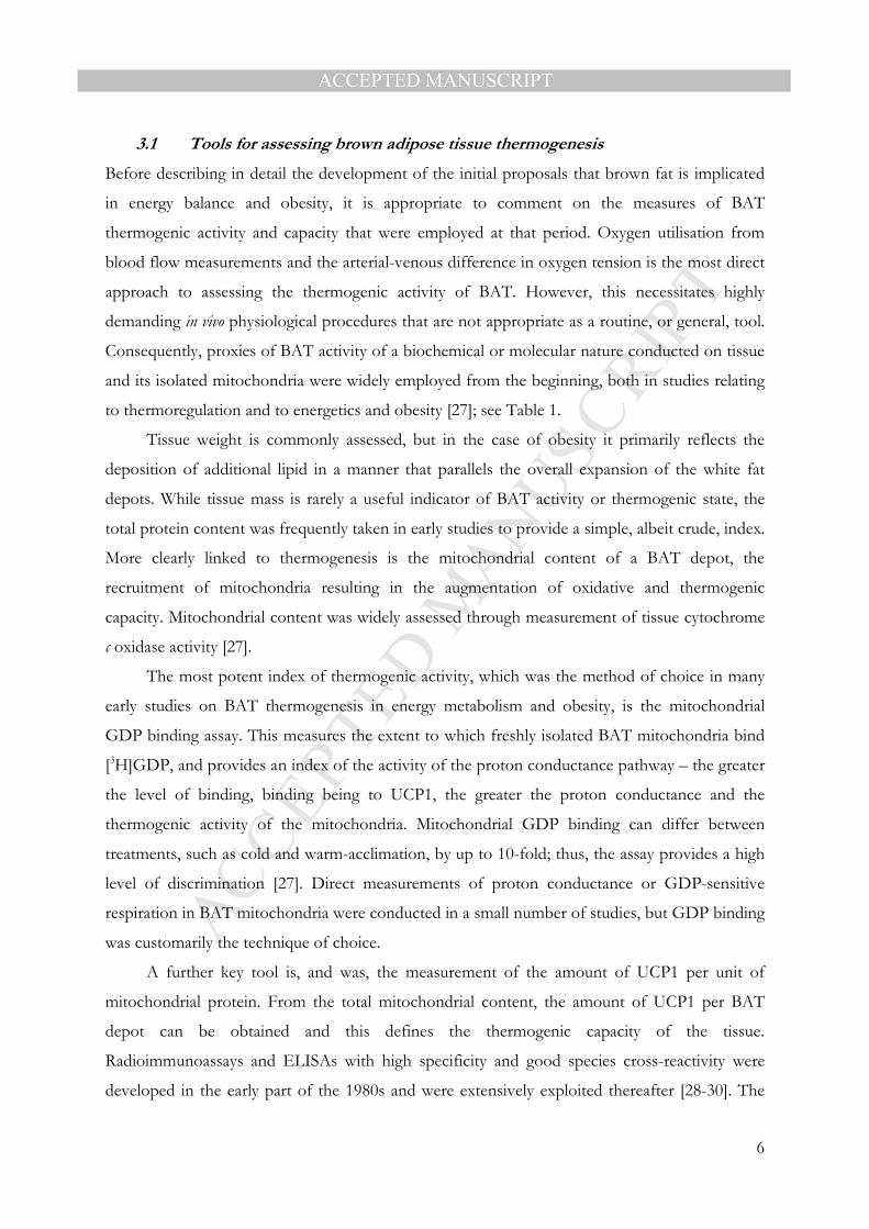
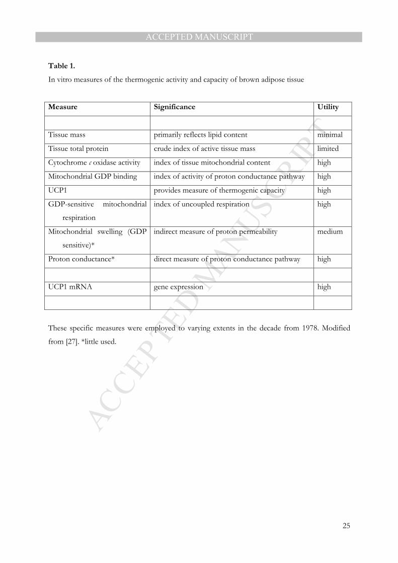
3.1 Tools for assessing brown adipose tissue thermogenesis
Before describing in detail the development of the initial proposals that brown fat is implicated
in energy balance and obesity, it is appropriate to comment on the measures of BAT
thermogenic activity and capacity that were employed at that period. Oxygen utilisation from
blood flow measurements and the arterial-venous difference in oxygen tension is the most direct
approach to assessing the thermogenic activity of BAT. However, this necessitates highly
demanding in vivo physiological procedures that are not appropriate as a routine, or general, tool.
Consequently, proxies of BAT activity of a biochemical or molecular nature conducted on tissue
and its isolated mitochondria were widely employed from the beginning, both in studies relating
to thermoregulation and to energetics and obesity [27]; see Table 1.
Tissue weight is commonly assessed, but in the case of obesity it primarily reflects the
deposition of additional lipid in a manner that parallels the overall expansion of the white fat
depots. While tissue mass is rarely a useful indicator of BAT activity or thermogenic state, the
total protein content was frequently taken in early studies to provide a simple, albeit crude, index.
More clearly linked to thermogenesis is the mitochondrial content of a BAT depot, the
recruitment of mitochondria resulting in the augmentation of oxidative and thermogenic
capacity. Mitochondrial content was widely assessed through measurement of tissue cytochrome
c oxidase activity [27].
The most potent index of thermogenic activity, which was the method of choice in many
early studies on BAT thermogenesis in energy metabolism and obesity, is the mitochondrial
GDP binding assay. This measures the extent to which freshly isolated BAT mitochondria bind
[3H]GDP, and provides an index of the activity of the proton conductance pathway – the greater
the level of binding, binding being to UCP1, the greater the proton conductance and the
thermogenic activity of the mitochondria. Mitochondrial GDP binding can differ between
treatments, such as cold and warm-acclimation, by up to 10-fold; thus, the assay provides a high
level of discrimination [27]. Direct measurements of proton conductance or GDP-sensitive
respiration in BAT mitochondria were conducted in a small number of studies, but GDP binding
was customarily the technique of choice.
A further key tool is, and was, the measurement of the amount of UCP1 per unit of
mitochondrial protein. From the total mitochondrial content, the amount of UCP1 per BAT
depot can be obtained and this defines the thermogenic capacity of the tissue.
Radioimmunoassays and ELISAs with high specificity and good species cross-reactivity were
developed in the early part of the 1980s and were extensively exploited thereafter [28-30]. The

MANUSCRIP
T
ACCEPTED
ACCEPTED MANUSCRIPT
7
cloning of the UCP1 gene in the mid 1980s provided the ability to measure mRNA levels,
enabling the factors that regulate the expression of the gene to be investigated [31-32]. One of
the first observations was that cold exposure induces a rapid stimulation of UCP1 gene
expression [33].
3.2 BAT and diet-induced thermogenesis
Following the initial proposition - based on tissue mass, temperature, and lipolytic sensitivity -
that BAT is involved in DIT, in a follow-up study molecular indices of thermogenesis were
assessed in cafeteria-fed and normal rats. The BAT of animals exhibiting facultative DIT had
increased total protein, mitochondrial content and oxidative capacity (cytochrome c oxidase and
α-glycerophosphate dehydrogenase activities); critically, they also had increased mitochondrial
GDP binding and exhibited an increase in GDP-sensitive respiration [34]. Later studies,
employing either an ELISA for UCP1 or densitometric analysis of SDS-PAGE gels,
demonstrated a marked recruitment of UCP1 in BAT of cafeteria-fed rats [35-36]. The levels of
the protein increased both ‘per mg of mitochondrial protein’ and ‘per depot’, confirming that the
capacity for BAT thermogenesis is increased in DIT. UCP1 gene expression, from
measurements of the mRNA level, was also soon shown to be elevated in cafeteria-fed rats [36].
Collectively, these changes indicate extensive activation of BAT and substantial increases in
thermogenic capacity in animals exhibiting high levels of DIT; such changes parallel those that
take place following adaptation, or acclimation, to the cold [37]. Further early studies
documented additional changes in the metabolic activity of BAT in cafeteria-diet rats, including
reduced lipogenesis which is likely to reflect the high fat content that characterises such diets
[38]. It should be noted that in practise not all studies in the 1980’s supported a role for BAT in
DIT [39]; however, in the past decade reports involving the genetic ablation of UCP1 have
provided unequivocal evidence for a central role for the tissue in this form of thermogenesis
[40].
3.3 BAT in obese animals
The initial observations of a reduction in the thermogenic activity of BAT in ob/ob mice were
followed by further studies on these leptin-deficient obese mutants as well as on several other
animal models of obesity. A blood flow study employing radioactively labelled microspheres to
map regional blood flow in young (5 week-old) mice found that BAT was a major site of
noradrenaline-stimulated NST in normal, lean mice consistent with the observations on cold-
acclimated rats [41]. Importantly, it also demonstrated that the reduced thermogenic capacity of

MANUSCRIP
T
ACCEPTED
ACCEPTED MANUSCRIPT
8
ob/ob mice in response to noradrenaline was almost entirely due to decreased heat production by
BAT. Blood flow studies on Zucker fa/fa (Leprfa/Leprfa) rats resulted in a similar conclusion, the
effect being attenuated by adrenalectomy [42].
Among the reported changes in BAT of mature ob/ob mice was a reduced concentration of
UCP1 in the mitochondria relative to lean siblings [43]. However, the UCP1 concentration was
found to be normal in ob/ob mice during the suckling period and shortly after weaning, indicating
that the reduced thermogenic capacity of the older mutant animals is not an intrinsic defect [43].
Since GDP binding is reduced even in suckling ob/ob mice, activity is nevertheless reduced.
Again, similar results were evident in fa/fa rats [43] with a decrease in GDP binding being
evident from the early days (day 2) of postnatal life [44]. Reduced GDP binding under basal
conditions and a lack of activation by diet, but a normal activation by cold, was additionally
reported for fa/fa rats [45]. Decreased GDP binding was also shown in both adult and suckling
db/db mice which, like the fa/fa rat, are characterised by a mutation in the leptin receptor [46].
Further studies on obese mutants demonstrated lower sympathetic activity in BAT, based on
noradrenaline turnover studies, when the animals are housed under normal environmental
conditions, but both acute cold-exposure and cold-acclimation induce a similar activation in
obese mice as in their lean siblings [47-48].
In addition to extensive studies on the single gene obese mutants, the properties of BAT
were widely explored in several non-genetic models of obesity. The attractions of the obese
mutants are, of course, the combination of the extreme obesity together with its early onset. In
general, the obesity exhibited by non-genetic models is less dramatic, and unlike the main obese
mutants does not relate specifically to abnormalities in the leptin system – whether of the
hormone itself (Lepob/Lepob) or of the receptor (Leprdb/Leprdb, Leprfa/Leprfa). Among the
experimentally-derived obesities that were initially investigated are those in which obesity is
induced by surgical lesioning of the ventromedial hypothalamus (VMH) – at the time a classical
model of obesity – or by the administration gold thioglucose (which also results in a lesion of the
VMH). In both these models, increased BAT mass, reflecting the deposition of excess lipid, and
reduced GDP binding were evident, together with a normal response to cold in terms of the
acute activation of thermogenesis [49-50]. Decreased mitochondrial content was observed in the
VMH-lesioned rats [51], though not in gold thioglucose-induced obese mice [52].
A role for glucocorticoids in the impaired thermogenic activity of obesity was a common
focus in several early studies and this followed particularly from the notable effects of
adrenalectomy on energy balance in both ob/ob mice and fa/fa rats; adrenalectomy leads to the
attenuation of hyperphagia and obesity itself, as well as of insulin resistance and other metabolic

MANUSCRIP
T
ACCEPTED
ACCEPTED MANUSCRIPT
9
abnormalities [53-54]. The effect of adrenalectomy on BAT in the obese mutants is to normalise
the reduced GDP binding and the mitochondrial content, responses that are rapid and which are
reversed by the administration of corticosterone [55-56]. It was noted that adrenalectomy had no
effect on lean animals [56]. Two other key observations were part of the early reports;
adrenalectomy was shown to lead to a normalisation of sympathetic activity in BAT of fa/fat rats
[57], and both the response to overfeeding and the immediate thermic effect of a single meal
were restored in brown fat of the obese animals [58].
A second approach to examining the role of glucocorticoids came from studies in which
corticosterone was directly administered to lean mice. Administration of the hormone leads to a
moderate obesity, with a reduction in mitochondrial content in BAT and a fall in GDP binding
[59]. This inhibition of BAT activity by corticosterone was consistent with the studies on
adrenalectomy in obese rodents.
3.4 Further models of altered energetics
In addition to investigations on overt obese models, one of the alternative strands in early studies
on the role of BAT in nutritional energetics came from the exploration of changes in the tissue
in physiological situations where there are substantial alterations in energy flux and/or energy
balance. These included pregnancy and lactation, photoperiod, seasonally-induced changes in
body fat, and marked alterations in the quantity or composition of the diet (Fig. 4). In each case,
changes in BAT consistent with the concept that the tissue is an important component in energy
balance and its regulation were observed (see [60-61]). Two specific examples will be summarised
here which were of particular interest to the present author: lactation and fasting.
Lactation in small animals is characterised by a very substantial increase in food intake in
order to fuel the high energy costs of milk production, intake in mice being at least twice that of
the pre-pregnant state [62]. The amount of lipid stored in rodents in pregnancy is small and its
subsequent mobilisation during lactation makes only a very limited contribution to the energy
costs of milk production – in contrast to larger mammals. Lactation in mice was shown to lead
to a major atrophy of BAT, the total protein and mitochondrial contents being markedly
decreased, as is GDP binding and GDP-sensitive respiration [63]. Later studies demonstrated
that there is a marked fall in UCP1 concentration in lactation [64] and the decrease in
thermogenic activity and capacity in BAT reflects a fall in the sympathetic drive to the tissue [65].
The functional atrophy of BAT in lactating rodents begins in late pregnancy, peaks in mid-late
lactation, and is reversed following weaning [63-64].

MANUSCRIP
T
ACCEPTED
ACCEPTED MANUSCRIPT
10
The scale of the atrophy of BAT in lactation is such that the thermogenic activity and
capacity of the tissue is similar to that in mice acclimated to thermoneutrality [62]. The energetic
implications of the near total suppression of BAT thermogenesis during lactation are
considerable; it has been estimated that at an environmental temperature of 21oC some 40% of
the maintenance energy expenditure of non-pregnant animals is ‘saved’ in lactating mice, this
reflecting the energy costs of thermoregulatory NST. In effect, the additional heat generated by
the obligatory DIT associated with the increased food intake in lactation, together with that
consequent to the synthesis of milk, negates the requirement for NST through BAT; heat from
rapid foetal growth would have a similar effect in late pregnancy [66]. It is also noteworthy that
the hyperphagia of lactating rodents does not lead to the stimulation of facultative DIT in BAT.
Fasting represents the most extreme of nutritional manipulations and several early studies
examined the effects of total food deprivation in rats and mice. Although some differences were
observed which are likely to be due to variations in the length of the period of food deprivation,
the caging conditions (number of animals per cage) and whether the animals were able to
undergo coprophagy, a reduction in mass (due to loss of lipid) and GDP binding was widely
observed which is reversible on re-feeding [67-71]. Fasting was also found to lead to a reduction
in mitochondrial content and in UCP1 (per unit of mitochondrial protein and per depot), and
thus of thermogenic capacity – effects that were again reversed on re-feeding [70-71].
Despite these observations, with prolonged starvation thermoregulatory needs begin to
predominant. While fasting rats for 24-48 h was shown to lead to a fall in UCP1 mRNA level,
which was reversed on refeeding, longer fasting resulted in increases in UCP1 mRNA [72]. This
rise in UCP1 mRNA on prolonged fasting was not observed, however, if the rats were at
thermoneutrality (28oC) rather than room temperature (23oC), suggesting that in starved animals
the drive to maintain body temperature through NST ultimately counteracts the short-term loss
of BAT resulting from food-deprivation [72].
Overall, the changes in BAT in the specific situations illustrated, as well as in a number of
other physiological conditions, were consistent with the core concept of a central role of the
tissue in nutritional energetics and in the aetiology of obesity in experimental animals (Fig. 4). In
the early 1990s the link between decreased BAT thermogenesis and obesity was further
established through genetic ablation studies in which the knockdown of UCP1 in transgenuc
mice was shown to result in obesity [40, 73].

MANUSCRIP
T
ACCEPTED
ACCEPTED MANUSCRIPT
11
4. Early evidence for active BAT in humans
A critical question was - and in part still is - the extent to which the animal data on BAT is
applicable to human energy metabolism, and to obesity in man in particular. This has
encompassed two distinct issues: (i) whether facultative thermogenesis is more than a very minor
component of energy expenditure in adults, and (ii) whether active BAT is present in humans
beyond the early years of life. The first question has been a matter of continuing debate, indeed
controversy, as has the extent to which reduced facultative thermogenesis may play a role in the
aetiology of obesity. The energy expenditure of mice at room temperature (21oC) is up to twice
that thermoneutrality (32oC), while at 4oC it is some 3 times higher, these differences reflecting
the energy costs of thermoregulation (primarily NST) [18]. There is no doubt that the energy
expenditure of adult humans on thermogenesis is proportionately considerably less, even on
exposure to low environmental temperatures.
Studies by Hull and colleagues, in particular, in the 1960s identified plentiful amounts of
BAT in newborn infants, the earliest unequivocal description of BAT in humans being at the
beginning of the last century [74-76]. The general consensus at the time was that BAT disappears
from humans over the first few years of postnatal life, though there was some evidence for the
persistence of the tissue in adults [77] and that when present it can be activated by stimuli such
as cold. However, these investigations were anatomical and histological in nature, largely centring
on the visualisation of multilocular fat cells. Given that they were made prior to the discovery of
UCP1, the identification of BAT was not based on the presence of the critical diagnostic feature
of a thermogenic adipocyte – whether brown or brite (beige).
UCP1 was soon identified and isolated from human adipose tissue and antibodies raised
against it to provide a tool for the subsequent exploration of the extent to which BAT is present
in adult humans [78-79]. A radioimmunoassay based on these antibodies demonstrated that
immunoreactive UCP1 is present in specific adipose tissue depots (perirenal and axillary) of
many adults, albeit at lower amounts than in children [80]. Patients with phaeochromocytoma, in
which there are high circulating levels of noradrenaline, have much higher UCP1 concentrations
in perirenal adipose tissue as well as a high mitochondrial content and GDP-sensitive respiration
[81]. These observations followed an earlier study on phaeochromocytoma patients in which
adipose tissue from around the adrenals and the kidneys was found to be rich in mitochondria
with a well-developed cristae structure and which exhibited GDP-sensitive respiration [82]. The
human UCP1 gene was subsequently cloned and found to be expressed in perirenal adipose
tissue of phaeochromocytoma patients [83].

MANUSCRIP
T
ACCEPTED
ACCEPTED MANUSCRIPT
12
Despite this clear evidence for BAT in adult humans and its capacity for activation
following an appropriate stimulus, there was little recognition that the tissue is present and a
potential component of energy expenditure in adults. As a consequence, a decade after a link
between brown fat and obesity was unequivocally established in rodents, interest in the idea that
impaired BAT thermogenesis plays a role in the aetiology of human obesity faded sharply.
Indeed, interest in brown fat at all levels waned during the 1990s.
5. The past decade: 2007 to 2016
In the late 2000s there was a major renaissance of interest in brown fat. This was partly because
of two key discoveries relating to the fundamental biology of the tissue – that brown adipocytes,
in contrast to white fat cells, are derived from myogenic precursors in skeletal muscle [84-85],
and that there is a third type of adipocyte, the brite (or beige) fat cell, which expresses UCP1
together with other, though not all, of the molecular markers of brown adipocytes [86-87].
However, the critical factor underlying the renewed focus on BAT has come from the
application of a procedure - fluorodeoxyglucose positron emission tomography (FDG-PET) -
which is employed in cancer investigations to track the metastasis of tumours by localising areas
that exhibit a high rate of glucose uptake. FDG-PET studies found high levels of glucose uptake
in fat tissue sites which had a distribution pattern similar to the presumptive pattern of BAT in
adult humans based on earlier anatomical observations [77].
Firm evidence that the fat tissue identified as having high glucose uptake is indeed BAT
was presented in 2009; the tissue exhibited clear immunostaining for UCP1 [88-89]. These
studies thus confirmed what the pioneering work in the 1980s had indicated – that BAT is clearly
present and functional in adult humans. The continuing application of FDG-PET has
demonstrated that not only is BAT in adults stimulated by cold and by insulin [89-92], but that it
is less active in older subjects - and importantly, the activity is lower in obese than in lean
individuals, being inversely proportional to BMI (body mass index) [88, 90, 93-94]. Thus, some
forty years after the initial proposal that BAT thermogenesis is impaired in the obese, the tissue
has again become a focus of research into the causes of obesity. There is correspondingly a
renewed interest in the activation and/or recruitment of BAT as a therapeutic route for the
treatment of obesity [95-97].
Important metabolic roles for BAT have also been recently suggested, specifically in
triglyceride clearance, insulin sensitivity, and in glucose homeostasis where the tissue has been
proposed as a major organ in glucose disposal [98-100]. As a consequence, reduced activity in
BAT has now been linked to the development of the metabolic syndrome [99, 101]. An

MANUSCRIP
T
ACCEPTED
ACCEPTED MANUSCRIPT
13
important role for brown fat in glucose removal was in practise first suggested in the early 1980s
- on the basis of the high activity of key glycolytic enzymes [102], together with high rates of 2-
deoxyglucose uptake in the tissue, uptake being stimulated by both insulin and noradrenaline
[103]. It is noteworthy that in the late 1970s very high rates of lipogenesis were also documented
in rodent BAT, particularly following cold-acclimation where the tissue is the major site of fatty
acid synthesis in whole-body terms [104-106].
The importance of insulin sensitivity in BAT thermogenesis was also noted at that time,
with the development of insulin resistance in the tissue in ob/ob mice being associated with a loss
in the ability to stimulate thermogenic activity on exposure to cold [107]. The reversal of insulin
resistance through administration of ciglitazone, the prototype thiazolidinedione, was further
shown to restore the normal cold-induced increase in GDP binding in the obese mutants [108]
6. Conclusions
The studies that were initiated on BAT thermogenesis towards the end of the 1970s resulted in a
paradigm shift in the understanding of nutritional energetics and the development of obesity.
They also resulted in a substantial shift in our comprehension of the physiological functions of
brown fat itself. Although the involvement of BAT in energy balance and obesity was incidental
to the exploration of the fundamental cellular and molecular mechanisms by which heat is
generated in brown adipocytes, considerable attention was drawn to the tissue because of this
link and interest in the more basic aspects of how these particular adipocytes function was
heightened. Certainly, the later discovery of a family of UCPs based on UCP1 would in all
probability have taken rather longer.
The demonstration in the 1980s that UCP1 is present in adipose tissue depots of adult
humans and that human BAT can be activated, at least in the present of the hypersecretion of
noradrenaline in phaeochromocytoma, was an important backdrop to the studies on human
BAT that emerged in the late 2000s following the application of the technique of FDG-PET. In
the case of human BAT, there is still uncertainty as to whether the tissue can make more than a
minor contribution to overall energy expenditure in adults. There is, nonetheless, renewed
interest in BAT as a therapeutic target for the treatment of obesity – whether by activating
existing brown adipocytes, recruiting new brown fat cells, or by the ‘beiging’ of specific adipose
tissue depots [97]. Some of the proposed routes by which these options might be achieved are
extremely challenging, and are in all probablility unrealistic.
Adult humans are not the only species where there has been uncertainty as to the
quantitative importance of BAT to energy expenditure. Despite the early demonstration that

MANUSCRIP
T
ACCEPTED
ACCEPTED MANUSCRIPT
14
BAT is activated in rats exhibiting DIT, the extent to which the tissue accounts for the
expenditure associated with this facultative process even in rodents is debated. Indeed, it has
been argued that this is not a function of the tissue [109]; nevertheless, genetic ablation studies
would seem to provide unequivocal evidence that BAT is central to DIT in rodents [40].
7. Coda – A Personal Note
Between 29 July and 3 August 1979, the Fourth International Symposium on the Pharmacology
of Thermoregulation was held at St Catherine’s College, Oxford. One of the organisers, Dr
Eduard Schönbaum, had become aware of the emerging interest in brown fat as a factor in the
regulation of energy balance and the development of obesity. He then suggested that some of us
involved should attend the conference and present our work. When the meeting finished on the
Friday afternoon, four of us – Jean Himms-Hagen, Nancy Rothwell, Michael (Mike) Stock and I
- adjourned to the ‘Turf Tavern’, an iconic Oxford pub, for beer and talk. I remember vividly the
intensity and excitement of that afternoon as we shared thoughts and speculations on the
possible relationship between brown fat and obesity. For me, there has been nothing quite like
that afternoon throughout my scientific career, neither before nor since.
One of the immediate outcomes of our discussions was that Mike Stock, Nancy Rothwell
and I agreed to collaborate on examining whether the mitochondrial proton conductance
pathway was activated in rats exhibiting DIT when fed a cafeteria diet. Their iconic paper
proposing a role for brown fat in DIT was to appear in Nature just a month later (6 September
1979) [15], and my PhD student Anne Goodbody and I had recently set-up at the MRC Dunn
Nutrition Laboratory in Cambridge the GDP binding assay to assess thermogenic activity. Mike
and Nancy subsequently transported rats in the boot of a car (something that would not now be
permitted) from London to Cambridge where we undertook the GDP binding measurements
and showed that DIT was indeed associated with an activation of the proton conductance
pathway in BAT mitochondria. On 20 February 1980 we submitted the findings as a ‘Letter to
Nature’, and this was published just 4 months later on 17 July [34].
This collaboration featured in the BBC Horizon science documentary on brown fat - ‘The
Fat in the Fire’ - which was broadcast in the UK on 10 December 1979.

MANUSCRIP
T
ACCEPTED
ACCEPTED MANUSCRIPT
15
Conflicts of interest
The author declares that he has no conflicts of interest.
Acknowledgements
I am grateful to my students and collaborators at the former Medical Research Council Dunn
Nutrition Laboratory in Cambridge, UK, who between 1977 and 1985 made pivotal
contributions to our programme on brown adipose tissue in energy metabolism and obesity. I
also thank the Distinguished Scientist Fellowship programme at King Saud University, Saudi
Arabia, for current funding.

MANUSCRIP
T
ACCEPTED
ACCEPTED MANUSCRIPT
16
References
[1] B.A. Afzelius, Brown adipose tissue: Its gross anatomy, histology , and cytology, In Brown
Adipose Tissue Edited by O Lindberg; American Elsevier, New York (1970) 1-31.
[2] R.E. Smith, B.A. Horwitz, Brown fat and thermogenesis, Physiol. Rev. 49 (1969) 330-425.
[3] D.G. Nicholls, R.M. Locke, Thermogenic mechanisms in brown fat, Physiol. Rev. 64
(1984) 1-64.
[4] D. Ricquier, J.C. Kader, Mitochondrial protein alterations in active brown fat: A sodium
dodecyl sulfate-polyacrylamide gel electrophoretic study, Biochem. Biophys. Res.
Commun. 73 (1976) 577-583.
[5] C. Fleury, M. Neverova, S. Collins, S. Raimbault, O. Champigny, C. Levi Meyrueis, F.
Bouillaud, M.F. Seldin, R.S. Surwit, D. Ricquier, C.H. Warden, Uncoupling protein-2: A
novel gene linked to obesity and hyperinsulinemia, Nature Genet. 15 (1997) 269-272.
[6] O. Boss, S. Samec, A. Paoloni-Giacobino, C. Rossier, A. Dulloo, J. Séydoux, P. Muzzin,
J.P. Giacobino, Uncoupling protein-3: A new member of the mitochondrial carrier family
with tissue-specific expression, FEBS Lett. 408 (1997) 39-42.
[7] D. Ricquier, F. Bouillaud, The mitochondrial uncoupling proteins, M S-Medecine Sciences
14 (1998) 889-897.
[8] L. Jansky, Non-shivering thermogenesis and its thermoregulatory significancce, Biol. Rev.
48 (1973) 85-132.
[9] D.O. Foster, M.L. Frydman, Nonshivering thermogenesis in the rat. II. Measurements of
blood flow with microspheres point to brown adipose tissue as the dominant site of the
calorigenesis induced by noradrenaline, Can J Physiol Pharmacol 56 (1978) 110-122.
[10] D.O. Foster, M.L. Frydman, Tissue distribution of cold-induced thermogenesis in
conscious warm- or cold-acclimated rats re-evaluated from changes in tissue blood flow:
The dominant role of brown adipose tissue in the replacement of shivering by non-
shivering thermogenesis, Can. J. Physiol. Pharmacol. 57 (1979) 257-270.
[11] D.O. Foster, M.L. Frydman, Comparison of microspheres and 86Rb+ as tracers of the
distribution of cardiac output in rats indicates invalidity of 86Rb+-based measurements,
Can. J. Physiol. Pharmacol. 56 (1977) 97-109.
[12] D.S. Miller, P. Mumford, Gluttony (1): An experimental study of overeating low- or high-
protein diets, Am. J. Clin. Nutr. 20 (1967) 1212-1222.
[13] D.S. Miller, P. Mumford, M.J. Stock, Gluttony. 2. Thermogenesis in overeating man, Am.
J. Clin. Nutr. 20 (1967) 1223-1229.

MANUSCRIP
T
ACCEPTED
ACCEPTED MANUSCRIPT
17
[14] A. Djazayery, D.S. Miller, M.J. Stock, Energy balances in obese mice, Nutr. Metab. 23
(1979) 357-367.
[15] N.J. Rothwell, M.J. Stock, A role for brown adipose tissue in diet-induced thermogenesis,
Nature 281 (1979) 31-35.
[16] N.J. Rothwell, M.J. Stock, Regulation of energy balance, Ann. Rev. Nutr 1 (1981) 235-256.
[17] P.L. Thurlby, P. Trayhurn, The role of thermoregulatory thermogenesis in the
development of obesity in genetically obese (ob/ob) mice pair-fed with lean siblings, Br. J.
Nutr. 42 (1979) 377-385.
[18] P. Trayhurn, W.P.T. James, Thermoregulation and non-shivering thermogenesis in the
genetically obese (ob/ob) mouse, Pflügers Archiv Eur. J. Physiol. 373 (1978) 189-193.
[19] P. Trayhurn, L. Fuller, The development of obesity in genetically diabetic-obese (db/db)
mice pair-fed with lean siblings. The importance of thermoregulatory thermogenesis,
Diabetologia 19 (1980) 148-153.
[20] B.G. Miller, R.F. Grimble, T.G. Taylor, Liver protein metabolism response to cold in
genetically obese (ob/ob) mice, Nature 266 (1977) 184-186.
[21] G.A. Bray, D.A. York, Y. Yukimara, Activity of (Na+ + K+)-ATPase in the liver of animals
with experimental obesity, Life Sci. 22 (1978) 1637-1642.
[22] E.A. Newsholme, B. Crabtree, Substrate cycles in metabolic regulation and in heat
generation, Biochem. Soc. Symp. 41 (1976) 61-109.
[23] E.A. Newsholme, Substrate cycles: Their metabolic, energetic and thermic consequences in
man, Biochem. Soc. Symp. 43 (1978) 183-205.
[24] D.A. York, G.A. Bray, Y. Yukimura, An enzymatic defect in the obese (ob/ob) mouse; loss
of thyroid-induced sodium - and potassium-dependent adenosinetriphosphatase, Proc.
Natl. Acad. Sci. U.S.A. 75 (1978) 477-481.
[25] M.H. Lin, D.R. Romsos, T. Akera, G.A. Leveille, Na+; K+-ATPase enzyme units in skeletal
muscle and liver of 14-day-old lean and obese (ob/ob) mice, Proc. Soc. Exp. Biol. Med. 161
(1979) 235-238.
[26] J. Himms-Hagen, M. Desautels, A mitochondrial defect in brown adipose tissue of the
obese (ob/ob) mouse: Reduced binding of purine nucleotides and a failure to respond to
cold by an increase in binding, Biochem. Biophys. Res. Commun. 83 (1978) 628-634.
[27] P. Trayhurn, R.E. Milner, A commentary on the interpretation of invitro biochemical
measures of brown adipose tissue thermogenesis, Can J. Physiol. Pharmacol. 67 (1989)
811-819.

MANUSCRIP
T
ACCEPTED
ACCEPTED MANUSCRIPT
18
[28] D. Ricquier, J.P. Barlet, J.M. Garel, G.M. Combes, M.P. Dubois, An immunological study
of the uncoupling protein of brown adipose tissue mitochondria, Biochem. J. 210 (1983)
859-866.
[29] M.E.J. Lean, W.J. Branch, W.P.T. James, G. Jennings, M. Ashwell, Measurement of rat
brown-adipose tissue mitochondrial uncoupling protein by radioimmunoassay. Increased
concentration after cold acclimation, Biosci. Rep. 3 (1983) 61-71.
[30] E.S. Hansen, J. Nedergaard, B. Cannon, J. Knudsen, Enzyme-linked immunosorbent assay
(ELISA) studies of the interaction between mammalian and avian anti-thermogenin
antibodies and brown-adipose tissue mitochondria from different species, Comp.
Biochem. Physiol. 79B (1984) 44l-445.
[31] F. Bouillaud, D. Ricquier, J. Thibault, J. Weissenbach, Molecular approach to
thermogenesis in brown adipose tissue: cDNA cloning of the mitochondrial uncoupling
protein, Proc. Natl. Acad. Sci. USA 82 (1985) 445-448.
[32] S. Klaus, L. Casteilla, F. Bouillaud, D. Ricquier, The uncoupling protein UCP - a
membraneous mitochondrial ion carrier exclusively expressed in brown adipose tissue, Int.
J. Biochem. 23 (1991) 791-801.
[33] D. Ricquier, G. Mory, F. Bouillaud, J. Thibault, J. Weissenbach, Rapid increase of
mitochondrial uncoupling protein and its mrna in stimulated brown adipose tissue, FEBS
Lett. 178 (1984) 240-244.
[34] S.L. Brooks, N.J. Rothwell, M.J. Stock, A.E. Goodbody, P. Trayhurn, Increased proton
conductance pathway in brown adipose tissue mitochondria of rats exhibiting diet-induced
thermogenesis, Nature 286 (1980) 274-276.
[35] J. Nedergaard, A. Raasmaja, B. Cannon, Parallel increases in amount of (3H)GDP binding
and thermogenin antigen in brown-adipose-tissue mitochondria of cafeteria-fed rats,
Biochem. Biophys. Res. Commun. 122 (1984) 1328-1336.
[36] R. Falcou, F. Bouillaud, G. Mory, M. Apfelbaum, D. Ricquier, Increase of uncoupling
protein and its mRNA in brown adipose tissue of rats fed on 'cafeteria' diet, Biochem. J.
231 (1985) 241-244.
[37] P. Trayhurn, M. Ashwell, G. Jennings, D. Richard, D.M. Stirling, Effect of warm or cold
exposure on GDP binding and uncoupling protein in rat brown fat, Am. J. Physiol.
Endocrinol. Metab. 252 (1987) E237-E243.
[38] N.J. Rothwell, M.J. Stock, P. Trayhurn, Reduced lipogenesis in cafeteria-fed rats exhibiting
diet-induced thermogenesis, Biosci. Rep. 3 (1983) 217-224.

MANUSCRIP
T
ACCEPTED
ACCEPTED MANUSCRIPT
19
[39] S. Ma, D.O. Foster, B.E. Nadeau, J. Triandafillou, Absence of increased oxygen
consumption in brown adipose tissue of rats exhibiting "Cafeteria" diet-induced
thermogenesis, Can J. Physiol. Pharmacol 66 (1988) 1347-1354.
[40] H.M. Feldmann, V. Golozoubova, B. Cannon, J. Nedergaard, UCP1 ablation induces
obesity and abolishes diet-induced thermogenesis in mice exempt from thermal stress by
living at thermoneutrality, Cell Metab. 9 (2009) 203-209.
[41] P.L. Thurlby, P. Trayhurn, Regional blood flow in genetically obese (ob/ob) mice: The
importance of brown adipose tissue to the reduced energy expenditure on non-shivering
thermogenesis, Pflügers Archiv Eur. J. Physiol. 385 (1980) 193-201.
[42] S.J. Wickler, B.A. Horwitz, J.S. Stern, Blood flow to brown fat in lean and obese
adrenalectomized Zucker rats, Am. J. Physiol. Reg. Integr. Comp. Physiol. 25l (1986) R85l-
R858.
[43] M. Ashwell, S. Holt, G. Jennings, D.M. Stirling, P. Trayhurn, D.A. York, Measurement by
radioimmunoassay of the mitochondrial uncoupling protein from brown adipose tissue of
obese (ob/ob) mice and Zucker (fa/fa) rats at different ages, FEBS Lett. 179 (1985) 233-
237.
[44] R. Bazin, D. Eteve, M. Lavau, Evidence for decreased gdp binding to brown-adipose-
tissue mitochondria of obese zucker (fa/fa) rats in the very first days of life, Biochem. J.
221 (1984) 241-245.
[45] J. Triandafillou, J. Himms-Hagen, Brown adipose tissue in genetically obese (fa/fa) rats:
Response to cold and diet, Am. J. Physiol. Endocrinol. Metab. 244 (1983) E145-E150.
[46] A.E. Goodbody, P. Trayhurn, GDP binding to brown-adipose-tissue mitochondria of
diabetic-obese (db/db) mice. Decreased binding in both the obese and pre-obese states,
Biochem. J. 194 (1981) 1019-1022.
[47] A.W. Knehans, D.R. Romsos, Reduced norepinephrine turnover in brown adipose tissue
of ob/ob mice, Am. J. Physiol. Endocrinol. Metab. 242 (1982) E253-E26l .
[48] G. Zaror-Behrens, J. Himms-Hagen, Cold-stimulated sympathetic activity in brown
adipose tissue of obese (ob/ob) mice, Am. J. Physiol. Endocrinol. Metab. 244 (1983) E361-
E366.
[49] S. Hogan, D.V. Coscina, J. Himms-Hagen, Brown adipose tissue of rats with obesity-
inducing ventro-medial hypothalamic lesions, Am. J. Physiol. Endocrinol. Metab. 243
(1982) E338-E344.
[50] J. Seydoux, D. Ricquier, F. Rohner-Jeanrenaud, F. Assimacopoulos-Jeannet, J.P.
Giacobino, B. Jeanrenaud, L. Girardier, Decreased guanine nucleotide binding and reduced

MANUSCRIP
T
ACCEPTED
ACCEPTED MANUSCRIPT
20
equivalent production by brown adipose tissue in hypothalamic obesity: Recovery after
cold acclimation, FEBS Lett. 146 (1982) 161-164.
[51] J. Seydoux, F. Rohner-Jeanrenaud, F. Assimacopoulos-Jeannet, B. Jeanrenaud, Functional
disconnection of brown adipose tissue in hypothalamic obesity in rats, Pflügers Archiv
Eur. J. Physiol. 390 (1981) 1-4.
[52] S. Hogan, J. Himms-Hagen, Brown adipose tissue of mice with gold thioglucose-induced
obesity: Effect of cold and diet, Am. J. Physiol. Endocrinol. Metab. 244 (1983) E581-
E588.
[53] G.A. Bray, D.A. York, Hypothalamic and genetic obesity in experimental animals: An
autonomic and endocrine hypothesis, Physiol. Re.v 59 (1979) 719-809.
[54] G.A. Bray, Integration of energy intake and expenditure in animals and man: The
autonomic and adrenal hypothesis, Clinics Endocrinol. Metab. 13 (1984) 521-546.
[55] S. Holt, D.A. York, The effect of adrenalectomy on GDP binding to brown-adipose-tissue
mitochondria of obese rats, Biochem. J. 208 (1982) 819-822.
[56] S.J. Holt, D.A. York, Effect of adrenalectomy on brown adipose tissue of obese (ob/ob)
mice, Horm. Metab. Res. 16 (1984) 378-379.
[57] D.A. York, D. Marchington, S.J. Holt, J. Allars, Regulation of sympathetic activity in lean
and obese zucker (fa/fa) rats, Am. J. Physiol. Endocrinol. Metab. 249 (1985) E299-E305.
[58] D. Marchington, N.J. Rothwell, M.J. Stock, D.A. York, Energy balance; diet-induced
thermogenesis and brown adipose tissue in lean and obese (fa/fa) Zucker rats after
adrenalectomy, J. Nutr. 113 (1983) 1395-1402.
[59] K.S. Galpin, R.G. Henderson, W.P.T. James, P. Trayhurn, GDP binding to brown-
adipose-tissue mitochondria of mice treated chronically with corticosterone, Biochem. J.
214 (1983) 265-268.
[60] P. Trayhurn, Brown adipose tissue and energy balance, Brown Adipose Tissue (edited by P
Trayhurn and DG Nicholls) Edward Arnold, London (1986) 299-388.
[61] J. Himms-Hagen, Brown adipose tissue thermogenesis and obesity, Prog. Lipid Res. 28
(1989) 67-115.
[62] P. Trayhurn, Brown adipose tissue thermogenesis and the energetics of lactation in
rodents, Int. J. Obesity 9 Suppl 2 (1985) 81-88.
[63] P. Trayhurn, J.B. Douglas, M.M. McGuckin, Brown adipose tissue thermogenesis is
'suppressed' during lactation in mice, Nature 298 (1982) 59-60.

MANUSCRIP
T
ACCEPTED
ACCEPTED MANUSCRIPT
21
[64] P. Trayhurn, G. Jennings, Functional atrophy of brown adipose tissue in mice: Effects of
lactation and weaning on mitochondrial gdp binding and uncoupling protein, Biochem. J.
248 (1987) 273-276.
[65] P. Trayhurn, M.C. Wusteman, Sympathetic activity in brown adipose tissue in lactating
mice, Am. J. Physiol. Endocrinol. Metab. 253 (1987) E515-E520.
[66] B. Cannon, J. Nedergaard, Brown adipose tissue: Function and physiological significance,
Physiol. Rev. 84 (2004) 277-359.
[67] M. Hayashi, T. Nagasaka, Suppression of norepinephrine-induced thermogenesis in brown
adipose tissue by fasting, Am. J. Physiol. Endcorinol. Metab. 245 (1983) E582-E586.
[68] N.J. Rothwell, M.E. Saville, M.J. Stock, Brown fat activity in fasted and refed rats, Biosci.
Rep. 4 (1984) 35l-357.
[69] M. Desautels, Mitochondrial thermogenin content is unchanged during atrophy of bat of
fasting mice, Am. J. Physiol. Endcorinol. Metab. 249 (1985) E99-E106.
[70] P. Trayhurn, G. Jennings, Evidence that fasting can induce a selective loss of uncoupling
protein from brown adipose tissue mitochondria of mice, Biosci. Rep. 6 (1986) 805-810.
[71] P. Trayhurn, G. Jennings, Nonshivering thermogenesis and the thermogenic capacity of
brown fat in fasted and/or refed mice, Am. J. Physiol. Reg. Integr. Comp. Physiol. 254
(1988) R11-R16.
[72] O. Champigny, D. Ricquier, Effects of fasting and refeeding on the level of uncoupling
protein mRNA in rat brown adipose tissue - evidence for diet-induced and cold-induced
responses, J. Nutr. 120 (1990) 1730-1736.
[73] B.B. Lowell, V. S-Susulic, A. Hamann, J.A. Lawitts, J. Himms-Hagen, B.B. Boyer, L.P.
Kozak, J.S. Flier, Development of obesity in transgenic mice after genetic ablation of
brown adipose tissue, Nature 366 (1993) 740-742.
[74] W. Aherne, D. Hull, Brown adipose tissue and heat production in the newborn infant, J.
Path. Bact. 91 (1966) 223-234.
[75] D. Hull, The structure and function of brown adipose tissue, Br. Med. Bull. 22 (1966) 92-
96.
[76] M.E.J. Lean, W.P.T. James, Brown adipose tissue in man, Brown Adipose Tissue (edited
by P Trayhurn and DG Nicholls) Edward Arnold, London (1986) 339-365.
[77] J. Heaton, The distribution of brown adipose tissue in the human, J. Anat. ll2 (1972) 35-39.
[78] F. Bouillaud, G.M. Combes, D. Ricquier, Mitochondria of adult human brown adipose
tissue contain a 32,000-Mr uncoupling protein, Biosci. Rep. 3 (1983) 775-780.
MANUSCRIP
T
ACCEPTED
ACCEPTED MANUSCRIPT
22
[79] M.E.J. Lean, W.P.T. James, Uncoupling protein in human brown adipose tissue
mitochondria: Isolation and detection by specific antiserum, FEBS Lett. 163 (1983) 235-
240.
[80] M.E.J. Lean, W.P.T. James, G. Jennings, P. Trayhurn, Brown adipose tissue uncoupling
protein content in human infants, children and adults, Clin. Sci. 7l (1986) 29l-297.
[81] M.E.J. Lean, W.P.T. James, G. Jennings, P. Trayhurn, Brown adipose tissue in patients
with phaeochromocytoma, Int. J. Obesity l0 (1986) 2l9-227.
[82] D. Ricquier, M. Néchad, G. Mory, Ultrastructural and biochemical characterization of
human brown adipose tissue in pheochromocytoma, J. Clin. Endocrinol. Metab. 54 (1982)
803-807.
[83] F. Bouillaud, F. Villarroya, E. Hentz, S. Raimbault, A.M. Cassard, D. Ricquier, Detection
of brown adipose tissue uncoupling protein mrna in adult humans by a genomic probe,
Clin. Sci. 75 (1988) 21-27.
[84] J.A. Timmons, K. Wennmalm, O. Larsson, T.B. Walden, T. Lassmann, N. Petrovic, D.L.
Hamilton, R.E. Gimeno, C. Wahlestedt, K. Baar, J. Nedergaard, B. Cannon, Myogenic
gene expression signature establishes that brown and white adipocytes originate from
distinct cell lineages, Proc. Natl. Acad. Sci. USA 104 (2007) 4401-4406.
[85] P. Seale, B. Bjork, W. Yang, S. Kajimura, S. Chin, S. Kuang, A. Scime, S. Devarakonda,
H.M. Conroe, H. Erdjument-Bromage, P. Tempst, M.A. Rudnicki, D.R. Beier, B.M.
Spiegelman, PRDM16 controls a brown fat/skeletal muscle switch, Nature 454 (2008) 961-
967.
[86] N. Petrovic, T.B. Walden, I.G. Shabalina, J.A. Timmons, B. Cannon, J. Nedergaard,
Chronic peroxisome proliferator-activated receptor γ (PPARγ) activation of epididymally
derived white adipocyte cultures reveals a population of thermogenically competent,
UCP1-containing adipocytes molecularly distinct from classic brown adipocytes, J. Biol.
Chem. 285 (2010) 7153-7164.
[87] J. Wu, P. Boström, Lauren M. Sparks, L. Ye, Jang H. Choi, A.-H. Giang, M. Khandekar,
Kirsi A. Virtanen, P. Nuutila, G. Schaart, K. Huang, H. Tu, Wouter D.
van Marken Lichtenbelt, J. Hoeks, S. Enerbäck, P. Schrauwen, Bruce M. Spiegelman, Beige
adipocytes are a distinct type of thermogenic fat cell in mouse and human, Cell 150 (2012)
366-376.
[88] A.M. Cypess, S. Lehman, G. Williams, I. Tal, D. Rodman, A.B. Goldfine, F.C. Kuo, E.L.
Palmer, Y.H. Tseng, A. Doria, G.M. Kolodny, C.R. Kahn, Identification and importance
of brown adipose tissue in adult humans, New Engl. J. Med. 360 (2009) 1509-1517.

MANUSCRIP
T
ACCEPTED
ACCEPTED MANUSCRIPT
23
[89] K.A. Virtanen, M.E. Lidell, J. Orava, M. Heglind, R. Westergren, T. Niemi, M. Taittonen,
J. Laine, N.J. Savisto, S. Enerback, P. Nuutila, Functional brown adipose tissue in healthy
adults, New Engl. J. Med. 360 (2009) 1518-1525.
[90] W.D. van Marken Lichtenbelt, J.W. Vanhommerig, N.M. Smulders, J.M. Drossaerts, G.J.
Kemerink, N.D. Bouvy, P. Schrauwen, G.J. Teule, Cold-activated brown adipose tissue in
healthy men, New Engl. J. Med. 360 (2009) 1500-1508.
[91] J. Orava, P. Nuutila, Martin E. Lidell, V. Oikonen, T. Noponen, T. Viljanen, M. Scheinin,
M. Taittonen, T. Niemi, S. Enerbäck, Kirsi A. Virtanen, Different metabolic responses of
human brown adipose tissue to activation by cold and insulin, Cell Metab. 14 (2011) 272-
279.
[92] V. Ouellet, S.M. Labbe, D.P. Blondin, S. Phoenix, B. Guerin, F. Haman, E.E. Turcotte, D.
Richard, A.C. Carpentier, Brown adipose tissue oxidative metabolism contributes to energy
expenditure during acute cold exposure in humans, J. Clin. Invest. 122 (2012) 545-552.
[93] C. Pfannenberg, M.K. Werner, S. Ripkens, I. Stef, A. Deckert, M. Schmadl, M. Reimold,
H.-U. Häring, C.D. Claussen, N. Stefan, Impact of age on the relationships of brown
adipose tissue with sex and adiposity in humans, Diabetes 59 (2010) 1789-1793.
[94] T. Yoneshiro, S. Aita, M. Matsushita, T. Kameya, K. Nakada, Y. Kawai, M. Saito, Brown
adipose tissue, whole-body energy expenditure, and thermogenesis in healthy adult men,
Obesity 19 (2011) 13-16.
[95] M. Harms, P. Seale, Brown and beige fat: Development, function and therapeutic
potential, Nature Med. 19 (2013) 1252-1263.
[96] F. Villarroya, A. Vidal-Puig, Beyond the sympathetic tone: The new brown fat activators,
Cell Metab. 17 (2013) 638-643.
[97] P. Trayhurn, J.S. Arch, New physiological aspects of brown adipose tissue, Curr. Obesity
Rep. 3 (2014) 414-421.
[98] A. Bartelt, O.T. Bruns, R. Reimer, H. Hohenberg, H. Ittrich, K. Peldschus, M.G. Kaul,
U.I. Tromsdorf, H. Weller, C. Waurisch, A. Eychmuller, P.L.S.M. Gordts, F. Rinninger, K.
Bruegelmann, B. Freund, P. Nielsen, M. Merkel, J. Heeren, Brown adipose tissue activity
controls triglyceride clearance, Nature Med. 17 (2011) 200-205.
[99] A. Bartelt, J. Heeren, The holy grail of metabolic disease: Brown adipose tissue, Curr.
Opin. Lipidol. 23 (2012) 190-195.
[100] K.I. Stanford, R.J.W. Middelbeek, K.L. Townsend, D. An, E.B. Nygaard, K.M. Hitchcox,
K.R. Markan, K. Nakano, M.F. Hirshman, Y.-H. Tseng, L.J. Goodyear, Brown adipose
MANUSCRIP
T
ACCEPTED
ACCEPTED MANUSCRIPT
24
tissue regulates glucose homeostasis and insulin sensitivity, J. Clin. Invest. 123 (2013) 215-
223.
[101] J. Nedergaard, T. Bengtsson, B. Cannon, New powers of brown fat: Fighting the
metabolic syndrome, Cell Metab. 13 (2011) 238-240.
[102] G.J. Cooney, E.A. Newsholme, The maximum capacity of glycolysis in brown adipose
tissue and its relationships to control of the blood glucose concentration, FEBS Lett. 148
(1982) 198-200.
[103] G.J. Cooney, I.D. Caterson, E.A. Newsholme, The effect of insulin and noradrenaline on
the uptake of 2-[1-14c}deoxyglucose in vivo by brown adipose tissue and other glucose-
utilizing tissues of the mouse, FEBS Lett. 188 (1985) 257-261.
[104] J.G. McCormack, R.M. Denton, Evidence that fatty acid synthesis in the inter-scapular
brown adipose tissue of cold-adapted rats is increased in vivo by insulin by mechanisms
involving paralleled activation of pyruvate dehydrogenase and acetyl-coenzyme a
carboxylase, Biochem. J. 166 (1977) 627-630.
[105] P. Trayhurn, Fatty acid synthesis in vivo in brown adipose tissue liver and white adipose
tissue of the cold-acclimated rat, FEBS Lett. 104 (1979) 13-16.
[106] P. Trayhurn, Fatty acid synthesis in brown adipose tissue in relation to whole body
synthesis in the cold-acclimated golden hamster (mesocricetus auratus), Biochim. Biophys.
Acta 620 (1980) 10-17.
[107] S.W. Mercer, P. Trayhurn, The development of insulin resistance in brown adipose tissue
may impair the acute cold-induced activation of thermogenesis in genetically obese (ob/ob)
mice, Biosci. Rep. 4 (1984) 933-940.
[108] S.W. Mercer, P. Trayhurn, Effects of ciglitazone on insulin resistance and thermogenic
responsiveness to acute cold in brown adipose tissue of genetically obese (ob/ob) mice,
FEBS Lett. 195 (1986) 12-16.
[109] L.P. Kozak, Brown fat and the myth of diet-induced thermogenesis, Cell Metab. 11 (2010)
263-267.
MANUSCRIP
T
ACCEPTED
ACCEPTED MANUSCRIPT
25
Table 1.
In vitro measures of the thermogenic activity and capacity of brown adipose tissue
Measure Significance Utility
Tissue mass primarily reflects lipid content minimal
Tissue total protein crude index of active tissue mass limited
Cytochrome c oxidase activity index of tissue mitochondrial content high
Mitochondrial GDP binding index of activity of proton conductance pathway high
UCP1 provides measure of thermogenic capacity high
GDP-sensitive mitochondrial
respiration
index of uncoupled respiration high
Mitochondrial swelling (GDP
sensitive)*
indirect measure of proton permeability medium
Proton conductance* direct measure of proton conductance pathway high
UCP1 mRNA gene expression high
These specific measures were employed to varying extents in the decade from 1978. Modified
from [27]. *little used.

MANUSCRIP
T
ACCEPTED
ACCEPTED MANUSCRIPT
26
Legends to Figures
Fig. 1. A schematic representation of energy flux and energy expenditure in mammals, with the
buffering role of white adipose tissue. BMR, basal metabolic rate; NEAT, non-exercise activity
thermogenesis (which has been considered a component of expenditure.
Fig. 2. Illustration of the stimulation of facultative diet-induced thermogenesis in rats during
voluntary overfeeding on a cafeteria diet. The metabolisable energy intake, energy deposited and
energy expenditure of the control (Con) animals fed a standard laboratory diet and the cafeteria-
fed (Cafe) rats is shown. Data adapted from Rothwell and Stock [15].
Fig. 3. Development of obesity in young ob/ob mice pair-fed to the ad libitum food intake of
lean (+/+, ob/+) siblings. (a) energy gain, (b) gross efficiency (kJ energy gain/kJ energy intake).
The difference between lean and ob/ob in energy gain on precisely the same intake is due to the
reduced energy expenditure of the obese mutants. Data taken from [17].
Fig. 4. Schematic representation of how different physiological and pathological conditions in
which energy flux and balance alter are characterised by increased or decreased brown adipose
tissue thermogenesis in experimental animals. The diagram summarises the major situations in
which BAT thermogenesis changes, which in most cases reflect long-term adaptations.
Highlights:
1. BAT was shown in the late 1970s to be linked to energetics and obesity as well as
thermoregulation
2. Studies in the 1980s demonstrated reduced BAT thermogenesis in a range of animal
obesities
3. Active BAT was found in adult humans in the 1980s, underpinning the current interest in
the tissue

MANUSCRIP
T
ACCEPTED
ACCEPTED MANUSCRIPT
BMR activity thermogenesis(exercise, NEAT) (diet – DIT, cold – NST)
Energy intake
..… Genetic
….. Environmental
Fig. 1

MANUSCRIP
T
ACCEPTED
ACCEPTED MANUSCRIPT
ConCafe
0.0
2.5
5.0
7.5
10.0
12.5 ***
En
erg
y I
nta
ke
(M
J)
IntakeCon
Cafe0.0
0.5
1.0
1.5
2.0
2.5 *
Deposition
En
erg
y G
ain
(k
J)
ConCafe
0
2
4
6
8
10 ***
Expenditure
En
erg
y e
xp
en
dit
ure
(M
J)
Fig. 2

MANUSCRIP
T
ACCEPTED
ACCEPTED MANUSCRIPT
Lean Obese0
50
100
150
200
250E
ne
rgy
Ga
in (
kJ)
Lean Obese0
10
20
30
40
Gro
ss E
ffic
ien
cy (
%)
Fig. 3
(a)
(b)

MANUSCRIP
T
ACCEPTED
ACCEPTED MANUSCRIPT
Increased ↑:
Cold
Overfeeding (DIT)
Catecholamines
Arousal from hibernation
Cancer cachexia
Low protein diets
Decreased ↓:
Obesity (ob/ob, fa/fa, VMH)
Fasting
Lactation
Diabetes
Hibernation
Fig. 4

MANUSCRIP
T
ACCEPTED
ACCEPTED MANUSCRIPT
Highlights:
1. BAT was shown in the late 1970s to be linked to energetics and obesity as well as
thermoregulation
2. Studies in the 1980s demonstrated reduced BAT thermogenesis in a range of animal
obesities
3. Active BAT was found in adult humans in the 1980s, underpinning the current interest in
the tissue

Annex: Acceptance Information
Subject: Your Submission
Resent-Date: Tue, 6 Sep 2016 13:08:37 +0100
Resent-From: [email protected]
Date: Tue, 6 Sep 2016 12:07:31 +0000
From: Biochimie <[email protected]>
To: Paul Trayhurn <[email protected]>
CC: [email protected] <[email protected]>, [email protected]
Ref.: Ms. No. BIOCHI-D-16-00292R1
Origins and early development of the concept that brown adipose tissue
thermogenesis is linked to energy balance and obesity
Biochimie
Dear Professor Trayhurn,
I am happy to inform you that your manuscript is now accepted for
publication in Biochimie.
Thank you again for submitting your work to our Journal.
With kind regards,
Claude Forest, Ph.D.
Editor of Biochimie
For further assistance, please visit our customer support site at
http://help.elsevier.com/app/answers/list/p/7923. Here you can search for
solutions on a range of topics, find answers to frequently asked questions
and learn more about EES via interactive tutorials. You will also find our
24/7 support contact details should you need any further assistance from
one of our customer support representatives.
When your paper is published on ScienceDirect, you want to make sure it
gets the attention it deserves. To help you get your message across,
Elsevier has developed a new, free service called AudioSlides: brief,
webcast-style presentations that are shown (publicly available) next to
your published article. This format gives you the opportunity to explain
your research in your own words and attract interest. You will receive an
invitation email to create an AudioSlides presentation shortly. For more
information and examples, please visit http://www.elsevier.com/audioslides.