Embed Size (px)
Citation preview

Biochimica et Biophysica Acta 1857 (2016) 1139–1146
Contents lists available at ScienceDirect
Biochimica et Biophysica Acta
j ourna l homepage: www.e lsev ie r .com/ locate /bbab io
The Crabtree and Warburg effects: Do metabolite-induced regulationsparticipate in their induction?☆
Noureddine Hammad a,b,1, Monica Rosas-Lemus a,b,c,1, Salvador Uribe-Carvajal c,Michel Rigoulet a,b, Anne Devin a,b,⁎a Université Bordeaux, IBGC, UMR 5095, Bordeaux, Franceb Institut de Biochimie et Génétique Cellulaires, CNRS UMR 5095, Bordeaux, Francec Department of Molecular Genetics, Instituto de Fisiología Celular, Universidad Nacional Autónoma de México, 04510 Mexico DF, Mexico
Abbreviations: 2HG, 2-hydroxyglutarate; 6PGD, 6-phoACC, acetyl-CoA carboxylase; ANT, adenine nucleotide tphosphate; AMP, adenosinemonophosphate; AMPK, AMPphate; Fru6P, fructose 6 phosphate; Fru1,6BP, fructosehydratase; G6PDH, glucose 6 phosphate dehydrogenaseHIF, hypoxia inducible factor; Hxk2, type 2 hexokinase;LKB1, tumor suppressor liver kinase B1; PKM2, M2 isofoscaffolding-like adaptor protein mouse protein 25; MPC,NADPH, nicotinamide adenine nucleotide phosphate redtose phosphate pathway; PTP, permeability transition porekinase; PHDs, prolyl hydroxylase enzymes; Ru-5-P, Ribuloxygen species; STRAD, pseudokinase Ste20-related adapnase; TCA, tricarboxylic acid cycle.☆ This article is part of a Special Issue entitled ‘EBEC 201
Conference, Riva del Garda, Italy, July 2–6, 2016’, edited b⁎ Corresponding author at: Institut de Biochimie et Gé
5095, 1, rue Camille Saint Saëns, 33077 Bordeaux Cedex, FE-mail address: [email protected] (A. Devin).
1 Both authors contributed equally.
http://dx.doi.org/10.1016/j.bbabio.2016.03.0340005-2728/© 2016 Elsevier B.V. All rights reserved.
a b s t r a c t
a r t i c l e i n f oArticle history:Received 22 January 2016Received in revised form 24 March 2016Accepted 25 March 2016Available online 9 April 2016
The Crabtree and Warburg effects are two well-known deviations of cell energy metabolism that will be de-scribed herein. A number of hypotheses have been formulated regarding the molecular mechanisms leading tothese cellular energy metabolism deviations. In this review, we will focus on the emerging notion thatmetabolite-induced regulations participate in the induction of these effects. All throughout this review, it shouldbe kept inmind that no regulatorymechanism is exclusive and that it may vary in cancer cells owing to differentcell types or oncogenic background. This article is part of a Special Issue entitled ‘EBEC 2016: 19th European Bio-energetics Conference, Riva del Garda, Italy, July 2–6, 2016’, edited by Prof. Paolo Bernardi.
© 2016 Elsevier B.V. All rights reserved.
Keywords:CrabtreeWarburgOxidative phosphorylationGlycolysisMetabolites
1. The Crabtree effect [1]
A careful review of the recent literature did not show that this fieldhas much evolved since our previous review [1]. Consequently, the par-agraph below is largely taken from our previous work [1] withpermission.
The Crabtree effect is defined as the glucose-induced repression ofrespiratory flux [2]. The addition of external glucose to Crabtree-sensitive cells triggers in a few seconds the partial inhibition of O2
sphogluconate dehydrogenase;ranslocase; ADP, adenosine di-Kinase; ATP, adenosine triphos-1,6-biphosphate; FH, fumarate; Glc6P, glucose 6 phosphate;IDH, isocitrate dehydrogenase;rm of pyruvate kinase; MO25,mitochondrial pyruvate carrier;uced form; PPP, oxidative pen-; PKM1,M1 isoformof pyruvateose-5-phosphate; ROS, reactivetor; SDH, succinate dehydroge-
6: 19th European Bioenergeticsy Prof. Paolo Bernardi.nétique Cellulaires, CNRS UMRrance.
consumption, which discards the involvement of gene expression andde novo protein synthesis. Even though a number of hypotheses havebeen formulated, its triggering mechanisms are still unknown. It isalso possible that its induction is due to a combination of several factors[3]. One of themost accepted hypothesis is that two glycolysis enzymes(phosphoglycerate kinase and pyruvate kinase) andmitochondria com-pete for free cytosolic ADP [3,4]. If glycolysis is overactive it could, intheory, override mitochondria regarding ADP uptake. As the latter isone of the substrates of oxidative phosphorylation, this would limitone of the substrates of the ATP synthase and consequently respirationwould be decreased. Nonetheless, it is unlikely that this could occurin vivo as the Km for the mitochondrial adenine nucleotide translocase(ANT) is almost 100-times lower than that of the glycolysis enzymes[5]. This led authors to conclude that mitochondria would still usecytosolic ADP even if the glycolysis enzymes increase their activity.However, it should be stressed here that glycolysis enzymes useMg2+-chelated nucleotides as substrates whereas the actual substratesfor ANT are non-chelated nucleotides. Thus this eventual competitionfor free ADP between glycolysis and mitochondria will highly dependon the ratio between chelated and non-chelated ADP.
The Crabtree effect on some tumor cells could be eliminated byadding an excess of phosphate (Pi) in vitro. It thus has been proposedthat a decrease in Pi was the actual trigger of this metabolic phenome-non [6]. This seems in accordancewith thedramatic decrease in Pi levelsobserved after glucose addition in tumor cells [3]. The thermodynamicphosphate potential (i.e. [ATP/ADP ⋅Pi]) [5], may be crucial during the
1140 N. Hammad et al. / Biochimica et Biophysica Acta 1857 (2016) 1139–1146
Crabtree effect [7], changes in this parameter have been detected in re-sponse to glucose addition to sarcoma ascites tumor cells [7]. Further,Ca2+ has been proposed to be involved in the induction of the Crabtreeeffect. Cytosolic Ca2+ levels could increase depending on physiologicalconditions and in response to specific stimuli. One study showed an in-creased mitochondrial Ca2+ uptake in response to glucose [8]. In theseconditions, this cation inhibited the mitochondrial ATP synthase induc-ing a decrease of respiration [9]. However, this could not be taken as acommon mechanism for Crabtree-positive cells as (i) Ca2+ levels wereshown to be constant in response to glucose in an hepatoma cell line[3] (ii) Saccharomyces cerevisiae cells are well known to lack a mito-chondrial Ca2+ uniporter [10]. Another proposedmechanism is the per-meability of the mitochondrial outer membrane through the porinchannel. Indeed this channel regulates the access of substrates to the in-termembrane space and thus it could regulate oxidative phosphoryla-tion [11]. If ADP or the respiratory substrates were kept in thecytoplasm this would induce a decreased respiratory flux. This possibil-ity has been overlooked, although it has been suggested that respiratorysubstrate availability may decrease upon induction of the Crabtree ef-fect [12]. However, it has also been shown that in normal adultcardiomyocytes and HL-1 cardiac cell line, intracellular local restrictionsof diffusion of adenine nucleotides andmetabolic feedback regulation ofrespiration via phosphotransfer networks are different, most probablyrelated to differences in structural organization of these cells [13]. Con-trary to cardiomyocytes where mitochondria and CaMgATPases are or-ganized into tight complexeswhich ensure effective energy transfer andfeedback signaling mediated by Creatine Kinase and Adenylate Kinaseisoforms, in HL-1 cells energy metabolism is less organized [14]. Inthese cells the permeability of the outer membrane for ADP and othersubstrates is increased and the mitochondrial compartment is very dy-namic leading to an increase in ATP consumption [15,16].
Our laboratory has shown that onemetabolite that links glycolysis tothe inhibition of respiration is fructose 1,6-biphosphate (Fru1,6BP) [17].Indeed, we have shown that three glycolysis hexoses phosphates,namely Glc6P, Fru6P and Fru1,6BP regulate the respiratory flux. Atlow, physiological concentrations, whereas Glc6P and Fru6P slightlystimulated the respiratory flux, Fru1,6BP inhibitsmitochondrial respira-tory rate at the level of respiratory complexes III and IV.Moreover,whenadded in the presence of Glc6P, Fru1,6BP strongly antagonized theGlc6P-induced increase in respiration. In permeabilized spheroplastsand at physiological concentrations of hexoses phosphates, the only ob-served effect was an inhibition of respiration by Fru1,6BP. The Fru1,6BPmediated inhibition of mitochondrial respiration was observed inmito-chondria isolated from Crabtree positive yeast whereas no inhibitionwas observed in mitochondria isolated from Crabtree negative yeast.Last but not least, we were able to show that this inhibition is observedin isolated rat liver mitochondria, which shows that this process is con-served and extends to mammalian cells. Altogether, these results led usto propose that Fru1,6BP participates in the establishment of theCrabtree effect. This regulation points to the fact that an impairmentof mitochondrial function is not a prerequisite for Crabtree effect induc-tion since the same decrease of respiration caused by Fru1,6BP can beobserved in mitochondria obtained from a non-tumor source. In addi-tion a protective role of Fru1,6BP was observed in the brain [18], liverand heart in ischemia reperfusion injury [19] and in hypoxic astrocytes[20]. In these studies the authors proposed that Fru1,6BP could bemetabolized and buffer the ATP ratio, as well as decrease the ROSproduction through activation of G6PDH, which increases the levels ofNADPH leading to an increase in reduced glutathione ratio (Fig. 1). Fur-ther studies are needed in order to know if these effects are relatedwithan inhibition of respiration. Recently it has been shown that Fru1,6BPinhibits the permeability transition pore (PTP) in S. cerevisiae [21], butits influence on the PTP of mammalian cells has not yet been investi-gated. Last, an antiapoptotic effect of Fru1,6BP through NO overproduc-tion was proposed [22]. Glc6P is another hexose phosphate that seemsto have an impact on mitochondrial function. The presence of this
hexose-monophosphate is important for the release of mitochondria-bound hexokinase and to enhance Bax-mediated release of cytochromec [23]. The detachment of mitochondrial Hxk2 by Glc6P overproductionmay play an important role in the promotion of apoptosis and respira-tion [24]. In addition it was proposed that in oocytes of Xenopus laevis,Glc6P inhibits apoptotic signals through overproduction of NADPH,which prevents the activation of caspase II [25].
As a conclusion, from the above-mentioned data (Fig. 1),we proposethat the participation of hexose phosphates as metabolic messengersand their influence on tumor cell oxidative metabolism needs to be fur-ther evaluated.
2. The Warburg effect
TheWarburg effect in cancer cells may require several hours or evendays to develop, and hence transcriptional/translational regulations arenecessarily involved. However, we will focus here on what seems to bean overlookedmechanism involved in the phenotypic expression of thiseffect, which is metabolite-induced regulations.
In order to proliferate, cells must comply with the energy demandimposed by vital processes such as macromolecule biosynthesis, DNAreplication, ion gradients generation and cell structure maintenance.Mitochondria play an important role in the response to this energy de-mand as they synthesize most of the cellular ATP through oxidativephosphorylation. However, it was observed early on by Otto Warburgthat cancer cells exhibit a decreased mitochondrial oxidative metabo-lism [26]. The early discoveries fromO.Warburg pointed out that cancercells display a decreased respiration along with an enhanced lactateproduction, whose respective rates correlate with the increase in cellu-lar proliferation i.e. the faster the cell proliferate the higher the lactateproduction rate, suggesting that cancer cells dependmainly on fermen-tative metabolism for ATP generation [26]. Despite the decrease in en-ergy yield as a consequence of the “glycolytic phenotype” this seemsto allow an increase in cell proliferation rate and be applicable toother fast growing cells [27]. Because the repression of oxidativemetab-olism occurs even in the presence of oxygen, this metabolic phenome-non is known as “aerobic glycolysis” or the “Warburg effect”. Thespecific advantages that cancer cells acquire by undergoing this meta-bolic switch have been suggested to be an acceleration through anabolicpathways, most of which arise from glycolysis intermediates. It is alsopossible that cells use this mechanism in order to proliferate in hypoxicenvironments, such as conditions prevailing within solid tumors [28]. Acorrelation between the glycolytic phenotype and tumor invasivenesshas also been suggested [4]. Nonetheless, there is a considerable bodyof evidence that challenges the paradigm of the purely “glycolytic” can-cer cell [29]. It has been demonstrated that some glioma, hepatoma andbreast cancer cell lines possess functional mitochondria and that theyobtain their ATP mainly from oxidative phosphorylation [30,31,32,33].Moreover, it has been demonstrated that some cancer cells can revers-ibly switch between fermentation and oxidative metabolism, depend-ing on the absence (where glucose is replaced by galactose) or thepresence of glucose and the environmental conditions [34,35,36]. Thissuggests that themetabolic plasticity observed in vitromay have an im-pact on tumor physiology in vivo. Therefore, it is crucial to understandthemolecularmechanismsbywhich cancer cells can reversibly regulatetheir energy metabolism.
Amajormitochondrial metabolic pathway that ismodified in cancercells is glutamine metabolism. Glutamine is the most abundant freealpha-amino acid in plasma (0.6–0.9 mmol/l) and skeletal muscle.This nutrient plays an important role in regulating gene expression, pro-tein turnover, anti-oxidative function, nutrient metabolism, and acid–base balance. In proliferating cells, where anabolic pathways are veryactive, the tricarboxylic acid (TCA) cycle furnishes intermediates for bio-synthesis pathways [37]. In this scheme, glutamine plays a majoranaplerotic role since its degradation to α-ketoglutarate allows themaintenance of intermediates in the TCA cycle [38]. Moreover, it has

Fig. 1. (A) scheme illustrating the glucose 6 phosphate-induced regulations on mitochondria and PPP. Physiological concentrations of glucose 6-phosphate stimulate the respiratory flux.Glucose 6 phosphate is important for the release of mitochondria-bound hexokinase and to enhance Bax-mediated release of cytochrome c. In oocytes of Xenopus laevis, G6P inhibitsapoptotic signals through overproduction of NADPH, which prevents the activation of caspase II (B) scheme illustrating the fructose-1,6-bisphosphate-induced regulations onmetabolism. PPP: pentose phosphate pathway, G6PDH, glucose-6-phosphatedehydrogenase. Fru1,6BP inhibits mitochondrial respiration. Fru16BP has a protective role in ischemiareperfusion injury.
1141N. Hammad et al. / Biochimica et Biophysica Acta 1857 (2016) 1139–1146
been shown that NADPH levels can be a controlling factor for cell prolif-eration [39]. Using 13C NMR spectroscopy, DeBerardinis et al. [37] stud-ied the metabolism of glioblastoma cells that exhibit aerobic glycolysis.They were able to show that in these cells, the TCA cycle was active andthat intermediates of this cycle were used in biosynthetic pathwayssuch as fatty acid synthesis. Moreover, they clearly showed that thisprocess, that requires both NADPH and oxaloacetate, was ascribable toa high rate of glutamine metabolism. [3-13C] glutamine consumption(glutaminolysis) showed oxidation of glutamine-derived malate bymalic enzyme, which produces NADPH. Moreover, they were able toshow that the glutaminolytic flux was at least as high as the G6PGHflux (the other major source of NADPH). Since this flux is higher thanwhat is needed for fatty acid synthesis, they proposed thatglutaminolysis flux is enough to provide the NADPH required for fattyacid synthesis. Moreover, in their conditions, anaplerotic acetate is
derived from glutamine. In conclusion, their 13C NMR study clearlyshows that glutamine catabolism provides a carbon source that sup-ports cell proliferation. Glutamine can also be the main source ofacetyl-coA and citrate in proliferating cells when pyruvate transport inthemitochondria is reduced. Indeed, pyruvate enters themitochondrialmatrix through to the MPC carrier [40,41]. This carrier is a heterodimercomposed of two subunits: MPC1 and MPC2. Whenever one of thesesubunits is dysfunctional, matricial citrate decreases [40,41]. This de-crease can be reversed by synthesis of acetyl-CoA and citrate from glu-tamine [37]. Consequently, it should be taken in account that(i) glutamine metabolism might be a significant source of Krebs cycleintermediates in cells proliferating at a high rate (ii) lactate might notonly arise from glucose metabolism but also from glutamine metabo-lism, thus, lactate production flux is not necessarily representative ofthe flux through glycolysis.

1142 N. Hammad et al. / Biochimica et Biophysica Acta 1857 (2016) 1139–1146
As stated above, cancer cells mainly depend on fermentative metab-olism for ATP generation. This occurs even in the presence of oxygen (theWarburg effect). Below, we will review the metabolite-induced regula-tions that lead to a modulation of fermentative metabolism to achieveATP production. First, it has been shown that theM2 isoform of pyruvatekinase (PKM2) is positively regulated by serine. Consequently, in thepresence of high levels of serine (that is necessary for biomass produc-tion), PKM2 is fully active and glycolysis rate is high –a hallmark of theWarburg effect-.Moreover, the activity of themaster regulator of cell en-ergy homeostasis, AMPK is negatively regulated by ribulose 5Pwhich di-verts carbons away from glycolysis – “a counter Warburg effect”-.Phosphofructokinase, that is a major glycolysis controlling enzyme innon-transformed cells is expressed andmodulated in such a way in can-cer cells that its allosteric inhibition by citrate is alleviated. This allows anincrease in the flux through this enzyme, a decrease in its control overthe glycolytic flux and thus an increase in glycolytic rate –a hallmark ofthe Warburg effect-. Last, oncometabolites have been shown to inhibitcompetitively α-ketoglutarate dependent dehydrogenases. This leadsto a stabilization of the transcription factor HIF1α and induces thechanges in gene expression in response to hypoxia (regarding energymetabolism:mainly an increase in glycolysis and a decrease in oxidativephosphorylation). Consequently the build-up of thesemetabolites elicitsa hypoxia-like response in the presence of oxygen –theWarburg effect-.
2.1. Pyruvate kinase M2 is allosterically activated by metabolicintermediates [42]
Pyruvate kinase catalyzes the final step of glycolysis, the synthesis ofATP and pyruvate from phosphoenol pyruvate and ADP. In mammals,there are four PK isoforms (PKM1, PKM2, PKL — liver, and PKR — redblood cells). PKM1 is present in many terminally differentiated tissues,whereas PKM2 is present in tissues with anabolic functions, proliferat-ing cells, and cancer cells. It has been shown that cancer cells engineeredto express PKM1 fail to support tumor growth [39,43]. However, a con-flicting report shows that M2 isoform of pyruvate kinase is dispensablefor tumor maintenance and growth [44]. PKM1 and PKM2 are alterna-tive splice isoforms of the Pykm gene and differ only by the inclusionof onemutually exclusive exon. Despite their substantial sequence sim-ilarity, PKM1 and PKM2 have very different catalytic and regulatoryproperties. PKM1 has constitutively high catalytic activity whereasPKM2 activity is subject to complex allosteric regulation. Thus, pyruvatekinase catalyzes a step in glucosemetabolism that can be crucial for con-trolling cell proliferation. The regulation of PKM2 allows cells access tothe ATP producing function of the enzyme when it is in the activestate and allows the use of glycolysis to support biosynthesis when inthe off state (increase in glycolysis intermediates concentrations).PKM1 is constitutively active because it forms a stable tetramerwhereasPKM2 can exist either as an active tetramer or an inactive non-tetramer.
Several post translational modifications of PKM2 have beendescribed [45]. However, we will focus here on metabolite-inducedmodifications. A well-known activator of PKM2 is the glycolytic inter-mediate fructose 1,6 biphosphate (Fru1,6BP) that promotes thetetramerization of the enzyme in its active conformation. Consequently,PMK2 exists in a monomer–tetramer equilibrium that can be altered bythe presence of Fru1,6BP. PKM2 activatorsmay impair tumor cell prolif-eration by interfering with anabolic metabolism indeed, activator treat-ment in vitro and in vivo results in decreased pools of ribose-phosphateand serine, which are key precursors for nucleotide and amino acidmetabolism [43]. Consequently, an activation of PKM2 throughFru1,6BP alleviates the building up of glycolytic intermediates andtheir engulfment in anabolic pathways.
The amino acid L-serine, one of the so-called non-essential aminoacids, plays a central role in cellular proliferation. L-Serine is the pre-dominant source of one-carbon groups for de novo synthesis of purinenucleotides and deoxythymidine monophosphate. Its metabolism isan anabolic pathway required for growth and proliferation [46]. It
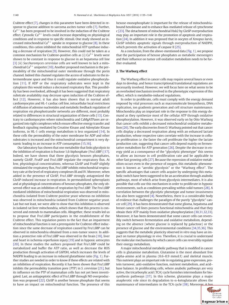
recruits carbon away from the energy production pathway of glucoseutilization. While investigating the relationship between glycolysisand the serine biosynthesis pathway (that arises from glycolysis),Chaneton et al. [47] were able to show that serine directly activatesPKM2, independently of Fru1,6BP mediated activation. This activationoccurs within the physiological range of serine concentration. Further,this activation is specific of PKM2 since PKM1, that exhibits a highbasal activity is refractory to both serine and Fru1,6BP activation.When serine is abundant PKM2 is fully active enabling the maximaluse of glucose through glycolysis andwhen the steady state level of ser-ine drops an attenuation of PKM2 activity occurs which redirects glu-cose derived carbons back in the serine biosynthetic pathway (Fig. 2).
Last, it was shown that SAICAR (succinylaminoimidazolecarboxamideribose-5′-phosphate, an intermediate of the de novo purine nucleotidesynthesis pathway) specifically stimulates PKM2 [48]. Using liquid-chromatography mass spectrometry, the authors were able to showthat SAICAR is a metabolite that interacts with PKM2. Moreover, assess-ment of PKM2 in vitro activity in the presence or absence of SAICARshowed that the binding of SAICAR to this enzyme activates it in such away that its activity became close to that of PKM1. They were able toshow that this regulation has an in vivo relevance since in cancer cells,metabolic flux and cellular energy balance were altered by changes inSAICAR concentration.
2.2. Role of Ru-5-P concentration in the balance between fatty aciddegradation and synthesis through AMPK inactivation; link between thepentose-phosphate pathway and lipogenesis
In cancer cells, the high activity of aerobic glycolysis is associated to arise in different intermediates of this pathway leading to an increase insome precursors for anabolic biosynthesis of macromolecules thatallow rapid cell proliferation and an activation of the oxidative pentosephosphate pathway (PPP) – through glucose-6-phosphate – that pro-duces ribose-5-phosphate (a precursor for nucleotide synthesis) andNADPH which is required for biosynthesis of lipids, appropriate redoxstatus and antioxidant defense (Fig. 1A). Thus PPP plays an importantrole in the metabolic reorganization that favors tumor cell proliferationand disease development. A molecular mechanism that links PPP activa-tion and the increase in lipogenesis has been recently evidenced [49]. Athigh concentration, Ribulose-5-phosphate (Ru-5-P) produced by the 6-phosphogluconate dehydrogenase (6PGD) disrupts the association be-tween LKB1 (tumor suppressor liver kinase B1), MO25 (scaffolding-likeadaptor protein mouse protein 25) and STRAD (pseudokinase Ste20-related adaptor) that is required for full AMPK (AMPKinase) activation.Such a decrease in AMPK activity leads to an increase in fatty-acid andcholesterol synthesis and inhibition of two main metabolic enzymesthat participate in fatty-acids degradation i.e. acetyl-CoA carboxylase(ACC) 1 and 2. The above-mentioned study suggests that 6PGD pro-vides an additional link between the oxidative PPP and lipogenesisthrough Ru-5-P-dependent inhibition of LKB1/AMPK signaling. Al-though it was shown in that study that the intracellular level of Ru-5-P decreased in 6PGD knockdown cells, which suggests an importantrole for 6PGD inmaintaining the intracellular Ru-5-P level that cannotbe compensated by other pathways. This does not exclude the poten-tial contribution of non-oxidative PPP in maintaining physiologicalRu5-P levels to regulate lipogenesis. In addition, these findings re-garding a commonly important role of 6PGD in the oxidative PPP incancer cells are different from the previous report using H1975 lungcancer cells suggesting that 6PGD functions may vary in cancer cellsowing to different cell types or oncogenic background [50].
2.3. Regulation of phosphofructokinase (PFK) activity by citrate andfructose 2,6-biphosphate (Fru2,6BP)
In the carbohydrate catabolism pathway, PFK is one of themost reg-ulated enzymes and it has been shown that, in different types of cancer,
Fig. 2. Scheme illustrating the PKM regulations and the consequences of these regulations on anabolic pathways. PKM1 has constitutively high catalytic activity. PKM2 activity is subject toallosteric regulation. PKM1 is constitutively active because it forms a stable tetramer whereas PKM2 can exist either as an active tetramer or an inactive non-tetramer. When serine isabundant PKM2 is fully active enabling the maximal use of glucose through glycolysis. When the steady state level of serine drops an attenuation of PKM2 activity occurs whichredirects glucose-derived carbons back in the serine biosynthetic pathway.
1143N. Hammad et al. / Biochimica et Biophysica Acta 1857 (2016) 1139–1146
the rate of glycolysis is, at least in part, determined by PFK activity. Theallosteric regulation of PFK is essentially due to 5metabolites: AMP, ADPand Fru2,6BP are activators while ATP and citrate are inhibitors. In non-transformed cells one of the most important regulation concerns the al-losteric response of PFK activity to citrate, a metabolite that is at thecrossroad of the Krebs cycle and lipid metabolism. Increase in this me-tabolite concentration strongly inhibits glycolysis through PFK inhibi-tion and favors fatty acid beta-oxidation. In many kinds of tumors, ithas been shown that PFK allosteric regulation is largely altered in sucha way that this enzyme is more sensitive to Fru2,6BP and less sensitiveto citrate. Indeed, in mammalian cells, PFK is an homo or hetero-tetramer of L, M and C isoforms which are differently expressed in var-ious tissues i.e. L is themain isoform expressed in liver and kidney, M isthe sole isoform found in skeletal muscle while C is predominantlyexpressed is platelets. These isoforms possess different kinetic proper-ties, particularly concerning the sensitivity towards allosteric effectors(for a detailed description of these kinetic properties of PFK isoforms,see [51]. For instance, the M isoform is the most sensitive to Fru2,6BPand the L isoform is the less sensitive to the citrate induced-inhibition.In cancer cells, the over-expressed isoforms are preferentially the onesfor which the sensitivity to citrate is low (L isoform essentially). It hasbeen proposed that in some human gliomas this L isoformmay be spe-cifically phosphorylated [52]. However this phosphorylation has notbeen associated to any significant modification in the kinetic propertiesof PFK.
Another process leading to a large decrease in PFK sensitivity to ATPand citrate and to an increase in Fru2,6BP induced PFK activation hasbeendescribed in a tumor tissuedeveloped inmice after subcutaneous in-fection with tumorigenic B16-F10 cells. In this case, a posttranslationalmodification of M isoform of PFK induced by proteolysis leads to a shortfragment (47 kD instead 87 kD) in which the regulatory properties arechanged: its activity is insensitive to citrate, and compared to the nativeisoform it presents a low sensitivity to ATP and is strongly activated byFru2,6BP [53]. Such regulations of PFK activity in rapidly proliferatingcells allow an increase in the flux through glycolysis. Indeed, it has been
shown that the regulations of PFK in cancer cells are such that this en-zyme does not exert significant control over the glycolytic flux [54,55].
2.4. Oncometabolites and their role in metabolism deviation
The term oncometabolite has recently been coined and assignedwith confidence to 2-hydroxyglutarate, the reduced form of 2-oxoglutarate (2OG). An oncometabolite is a small molecule component(or enantiomer) of normal metabolism whose accumulation causesmetabolic dysregulation and consequently primes cells allowing futureprogression to cancer.Wewill here focus on the dysregulations inducedby these oncometabolites that arise from the Krebs cycle.
2.4.1. α-ketoglutarate (αKG)-dependent dioxygenasesα-ketoglutarate (αKG)-dependent dioxygenases are important
modulators of both the oxygen-sensing machinery and the epigenome.Regarding the oxygen-sensing machinery, the majority of the changesin gene expression observed in response to hypoxia are due toHIFs, het-erodimeric transcription factors that are tightly linked to tumorigenesis[56]. HIF transcription factors consist of an α- and a β-subunit that areconstitutively expressed. However, under normoxia the α-subunit ishighly labile [57], [58] and the rapid degradation of HIFα is regulatedby oxygen-dependent prolyl hydroxylase enzymes (PHDs), which hy-droxylate twodefined prolyl residues in its oxygen-dependent degrada-tion (ODD) domain. This in turn recruits an E3 ubiquitin ligase complexcontaining pVHL, resulting in HIFα ubiquitylation and degradation bythe proteasome. Consequently, any dysregulation of the hydroxylationprocess would lead to a stabilization of HIF. Recently, metabolic changesdriven bymutations in genes related to the TCA cycle, have indicated analternative role, that of the ‘oncometabolite’ [59]. A particular metabo-lite builds upwithin the cell and contributes to the tumorigenic process.Three enzymes of the TCA cycle have been shown to be involved in thisprocess: fumarate hydratase, succinate dehydrogenase and isocitratedehydrogenase. Mutations in fumarate hydratase (FH) and succinatedehydrogenase (SDH) subunits lead to loss of gene function, and

Fig. 3. Scheme illustrating the regulations induced by oncometabolites. The majority of the changes in gene expression observed in response to hypoxia are due to HIFs, heterodimerictranscription factors that are tightly linked to tumorigenesis. The rapid degradation of HIFα is down-regulated by the competitive inhibition of oxygen-dependent prolyl hydroxylaseenzymes (PHDs) by the three oncometabolites succinate, fumarate and 2-HG. The build up of these metabolites is due to mutations in fumarate hydratase, succinate dehydrogenaseand isocitrate dehydrogenase. The inhibition of these enzymes leads to the stabilization of HIF1α through a decrease in its hydroxylation rate and induce the majority of the changesin gene expression observed in response to hypoxia. This allows a “hypoxia-like” cellular response even in the presence of oxygen.
1144 N. Hammad et al. / Biochimica et Biophysica Acta 1857 (2016) 1139–1146
accumulation of the substrates fumarate and succinate, respectively.Conversely, for isocitrate dehydrogenase (IDH), a single-allele mutationconfers a gain of function, producing an excess of a new metabolite, 2-hydroxyglutarate (2HG) [59]. These three oncometabolites inhibit com-petitively the α-ketoglutarate (αKG)-dependent dioxygenases. Succi-nate and fumarate, which are structurally similar, inhibit theseenzymes through product inhibition, since it has been shown that theeffects of both metabolites can be reversed by the addition of excessαKG in vitro and in vivo [60]. 2HG is structurally very similar to αKGand glutamate, the only difference is the presence of a hydroxyl groupinstead of a ketone or amine group, respectively. It is this similaritythat results in the competitive inhibition of αKG-dependentdioxygenases by 2HG, as it occupies the same binding site as αKG[61]. As stated above, the inhibition of these enzymes leads to the stabi-lization of HIF1α through a decrease in its hydroxylation rate and in-duce the majority of the changes in gene expression observed inresponse to hypoxia. This allows a “hypoxia-like” cellular responseeven in the presence of oxygen (Fig. 3).
2.4.2. Histone methylation [62]Recent studies have shown that Krebs cycle intermediates can shape
the epigenetic landscape of chromatin by regulating DNA and histonemethylation. α-ketoglutarate is a key metabolite in the Krebs cycleand is also an obligatory substrate for α-oxoglutarate-dependentdioxygenases. This enzyme family includes the major enzymes of DNAand histone demethylation, i.e. Ten-Eleven Translocation and JumonjiC domain containing demethylases. As stated above, succinate and fu-marate, are potent inhibitors of α-ketoglutarate (αKG)-dependentdioxygenases enzymes, i.e. the balance of the Krebs cycle reactions canaffect the level of DNA and histone methylation and thus control geneexpression.
3. Conclusion
Metabolites i.e., the intermediaries from diverse metabolic path-ways, are demonstrating their role as metabolic messengers that
regulate the activity of diverse enzymes and impinge on the half-lifeof enzymes. This concept opens a whole new field on the control ofcell survival and metabolism.
Transparency Document
The Transparency document associated with this article can befound in online version.
Acknowledgements
This work was supported by the Ligue contre le Cancer, the AgenceNationale de la Recherche (ANR2011BSV502501), the CNRS (ConseilNational de la Recherche Scientifique) and the Comité de Dordogne &Gironde de la Ligue Nationale Contre le Cancer. The authors wish tothank Dr. Manuel Rojo and Dr. ArnaudMourier for careful proofreadingof the manuscript and constructive discussions.
References
[1] R. Diaz-Ruiz, M. Rigoulet, A. Devin, The Warburg and Crabtree effects: on the originof cancer cell energy metabolism and of yeast glucose repression, Biochim. Biophys.Acta 1807 (2011) 568–576.
[2] H.G. Crabtree, Observations on the carbohydrate metabolism of tumours, Biochem. J.23 (1929) 536–545.
[3] S. Rodríguez-Enríquez, O. Juárez, J.S. Rodríguez-Zavala, R. Moreno-Sánchez, Multi-site control of the Crabtree effect in ascites hepatoma cells, Eur. J. Biochem. FEBS268 (2001) 2512–2519.
[4] S. Weinhouse, Glycolysis, respiration, and anomalous gene expression in experi-mental hepatomas: GHA Clowes memorial lecture, Cancer Res. 32 (1972)2007–2016.
[5] R.L. Veech, J.W. Lawson, N.W. Cornell, H.A. Krebs, Cytosolic phosphorylation poten-tial, J. Biol. Chem. 254 (1979) 6538–6547.
[6] D.H. Koobs, Phosphate mediation of the Crabtree and Pasteur effects, Science 178(1972) 127–133.
[7] I. Sussman, M. Erecińska, D.F. Wilson, Regulation of cellular energy metabolism: theCrabtree effect, Biochim. Biophys. Acta 591 (1980) 209–223.
[8] L. Wojtczak, V.V. Teplova, K. Bogucka, A. Czyż, A. Makowska, M.R. Więckowski, J.Duszyński, Y.V. Evtodienko, Effect of glucose and deoxyglucose on the redistributionof calcium in Ehrlich ascites tumour and Zajdela hepatoma cells and its conse-quences for mitochondrial energetics, Eur. J. Biochem. 263 (1999) 495–501.

1145N. Hammad et al. / Biochimica et Biophysica Acta 1857 (2016) 1139–1146
[9] YuV Evtodienko, null, V.V. Teplova, J. Duszyński, K. Bogucka, L. Wojtczak, The role ofcytoplasmic [Ca2+] in glucose-induced inhibition of respiration and oxidative phos-phorylation in Ehrlich ascites tumour cells: a novel mechanism of the Crabtree ef-fect, Cell Calcium 15 (1994) 439–446.
[10] S. Uribe, P. Rangel, J.P. Pardo, Interactions of calcium with yeast mitochondria, CellCalcium 13 (1992) 211–217.
[11] V. Saks, Y. Belikova, E. Vasilyeva, A. Kuznetsov, E. Fontaine, C. Keriel, X. Leverve, Cor-relation between degree of rupture of outer mitochondrial membrane and changesof kinetics of regulation of respiration by ADP in permeabilized heart and liver cells,Biochem. Biophys. Res. Commun. 208 (1995) 919–926.
[12] E.F. Greiner, M. Guppy, K. Brand, Glucose is essential for proliferation and the glyco-lytic enzyme induction that provokes a transition to glycolytic energy production, J.Biol. Chem. 269 (1994) 31484–31490.
[13] T. Anmann, R. Guzun, N. Beraud, S. Pelloux, A.V. Kuznetsov, L. Kogerman, T.Kaambre, P. Sikk, K. Paju, N. Peet, E. Seppet, C. Ojeda, Y. Tourneur, V. Saks, Differentkinetics of the regulation of respiration in permeabilized cardiomyocytes and in HL-1 cardiac cells: importance of cell structure/organization for respiration regulation,Biochim. Biophys. Acta BBA Bioenerg. 1757 (2006) 1597–1606.
[14] M. Eimre, K. Paju, S. Pelloux, N. Beraud, M. Roosimaa, L. Kadaja, M. Gruno, N. Peet, E.Orlova, R. Remmelkoor, A. Piirsoo, V. Saks, E. Seppet, Distinct organization of energymetabolism in HL-1 cardiac cell line and cardiomyocytes, Biochim. Biophys. ActaBBA Bioenerg. 1777 (2008) 514–524.
[15] N. Beraud, S. Pelloux, Y. Usson, A.V. Kuznetsov, X. Ronot, Y. Tourneur, V. Saks, Mito-chondrial dynamics in heart cells: very low amplitude high frequency fluctuationsin adult cardiomyocytes and flow motion in non beating Hl-1 cells, J. Bioenerg.Biomembr. 41 (2009) 195–214.
[16] V. Saks, R. Guzun, N. Timohhina, K. Tepp, M. Varikmaa, C. Monge, N. Beraud, T.Kaambre, A. Kuznetsov, L. Kadaja, M. Eimre, E. Seppet, Structure–function relation-ships in feedback regulation of energy fluxes in vivo in health and disease: mito-chondrial interactosome, Biochim. Biophys. Acta BBA Bioenerg. 1797 (2010)678–697.
[17] R. Díaz-Ruiz, N. Avéret, D. Araiza, B. Pinson, S. Uribe-Carvajal, A. Devin, M.Rigoulet, Mitochondrial oxidative phosphorylation is regulated by fructose1,6-bisphosphate. A possible role in Crabtree effect induction? J. Biol. Chem.283 (2008) 26948–26955.
[18] Z.S. Vexler, A. Wong, C. Francisco, C. Manabat, S. Christen, M. Täuber, D.M. Ferriero,G. Gregory, Fructose-1,6-bisphosphate preserves intracellular glutathione and pro-tects cortical neurons against oxidative stress, Brain Res. 960 (2003) 90–98.
[19] W. Sano, F. Watanabe, H. Tamai, E. Furuya, M. Mino, Beneficial effect of fructose-1,6-bisphosphate on mitochondrial function during ischemia–reperfusion of rat liver,Gastroenterology 108 (1995) 1785–1792.
[20] J.A. Kelleher, P.H. Chan, T.Y. Chan, G.A. Gregory, Energymetabolism in hypoxic astro-cytes: protective mechanism of fructose-1,6-bisphosphate, Neurochem. Res. 20(1995) 785–792.
[21] M. Rosas-Lemus, C. Uribe-Alvarez, N. Chiquete-Félix, S. Uribe-Carvajal, In Saccharo-myces cerevisiae fructose-1,6-bisphosphate contributes to the Crabtree effectthrough closure of the mitochondrial unspecific channel, Arch. Biochem. Biophys.555-556 (2014) 66–70.
[22] R. Calafell, J. Boada, A.F. Santidrian, J. Gil, T. Roig, J.C. Perales, J. Bermudez, Fructose1,6-bisphosphate reduced TNF-alpha-induced apoptosis in galactosamine sensitizedrat hepatocytes through activation of nitric oxide and cGMP production, Eur. J.Pharmacol. 610 (2009) 128–133.
[23] J.G. Pastorino, N. Shulga, J.B. Hoek, Mitochondrial binding of hexokinase II inhibitsBax-induced cytochrome c release and apoptosis, J. Biol. Chem. 277 (2002)7610–7618.
[24] V.V. Lemeshko, VDAC electronics: 1. VDAC-hexo(gluco)kinase generator of the mito-chondrial outermembrane potential, Biochim. Biophys. Acta 1838 (2014) 1362–1371.
[25] L.K. Nutt, The Xenopus oocyte: a model for studying themetabolic regulation of can-cer cell death, Semin. Cell Dev. Biol. 23 (2012) 412–418.
[26] O. Warburg, On the origin of cancer cells, Science 123 (1956) 309–314.[27] K. Brand, Aerobic glycolysis by proliferating cells: protection against oxidative stress
at the expense of energy yield, J. Bioenerg. Biomembr. 29 (1997) 355–364.[28] R.A. Gatenby, R.J. Gillies, Why do cancers have high aerobic glycolysis? Nat. Rev.
Cancer 4 (2004) 891–899.[29] R. Moreno-Sánchez, S. Rodríguez-Enríquez, A. Marín-Hernández, E. Saavedra, En-
ergy metabolism in tumor cells, FEBS J. 274 (2007) 1393–1418.[30] M. Guppy, P. Leedman, X. Zu, V. Russell, Contribution by different fuels and meta-
bolic pathways to the total ATP turnover of proliferating MCF-7 breast cancercells, Biochem. J. 364 (2002) 309–315.
[31] S. Rodríguez-Enríquez, P.A. Vital-González, F.L. Flores-Rodríguez, A. Marín-Hernández, L. Ruiz-Azuara, R. Moreno-Sánchez, Control of cellular proliferation bymodulation of oxidative phosphorylation in human and rodent fast-growingtumor cells, Toxicol. Appl. Pharmacol. 215 (2006) 208–217.
[32] M. Martin, B. Beauvoit, P.J. Voisin, P. Canioni, B. Guérin, M. Rigoulet, Energetic andmorphological plasticity of C6 glioma cells grown on 3-D support; effect of transientglutamine deprivation, J. Bioenerg. Biomembr. 30 (1998) 565–578.
[33] P. Pasdois, C. Deveaud, P. Voisin, V. Bouchaud, M. Rigoulet, B. Beauvoit, Contributionof the phosphorylable complex I in the growth phase-dependent respiration of C6glioma cells in vitro, J. Bioenerg. Biomembr. 35 (2003) 439–450.
[34] S. Rodríguez-Enríquez, J.C. Gallardo-Pérez, A. Avilés-Salas, A. Marín-Hernández, L.Carreño-Fuentes, V. Maldonado-Lagunas, R. Moreno-Sánchez, Energy metabolismtransition in multi-cellular human tumor spheroids, J. Cell. Physiol. 216 (2008)189–197.
[35] K. Smolková, N. Bellance, F. Scandurra, E. Génot, E. Gnaiger, L. Plecitá-Hlavatá, P.Ježek, R. Rossignol, Mitochondrial bioenergetic adaptations of breast cancer cellsto aglycemia and hypoxia, J. Bioenerg. Biomembr. 42 (2010) 55–67.
[36] R. Rossignol, R. Gilkerson, R. Aggeler, K. Yamagata, S.J. Remington, R.A. Capaldi, En-ergy substrate modulates mitochondrial structure and oxidative capacity in cancercells, Cancer Res. 64 (2004) 985–993.
[37] C. Yang, B. Ko, C.T. Hensley, L. Jiang, A.T. Wasti, J. Kim, J. Sudderth, M.A. Calvaruso, L.Lumata, M. Mitsche, J. Rutter, M.E. Merritt, R.J. DeBerardinis, Glutamine oxidationmaintains the TCA cycle and cell survival during impaired mitochondrial pyruvatetransport, Mol. Cell 56 (2014) 414–424.
[38] R.J. DeBerardinis, A. Mancuso, E. Daikhin, I. Nissim, M. Yudkoff, S. Wehrli, C.B.Thompson, Beyond aerobic glycolysis: transformed cells can engage in glutaminemetabolism that exceeds the requirement for protein and nucleotide synthesis,Proc. Natl. Acad. Sci. U. S. A. 104 (2007) 19345–19350.
[39] H.R. Christofk, M.G. Vander Heiden, M.H. Harris, A. Ramanathan, R.E. Gerszten, R.Wei, M.D. Fleming, S.L. Schreiber, L.C. Cantley, The M2 splice isoform of pyruvate ki-nase is important for cancer metabolism and tumour growth, Nature 452 (2008)230–233.
[40] S. Herzig, E. Raemy, S. Montessuit, J.-L. Veuthey, N. Zamboni, B. Westermann, E.R.S.Kunji, J.-C. Martinou, Identification and functional expression of the mitochondrialpyruvate carrier, Science 337 (2012) 93–96.
[41] D.K. Bricker, E.B. Taylor, J.C. Schell, T. Orsak, A. Boutron, Y.-C. Chen, J.E. Cox, C.M.Cardon, J.G. Van Vranken, N. Dephoure, C. Redin, S. Boudina, S.P. Gygi, M. Brivet,C.S. Thummel, J. Rutter, A mitochondrial pyruvate carrier required for pyruvate up-take in yeast, Drosophila, and humans, Science 337 (2012) 96–100.
[42] D.Y. Gui, C.A. Lewis, M.G. Vander Heiden, Allosteric regulation of PKM2 allows cellu-lar adaptation to different physiological states, Sci. Signal. 6 (2013) pe7.
[43] D. Anastasiou, Y. Yu, W.J. Israelsen, J.-K. Jiang, M.B. Boxer, B.S. Hong, W. Tempel, S.Dimov, M. Shen, A. Jha, H. Yang, K.R. Mattaini, C.M. Metallo, B.P. Fiske, K.D. Courtney,S. Malstrom, T.M. Khan, C. Kung, A.P. Skoumbourdis, H. Veith, N. Southall, M.J.Walsh, K.R. Brimacombe, W. Leister, S.Y. Lunt, Z.R. Johnson, K.E. Yen, K. Kunii, S.M.Davidson, H.R. Christofk, C.P. Austin, J. Inglese, M.H. Harris, J.M. Asara, G.Stephanopoulos, F.G. Salituro, S. Jin, L. Dang, D.S. Auld, H.-W. Park, L.C. Cantley, C.J.Thomas, M.G. Vander Heiden, Pyruvate kinase M2 activators promote tetramer for-mation and suppress tumorigenesis, Nat. Chem. Biol. 8 (2012) 839–847.
[44] M. Cortes-Cros, C. Hemmerlin, S. Ferretti, J. Zhang, J.S. Gounarides, H. Yin, A. Muller,A. Haberkorn, P. Chene, W.R. Sellers, F. Hofmann, M2 isoform of pyruvate kinase isdispensable for tumor maintenance and growth, Proc. Natl. Acad. Sci. 110 (2013)489–494.
[45] Z. Li, P. Yang, Z. Li, The multifaceted regulation and functions of PKM2 in tumor pro-gression, Biochim. Biophys. Acta 1846 (2014) 285–296.
[46] T.J. De Koning, K. Snell, M. Duran, R. Berger, B.-T. Poll-The, R. Surtees, L-serine in dis-ease and development, Biochem. J. 371 (2003) 653–661.
[47] B. Chaneton, P. Hillmann, L. Zheng, A.C.L. Martin, O.D.K. Maddocks, A.Chokkathukalam, J.E. Coyle, A. Jankevics, F.P. Holding, K.H. Vousden, C. Frezza, M.O'Reilly, E. Gottlieb, Serine is a natural ligand and allosteric activator of pyruvate ki-nase M2, Nature 491 (2012) 458–462.
[48] K.E. Keller, I.S. Tan, Y.-S. Lee, SAICAR stimulates pyruvate kinase isoform M2 andpromotes cancer cell survival in glucose-limited conditions, Science 338 (2012)1069–1072.
[49] R. Lin, S. Elf, C. Shan, H.-B. Kang, Q. Ji, L. Zhou, T. Hitosugi, L. Zhang, S. Zhang, J.H. Seo, J.Xie, M. Tucker, T.-L. Gu, J. Sudderth, L. Jiang, M. Mitsche, R.J. DeBerardinis, S. Wu, Y. Li,H. Mao, P.R. Chen, D.Wang, G.Z. Chen, S.J. Hurwitz, S. Lonial, M.L. Arellano, H.J. Khoury,F.R. Khuri, B.H. Lee, Q. Lei, D.J. Brat, K. Ye, T.J. Boggon, C. He, S. Kang, J. Fan, J. Chen, 6-Phosphogluconate dehydrogenase links oxidative PPP, lipogenesis and tumourgrowth by inhibiting LKB1-AMPK signalling, Nat. Cell Biol. 17 (2015) 1484–1496.
[50] V.P. Sukhatme, B. Chan, Glycolytic cancer cells lacking 6-phosphogluconate dehy-drogenase metabolize glucose to induce senescence, FEBS Lett. 586 (2012)2389–2395.
[51] R. Moreno-Sánchez, A. Marín-Hernández, J.C. Gallardo-Pérez, H. Quezada, R.Encalada, S. Rodríguez-Enríquez, E. Saavedra, Phosphofructokinase type 1 kinetics,isoform expression, and gene polymorphisms in cancer cells, J. Cell. Biochem. 113(2012) 1692–1703.
[52] G.E. Staal, A. Kalff, E.C. Heesbeen, C.W. van Veelen, G. Rijksen, Subunit composition,regulatory properties, and phosphorylation of phosphofructokinase from humangliomas, Cancer Res. 47 (1987) 5047–5051.
[53] A. Šmerc, E. Sodja, M. Legiša, Posttranslational modification of 6-phosphofructo-1-kinase as an important feature of cancer metabolism, PLoSOne 6 (2011) e19645.
[54] A. Marín-Hernández, S. Rodríguez-Enríquez, P.A. Vital-González, F.L. Flores-Rodríguez, M. Macías-Silva, M. Sosa-Garrocho, R. Moreno-Sánchez, Determiningand understanding the control of glycolysis in fast-growth tumor cells, FEBS J. 273(2006) 1975–1988.
[55] A. Marín-Hernández, J.C. Gallardo-Pérez, S. Rodríguez-Enríquez, R. Encalada, R.Moreno-Sánchez, E. Saavedra, Modeling cancer glycolysis, Biochim. Biophys. ActaBBA Bioenerg. 1807 (2011) 755–767.
[56] P.H. Maxwell, C.W. Pugh, P.J. Ratcliffe, Activation of the HIF pathway in cancer, Curr.Opin. Genet. Dev. 11 (2001) 293–299.
[57] G.L. Wang, B.H. Jiang, E.A. Rue, G.L. Semenza, Hypoxia-inducible factor 1 is a basic-helix–loop–helix–PAS heterodimer regulated by cellular O2 tension, Proc. Natl.Acad. Sci. U. S. A. 92 (1995) 5510–5514.
[58] B.H. Jiang, E. Rue, G.L. Wang, R. Roe, G.L. Semenza, Dimerization, DNA binding, andtransactivation properties of hypoxia-inducible factor 1, J. Biol. Chem. 271 (1996)17771–17778.
[59] L. Dang, D.W. White, S. Gross, B.D. Bennett, M.A. Bittinger, E.M. Driggers, V.R. Fantin,H.G. Jang, S. Jin, M.C. Keenan, K.M. Marks, R.M. Prins, P.S. Ward, K.E. Yen, L.M. Liau,J.D. Rabinowitz, L.C. Cantley, C.B. Thompson, M.G. Vander Heiden, S.M. Su, Cancer-associated IDH1 mutations produce 2-hydroxyglutarate, Nature 462 (2009)739–744.

1146 N. Hammad et al. / Biochimica et Biophysica Acta 1857 (2016) 1139–1146
[60] M.A. Selak, S.M. Armour, E.D. MacKenzie, H. Boulahbel, D.G. Watson, K.D. Mansfield,Y. Pan, M.C. Simon, C.B. Thompson, E. Gottlieb, Succinate links TCA cycle dysfunctionto oncogenesis by inhibiting HIF-alpha prolyl hydroxylase, Cancer Cell 7 (2005)77–85.
[61] W. Xu, H. Yang, Y. Liu, Y. Yang, P. Wang, S.-H. Kim, S. Ito, C. Yang, P.Wang, M.-T. Xiao,L. Liu, W. Jiang, J. Liu, J. Zhang, B. Wang, S. Frye, Y. Zhang, Y. Xu, Q. Lei, K.-L. Guan, S.
Zhao, Y. Xiong, Oncometabolite 2-hydroxyglutarate is a competitive inhibitor of α-ketoglutarate-dependent dioxygenases, Cancer Cell 19 (2011) 17–30.
[62] A. Salminen, A. Kauppinen, M. Hiltunen, K. Kaarniranta, Krebs cycle intermediatesregulate DNA and histone methylation: epigenetic impact on the aging process,Ageing Res. Rev. 16 (2014) 45–65.


![Biochimica et Biophysica Acta - immed.org considerations/09.07.2017 updates/Membrane... · G.L. Nicolson, M.E. Ash / Biochimica et Biophysica Acta 1859 (2017) 1704–1724 1705 [8]](https://img.pdfslide.us/doc/110x75/5c684f1e09d3f2f5638b5509/biochimica-et-biophysica-acta-immed-considerations09072017-updatesmembrane.jpg)