Embed Size (px)
Citation preview
Biochimica et Biophysica Acta 1818 (2012) 1613–1624
Contents lists available at SciVerse ScienceDirect
Biochimica et Biophysica Acta
j ourna l homepage: www.e lsev ie r .com/ locate /bbamem
Structure, activity and interactions of the cysteine deleted analog of tachyplesin-1with lipopolysaccharide micelle: Mechanistic insights into outer-membranepermeabilization and endotoxin neutralization
Rathi Saravanan, Harini Mohanram, Mangesh Joshi, Prerna N. Domadia, Jaume Torres,Christiane Ruedl, Surajit Bhattacharjya ⁎School of Biological Sciences, Division of Structural and Computational Biology, Nanyang Technological University, Singapore 637551, Singapore
Abbreviations: AMPs, antimicrobial peptides; CDTtachyplesin-1; LPS, lipopolysaccharide; NMR, nucleartransferred nuclear Overhauser effect; NOESY, nuclear OTOCSY, total correlation spectroscopy; CD, circular dichrenhancement; STD-NMR, saturation transfer difference⁎ Corresponding author at: 60 Nanyang Drive, 6375
3856.E-mail address: [email protected] (S. Bhattacharjya
0005-2736/$ – see front matter © 2012 Elsevier B.V. Alldoi:10.1016/j.bbamem.2012.03.015
a b s t r a c t
a r t i c l e i n f oArticle history:Received 31 January 2012Received in revised form 14 March 2012Accepted 16 March 2012Available online 23 March 2012
Keywords:Antimicrobial peptideLipopolysaccharideNMRStructureEndotoxinSTD-NMR
Tachyplesin-1, a disulfide stabilized β-hairpin antimicrobial peptide, can be found at the hemocytes of horseshoe crab Tachypleus tridentatus. A cysteine deleted linear analog of tachyplesin-1 or CDT (KWFRVYRGIYRRR-NH2) contains a broad spectrum of bactericidal activity with a reduced hemolytic property. The bactericidalactivity of CDT stems from selective interactions with the negatively charged lipids including LPS. In thiswork, CDT–LPS interactions were investigated using NMR spectroscopy, optical spectroscopy and functionalassays. We found that CDT neutralized LPS and disrupted permeability barrier of the outer membrane. Zetapotential and ITC studies demonstrated charge compensation and hydrophobic interactions of CDT withthe LPS-outer membrane, respectively. Secondary structure of the peptide was probed by CD and FT-IR exper-iments indicating β-strands and/or β-turn conformations in the LPS micelle. An ensemble of structures, de-termined in LPS micelle by NMR, revealed a β-hairpin like topology of the CDT peptide that was typified byan extended cationic surface and a relatively shorter segment of hydrophobic region. Interestingly, at thenon-polar face, residue R11 was found to be in a close proximity to the indole ring of W2, suggesting acation–π type interactions. Further, saturation transfer difference (STD) NMR studies established intimatecontacts among the aromatic and cationic residues of CDT with the LPS micelle. Fluorescence and dynamiclight scattering experiments demonstrated that CDT imparted structural destabilization to the aggregatedstates of LPS. Collectively, atomic resolution structure and interactions of CDT with the outer membrane-LPS could be exploited for developing potent broad spectrum antimicrobial and anti-sepsis agents.
© 2012 Elsevier B.V. All rights reserved.
1. Introduction
The emergence of drug-resistant bacteria has caused a major chal-lenge to the human health care [1,2]. There are now a number of bac-teria, termed as multi drug resistant strains or superbugs, withprotection against a number of conventional antibiotics [3–5]. Amongthese superbugs, as comprehended, the Gram-negative ones e.g.Pseudomonas aeruginosa, Acinetobacter baumannii, and Klebsiellapneumoniae thought to be a major threat due to the deficiency ofthe active drug candidates in the pipeline [5]. Therefore, novel anti-biotics with different modes of actions are highly desirable to combatinfectious diseases. Cationic antimicrobial peptides (AMPs) are an
, cysteine deleted analog ofmagnetic resonance; Tr-NOE,verhauser effect spectroscopy;oism; NOE, nuclear OverhauserNMR51, Singapore. Fax: +65 6791
).
rights reserved.
integral component of the innate immune system. AMPs serve asthe first line of defense with a broad spectrum of activity againstbacteria, viruses and fungi [6–9]. The evolutionary preserved AMPshave been recognized as a promising lead for the development ofnew antibiotics [6–9]. In humans, AMPs are involved for the probablestimulation andmodulation of adaptive immunity [10,11]. As amodeof action, most of the AMPs disrupt the integrity of bacterial mem-branes [12–14]. The amphipathic sequence features, typified by thepreponderance of cationic and hydrophobic amino acids, of AMPsfacilitate membrano-lytic activity toward microorganisms [12–14].The disruption of cytosolic membrane by AMPs, causing cell lysis,may occur following one or several mechanisms e.g. barrel stave, to-roidal pore, carpeting sinking raft and lipid clustering [15–18]. Thus,structural or SAR studies of AMPs are largely preformed with modelmembranes consisted of negatively charged phospholipids plausiblymimicking the plasma membrane of bacteria [15–18]. It may benoted that while a variety of biophysical methods has been utilizedto understand interactions of AMPs with model lipid membranes,the use of NMR techniques provided atomic-resolution insights interms of structure, membrane orientation and mechanisms [18,19].

1614 R. Saravanan et al. / Biochimica et Biophysica Acta 1818 (2012) 1613–1624
The outer membrane of Gram-negative organisms establishes anefficient permeability barrier for antibiotics, AMPs and host defenseproteins [19–22]. A higher lethal concentration of AMPs is oftennecessary to prevent growth of Gram-negative organisms as comparedto Gram-positive counterparts [23,24]. Some AMPs are known to bequite ineffective against Gram-negative strains [23,24]. As observed,in the context of LPS these AMPs undergo self-association preventingtheir translocation through the outer membrane [24–26]. Therefore,AMPs need to disrupt LPS mediated barrier of the outer membranein order to achieve the broad spectrum activity. Chemically, LPS consistsof three different segments, a distally located variable polysaccharide orO-antigen domain, a relatively conserved hexa-acylated lipid A moietyand an oligosaccharide domain covalently linking the lipid A andO-antigen domains. Consequently, atomic-resolution structures andinteractions of AMPs in LPS lipids are vital to realize the mechanism ofaction and for the further development of highly active peptide basedantibiotics [27–32]. Moreover, LPS, as termed endotoxin, is the causativeagent of fatal septic shock syndromes in human [33,34]. Thus, AMPswithLPS binding and neutralizing ability may endow with scaffolds for thegeneration of anti-endotoxic drugs [27,35,36].
Tachyplesin-1 or TP-1 (KWCFRVCYRGICYRRCR-NH2) is a well char-acterized, structurally and mechanistically, AMP from the hemocyte ofhorse shoe crab [37–39]. TP-1 has a broad spectrum of antimicrobial ac-tivity against several strains of Gram-negative andGram-positive bacte-ria, fungi and viruses [40,41]. The antimicrobial activity of TP-1 appearsto stem from the ability of the peptide to disorganize bacterial cellmembranes [37–39]. NMR studies demonstrated that TP-1 adopts β-hairpin structure in solution and also in lipid micelles and bilayers[38,42,43]. The antiparallel β-strands of β-hairpin structure of TP-1are constrained by the disulfide bridges between residues Cys3–Cys15and Cys7–Cys12 [37,38]. Replacement of disulfide bonds with variousamino acids has yielded important insight toward structure–activity re-lationship for β-hairpin or β-sheet AMPs including TP-1 [44–47]. How-ever, the consequences, in terms of activity and structures, of acomplete deletion of Cys residues from the amino acid sequences ofAMPs had been less frequently examined. In this regard, a shorter ana-log of TP-1, 13-residue long, termed CDT, whereby all of the four Cysresidues were deleted from the primary structure of AMP, was investi-gated for antibacterial activity, red blood cell lysis and liposome leakageand vesicle binding [48]. CDT demonstrated a marked inhibition ofgrowth of Gram negative and Gram positive bacterial strains akin toTP-1. The MIC (minimum inhibitory concentration) values of CDTwere even found to be lower against Escherichia coli and Listeria mono-cytogenes in comparison to the native TP-1 peptide. In addition, CDT didnot cause any measurable hemolysis of erythrocytes at a concentrationas high as 200 μg/mL. Using a variety of biophysical methods includingsolid state NMR studies, Ramamoorthy and coworkers demonstratedselective interactions of CDT with the negatively charged lipid vesiclese.g. POPE/POPG, POPC/POPG and POPC/LPS [48]. Based on the solidstate 31P NMR experiments with various lipid bilayers (POPC, POPC/POPG), it was proposed that CDT may destabilize head group packingof the lipid bilayers as a probable mode for bacterial cell lysis. However,atomic resolution structures of CDT in any of these lipid environments,in particularwith the LPS-outermembrane, are yet to be known. To gaininsights into the outer membrane permeabilization and endotoxin neu-tralization, in this work, we have characterized atomic resolution struc-ture and interactions of CDT in LPSmicelle by use of NMR spectroscopy,ITC, dynamic light scattering and optical spectroscopy methods.
2. Materials and methods
2.1. Peptide, lipids and chemicals
LPS from E. coli 0111:B4, fluorescein isothiocyanate (FITC) conju-gated LPS from E. coli 055:B5 and 1-N-phenylnaph-thylamine (NPN)were purchased from Sigma-Aldrich (St. Louis, MO). POPC (1-
palmitoyl-2-oleoyl-sn-glycero-3-phosphatidylcholine) was obtainedfrom Avanti Polar Lipids™. CDT peptide was synthesized commercial-ly by GL-Biochem (Shanghai, China) and was further purified by thereverse-phase HPLC, Waters™, using a C18 column (300 Å pore size,5 μm particle size) with a linear gradient of acetonitrile/water mix-ture. The molecular weight of the peptide was confirmed by massspectrometry.
2.2. Outer membrane permeabilization assay
Outer membrane permeabilization activity of CDT was analyzedby 1-N-phenylnaph-thylamine (NPN) dye uptake. Mid-log phase E.coli cells, obtained from an overnight culture, were centrifuged at3000 g and resuspended in 10 mM sodium phosphate buffer to an op-tical density of 0.6 at 600 nm. A stock solution of NPN previously pre-pared in acetone was added to a final concentration of 10 μM in500 μL of E. coli cells and a basal fluorescence was recorded. TheNPN dye was excited at 350 nm and the increase in fluorescence in-tensity (emission maximum) at 410 nm as a function of increasingpeptide concentrations (0.5–10 μM) was recorded. The maximalvalue of NPN uptake was determined after the addition of 10 μL poly-myxin B sulfate (0.64 mg/mL stock). The results were presented aspercentage membrane permeabilization relative to the membranepermeabilization observed for polymyxin B.
2.3. TNF-α release assay
Murine macrophage (Raw 264.7) cells were revived to confluencyin IMDM medium supplemented with FCS, glutamine and penicillin:streptomycin in a 96 well micro-titer plate. A serial dilution of CDTpeptide from 1 μM to 10 μM was incubated with different concentra-tions of LPS (1 ng/mL to 100 ng/mL) for 30 min. Peptide–LPS complexwas then added to confluent macrophage cells and further incubatedfor 6 h. The supernatant from each well was then analyzed by con-ventional ELISA according to the manufacturer's instruction. The re-lease of TNF-α was then detected spectrophotometrically at 540 nm.
2.4. Measurement of surface charge
Zeta potential studies for surface charge measurements were car-ried out on a Zetasizer Nano ZS (Malvern Instruments, UK) equippedwith a 633 nm HeNe laser. E. coli cells were grown to mid-log phasefrom overnight grown culture and suspended in 10 mM sodium phos-phate buffer, pH 6.8 to a final O.D. of 0.2. POPC:LPS (80:20 w/w) LUVswere prepared by extrusion method. E. coli cells or LUVs were loadedinto a disposable zeta cell with gold electrode and the samples wereallowed to equilibrate for 3 min at 25 °C. Measurements were madeas a function of increasing concentration of CDT. A total of 5 measure-ments of 100 runs each were carried out for all the concentrations ofpeptide.
2.5. Fluorescence studies
All of the fluorescence experiments were carried out in a CaryEclipse fluorescence spectrophotometer (Varian, Inc., Australia)equipped with a dual monochromator. Measurements were per-formed using a 0.1 cm path length cuvette and a band width of5 nm for excitation and emission. Intrinsic tryptophan fluorescencespectra of CDT were recorded by titrating 5 μM peptide with increas-ing concentrations of E. coli 0111:B4 LPS. Fluorescence spectra wererecorded in 10 mM sodium phosphate buffer at pH 6.0. Fluorescencequenching experiments for free and LPS micelle bound peptidewere recorded by stepwise addition of (0.02–0.2 M) acrylamide ei-ther into the peptide or peptide–lipid solutions of 5 μM LPS. Quench-ing of fluorescence intensity at the emission maximum for the freeand LPS bound forms of CDT was noted and results were analyzed
1615R. Saravanan et al. / Biochimica et Biophysica Acta 1818 (2012) 1613–1624
using Stern–Volmer's quenching equation Fo/F=1+KSV[Q], where Foand F are the fluorescence intensity at the emission maximum in theabsence and presence of quencher, respectively, KSV is the Stern–Vol-mer quenching constant and [Q] is the molar concentration of acryl-amide. The emission spectra, from 500 to 600 nm, of FITCconjugated LPS, in 10 mM sodium phosphate buffer, pH 6.0, wererecorded with the excitation at 480 nm in samples containing0.5 μM FITC-LPS.
2.6. Isothermal titration calorimetry (ITC) experiments
Binding affinity of CDT with LPS and thermodynamic parametersof interactions were determined using ITC experiments on a Nano-ITC calorimeter (Micro Cal Inc, Northampton, MA). CDT and LPSwere separately dissolved in a 10 mM sodium phosphate buffer, pH6.0. LPS samples were further vortexed for 10 min and sonicated for5 min prior to use. Twenty injections, 0.4 μL each, from 550 μMstock of CDT were made into a solution of 50 μM LPS, at an intervalof 4 min, with a stirring speed of 300 rpm and the heat exchangewas recorded at 20 °C. The heat of dilution of the peptide alone intothe buffer was subtracted from the titration data. The resulting datawere integrated with Micro Cal Origin 7.0. The data was fitted withmodels provided in the software and analyzed to determine the asso-ciation constant Ka and the enthalpy change ΔH. The Gibb's free ener-gy change ΔG and entropy change ΔS were calculated from thefundamental thermodynamic equations: ΔG=−RT ln Ka andTΔS=(ΔH−ΔG), respectively.
2.7. Dynamic light scattering (DLS) studies
Particle size distribution of LPS micelle in the presence and ab-sence of CDT was determined using DLS measurements, performedon a BI-200SM instrument equipped with a He–Ne laser and a BI-900 AT auto-correlation system (Brookhaven Instruments Corp.,Holtsville, NY). For DLS measurement, 1 μM LPS was dissolved in a10 mM sodium phosphate buffer, pH 6.0 and was exposed to 90°light scattering for 3 min. DLS measurements were recorded for LPSin the presence of 2 μM CDT, under similar conditions. All sampleswere filtered, degassed and scanned using a 1-mm path length quartzcuvette. Data were analyzed by the CONTINmethod supplied with theinstrument.
2.8. Circular dichroism (CD) and IR spectroscopy
Far-UV CD spectra of CDT were recorded using a Chirascan CDspectrophotometer (Applied Photophysics Ltd., UK) with a 0.1 mmpath length of cuvette. CD data were collected with a spectral band-width of 1 nm and a time constant of 1 s. Peptide concentration inall experiments was fixed to 100 μM. 50 μM LPS was used to recordsecondary structural changes in the presence of lipid. Samples wereprepared in 10 mM sodium phosphate buffer at pH 6.0. FTIR spectrawere recorded on a Nicolet Nexus 560 spectrometer (Madison, USA)purged with N2 and equipped with a MCT/A detector cooled with liq-uid nitrogen. Attenuated total reflection (ATR) spectra were mea-sured with a 25-reflection ATR accessory from Graseby Specac(Kent, UK) and a wire grid polarizer (0.25 μM, Graseby Specac).Approximately 200 μL of a D2O solution of LPS alone or in the pres-ence of peptide in a 20:1 lipid/peptide molar ratio was applied ontoa trapezoidal (50 mm×2 mm×20 mm) Ge internal reflection ele-ment (IRE). A dry, or D2O saturated, N2 stream flowing through theATR compartment was used to remove bulk water (low hydration)or to fully hydrate the sample (high hydration), respectively. A totalof 200 scans were collected at a resolution of 4 cm−1, averaged andprocessed with one-point zero filling and Happ-Genzel apodization.Percentages of secondary structure were estimated by fittingthe amide I region (from 1700 to 1600 cm−1) after mild Fourier
deconvolution. The amide I band was Fourier self-deconvolved(FSD) using a full-width at a half-height (FWHH) of 20 cm−1 andan enhancement factor, k, of 2.0.
2.9. NMR experiments and structure determination
NMR experiments were performed on a Bruker DRX 600 MHzspectrometer (Germany), equipped with a cryo-probe and pulsefield gradients. NMR data processing and analysis were carried outusing the programs Topspin (Bruker) and SPARKY (Goddard, T. D.,and Kneller, D. G., University of California, San Francisco), respective-ly. A series of one dimensional 1H NMR spectra of CDT was obtainedby titrating aliquots of LPS, from a stock solution of 0.5 mM, in aque-ous solution at pH 4.5 at 298 K. Two dimensional 1H–1H NOESY (mix-ing time: 150 and 200 ms) and TOCSY (mixing time: 80 ms) spectraof CDT, at 0.5 mM, in the presence of 10 μM LPS were acquired.NOESY spectra of CDT in free solution were also obtained in an aque-ous solution at pH 4.5 at 298 K. Saturation transfer difference (STD)NMR experiments were carried out as described previously [49,50].Typically, CDT (0.5 mM) was dissolved in 550 μL of D2O and pH wasadjusted to 4.5. Stock solution, 5 mg/mL, of LPS was prepared inD2O solution at pH 4.5. All STD experiments were performed at298 K in the presence of 10 μM LPS concentration using standardSTD pulse sequences and WATERGATE 3-9-19 sequence for watersuppression [49,50]. The on-resonance frequency of STD studies wasfixed by acquiring a series of one-dimensional STD spectra at differenton-resonances in the range of 0 to −10 ppm with an interval of0.5 ppm. Finally, an on-resonance frequency was set to −3.5 ppmwhile the off-resonance frequency was fixed to 40 ppm for STD ex-periments. One dimensional STD NMR experiments were performedwith a scan number of 1024 for a typical spectral width of 12 ppm.All STD spectra were multiplied by an exponential line broadeningfunction of 3 Hz, prior to Fourier transformation. Two-dimensionalSTD-TOCSY spectra were recorded with 350 increments in t1 and 80transients using a MLEV-17 spin lock field of 80 ms. The relaxationdelay was set to 2.1 s. Saturation transfer was achieved by using 40selective Gaussian 270 pulses with a duration of 50 ms.
LPS-bound structure of CDT was calculated based on NOE-drivendistance restraints. The upper bound of the inter proton distancewas determined from the volume of NOESY cross-peaks. The lowerbound of the inter proton distance was fixed to 2.0 Å. It should benoted that determination of the three-dimensional structure of thepeptide was largely driven by the medium and long-range NOEs, ex-clusively present in the LPS bound state. The backbone dihedral angle(Φ) was varied from −30 to −120° for non-glycine residues to limitthe conformational search. NMR structures of CDT were iterativelycalculated from an extended polypeptide chain in 7 cycles of simulat-ed annealing using Cyana 2.1 [51] program. The 20 lowest-energystructures without distance violations greater than 0.3 Å and noangle violations greater than 5° were accepted into the final ensem-ble. Structures were visualized using MOLMOL and PyMOL software.The quality of structures was determined using Procheck [52] andthe Protein Structure Validation Suite [53].
3. Results
3.1. Neutralization of LPS toxicity and disruption of the LPS-outermembrane barrier by CDT
Owing to the interactions with the outer membrane of Gram-negative bacteria, AMPs may confer neutralization of the toxicity ofLPS or endotoxin [19,27]. However, it may be noted that a numberof AMPs despite their interactions with LPS or outer membrane areincapable of neutralizing endotoxin [27,54]. Typically, binding ofendotoxin neutralizing molecules with LPS would inhibit the activa-tion of a cell surface receptor, TLR4, causing an inhibition of the
1616 R. Saravanan et al. / Biochimica et Biophysica Acta 1818 (2012) 1613–1624
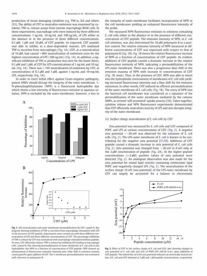
production of tissue damaging cytokines e.g. TNF-α, IL6 and others[55]. The ability of CDT to neutralize endotoxin was examined by cy-tokine, TNF-α, release assays from murine macrophage RAW cells. Inthese experiments, macrophage cells were induced by three differentconcentrations, 1 ng/mL, 10 ng/mL and 100 ng/mL, of LPS either inthe absence or in the presence of three different concentrations,0.1 μM, 1 μM and 10 μM, of CDT peptide. As expected, CDT peptidewas able to inhibit, in a dose-dependent manner, LPS mediatedTNF-α secretion from macrophages (Fig. 1A). CDT, at a concentrationof 10 μM, had caused >80% neutralization of endotoxin even for thehighest concentration of LPS (100 ng/mL) (Fig. 1A). In addition, a sig-nificant inhibition of TNF-α production was seen for the lower doses,0.1 μM and 1 μM, of CDT for LPS concentrations of 1 ng/mL and 10 ng/mL (Fig. 1A). There was >70% neutralization of endotoxin by CDT, atconcentrations of 0.1 μM and 1 μM, against 1 ng/mL and 10 mg/mLLPS, respectively (Fig. 1A).
In order to exert lethal effect against Gram-negative pathogens,potent AMPs should disrupt the integrity of the outer-membrane. 1-N-phenylnaphthylamine (NPN) is a fluorescent hydrophobic dyewhich shows a low intensity of fluorescence emission in aqueous so-lution. NPN is excluded by the outer-membrane; however, a loss in
Fig. 1. LPS neutralization and outer membrane permeabilization by CDT. (panel A) Bardiagram showing inhibition of TNF-α secretion from macrophage stimulated with LPSin the presence of CDT peptide. Experiments were carried out with three different con-centrations of LPS and three different concentrations of CDT. The percentage inhibitionof TNF-α release by CDT was normalized with macrophage stimulated without peptide.As seen, CDT efficiently reduces TNF-α release by inhibiting LPS binding to macrophagecells. (panel B) Plot showing permeabilization of outer membrane of E. coli cells to thehydrophobic dye NPN as a function of concentration of CDT peptide. NPN showed anenhancement in fluorescence emission intensity and a blue shift in emission maxima(inset panel B) upon addition of CDT. The % membrane permeabilization was estimatedwith reference to polymyxin B.
the integrity of outer-membrane facilitates incorporation of NPN tothe cell membranes yielding an enhanced fluorescence intensity ofthe probe.
We measured NPN fluorescence emission in solutions containingE. coli cells either in the absence or in the presence of different con-centrations of CDT peptide. The emission intensity of NPN, in E. colicell solutions, was also determined for 10 μM polymyxin B as a posi-tive control. The relative emission intensity of NPN measured at dif-ferent concentrations of CDT was expressed with respect to that ofpolymyxin B (Fig. 1B). Fig. 1B shows the relative fluorescence increaseof NPN as a function of concentrations of CDT peptide. As evident,additions of CDT peptide caused a dramatic increase in the relativefluorescence intensity of NPN, indicating a permeabilization of theLPS-outer membrane. There was also a progressive blue shift in theemission maxima of NPN with increase in concentrations of CDT(Fig. 1B, inset). Thus, in the presence of CDT, NPN was able to insertinto the hydrophobic environment of membranes of E. coli cells yield-ing increased fluorescence intensity and a blue shift for the emissionmaximum. In other words, CDT induced an efficient permeabilizationof the outer-membrane of E. coli cells (Fig. 1B). The entry of NPN intothe bacterial cell membranes was considered as a signature of thepermeabilization of the outer membrane mediated by the cationicAMPs, as termed ‘self-promoted’ uptake process [56]. Taken together,cytokine release and NPN fluorescence experiments demonstratedthat CDT efficiently neutralizes toxicity of LPS and also disrupts integ-rity of the outer-membrane.
3.2. Surface charge neutralization of E. coli cells by CDT
Zeta potential was measured for E. coli cells and LUV composed ofPOPC and LPS at various concentrations of CDT (Fig. 2). A negativezeta potential ~−20 mV was observed for the solutions of E. colicells (Fig. 2). The LPS-outer membrane of E. coli is known to be con-tributing for the negative zeta potential [57,58]. Additions of CDTpeptide caused a dramatic increase in zeta potential of E. coli cells(Fig. 2). Zeta potential was changed from −20 mV to 0 mV only atthe 2 μM concentration of peptide (Fig. 2A). At the higher peptideconcentrations (>2 μM) positive values of zeta potential weredetected (Fig. 2). An analogous observation was also made for thezeta potential for mixed lipid vesicles containing zwitterionic lipidPOPC and negatively charged LPS (Fig. 2). The neutralization of thesurface charge (0 mV zeta potential) of the LPS-outer membrane byCDT can largely be accounted for a balance in electrostatic
Fig. 2. Effect of CDT to the surface charge of E. coli and LUV: plot showing changes inzeta potential of E. coli cells and LUV of POPC-LPS (80:20 w/w) upon additions ofCDT peptide. The dotted line at 0 mV zeta potential indicates an electrically neutral sur-face of E. coli and LUV obtained at 2 μM and 1 μM peptide concentrations, respectively.

1617R. Saravanan et al. / Biochimica et Biophysica Acta 1818 (2012) 1613–1624
interactions among the cationic residues of CDT with the negativelycharged LPS lipids. However, the positive zeta potential, observed at>2 μM concentration of CDT, of E. coli cells may be interpreted as in-sertion of the peptide into the hydrophobic milieu of the LPS-outermembrane membranes [57,58]. These data demonstrated that as amode of action CDT efficiently neutralizes surface charges of theouter membrane of E. coli cells and inserts into the hydrophobic envi-ronment of lipids.
3.3. Localization of Trp residue of CDT in LPS micelle
CDT contains a single Trp residue at position 2 in the amino acidsequence. The interactions between CDT and LPS were probed bymeans of intrinsic Trp emission and quenching experiments. In thefree solutions, Trp residue of CDT showed an emission maximum at~355 nm, indicating that Trp is highly solvent exposed (Fig. 3A). In-clusion of LPS at various concentrations into solutions containingCDT yielded a marked enhancement in the fluorescence emission in-tensity of Trp residue along with a blue shift in the emission maxi-mum (Fig. 3A). At a 10 μM of LPS concentration, Trp residue of thepeptide had an emission maximum of ~345 nm (Fig. 3A). These ob-servations delineated the incorporation of Trp residue of CDT intothe hydrophobic environment of LPS micelles. The extent of solventaccessibility of Trp was further determined by fluorescence quench-ing using acrylamide as a quencher. There was a limited quenchingof Trp of CDT in complex with LPS in comparison to the free peptide
Fig. 3. Trp fluorescence emission and quenching of CDT. (panel A) Fluorescence emis-sion spectra of Trp residue of CDT at different concentrations of LPS. (panel B) Plotshowing a comparison of quenching of Trp fluorescence by acrylamide. Trp experi-enced both a blue shift in emission maxima (panel A) and a limited acrylamidequenching in the presence of LPS (panel B).
(Fig. 3B). The Stern–Volmer quenching plots estimated a quenchingconstant (Ksv) of 20.0 and 8.4 for free and LPS-bound CDT, respective-ly (Fig. 3B). Collectively, fluorescence studies established that Trp res-idue of CDT is inserted into the non-polar milieu of the LPS lipidmicelles with a restricted exposure to the aqueous solvent.
3.4. Secondary structures of CDT in LPS micelle
The global secondary structures of CDT were examined by CD andIR spectroscopic methods. Fig. 4A shows far UV CD spectra of CDT infree solution and in the presence of 50 μM LPS. The far UV CD spectraof CDT in buffer solutions were characterized by an intense singlenegative band at 200 nm, indicating random conformations of thepeptide (Fig. 4A). By contrast, the far UV CD spectra of CDT obtainedin a solution containing LPS micelles were dominated by the presenceof two bands, with negative ellipticity, at 215 nm and 202 nm, imply-ing structural transitions (Fig. 4A). The CD band detected at the wave-length at 215 nm could be interpreted in terms of populated β-sheetlike structures of CDT in LPS micelles. Note that the estimation of sec-ondary structures from CD spectra appeared to be complicated by thepotential contribution to the far UV CD bands from the number of ar-omatic residues in CDT. A more definitive assignment of secondarystructures of CDT in LPS micelles was further obtained from FT-IRstudies. The secondary structure of CDT was determined followingprevious assignments in the amide I [59]. The spectra in the amide Iregion of CDT is shown after the subtraction of the LPS contribution
Fig. 4. Secondary structure of CDT in LPS by CD and IR spectroscopy: (panel A) far-UVCD spectra of CDT in the absence (solid line) and presence (dashed line) of 50 μM LPS.(panel B) Amide I and II regions corresponding to peptide CDT mixed with LPS in thepresence of H2O (red) and D2O (blue). The wavenumbers (in cm−1) of the mainbands of both spectra are indicated.
1618 R. Saravanan et al. / Biochimica et Biophysica Acta 1818 (2012) 1613–1624
in the presence of either H2O or D2O (Fig. 4B). The main bands in thespectrum in H2O were centered at 1668 and 1635 cm−1. The formerband is indicative of β-turns or β-structures [59], whereas the bandat 1635 cm−1 originates solely from β-structures. In any case, noneof these bands can be assigned to α-helical structures or to randomcoils. The amide II band is visible at 1542 cm−1, whereas the bandat 1517 cm−1 corresponds to the tyrosine side chain of the peptide.After D2O exchange (blue spectrum), the amide II appears to becompletely shifted to lower wave numbers, which indicates a com-plete exchange (Fig. 4B). In the amide I, there was a small shift,from 1668 to 1671 cm−1, and from 1635 to 1632 cm−1. Due to theuncertainty in the assignment of the high frequency band, it is diffi-cult to estimate the secondary structure composition, but one canrefer to two limit situations; if the assignment of the 1668 cm−1
band corresponds to β-structure, then the peptide has 100% β-structure in LPS. If this band is assigned to β-turns, these would rep-resent 58±8% of the peptide, whereas 42±12% is β-structure. Theseresults apply regardless of the level of hydration: in the IR spectrumobtained in H2O, bulk water was removed by nitrogen, whereas thesample in D2O was hydrated in the ATR conditions. Yet, in thesetwo conditions the amide I spectra appeared similar, except forsmall shifts due to H/D exchange.
3.5. Interactions of CDT and LPS by ITC
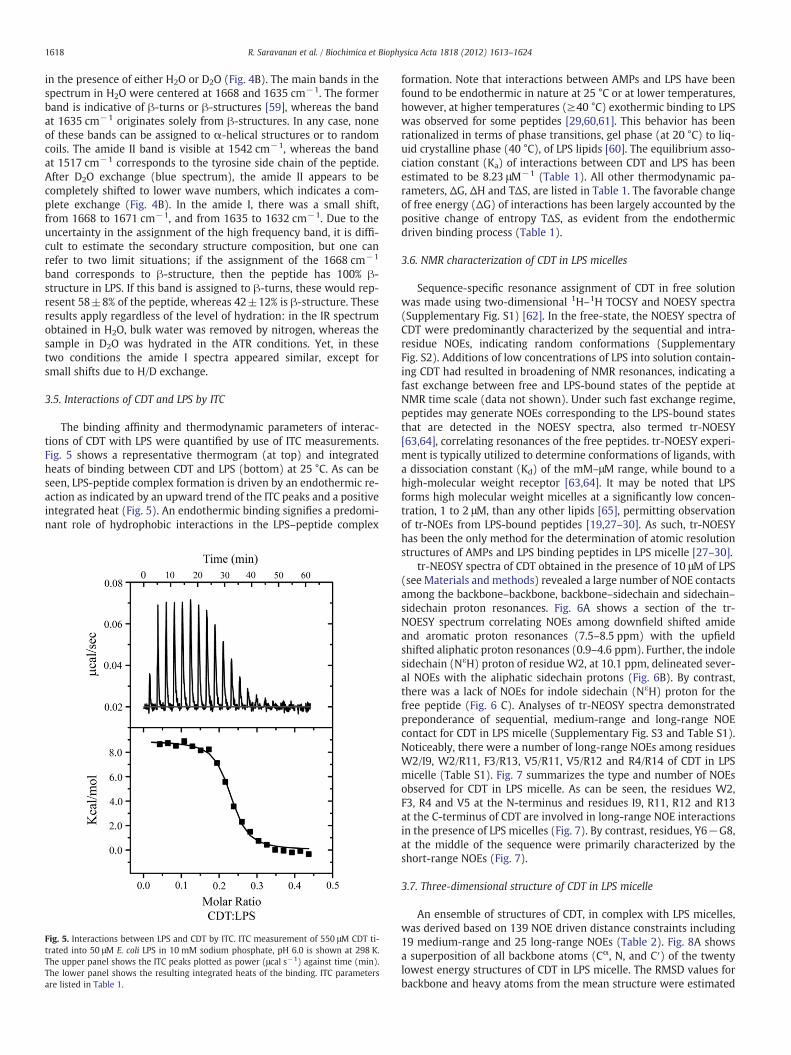
The binding affinity and thermodynamic parameters of interac-tions of CDT with LPS were quantified by use of ITC measurements.Fig. 5 shows a representative thermogram (at top) and integratedheats of binding between CDT and LPS (bottom) at 25 °C. As can beseen, LPS-peptide complex formation is driven by an endothermic re-action as indicated by an upward trend of the ITC peaks and a positiveintegrated heat (Fig. 5). An endothermic binding signifies a predomi-nant role of hydrophobic interactions in the LPS–peptide complex
Fig. 5. Interactions between LPS and CDT by ITC. ITC measurement of 550 μM CDT ti-trated into 50 μM E. coli LPS in 10 mM sodium phosphate, pH 6.0 is shown at 298 K.The upper panel shows the ITC peaks plotted as power (μcal s−1) against time (min).The lower panel shows the resulting integrated heats of the binding. ITC parametersare listed in Table 1.
formation. Note that interactions between AMPs and LPS have beenfound to be endothermic in nature at 25 °C or at lower temperatures,however, at higher temperatures (≥40 °C) exothermic binding to LPSwas observed for some peptides [29,60,61]. This behavior has beenrationalized in terms of phase transitions, gel phase (at 20 °C) to liq-uid crystalline phase (40 °C), of LPS lipids [60]. The equilibrium asso-ciation constant (Ka) of interactions between CDT and LPS has beenestimated to be 8.23 μM−1 (Table 1). All other thermodynamic pa-rameters, ΔG, ΔH and TΔS, are listed in Table 1. The favorable changeof free energy (ΔG) of interactions has been largely accounted by thepositive change of entropy TΔS, as evident from the endothermicdriven binding process (Table 1).
3.6. NMR characterization of CDT in LPS micelles
Sequence-specific resonance assignment of CDT in free solutionwas made using two-dimensional 1H–1H TOCSY and NOESY spectra(Supplementary Fig. S1) [62]. In the free-state, the NOESY spectra ofCDT were predominantly characterized by the sequential and intra-residue NOEs, indicating random conformations (SupplementaryFig. S2). Additions of low concentrations of LPS into solution contain-ing CDT had resulted in broadening of NMR resonances, indicating afast exchange between free and LPS-bound states of the peptide atNMR time scale (data not shown). Under such fast exchange regime,peptides may generate NOEs corresponding to the LPS-bound statesthat are detected in the NOESY spectra, also termed tr-NOESY[63,64], correlating resonances of the free peptides. tr-NOESY experi-ment is typically utilized to determine conformations of ligands, witha dissociation constant (Kd) of the mM–μM range, while bound to ahigh-molecular weight receptor [63,64]. It may be noted that LPSforms high molecular weight micelles at a significantly low concen-tration, 1 to 2 μM, than any other lipids [65], permitting observationof tr-NOEs from LPS-bound peptides [19,27–30]. As such, tr-NOESYhas been the only method for the determination of atomic resolutionstructures of AMPs and LPS binding peptides in LPS micelle [27–30].
tr-NEOSY spectra of CDT obtained in the presence of 10 μM of LPS(see Materials and methods) revealed a large number of NOE contactsamong the backbone–backbone, backbone–sidechain and sidechain–sidechain proton resonances. Fig. 6A shows a section of the tr-NOESY spectrum correlating NOEs among downfield shifted amideand aromatic proton resonances (7.5–8.5 ppm) with the upfieldshifted aliphatic proton resonances (0.9–4.6 ppm). Further, the indolesidechain (NεH) proton of residue W2, at 10.1 ppm, delineated sever-al NOEs with the aliphatic sidechain protons (Fig. 6B). By contrast,there was a lack of NOEs for indole sidechain (NεH) proton for thefree peptide (Fig. 6 C). Analyses of tr-NEOSY spectra demonstratedpreponderance of sequential, medium-range and long-range NOEcontact for CDT in LPS micelle (Supplementary Fig. S3 and Table S1).Noticeably, there were a number of long-range NOEs among residuesW2/I9, W2/R11, F3/R13, V5/R11, V5/R12 and R4/R14 of CDT in LPSmicelle (Table S1). Fig. 7 summarizes the type and number of NOEsobserved for CDT in LPS micelle. As can be seen, the residues W2,F3, R4 and V5 at the N-terminus and residues I9, R11, R12 and R13at the C-terminus of CDT are involved in long-range NOE interactionsin the presence of LPS micelles (Fig. 7). By contrast, residues, Y6−G8,at the middle of the sequence were primarily characterized by theshort-range NOEs (Fig. 7).
3.7. Three-dimensional structure of CDT in LPS micelle
An ensemble of structures of CDT, in complex with LPS micelles,was derived based on 139 NOE driven distance constraints including19 medium-range and 25 long-range NOEs (Table 2). Fig. 8A showsa superposition of all backbone atoms (Cα, N, and C′) of the twentylowest energy structures of CDT in LPS micelle. The RMSD values forbackbone and heavy atoms from the mean structure were estimated
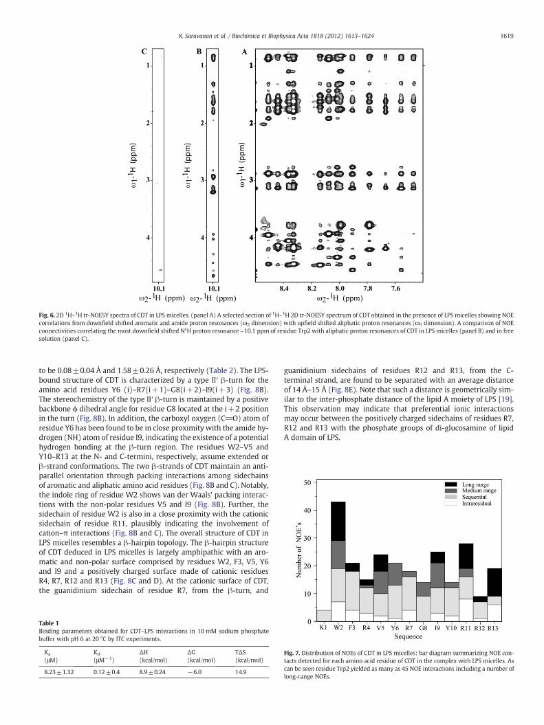
Fig. 6. 2D 1H–1H tr-NOESY spectra of CDT in LPS micelles. (panel A) A selected section of 1H–1H 2D tr-NOESY spectrum of CDT obtained in the presence of LPS micelles showing NOEcorrelations from downfield shifted aromatic and amide proton resonances (ω2 dimension) with upfield shifted aliphatic proton resonances (ω1 dimension). A comparison of NOEconnectivities correlating the most downfield shifted NεH proton resonance ~10.1 ppm of residue Trp2 with aliphatic proton resonances of CDT in LPS micelles (panel B) and in freesolution (panel C).
1619R. Saravanan et al. / Biochimica et Biophysica Acta 1818 (2012) 1613–1624
to be 0.08±0.04 Å and 1.58±0.26 Å, respectively (Table 2). The LPS-bound structure of CDT is characterized by a type II′ β-turn for theamino acid residues Y6 (i)–R7(i+1)–G8(i+2)–I9(i+3) (Fig. 8B).The stereochemistry of the type II′ β-turn is maintained by a positivebackbone ϕ dihedral angle for residue G8 located at the i+2 positionin the turn (Fig. 8B). In addition, the carboxyl oxygen (C_O) atom ofresidue Y6 has been found to be in close proximity with the amide hy-drogen (NH) atom of residue I9, indicating the existence of a potentialhydrogen bonding at the β-turn region. The residues W2–V5 andY10–R13 at the N- and C-termini, respectively, assume extended orβ-strand conformations. The two β-strands of CDT maintain an anti-parallel orientation through packing interactions among sidechainsof aromatic and aliphatic amino acid residues (Fig. 8B and C). Notably,the indole ring of residue W2 shows van der Waals' packing interac-tions with the non-polar residues V5 and I9 (Fig. 8B). Further, thesidechain of residue W2 is also in a close proximity with the cationicsidechain of residue R11, plausibly indicating the involvement ofcation–π interactions (Fig. 8B and C). The overall structure of CDT inLPS micelles resembles a β-hairpin topology. The β-hairpin structureof CDT deduced in LPS micelles is largely amphipathic with an aro-matic and non-polar surface comprised by residues W2, F3, V5, Y6and I9 and a positively charged surface made of cationic residuesR4, R7, R12 and R13 (Fig. 8C and D). At the cationic surface of CDT,the guanidinium sidechain of residue R7, from the β-turn, and
Table 1Binding parameters obtained for CDT–LPS interactions in 10 mM sodium phosphatebuffer with pH 6 at 20 °C by ITC experiments.
Ka Kd ΔH ΔG TΔS(μM) (μM−1) (kcal/mol) (kcal/mol) (kcal/mol)
8.23±1.32 0.12±0.4 8.9±0.24 −6.0 14.9
guanidinium sidechains of residues R12 and R13, from the C-terminal strand, are found to be separated with an average distanceof 14 Å–15 Å (Fig. 8E). Note that such a distance is geometrically sim-ilar to the inter-phosphate distance of the lipid A moiety of LPS [19].This observation may indicate that preferential ionic interactionsmay occur between the positively charged sidechains of residues R7,R12 and R13 with the phosphate groups of di-glucosamine of lipidA domain of LPS.
Fig. 7. Distribution of NOEs of CDT in LPS micelles: bar diagram summarizing NOE con-tacts detected for each amino acid residue of CDT in the complex with LPS micelles. Ascan be seen residue Trp2 yielded as many as 45 NOE interactions including a number oflong-range NOEs.
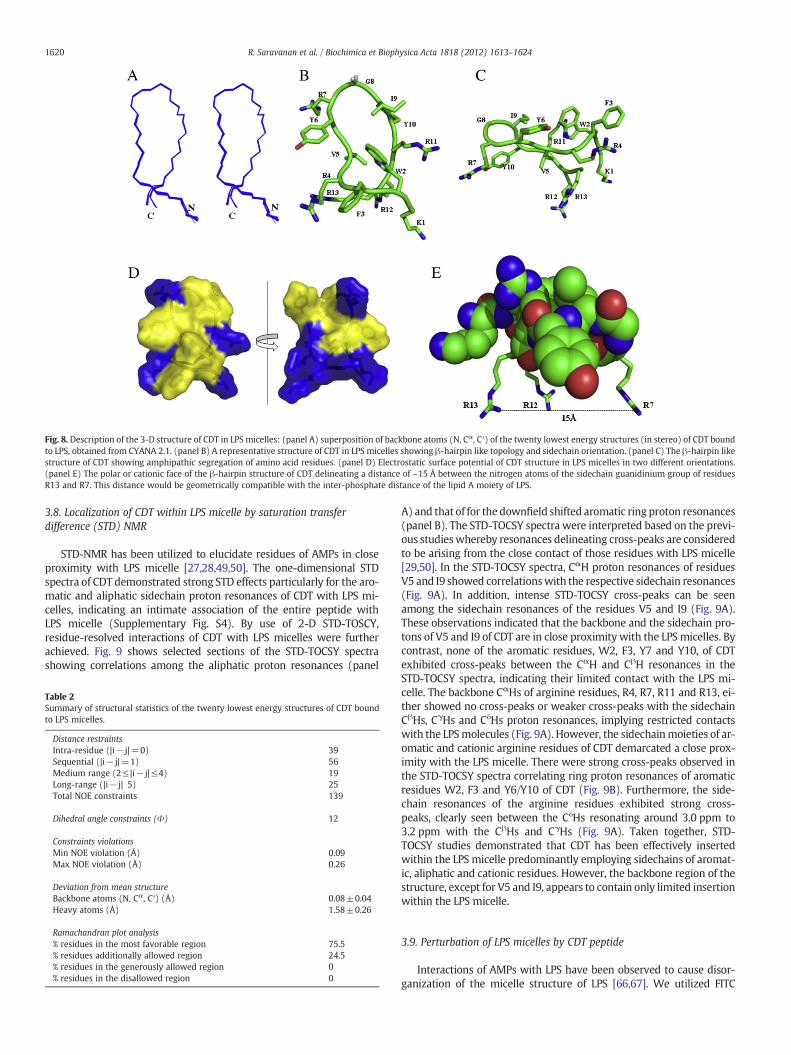
Fig. 8. Description of the 3-D structure of CDT in LPS micelles: (panel A) superposition of backbone atoms (N, Cα, C′) of the twenty lowest energy structures (in stereo) of CDT boundto LPS, obtained from CYANA 2.1. (panel B) A representative structure of CDT in LPS micelles showing β-hairpin like topology and sidechain orientation. (panel C) The β-hairpin likestructure of CDT showing amphipathic segregation of amino acid residues. (panel D) Electrostatic surface potential of CDT structure in LPS micelles in two different orientations.(panel E) The polar or cationic face of the β-hairpin structure of CDT delineating a distance of ~15 Å between the nitrogen atoms of the sidechain guanidinium group of residuesR13 and R7. This distance would be geometrically compatible with the inter-phosphate distance of the lipid A moiety of LPS.
1620 R. Saravanan et al. / Biochimica et Biophysica Acta 1818 (2012) 1613–1624
3.8. Localization of CDT within LPS micelle by saturation transferdifference (STD) NMR
STD-NMR has been utilized to elucidate residues of AMPs in closeproximity with LPS micelle [27,28,49,50]. The one-dimensional STDspectra of CDT demonstrated strong STD effects particularly for the aro-matic and aliphatic sidechain proton resonances of CDT with LPS mi-celles, indicating an intimate association of the entire peptide withLPS micelle (Supplementary Fig. S4). By use of 2-D STD-TOSCY,residue-resolved interactions of CDT with LPS micelles were furtherachieved. Fig. 9 shows selected sections of the STD-TOCSY spectrashowing correlations among the aliphatic proton resonances (panel
Table 2Summary of structural statistics of the twenty lowest energy structures of CDT boundto LPS micelles.
Distance restraintsIntra-residue (|i− j|=0) 39Sequential (|i− j|=1) 56Medium range (2≤ |i− j|≤4) 19Long-range (|i− j| 5) 25Total NOE constraints 139
Dihedral angle constraints (Φ) 12
Constraints violationsMin NOE violation (Å) 0.09Max NOE violation (Å) 0.26
Deviation from mean structureBackbone atoms (N, Cα, C′) (Å) 0.08±0.04Heavy atoms (Å) 1.58±0.26
Ramachandran plot analysis% residues in the most favorable region 75.5% residues additionally allowed region 24.5% residues in the generously allowed region 0% residues in the disallowed region 0
A) and that of for the downfield shifted aromatic ring proton resonances(panel B). The STD-TOCSY spectra were interpreted based on the previ-ous studieswhereby resonances delineating cross-peaks are consideredto be arising from the close contact of those residues with LPS micelle[29,50]. In the STD-TOCSY spectra, CαH proton resonances of residuesV5 and I9 showed correlationswith the respective sidechain resonances(Fig. 9A). In addition, intense STD-TOCSY cross-peaks can be seenamong the sidechain resonances of the residues V5 and I9 (Fig. 9A).These observations indicated that the backbone and the sidechain pro-tons of V5 and I9 of CDT are in close proximity with the LPS micelles. Bycontrast, none of the aromatic residues, W2, F3, Y7 and Y10, of CDTexhibited cross-peaks between the CαH and CβH resonances in theSTD-TOCSY spectra, indicating their limited contact with the LPS mi-celle. The backbone CαHs of arginine residues, R4, R7, R11 and R13, ei-ther showed no cross-peaks or weaker cross-peaks with the sidechainCβHs, CγHs and CδHs proton resonances, implying restricted contactswith the LPSmolecules (Fig. 9A). However, the sidechainmoieties of ar-omatic and cationic arginine residues of CDT demarcated a close prox-imity with the LPS micelle. There were strong cross-peaks observed inthe STD-TOCSY spectra correlating ring proton resonances of aromaticresidues W2, F3 and Y6/Y10 of CDT (Fig. 9B). Furthermore, the side-chain resonances of the arginine residues exhibited strong cross-peaks, clearly seen between the CδHs resonating around 3.0 ppm to3.2 ppm with the CβHs and CγHs (Fig. 9A). Taken together, STD-TOCSY studies demonstrated that CDT has been effectively insertedwithin the LPS micelle predominantly employing sidechains of aromat-ic, aliphatic and cationic residues. However, the backbone region of thestructure, except for V5 and I9, appears to contain only limited insertionwithin the LPS micelle.
3.9. Perturbation of LPS micelles by CDT peptide
Interactions of AMPs with LPS have been observed to cause disor-ganization of the micelle structure of LPS [66,67]. We utilized FITC
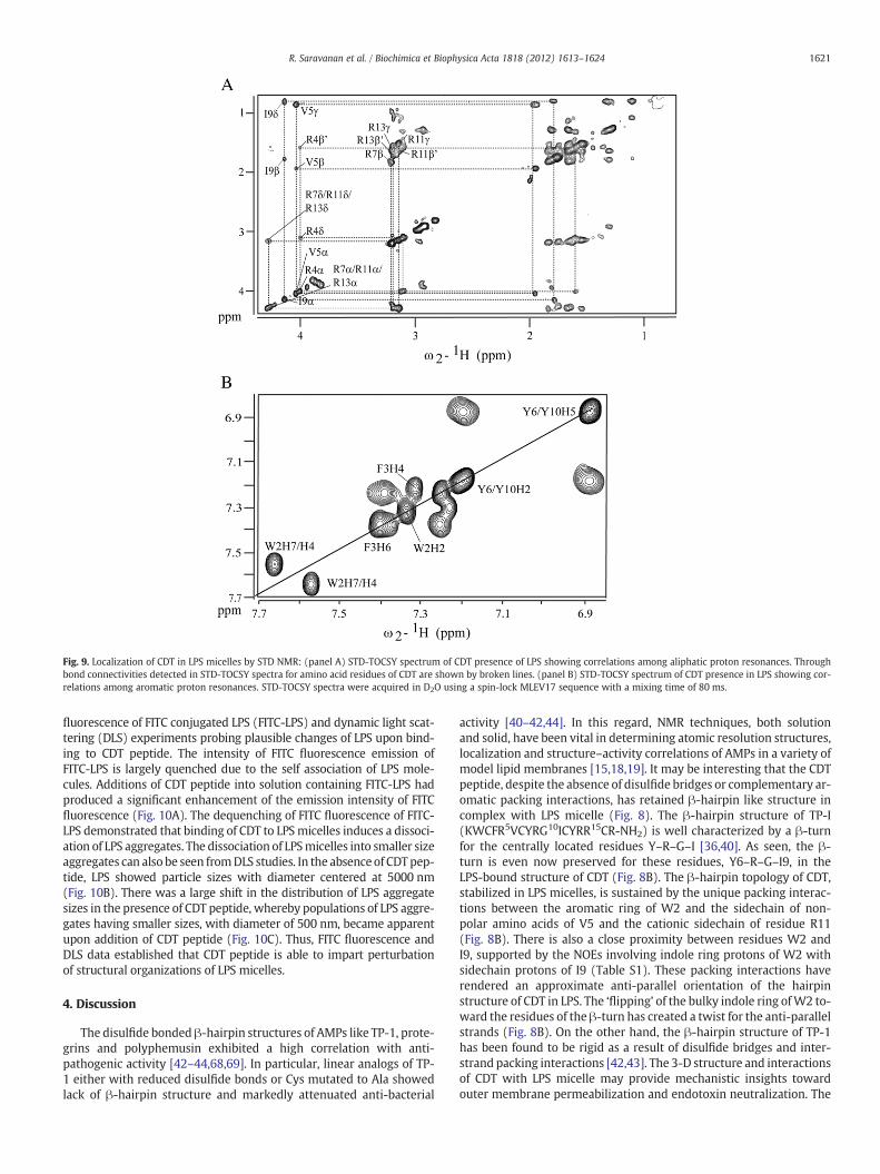
Fig. 9. Localization of CDT in LPS micelles by STD NMR: (panel A) STD-TOCSY spectrum of CDT presence of LPS showing correlations among aliphatic proton resonances. Throughbond connectivities detected in STD-TOCSY spectra for amino acid residues of CDT are shown by broken lines. (panel B) STD-TOCSY spectrum of CDT presence in LPS showing cor-relations among aromatic proton resonances. STD-TOCSY spectra were acquired in D2O using a spin-lock MLEV17 sequence with a mixing time of 80 ms.
1621R. Saravanan et al. / Biochimica et Biophysica Acta 1818 (2012) 1613–1624
fluorescence of FITC conjugated LPS (FITC-LPS) and dynamic light scat-tering (DLS) experiments probing plausible changes of LPS upon bind-ing to CDT peptide. The intensity of FITC fluorescence emission ofFITC-LPS is largely quenched due to the self association of LPS mole-cules. Additions of CDT peptide into solution containing FITC-LPS hadproduced a significant enhancement of the emission intensity of FITCfluorescence (Fig. 10A). The dequenching of FITC fluorescence of FITC-LPS demonstrated that binding of CDT to LPSmicelles induces a dissoci-ation of LPS aggregates. The dissociation of LPSmicelles into smaller sizeaggregates can also be seen fromDLS studies. In the absence of CDTpep-tide, LPS showed particle sizes with diameter centered at 5000 nm(Fig. 10B). There was a large shift in the distribution of LPS aggregatesizes in the presence of CDT peptide, whereby populations of LPS aggre-gates having smaller sizes, with diameter of 500 nm, became apparentupon addition of CDT peptide (Fig. 10C). Thus, FITC fluorescence andDLS data established that CDT peptide is able to impart perturbationof structural organizations of LPS micelles.
4. Discussion
The disulfide bonded β-hairpin structures of AMPs like TP-1, prote-grins and polyphemusin exhibited a high correlation with anti-pathogenic activity [42–44,68,69]. In particular, linear analogs of TP-1 either with reduced disulfide bonds or Cys mutated to Ala showedlack of β-hairpin structure and markedly attenuated anti-bacterial
activity [40–42,44]. In this regard, NMR techniques, both solutionand solid, have been vital in determining atomic resolution structures,localization and structure–activity correlations of AMPs in a variety ofmodel lipid membranes [15,18,19]. It may be interesting that the CDTpeptide, despite the absence of disulfide bridges or complementary ar-omatic packing interactions, has retained β-hairpin like structure incomplex with LPS micelle (Fig. 8). The β-hairpin structure of TP-I(KWCFR5VCYRG10ICYRR15CR-NH2) is well characterized by a β-turnfor the centrally located residues Y–R–G–I [36,40]. As seen, the β-turn is even now preserved for these residues, Y6–R–G–I9, in theLPS-bound structure of CDT (Fig. 8B). The β-hairpin topology of CDT,stabilized in LPS micelles, is sustained by the unique packing interac-tions between the aromatic ring of W2 and the sidechain of non-polar amino acids of V5 and the cationic sidechain of residue R11(Fig. 8B). There is also a close proximity between residues W2 andI9, supported by the NOEs involving indole ring protons of W2 withsidechain protons of I9 (Table S1). These packing interactions haverendered an approximate anti-parallel orientation of the hairpinstructure of CDT in LPS. The ‘flipping’ of the bulky indole ring ofW2 to-ward the residues of the β-turn has created a twist for the anti-parallelstrands (Fig. 8B). On the other hand, the β-hairpin structure of TP-1has been found to be rigid as a result of disulfide bridges and inter-strand packing interactions [42,43]. The 3-D structure and interactionsof CDT with LPS micelle may provide mechanistic insights towardouter membrane permeabilization and endotoxin neutralization. The
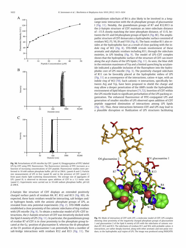
Fig. 10. Perturbations of LPS micelles by CDT. (panel A) Disaggregation of FITC labeledLPS by CDT using FITC fluorescence. The fluorescence intensity of FITC increases as afunction of increasing concentrations of CDT peptide. Fluorescence studies were per-formed in 10 mM sodium phosphate buffer, pH 6.0 at 298 K. (panels B and C) Particlesize measurements of LPS in free (panel B) and in the presence of CDT (panel C)from quasi elastic light scattering measurements. The average size of aggregates ofLPS (panel B) is observed to decrease upon addition of CDT in a 1:2 molar ratio(panel C). Experiments were carried out in 10 mM sodium phosphate buffer, pH 6.0at 298 K.
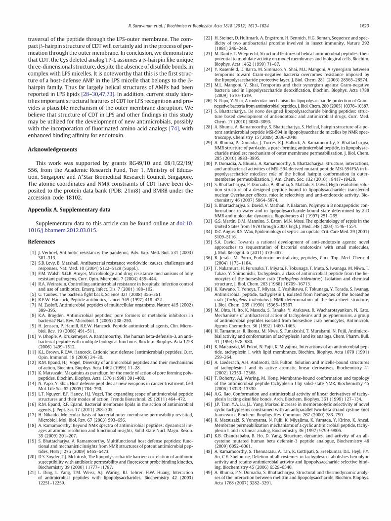
Fig. 11. Mode of interaction of CDT with LPS: a molecular model of CDT–LPS complexshowing close proximity of the negatively charged phosphate groups of glucosamine1 and glucosamine 2 of the lipid A moiety of LPS with the sidechains of cationic resi-dues R7, R12 and R13. The sidechains of residues R11 and W4, with probable cation–πinteractions, are rather deeply inserted, along with other aromatic and non-polar resi-dues, to the hydrophobic acyl region of LPS. The image was produced using INSIGHTII.
1622 R. Saravanan et al. / Biochimica et Biophysica Acta 1818 (2012) 1613–1624
β-hairpin like structure of CDT displays an extended positivelycharged surface patch of residues R4, R7, R12 and R13 (Fig. 8D). Asexpected, these basic residues would be interacting, salt bridges and/or hydrogen bonds, with the anionic phosphate groups of LPS, asrevealed from zeta potential experiments (Fig. 2). STD-NMR studiesestablished a close proximity of the cationic sidechains of Arg residueswith LPS micelle (Fig. 9). To obtain a molecular model of LPS–CDT in-teractions, the β-hairpin structure of CDT was iteratively docked withthe lipid Amoiety of LPS (Fig. 11). In particular, the guanidiniumgroupof residue R7 of CDT is in close proximity to the phosphate group lo-cated at the O4′ position of glucosamine II, whereas the di-phosphateat the O1 position of glucosamine I can potentially form a number ofsalt-bridge interactions with residues R12 and R13 (Fig. 11). The
guanidinium sidechain of R4 is also likely to be involved in a long-range ionic interaction with the di-phosphate groups of glucosamineI (Fig. 11). Notably, the guanidinium groups of R7 and R12/R13 inthe β-hairpin structure of CDT maintain an inter-sidechain distanceof ~15 Å closely matching the inter-phosphate distance, of 13 Å, be-tween the O1 andO4 phosphate groups of lipid A (Fig. 8E). The amphi-pathic structure of CDT demarcates a hydrophobic surface consisted ofresidues W2, F3, Y6, I9 and Y10 (Fig. 8). The basic residue R11 also re-sides at the hydrophobic face as a result of close packing with the in-dole ring of W2 (Fig. 8). STD-NMR reveals involvement of thesearomatic and aliphatic residues including R11, through the sidechainmoieties, in LPS binding (Fig. 9). The model of LPS–CDT complexshows that the hydrophobic surface of the structure of CDT can insertalong the acyl chains of the LPS lipids (Fig. 11). As seen, the blue shiftin the emission maximum of Trp and a limited quenching by acrylam-ide indicated a plausible inclusion of the fluorophore into the hydro-phobic core of LPS micelle (Fig. 3). The positively charged sidechainof R11 can be favorably placed at the hydrophobic milieu of LPS(Fig. 11) as a consequence of the interactions, cation–π type, with anindole ring of W2 [70]. Such cationic–π interactions, specifically be-tween Arg and Trp, have been proposed to shield the charge andmay allow a deeper penetration of the AMPs inside the hydrophobicenvironment of lipid bilayer structure [71,72]. Insertion of CDT withinthe LPS micelle leads to significant perturbation of the LPS packing or-ganization. The enhanced fluorescence of FITC-conjugated LPS andgeneration of smaller micelles of LPS observed upon addition of CDTpeptide suggested diminution of interactions among LPS lipids(Fig. 10). Thus, these interactions between CDT and LPS may lead toa plausible disruption or fluidization of LPS structures facilitating
1623R. Saravanan et al. / Biochimica et Biophysica Acta 1818 (2012) 1613–1624
traversal of the peptide through the LPS-outer membrane. The com-pact β-hairpin structure of CDT will certainly aid in the process of per-meation through the outer membrane. In conclusion, we demonstratethat CDT, the Cys deleted analog TP-I, assumes a β-hairpin like uniquethree-dimensional structure, despite the absence of disulfide bonds, incomplex with LPS micelles. It is noteworthy that this is the first struc-ture of a host-defense AMP in the LPS micelle that belongs to the β-hairpin family. Thus far largely helical structures of AMPs had beenreported in LPS lipids [28–30,47,73]. In addition, current study iden-tifies important structural features of CDT for LPS recognition and pro-vides a plausible mechanism of the outer membrane disruption. Webelieve that structure of CDT in LPS and other findings in this studymay be utilized for the development of new antimicrobials, possiblywith the incorporation of fluorinated amino acid analogs [74], withenhanced binding affinity for endotoxin.
Acknowledgements
This work was supported by grants RG49/10 and 08/1/22/19/556, from the Academic Research Fund, Tier 1, Ministry of Educa-tion, Singapore and A*Star Biomedical Research Council, Singapore.The atomic coordinates and NMR constraints of CDT have been de-posited to the protein data bank (PDB: 21m8) and BMRB under theaccession code 18102.
Appendix A. Supplementary data
Supplementary data to this article can be found online at doi:10.1016/j.bbamem.2012.03.015.
References
[1] J. Verhoef, Antibiotic resistance: the pandemic, Adv. Exp. Med. Biol. 531 (2003)301–313.
[2] S.B. Levy, B. Marshall, Antibacterial resistance worldwide: causes, challenges andresponses, Nat. Med. 10 (2004) S122–S129 (Suppl.).
[3] F.M. Walsh, S.G.B. Amyes, Microbiology and drug resistance mechanisms of fullyresistant pathogens, Curr. Opin. Microbiol. 7 (2004) 439–444.
[4] R.A. Weinstein, Controlling antimicrobial resistance in hospitals: infection controland use of antibiotics, Emerg. Infect. Dis. 7 (2001) 188–192.
[5] G. Taubes, The bacteria fight back, Science 321 (2008) 356–361.[6] R.E.W. Hancock, Peptide antibiotics, Lancet 349 (1997) 418–422.[7] M. Zasloff, Antimicrobial peptides of multicellular organisms, Nature 415 (2002)
389–395.[8] K.A. Brogden, Antimicrobial peptides: pore formers or metabolic inhibitors in
bacteria? Nat. Rev. Microbiol. 3 (2005) 238–250.[9] H. Jenssen, P. Hamill, R.E.W. Hancock, Peptide antimicrobial agents, Clin. Micro-
biol. Rev. 19 (2006) 491–511.[10] V. Dhople, A. Krukemeyer, A. Ramamoorthy, The human beta-defensin-3, an anti-
bacterial peptide with multiple biological functions, Biochim. Biophys. Acta 1758(2006) 1499–1512.
[11] K.L. Brown, R.E.W. Hancock, Cationic host defense (antimicrobial) peptides, Curr.Opin. Immunol. 18 (2006) 24–30.
[12] R.M. Epand, H.J. Vogel, Diversity of antimicrobial peptides and their mechanismsof action, Biochim. Biophys. Acta 1462 (1999) 11–28.
[13] K. Matsuzaki, Magainins as paradigm for the mode of action of pore forming poly-peptides, Biochim. Biophys. Acta 1376 (1998) 391–400.
[14] N. Papo, Y. Shai, Host defense peptides as new weapons in cancer treatment, CellMol. Life Sci. 62 (2005) 784–790.
[15] L.T. Nguyen, E.F. Haney, H.J. Vogel, The expanding scope of antimicrobial peptidestructures and their modes of action, Trends Biotechnol. 29 (2011) 464–472.
[16] R.M. Epand, R.F. Epand, Bacterial membrane lipids in the action of antimicrobialagents, J. Pept. Sci. 17 (2011) 298–305.
[17] H. Nikaido, Molecular basis of bacterial outer membrane permeability revisited,Microbiol. Mol. Biol. Rev. 67 (2003) 593–656.
[18] A. Ramamoorthy, Beyond NMR spectra of antimicrobial peptides: dynamical im-ages at atomic resolution and functional insights, Solid State Nucl. Magn. Reson.35 (2009) 201–207.
[19] S. Bhattacharjya, A. Ramamoorthy, Multifunctional host defense peptides: func-tional and mechanistic insights from NMR structures of potent antimicrobial pep-tides, FEBS J. 276 (2009) 6465–6473.
[20] D.S. Snyder, T.J. McIntosh, The lipopolysaccharide barrier: correlation of antibioticsusceptibility with antibiotic permeability and fluorescent probe binding kinetics,Biochemistry 39 (2000) 11777–11787.
[21] L. Ding, L. Yang, T.M. Weiss, A.J. Waring, R.I. Lehrer, H.W. Huang, Interactionof antimicrobial peptides with lipopolysaccharides, Biochemistry 42 (2003)12251–12259.
[22] H. Steiner, D. Hultmark, A. Engstrom, H. Bennich, H.G. Boman, Sequence and spec-ificity of two antibacterial proteins involved in insect immunity, Nature 292(1981) 246–248.
[23] M. Dante, T. Wieprecht, Structural features of helical antimicrobial peptides: theirpotential to modulate activity on model membranes and biological cells, Biochim.Biophys. Acta 1462 (1999) 71–87.
[24] Y. Rosenfeld, D. Barra, M. Simmaco, Y. Shai, M.L. Mangoni, A synergism betweentemporins toward Gram-negative bacteria overcomes resistance imposed bythe lipopolysaccharide protective layer, J. Biol. Chem. 281 (2006) 28565–28574.
[25] M.L. Mangoni, Y. Shai, Temporins and their synergism against Gram-negativebacteria and in lipopolysaccharide detoxification, Biochim. Biophys. Acta 1788(2009) 1610–1619.
[26] N. Papo, Y. Shai, A molecular mechanism for lipopolysaccharide protection of Gram-negative bacteria from antimicrobial peptides, J. Biol. Chem. 280 (2005) 10378–10387.
[27] S. Bhattacharjya, De novo designed lipopolysaccharide binding peptides: struc-ture based development of antiendotoxic and antimicrobial drugs, Curr. Med.Chem. 17 (2010) 3080–3093.
[28] A. Bhunia, A. Ramamoorthy, S. Bhattacharjya, S. Helical, hairpin structure of a po-tent antimicrobial peptide MSI-594 in lipopolysaccharide micelles by NMR spec-troscopy, Chemistry 15 (2009) 2036–2040.
[29] A. Bhunia, P. Domadia, J. Torres, K.J. Hallock, A. Ramamoorthy, S. Bhattacharjya,NMR structure of pardaxin, a pore-forming antimicrobial peptide, in lipopolysac-charide micelles: mechanism of outer membrane permeabilization, J. Biol. Chem.285 (2010) 3883–3895.
[30] P. Domadia, A. Bhunia, A. Ramamoorthy, S. Bhattacharjya, Structure, interactions,and antibacterial activities of MSI-594 derived mutant peptide MSI-594F5A in li-popolysaccharide micelles: role of the helical hairpin conformation in outer-membrane permeabilization, J. Am. Chem. Soc. 132 (2010) 18417–18428.
[31] S. Bhattacharjya, P. Domadia, A. Bhunia, S. Malladi, S. David, High resolution solu-tion structure of a designed peptide bound to lipopolysaccharide: transferrednuclear Overhauser effects, micelle selectivity and anti-endotoxic activity, Bio-chemistry 46 (2007) 5864–5874.
[32] S. Bhattacharjya, S. David, V. Mathan, P. Balaram, Polymyxin B nonapeptide: con-formations in water and in lipopolysaccharide-bound state determined by 2-DNMR and molecular dynamics, Biopolymers 41 (1997) 251–265.
[33] G.S. Martin, D.M. Mannino, S. Eaton, M.N. Moss, The epidemiology of sepsis in theUnited States from 1979 through 2000, Engl. J. Med. 348 (2003) 1546–1554.
[34] D.C. Angus, R.S. Wax, Epidemiology of sepsis: an update, Crit. Care Med. 29 (2001)S109–S116.
[35] S.A. David, Towards a rational development of anti-endotoxin agents: novelapproaches to sequestration of bacterial endotoxins with small molecules,J. Mol. Recognit. 6 (2011) 370–387.
[36] R. Jerala, M. Porro, Endotoxin neutralizing peptides, Curr. Top. Med. Chem. 4(2004) 1173–1184.
[37] T. Nakamura, H. Furunaka, T. Miyata, F. Tokunaga, T. Muta, S. Iwanaga, M. Niwa, T.Takao, Y. Shimonishi, Tachyplesin, a class of antimicrobial peptide from the he-mocytes of the horseshoe crab (Tachypleus tridentatus). Isolation and chemicalstructure, J. Biol. Chem. 263 (1988) 16709–16713.
[38] K. Kawano, T. Yoneya, T. Miyata, K. Yoshikawa, F. Tokunaga, Y. Terada, S. Iwanag,Antimicrobial peptide, tachyplesin I, isolated from hemocytes of the horseshoecrab (Tachypleus tridentatus). NMR determination of the beta-sheet structure,J. Biol. Chem. 265 (1990) 15365–15367.
[39] M. Ohta, H. Ito, K. Masuda, S. Tanaka, Y. Arakawa, R. Wacharotayankun, N. Kato,Mechanisms of antibacterial action of tachyplesins and polyphemusins, a groupof antimicrobial peptides isolated from horseshoe crab hemocytes, Antimicrob.Agents Chemother. 36 (1992) 1460–1465.
[40] H. Tamamura, R. Ikoma, M. Niwa, S. Funakoshi, T. Murakami, N. Fujii, Antimicro-bial activity and conformation of tachyplesin I and its analogs, Chem. Pharm. Bull.41 (1993) 978–980.
[41] K. Matsuzaki, M. Fukui, N. Fujii, K. Miyajima, Interactions of an antimicrobial pep-tide, tachyplesin I, with lipid membranes, Biochim. Biophys. Acta 1070 (1991)259–264.
[42] A. Laederach, A.H. Andreotti, D.B. Fulton, Solution and micelle-bound structuresof tachyplesin I and its active aromatic linear derivatives, Biochemistry 41(2002) 12359–12368.
[43] T. Doherty, A.J. Waring, M. Hong, Membrane-bound conformation and topologyof the antimicrobial peptide tachyplesin I by solid-state NMR, Biochemistry 45(2006) 13323–13330.
[44] A.G. Rao, Conformation and antimicrobial activity of linear derivatives of tachy-plesin lacking disulfide bonds, Arch. Biochem. Biophys. 361 (1999) 127–134.
[45] J.P. Tam, Y.A. Lu, J.L. Yang, Marked increase in membranolytic selectivity of novelcyclic tachyplesins constrained with an antiparallel two-beta strand cystine knotframework, Biochem. Biophys. Res. Commun. 267 (2000) 783–790.
[46] K. Matsuzaki, S. Yoneyama, N. Fujii, K. Miyajima, K. Yamada, Y. Kirino, K. Anzai,Membrane permeabilization mechanisms of a cyclic antimicrobial peptide, tachy-plesin I, and its linear analog, Biochemistry 36 (1997) 9799–9806.
[47] K.B. Chandrababu, B. Ho, D. Yang, Structure, dynamics, and activity of an all-cysteine mutated human beta defensin-3 peptide analogue, Biochemistry 48(2009) 6052–6061.
[48] A. Ramamoorthy, S. Thennarasu, A. Tan, K. Gottipati, S. Sreekumar, D.L. Heyl, F.Y.An, C.E. Shelburne, Deletion of all cysteines in tachyplesin I abolishes hemolyticactivity and retains antimicrobial activity and lipopolysaccharide selective bind-ing, Biochemistry 45 (2006) 6529–6540.
[49] A. Bhunia, P.N. Domadia, S. Bhattacharjya, Structural and thermodynamic analy-ses of the interaction between melittin and lipopolysaccharide, Biochim. Biophys.Acta 1768 (2007) 3282–3291.
1624 R. Saravanan et al. / Biochimica et Biophysica Acta 1818 (2012) 1613–1624
[50] A. Bhunia, S. Bhattacharjya, Mapping residue-specific contacts of polymyxin Bwith lipopolysaccharide by saturation transfer difference NMR: insights intoouter-membrane disruption and endotoxin neutralization, Biopolymers (PeptideSci.) 96 (2010) 273–287.
[51] P. Guntert, C. Mumenthaler, K.Wuthrich, Torsion angle dynamics for NMR structurecalculation with the new program DYANA, J. Mol. Biol. 273 (1997) 283–298.
[52] R.A. Laskowski, J.A. Rullmannn, M.W. MacArthur, R. Kaptein, J.M. Thornton, AQUAand PROCHECK-NMR: programs for checking the quality of protein structuressolved by NMR, J. Biomol. NMR 8 (1996) 477–486.
[53] Y.J. Huang, R. Powers, G.T. Montelione, Protein NMR recall, precision, and F-measure scores (RPF scores): structure quality assessment measures based on in-formation retrieval statistics, J. Am. Chem. Soc. 127 (2005) 1665–1674.
[54] M.G. Scott, A.C. Vreugdenhil, W.A. Buurman, R.E.W. Hancock, M.R. Gold, Cuttingedge: cationic antimicrobial peptides block the binding of lipopolysaccharide(LPS) to LPS binding protein, J. Immunol. 164 (2000) 549–553.
[55] B. Beutler, Tlr4: central component of the sole mammalian LPS sensor, Curr. Opin.Immunol. 12 (2000) 20–26.
[56] R.E.W. Hancock, Alterations in outer membrane permeability, Annu. Rev. Micro-biol. 38 (1984) 237–264.
[57] C.S. Alves, M.N. Melo, H.G. Franquelim, R. Ferre, M. Planas, L. Feliu, E. Bardají, W.Kowalczyk, D. Andreu, N.C. Santos, M.X. Fernandes, M.A. Castanho, Escherichiacoli cell surface perturbation and disruption induced by antimicrobial peptidesBP100 and pepR, J. Biol. Chem. 285 (2010) 27536–27544.
[58] M.M. Domingues, M.A. Castanho, N.C. Santos, rBPI(21) promotes lipopolysaccha-ride aggregation and exerts its antimicrobial effects by (hemi)fusion of PG-containing membranes, PLoS One 4 (2009) e8385.
[59] D.M. Byler, H. Susi, Examination of the secondary structure of proteins by decon-volved FTIR spectra, Biopolymers 25 (1986) 469–487.
[60] J. Howe, J. Andrä, R. Conde, M. Iriarte, P. Garidel, M.H. Koch, T. Gutsmann, I.Moriyón, K. Brandenburg, Thermodynamic analysis of the lipopolysaccharide-dependent resistance of gram-negative bacteria against polymyxin B, Biophys.J. 92 (2007) 2796–2805.
[61] A. Bhunia, H. Mohanram, P.N. Domadia, J. Torres, S. Bhattacharjya, Designed beta-boomerang antiendotoxic and antimicrobial peptides: structures and activities inlipopolysaccharide, J. Biol. Chem. 284 (2009) 21991–22004.
[62] K.Wüthrich, NMR of Protein and Nucleic Acids, JohnWiley & Sons, New York, 1986.
[63] G.M. Clore, A.M. Gronenborn, Theory and applications of the transferred nuclearOverhauser effect to the study of the conformations of small ligands bound toproteins, J. Magn. Reson. 48 (1982) 402–417.
[64] M. Mayer, B. Meyer, Characterization of ligand binding by saturation transferdifference NMR spectroscopy, Angew. Chem. Int. Ed. Engl. 38 (1999)1784–1788.
[65] N.C. Santos, A.C. Silva, M.A. Castanho, J. Martins-Silva, C. Saldanha, Evaluation oflipopolysaccharide aggregation by light scattering spectroscopy, ChemBioChem4 (2003) 96–100.
[66] P.S. Tobias, K. Soldau, J.A. Gegner, D. Mintz, R.J. Ulevitch, Lipopolysaccharidebinding protein-mediated complexation of lipopolysaccharide with solubleCD14, J. Biol. Chem. 270 (1995) 10482–10488.
[67] Y. Rosenfeld, H.G. Sahl, Y. Shai, Parameters involved in antimicrobial and endo-toxin detoxification activities of antimicrobial peptides, Biochemistry 47 (2008)6468–6478.
[68] J.-P. Power, A. Tan, A. Ramamoorthy, R.E.W. Hancock, Solution structure and in-teractions of the antimicrobial polyphemusins with lipid membranes, Biochemis-try 44 (2005) 15504–15513.
[69] L. Gottler, R. Bea, C. Shelburne, A. Ramamoorthy, E.N. Marsh, Using fluorousamino acids to probe the effects of changing hydrophobicity on the physicaland biological properties of the β-hairpin antimicrobial peptide protegrins-1, Bio-chemistry 47 (2008) 9243–9250.
[70] D.I. Chan, E.J. Prenner, H.J. Vogel, Tryptophan- and arginine-rich antimicrobialpeptides: structures and mechanisms of action, Biochim. Biophys. Acta 1758(2006) 1184–1202.
[71] D.A. Dougherty, Cation–pi interactions in chemistry and biology: a new view ofbenzene, Phe Tyr Trp Sci. 271 (1996) 163–168.
[72] W. Jing, A.R. Demcoe, H.J. Vogel, Conformation of a bactericidal domain of puroin-doline a: structure and mechanism of action of a 13-residue antimicrobial pep-tide, J. Bacteriol. 185 (2003) 4938–4947.
[73] A. Bhunia, R. Saravanan, H. Mohanram, M.L. Mangoni, S. Bhattacharjya, NMR struc-tures and interactions of temporin-1Tl and temporin-1 Tb with lipopolysaccharidemicelles: mechanistic insights into outer membrane permeabilization and synergis-tic activity, J. Biol. Chem. 286 (2011) 24394–24406.
[74] E.N. Marsh, B. Buer, A. Ramamoorthy, Fluorine—a new element in the design ofmembrane-active peptides, Mol. Biosyst. 5 (2009) 1143–1147.