Embed Size (px)
Citation preview
Biochimica et Biophysica Acta 1842 (2014) 1413–1422
Contents lists available at ScienceDirect
Biochimica et Biophysica Acta
j ourna l homepage: www.e lsev ie r .com/ locate /bbad is
Sulfite disrupts brain mitochondrial energy homeostasis and inducesmitochondrial permeability transition pore opening via thiolgroup modification
Mateus Grings a, Alana P. Moura a, Alexandre U. Amaral a, Belisa Parmeggiani a, Juciano Gasparotto a,José C.F. Moreira a, Daniel P. Gelain a, Angela T.S. Wyse a, Moacir Wajner a,b, Guilhian Leipnitz a,⁎a Departamento de Bioquímica, Instituto de Ciências Básicas da Saúde, Universidade Federal do Rio Grande do Sul, Rua Ramiro Barcelos, 2600-Anexo, CEP 90035-003 Porto Alegre, RS, Brazilb Serviço de Genética Médica, Hospital de Clínicas de Porto Alegre, Rua Ramiro Barcelos, 2350, CEP 90035-903 Porto Alegre, RS, Brazil
Abbreviations: AASA, α-aminoadipic semialdehydtranslocator; Alam, alamethicin; BSA, bovine serum albumyl cyanide m-chlorophenyl hydrazine; CoQ, coenzyme Q1
closporin A; DTT, dithiothreitol; FAU, fluorescence arcyanide-4-(trifluoromethoxy) phenylhydrazone; GDH, gglutamate plusmalate;α-KG,α-ketoglutarate;α-KGDH,αMDH, malate dehydrogenase; MEL, melatonin; MPT, mittion; MTT, 3-(4,5-dimethylthiazol-2-yl)-2,5-diphenyltacetylcysteine; NEM, N-ethylmaleimide; P6C, piperideinplus malate; QUIN, quinacrine; RCR, respiratory control rafite oxidase; SUC, succinate; TFZ, trifluoperazine; TNB, 5-mitochondrial membrane potential⁎ Corresponding author. Tel.: +55 51 3308 5551; fax: +
E-mail address: [email protected] (G. Leipnitz).
http://dx.doi.org/10.1016/j.bbadis.2014.04.0220925-4439/© 2014 Elsevier B.V. All rights reserved.
a b s t r a c t
a r t i c l e i n f oArticle history:Received 26 November 2013Received in revised form 23 April 2014Accepted 25 April 2014Available online 2 May 2014
Keywords:Sulfite oxidase deficiencySulfiteBioenergetic dysfunctionMitochondrial permeability transitionCalciumBrain mitochondria
Sulfite oxidase (SO) deficiency is biochemically characterized by the accumulation of sulfite, thiosulfateand S-sulfocysteine in tissues and biological fluids of the affected patients. The main clinical symptoms includesevere neurological dysfunction and brain abnormalities, whose pathophysiology is still unknown. The presentstudy investigated the in vitro effects of sulfite and thiosulfate on mitochondrial homeostasis in rat brain mito-chondria. It was verified that sulfite per se, but not thiosulfate, decreased state 3, CCCP-stimulated state and re-spiratory control ratio in mitochondria respiring with glutamate plus malate. In line with this, we found thatsulfite inhibited the activities of glutamate and malate (MDH) dehydrogenases. In addition, sulfite decreasedthe activity of a commercial solution of MDH, that was prevented by antioxidants and dithiothreitol. Sulfitealso induced mitochondrial swelling and reduced mitochondrial membrane potential, Ca2+ retention capacity,NAD(P)H pool and cytochrome c immunocontent when Ca2+ was present in the medium. These alterationswere prevented by ruthenium red, cyclosporine A (CsA) and ADP, supporting the involvement of mitochondrialpermeability transition (MPT) in these effects. We further observed that N-ethylmaleimide prevented thesulfite-elicited swelling and that sulfite decreased free thiol group content in brain mitochondria. These findingsindicate that sulfite acts directly onMPT pore containing thiol groups. Finally, we verified that sulfite reduced cellviability in cerebral cortex slices and that this effect was prevented by CsA. Therefore, it may be presumed thatdisturbance of mitochondrial energy homeostasis and MPT induced by sulfite could be involved in the neuronaldamage characteristic of SO deficiency.
© 2014 Elsevier B.V. All rights reserved.
1. Introduction
Sulfite, thiosulfate and S-sulfocysteine accumulate in tissues and bi-ological fluids of patients affected by sulfite oxidase (SO) deficiency, anautosomal recessive disorder which can arise either from the isolated
e; ANT, adenine nucleotidein; CAT, catalase; CCCP, carbon-0; CP, chlorpromazine; CsA, cy-bitratry units; FCCP, carbonyllutamate dehydrogenase; GM,-ketoglutarate dehydrogenase;ochondrial permeability transi-etrazolium bromide; NAC, N-e-6-carboxylate; PM, pyruvatetio; RR, ruthenium red; SO, sul-thio-2-nitrobenzoic acid; ΔΨm,
55 51 3308 5540.
deficiency of the enzymeSOor fromdefects in the biosynthetic pathwayof its essential cofactor, amolybdenum containing pterinmolecule [1,2].SO is a mitochondrial enzyme that catalyzes the final step in the oxida-tive degradation of the sulfur containing amino acids cysteine and me-thionine, playing also an important role in detoxifying exogenouslysupplied sulfite, since this metabolite may be generated from com-pounds that are used in food andpharmaceutical industries as preserva-tives and antimicrobial agents [3–5].
Both forms of SO deficiency are clinically characterized by progres-sive neurological dysfunction, severe neonatal seizures, lens subluxa-tion, axial hypotonia, limb hypertonicity and failure to thrive, resultingoften in early childhood death [1,2,6]. Neuropathological studies revealsevere encephalopathy with neuronal loss and demyelination in thecerebral white matter accompanied by gliosis and diffuse spongiosis.Marked atrophy of the cerebral cortex, basal ganglia, thalami, as wellas myelin loss in the cerebellum are also reported [1,6–8]. Furthermore,MRI scans show hypoplasia of the corpus callosum, basal gangliaand brainstem, and cystic changes and calcifications in the basal ganglia[1,9].
1414 M. Grings et al. / Biochimica et Biophysica Acta 1842 (2014) 1413–1422
Although brain abnormalities are predominant features in patientsaffected by SO deficiency, the biochemical basis of the pathogenesischaracteristic of this disorder is still unclear. Nevertheless, there are ev-idences that accumulation of sulfite and its derivatives induces oxida-tive stress [10,11] and disturbs mitochondrial function [12,13] in ratbrain, which may contribute for the clinical findings observed in affect-ed patients.
Calcium homeostasis dysregulation has been suggested to play animportant role in the pathophysiology of neurodegenerative disordersthat are associatedwith excitotoxicity, bioenergetic dysfunction and ox-idative stress [14–16]. Under such pathological conditions, excessiveCa2+ uptake by the mitochondrion directly results in organelle dys-function characterized by exacerbated reactive oxygen species forma-tion, dissipation of the membrane potential, altered redox potentialand opening of the mitochondrial permeability transition (MPT) pore,which may lead to cell death [17,18].
Since the effects of sulfite and thiosulfate on mitochondrial functionare not totally elucidated, we examined the in vitro effects of these com-pounds on ADP-stimulated state (state 3), resting state (state 4) andcarbonyl cyanide m-chlorophenyl hydrazine (CCCP)-stimulated state(uncoupled state) of mitochondrial respiration, the respiratory controlratio (RCR), as well as the activities of glutamate, malate and α-ketoglutarate dehydrogenases in brain mitochondrial preparationsfrom young rats. Considering that a mitochondrial dysfunction cancompromise Ca2+ buffering system and that this may be involved inthe pathogenesis of SO deficiency, we also investigated the influenceof sulfite in the presence ofmicromolar concentrations of Ca2+ onmito-chondrial membrane potential, swelling, Ca2+ retention capacity, ma-trix NAD(P)H content, membrane protein thiol group content andcytochrome c release. We further tested the effect of sulfite on cell via-bility in cerebral cortex slices.
2. Material and methods
2.1. Reagents
All chemicals, including sodium sulfite, sodium thiosulfate, glutamicacid and malic acid, were purchased from Sigma (St. Louis, MO, USA),except for calcium green-5N that was obtained from Molecular Probes,Invitrogen (Carlsbad, CA), and mouse anti-cytochrome c monoclonalantibody and anti-mouse IgG peroxidase-linked antibody from Abcam(Cambridge, UK). Sulfite and thiosulfate were dissolved in the bufferused for each technique and the pHwas adjusted to 7.4 immediately be-fore the experiments. The final concentrations of these metabolites inthe incubation medium ranged from 1 to 500 μM.
2.2. Animals
Thirty-day-old male Wistar rats, obtained from the Central AnimalHouse of the Department of Biochemistry, ICBS, Universidade Federal doRio Grande do Sul, Porto Alegre, RS, Brazil, were used. The animals weremaintained on a 12:12 h light/dark cycle (lights on 07:00–19:00 h) inair conditioned constant temperature (22 ± 1 °C) colony room, withfree access to water and 20% (w/w) protein commercial chow (SUPRA,Porto Alegre, RS, Brazil). The experimental protocol was approved bythe Ethics Committee for Animal Research of the Universidade Federaldo Rio Grande do Sul, Porto Alegre, Brazil, and followed the NIH Guidefor the Care and Use of Laboratory Animals (NIH publication 85-23, re-vised 1996). All efforts were made to minimize the number of animalsused and their suffering.
2.3. Preparation of mitochondrial fractions
Forebrain and livermitochondriawere isolated from 30-day-old ratsas previously described [19] with slight modifications [20]. Animalswere killed by decapitation, had their brain and liver rapidly removed
and put into ice-cold isolation buffer containing 225 mM mannitol,75 mM sucrose, 1 mM EGTA, 0.1% bovine serum albumin (BSA; fattyacid free) and 10 mM HEPES, pH 7.2. Regarding to the brain, the cere-bellum, pons, medulla and olfactory bulbs were removed and the re-maining material was used as the forebrain. Both tissues were cutinto small pieces using surgical scissors, extensively washed to removeblood and homogenized 1:10 in a Dounce homogenizer using both aloose-fitting and a tight-fitting pestle. The homogenatewas centrifugedfor 3 min at 2000 g. After centrifugation, the supernatant was againcentrifuged for 8 min at 12,000 g. The pellet was suspended in isolationbuffer containing 10 μL of 10% digitonin and centrifuged for 8 min at12,000 g. The final pellet containing the mitochondria was gentlywashed and suspended in isolation buffer devoid of EGTA, at an ap-proximate protein concentration of 20 mg·mL−1. For the measure-ment of malate dehydrogenase (MDH), glutamate dehydrogenase(GDH) and α-ketoglutarate dehydrogenase (α-KGDH) activities, mito-chondrial preparations were submitted to a pre-incubation at 37 °C for30 min in the absence or presence of sulfite. All experiments were per-formed in mitochondria with high RCR values, guarantying the full in-tegrity of the preparations.
We carried out parallel experiments with various blanks (controls)in the presence or absence of sulfite and thiosulfate, and also with orwithout mitochondrial preparations in the incubation medium inorder to detect any interference (artifacts) in the techniques utilizedto measure the mitochondrial parameters.
2.4. Determination of mitochondrial respiratory parameters by oxygenconsumption
Oxygen consumption rate was measured according to Amaralet al. [21] using a Clark-type electrode in a thermostatically con-trolled (37 °C) and magnetically stirred incubation chamber usingglutamate plus malate (2.5 mM each), succinate (5 mM) plus rote-none (2 μg·mL−1), α-ketoglutarate (5 mM) or pyruvate plus malate(2.5 mM each) as substrates. Sulfite or thiosulfate was added to the re-action medium consisted of 0.3 M sucrose, 5 mMMOPS, 5 mM potassi-um phosphate, 1 mM EGTA and 0.1% BSA, pH 7.4, and mitochondrialpreparations (0.75 mg protein·mL−1 using glutamate plus malate,α-ketoglutarate or pyruvate plus malate and 0.5 mg protein·mL−1
using succinate). State 3 respiration was measured after the additionof 1 mM ADP to the incubation medium. In order to measure resting(state 4) respiration, 1 μg·mL−1 oligomycin A was added to the incu-bation medium. The respiratory control ratio (RCR: state 3/state 4)was then calculated. The uncoupled state was induced by the additionof the classical uncoupler CCCP (1 μM). States 3, 4 and CCCP-inducedstate were calculated as nmol O2 consumed·min−1·mg protein−1
and the results were expressed as percentage of control.
2.5. Determination of glutamate dehydrogenase (GDH) activity
GDH activity was assayed according to Colon et al. [22]. The reactionmixture contained mitochondrial preparations (60 μg protein·mL−1),50mMtriethanolamine buffer, pH 7.8, 2.6mMEDTA, 105mMammoni-um acetate, 0.2 mM NADH, 10 mM α-ketoglutarate and 1.0 mM ADP.The reduction of NADH absorbance was monitored spectrophotometri-cally at 340 nm. GDH activity was calculated as μmol NADH·min−1·mgprotein−1 and expressed as percentage of control.
2.6. Determination of malate dehydrogenase (MDH) activity
MDH activity was measured according to Kitto [23]. The incubationmedium consisted of mitochondrial preparations (7 μg protein·mL−1),10 μM rotenone, 0.1% Triton X-100, 0.14 mM NADH, 0.3 mM oxaloace-tate and 50mM potassium phosphate, pH 7.4. MDH activity was deter-mined following the reduction of NADH fluorescence at wavelengths ofexcitation and emission of 366 and 450 nm, respectively. MDH activity
1415M. Grings et al. / Biochimica et Biophysica Acta 1842 (2014) 1413–1422
was calculated as μmol NADH·min−1·mg protein−1 and expressed aspercentage of control.
2.7. Determination of α-ketoglutarate dehydrogenase (α-KGDH) complexactivity
The activity ofα-KGDH complex was evaluated according to Lai andCooper [24] and Tretter and Adam-Vizi [25] with some modifications.The incubation medium contained mitochondrial preparations(0.25 mg protein·mL−1), 1 mM MgCl2, 0.2 mM thiamine pyrophos-phate, 0.4 mMADP, 10 μM rotenone, 0.2mMEGTA, 0.12 mM coenzymeA-SH, 1 mM α-ketoglutarate, 2 mM NAD+, 0.1% Triton X-100 and50 mM potassium phosphate, pH 7.4. The reduction of NAD+ was re-corded at wavelengths of excitation and emission of 366 and450 nm, respectively. α-KGDH activity was calculated as nmolNADH·min−1·mg protein−1 and expressed as percentage of control.
2.8. Determination of mitochondrial membrane potential (ΔΨm)
The ΔΨm was estimated according to Akerman and Wikstrom [26]and Figueira et al. [27], following the fluorescence of the cationic dyeSafraninO (5 μM)on aHitachi F-4500 spectrofluorometerwithmagnet-ic stirring at an excitation and emission of 495 and 586 nm, respectively,using 2.5 mM glutamate plus 2.5 mM malate as substrates. Mitochon-drial preparations (0.5 mg protein·mL−1) were incubated at 37 °Cwith sulfite in 5 mMHEPES buffer, pH 7.2, containing 150 mM potassi-um chloride, 5 mM magnesium chloride, 0.015 mM EGTA, 5 mMpotassium phosphate, 0.01% BSA and 1 μg·mL−1 oligomycin A. CaCl2(10–30 μM)was added to the reactionmedium 50 s after the beginningof the assay. In some experiments, themitochondrial preparationswereincubated with cyclosporin A (CsA; 1 μM), ADP (300 μM) or rutheniumred (RR; 1 μM). In the end of each measurement, maximal depolariza-tion was induced by 1 μM carbonyl cyanide-4-(trifluoromethoxy)phenylhydrazone (FCCP). The results are shown as traces representingfluorescence arbitrary units (FAU).
2.9. Determination of mitochondrial swelling
Mitochondrial swelling was assessed by measuring light scatteringchanges on a Hitachi F-4500 spectrofluorometer with magnetic stirringoperating at excitation and emission of 540 nm, using 2.5 mM gluta-mate plus 2.5 mM malate as substrates. Mitochondrial preparations(0.5 mg protein·mL−1) were incubated at 37 °C with 500 μM sulfitein 5 mM HEPES buffer, pH 7.2, containing 150 mM potassium chloride,5 mM magnesium chloride, 0.015 mM EGTA, 5 mM potassium phos-phate, 0.01% BSA and 1 μg·mL−1 oligomycin A. CaCl2 (40 μM) wasadded to the reaction medium 50 s after the beginning of the assay. Insome experiments the mitochondrial preparations were incubatedwith CsA (1 μM), ADP (300 μM), RR (1 μM), N-acetylcysteine (NAC;1 mM), catalase (CAT; 500 U·mL−1), melatonin (MEL; 1 mM), coen-zyme Q10 (CoQ; 50 μM), trifluoperazine (TFZ; 20 μM), chlorpromazine(CP; 20 μM), quinacrine (QUIN; 200 μM), dithiothreitol (DTT; 3 mM)or N-ethylmaleimide (NEM; 20 μM). In the end of each measurement,maximal swelling was induced by the addition of alamethicin(40 μg·mL−1), a pore-forming compound. The results are shown astraces representing FAU.
2.10. Mitochondrial Ca2+ retention capacity
Ca2+ retention capacity was determined in mitochondria incubatedat 37 °C with 500 μM sulfite in 5 mM HEPES buffer, pH 7.2, containing150 mM potassium chloride, 5 mM magnesium chloride, 0.015 mMEGTA, 5 mM potassium phosphate, 0.01% BSA, 30 μM ADP, 1 μg·mL−1
oligomycin A, 2.5mMglutamate and2.5mMmalate, and supplementedwith 0.2 μMcalcium green-5N. A low concentration of ADP (30 μM)waspresent in the incubation medium to achieve more consistent
mitochondrial Ca2+ uptake responses [28]. Levels of external freeCa2+ were measured by recording the fluorescence of calcium green-5N on a temperature-controlled Hitachi F-4500 spectrofluorometerwith magnetic stirring operating at excitation and emission wave-lengths of 506 and 532 nm, respectively, and slit width of 5 nm. Twomi-nutes after the addition of mitochondria (0.5 mg·mL−1) to the cuvette,20 μM CaCl2 was added. In some experiments, the mitochondrial prep-arationswere incubatedwith CsA (1 μM) andADP (300 μM). The resultsare shown as traces representing FAU.
2.11. Determination of NAD(P)H fluorescence
Mitochondrial matrix NAD(P)H autofluorescence was measured at37 °C on a Hitachi F-4500 spectrofluorometer withmagnetic stirring op-erating at an excitation wavelength of 366 nm and emission wavelengthof 450 nm, using 2.5 mM glutamate plus 2.5 mM malate as substrates.Mitochondrial preparations (0.5 mg protein·mL−1) were incubated at37 °C with 500 μM sulfite in 5 mM HEPES buffer, pH 7.2, containing150 mM potassium chloride, 5 mM magnesium chloride, 0.015 mMEGTA, 5mMpotassiumphosphate, 0.01% BSA and 1 μg·mL−1 oligomycinA. CaCl2 (20 μM)was added to the reactionmedium 50 s after the begin-ning of the assay. In some experiments, the mitochondrial preparationswere incubated with CsA (1 μM), ADP (300 μM) or RR (1 μM). In theend of the measurements, maximal NAD(P)H oxidation was induced by1 μM FCCP. The results are shown as traces representing FAU.
2.12. Determination of mitochondrial membrane protein thiol groupcontent
The mitochondrial membrane protein thiol content in rat brain wasmeasured according to Kowaltowski et al. [29] with slight modifica-tions. After mitochondrial swelling experiments, the medium was cen-trifuged at 15,000 g for 2 min in order to sediment the mitochondria.The resultant pellet was resuspended in 5 mM HEPES buffer, pH 7.2,containing 150 mM potassium chloride, 5 mM magnesium chloride,0.015 mM EGTA, 5 mM potassium phosphate and 0.01% BSA and sub-mitted to three subsequent freeze-thawing procedures to release ma-trix proteins. A centrifugation was then carried out during 2 min at15,000 g. The pellet was treated with 200 μL of 6.5% trichloroaceticacid and centrifuged at 15,000 g during 2 min in order to precipitatethe proteins. This procedure was repeated twice. The final pellet wassuspended in 200 μL of a medium containing 0.5 mM EDTA and 0.5 MTris, pH 8.3. Three hundred and forty micrograms of protein wereadded to 1 mL of a solution containing 100 μM 5,5′-dithio-bis(2-nitrobenzoic acid), 0.5 mM EDTA and 0.5 M Tris, pH 8.3. Absorptionwas measured at 412 nm. Thiol group content was calculated as nmol5-thio-2-nitrobenzoic acid (TNB)·mg protein−1 and expressed as per-centage of control.
2.13. Cytochrome c immunocontent measurement
After mitochondrial swelling experiments, the medium was centri-fuged at 12,000 g for 10 min in order to sediment the mitochondria.The resultant pellet was resuspended in 1 × RIPA buffer and centrifuged(10,000 g for 5 min at 4 °C). Equal amounts of protein (30 μg per well)for each sample prepared in Laemmli-sample buffer (62.5 mM Tris–HCl, pH 6.8, 1% (w/v) SDS, 10% (v/v) glycerol) were fractionated bySDS-PAGE and electro-blotted onto nitrocellulose membranes withTrans-Blot® SD Semi-Dry Electrophoretic Transfer Cell, Bio-Rad (Hercu-les, CA, USA). Protein loading and electro-blotting efficiency were veri-fied through Ponceau S staining, and the membrane was washed withTween–Tris buffered saline (Tris 100 mM, pH 7.5, 0.9% NaCl and 0.1%Tween-20).Membraneswere incubated for 20min at room temperaturein SNAP i.d.® 2.0 Protein Detection System Merck Millipore (Billerica,MA, USA) with cytochrome c primary antibody (1:500 dilution range)and washed with TTBS afterwards. Anti-mouse IgG peroxidase-linked

1416 M. Grings et al. / Biochimica et Biophysica Acta 1842 (2014) 1413–1422
secondary antibodywas incubatedwithmembrane for additional 20minin SNAP (1:5000 dilution range), washed again and the immunoreactiv-ity was detected by enhanced chemiluminescence using SupersignalWest Pico Chemiluminescent kit from Thermo Scientific (Luminol/Enhancer and Stable Peroxide Buffer). Densitometric analysis of thefilms was performed with Image J software. Blots were developed tobe linear in the range used for densitometry. The resultswere expressedas percentage of control.
2.14. Brain slice preparation
Animals were killed by decapitation and their brains were removed.Four hundredmicrometerswide cerebral cortex sliceswere obtained byusing a McIlwain tissue chopper. The slices were washed with 500 μLHanks' balanced salt solution (HBSS) containing 137 mM NaCl,0.63 mM Na2HPO4, 4.17 mM NaHCO3, 5.36 mM KCl, 0.44 mM KH2PO4,0.41 mM MgSO4, 5.55 mM glucose, 0.49 mM MgCl2 and 1.26 mMCaCl2 and pre-incubated at 37 °C for 3 h with sulfite at the presence of5% CO2. The pH was maintained at 7.4.
Fig. 1. Effect of sulfite on (A) ADP-stimulated respiration (state 3), (B) resting respiration (sta(RCR) using brain mitochondria supported by glutamate plus malate (GM), succinate (SUC)added at the beginning of incubation to the reaction medium containing the mitochondrialusing SUC). (E) Representative curve of oxygen concentration in the absence (trace a) or preseindicate the addition of ADP, oligomycin and CCCP. The slopes of the curve during states 3 and 4four independent experiments (animals) and are expressed as percentage of controls (Controls:41.4 ± 6.54; PM: 124 ± 12.5; (B) State 4 [nmol O2·min−1·mg of protein−1]: GM: 8.43 ± 1.54of protein−1]: GM: 85.9 ± 13.9; SUC: 67.0 ± 6.46; α-KG: 27.5 ± 2.59; PM: 124 ± 35.2;*P b 0.05, ** P b 0.01, *** P b 0.001, compared to controls (Duncan multiple range test).
2.15. MTT reduction
Cell viability was determined in cerebral cortex slices by measuringthe reduction of 3-(4,5-dimethylthiazol-2-yl)-2,5-diphenyltetrazoliumbromide (MTT) to a dark violet formazan product [30]. After pre-incubation, the slices were washed twicewith 500 μL HBSS. MTT reduc-tion assay was performed in plates containing 300 μL HBSS and the re-action was started with the addition of 0.5 mg·mL−1 MTT. After45min incubation at 37 °C themediumwas removed and the slices dis-solved in dimethyl sulphoxide. The rate ofMTT reductionwasmeasuredspectrophotometrically at a wavelength of 570 nm and a referencewavelength of 630 nm. Results were compared to control samples towhich 100% viability was attributed.
2.16. Protein determination
Protein contentwasmeasured by themethod of Bradford [31], usingbovine serum albumin as a standard.
te 4), (C) CCCP-stimulated respiration (uncoupled state) and (D) respiratory control ratio, α-ketoglutarate (α-KG) and pyruvate plus malate (PM). Sulfite (100 or 500 μM) waspreparations (0.75 mg protein·mL−1 using GM, α-KG or PM and 0.5 mg protein·mL−1
nce of sulfite (500 μM) (trace b) using brain mitochondria supported by GM. The arrowswere used for direct calculation of RCR. Values aremeans± standard deviation for three to(A) State 3 [nmol O2·min−1·mgof protein−1]: GM: 80.7± 12.7; SUC: 72.2± 6.45;α-KG:; SUC: 13.5 ± 0.87; α-KG: 11.6 ± 0.99; PM: 9.09 ± 0.50; (C) CCCP [nmol O2·min−1·mg(D) RCR: GM: 9.67 ± 0.98; SUC: 5.32 ± 0.19; α-KG: 3.56 ± 0.31; PM: 13.7 ± 2.12).
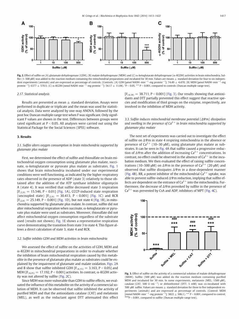
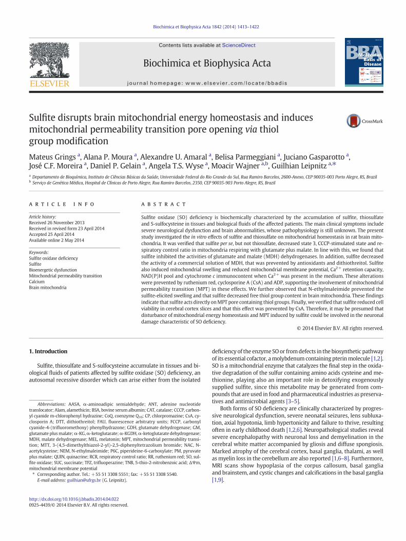
Fig. 2. Effect of sulfite on (A) glutamate dehydrogenase (GDH), (B)malate dehydrogenase (MDH) and (C)α-ketoglutarate dehydrogenase (α-KGDH) activities in brainmitochondria. Sul-fite (1–500 μM)was added to the reaction medium containing the mitochondrial preparations and incubated for 30 min. Values are means± standard deviation for four to six indepen-dent experiments (animals) and are expressed as percentage of controls. (Controls: (A) GDH [μmol NADH·min−1·mg protein−1]: 74.49 ± 4.470; (B) MDH [μmol NADH·min−1·mgprotein−1]: 6377 ± 570.5; (C) α-KGDH [nmol NADH·min−1·mg protein−1]: 54.17 ± 11.04). *P b 0.05, *** P b 0.001, compared to controls (Duncan multiple range test).
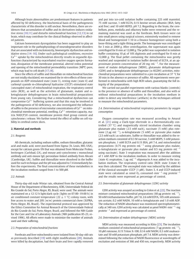
Fig. 3. Effect of sulfite on the activity of a commercial solution of malate dehydrogenase(MDH). Sulfite (500 μM) was added do the reaction medium containing purifiedMDH and incubated for 30 min. In some experiments, melatonin (MEL; 1500 μM),catalase (CAT; 500 U·mL−1) or dithiothreitol (DTT; 5 mM) was co-incubated with500 μM sulfite. Values are means ± standard deviation for three to five independent ex-periments (animals) and are expressed as percentage of controls. (Control: MDH[mmol NADH·min−1·mg protein−1]: 3822± 506.1). *** P b 0.001, compared to control;###P b 0.001, compared to sulfite (Duncan multiple range test).
1417M. Grings et al. / Biochimica et Biophysica Acta 1842 (2014) 1413–1422
2.17. Statistical analysis
Results are presented as mean ± standard deviation. Assays wereperformed in duplicate or triplicate and the mean was used for statisti-cal analysis. Data were analyzed by one-way ANOVA, followed by thepost hoc Duncanmultiple range testwhen Fwas significant. Only signif-icant F values are shown in the text. Differences between groups wererated significant at P b 0.05. All analyses were carried out using theStatistical Package for the Social Sciences (SPSS) software.
3. Results
3.1. Sulfite alters oxygen consumption in brain mitochondria supported byglutamate plus malate
First, we determined the effect of sulfite and thiosulfate on brainmi-tochondrial oxygen consumption using glutamate plus malate, succi-nate, α-ketoglutarate or pyruvate plus malate as substrates. Fig. 1shows that brain mitochondria incubated under our experimentalconditionswere well functioning, as indicated by the higher respiratoryrates observed in the presence of ADP (state 3) relatively to those ob-tained after the addition of the ATP synthase inhibitor oligomycinA (state 4). It was verified that sulfite decreased state 3 respiration[F(2,9) = 15.546, P b 0.01] (Fig. 1A), CCCP-induced state respiration(uncoupled state) [F(2,9) = 30.413, P b 0.001] (Fig. 1C) and RCR[F(2,9) = 25.149, P b 0.001] (Fig. 1D), but not state 4 (Fig. 1B), inmito-chondria supported by glutamate plus malate. In contrast, sulfite did notaltermitochondrial respirationwhen succinate,α-ketoglutarate or pyru-vate plus malate were used as substrates. Moreover, thiosulfate did notaffect mitochondrial oxygen consumption regardless of the substrateused (results not shown). Fig. 1E shows a representative respirationcurve demonstrating the transition from state 3 to state 4. This figure al-lows a direct calculation of state 3, state 4 and RCR.
3.2. Sulfite inhibits GDH and MDH activities in brain mitochondria
We assessed the effect of sulfite on the activities of GDH, MDH andα-KGDH in mitochondrial preparations in order to evaluate whetherthe inhibition of brain mitochondrial respiration caused by this metab-olite in the presence of glutamate plus malate as substrates could be ex-plained by the impairment of glutamate and malate oxidation. Figs. 2Aand B show that sulfite inhibited GDH [F(4,15) = 3.163, P b 0.05] andMDH [F(4,25) = 17.192, P b 0.001] activities. In contrast, α-KGDH activ-ity was not altered by sulfite (Fig. 2C).
SinceMDHwasmore vulnerable thanGDH to sulfite effects,we eval-uated the influence of thismetabolite on the activity of a commercial so-lution of MDH. It can be observed that sulfite inhibited the activity ofpurified MDH and that the antioxidants catalase (CAT) and melatonin(MEL), as well as the reductant agent DTT attenuated this effect
[F(4,18) = 38.711, P b 0.001] (Fig. 3). Our results showing that antioxi-dants and DTT partially prevented this effect suggest that reactive spe-cies and modification of thiol groups on the enzyme, respectively, areinvolved in the inhibition of MDH activity.
3.3. Sulfite induces mitochondrial membrane potential (ΔΨm) dissipationand swelling in the presence of Ca2+ in brain mitochondria supported byglutamate plus malate
The next set of experiments was carried out to investigate the effectof sulfite on ΔΨm in state 4 respiring mitochondria in the absence orpresence of Ca2+ (10–30 μM), using glutamate plus malate as sub-strates. It can be seen in Fig. 4A that sulfite caused a progressive reduc-tion of ΔΨm after the addition of increasing Ca2+ concentrations. Incontrast, no effect could be observed in the absence of Ca2+ in the incu-bation medium. We then evaluated the effect of raising sulfite concen-trations (10–500 μM) on ΔΨm in the presence of Ca2+ (20 μM) andobserved that sulfite dissipates ΔΨm in a dose-dependent manner(Fig. 4B). RR, a potent inhibitor of the mitochondrial Ca2+ uptake, wasable to prevent sulfite-inducedΔΨm reduction, implying that sulfite ef-fects are dependent on the entrance of Ca2+ into themitochondria. Fur-thermore, the decrease of ΔΨm provoked by sulfite in the presence ofCa2+ was prevented by CsA and ADP, inhibitors of MPT (Fig. 4C).

Fig. 4. Effect of sulfite onmitochondrialmembrane potential (ΔΨm)using brainmitochondria supported by glutamate plusmalate in the absence or presenceof Ca2+. (A) Sulfite (500 μM)(trace b) was added at the beginning of incubation to the reaction medium containing the mitochondrial preparations (0.5 mg protein·mL−1). Increasing concentrations of Ca2+ (0 μM,trace b; 10 μM, trace c; 20 μM, trace d; 30 μM, trace e)were added 50 s afterwards. (B) Increasing concentrations of sulfite (10 μM, trace b; 100 μM, trace c; 250 μM, trace d; 500 μM, trace e)were added at the beginning of incubation to the reactionmediumcontaining themitochondrial preparations (0.5mg protein·mL−1). Ca2+ (20 μM)was added 50 s afterwards. (C) Sulfite(500 μM) (trace b) was added at the beginning of incubation to the reaction medium containing the mitochondrial preparations (0.5 mg protein·mL−1). Ca2+ (20 μM) was added 50 safterwards. In some experiments, the mitochondrial preparations were incubated with sulfite plus ruthenium red (RR; 1 μM; trace c), cyclosporine A (CsA; 1 μM; trace d) orADP (300 μM; trace e). Control (trace a) in graphic A was performed in the absence of Ca2+ and sulfite, whereas controls (traces a) in graphics B and C were performed inthe presence of Ca2+ (20 μM) and did not contain sulfite. FCCP (1 μM) was added at the end of the experiment, as indicated. Traces are representative of three independent ex-periments and express fluorescence arbitrary units (FAU).
1418 M. Grings et al. / Biochimica et Biophysica Acta 1842 (2014) 1413–1422
In order to further study a possible involvement of the non-selectiveinner membrane permeabilization due to MPT pore opening withsulfite-induced ΔΨm decrease, we measured mitochondrial swellingby following light scattering changes. It was verified that sulfite causedswelling in the presence of Ca2+ in brain mitochondria and that this ef-fect was fully prevented by RR, CsA and ADP, reinforcing therefore theinduction of MPT by sulfite (Fig. 5A). In contrast, sulfite did not induceswelling in mitochondrial preparations from rat liver, implying thatbrain is more vulnerable to the toxic effects of this metabolite (Fig. 5B).
3.4. Sulfite reduces Ca2+ retention capacity in brain mitochondriasupported by glutamate plus malate
We then investigated the influence of sulfite on mitochondrial Ca2+
retention, sinceMPT induction leads to a rapid release of Ca2+ frommi-tochondria. As shown in Fig. 6, sulfite reduced the mitochondrial Ca2+
retention capacity compared with control mitochondria supported byglutamate plus malate. Furthermore, CsA and ADP prevented this effect,
Fig. 5.Effect of sulfite onmitochondrial swellingusing (A)brain or (B) livermitochondria suppoat the beginning of incubation to the reactionmedium containing themitochondrial preparatiothemitochondrial preparationswere incubatedwith sulfite plus ruthenium red (RR; 1 μM; tracperformed in the presence of Ca2+ (40 μM) and did not contain sulfite. Alamethicin (Alam; 40resentative of three independent experiments and express fluorescence arbitrary units (FAU).
indicating the involvement of MPT induction in themitochondrial Ca+2
handling disruption induced by sulfite.
3.5. Sulfite decreasesmitochondrial matrix NAD(P)Hpool in the presence ofCa2+ in brain mitochondria supported by glutamate plus malate
Sulfite reducedNAD(P)H fluorescence in the presence of Ca2+ inmi-tochondria supported by glutamate plus malate (Fig. 7), suggestingthat this metabolite decreased NAD(P)H pool due either to a oxidationof the reduced equivalents or their loss from the matrix. CsA, ADP andRR were able to prevent this effect, suggesting the involvement ofMPT induction.
3.6. NEM prevents sulfite-induced swelling in the presence of Ca2+ in brainmitochondria supported by glutamate plus malate
In the next step, we evaluated whether compounds reported to in-hibit the MPT, such as phospholipase A2 inhibitors (TFZ, CP and QUIN)
rted by glutamate plusmalate in thepresenceof Ca2+. Sulfite (500 μM) (traceb)was addedns (0.5mg protein·mL−1). Ca2+ (40 μM)was added 50 s afterwards. In some experiments,e c), cyclosporine A (CsA; 1 μM; trace d) or ADP (300 μM; trace e). Controls (traces a) wereμg·mg of protein−1) was added at the end of the experiment, as indicated. Traces are rep-
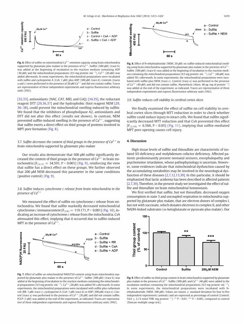
Fig. 6. Effect of sulfite onmitochondrial Ca2+ retention capacity using brain mitochondriasupported by glutamate plus malate in the presence of Ca2+. Sulfite (500 μM) (trace b)was added at the beginning of incubation to the reaction medium containing ADP(30 μM) and the mitochondrial preparations (0.5 mg protein·mL−1). Ca2+ (20 μM) wasadded afterwards. In some experiments, the mitochondrial preparations were incubatedwith sulfite and cyclosporine A (CsA; 1 μM) plus ADP (300 μM; trace d). Controls (tracesa and c)were performed in the presence of 20 μMCa2+ and did not contain sulfite. Tracesare representative of three independent experiments and express fluorescence arbitraryunits (FAU).
Fig. 8. Effect of N-ethylmaleimide (NEM; 20 μM) on sulfite-induced mitochondrial swell-ing using brainmitochondria supported by glutamate plusmalate in the presence of Ca2+.Sulfite (500 μM) (trace b) was added at the beginning of incubation to the reaction medi-um containing themitochondrial preparations (0.5 mg protein·mL−1). Ca2+ (40 μM)wasadded 50 s afterwards. In some experiments, the mitochondrial preparations were incu-bated with sulfite plus NEM (trace c). Control (trace a) was performed in the presenceof Ca2+ (40 μM) and did not contain sulfite. Alamethicin (Alam; 40 μg·mg of protein−1)was added at the end of the experiment, as indicated. Traces are representative of threeindependent experiments and express fluorescence arbitrary units (FAU).
1419M. Grings et al. / Biochimica et Biophysica Acta 1842 (2014) 1413–1422
[32,33], antioxidants (NAC, CAT, MEL and CoQ) [34,35], the reductantreagent DTT [29,36,37] and the hydrophobic thiol reagent NEM [29,36–38], could prevent the mitochondrial swelling induced by sulfite.We found that the inhibitors of phospholipase A2, antioxidants andDTT did not alter this effect (results not shown). In contrast, NEMprevented sulfite-induced swelling in the presence of Ca2+, suggestingthat sulfite exerts a direct effect on thiol groups of proteins involved inMPT pore formation (Fig. 8).
3.7. Sulfite decreases the content of thiol groups in the presence of Ca2+ inbrain mitochondria supported by glutamate plus malate
Our results also demonstrate that 500 μM sulfite significantly de-creased the content of thiol groups in the presence of Ca2+ in brain mi-tochondria [F(2,11) = 34.591, P b 0.001] (Fig. 9), reinforcing the viewthat sulfite has a direct effect on these groups. We further observedthat 200 μM NEM decreased this parameter in the same conditions(positive control) (Fig. 9).
3.8. Sulfite induces cytochrome c release from brain mitochondria in thepresence of Ca2+
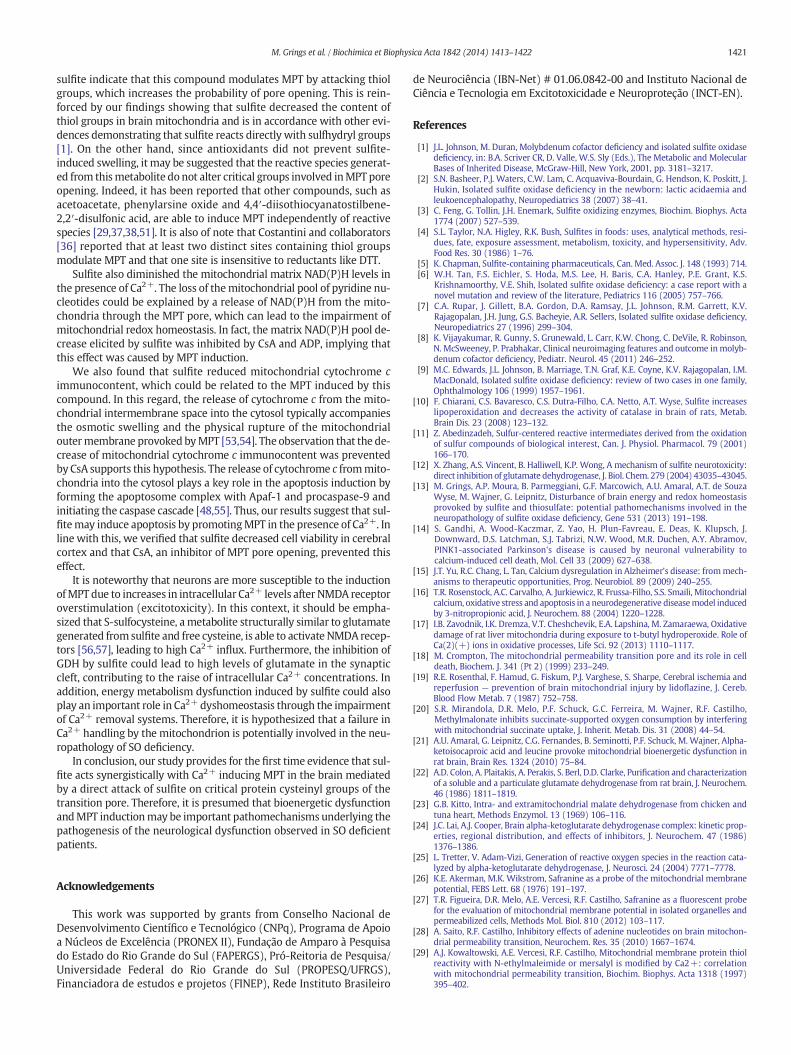
Wemeasured the effect of sulfite on cytochrome c release frommi-tochondria. We found that sulfite markedly decreased mitochondrialcytochrome c immunocontent [F(2,6)= 119.171, P b 0.001] (Fig. 10), in-dicating an increase of cytochrome c release from themitochondria. CsAattenuated this effect, implying that it occurred due to sulfite-inducedMPT in the presence of Ca2+.
Fig. 7. Effect of sulfite on mitochondrial NAD(P)H content using brain mitochondria sup-ported by glutamate plus malate in the presence of Ca2+. Sulfite (500 μM) (trace b) wasadded at the beginning of incubation to the reactionmedium containing themitochondri-al preparations (0.5mg protein·mL−1). Ca2+ (20 μM)was added 50 s afterwards. In someexperiments, the mitochondrial preparations were incubated with sulfite plus rutheniumred (RR; 1 μM; trace c), cyclosporine A (CsA; 1 μM; trace d) or ADP (300 μM; trace e). Con-trol (trace a) was performed in the presence of Ca2+ (20 μM) and did not contain sulfite.FCCP (1 μM)was added at the end of the experiment, as indicated. Traces are representa-tive of three independent experiments and express fluorescence arbitrary units (FAU).
3.9. Sulfite reduces cell viability in cerebral cortex slices
We finally examined the effect of sulfite on cell viability in cere-bral cortex slices through MTT reduction in order to check whethersulfite could induce injury to intact cells. We found that sulfite signif-icantly decreased MTT reduction and that CsA prevented this effect[F(2,10) = 6.566, P b 0.05] (Fig. 11), implying that sulfite-mediatedMPT pore opening causes cell injury.
4. Discussion
High tissue levels of sulfite and thiosulfate are characteristic of iso-lated SO deficiency and molybdenum cofactor deficiency. Affected pa-tients predominantly present neonatal seizures, encephalopathy andpsychomotor retardation, whose pathophysiology is uncertain. Howev-er, some evidences indicate that mitochondrial dysfunction caused bythe accumulating metabolites may be involved in the neurological dys-function of these diseases [2,7,12,13,39]. In this particular, it should beemphasized that lactic acidemia has been described in affected patients[2,7,39]. Therefore, in the present studywe investigated the effect of sul-fite and thiosulfate on brain mitochondrial homeostasis.
We first verified that sulfite, but not thiosulfate, decreased oxygenconsumption in state 3 and uncoupled respiration in mitochondria sup-ported by glutamate plus malate, that are electron donors of complex I,but notwith succinate, which donates electrons to complex II, and otherNADH-linked substrates (α-ketoglutarate or pyruvate plusmalate). Our
Fig. 9. Effect of sulfite on thiol group content in brainmitochondria supported by glutamateplus malate in the presence of Ca2+. Sulfite (500 μM) and Ca2+ (40 μM)were added to theincubation medium containing the mitochondrial preparations (0.5 mg protein·mL−1).In some experiments, the mitochondrial preparations were incubated with N-ethylmaleimide (NEM; 200 μM). Values are means ± standard deviation for four to fiveindependent experiments (animals) and are expressed as percentage of control (Control:54.0 ± 2.13 nmol TNB·mg protein−1). ** P b 0.01, *** P b 0.001, compared to control(Duncan multiple range test).
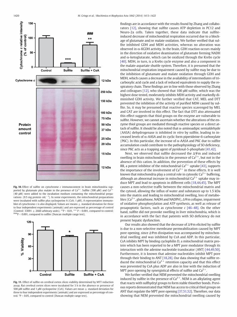
Fig. 10. Effect of sulfite on cytochrome c immunocontent in brain mitochondria sup-ported by glutamate plus malate in the presence of Ca2+. Sulfite (500 μM) and Ca2+
(40 μM) were added to the incubation medium containing the mitochondrial prepa-rations (0.5 mg protein·mL−1). In some experiments, the mitochondrial preparationswere incubated with sulfite plus cyclosporine A (CsA; 1 μM). A representative immuno-blot of cytochrome c is also displayed. Values are means ± standard deviation for threeto four independent experiments (animals) and are expressed as percentage of control(Control: 33091 ± 2660 arbitrary units). **P b 0.01, *** P b 0.001, compared to control;###P b 0.001, compared to sulfite (Duncan multiple range test).
Fig. 11. Effect of sulfite on cerebral cortex slices viability determined by MTT reductionassay. Rat cerebral cortex slices were incubated for 3 h in the absence or presence of500 μM sulfite and 1 μM cyclosporine (CsA). Values are mean ± standard deviation forthree to four independent experiments (animals) and are expressed as percentage of con-trol. *P b 0.05, compared to control (Duncan multiple range test).
1420 M. Grings et al. / Biochimica et Biophysica Acta 1842 (2014) 1413–1422
findings are in accordancewith the results found by Zhang and collabo-rators [12], showing that sulfite causes ATP depletion in PC12 andNeuro-2a cells. Taken together, these data indicate that sulfite-induced decrease of mitochondrial respiration occurred due to a block-age of glutamate and/or malate oxidation. We further verified that sul-fite inhibited GDH and MDH activities, whereas no alteration wasobserved in α-KGDH activity. In the brain, GDH reaction occurs mainlyin the direction of oxidative deamination of glutamate forming NADHand α-ketoglutarate, which can be oxidized through the Krebs cycle[40]. MDH, in turn, is a Krebs cycle enzyme and also a component inthe malate-aspartate shuttle system. Therefore, it is presumed that themitochondrial respiration impairment caused by sulfite may be due tothe inhibition of glutamate and malate oxidation through GDH andMDH, which causes a decrease in the availability of intermediates of tri-carboxylic acid cycle and a lack of reduced equivalents to supply the re-spiratory chain. These findings are in line with those observed by Zhangand colleagues [12], who showed that 100 μM sulfite, which was thehighest dose tested, moderately inhibits MDH activity andmarkedly di-minished GDH activity. We further verified that CAT, MEL and DTTprevented the inhibition of the activity of purified MDH caused by sul-fite. So, it may be presumed that reactive species scavenged by MELand CAT are involved in this effect. The fact that DTT also attenuatedthis effect suggests that thiol groups on the enzyme are vulnerable tosulfite. However, we cannot ascertainwhether the alterations of the en-zyme thiol groups are mediated through reactive species or a direct at-tack of sulfite. It should be also noted thatα-aminoadipic semialdehyde(AASA) dehydrogenase is inhibited in vitro by sulfite, leading to in-creased levels of α-AASA and its cyclic form piperideine-6-carboxylate(P6C). In this particular, the increase of α-AASA and P6C due to sulfiteaccumulation could contribute to the pathophysiology of SO deficiency,since P6C acts as a trapping agent of pyridoxal-5-phosphate [41,42].
Next, we observed that sulfite decreased the ΔΨm and inducedswelling in brain mitochondria in the presence of Ca2+, but not in theabsence of this cation. In addition, the prevention of these effects byRR, a potent inhibitor of the mitochondrial Ca2+ uptake [43], supportsthe importance of the involvement of Ca2+ in these effects. It is wellknown thatmitochondria play a central role in cytosolic Ca2+ buffering,although an abnormal increase in mitochondrial Ca2+ uptake may in-duce MPT and lead to apoptosis in different cells [18,44,45]. The MPTcauses a non-selective traffic between the mitochondrial matrix andthe cytosol, allowing the influx of water and substances up to 1.5 kDainto the matrix and leading to mitochondrial swelling, loss of metabo-lites (Ca2+, glutathione, NADHandNADPH),ΔΨmcollapse, impairmentof oxidative phosphorylation and ATP synthesis, as well as release ofproapoptotic factors, such as cytochrome c [46–48]. On the otherhand, sulfite did not provoke swelling in liver mitochondria, which isin accordance with the fact that patients with SO deficiency do notpresent hepatic dysfunction.
Our results also showed that the decrease of ΔΨm elicited by sulfiteis due to a non-selective membrane permeabilization caused by MPTpore opening, since ΔΨm dissipation was accompanied by mitochon-drial swelling and was inhibited by CsA and ADP. In this particular,CsA inhibits MPT by binding cyclophilin D, a mitochondrial matrix pro-tein which has been reported to be a MPT pore modulator through itsinteraction with the adenine nucleotide translocator (ANT) [44,49,50].Furthermore, it is known that adenine nucleotides inhibit MPT porethrough their binding to ANT [18,28]. Our data showing that sulfite re-duced the mitochondrial Ca2+ retention capacity and that this effectwas prevented by CsA plus ADP are also in line with the induction ofMPT pore opening by synergistical effects of sulfite and Ca2+.
We further verified that NEM prevented the mitochondrial swellinginduced by sulfite in the presence of Ca2+. NEM is an alkylating agentthat reacts with sulfhydryl groups to form stable thioether bonds. Previ-ous reports demonstrated that NEMhas access to critical thiol groups onANT that regulate the MPT pore opening [37,51,52]. Therefore, our datashowing that NEM prevented the mitochondrial swelling caused by
1421M. Grings et al. / Biochimica et Biophysica Acta 1842 (2014) 1413–1422
sulfite indicate that this compound modulates MPT by attacking thiolgroups, which increases the probability of pore opening. This is rein-forced by our findings showing that sulfite decreased the content ofthiol groups in brain mitochondria and is in accordance with other evi-dences demonstrating that sulfite reacts directly with sulfhydryl groups[1]. On the other hand, since antioxidants did not prevent sulfite-induced swelling, it may be suggested that the reactive species generat-ed from thismetabolite do not alter critical groups involved inMPT poreopening. Indeed, it has been reported that other compounds, such asacetoacetate, phenylarsine oxide and 4,4′-diisothiocyanatostilbene-2,2′-disulfonic acid, are able to induce MPT independently of reactivespecies [29,37,38,51]. It is also of note that Costantini and collaborators[36] reported that at least two distinct sites containing thiol groupsmodulate MPT and that one site is insensitive to reductants like DTT.
Sulfite also diminished the mitochondrial matrix NAD(P)H levels inthe presence of Ca2+. The loss of themitochondrial pool of pyridine nu-cleotides could be explained by a release of NAD(P)H from the mito-chondria through the MPT pore, which can lead to the impairment ofmitochondrial redox homeostasis. In fact, the matrix NAD(P)H pool de-crease elicited by sulfite was inhibited by CsA and ADP, implying thatthis effect was caused by MPT induction.
We also found that sulfite reduced mitochondrial cytochrome cimmunocontent, which could be related to the MPT induced by thiscompound. In this regard, the release of cytochrome c from the mito-chondrial intermembrane space into the cytosol typically accompaniesthe osmotic swelling and the physical rupture of the mitochondrialoutermembrane provoked byMPT [53,54]. The observation that the de-crease of mitochondrial cytochrome c immunocontent was preventedby CsA supports this hypothesis. The release of cytochrome c frommito-chondria into the cytosol plays a key role in the apoptosis induction byforming the apoptosome complex with Apaf-1 and procaspase-9 andinitiating the caspase cascade [48,55]. Thus, our results suggest that sul-fitemay induce apoptosis by promotingMPT in the presence of Ca2+. Inline with this, we verified that sulfite decreased cell viability in cerebralcortex and that CsA, an inhibitor of MPT pore opening, prevented thiseffect.
It is noteworthy that neurons are more susceptible to the inductionofMPT due to increases in intracellular Ca2+ levels after NMDA receptoroverstimulation (excitotoxicity). In this context, it should be empha-sized that S-sulfocysteine, ametabolite structurally similar to glutamategenerated from sulfite and free cysteine, is able to activate NMDA recep-tors [56,57], leading to high Ca2+ influx. Furthermore, the inhibition ofGDH by sulfite could lead to high levels of glutamate in the synapticcleft, contributing to the raise of intracellular Ca2+ concentrations. Inaddition, energy metabolism dysfunction induced by sulfite could alsoplay an important role in Ca2+ dyshomeostasis through the impairmentof Ca2+ removal systems. Therefore, it is hypothesized that a failure inCa2+ handling by the mitochondrion is potentially involved in the neu-ropathology of SO deficiency.
In conclusion, our study provides for the first time evidence that sul-fite acts synergistically with Ca2+ inducing MPT in the brain mediatedby a direct attack of sulfite on critical protein cysteinyl groups of thetransition pore. Therefore, it is presumed that bioenergetic dysfunctionandMPT inductionmay be important pathomechanisms underlying thepathogenesis of the neurological dysfunction observed in SO deficientpatients.
Acknowledgements
This work was supported by grants from Conselho Nacional deDesenvolvimento Científico e Tecnológico (CNPq), Programa de Apoioa Núcleos de Excelência (PRONEX II), Fundação de Amparo à Pesquisado Estado do Rio Grande do Sul (FAPERGS), Pró-Reitoria de Pesquisa/Universidade Federal do Rio Grande do Sul (PROPESQ/UFRGS),Financiadora de estudos e projetos (FINEP), Rede Instituto Brasileiro
de Neurociência (IBN-Net) # 01.06.0842-00 and Instituto Nacional deCiência e Tecnologia em Excitotoxicidade e Neuroproteção (INCT-EN).
References
[1] J.L. Johnson, M. Duran, Molybdenum cofactor deficiency and isolated sulfite oxidasedeficiency, in: B.A. Scriver CR, D. Valle, W.S. Sly (Eds.), The Metabolic and MolecularBases of Inherited Disease, McGraw-Hill, New York, 2001, pp. 3181–3217.
[2] S.N. Basheer, P.J. Waters, C.W. Lam, C. Acquaviva-Bourdain, G. Hendson, K. Poskitt, J.Hukin, Isolated sulfite oxidase deficiency in the newborn: lactic acidaemia andleukoencephalopathy, Neuropediatrics 38 (2007) 38–41.
[3] C. Feng, G. Tollin, J.H. Enemark, Sulfite oxidizing enzymes, Biochim. Biophys. Acta1774 (2007) 527–539.
[4] S.L. Taylor, N.A. Higley, R.K. Bush, Sulfites in foods: uses, analytical methods, resi-dues, fate, exposure assessment, metabolism, toxicity, and hypersensitivity, Adv.Food Res. 30 (1986) 1–76.
[5] K. Chapman, Sulfite-containing pharmaceuticals, Can. Med. Assoc. J. 148 (1993) 714.[6] W.H. Tan, F.S. Eichler, S. Hoda, M.S. Lee, H. Baris, C.A. Hanley, P.E. Grant, K.S.
Krishnamoorthy, V.E. Shih, Isolated sulfite oxidase deficiency: a case report with anovel mutation and review of the literature, Pediatrics 116 (2005) 757–766.
[7] C.A. Rupar, J. Gillett, B.A. Gordon, D.A. Ramsay, J.L. Johnson, R.M. Garrett, K.V.Rajagopalan, J.H. Jung, G.S. Bacheyie, A.R. Sellers, Isolated sulfite oxidase deficiency,Neuropediatrics 27 (1996) 299–304.
[8] K. Vijayakumar, R. Gunny, S. Grunewald, L. Carr, K.W. Chong, C. DeVile, R. Robinson,N. McSweeney, P. Prabhakar, Clinical neuroimaging features and outcome inmolyb-denum cofactor deficiency, Pediatr. Neurol. 45 (2011) 246–252.
[9] M.C. Edwards, J.L. Johnson, B. Marriage, T.N. Graf, K.E. Coyne, K.V. Rajagopalan, I.M.MacDonald, Isolated sulfite oxidase deficiency: review of two cases in one family,Ophthalmology 106 (1999) 1957–1961.
[10] F. Chiarani, C.S. Bavaresco, C.S. Dutra-Filho, C.A. Netto, A.T. Wyse, Sulfite increaseslipoperoxidation and decreases the activity of catalase in brain of rats, Metab.Brain Dis. 23 (2008) 123–132.
[11] Z. Abedinzadeh, Sulfur-centered reactive intermediates derived from the oxidationof sulfur compounds of biological interest, Can. J. Physiol. Pharmacol. 79 (2001)166–170.
[12] X. Zhang, A.S. Vincent, B. Halliwell, K.P. Wong, A mechanism of sulfite neurotoxicity:direct inhibition of glutamate dehydrogenase, J. Biol. Chem. 279 (2004) 43035–43045.
[13] M. Grings, A.P. Moura, B. Parmeggiani, G.F. Marcowich, A.U. Amaral, A.T. de SouzaWyse, M. Wajner, G. Leipnitz, Disturbance of brain energy and redox homeostasisprovoked by sulfite and thiosulfate: potential pathomechanisms involved in theneuropathology of sulfite oxidase deficiency, Gene 531 (2013) 191–198.
[14] S. Gandhi, A. Wood-Kaczmar, Z. Yao, H. Plun-Favreau, E. Deas, K. Klupsch, J.Downward, D.S. Latchman, S.J. Tabrizi, N.W. Wood, M.R. Duchen, A.Y. Abramov,PINK1-associated Parkinson's disease is caused by neuronal vulnerability tocalcium-induced cell death, Mol. Cell 33 (2009) 627–638.
[15] J.T. Yu, R.C. Chang, L. Tan, Calcium dysregulation in Alzheimer's disease: frommech-anisms to therapeutic opportunities, Prog. Neurobiol. 89 (2009) 240–255.
[16] T.R. Rosenstock, A.C. Carvalho, A. Jurkiewicz, R. Frussa-Filho, S.S. Smaili, Mitochondrialcalcium, oxidative stress and apoptosis in a neurodegenerative diseasemodel inducedby 3-nitropropionic acid, J. Neurochem. 88 (2004) 1220–1228.
[17] I.B. Zavodnik, I.K. Dremza, V.T. Cheshchevik, E.A. Lapshina, M. Zamaraewa, Oxidativedamage of rat liver mitochondria during exposure to t-butyl hydroperoxide. Role ofCa(2)(+) ions in oxidative processes, Life Sci. 92 (2013) 1110–1117.
[18] M. Crompton, The mitochondrial permeability transition pore and its role in celldeath, Biochem. J. 341 (Pt 2) (1999) 233–249.
[19] R.E. Rosenthal, F. Hamud, G. Fiskum, P.J. Varghese, S. Sharpe, Cerebral ischemia andreperfusion — prevention of brain mitochondrial injury by lidoflazine, J. Cereb.Blood Flow Metab. 7 (1987) 752–758.
[20] S.R. Mirandola, D.R. Melo, P.F. Schuck, G.C. Ferreira, M. Wajner, R.F. Castilho,Methylmalonate inhibits succinate-supported oxygen consumption by interferingwith mitochondrial succinate uptake, J. Inherit. Metab. Dis. 31 (2008) 44–54.
[21] A.U. Amaral, G. Leipnitz, C.G. Fernandes, B. Seminotti, P.F. Schuck, M. Wajner, Alpha-ketoisocaproic acid and leucine provoke mitochondrial bioenergetic dysfunction inrat brain, Brain Res. 1324 (2010) 75–84.
[22] A.D. Colon, A. Plaitakis, A. Perakis, S. Berl, D.D. Clarke, Purification and characterizationof a soluble and a particulate glutamate dehydrogenase from rat brain, J. Neurochem.46 (1986) 1811–1819.
[23] G.B. Kitto, Intra- and extramitochondrial malate dehydrogenase from chicken andtuna heart, Methods Enzymol. 13 (1969) 106–116.
[24] J.C. Lai, A.J. Cooper, Brain alpha-ketoglutarate dehydrogenase complex: kinetic prop-erties, regional distribution, and effects of inhibitors, J. Neurochem. 47 (1986)1376–1386.
[25] L. Tretter, V. Adam-Vizi, Generation of reactive oxygen species in the reaction cata-lyzed by alpha-ketoglutarate dehydrogenase, J. Neurosci. 24 (2004) 7771–7778.
[26] K.E. Akerman, M.K. Wikstrom, Safranine as a probe of the mitochondrial membranepotential, FEBS Lett. 68 (1976) 191–197.
[27] T.R. Figueira, D.R. Melo, A.E. Vercesi, R.F. Castilho, Safranine as a fluorescent probefor the evaluation of mitochondrial membrane potential in isolated organelles andpermeabilized cells, Methods Mol. Biol. 810 (2012) 103–117.
[28] A. Saito, R.F. Castilho, Inhibitory effects of adenine nucleotides on brain mitochon-drial permeability transition, Neurochem. Res. 35 (2010) 1667–1674.
[29] A.J. Kowaltowski, A.E. Vercesi, R.F. Castilho, Mitochondrial membrane protein thiolreactivity with N-ethylmaleimide or mersalyl is modified by Ca2+: correlationwith mitochondrial permeability transition, Biochim. Biophys. Acta 1318 (1997)395–402.
1422 M. Grings et al. / Biochimica et Biophysica Acta 1842 (2014) 1413–1422
[30] T. Mosmann, Rapid colorimetric assay for cellular growth and survival: applicationto proliferation and cytotoxicity assays, J. Immunol. Methods 65 (1983) 55–63.
[31] M.M. Bradford, A rapid and sensitive method for the quantitation of microgramquantities of protein utilizing the principle of protein-dye binding, Anal. Biochem.72 (1976) 248–254.
[32] J.G. Pastorino, G. Simbula, K. Yamamoto, P.A. Glascott Jr., R.J. Rothman, J.L. Farber,The cytotoxicity of tumor necrosis factor depends on induction of the mitochondrialpermeability transition, J. Biol. Chem. 271 (1996) 29792–29798.
[33] K.M. Broekemeier, J.R. Iben, E.G. LeVan, E.D. Crouser, D.R. Pfeiffer, Pore formationand uncoupling initiate a Ca2 +−independent degradation of mitochondrial phos-pholipids, Biochemistry 41 (2002) 7771–7780.
[34] M. Nishimura, Y. Okimura, H. Fujita, H. Yano, J. Lee, E. Suzaki, M. Inoue, K. Utsumi, J.Sasaki, Mechanism of 3-nitropropionic acid-induced membrane permeability tran-sition of isolated mitochondria and its suppression by L-carnitine, Cell Biochem.Funct. 26 (2008) 881–891.
[35] C. Chinopoulos, A.A. Starkov, G. Fiskum, Cyclosporin A-insensitive permeability tran-sition in brain mitochondria: inhibition by 2-aminoethoxydiphenyl borate, J. Biol.Chem. 278 (2003) 27382–27389.
[36] P. Costantini, B.V. Chernyak, V. Petronilli, P. Bernardi, Modulation of the mitochon-drial permeability transition pore by pyridine nucleotides and dithiol oxidation attwo separate sites, J. Biol. Chem. 271 (1996) 6746–6751.
[37] V. Petronilli, P. Costantini, L. Scorrano, R. Colonna, S. Passamonti, P. Bernardi, Thevoltage sensor of the mitochondrial permeability transition pore is tuned by theoxidation-reduction state of vicinal thiols. Increase of the gating potential by oxi-dants and its reversal by reducing agents, J. Biol. Chem. 269 (1994) 16638–16642.
[38] T.S. Kim, D.W. Jeong, B.Y. Yun, I.Y. Kim, Dysfunction of rat liver mitochondria by sel-enite: induction of mitochondrial permeability transition through thiol-oxidation,Biochem. Biophys. Res. Commun. 294 (2002) 1130–1137.
[39] F. Eichler, W.H. Tan, V.E. Shih, P.E. Grant, K. Krishnamoorthy, Proton magnetic reso-nance spectroscopy and diffusion-weighted imaging in isolated sulfite oxidase defi-ciency, J. Child Neurol. 21 (2006) 801–805.
[40] A. Kelly, C.A. Stanley, Disorders of glutamate metabolism, Ment. Retard. Dev. Disabil.Res. Rev. 7 (2001) 287–295.
[41] P.B. Mills, E.J. Footitt, S. Ceyhan, P.J. Waters, C. Jakobs, P.T. Clayton, E.A. Struys,Urinary AASA excretion is elevated in patients with molybdenum cofactor deficien-cy and isolated sulphite oxidase deficiency, J. Inherit. Metab. Dis. 35 (2012)1031–1036.
[42] E.A. Struys, B. Nota, A. Bakkali, S. Al Shahwan, G.S. Salomons, B. Tabarki, Pyridoxine-dependent epilepsy with elevated urinary alpha-amino adipic semialdehyde in mo-lybdenum cofactor deficiency, Pediatrics 130 (2012) e1716–e1719.
[43] C.L. Moore, Specific inhibition of mitochondrial Ca++ transport by ruthenium red,Biochem. Biophys. Res. Commun. 42 (1971) 298–305.
[44] C. Yarana, J. Sripetchwandee, J. Sanit, S. Chattipakorn, N. Chattipakorn, Calcium-induced cardiac mitochondrial dysfunction is predominantly mediated by cyclo-sporine A-dependent mitochondrial permeability transition pore, Arch. Med. Res.43 (2012) 333–338.
[45] G. Hajnoczky, G. Csordas, S. Das, C. Garcia-Perez, M. Saotome, S. Sinha Roy, M. Yi,Mitochondrial calcium signalling and cell death: approaches for assessing the roleof mitochondrial Ca2+ uptake in apoptosis, Cell Calcium 40 (2006) 553–560.
[46] D.B. Zorov, M. Juhaszova, Y. Yaniv, H.B. Nuss, S. Wang, S.J. Sollott, Regulation andpharmacology of the mitochondrial permeability transition pore, Cardiovasc. Res.83 (2009) 213–225.
[47] M. Zoratti, I. Szabo, The mitochondrial permeability transition, Biochim. Biophys.Acta 1241 (1995) 139–176.
[48] A. Rasola, P. Bernardi, Mitochondrial permeability transition in Ca(2+)-dependentapoptosis and necrosis, Cell Calcium 50 (2011) 222–233.
[49] J. Pottecher, M. Guillot, E. Belaidi, A.L. Charles, A. Lejay, A. Gharib, P. Diemunsch, B.Geny, Cyclosporine A normalizes mitochondrial coupling, reactive oxygen speciesproduction, and inflammation and partially restores skeletal muscle maximal oxida-tive capacity in experimental aortic cross-clamping, J. Vasc. Surg. 57 (2013)1100–1108 (e1102).
[50] E. Basso, L. Fante, J. Fowlkes, V. Petronilli, M.A. Forte, P. Bernardi, Properties of thepermeability transition pore in mitochondria devoid of Cyclophilin D, J. Biol.Chem. 280 (2005) 18558–18561.
[51] G.P. McStay, S.J. Clarke, A.P. Halestrap, Role of critical thiol groups on the matrix sur-face of the adenine nucleotide translocase in the mechanism of the mitochondrialpermeability transition pore, Biochem. J. 367 (2002) 541–548.
[52] A.P. Halestrap, K.Y. Woodfield, C.P. Connern, Oxidative stress, thiol reagents, andmembrane potential modulate the mitochondrial permeability transition by affect-ing nucleotide binding to the adenine nucleotide translocase, J. Biol. Chem. 272(1997) 3346–3354.
[53] M. Crompton, Mitochondrial intermembrane junctional complexes and their role incell death, J. Physiol. 529 (Pt 1) (2000) 11–21.
[54] P.X. Petit, M. Goubern, P. Diolez, S.A. Susin, N. Zamzami, G. Kroemer, Disruption ofthe outer mitochondrial membrane as a result of large amplitude swelling: the im-pact of irreversible permeability transition, FEBS Lett. 426 (1998) 111–116.
[55] D.R. Green, J.C. Reed, Mitochondria and apoptosis, Science 281 (1998) 1309–1312.[56] J.W. Olney, C.H. Misra, T. de Gubareff, Cysteine-S-sulfate: brain damaging metabolite
in sulfite oxidase deficiency, J. Neuropathol. Exp. Neurol. 34 (1975) 167–177.[57] B. Kagedal, M. Kallberg, B. Sorbo, A possible involvement of glutathione in the detox-
ication of sulfite, Biochem. Biophys. Res. Commun. 136 (1986) 1036–1041.