Embed Size (px)
Citation preview
Biochimica et Biophysica Acta 1838 (2014) 3078–3087
Contents lists available at ScienceDirect
Biochimica et Biophysica Acta
j ourna l homepage: www.e lsev ie r .com/ locate /bbamem
HIV-1 Tat membrane interactions probed using X-ray and neutronscattering, CD spectroscopy and MD simulations
Kiyotaka Akabori a, Kun Huang b, Bradley W. Treece a, Michael S. Jablin a, Brian Maranville c, Arthur Woll d,John F. Nagle a, Angel E. Garcia b, Stephanie Tristram-Nagle a,⁎a Department of Physics, Carnegie Mellon University, 5000 Forbes Avenue, Pittsburgh, PA, United Statesb Department of Physics, Rensselaer Polytechnic Institute, 110 Eighth Street, Troy, NY 12180, United Statesc NIST Center for Neutron Research, 100 Bureau Drive, Stop 6102, Gaithersburg, MD 20899, United Statesd CHESS, Cornell University, 161 Synchrotron Drive, Ithaca, NY 14853, United States
Abbreviations: (DOPC), dioleoylphosphocholine;ethanolamine; (DOPS), dioleoylphosphoserine; (POPC), p(POPE), palmitoyloleoylphosphoethanolamine; (POPS),(Soy PI), soy phosphoinositol; (SM), sphingomyelinbis(monoacylglycerol)-phosphate); (PG), phosphoglycerol;(GUVs), giant unilamellar vesicles; (LUVs), large unilameX-ray scattering; (WAXS), wide-angle X-ray scattering;(TFE), trifluoroethanol; (SDP) modeling program, ScatterinSecondary Structure of Proteins; P, peptide; L, lipid⁎ Corresponding author.
E-mail address: [email protected] (S. Tristram-Nagle).
http://dx.doi.org/10.1016/j.bbamem.2014.08.0140005-2736/© 2014 Elsevier B.V. All rights reserved.
a b s t r a c t
a r t i c l e i n f oArticle history:Received 26 April 2014Received in revised form 8 July 2014Accepted 5 August 2014Available online 19 August 2014
Keywords:Low-angle X-ray scattering (LAXS)Wide-angle X-ray scattering (WAXS)Molecular dynamics (MD) simulationsLipid bilayer structure cell-penetrating peptidePeptide translocationMembrane pore
We report the effect on lipid bilayers of the Tat peptide Y47GRKKRRQRRR57 from theHIV-1 virus transactivator oftranslation (Tat) protein. Synergistic use of low-angle X-ray scattering (LAXS) and atomistic molecular dynamicsimulations (MD) indicate Tat peptide binding to neutral dioleoylphosphocholine (DOPC) lipid headgroups. Thisbinding induced the local lipid phosphate groups to move 3 Å closer to the center of the bilayer. Many of thepositively charged guanidinium components of the arginines were as close to the center of the bilayer as thelocally thinned lipid phosphate groups. LAXS data for DOPC, DOPC/dioleoylphosphoethanolamine (DOPE),DOPC/dioleoylphosphoserine (DOPS), and a mimic of the nuclear membrane gave similar results. Generally,the Tat peptide decreased the bilayer bending modulus KC and increased the area/lipid. Further indicationsthat Tat softens a membrane, thereby facilitating translocation, were provided by wide-angle X-ray scattering(WAXS) and neutron scattering. CD spectroscopy was also applied to further characterize Tat/membrane inter-actions. Although a mechanism for translation remains obscure, this study suggests that the peptide/lipid inter-action makes the Tat peptide poised to translocate from the headgroup region.
© 2014 Elsevier B.V. All rights reserved.
1. Introduction
The name cell-penetrating peptide (CPP) connotes a peptide thateasily penetrates cell membranes (for Reviews see [1–3]). The presentwork focuses on the transactivator of translation, Tat, from the HIV-1virus, which plays a role in AIDS progression. Early work showed thatthe HIV-Tat transactivator protein (86 amino acids) was efficientlytaken up by cells, and concentrations as low as 1 nM were sufficientto transactivate a reporter gene expressed from the HIV-1 promoter[4,5]. It has been reported that Tat protein uptake does not requireATP [6]. Studies using inhibitors of different types of endocytosis, in-cluding clathrin- and caveolae-mediated, or receptor-independentmacropinocytosis reached the same conclusion that ATP mediated
(DOPE), dioleoylphospho-almitoyloleoylphosphocholine;palmitoyloleoylphosphoserine;; (Chol), cholesterol; (BMP),(CPP), cell-penetrating peptide;llar vesicles; (LAXS), low-angle(HFP), hexafluoroisopropanol;g Density Profile; (DSSP), Define
endocytosis is not involved in Tat protein permeation [7–10]. However,this issue is controversial, as other studies found evidence for endocyto-sis in Tat protein import [11–19]. Still other studies have concluded thatan ATP requirement for Tat protein entry depends on the size of thecargo attached to Tat protein, or on the specific cell type [20–22].
The part of the Tat protein responsible for cellular uptake wasassigned to a short region Tat (48–60), G48RKKRRQRRRPPQ60, which isparticularly rich in basic amino acids [6]. Deletion of three out of eightpositive charges in this region caused loss of its ability to translocate[6]. In this manuscript short basic regions will be called Tat, while theentire 86-amino acid protein will be called Tat protein. Tat was shownto be responsible for the Tat protein's permeation into the cell nucleusand the nucleoli [6], and this was confirmed using live cell fluorescencein SVGA cells [23]. Tat (48–60)was shown to have little toxicity onHeLacells at 100 μM concentration [6], but the longer Tat protein (2–86) wastoxic to rat brain glioma cells at 1–10 μM [24]. Interestingly, no hemo-lytic activity was found when human erythrocytes were incubatedwith a highly neurotoxic concentration (40 μM) of Tat (2–86) [24].These results prompt the question, what is the mechanism of Tat'stranslocation through membranes?
To address this question, many biophysical studies have used simplemodels of biological membranes composed of a small number of lipidtypes. These studies are valuable because there is no possibility for
3079K. Akabori et al. / Biochimica et Biophysica Acta 1838 (2014) 3078–3087
ATP-dependent translocation, thus ruling out endocytosis if transloca-tion occurs. For example, Mishra et al. reported that the rate of entryinto giant unilamellar vesicles (GUVs) composed of PS/PC (1:4 moleratio) lipids of rhodamine-tagged Tat is immeasurably slow, but itcrosses a GUV composed of PS/PC/PE (1:2:1) lipids within 30 s [25].This study suggests that negative curvature induced by the inclusionof PE facilitates translocation. In a subsequent study usingmuch smallerunilamellar vesicles (LUVs), Tat did not release an encapsulated fluores-cent probe in LUVs composed of lipidsmodeling the outer plasmamem-brane, PC/PE/SM/Chol (1:1:1:1.5), but did release the probe in LUVscomposed of BMP/PC/PE (77:19:4) [26]; BMP (bis(monoacylglycero)-phosphate) is an anionic lipid specific to late endosomes. In that study[26], the inclusion of PE did not suffice to cause leaky fusion in LUVs inthe absence of a negatively charged lipid. The contrasting results inthese two experiments may also be due to the use of LUVs instead ofGUVs since it was reported that Tat does not translocate across LUVSof PC/PG (3:2) but does translocate across GUVs of the same lipid com-position [27]. In a similar experiment, Tat did not translocate into egg PCLUVs [28]. In another experiment confirming these results, Tat did nottranslocate into GUVS containing only PC with 20 mol.% cholesterol,but when PS or PE was included with PC, then rapid translocation ofTat was observed [29]. These experiments demonstrate that the choiceof lipids and model systems influences Tat translocation.
Is a pore formed during Tat translocation? Although direct conduc-tance measurements of Tat and lipid membranes have not been carriedout, two studies measured conductancewith the somewhat similar CPPoligoarginine cR9 peptide. Using single-channel conductance of grami-cidin A in planar lipid membranes consisting of anionic, neutral orpositively charged lipids, c9R did not increase conductance, even in an-ionic lipid membranes [30]. By contrast, in a similar experiment usingplanar lipid membranes, a current was induced by c9R in PC/PG (3:1)membranes, with increasing destabilization over time [31]. Thus ques-tions remain about pore formation of Tat in membranes. In the GUVexperiment with Tat mentioned above [29], Ciobanasu et al., using sizeexclusion methods, suggested a pore in the nanometer range, whichcould only be passed by small dye tracer molecules. Thus, if a true poreforms, it is likely to be small and transitory.
What is the secondary structure of Tat in membranes? Circulardichroism (CD) spectroscopy was carried out on, where the penul-timate proline on Tat (48–60) was replaced by a tryptophan [27].That study found a random coil secondary structure in aqueous so-lution as well as when mixed with PC/PG/PE (65:35:5) LUVs. Thesame result was obtained using CD in PC/PG (3:1) vesicles byZiegler et al. [10], indicating that an alpha helix is not required forTat's translocation ability. In addition, solid state NMR has identifieda random coil structure of Tat in DMPC/DMPG (8:7 mole ratio)multibilayers [32]. In the larger Tat (1–72) protein NMR measure-ments at pH 4 have determined that there is no secondary struc-ture, with a dynamical basic region [33]. Similarly, NMR was usedto study the full Tat protein and found a highly flexible basic region[34].
Regarding the mechanism of translocation of this randomly struc-tured, short basic peptide, many models have been proposed based onthe conflicting results listed above. Molecular dynamic simulationsoffer some insight into the molecular details of translocation. Herceand Garcia simulated the translocation of Tat (Y47GRKKRRQRRR57)across DOPC at various lipid:peptidemolar ratios [35]. Their simulationsindicated that Tat binds to the phosphate headgroups, with 1 Tat bind-ing with 14 lipids, each positive charge on Tat associated with nearly 2phosphate groups [35]. Translocation involved a localized thinning,and snorkeling of arginine side chains through the hydrophobic layerto interact with phosphates on the other side of the membrane. Thisallowed some water molecules to penetrate the membrane along withTat, forming a pore [35]. In this simulation, performedwithout inclusionof counterions, pore formation was only observed at high ratios of pep-tide:lipid (1:18) or at elevated temperature. However, a subsequent
Gromacs simulation with counterions found no thinning and no poreformation when Tat was added to DOPC membranes [36]. Instead itfound a membrane invagination associated with a cluster of Tat pep-tides, suggesting thatmicropinocytosis could be themodel for Tat trans-location across membranes [36].
In this work we primarily combine experimental low-angle X-rayscattering (LAXS) data with MD simulations to obtain the structure offully hydrated, oriented lipid bilayers with Tat (47–57) added at severalmole ratios. The lipid systemswere DOPC, DOPC/DOPE (3:1mole ratio),DOPC/DOPS (3:1), DOPC/DOPE (1:1) and a mimic of the nuclearmembrane (POPC/POPE/POPS/SoyPI/Chol, 69:15:2:4:11). Accessorytechniques, densitometry, wide-angle X-ray scattering (WAXS), neu-tron scattering, and CD spectroscopy were also applied to further char-acterize Tat/membrane interactions.
2. Materials and methods
2.1. Lipids and peptides
Synthesized lipids were purchased from Avanti Polar Lipids(Alabaster, AL) and used without further purification. Membranemimics were prepared by first dissolving lyophilized lipids in chloro-form and thenmixing these stock solutions to create the lipid composi-tions DOPC, DOPC/DOPE (3:1), DOPC/DOPE (1:1), DOPC/DOPS (3:1)and nuclear membrane mimic (POPC/POPE/POPS/SoyPI/Cholesterol,69:15:2:4:11) (based on Ref. [37]). Peptide (Y47GRKKRRQRRR57) waspurchased in three separate lots from the Peptide Synthesis Facility(University of Pittsburgh, Pittsburgh, PA); mass spectroscopy revealedN95% purity. This Tat peptide corresponds to residues (47–57) ofthe 86 residues in the Tat protein [6]. Tat was dissolved in HPLCtrifluoroethanol (TFE) and then mixed with lipid stock solutions inchloroform to form mole fractions between 0.0044 and 0.108. Theweight of Tat in these mole fractions was corrected for protein content(the remainder being 8 trifluoroacetate counter-ions from the peptidesynthesis). Solvents were removed by evaporation in the fume hoodfollowed by 2 h in a vacuum chamber at room temperature.
2.2. Samples for X-ray and neutron scattering
Four mg dried lipid/peptide mixture was re-dissolved in HPLC chlo-roform/TFE (2:1 v:v) for most of the lipid compositions. However,DOPC/DOPS (3:1) mixtures required chloroform/HFP (1:1 v:v) inorder to solubilize the negatively chargedDOPS. 200 μl of 4mgmixturesin solventswas plated onto siliconwafers (15 × 30×1mm) via the rockand roll method [38] to produce stacks of ~1800 well-aligned bilayers;solvents were removed by evaporation in the fume hood, followed by2 h under vacuum. 2H NMR revealed ~1% residual solvent after theseprocedures. Samples were prehydrated through the vapor in polypro-pylene hydration chambers at 37 °C for 2–6 h directly before hydratingin the thick-walled X-ray hydration chamber [39] for 0.5–1 h. Pre-equilibration allowed sufficient time for equilibriumbinding of peptideswith membrane mimics.
2.3. Samples for densimetry
Multilamellar vesicles (MLVs) were prepared by mixing dried lipidmixtures with MilliQ water to a final concentration of 2–5 wt.% innalgene vials and cycling three times between −20 °C and 60 °C for10 min at each temperature with vortexing. Pure Tat was dissolved inwater at 0.4 wt.%.
2.4. Samples for circular dichroism (CD)
Thin films were prepared by spreading ~1 mg, x = 0.11 (Peptide/(Lipid + Peptide)), in chloroform/TFE (1:1) onto one inside face of aquartz cuvette (Fisher Scientific, Pittsburgh, PA) and solvents were
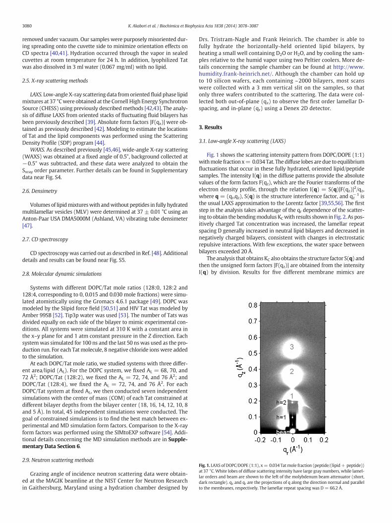
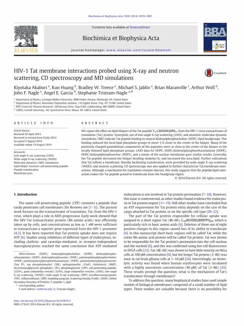
Fig. 1. LAXS of DOPC/DOPE (1:1), x= 0.034 Tatmole fraction (peptide/(lipid+ peptide))at 37 °C. White lobes of diffuse scattering intensity have large gray numbers, while lamel-lar orders and beam are shown to the left of the molybdenum beam attenuator (short,dark rectangle). qz and qr are the projections of q along the direction normal and parallelto the membranes, respectively. The lamellar repeat spacing was D = 66.2 Å.
3080 K. Akabori et al. / Biochimica et Biophysica Acta 1838 (2014) 3078–3087
removed under vacuum. Our samples were purposely misoriented dur-ing spreading onto the cuvette side to minimize orientation effects onCD spectra [40,41]. Hydration occurred through the vapor in sealedcuvettes at room temperature for 24 h. In addition, lyophilized Tatwas also dissolved in 3 ml water (0.067 mg/ml) with no lipid.
2.5. X-ray scattering methods
LAXS. Low-angle X-ray scatteringdata fromoriented fluid phase lipidmixtures at 37 °Cwere obtained at the Cornell High Energy SynchrotronSource (CHESS) using previously describedmethods [42,43]. The analy-sis of diffuse LAXS from oriented stacks of fluctuating fluid bilayers hasbeen previously described [39]. Absolute form factors |F(qz)| were ob-tained as previously described [42]. Modeling to estimate the locationsof Tat and the lipid components was performed using the ScatteringDensity Profile (SDP) program [44].
WAXS. As described previously [45,46], wide-angle X-ray scattering(WAXS) was obtained at a fixed angle of 0.5°, background collected at−0.5° was subtracted, and these data were analyzed to obtain theSxray order parameter. Further details can be found in Supplementarydata near Fig. S4.
2.6. Densimetry
Volumes of lipidmixtureswith andwithout peptides in fully hydratedmultilamellar vesicles (MLV) were determined at 37 ± 0.01 °C using anAnton-Paar USA DMA5000M (Ashland, VA) vibrating tube densimeter[47].
2.7. CD spectroscopy
CD spectroscopy was carried out as described in Ref. [48]. Additionaldetails and results can be found near Fig. S5.
2.8. Molecular dynamic simulations
Systems with different DOPC/Tat mole ratios (128:0, 128:2 and128:4, corresponding to 0, 0.015 and 0.030 mole fractions) were simu-lated atomistically using the Gromacs 4.6.1 package [49]. DOPC wasmodeled by the Slipid force field [50,51] and HIV Tat was modeled byAmber 99SB [52]. Tip3p water was used [53]. The number of Tats wasdivided equally on each side of the bilayer to mimic experimental con-ditions. All systems were simulated at 310 K with a constant area inthe x–y plane for and 1 atm constant pressure in the Z direction. Eachsystemwas simulated for 100 ns and the last 50 nswas used as the pro-duction run. For each Tat molecule, 8 negative chloride ions were addedto the simulation.
At each DOPC/Tat mole ratio, we studied systems with three differ-ent area/lipid (AL). For the DOPC system, we fixed AL = 68, 70, and72 Å2; DOPC/Tat (128:2), we fixed the AL = 72, 74, and 76 Å2; andDOPC/Tat (128:4), we fixed the AL = 72, 74, and 76 Å2. For eachDOPC/Tat system at fixed AL, we then conducted seven independentsimulations with the center of mass (COM) of each Tat constrained atdifferent bilayer depths from the bilayer center (18, 16, 14, 12, 10, 8and 5 Å). In total, 45 independent simulations were conducted. Thegoal of constrained simulations is to find the best match between ex-perimental and MD simulation form factors. Comparison to the X-rayform factors was performed using the SIMtoEXP software [54]. Addi-tional details concerning the MD simulation methods are in Supple-mentary Data Section 6.
2.9. Neutron scattering methods
Grazing angle of incidence neutron scattering data were obtain-ed at the MAGIK beamline at the NIST Center for Neutron Researchin Gaithersburg, Maryland using a hydration chamber designed by
Drs. Tristram-Nagle and Frank Heinrich. The chamber is able tofully hydrate the horizontally-held oriented lipid bilayers, byheating a small well containing D2O or H2O, and by cooling the sam-ples relative to the humid vapor using two Peltier coolers. More de-tails concerning the sample chamber can be found at http://www.humidity.frank-heinrich.net/. Although the chamber can hold upto 10 silicon wafers, each containing ~2000 bilayers, most scanswere collected with a 3 mm vertical slit on the samples, so thatonly three wafers contributed to the scattering. The data were col-lected both out-of-plane (qz) to observe the first order lamellar D-spacing, and in-plane (qr) using a Denex 2D detector.
3. Results
3.1. Low-angle X-ray scattering (LAXS)
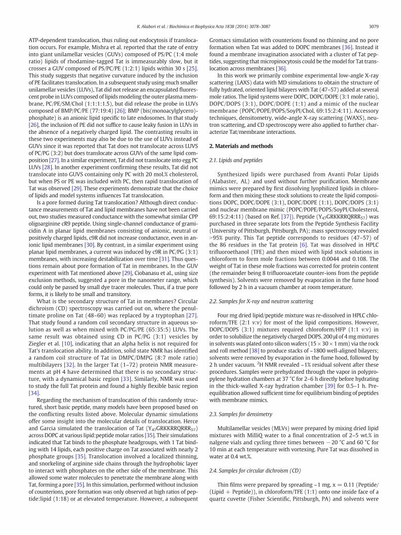
Fig. 1 shows the scattering intensity pattern from DOPC/DOPE (1:1)withmole fraction x= 0.034 Tat. The diffuse lobes are due to equilibriumfluctuations that occur in these fully hydrated, oriented lipid/peptidesamples. The intensity I(q) in the diffuse patterns provide the absolutevalues of the form factors F(qz), which are the Fourier transforms of theelectron density profile, through the relation I(q) = S(q)|F(qz)|2/qz,where q = (qr,qz), S(q) is the structure interference factor, and qz−1 isthe usual LAXS approximation to the Lorentz factor [39,55,56]. The firststep in the analysis takes advantage of the qr dependence of the scatter-ing to obtain the bendingmodulus KCwith results shown in Fig. 2. As pos-itively charged Tat concentration was increased, the lamellar repeatspacing D generally increased in neutral lipid bilayers and decreased innegatively charged bilayers, consistent with changes in electrostaticrepulsive interactions. With few exceptions, the water space betweenbilayers exceeded 20 Å.
The analysis that obtainsKC also obtains the structure factor S(q) andthen the unsigned form factors |F(qz)| are obtained from the intensityI(q) by division. Results for five different membrane mimics are
Fig. 2. Bilayer bending modulus, KC, vs. P/(L + P) mole fraction. D-spacings for DOPC/Tatmixtures varied from 64 to 68 Å, for DOPC/DOPE/Tat mixtures from 64 to 69 Å, forDOPC/DOPS/Tat (3:1) mixtures from 57 Å to N100 Å (pure DOPS was unbound), and fornuclearmimic/Tatmixtures from unbound (nuclearmembranemimic) to 64Å. Estimateduncertainty in all values is ±2.
3081K. Akabori et al. / Biochimica et Biophysica Acta 1838 (2014) 3078–3087
shown in Fig. 3. Vertical lines indicate the “zero” position between thelobes of diffuse data where F(qz) change sign. In every sample, thezero positions shift to larger qz, indicating a thinning of themembranes.
3.2. MD simulations
Due to the slow relaxation in lipid bilayers and limited accuracy ofthe force field, a good agreement between experimental and MD
Fig. 3. Form factors of lipidmixtures (arbitrarily scaled and vertically displaced)with increasingDOPE (3:1) C. DOPC/DOPE (1:1) D. DOPC/DOPS (3:1) E. Nuclear mimic. The entire qz range is shthe form factors equal zero between the lobes of diffuse data.
simulation calculated form factors may be difficult to reach. Conse-quently, we carried out several constrained simulations at AL and ZTatas described in Materials andmethods. We then compared the simulat-ed form factor F(qz) with the experimental form factor. The best matchfor DOPC/Tat (128:4)was foundwhen the Tatswere constrained at 18Åaway from the bilayer center (Fig. 4A,B). The other best fit results were:DOPC AL= 70 Å2 and DOPC/Tat(128:2) AL=72Å2, ZTat= 18 Å. It clear-ly indicates that with increasing Tat concentration, AL increases. Theagreement worsened as Tat was constrained to be closer to the centerof the bilayer. When Tats were constrained at 5 Å away from the bilayercenter, we observed a spontaneous formation of water pores in the MDsimulation. However, as shown in Fig. 4.C the corresponding form factorcalculated from MD simulations does not match well with the experi-mental form factor. Thus, by comparing the experimental and simulatedform factors, Tat's headgroup position is validated, while the hydrocar-bon position is ruled out. A similar comparison of form factors from X-ray scattering and MD simulation previously allowed us to determinethat the pore-forming alamethicin peptide locates in a transmembrane,not headgroup position [57].
3.3. SDP modeling
We also estimate structure by fitting the experimental form factorsusing the SDP method [44] with the component groups identified inFig. 5. The positions of these groups were free parameters and the agree-ment with the experimental form factors was excellent. Absolute totalelectron density profiles and the Tat profiles are shown formany samplesin Fig. 6(A–C). It must be emphasized, however, that, while the total EDPiswell determined by this fitting procedure, the values of the parametersfor the components are not as well determined as they would be if one
Tatmole fractions, P/(L+P), indicated on figure legends. Lipidmixtures: A. DOPC B. DOPC/own in C,while others show partial ranges. Solid vertical lines indicate the qz valueswhere
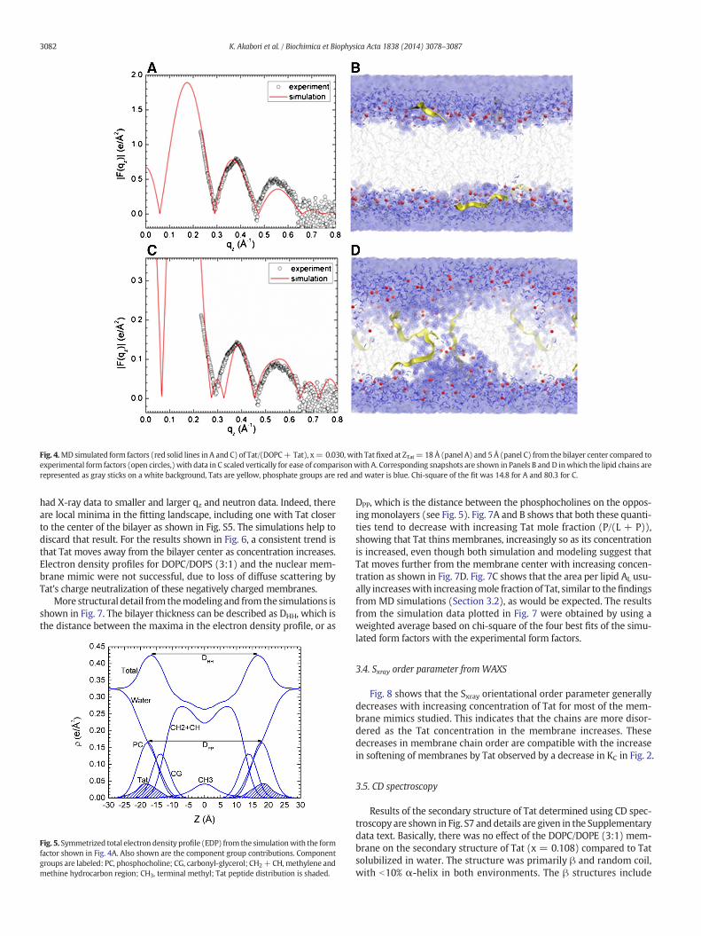
Fig. 4.MDsimulated form factors (red solid lines in A and C) of Tat/(DOPC+ Tat), x= 0.030, with Tat fixed at ZTat= 18 Å (panel A) and 5 Å (panel C) from the bilayer center compared toexperimental form factors (open circles,) with data in C scaled vertically for ease of comparisonwith A. Corresponding snapshots are shown in Panels B and D inwhich the lipid chains arerepresented as gray sticks on a white background, Tats are yellow, phosphate groups are red and water is blue. Chi-square of the fit was 14.8 for A and 80.3 for C.
3082 K. Akabori et al. / Biochimica et Biophysica Acta 1838 (2014) 3078–3087
had X-ray data to smaller and larger qz and neutron data. Indeed, thereare local minima in the fitting landscape, including one with Tat closerto the center of the bilayer as shown in Fig. S5. The simulations help todiscard that result. For the results shown in Fig. 6, a consistent trend isthat Tat moves away from the bilayer center as concentration increases.Electron density profiles for DOPC/DOPS (3:1) and the nuclear mem-brane mimic were not successful, due to loss of diffuse scattering byTat's charge neutralization of these negatively charged membranes.
More structural detail from themodeling and from the simulations isshown in Fig. 7. The bilayer thickness can be described as DHH, which isthe distance between the maxima in the electron density profile, or as
Fig. 5. Symmetrized total electron density profile (EDP) from the simulationwith the formfactor shown in Fig. 4A. Also shown are the component group contributions. Componentgroups are labeled: PC, phosphocholine; CG, carbonyl-glycerol; CH2+ CH,methylene andmethine hydrocarbon region; CH3, terminal methyl; Tat peptide distribution is shaded.
DPP, which is the distance between the phosphocholines on the oppos-ingmonolayers (see Fig. 5). Fig. 7A and B shows that both these quanti-ties tend to decrease with increasing Tat mole fraction (P/(L + P)),showing that Tat thins membranes, increasingly so as its concentrationis increased, even though both simulation and modeling suggest thatTat moves further from the membrane center with increasing concen-tration as shown in Fig. 7D. Fig. 7C shows that the area per lipid AL usu-ally increaseswith increasingmole fraction of Tat, similar to thefindingsfrom MD simulations (Section 3.2), as would be expected. The resultsfrom the simulation data plotted in Fig. 7 were obtained by using aweighted average based on chi-square of the four best fits of the simu-lated form factors with the experimental form factors.
3.4. Sxray order parameter fromWAXS
Fig. 8 shows that the Sxray orientational order parameter generallydecreases with increasing concentration of Tat for most of the mem-brane mimics studied. This indicates that the chains are more disor-dered as the Tat concentration in the membrane increases. Thesedecreases in membrane chain order are compatible with the increasein softening of membranes by Tat observed by a decrease in KC in Fig. 2.
3.5. CD spectroscopy
Results of the secondary structure of Tat determined using CD spec-troscopy are shown in Fig. S7 anddetails are given in the Supplementarydata text. Basically, there was no effect of the DOPC/DOPE (3:1) mem-brane on the secondary structure of Tat (x = 0.108) compared to Tatsolubilized in water. The structure was primarily β and random coil,with b10% α-helix in both environments. The β structures include
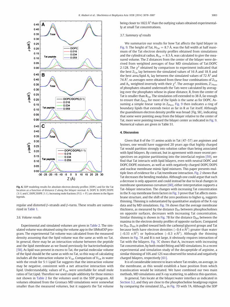
Fig. 6. SDP modeling results for absolute electron density profiles (EDPs) and for the Tatlocation as a function of distance Z along the bilayer normal. A. DOPC B. DOPC/DOPE(3:1) and C. DOPC/DOPE (1:1). Increasingmole fractions (P/(L+P)) are shown in the figurelegends.
3083K. Akabori et al. / Biochimica et Biophysica Acta 1838 (2014) 3078–3087
regular and distorted β-strands and β-turns. These results are summa-rized in Table 1.
3.6. Volume results
Experimental and simulated volumes are given in Table 2. The sim-ulated volumewas obtained using the volume app in the SIMtoEXPpro-gram. The experimental Tat volume was calculated from the measureddensity assuming that the lipid volume was the same as with no Tat.In general, there may be an interaction volume between the peptideand the lipid membrane as we found previously for bacteriorhodopsin[58]. As lipid was present in excess to Tat, the partial molecular volumeof the lipid should be the same as with no Tat, so this way of calculatingincludes all the interaction volume in VTat. Comparison of VTat in waterwith the result for 5:1 Lipid:Tat suggests that the interaction volumemay be negative, consistent with a net attractive interaction withlipid. Understandably, values of VTat were unreliable for small moleratios of Tat:Lipid. Thereforewe used simple additivity for thosemimicsnot shown in Table 2 for the volumes used in the SDP program. Allvolumes obtained from the Gromacs MD simulations were somewhatsmaller than the measured volumes, but it supports the Tat volume
being closer to 1822Å3 than the outlying values obtained experimental-ly at small Tat concentrations.
3.7. Summary of results
We summarize our results for how Tat affects the lipid bilayer inFig. 9. The height of Tat, HTat = 8.7 Å, was the full width at half maxi-mum of the Tat electron density profiles obtained from simulationsand the cylindrical radius, RTat = 8.3 Å, was calculated to give the mea-sured volume. The Z distances from the center of the bilayer were de-rived from weighted averages of four MD simulations of Tat:DOPC2:128. The χ2 obtained by comparison to experiment indicated thatthe best ZTat lay between the simulated values of 16 Å and 18 Å andthe best area/lipid AL lay between the simulated values of 72 Å2 and74 Å2, so averages were obtained from these four combinations of ZTatand AL, weighted inversely with their χ2. The average positions, Z′Phos,of phosphates situated underneath the Tats were calculated by averag-ing over the phosphates whose in-plane distance, R, from the center ofTat is smaller than RTat. The simulation cell extended to 38 Å, far enoughto ensure that ZPhos for most of the lipids is the same as for DOPC. As-suming a simple linear ramp in ZPhos, Fig. 9 then indicates a ring ofboundary lipids that extends twice as far in R as Tat itself. Althoughthe guanidinium electron density profile was broad (Fig. S8), indicatingthat some were pointing away from the bilayer relative to the center ofTat, more were pointing toward the bilayer center as indicated in Fig. 9.Numerical values are given in Table S1.
4. Discussion
Given that 8 of the 11 amino acids in Tat (47–57) are arginines andlysines, one would have suggested 20 years ago that highly chargedTat would partition strongly into solution rather than being associatedwith lipid bilayers. By contrast, but in agreement with more recent per-spectives on arginine partitioning into the interfacial region [59], wefind that Tat interacts with lipid bilayers, even with neutral DOPC andDOPC/DOPE mixtures, as well as with negatively charged DOPC/DOPSand nuclear membrane mimic lipid mixtures. This paper presents mul-tiple lines of evidence for a Tat/membrane interaction. Fig. 2 shows thatTat decreases the bendingmodulus. Although one could argue that sucha decrease is only apparent and could instead be due to local changes inmembrane spontaneous curvature [60], either interpretation supports aTat–bilayer interaction. The changes with increasing Tat concentrationin theX-raymembrane form factors in Fig. 3 prove that Tat affectsmem-brane structure, and the shift of the zero positions to higher qz suggeststhinning. Thinning is substantiated by quantitative analysis of the X-raydata and by MD simulations. Fig. 7A shows that the average membranethickness, as measured by the distance DPP between phosphocholineson opposite surfaces, decreases with increasing Tat concentration.Similar thinning is shown in Fig. 7B for the distance DHH between themaxima in the electron density profiles of opposite surfaces. Comparedto DPP, DHH is pulled toward both the carbonyl/glycerol groups and Tatbecause both have electron densities (~0.4 e/Å3) greater than water(~0.33 e/Å3) or hydrocarbon (~0.3 e/Å3). Although the thinningshown in Fig. 7A and B is not large, it obviously requires interaction ofTat with the bilayers. Fig. 7C shows that AL increases with increasingTat concentration, by bothmodel fitting andMD simulations. In a recentexperimental and simulation study of the decapeptide of arginine, asimilar thinning of 10% and 12%was observed for neutral and negativelycharged bilayers, respectively [61].
It is of considerable interest to learnwhere Tat resides, on average, inthe membrane, as this would establish a base position from whichtranslocation would be initiated. We have combined our two mainmethods, MD simulations and X-ray scattering, to address this question.
In general, Tats locate at the bilayer/water interface as indicated inSection 3.2, and they are close to the phosphocholine headgroup regionby comparing the simulated 2ZTat in Fig. 7D with 7A. Although the SDP
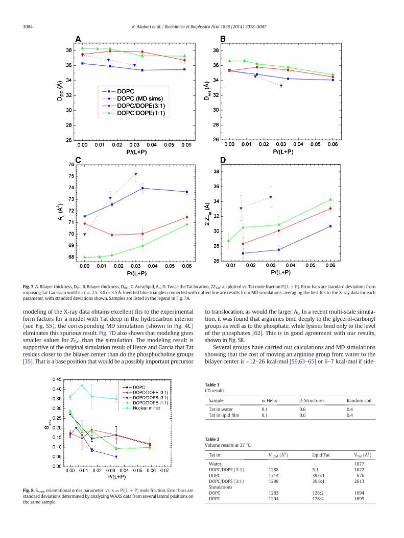
Fig. 7.A. Bilayer thickness, DPP; B. Bilayer thickness, DHH; C. Area/lipid, AL; D. Twice the Tat location, 2ZTat: all plotted vs. Tatmole fraction P/(L+P). Error bars are standard deviations fromimposing Tat Gaussian widths, σ= 2.5, 3.0 or 3.5 Å. Inverted blue triangles connected with dotted line are results fromMD simulations, averaging the best fits to the X-ray data for eachparameter, with standard deviations shown. Samples are listed in the legend in Fig. 7A.
3084 K. Akabori et al. / Biochimica et Biophysica Acta 1838 (2014) 3078–3087
modeling of the X-ray data obtains excellent fits to the experimentalform factors for a model with Tat deep in the hydrocarbon interior(see Fig. S5), the corresponding MD simulation (shown in Fig. 4C)eliminates this spurious result. Fig. 7D also shows that modeling givessmaller values for ZTat than the simulation. The modeling result issupportive of the original simulation result of Herce and Garcia that Tatresides closer to the bilayer center than do the phosphocholine groups[35]. That is a base position that would be a possibly important precursor
Fig. 8. Sxray, orientational order parameter, vs. x = P/(L + P) mole fraction. Error bars arestandard deviations determined by analyzingWAXS data from several lateral positions onthe same sample.
to translocation, as would the larger AL. In a recent multi-scale simula-tion, it was found that arginines bind deeply to the glycerol-carbonylgroups as well as to the phosphate, while lysines bind only to the levelof the phosphates [62]. This is in good agreement with our results,shown in Fig. S8.
Several groups have carried out calculations and MD simulationsshowing that the cost of moving an arginine group from water to thebilayer center is ~12–26 kcal/mol [59,63–65] or 6–7 kcal/mol if side-
Table 1CD results.
Sample α-Helix β-Structures Random coil
Tat in water 0.1 0.6 0.4Tat in lipid film 0.1 0.6 0.4
Table 2Volume results at 37 °C.
Tat in: Vlipid (Å3) Lipid:Tat VTat (Å3)
Water 1877DOPC/DOPE (3:1) 1288 5:1 1822DOPC 1314 39.6:1 676DOPC/DOPS (3:1) 1298 39.6:1 2613SimulationsDOPC 1283 128:2 1694DOPC 1294 128:4 1699
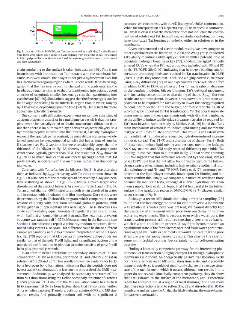
Fig. 9. Location of Tat in DOPC bilayer. Tat is represented as a cylinder, Z is the distancefrom the bilayer center, and R is the in-plane distance from the center of Tat. The averageZ of the lipid phosphates as a function of R and the arginine guanidiniums are shown in redand blue, respectively.
3085K. Akabori et al. / Biochimica et Biophysica Acta 1838 (2014) 3078–3087
chain snorkeling to the surface is taken into account [66]. This is notinconsistent with our result that Tat interacts with the membrane be-cause, as is well known, the bilayer is not just a hydrocarbon slab, buthas interfacial headgroup regions where Tat can reside. It has been sug-gested that the free energy cost for charged amino acids entering theheadgroup region is similar to that for partitioning into octanol, aboutan order of magnitude smaller free energy cost than partitioning intocyclohexane [67–69]. Simulations suggest that the free energy is smallerfor an arginine residing in the interfacial region than in water, roughlyby 3 kcal/mole, depending upon the lipid [59,69]. Our results thereforeappear energetically reasonable.
One concern with diffraction experiments on samples consisting ofadjacent bilayers in a stack or in a multilamellar vesicle is that the sam-ples have to be partially dried to obtain conventional diffraction data.But then there is no pure water layer between adjacent bilayers, so ahydrophilic peptide is forced into the interfacial, partially hydrophilicregion of the lipid bilayer. In contrast, by using diffuse scattering, we ob-tained structure from experimental samples that had a range of lamellarD spacings (see Fig. 2 caption) that were considerably larger than thethickness of the bilayer in Fig. 7A, thereby providing an ample purewater space, typically greater than 20 Å. The result that 2ZTat shown inFig. 7D is so much smaller than our repeat spacings shows that Tatpreferentially associates with the membrane rather than dissociatinginto water.
Consistent with Tat softening the bilayers (Fig. 2), it also disordersthem as indicated by Sxray decreasing with Tat concentration shown inFig. 8. Tat also increases the mosaic spread observed by X-ray and neu-tron scattering as shown in Figs. S1–3; this is a much larger scaledisordering of the stack of bilayers. As shown in Table 1 and in Fig. S7,Tat assumed slightly N50% β structures, both when dissolved in waterand in contact with a hydrated thin film membrane. Our results weredetermined using the DichroWEB program, which compares the meanresidue ellipticity with that from standard globular proteins, withdetails given in Supplementary data near Fig. S7. These structures in-clude approximately equal amounts of regular β strands and turns,with ~half that amount of distorted β strands. The next most prevalentstructure was random coil (~37%). Measurements in the literature (seeSection 1. Introduction) report a primarily random structure, deter-mined using either CD or NMR. This difference could be due to differentsample preparations, or due to a different interpretation of the CD spec-tra. Ref. [70] reported that CD spectra of unordered polypeptides aresimilar to that of the poly(Pro)II helix, and a significant fraction of theunordered conformation in globular proteins consists of poly(Pro)IIhelix plus distorted β strands.
In an effort to better determine the secondary structure of Tat, ourcollaborator, Dr. Rieko Ishima, performed 1D and 2D-NMR of Tat insolution at 10, 20 and 30 °C. Her results showed no evidence for back-bone hydrogen bond formation, indicating that the peptide does nothave a stable β conformation, at least on the time scale of theNMRmea-surement. Additionally, we analyzed the secondary structures of Tatsfrom MD simulations using the Define Secondary Structure of Proteins(DSSP) program [71]. Data from the MD simulation which has the bestfit to experimental X-ray form factors show that Tat contains neitherβ norα-helix structures. Therefore, both our solutionNMR andMD sim-ulation results find primarily random coil, with no significant β
structure,which contrastswith our CDfindings of N50%β conformation.While the interpretation of CD spectra as β, P2 helix or coil is controver-sial, what is clear is that the membrane does not influence the confor-mation of solubilized Tat. In addition, no studies including our own,have implicated Tat forming an α-helix, either in solution or in themembrane.
Given our structural and elastic moduli results, we now compare toother experiments in the literature. In 2008, theWong group implicatedTat's ability to induce saddle-splay curvature with a potential role ofbidentate hydrogen bonding as key [72]. Rhodamine-tagged Tat onlyentered GUVs when the PE headgroup was included with PS and PClipids (PS/PC/PE, 20:40:40), indicating that hydrogen-bonding, and/orcurvature-promoting lipids are required for Tat translocation. In PS/PE(20:80) lipids, they found that Tat caused a highly curved cubic phaseusing X-ray diffraction [72]. In our experiments, there was little effectof adding DOPE to DOPC at either a 3:1 or 1:1 mole ratio on decreasein the bending modulus, bilayer thinning, Tat's outward movementwith increasing concentration or disordering of chains (Sxray). Our tworesults are not inconsistent, however, since curvature-promotion ap-pears not to be required for Tat's ability to lower the energy requiredto bend, nor to locate Tat in the bilayer, nor to disorder chains, all ofwhich may be important for Tat translocation. Yet Tat does translocateacross membranes in their experiments only with PE in themembrane,so the ability to induce saddle-splay curvature may also be required forTat's translocation. Another study byMelikov et al. [26] found that Tat'smain mechanism of action is to induce lipid mixing and membraneleakage with lipids of late endosomes. This result is consistent withour results that Tat induced a reversible, hydration-induced increasein mosaic spread (Figs. S1–3) and a disordering of chains (Fig. 8). Bothof these could induce lipid mixing and perhaps, membrane leakage.An X-ray, neutron and AFM study reported thickening upon initial Tatbinding, in contradiction to our result in Fig. 7B that shows thinning[73]. We suggest that this difference was caused by their using stiff gelphase DPPC lipid that did not allow bound Tat to perturb the bilayer.Using a variety of techniques, including high sensitivity isothermal titra-tion calorimetry and 2H- and 31P-NMR, Seelig et al. [74] presented evi-dence that the lipid bilayer remains intact upon Tat binding and ourresults confirm this. Finally, we compare our structural results to thoseobtained by solid state NMR, although at a lower hydration level thanin our sample. Hong et al. [32] found that Tat lies parallel to the bilayersurface in the headgroup region of DMPC/DMPG (8:7) bilayers, similarto our cartoon in Fig. 9.
Although a recent MD simulation using umbrella sampling [75]found that the free energy required for cR9 to traverse a membranewas smaller if a water pore was present, we cannot directly testthe existence of a transient water pore from our X-ray or neutronscattering experiments. This is because, even with a water pore, thetranslocation process still requires crossing a free energy barrierwhich is a non-equilibrium process. X-ray form factors measure anequilibrium state. If the form factors obtained fromwater pore struc-tures agreed well with experiments, it would indicate that the porestructure was thermodynamically stable. This may be the case forsome antimicrobial peptides, but certainly not for cell-penetratingpeptides.
Finding a kinetically competent pathway for the interesting phe-nomenon of translocation of highly charged Tat through hydrophobicmembranes is difficult. An energetically passive translocation likelyoccurs very seldom on an MD simulation time scale, and it probablyhappens quickly, so it would not significantly change the average struc-ture of the membrane in which it occurs. Although our results in thispaper do not reveal a kinetically competent pathway, they do showthat Tat is drawn to the surface of the membrane, and is thereforeready for translocation at a region of local thinning. And they showthat these interactions tend to soften (Fig. 2) and disorder (Fig. 8) themembrane and increase the AL, thereby likely reducing the energy barrierfor passive translocation.
3086 K. Akabori et al. / Biochimica et Biophysica Acta 1838 (2014) 3078–3087
5. Conclusions
In this work we have used X-ray scattering to show that Tat thinsmembranes: 1–2 Å globally for three neutral membrane mimics(DOPC, DOPC:DOPE (3:1) and DOPC:DOPE (1:1). In addition, the X-rayform factors from DOPC:DOPS (3:1) and nuclear membrane mimicssuggest a similar global thinning. MD simulations showed that Tatcauses a 3 Å local thinning in DOPC membranes, with the lipid phos-phate groups closer to the bilayer center than most of the guanidiniumgroups. By comparing the form factors generated from X-ray experi-ment and MD simulations, we can rule out a pore with Tat and waterspanning the membrane. Our X-ray results also provide materialconstants; Tat caused softening and chain disordering. CD spectroscopyrevealed either a β- or random coil structure that did not change uponmembrane binding. Therefore, the mechanism of Tat translocationmust involve a lipid perturbation and local membrane thinning. Fromits headgroup position, Tat must translocate quickly across the mem-brane, without the formation of a permanent water pore.
Acknowledgments
Research reported in this publication was supported by the NationalInstitute of General Medical Sciences of the National Institutes of Healthunder award GM44976 (PIs JFN, STN) and GM86801 (PI AG). The con-tent is solely the responsibility of the authors and does not necessarilyrepresent the official views of the National Institutes of Health. Weacknowledge the Cornell High Energy Synchrotron Source (CHESS)which is supported by the National Science Foundation and the NIH/NIGMS under NSF award DMR-0936384. We acknowledge the Centerfor Molecular Analysis at Carnegie Mellon University for use of theJasco 715 and for mass spectrometry analysis and the Protein SynthesisCore of the University of Pittsburgh for peptide production. The MDsimulations in this work used the Extreme Science and EngineeringDiscovery Environment (XSEDE # MCB130178), which is supportedby NSF # ACI-1053575. The authors also acknowledge Dr. Rieko Ishimafor carrying out NMRmeasurements, Leah Langer for help usingMatlabwith the neutron scattering data and Laura Carroll for carrying out CDmeasurements. We would also like to acknowledge Dr. Jeffery Krzywonat NIST for technical help with our hydration chamber.
Appendix A. Supplementary data
Supplementary data to this article can be found online at http://dx.doi.org/10.1016/j.bbamem.2014.08.014.
References
[1] R. Fischer, M. Fotin-Mleczek, H. Hufnagel, R. Brock, Break on through to the other side-biophysics and cell biology shed light on cell-penetrating peptides, Chembiochem 6(2005) 2126–2142.
[2] A. Joliot, A. Prochiantz, Transduction peptides: from technology to physiology, Nat.Cell Biol. 6 (2004) 189–196.
[3] M. Lindgren, M. Hallbrink, A. Prochiantz, U. Langel, Cell-penetrating peptides, TrendsPharmacol. Sci. 21 (2000) 99–103.
[4] A.D. Frankel, C.O. Pabo, Cellular uptake of the tat protein from human immunodefi-ciency virus, Cell 55 (1988) 1189–1193.
[5] M. Green, P.M. Loewenstein, Autonomous functional domains of chemically synthe-sized human immunodeficiency virus tat trans-activator protein, Cell 55 (1988)1179–1188.
[6] E. Vives, P. Brodin, B. Lebleu, A truncated HIV-1 Tat protein basic domain rapidlytranslocates through the plasma membrane and accumulates in the cell nucleus, J.Biol. Chem. 272 (1997) 16010–16017.
[7] G. Ter-Avetisyan, G. Tunnemann, D. Nowak, M. Nitschke, A. Herrmann, M. Drab, M.C.Cardoso, Cell entry of arginine-rich peptides is independent of endocytosis, J. Biol.Chem. 284 (2009) 3370–3378.
[8] F. Duchardt, M. Fotin-Mleczek, H. Schwarz, R. Fischer, R. Brock, A comprehensivemodel for the cellular uptake of cationic cell-penetrating peptides, Traffic 8 (2007)848–866.
[9] G. Tunnemann, R.M. Martin, S. Haupt, C. Patsch, F. Edenhofer, M.C. Cardoso, Cargo-dependent mode of uptake and bioavailability of TAT-containing proteins andpeptides in living cells, Faseb J 20 (2006) 1775–1784.
[10] A. Ziegler, P. Nervi, M. Durrenberger, J. Seelig, The cationic cell-penetrating peptideCpp(TAT) derived from the HIV-1 protein TAT is rapidly transported into livingfibroblasts: optical, biophysical, and metabolic evidence, Biochemistry 44 (2005)138–148.
[11] J.S. Wadia, R.V. Stan, S.F. Dowdy, Transducible TAT-HA fusogenic peptide enhancesescape of TAT-fusion proteins after lipid raft macropinocytosis, Nat. Med. 10(2004) 310–315.
[12] I.M. Kaplan, J.S.Wadia, S.F. Dowdy, Cationic TAT peptide transduction domain enterscells by macropinocytosis, J. Control. Release 102 (2005) 247–253.
[13] D.A. Mann, A.D. Frankel, Endocytosis and targeting of exogenous HIV-1 Tat protein,Embo J. 10 (1991) 1733–1739.
[14] J.P. Richard, K. Melikov, H. Brooks, P. Prevot, B. Lebleu, L.V. Chernomordik, Cellularuptake of unconjugated TAT peptide involves clathrin-dependent endocytosis andheparan sulfate receptors, J. Biol. Chem. 280 (2005) 15300–15306.
[15] S.W. Jones, R. Christison, K. Bundell, C.J. Voyce, S.M. Brockbank, P. Newham, M.A.Lindsay, Characterisation of cell-penetrating peptide-mediated peptide delivery,Br. J. Pharmacol. 145 (2005) 1093–1102.
[16] A. Vendeville, F. Rayne, A. Bonhoure, N. Bettache, P. Montcourrier, B. Beaumelle,HIV-1 Tat enters T cells using coated pits before translocating from acidifiedendosomes and eliciting biological responses, Mol. Biol. Cell 15 (2004) 2347–2360.
[17] C. Foerg, U. Ziegler, J. Fernandez-Carneado, E. Giralt, R. Rennert, A.G. Beck-Sickinger, H.P.Merkle, Decoding the entry of two novel cell-penetrating peptides in HeLa cells: lipidraft-mediated endocytosis and endosomal escape, Biochemistry-Us 44 (2005) 72–81.
[18] A. Fittipaldi, M. Giacca, Transcellular protein transduction using the Tat protein ofHIV-1, Adv. Drug Deliv. Rev. 57 (2005) 597–608.
[19] Y. Liu, M. Jones, C.M. Hingtgen, G.J. Bu, N. Laribee, R.E. Tanzi, R.D. Moir, A. Nath, J.J.He, Uptake of HIV-1 Tat protein mediated by low-density lipoprotein receptor-related protein disrupts the neuronal metabolic balance of the receptor ligands,Nat. Med. 6 (2000) 1380–1387.
[20] V.P. Torchilin, R. Rammohan, V. Weissig, T.S. Levchenko, TAT peptide on the surfaceof liposomes affords their efficient intracellular delivery even at low temperatureand in the presence of metabolic inhibitors, Proc. Natl. Acad. Sci. U. S. A. 98 (2001)8786–8791.
[21] V.P. Torchilin, T.S. Levchenko, R. Rammohan, N. Volodina, B. Papahadjopoulos-Sternberg, G.G. D'Souza, Cell transfection in vitro and in vivo with nontoxic TATpeptide-liposome-DNA complexes, Proc.Natl. Acad. Sci. U. S. A. 100 (2003) 1972–1977.
[22] C. Rudolph, C. Plank, J. Lausier, U. Schillinger, R.H. Muller, J. Rosenecker, Oligomers ofthe arginine-rich motif of the HIV-1 TAT protein are capable of transferring plasmidDNA into cells, J. Biol. Chem. 278 (2003) 11411–11418.
[23] A. Chauhan, A. Tikoo, A.K. Kapur, M. Singh, The taming of the cell penetratingdomain of the HIV Tat: myths and realities, J. Control. Release 117 (2007) 148–162.
[24] J.M. Sabatier, E. Vives, K. Mabrouk, A. Benjouad, H. Rochat, A. Duval, B. Hue, E.Bahraoui, Evidence for neurotoxic activity of tat from human immunodeficiencyvirus type 1, J. Virol. 65 (1991) 961–967.
[25] A.Mishra, V.D. Gordon, L. Yang, R. Coridan, G.C.Wong, HIV TAT forms pores inmem-branes by inducing saddle-splay curvature: potential role of bidentate hydrogenbonding, Angew. Chem. Int. Ed. Engl. 47 (2008) 2986–2989.
[26] S.T. Yang, E. Zaitseva, L.V. Chernomordik, K. Melikov, Cell-penetrating peptide in-duces leaky fusion of liposomes containing late endosome-specific anionic lipid,Biophys. J. 99 (2010) 2525–2533.
[27] P.E.G. Thoren, D. Persson, E.K. Esbjorner, M. Goksor, P. Lincoln, B. Norden, Membranebinding and translocation of cell-penetrating peptides, Biochemistry 43 (2004)3471–3489.
[28] S.D. Kramer, H. Wunderli-Allenspach, No entry for TAT(44-57) into liposomes andintact MDCK cells: novel approach to study membrane permeation of cell-penetrating peptides, Biochim. Biophys. Acta Biomembr. 1609 (2003) 161–169.
[29] C. Ciobanasu, J.P. Siebrasse, U. Kubitscheck, Cell-penetrating HIV1 TAT peptides cangenerate pores in model membranes, Biophys. J. 99 (2010) 153–162.
[30] P.A. Gurnev, S.T. Yang, K.C. Melikov, L.V. Chernomordik, S.M. Bezrukov, Cationic cell-penetrating peptide binds to planar lipid bilayers containing negatively chargedlipids but does not induce conductive pores, Biophys. J. 104 (2013) 1933–1939.
[31] H.D. Herce, A.E. Garcia, J. Litt, R.S. Kane, P. Martin, N. Enrique, A. Rebolledo, V. Milesi,Arginine-rich peptides destabilize the plasma membrane, consistent with a poreformation translocation mechanism of cell-penetrating peptides, Biophys. J. 97(2009) 1917–1925.
[32] Y.C. Su, A.J. Waring, P. Ruchala, M. Hong, Membrane-bound dynamic structure of anarginine-rich cell-penetrating peptide, the protein transduction domain of HIV Tat,from solid-state NMR, Biochemistry 49 (2010) 6009–6020.
[33] S. Shojania, J.D. O'Neil, HIV-1 Tat is a natively unfolded protein—the solution confor-mation and dynamics of reduced HIV-1 Tat-(1-72) by NMR spectroscopy, J. Biol.Chem. 281 (2006) 8347–8356.
[34] P. Bayer, M. Kraft, A. Ejchart, M. Westendorp, R. Frank, P. Rosch, Structural studies ofHiv-1 tat protein, J. Mol. Biol. 247 (1995) 529–535.
[35] H.D. Herce, A.E. Garcia, Molecular dynamics simulations suggest a mechanism fortranslocation of the HIV-1 TAT peptide across lipid membranes, Proc. Natl. Acad.Sci. U. S. A. 104 (2007) 20805–20810.
[36] S. Yesylevskyy, S.J. Marrink, A.E. Mark, Alternativemechanisms for the interaction ofthe cell-penetrating peptides penetratin and the TAT peptide with lipid bilayers,Biophys. J. 97 (2009) 40–49.
[37] E.D. Jarasch, C.E. Reilly, P. Comes, J. Kartenbeck, W.W. Franke, Isolation and charac-terization of nuclear membranes from calf and rat thymus, Hoppe Seylers Z. Physiol.Chem. 354 (1973) 974–986.
[38] S.A. Tristram-Nagle, Preparation of oriented, fully hydrated lipid samples for struc-ture determination using X-ray scattering, Methods Mol. Biol. 400 (2007) 63–75.
[39] N. Kučerka, Y.F. Liu, N.J. Chu, H.I. Petrache, S. Tristram-Nagle, J.F. Nagle, Structure offully hydrated fluid phase DMPC and DLPC lipid bilayers using X-ray scattering from
3087K. Akabori et al. / Biochimica et Biophysica Acta 1838 (2014) 3078–3087
oriented multilamellar arrays and from unilamellar vesicles, Biophys. J. 88 (2005)2626–2637.
[40] K. He, S.J. Ludtke, W.T. Heller, H.W. Huang, Mechanism of alamethicin insertion intolipid bilayers, Biophys. J. 71 (1996) 2669–2679.
[41] S. Schick, L.R. Chen, E. Li, J. Lin, I. Koper, K. Hristova, Assembly of the M2 tetramer isstrongly modulated by lipid chain length, Biophys. J. 99 (2010) 1810–1817.
[42] Y.F. Liu, J.F. Nagle, Diffuse scattering provides material parameters and electrondensity profiles of biomembranes, Phys. Rev. E. 69 (2004) 040901–040904 (R).
[43] Y. Lyatskaya, Y.F. Liu, S. Tristram-Nagle, J. Katsaras, J.F. Nagle, Method for obtainingstructure and interactions from oriented lipid bilayers, Phys. Rev. E. 63 (2001)0119071–0119079.
[44] N. Kučerka, J.F. Nagle, J.N. Sachs, S.E. Feller, J. Pencer, A. Jackson, J. Katsaras, Lipidbilayer structure determined by the simultaneous analysis of neutron and X-rayscattering data, Biophys. J. 95 (2008) 2356–2367.
[45] T.T. Mills, G.E.S. Toombes, S. Tristram-Nagle, D.M. Smilgies, G.W. Feigenson, J.F.Nagle, Order parameters and areas in fluid-phase oriented lipid membranes usingwide angle X-ray scattering, Biophys. J. 95 (2008) 669–681.
[46] T.T. Mills, S. Tristram-Nagle, F.A. Heberle, N.F. Morales, J. Zhao, J. Wu, G.E.S. Toombes,J.F. Nagle, G.W. Feigenson, Liquid–liquid domains in bilayers detected by wide angleX-ray scattering, Biophys. J. 95 (2008) 682–690.
[47] M. Raghunathan, Y. Zubovski, R.M. Venable, R.W. Pastor, J.F. Nagle, S. Tristram-Nagle, Structure and elasticity of lipid membranes with genistein and daidzeinbioflavinoids using X-ray scattering and MD simulations, J. Phys. Chem. B 116(2012) 3918–3927.
[48] A.L. Boscia, K. Akabori, Z. Benamram, J.A. Michel, M.S. Jablin, J.D. Steckbeck, R.C.Montelaro, J.F. Nagle, S. Tristram-Nagle, Membrane structure correlates to function ofLLP2 on the cytoplasmic tail of HIV-1 gp41 protein, Biophys. J. 105 (2013) 657–666.
[49] B. Hess, C. Kutzner, D. van der Spoel, E. Lindahl, GROMACS 4: algorithms for highlyefficient, load-balanced, and scalable molecular simulation, J. Chem. Theory Comput.4 (2008) 435–447.
[50] J.P. Jambeck, A.P. Lyubartsev, Derivation and systematic validation of a refined all-atom force field for phosphatidylcholine lipids, J. Phys. Chem. B 116 (2012)3164–3179.
[51] J.P.M. Jambeck, A.P. Lyubartsev, An extension and further validation of an all-atomisticforce field for biological membranes, J. Chem. Theory Comput. 8 (2012) 2938–2948.
[52] V. Hornak, R. Abel, A. Okur, B. Strockbine, A. Roitberg, C. Simmerling, Comparison ofmultiple amber force fields and development of improved protein backbone param-eters, Proteins 65 (2006) 712–725.
[53] W.L. Jorgensen, J. Chandrasekhar, J.D.Madura, R.W. Impey,M.L. Klein, Comparison ofsimple potential functions for simulating liquid water, J. Chem. Phys. 79 (1983)926–935.
[54] N. Kučerka, J. Katsaras, J.F. Nagle, Comparing membrane simulations to scatteringexperiments: introducing the SIMtoEXP software, J. Membr. Biol. 235 (2010) 43–50.
[55] N. Kučerka, S. Tristram-Nagle, J.F. Nagle, Closer look at structure of fully hydratedfluid phase DPPC bilayers, Biophys. J. 90 (2006) L83–L85.
[56] N. Kučerka, S. Tristram-Nagle, J.F. Nagle, Structure of fully hydrated fluid phase lipidbilayers with monounsaturated chains, J. Membr. Biol. 208 (2005) 193–202.
[57] J. Pan, D.P. Tieleman, J.F. Nagle, N. Kucerka, S. Tristram-Nagle, Alamethicin in lipidbilayers: combined use of X-ray scattering and MD simulations, Biochim. Biophys.Acta 1788 (2009) 1387–1397.
[58] S. Tristram-Nagle, C.P. Yang, J.F. Nagle, Thermodynamic studies of purplemembrane,Biochim. Biophys. Acta 854 (1986) 58–66.
[59] A.C.V. Johansson, E. Lindahl, The role of lipid composition for insertion and stabilizationof amino acids in membranes, J. Chem. Phys. 130 (2009).
[60] S. Tristram-Nagle, J.F. Nagle, HIV-1 fusion peptide decreases bending energy andpromotes curved fusion intermediates, Biophys. J. 93 (2007) 2048–2055.
[61] M. Vazdar, E. Wernersson, M. Khabiri, L. Cwiklik, P. Jurkiewicz, M. Hof, E. Mann, S.Kolusheva, R. Jelinek, P. Jungwirth, Aggregation of oligoarginines at phospholipidmembranes: molecular dynamics simulations, time-dependent fluorescence shift,and biomimetic colorimetric assays, J. Phys. Chem. B 117 (2013) 11530–11540.
[62] Z. Wu, Q. Cui, A. Yethiraj, Why do arginine and lysine organize lipids differently? In-sights from coarse-grained and atomistic simulations, J. Phys. Chem. B 117 (2013)12145–12156.
[63] L.B. Li, I. Vorobyov, T.W. Allen, Potential of mean force and pK(a) profile calculationfor a lipid membrane-exposed arginine side chain, J. Phys. Chem. B 112 (2008)9574–9587.
[64] I. Vorobyov, L.B. Li, T.W. Allen, Assessing atomistic and coarse-grained force fields forprotein–lipid interactions: the formidable challenge of an ionizable side chain in amembrane, J. Phys. Chem. B 112 (2008) 9588–9602.
[65] J.L. MacCallum, W.F.D. Bennett, D.P. Tieleman, Distribution of amino acids in a lipidbilayer from computer simulations, Biophys. J. 94 (2008) 3393–3404.
[66] E.V. Schow, J.A. Freites, P. Cheng, A. Bernsel, G. von Heijne, S.H. White, D.J. Tobias,Arginine in membranes: the connection between molecular dynamics simulationsand translocon-mediated insertion experiments, J. Membr. Biol. 239 (2011) 35–48.
[67] W.C. Wimley, T.P. Creamer, S.H. White, Solvation energies of amino acid side chainsand backbone in a family of host–guest pentapeptides, Biochemistry 35 (1996)5109–5124.
[68] W.C.Wimley, S.H.White, Experimentally determined hydrophobicity scale for proteinsat membrane interfaces, Nat. Struct. Biol. 3 (1996) 842–848.
[69] B. Roux, Lonely arginine seeks friendly environment, J. Gen. Physiol. 130 (2007)233–236.
[70] N. Sreerama, R.W. Woody, Structural composition of beta(I)- and beta(II)-proteins,Protein Sci. 12 (2003) 384–388.
[71] W. Kabsch, C. Sander, Dictionary of protein secondary structure: pattern recognitionof hydrogen-bonded and geometrical features, Biopolymers 22 (1983) 2577–2637.
[72] A. Mishra, V.D. Gordon, L.H. Yang, R. Coridan, G.C.L. Wong, HIV TAT forms pores inmembranes by inducing saddle-splay curvature: potential role of bidentate hydro-gen bonding, Angew. Chem. Int. Ed. 47 (2008) 2986–2989.
[73] D. Choi, J.H. Moon, H. Kim, B.J. Sung, M.W. Kim, G.Y. Tae, S.K. Satija, B. Akgun, C.J. Yu,H.W. Lee, D.R. Lee, J.M. Henderson, J.W. Kwong, K.L. Lam, K.Y.C. Lee, K. Shin, Insertionmechanism of cell-penetrating peptides into supported phospholipid membranesrevealed by X-ray and neutron reflection, Soft Matter 8 (2012) 8294–8297.
[74] A. Ziegler, X.L. Blatter, A. Seelig, J. Seelig, Protein transduction domains of HIV-1 andSIV TAT interact with charged lipid vesicles. Bindingmechanism and thermodynamicanalysis, Biochemistry 42 (2003) 9185–9194.
[75] K. Huang, A.E. Garcia, Free energy of translocating an arginine-rich cell-penetratingpeptide across a lipid bilayer suggests pore formation, Biophys. J. 104 (2013) 412–420.