Embed Size (px)
Citation preview

Biochemistry (Module IA)Structure and function of human body
Syllabus (see web page of Dpt. of biochemistry)
Practical trainings• A1 (Serum protein electrophoresis) – 2nd week• A2 (Chemical examination of urine) – 6th week• A3 (Determination of glycemia) – 8th week• A4 (Determination of ALP activity) – 10th week
Credit requirements• 4 practical trainings • 3 credit examinations
Blood
Methods used in biochemistry (centrifugation, electrophoresis, dialysis)
Pavla Balínová
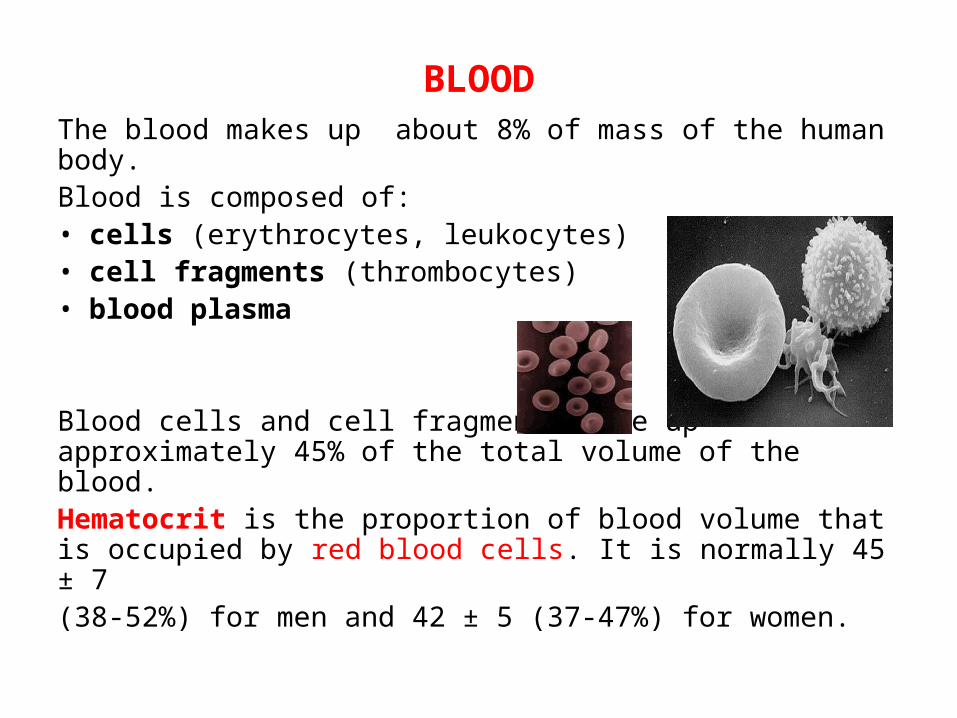
BLOODThe blood makes up about 8% of mass of the human body.Blood is composed of:• cells (erythrocytes, leukocytes)• cell fragments (thrombocytes)• blood plasma
Blood cells and cell fragments make up approximately 45% of the total volume of the blood.Hematocrit is the proportion of blood volume that is occupied by red blood cells. It is normally 45 ± 7 (38-52%) for men and 42 ± 5 (37-47%) for women.
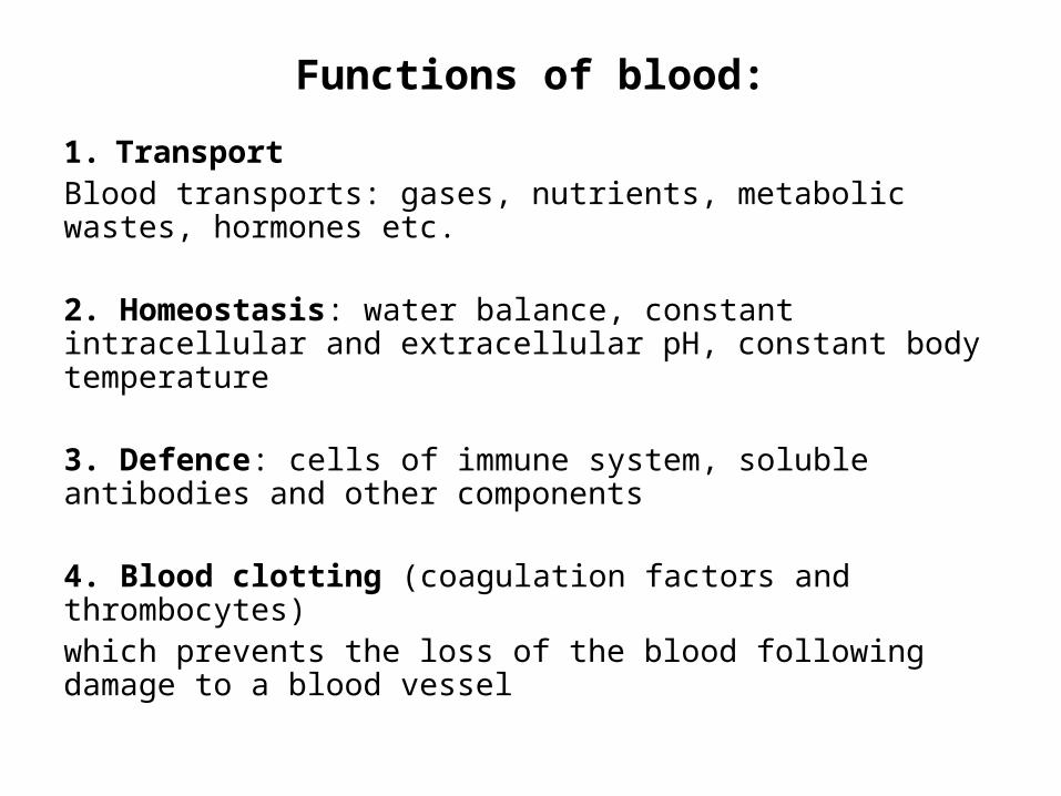
Functions of blood:
1. TransportBlood transports: gases, nutrients, metabolic wastes, hormones etc.
2. Homeostasis: water balance, constant intracellular and extracellular pH, constant body temperature
3. Defence: cells of immune system, soluble antibodies and other components
4. Blood clotting (coagulation factors and thrombocytes)which prevents the loss of the blood following damage to a blood vessel
Blood plasma
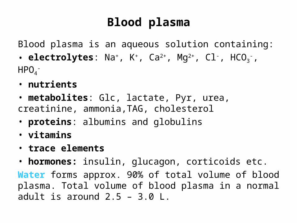
Blood plasma is an aqueous solution containing:
• electrolytes: Na+, K+, Ca2+, Mg2+, Cl-, HCO3-, HPO4
-
• nutrients• metabolites: Glc, lactate, Pyr, urea, creatinine, ammonia,TAG, cholesterol• proteins: albumins and globulins• vitamins• trace elements• hormones: insulin, glucagon, corticoids etc.Water forms approx. 90% of total volume of blood plasma. Total volume of blood plasma in a normal adult is around 2.5 – 3.0 L.
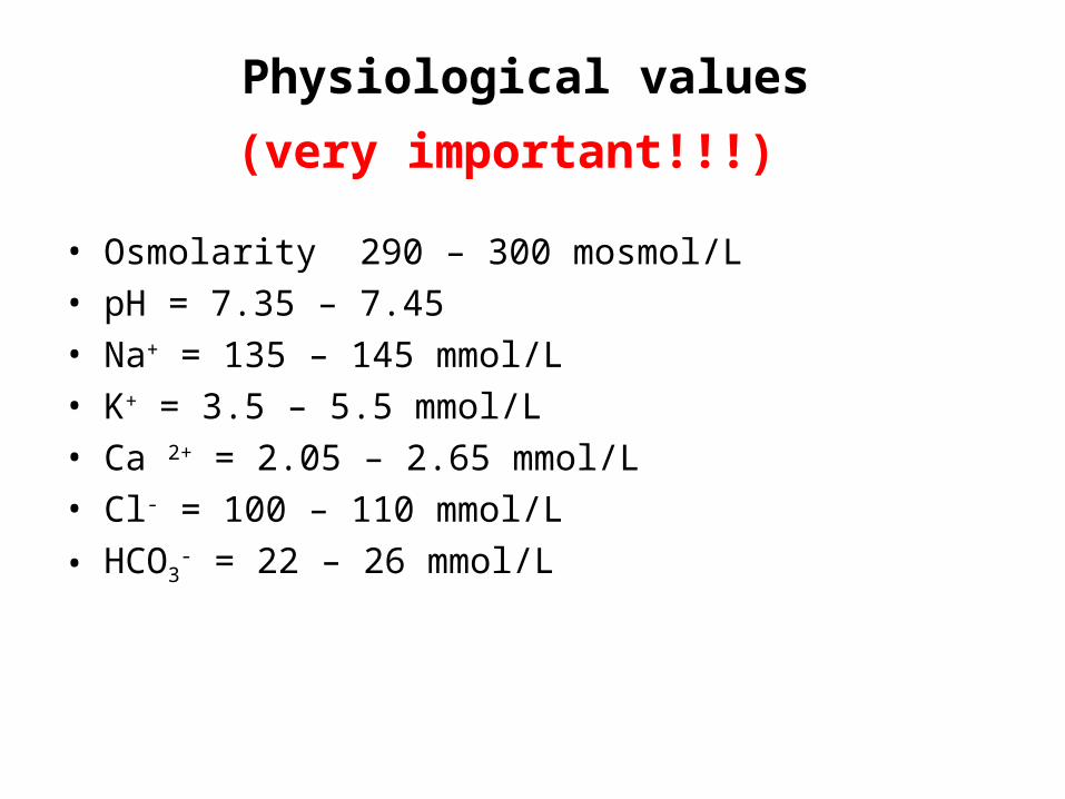
Physiological values
(very important!!!)
• Osmolarity 290 – 300 mosmol/L• pH = 7.35 – 7.45• Na+ = 135 – 145 mmol/L• K+ = 3.5 – 5.5 mmol/L• Ca 2+ = 2.05 – 2.65 mmol/L• Cl- = 100 – 110 mmol/L
• HCO3- = 22 – 26 mmol/L

Plasma proteins
The protein concentration in the plasma is 60 – 80 g/L. Blood plasma contains perhaps 100 different proteins. They are commonly divided into 5 different groups according to their behavior during electrophoresis: Albumins α1-globulins α2-globulins β-globulins γ-globulins
All globulins are dissolved in water in the presence of salts. All globulins are glycoproteins.
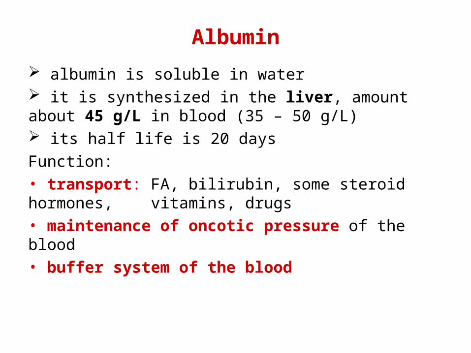
Albumin
albumin is soluble in water it is synthesized in the liver, amount about 45 g/L in blood (35 – 50 g/L) its half life is 20 daysFunction:• transport: FA, bilirubin, some steroid hormones, vitamins, drugs• maintenance of oncotic pressure of the blood• buffer system of the blood
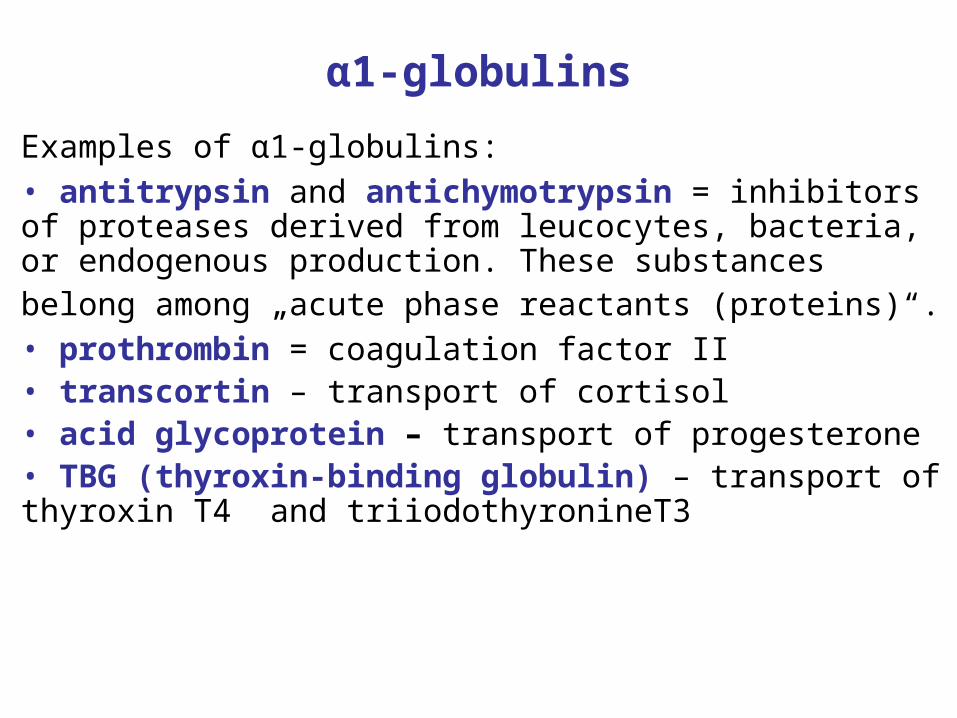
α1-globulins
Examples of α1-globulins:• antitrypsin and antichymotrypsin = inhibitors of proteases derived from leucocytes, bacteria, or endogenous production. These substances belong among „acute phase reactants (proteins)“. • prothrombin = coagulation factor II• transcortin – transport of cortisol• acid glycoprotein – transport of progesterone• TBG (thyroxin-binding globulin) – transport of thyroxin T4 and triiodothyronineT3

α2-globulins
Examples of α2-globulins: • ceruloplasmin – transport of Cu ions• haptoglobin – binding of a free hemoglobin• macroglobulin – binding of proteases, transport of
Zn2+ ions• retinol-binding protein – transport of vitamin A• vitamin D-binding protein – transport of calciols

β-globulins
• LDL – transport of cholesterol esters and TAG• transferrin – transport of Fe ions• fibrinogen = coagulation factor I• sex hormone-binding globulin – transport of testosterone and estradiol• transcobalamin – transport of vitamin B12• C-reactive protein – complement activation
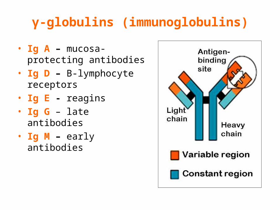
γ-globulins (immunoglobulins)
• Ig A – mucosa-protecting antibodies
• Ig D – B-lymphocyte receptors
• Ig E - reagins• Ig G – late antibodies• Ig M – early antibodies
Immunoglobulins
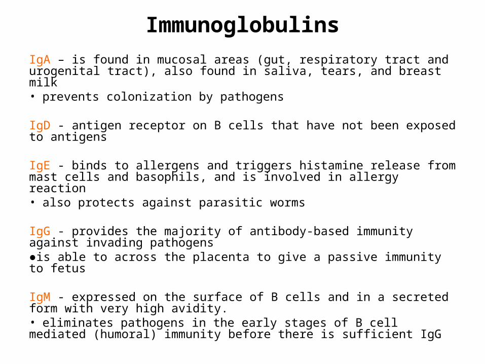
IgA – is found in mucosal areas (gut, respiratory tract and urogenital tract), also found in saliva, tears, and breast milk• prevents colonization by pathogens
IgD - antigen receptor on B cells that have not been exposed to antigens IgE - binds to allergens and triggers histamine release from mast cells and basophils, and is involved in allergy reaction• also protects against parasitic worms
IgG - provides the majority of antibody-based immunity against invading pathogens●is able to across the placenta to give a passive immunity to fetus
IgM - expressed on the surface of B cells and in a secreted form with very high avidity. • eliminates pathogens in the early stages of B cell mediated (humoral) immunity before there is sufficient IgG

Capillary blood collection from finger
• patient´s hand must be clean and dry• always puncture on the sides of the digit parallel
to the side edges of the nail. • try not to use the tip or pad of the finger - there
are more nerves there so it will hurt more.
Plasma x serum
Plasma is obtained from venous, arterial or capillary whole blood. An anticoagulant is used to prevent blood clotting (e. g. sodium citrate, heparin, potassium oxalate, sodium or potassium salt of EDTA).
Serum is obtained from whole blood. Whole blood clots for 20 minutes – 2 hrs and then it is centrifuged. Serum is an yellow liquid which doesn´t contain clotting factors.
!!! Serum is a blood plasma without fibrinogen and other clotting factors !!!
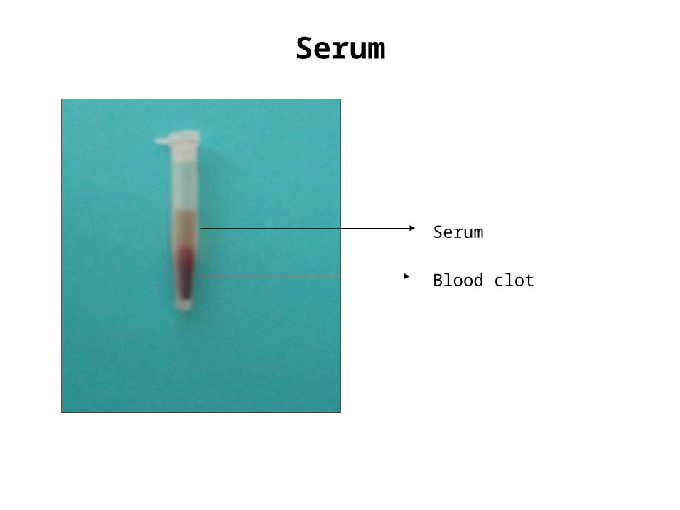
Serum
Serum
Blood clot

METHODS USED IN BIOCHEMISTRY
A) Centrifugation is a method used to separation of particles from solution according to their size, density, shape, viscosity of medium and rotor speed.
Cells, cellular organelles, viruses, large molecules (proteins, nucleic acids) can be separated by centrifugation.
Centrifugal force is a speed of sedimentation of a particle during centrifugation that depends on:● angular speed (ω)● an effective radius of rotor (r)● mass of the particle (m)

Rotor is the most important part of centrifuge.
Speed of centrifugation is expressed in RPM or RCF:• RPM = revolutions per minute (e. g. 10 000 RPM)
• RCF = relative centrifugal force, unit: multiple of g (g = 9.81 m/s2)RCF = how many times is acceleration higher than the gravitational acceleration g
!!! The centrifugal tubes in the rotor must be balanced before centrifugation !!!

Classification of centrifugation is based on the purpose of centrifugation:
a) analytical centrifugation is used to determination of the physical properties of the particles (sedimentation coefficient, molecular weight)
b) preparative centrifugation is used to separate particles from solution. We obtain 2 fractions called a pellet (= sediment, solid phase) and a supernatant (= liquid phase)

B) Electrophoresis is an analytical method for separation of electrically charged molecules (e. g. proteins).This method is based on migration of electrically charged molecules in an external electric field.Electrophoretic mobility of molecule depends on the charge, size and shape of substance and given applied voltage.
Classification of electrophoresis:● free electrophoresis – liquid phase (capillary elpho)● electrophoresis in a supporting medium – gel (agarose, polyacrylamide, cellulose acetate)

Application examples:serum protein electrophoresis
• Separation of serum proteins, isoenzymes, nucleic acids
• Immunoelectrophoresis (immunoglobulins)
Figure is found on http://www.rlbuht.nhs.uk
A = normal serum
B = acute phase response
C = paraproteinemia
D = normal plasma with fibrinogen band
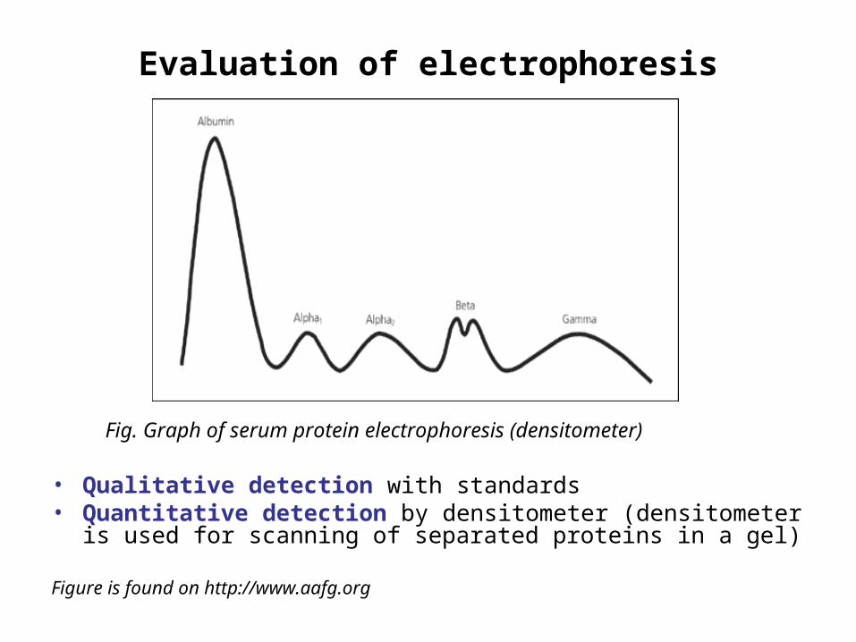
Evaluation of electrophoresis
Fig. Graph of serum protein electrophoresis (densitometer)
• Qualitative detection with standards• Quantitative detection by densitometer (densitometer is
used for scanning of separated proteins in a gel)
Figure is found on http://www.aafg.org
Using of electrophoresis in clinical practice
Serum protein electrophoresis is a simple technique used in the screening of the following diseases: immediate response late response hypogammaglobulinemia hepatic cirrhosis monoclonal gammopathies nephrotic syndrome
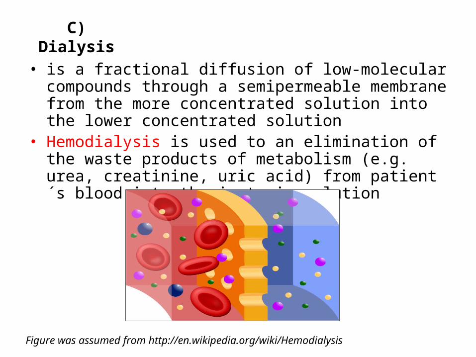
C) Dialysis
• is a fractional diffusion of low-molecular compounds through a semipermeable membrane from the more concentrated solution into the lower concentrated solution
• Hemodialysis is used to an elimination of the waste products of metabolism (e.g. urea, creatinine, uric acid) from patient´s blood into the isotonic solution
Figure was assumed from http://en.wikipedia.org/wiki/Hemodialysis
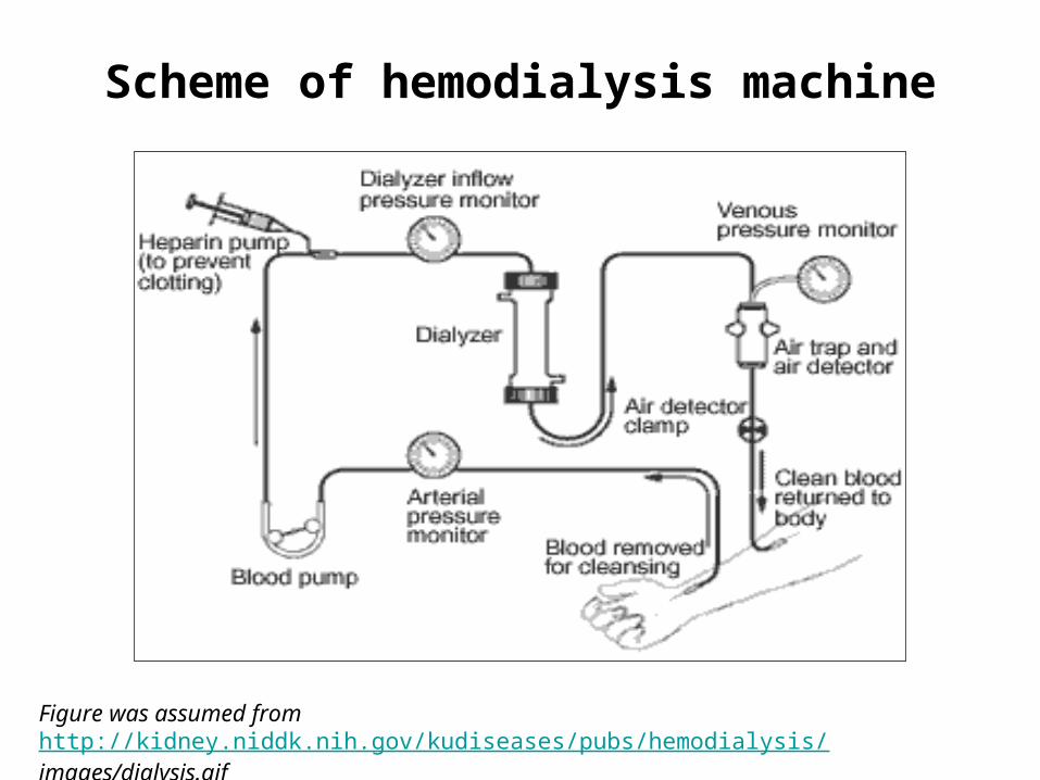
Scheme of hemodialysis machine
Figure was assumed from http://kidney.niddk.nih.gov/kudiseases/pubs/hemodialysis/images/dialysis.gif