Embed Size (px)
Citation preview
Biochemistry 463aSpring 2012
Chemical/thermal denaturation of Alkaline Phosphatase
Allen SuarezJuliana Liang
William Pinkston
In collaboration with:Allison Morley
Blake TyeDuyen Vo
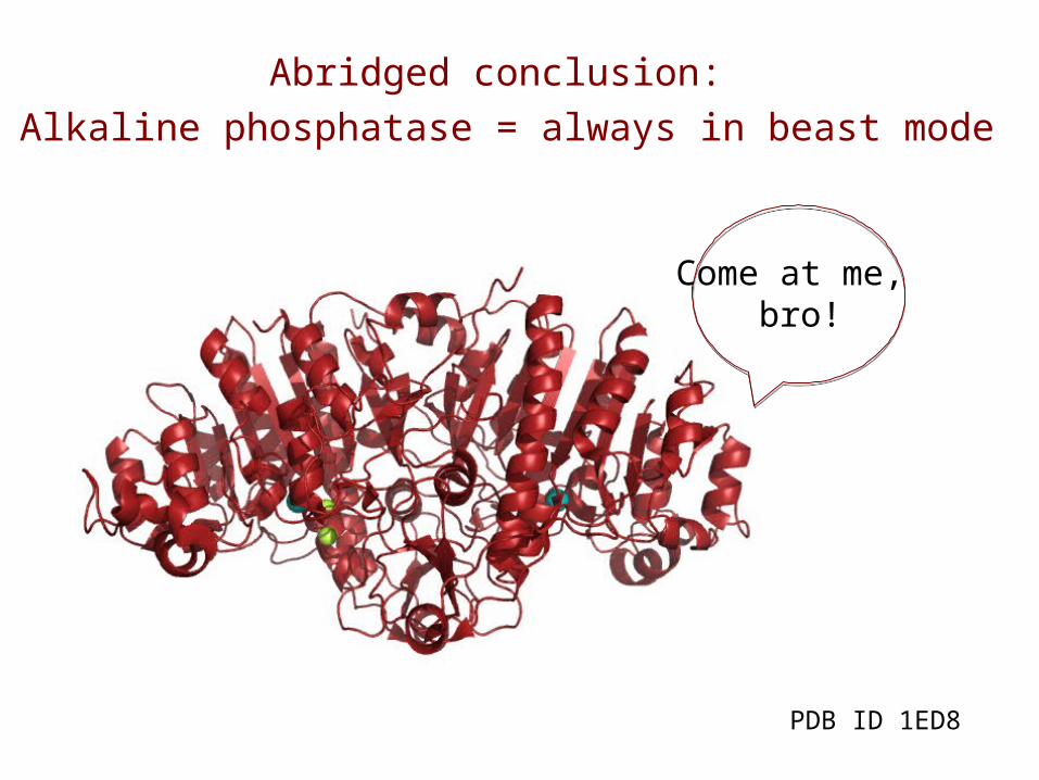
Abridged conclusion: Alkaline phosphatase = always in beast mode
Come at me, bro!
PDB ID 1ED8
Past Experiments
• In past studies of the denaturation of AP students have used 8M urea at room temperature and saw no significant unfolding of the phosphatase1.
• However, last Spring 2011, a student group showed that AP denatures near 90°C using a circular dichroism method2.
Goals & Hypothesis• We set out to denature AP using 8M urea at an elevated
temperature in order to assess the stability of this enzyme using the circular dichroism method of analyzing secondary structure.
• Initial hypothesis: Given that 8M urea denatures most proteins even at room temperature4, 5, we predict denaturation of AP will occur somewhere between ~40°C - 55°C in the presence of 8M urea.
• Revised hypothesis: Because no significant denaturation was observed in our initial range, we predict AP will denature after 30 minutes in 8M urea at ~70°C.
• Further revised hypothesis: After observation that no significant unfolding occurred at 70°C, we now predict denaturation will occur in 8M urea at 80°C.
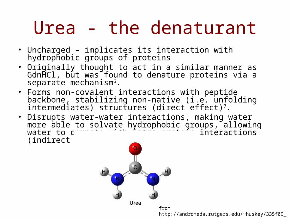
Urea - the denaturant• Uncharged – implicates its interaction with hydrophobic groups of
proteins• Originally thought to act in a similar manner as GdnHCl, but was
found to denature proteins via a separate mechanism6.• Forms non-covalent interactions with peptide backbone, stabilizing
non-native (i.e. unfolding intermediates) structures (direct effect)7.• Disrupts water-water interactions, making water more able to
solvate hydrophobic groups, allowing water to compete with intra-protein interactions (indirect effect)7.
from http://andromeda.rutgers.edu/~huskey/335f09_lec.html
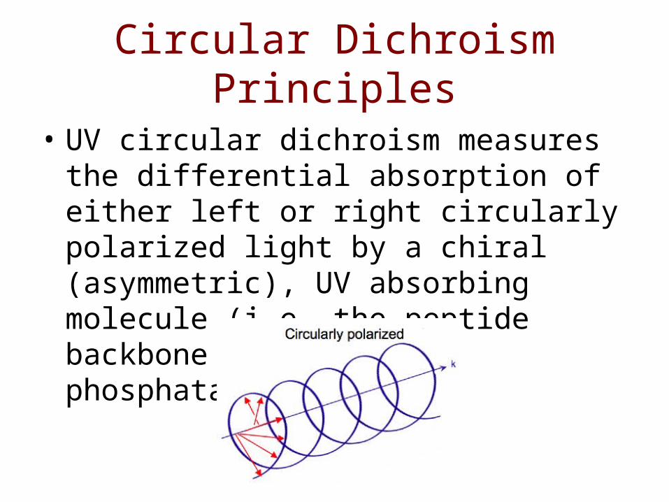
Circular Dichroism Principles
• UV circular dichroism measures the differential absorption of either left or right circularly polarized light by a chiral (asymmetric), UV absorbing molecule (i.e. the peptide backbone of alkaline phosphatase)
Circular Dichroism Principles
• Ordered structures such as alpha-helices and beta sheets display characteristic spectra between 260 nm and 200 nm. This principle is key to monitoring secondary structure of proteins and thus unfolding.
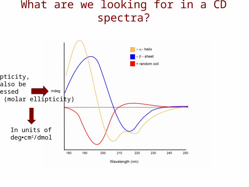
What are we looking for in a CD spectra?
Ellipticity, can also be expressed as θ (molar ellipticity)
In units of degcm2/dmol
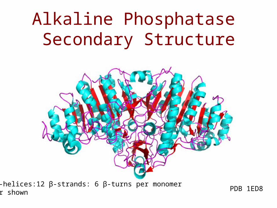
Alkaline Phosphatase Secondary Structure
17 α-helices:12 β-strands: 6 β-turns per monomerDimer shown PDB 1ED8
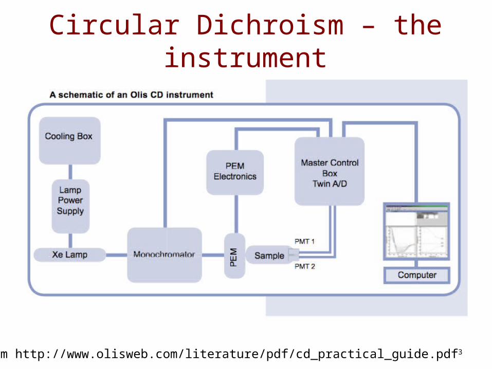
Circular Dichroism – the instrument
from http://www.olisweb.com/literature/pdf/cd_practical_guide.pdf3
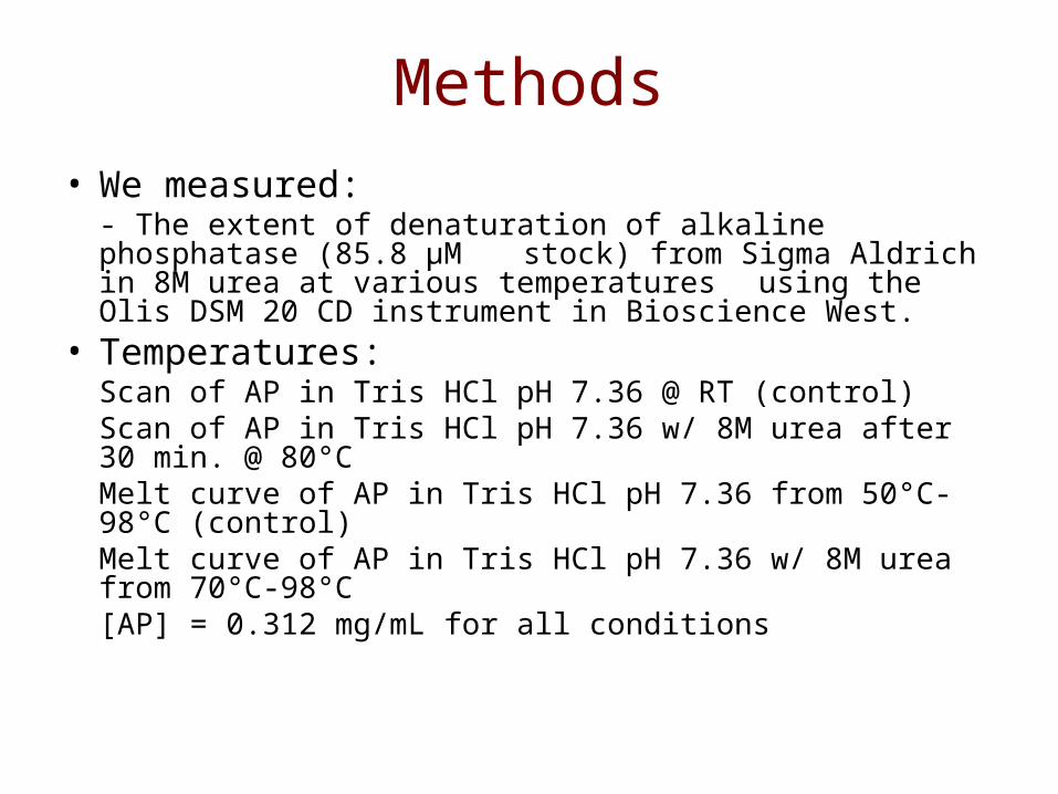
Methods• We measured:
- The extent of denaturation of alkaline phosphatase (85.8 μM stock) from Sigma Aldrich in 8M urea at various
temperatures using the Olis DSM 20 CD instrument in Bioscience West.
• Temperatures: Scan of AP in Tris HCl pH 7.36 @ RT (control)Scan of AP in Tris HCl pH 7.36 w/ 8M urea after 30 min. @ 80°C Melt curve of AP in Tris HCl pH 7.36 from 50°C-98°C (control)Melt curve of AP in Tris HCl pH 7.36 w/ 8M urea from 70°C-98°C
[AP] = 0.312 mg/mL for all conditions
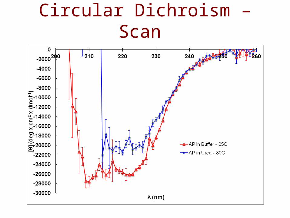
Circular Dichroism – Scan
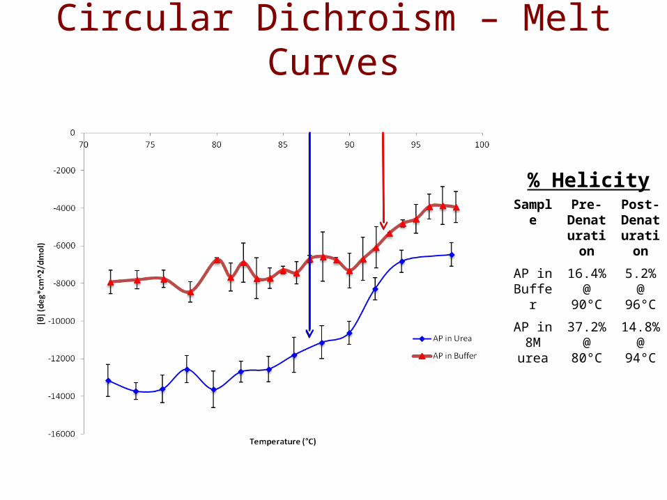
Circular Dichroism – Melt Curves
Sample Pre-Denaturation
Post-Denaturation
AP in Buffer
16.4% @ 90°C
5.2% @ 96°C
AP in 8M
urea
37.2% @ 80°C
14.8% @ 94°C
% Helicity
Conclusions
• 8M urea appears to contribute insignificantly to the denaturation process of Alkaline Phosphatase, even at temperatures above 80°C.
• The only effect 8M urea has on the melt curve of Alkaline Phosphatase compared to a solution containing no urea is to lower the Tm value by less than 5°C, indicating heating to these temps is sufficient to denature without chemical denaturants.
• Interestingly, we found the AP in urea produced larger negative values for θ than AP in only buffer. This indicates the enzyme was “more folded” in urea than when no urea was present.
Further Questions
• If time is a factor in the chemical denaturation of proteins, will AP denature completely in urea when it is allowed to incubate for longer than 30 minutes (i.e. 2 hours? 3 hours? Etc.)?
• What is the reason that AP in urea is “more folded” than AP only in buffer?
Acknowledgements
• We would like to thank our collaborative group of Blake Tye, Duyen Vo and Allison Morley for their patience and aide in searching for an appropriate temperature to run these experiments.
• We would especially like to thank Dr. Chad Park for his excellent assistance and guidance in using the CD instrument.
• Last, but not least, we would like to thank Kayla, Nicole, Swapna and Dr. Hazzard for their advice (and constructive criticisms) throughout the arduous process of attempting to denature the beast.
References1. Boyadjian, N., Childers, K., Luiten, R., and O’Neil, L. “Circular Dichroism of Alkaline Phosphatase” (lab report for BIOC 463a), 2011.
2. Louis, A., Mehlau, M., Nelson, M., and Sund, D. “Circular Dichroism” (lab report for BIOC 463a), 2010.
3. Copeland, R.A. Methods for Protein Analysis: A Practical Guide; Olis, Inc.: Georgia, 1994; p 3.
4. Pace, C. N. Determination and Analysis of Urea and Guanidine Hydrochloride Denaturation Curves. Methods Enzymol. [Online] 1986, 131, 266-280. http://www.biochem.arizona.edu/classes/bioc463a/special_res_proj/urea_gdnhcl_pace86.pdf (accessed Apr 20, 2012).
5. Pace, C.N., and Shaw, K.L. Linear Extrapolation Method of Analyzing Solvent Denaturation curves. Proteins: Struct., Funct., Genet. [Online] 2000, 4, 1-7. http://www.biochem.arizona.edu/classes/bioc463a/special_res_proj/solvent_denaturation_pace_shaw.pdf. (accessed Apr 20, 2012).
6. Monera, O.D., Kay, C.M., and Hodges, R.S. Protein denaturation with guanidine hydrochloride or urea provides a different estimate of stability depending on the contributions of electrostatic interactions. Protein Sci. [Online] 1994, 3 (11), 1984-1991. http://onlinelibrary.wiley.com/doi/10.1002/pro.5560031110/abstract;jsessionid=8218EC6F8BA4F6409948549627E1D4BE.d03t03. (accessed Apr 23, 2012).
7. Bennion, B.J., and Daggett, V. The molecular basis for the chemical denaturation of proteins by urea. Proc. Natl. Acad. Sci. U.S.A. [Online] 2003, 100 (9), 5142-5147. http://www.ncbi.nlm.nih.gov/pmc/articles/PMC154312/ (accessed Apr 23, 2012).