Embed Size (px)
Citation preview
Hindawi Publishing CorporationMediators of InflammationVolume 2012, Article ID 809801, 7 pagesdoi:10.1155/2012/809801
Research Article
Biochemical Analysis of Pentraxin 3 and Fibrinogen Levels inExperimental Periodontitis Model
Gonca Cayir Keles,1 Umut Balli,1 Burcu Ozkan Cetinkaya,1 Bulent Ayas,2 Arzu Findik,3
Zeynep Pinar Keles,1 and Ferda Pamuk4
1 Department of Periodontology, Faculty of Dentistry, Ondokuzmayis University, 55139 Samsun, Turkey2 Department of Histology and Embryology, Faculty of Medicine, Ondokuzmayis University, 55139 Samsun, Turkey3 Department of Microbiology, Faculty of Veterinary Medicine, Ondokuzmayis University, 55139 Samsun, Turkey4 Department of Periodontology, Faculty of Dentistry, Istanbul Aydin University, 34380 Istanbul, Turkey
Correspondence should be addressed to Gonca Cayir Keles, [email protected]
Received 22 March 2012; Revised 22 May 2012; Accepted 5 July 2012
Academic Editor: Hidde Bult
Copyright © 2012 Gonca Cayir Keles et al. This is an open access article distributed under the Creative Commons AttributionLicense, which permits unrestricted use, distribution, and reproduction in any medium, provided the original work is properlycited.
Objective. Pentraxin 3 (PTX3), newly discovered inflammation marker, is a member of acute-phase proteins. The hypothesis,synthesis of gingival tissue and serum PTX-3 increases in the experimental periodontitis model (with 10-day and 40-day periods),was tested by detecting gingival tissue and serum PTX-3 levels in rats with experimental periodontitis. Methods. Thirty ratswere randomly divided into three groups of ten animals each: ligature-induced experimental periodontitis groups (with 10-day(Group1) and 40-day periods (Group2)) and healthy group (Group3). At the end of experimental period, rats were sacrificed, andradiological and histomorphometric analyses were performed on the mandibles. PTX3 levels were measured in gingival tissue andserum samples using ELISA. Plasma fibrinogen levels were measured according to the nephelometric method. Results. Significantalveolar bone resorption and periodontal inflammation were evident in periodontitis groups. Levels of PTX3 in gingival tissuewere statistically higher in Group 1 than those in groups 2 and 3 (P < 0.01). No significant difference was found in serum PTX3levels between experimental periodontitis and control groups (P > 0.05). Plasma fibrinogen levels were significantly increasedin the experimental periodontitis groups (P < 0.001). Conclusion. PTX3 seems to be associated with tissue destruction in earlierperiods of inflammatory periodontal disease, contrary to the fibrinogen findings.
1. Introduction
Periodontal disease is a multifactorial infectious disease; al-though the main cause of periodontal disease is the presenceof periodontal microorganisms, subsequent progression anddisease severity are considered to be determined by the hostimmune response [1–4]. Mediators produced as a part ofhost response that contribute to tissue destruction includeacute-phase proteins, cytokines, and prostaglandins [5–8].The acute-phase response is a nonspecific process that mayoccur in the initial host response to injuries, infections,ischaemic necrosis, or malignancy [9]. It is initiated by theactivation of local macrophages and other cells (includingfibroblasts and endothelial cells) and has a variety of func-tions including proinflammatory properties, activation of
complement factors, neutralization of invasive pathogens,and stimulation of repair and regeneration of tissues [9, 10].Data show that acute-phase proteins, plasma proteins, notonly appear in acute inflammation, but also in longstanding,chronic conditions [10]. Acute-phase proteins are generallyincreased following a microbial infection [11]. It is, therefore,possible that acute-phase proteins are sensitive markers toevaluate inflammatory status of various microbial infectionsincluding periodontitis.
Pentraxins (PTXS), a superfamily of acute-phase pro-teins, are an essential component of the humoral arm ofinnate immunity [12, 13]. Short PTXS such as C-reactiveprotein and serum amyloid P component are acute-phaseproteins in man and mouse, respectively, and are producedmainly by the liver in response to inflammatory stimuli, such

2 Mediators of Inflammation
as IL-6 [14, 15]. PTX3 was the first long PTX described as anIL-1β inducible gene in endothelial cells [15]. It is producedby a variety of cells, mostly by cells abundant in peri-odontal tissues like neutrophils [8, 16], fibroblasts [8, 17],monocytes/macrophages [8, 17, 18], dendritic cells [8, 19],epithelial cells [8, 20], endothelial cells [8, 21], and vascularsmooth muscle cells [8, 22]. PTX3 behaves as an acute-phase response protein since its blood levels, low in normalconditions (about 25 ng/mL in the mouse, <2 ng/mL in theman), increase rapidly (peak at 6–8 h) and dramatically(200–800 ng/mL) during endotoxic shock, sepsis, and otherinflammatory and infectious conditions [23, 24].
There is evidence that PTX3 levels increase in variouschronic inflammatory diseases such as atherosclerotic lesions[22, 24, 25], coronary artery disease [24, 26], small vesselvasculitis, rheumatoid arthritis [24, 27, 28], and chronickidney disease [24, 29]. Moreover, in a recent clinical study,suggesting that PTX3 concentrations may have been a goodpredictive diagnostic tool for unstable angina pectoris,plasma, PTX3 levels have been significantly increased inpatients with unstable angina pectoris [30]. To date, data sug-gest a possible role of PTX3 as a marker of pathology, sincethere is a correlation between PTX3 plasma concentrationsand severity of diseases [24]. Periodontal disease is a low-grade local infection microorganisms and their products arethe principal etiological agents, and it is associated with amoderate systemic inflammatory response [31, 32].
In light of these observations, PTX-3 might probably playa role in the pathogenesis of periodontal disease. The tworecent clinical studies were published suggesting that PTX-3 in gingival crevicular fluid is considered a diagnosticmarker of periodontal disease inflammatory activity [8, 33].Fibrinogen, another acute-phase reactant, has been sug-gested to be a possible mediator in the pathogenesis of peri-odontal disease [34]. This is consistent with the report thatthere is an independent association between periodontaldisease and plasma fibrinogen levels [35]. The presentstudy was undertaken to test the hypothesis that synthe-sis of gingival tissue and serum PTX-3 increases in theexperimental periodontitis model (with 10 days and 40days periods) in rats which can easily be standardized.Plasma fibrinogen levels were also determined in rats withexperimental periodontitis.
2. Material and Methods
2.1. Animals. Thirty male Wistar rats weighing 200 to 250 gwere housed separately in plastic cages and kept in a tempera-ture controlled room with a standard light dark illuminationcycle (12 hours each). They received water and standard foodad libitum. All study protocols were in compliance withguidelines and with the approval of the Committee of Ethicsin Animal Research of the Ondokuzmayis University.
2.2. Experimental Design. Thirty rats were randomly dividedinto three groups of ten animals each: experimental perio-dontitis groups (with 10 days and 40 days periods) and peri-odontally healthy group. After systemic anesthesia with an
intraperitoneal injection of 60 mg/kg ketamine-HCL (Pfizer,Istanbul, Turkey), the rats were subjected to experimentalperiodontitis by tying 3.0 sterile silk ligatures around thecervical area of the right and left mandibular first molars, andthese were kept in position for 10 days (Group 1) and 40 days(Group 2) to promote microbial dental plaque accumulation,and inflammation [36]. At the end of the experimentalperiod, 5 mL venous blood was drawn by cardiac punctureand processed for serum and plasma analyses. After thatprocedure, the rats with experimental periodontitis andperiodontally healthy rats (Group 3) were decapitated. Themandibles were carefully removed together with the sur-rounding gingiva, and the gingival tissue samples wereextracted from the buccal region of the mandibular rightfirst molars. The left mandibles were soaked in neutral10% formalin for 48 hours. Standardized radiographs wereobtained by the long-cone technique at 70 kilovolt (peak),8 mA from all groups.
2.3. Histomorphometric Analysis. Following the radiographs,the left mandibles were decalcified in 10% formic acid,embedded paraffin; 6 μm thick sections were cut in a mesio-distal direction and stained with hematoxylin and eosin. Thelevel of the alveolar bone was determined by histometricmeasuring the distance from the cementoenamel junctionto the alveolar bone crest [36]. These measurements wereperformed on monitor images of the sections, which weretransferred via color camera (objective ×3.3, F10 CCDCamera, Panasonic, Osaka, Japan) attached to a microscope(BH2 Research Microscope, Olympus, Tokyo, Japan). In-flammatory cells were counted in systematically sampled;40× 40-μm areas.
2.4. Biochemical Analysis. Gingival tissues, removed from thealveolar bone, were placed immediately in a sterile salinesolution and frozen at −80◦C until biochemical analysis[37]. In brief, before grinding, tissue was blotted, weighedon a microbalance, and placed into a sufficient volume ofphosphate buffered saline (PBS; 4◦C; pH: 7.0) containinga protease inhibitor (5 μg/mL aprotinin, 1 mM EDTA) toa dilution of 10 mg tissue/mL PBS plus protease inhibitorsolution. The samples were homogenized at 8,500 revolu-tions per minute (rpm) for 30 seconds four times with 10-second intervals by homogenizer (Ultra Turrax. T25, IKALABORTECHNIK, Staufen, Germany). The homogenatewas processed with freeze-thawed procedures two times andthen sonicated three times by ultrasonicator (MSE Soniprep150, Sanyo Gallenkamp, Leicestershire, UK) at 4 to 5 μ for 30seconds with 10-second intervals. The homogenate was cen-trifuged (Refrigerated centrifuge, SIGMA 3K30, Osterode,Germany) at 15,000 rpm for 16 minutes [37], and super-natant was collected for PTX3 analysis. The supernatantpreparation processes were performed on ice medium at∼0◦C to 4◦C. All samples were brought to room temperaturefor enzyme-linked immunosorbent assay (ELISA). Gingivaltissue and serum PTX3 concentrations were analyzed in each50 μL sample by standard ELISA apparatus at 450 to 550 nmusing a PTX3 kit (Uscn Life Science Inc., Wuhan, China) thatdetects PTX3 levels. Plasma fibrinogen levels were measured
Mediators of Inflammation 3
Table 1: Alveolar bone level and numbers of inflammatory cells counted in the connective tissue and epithelium.
Alveolar bone level∗ Inflammatory cells∗ Inflammatory cells∗
(mm) connective tissue epithelium
(cells/1600 μm2) (cells/1600 μm2)
Group 1 1.08± 0.03 3.9± 0.7 3.4± 0.8
(Experimental periodontitis with 10 days period) 1.08 (1.03–1.13) 4.0 (3.0–5.0) 3.0 (2.0–5.0)
Group 2 1.37± 0.05 5.4± 1.2 4.5± 0.7
(Experimental periodontitis with 40 days period) 1.39 (1.29–1.44) 5.5 (4.0–7.0) 4.0 (4.0–6.0)
Group 3 0.93± 0.07 0.5± 0.5 0.3± 0.5
(Healthy control group) 0.96 (0.80–1.01) 0.5 (0.0–1.0) 0.0 (0.0–1.0)
Data were analyzed by one-way ANOVA and expressed as the means ± standard deviation and medians (minimum-maximum).∗Significant difference in histomorphometric findings between groups (P < 0.001).
(a) (b) (c)
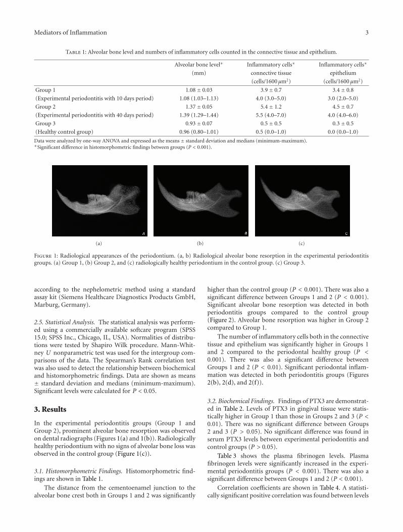
Figure 1: Radiological appearances of the periodontium. (a, b) Radiological alveolar bone resorption in the experimental periodontitisgroups. (a) Group 1, (b) Group 2, and (c) radiologically healthy periodontium in the control group. (c) Group 3.
according to the nephelometric method using a standardassay kit (Siemens Healthcare Diagnostics Products GmbH,Marburg, Germany).
2.5. Statistical Analysis. The statistical analysis was perform-ed using a commercially available softcare program (SPSS15.0; SPSS Inc., Chicago, IL, USA). Normalities of distribu-tions were tested by Shapiro Wilk procedure. Mann-Whit-ney U nonparametric test was used for the intergroup com-parisons of the data. The Spearman’s Rank correlation testwas also used to detect the relationship between biochemicaland histomorphometric findings. Data are shown as means± standard deviation and medians (minimum-maximum).Significant levels were calculated for P < 0.05.
3. Results
In the experimental periodontitis groups (Group 1 andGroup 2), prominent alveolar bone resorption was observedon dental radiographs (Figures 1(a) and 1(b)). Radiologicallyhealthy periodontium with no signs of alveolar bone loss wasobserved in the control group (Figure 1(c)).
3.1. Histomorphometric Findings. Histomorphometric find-ings are shown in Table 1.
The distance from the cementoenamel junction to thealveolar bone crest both in Groups 1 and 2 was significantly
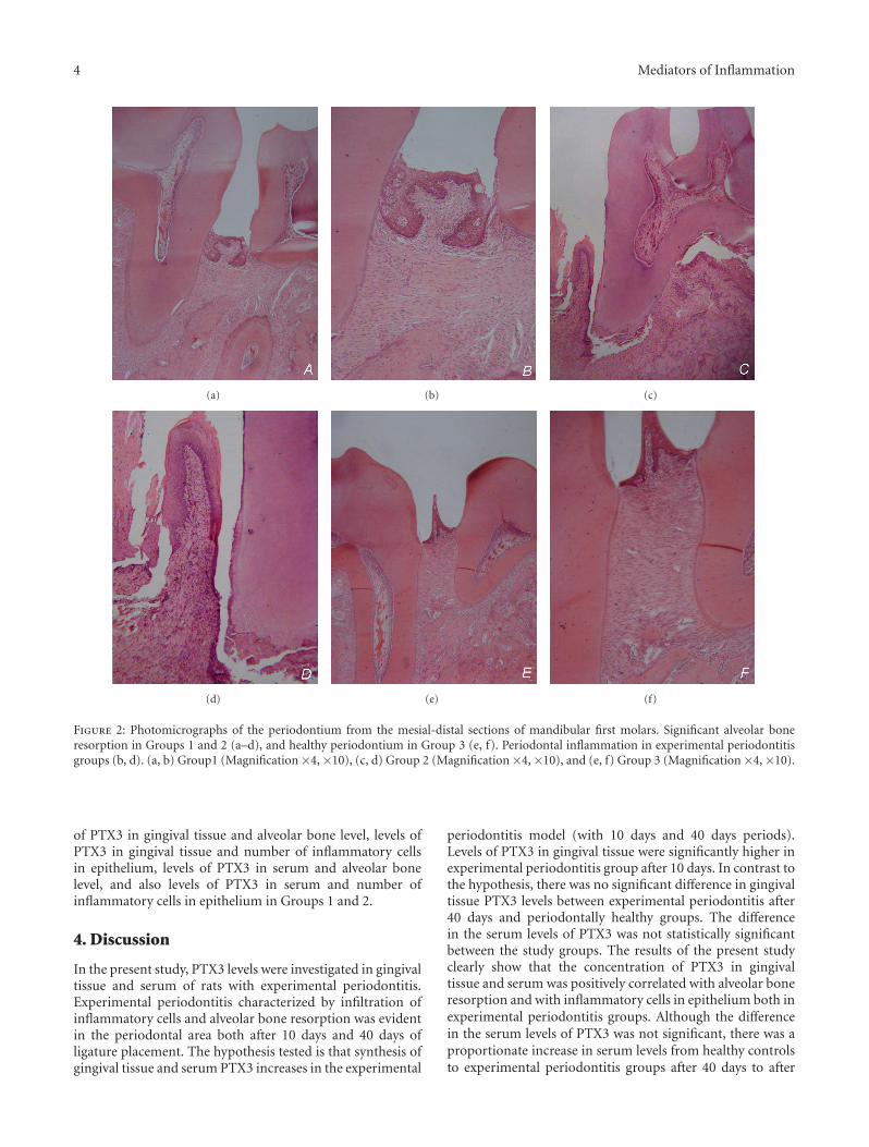
higher than the control group (P < 0.001). There was also asignificant difference between Groups 1 and 2 (P < 0.001).Significant alveolar bone resorption was detected in bothperiodontitis groups compared to the control group(Figure 2). Alveolar bone resorption was higher in Group 2compared to Group 1.
The number of inflammatory cells both in the connectivetissue and epithelium was significantly higher in Groups 1and 2 compared to the periodontal healthy group (P <0.001). There was also a significant difference betweenGroups 1 and 2 (P < 0.01). Significant periodontal inflam-mation was detected in both periodontitis groups (Figures2(b), 2(d), and 2(f)).
3.2. Biochemical Findings. Findings of PTX3 are demonstrat-ed in Table 2. Levels of PTX3 in gingival tissue were statis-tically higher in Group 1 than those in Groups 2 and 3 (P <0.01). There was no significant difference between Groups2 and 3 (P > 0.05). No significant difference was found inserum PTX3 levels between experimental periodontitis andcontrol groups (P > 0.05).
Table 3 shows the plasma fibrinogen levels. Plasmafibrinogen levels were significantly increased in the experi-mental periodontitis groups (P < 0.001). There was also asignificant difference between Groups 1 and 2 (P < 0.001).
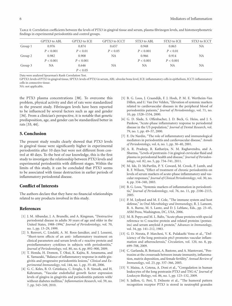
Correlation coefficients are shown in Table 4. A statisti-cally significant positive correlation was found between levels
4 Mediators of Inflammation
(a) (b) (c)
(d) (e) (f)
Figure 2: Photomicrographs of the periodontium from the mesial-distal sections of mandibular first molars. Significant alveolar boneresorption in Groups 1 and 2 (a–d), and healthy periodontium in Group 3 (e, f). Periodontal inflammation in experimental periodontitisgroups (b, d). (a, b) Group1 (Magnification ×4, ×10), (c, d) Group 2 (Magnification ×4, ×10), and (e, f) Group 3 (Magnification×4, ×10).
of PTX3 in gingival tissue and alveolar bone level, levels ofPTX3 in gingival tissue and number of inflammatory cellsin epithelium, levels of PTX3 in serum and alveolar bonelevel, and also levels of PTX3 in serum and number ofinflammatory cells in epithelium in Groups 1 and 2.
4. Discussion
In the present study, PTX3 levels were investigated in gingivaltissue and serum of rats with experimental periodontitis.Experimental periodontitis characterized by infiltration ofinflammatory cells and alveolar bone resorption was evidentin the periodontal area both after 10 days and 40 days ofligature placement. The hypothesis tested is that synthesis ofgingival tissue and serum PTX3 increases in the experimental
periodontitis model (with 10 days and 40 days periods).Levels of PTX3 in gingival tissue were significantly higher inexperimental periodontitis group after 10 days. In contrast tothe hypothesis, there was no significant difference in gingivaltissue PTX3 levels between experimental periodontitis after40 days and periodontally healthy groups. The differencein the serum levels of PTX3 was not statistically significantbetween the study groups. The results of the present studyclearly show that the concentration of PTX3 in gingivaltissue and serum was positively correlated with alveolar boneresorption and with inflammatory cells in epithelium both inexperimental periodontitis groups. Although the differencein the serum levels of PTX3 was not significant, there was aproportionate increase in serum levels from healthy controlsto experimental periodontitis groups after 40 days to after
Mediators of Inflammation 5
Table 2: Gingival tissue and serum PTX3 levels in experimental periodontitis and control groups (ng/mL).
Gingival tissue Serum∗
Group 1 1.6± 0.4a 1.2± 0.3
(Experimental periodontitis with 10 days period) 1.5 (1.3–2.4) 1.1 (0.9–1.6)
Group 2 1.2± 0.2b 1.1± 0.2
(Experimental periodontitis with 40 days period) 1.2 (1.0–1.5) 1.1 (0.9–1.4)
Group 3 1.1± 0.2b 1.0± 0.2
(Healthy control group) 1.1 (0.9–1.5) 1.0 (0.8–1.3)
Data were analyzed by one-way ANOVA and expressed as the means ± standard deviation and medians (minimum-maximum).aSignificant difference between groups 2 and 3 (P < 0.01).bNo significant difference between groups (P > 0.05).∗No significant difference in serum PTX3 levels between groups (P > 0.05).
Table 3: Plasma fibrinogen levels in experimental periodontitis andcontrol groups (mg/mL).
Plasma fibrinogen∗
Group 1 1.73± 0.21
(Experimental periodontitis with 10days period)
1.75 (1.36–2.10)
Group 2 0.99± 0.27
(Experimental periodontitis with 40days period)
0.93 (0.74–1.69)
Group 3 0.67± 0.07
(Healthy control group) 0.72 (0.54–0.73)
Data were analyzed by Mann-Whitney U Test and expressed as the means ±standard deviation and medians (minimum-maximum).∗Significant difference in fibrinogen levels between groups (P < 0.001).
10 days. In the present study, the quantitative detections ofPTX3 were performed by ELISA, which was generally usedto detect PTX3 in gingival crevicular fluid and plasma withhigh sensitivity and specificity [8, 30, 33, 38].
The results on plasma fibrinogen levels in the presentstudy confirm the role of this protein in the pathogenesis ofperiodontitis, which is higher in experimental periodontitisgroups both after 10 days and 40 days. It is important toalso consider that 40-day experimental period is sufficient forchronic periodontal inflammation in rats. This suggests thatfibrinogen appears also in chronic inflammation process,which seems not to agree with PTX3.
The acute-phase reactant PTX3 is expressed as an IL-1 inducible and TNF inducible gene in endothelial cellsand fibroblasts, respectively, [39–41]. PTX3 is mostly gen-erated by endothelial cells and mononuclear phagocyteson stimulation with lipopolysaccharides and inflammatorycytokines, such as IL-1β and TNF-α, but not IL-6 [39, 41,42]. CRP, IL-1β, IL-6, and TNF-α have been associatedwith the presence of various bacterial infections includingperiodontitis [9]. Periodontal disease is an inflammatorydisease in which microorganisms and their products are theprincipal etiologic agents. In a recent clinical work, PTX3is suggested to associate with the severity of periodontaldisease and considered as a marker of inflammatory activityin periodontal disease [8]. It is important to note that thereis no significant difference in plasma PTX3 concentrations
between periodontal disease and control groups, which is inagreement with our serum PTX3 results. The results of thepresent study clearly showed that gingival tissue PTX3 levelswere not increased in experimental periodontitis modelwith 40-day period, contrary to the gingival crevicular fluidfindings of Pradeep et al. which reported that concentra-tion of PTX3 in gingival crevicular fluid is increased inproportionately with the severity of periodontal disease [8].Same authors in another clinical study observed that plasmaPTX3 levels are higher in both patients with chronic kidneydisease and with chronic kidney disease + periodontal diseasethan healthy controls. There is no significant difference inplasma PTX3 levels between two groups with chronic kidneydisease [43]. Very recent clinical data suggested that gingivalcrevicular fluid PTX3 level is higher in periodontally diseasedsites as compared to healthy sites in the same patient withchronic periodontitis [33]. Although the split mouth designis a good model in periodontal clinical studies, it is thereforepossible that periodontopathogens in diseased sites maytranslocate to healthy sites.
Periodontal tissue destruction occurs in an episodic,intermittent manner, with periods of inactivity and destruc-tive periods that are associated with acute inflammatory reac-tion and result in loss of collagen and alveolar bone. It isimportant to also consider that the increased concentrationof PTX3 in these clinical studies [8, 33] might probablybe related to the destructive periods in periodontal disease.This is consistent with the results of the present study thatlevels of PTX3 in gingival tissue was significantly higher inexperimental periodontitis group after 10 days. When ourresults were taken into consideration, a significant increase inthe levels of PTX3 in gingival tissue occurred after 10 days ofligature placement due to the acute inflammatory reaction,contrary to the findings of experimental periodontitis groupwith 40-day period, which is chronic periodontal inflamma-tion model.
Some possible reasons for the inconsistent findings ofprevious clinical studies could be related to the charac-teristics of subjects. Existing knowledge demonstrated thatphysical activity and energy balance of individuals potentiallyinfluence inflammatory response [38]. Clinical data showthat there is an inverse relationship between plasma levels ofPTX3 and nutrient intake, and also body fat decreases. More-over, the findings clearly indicate that bed rest increases
6 Mediators of Inflammation
Table 4: Correlation coefficients between the levels of PTX3 in gingival tissue and serum, plasma fibrinogen levels, and histomorphometricfindings in experimental periodontitis and control groups.
GPTX3 to ABL GPTX3 to ICE GPTX3 to ICCT STX3 to ABL STX3 to ICE STX3 to ICCT
Group 1 0.976 0.874 0.657 0.948 0.863 NA
P < 0.001 P < 0.01 P < 0.05 P < 0.001 P < 0.01
Group 2 0.982 0.908 NA 0.966 0.914 NA
P < 0.001 P < 0.001 P < 0.001 P < 0.001
Group 3 NA 0.646 NA NA NA NA
P < 0.05
Data were analyzed Spearman’s Rank Correlation Test.GPTX3: levels of PTX3 in gingival tissue, SPTX3: levels of PTX3 in serum, ABL: alveolar bone level, ICE: inflammatory cells in epithelium, ICCT: inflammatorycells in connective tissue.NA: not applicable.
the PTX3 plasma concentrations [38]. To overcome thisproblem, physical activity and diet of rats were standardizedin the present study. Fibrinogen levels have been reportedto be influenced by several factors such as age and gender[34]. From a clinician’s perspective, it is notable that geneticpredisposition, age, and gender can be standardized better inrats [33, 44].
5. Conclusion
The present study results clearly showed that PTX3 levelsin gingival tissue were significantly higher in experimentalperiodontitis after 10 days but were not different from con-trol at 40 days. To the best of our knowledge, this is the firststudy to investigate the relationship between PTX3 levels andexperimental periodontitis with different stages. Within thelimits of this study, it can be concluded that PTX3 seemsto be associated with tissue destruction in earlier periods ofinflammatory periodontal disease.
Conflict of Interests
The authors declare that they have no financial relationshipsrelated to any products involved in this study.
References
[1] J. M. Albandar, J. A. Brunelle, and A. Kingman, “Destructiveperiodontal disease in adults 30 years of age and older in theUnited States, 1988–1994,” Journal of Periodontology, vol. 70,no. 1, pp. 13–29, 1999.
[2] S. Renvert, C. Lindahl, A. M. Roos-Jansaker, and J. Lessemi,“Short-term effects of an anti- inflammatory treatment onclinical parameters and serum levels of c-reactive protein andproinflammatory cytokines in subjects with periodontitis,”Journal of Periodontology, vol. 80, no. 6, pp. 892–900, 2009.
[3] T. Honda, H. Domon, T. Okui, K. Kajita, R. Amanuma, andK. Yamazaki, “Balance of inflammatory response in stable gin-givitis and progressive periodontitis lesions,” Clinical and Ex-perimental Immunology, vol. 144, no. 1, pp. 35–40, 2006.
[4] G. C. Keles, B. O. Cetinkaya, C. Eroglu, S. B. Simsek, and H.Kahraman, “Vascular endothelial growth factor expressionlevels of gingiva in gingivitis and periodontitis patients with/without diabetes mellitus,” Inflammation Research, vol. 59, no.7, pp. 543–549, 2010.
[5] B. G. Loos, J. Craandijk, F. J. Hoek, P. M. E. Wertheim-VanDillen, and U. Van Der Velden, “Elevation of systemic markersrelated to cardiovascular diseases in the peripheral blood ofperiodontitis patients,” Journal of Periodontology, vol. 71, no.10, pp. 1528–1534, 2000.
[6] G. D. Slade, S. Offenbacher, J. D. Beck, G. Heiss, and J. S.Pankow, “Acute-phase inflammatory response to periodontaldisease in the US population,” Journal of Dental Research, vol.79, no. 1, pp. 49–57, 2000.
[7] E. De Nardin, “The role of inflammatory and immunologicalmediators in periodontitis and cardiovascular disease,” Annalsof Periodontology, vol. 6, no. 1, pp. 30–40, 2001.
[8] A. R. Pradeep, R. Kathariya, N. M. Raghavendra, and A.Sharma, “Levels of pentraxin-3 in gingival crevicular fluid andplasma in periodontal health and disease,” Journal of Periodon-tology, vol. 82, no. 5, pp. 734–741, 2011.
[9] M. Ide, D. McPartlin, P. Y. Coward, M. Crook, P. Lumb, andR. F. Wilson, “Effect of treatment of chronic periodontitis onlevels of serum markers of acute-phase inflammatory and vas-cular responses,” Journal of Clinical Periodontology, vol. 30, no.4, pp. 334–340, 2003.
[10] B. G. Loos, “Systemic markers of inflammation in periodonti-tis,” Journal of Periodontology, vol. 76, no. 11, pp. 2106–2115,2005.
[11] P. M. Lydyard and M. F. Cole, “The immune system and hostdefense,” in Oral Microbiology and Immunology, R. J. Lamont,R. A. Burne, M. S. Lantz, and D. J. Leblanc, Eds., pp. 23–45,ASM Press, Washington, DC, USA, 2006.
[12] M. B. Pepys and M. L. Baltz, “Acute phase proteins with specialreference to C-reactive protein and related proteins (pentax-ins) and serum amyloid A protein,” Advances in Immunology,vol. 34, pp. 141–212, 1983.
[13] G. D. Norata, P. Marchesi, V. K. Pulakazhi Venu et al., “Def-iciency of the long pentraxin ptx3 promotes vascular inflam-mation and atherosclerosis,” Circulation, vol. 120, no. 8, pp.699–708, 2009.
[14] C. Garlanda, B. Bottazzi, A. Bastone, and A. Mantovani, “Pen-traxins at the crossroads between innate immunity, inflamma-tion, matrix deposition, and female fertility,” Annual Review ofImmunology, vol. 23, pp. 337–366, 2005.
[15] V. Maina, A. Cotena, A. Doni et al., “Coregulation in humanleukocytes of the long pentraxin PTX3 and TSG-6,” Journal ofLeukocyte Biology, vol. 86, no. 1, pp. 123–132, 2009.
[16] S. Jaillon, G. Peri, Y. Delneste et al., “The humoral patternrecognition receptor PTX3 is stored in neutrophil granules
Mediators of Inflammation 7
and localizes in extracellular traps,” Journal of ExperimentalMedicine, vol. 204, no. 4, pp. 793–804, 2007.
[17] A. R. Goodman, D. E. Levy, L. F. L. Reis, and J. Vilcek, “Differ-ential regulation of TSG-14 expression in murine fibroblastsand peritoneal macrophages,” Journal of Leukocyte Biology, vol.67, no. 3, pp. 387–395, 2000.
[18] V. V. Alles, B. Bottazzi, G. Peri, J. Golay, M. Introna, and A.Mantovani, “Inducible expression of PTX3, a new memberof the pentraxin family, in human mononuclear phagocytes,”Blood, vol. 84, no. 10, pp. 3483–3493, 1994.
[19] A. Doni, M. Michela, B. Bottazzi et al., “Regulation of PTX3,a key component of humoral innate immunity in human den-dritic cells: stimulation by IL-10 and inhibition by IFN-γ,”Journal of Leukocyte Biology, vol. 79, no. 4, pp. 797–802, 2006.
[20] A. J. Nauta, S. De Haij, B. Bottazzi et al., “Human renal epi-thelial cells produce the long pentraxin PTX3,” Kidney Inter-national, vol. 67, no. 2, pp. 543–553, 2005.
[21] C. Gustin, E. Delaive, M. Dieu, D. Calay, and M. Raes, “Up-regulation of pentraxin-3 in human endothelial cells after ly-sophosphatidic acid exposure,” Arteriosclerosis, Thrombosis,and Vascular Biology, vol. 28, no. 3, pp. 491–497, 2008.
[22] M. Klouche, G. Peri, C. Knabbe et al., “Modified atherogeniclipoproteins induce expression of pentraxin-3 by humanvascular smooth muscle cells,” Atherosclerosis, vol. 175, no. 2,pp. 221–228, 2004.
[23] A. A. M. Dias, A. R. Goodman, J. L. Dos Santos et al., “TSG-14transgenic mice have improved survival to endotoxemia andto CLP-induced sepsis,” Journal of Leukocyte Biology, vol. 69,no. 6, pp. 928–936, 2001.
[24] A. Mantovani, C. Garlanda, A. Doni, and B. Bottazzi, “Pen-traxins in innate immunity: from C-reactive protein to thelong pentraxin PTX3,” Journal of Clinical Immunology, vol. 28,no. 1, pp. 1–13, 2008.
[25] M. S. Rolph, S. Zimmer, B. Bottazzi, C. Garlanda, A. Manto-vani, and G. K. Hansson, “Production of the long pentraxinPTX3 in advanced atherosclerotic plaques,” Arteriosclerosis,Thrombosis, and Vascular Biology, vol. 22, no. 5, pp. e10–14,2002.
[26] N. Kotooka, T. Inoue, D. Fujimatsu et al., “Pentraxin3 is anovel marker for stent-induced inflammation and neointimalthickening,” Atherosclerosis, vol. 197, no. 1, pp. 368–374, 2008.
[27] M. M. Luchetti, G. Piccinini, A. Mantovani et al., “Expressionand production of the long pentraxin PTX3 in rheumatoidarthritis (RA),” Clinical and Experimental Immunology, vol.119, no. 1, pp. 196–202, 2000.
[28] F. Fazzini, G. Peri, A. Doni et al., “PTX3 in small-vessel vas-culitides: an independent indicator of disease activity pro-duced at sites of inflammation,” Arthritis and Rheumatism, vol.44, no. 12, pp. 2841–2850, 2001.
[29] M. Tong, J. J. Carrero, A. R. Qureshi et al., “Plasma pentraxin3 in patients with chronic kidney disease: associations withrenal function, protein-energy wasting, cardiovascular disease,and mortality,” Clinical Journal of the American Society ofNephrology, vol. 2, no. 5, pp. 889–897, 2007.
[30] K. Inoue, A. Sugiyama, P. C. Reid et al., “Establishment ofa high sensitivity plasma assay for human pentraxin3 as amarker for unstable angina pectoris,” Arteriosclerosis, Throm-bosis, and Vascular Biology, vol. 27, no. 1, pp. 161–167, 2007.
[31] M. A. Listgarten, “Nature of periodontal diseases: pathogenicmechanisms,” Journal of Periodontal Research, vol. 22, no. 3,pp. 172–178, 1987.
[32] G. C. Keles, B. O. Cetinkaya, S. B. Simsek, D. Koprulu, andH. Kahraman, “The role of periodontal disease on acute phaseproteins in patients with coronary heart disease and diabetes,”
Turkish Journal of Medical Sciences, vol. 37, no. 1, pp. 39–44,2007.
[33] Y. Fujita, H. Ito, S. Sekino, and Y. Numabe, “Correlationsbetween pentraxin 3 or cytokine levels in gingival crevicularfluid and clinical parameters of chronic periodontitis,” Odon-tology, vol. 100, no. 2, pp. 215–221, 2012.
[34] S. E. Sahingur, A. Sharma, R. J. Genco, and E. De Nardin,“Association of increased levels of fibrinogen ad the -455G/Afibrinogen gene polymorphism with chronic periodontitis,”Journal of Periodontology, vol. 74, no. 3, pp. 329–337, 2003.
[35] C. Schwahn, H. Volzke, D. M. Robinson et al., “Periodontaldisease, but not edentulism, is independently associated withincreased plasma fibrinogen levels. Results from a population-based study,” Thrombosis and Haemostasis, vol. 92, no. 2, pp.244–252, 2004.
[36] G. C. Keles, G. Acikgoz, B. Ayas, E. Sakallioglu, and E. Firatli,“Determination of systemically & locally induced periodontaldefects in rats,” Indian Journal of Medical Research, vol. 121,no. 3, pp. 176–184, 2005.
[37] B. O. Cetinkaya, G. Acikgoz, B. Ayas, E. Aliyev, and E.E. Sakallioglu, “Increased expression of vascular endothelialgrowth factor in cyclosporin A-induced gingival overgrowth inrats,” Journal of Periodontology, vol. 77, no. 1, pp. 54–60, 2006.
[38] A. Bosutti, G. Malaponte, M. Zanetti et al., “Calorie restrictionmodulates inactivity-induced changes in the inflammatorymarkers C-reactive protein and pentraxin-3,” Journal of Clin-ical Endocrinology and Metabolism, vol. 93, no. 8, pp. 3226–3229, 2008.
[39] F. Breviario, E. M. D’Aniello, J. Golay et al., “Interleukin-1-inducible genes in endothelial cells. Cloning of a new generelated to C-reactive protein and serum amyloid P com-ponent,” Journal of Biological Chemistry, vol. 267, no. 31, pp.22190–22197, 1992.
[40] G. W. Lee, A. R. Goodman, T. H. Lee, and J. Vilcek, “Rela-tionship of TSG-14 protein to the pentraxin family of majoracute phase proteins,” Journal of Immunology, vol. 153, no. 8,pp. 3700–3707, 1994.
[41] E. Napoleone, A. Di Santo, A. Bastone et al., “Long pentraxinPTX3 upregulates tissue factor expression in human endothe-lial cells: a novel link between vascular inflammation andclotting activation,” Arteriosclerosis, Thrombosis, and VascularBiology, vol. 22, no. 5, pp. 782–787, 2002.
[42] V. V. Alles, B. Bottazzi, G. Peri, J. Golay, M. Introna, and A.Mantovani, “Inducible expression of PTX3, a new memberof the pentraxin family, in human mononuclear phagocytes,”Blood, vol. 84, no. 10, pp. 3483–3493, 1994.
[43] A. R. Pradeep, R. Kathariya, P. Arjun Raju, R. Sushma Rani,A. Sharma, and N. M. Raghavendra, “Risk factors for chronickidney diseases may include periodontal diseases, as estimatedby the correlations of plasma pentraxin-3 levels: a case-controlstudy,” International Urology and Nephrology, pp. 1–11, 2011.
[44] S. Nishikawa, T. Nagata, I. Morisaki, T. Oka, and H. Ishida,“Pathogenesis of drug-induced gingival overgrowth. a reviewof studies in the rat model,” Journal of Periodontology, vol. 67,no. 5, pp. 463–471, 1996.

Submit your manuscripts athttp://www.hindawi.com
Stem CellsInternational
Hindawi Publishing Corporationhttp://www.hindawi.com Volume 2014
Hindawi Publishing Corporationhttp://www.hindawi.com Volume 2014
MEDIATORSINFLAMMATION
of
Hindawi Publishing Corporationhttp://www.hindawi.com Volume 2014
Behavioural Neurology
EndocrinologyInternational Journal of
Hindawi Publishing Corporationhttp://www.hindawi.com Volume 2014
Hindawi Publishing Corporationhttp://www.hindawi.com Volume 2014
Disease Markers
Hindawi Publishing Corporationhttp://www.hindawi.com Volume 2014
BioMed Research International
OncologyJournal of
Hindawi Publishing Corporationhttp://www.hindawi.com Volume 2014
Hindawi Publishing Corporationhttp://www.hindawi.com Volume 2014
Oxidative Medicine and Cellular Longevity
Hindawi Publishing Corporationhttp://www.hindawi.com Volume 2014
PPAR Research
The Scientific World JournalHindawi Publishing Corporation http://www.hindawi.com Volume 2014
Immunology ResearchHindawi Publishing Corporationhttp://www.hindawi.com Volume 2014
Journal of
ObesityJournal of
Hindawi Publishing Corporationhttp://www.hindawi.com Volume 2014
Hindawi Publishing Corporationhttp://www.hindawi.com Volume 2014
Computational and Mathematical Methods in Medicine
OphthalmologyJournal of
Hindawi Publishing Corporationhttp://www.hindawi.com Volume 2014
Diabetes ResearchJournal of
Hindawi Publishing Corporationhttp://www.hindawi.com Volume 2014
Hindawi Publishing Corporationhttp://www.hindawi.com Volume 2014
Research and TreatmentAIDS
Hindawi Publishing Corporationhttp://www.hindawi.com Volume 2014
Gastroenterology Research and Practice
Hindawi Publishing Corporationhttp://www.hindawi.com Volume 2014
Parkinson’s Disease
Evidence-Based Complementary and Alternative Medicine
Volume 2014Hindawi Publishing Corporationhttp://www.hindawi.com