Embed Size (px)
Citation preview
Biochemical Properties of Ectoine Hydroxylases fromExtremophiles and Their Wider Taxonomic Distributionamong MicroorganismsNils Widderich1,2., Astrid Hoppner3., Marco Pittelkow1, Johann Heider1,5, Sander H. J. Smits4*,
Erhard Bremer1,5*
1 Laboratory for Microbiology, Department of Biology, Philipps-University Marburg, Marburg, Germany, 2Max Planck Institute for Terrestrial Microbiology, Emeritus Group
R.K. Thauer, Marburg, Germany, 3X-Ray Facility and Crystal Farm, Heinrich-Heine-University Dusseldorf, Dusseldorf, Germany, 4 Institute of Biochemistry, Heinrich-Heine-
University Dusseldorf, Dusseldorf, Germany, 5 LOEWE-Center for Synthetic Microbiology, Philipps-University Marburg, Marburg, Germany
Abstract
Ectoine and hydroxyectoine are well-recognized members of the compatible solutes and are widely employed bymicroorganisms as osmostress protectants. The EctABC enzymes catalyze the synthesis of ectoine from the precursor L-aspartate-b-semialdehyde. A subgroup of the ectoine producers can convert ectoine into 5-hydroxyectoine through aregion-selective and stereospecific hydroxylation reaction. This compatible solute possesses stress-protective and function-preserving properties different from those of ectoine. Hydroxylation of ectoine is carried out by the EctD protein, a memberof the non-heme-containing iron (II) and 2-oxoglutarate-dependent dioxygenase superfamily. We used the signatureenzymes for ectoine (EctC) and hydroxyectoine (EctD) synthesis in database searches to assess the taxonomic distribution ofpotential ectoine and hydroxyectoine producers. Among 6428 microbial genomes inspected, 440 species are predicted toproduce ectoine and of these, 272 are predicted to synthesize hydroxyectoine as well. Ectoine and hydroxyectoine genesare found almost exclusively in Bacteria. The genome context of the ect genes was explored to identify proteins that arefunctionally associated with the synthesis of ectoines; the specialized aspartokinase Ask_Ect and the regulatory protein EctR.This comprehensive in silico analysis was coupled with the biochemical characterization of ectoine hydroxylases frommicroorganisms that can colonize habitats with extremes in salinity (Halomonas elongata), pH (Alkalilimnicola ehrlichii,Acidiphilium cryptum), or temperature (Sphingopyxis alaskensis, Paenibacillus lautus) or that produce hydroxyectoine veryefficiently over ectoine (Pseudomonas stutzeri). These six ectoine hydroxylases all possess similar kinetic parameters for theirsubstrates but exhibit different temperature stabilities and differ in their tolerance to salts. We also report the crystalstructure of the Virgibacillus salexigens EctD protein in its apo-form, thereby revealing that the iron-free structure existsalready in a pre-set configuration to incorporate the iron catalyst. Collectively, our work defines the taxonomic distributionand salient biochemical properties of the ectoine hydroxylase protein family and contributes to the understanding of itsstructure.
Citation: Widderich N, Hoppner A, Pittelkow M, Heider J, Smits SHJ, et al. (2014) Biochemical Properties of Ectoine Hydroxylases from Extremophiles and TheirWider Taxonomic Distribution among Microorganisms. PLoS ONE 9(4): e93809. doi:10.1371/journal.pone.0093809
Editor: Beom Seok Kim, Korea University, Republic of Korea
Received December 14, 2013; Accepted March 6, 2014; Published April 8, 2014
Copyright: � 2014 Widderich et al. This is an open-access article distributed under the terms of the Creative Commons Attribution License, which permitsunrestricted use, distribution, and reproduction in any medium, provided the original author and source are credited.
Funding: Funding for this study was generously provided by grants from the Deutsche Forschungsgemeinschaft through the SFB 987 and the LOEWE programof the state of Hessen (via the Centre for Synthetic Microbiology, SYNMIKRO; Marburg) (both to J.H. and E.B.), a contribution from the Max-Planck Institute forterrestrial Microbiology (Marburg) through the Emeritus Group of R.K. Thauer (to N.W.), by the Fonds der Chemischen Industrie (to E.B.) and by the initiative ‘‘Fitfor Excellence’’ of the Heinrich-Heine University of Dusseldorf (to A.H. and S.H.J.S.). N.W. war partly funded by the International Max Planck Research School forEnvironmental, Cellular and Molecular Microbiology (IMPRS-Mic, Marburg). The funders had no role in study design, data collection and analysis, decision topublish, or preparation of the manuscript.
Competing Interests: The authors have declared that no competing interests exist.
* E-mail: [email protected] (SHGS); [email protected] (EB)
. These authors contributed equally to this work.
Introduction
The ability to sensitively detect and respond in a timely manner
to changes in the external osmolarity through concerted genetic
and physiological adaptation reactions is critical for the wellbeing
and growth of most microorganisms [1,2]. The accumulation of
compatible solutes is a widely used strategy by members of both
the Bacteria and the Archaea to offset the detrimental effects of high
osmolarity on cellular hydration and physiology [3–5]. Compat-
ible solutes are operationally defined as small organic osmolytes,
highly water-soluble compounds whose physicochemical proper-
ties make them compliant with cellular biochemistry and
physiology [6–9]. As a consequence, microbial cells can build-up
compatible solute pools to exceedingly high intracellular levels,
either through synthesis or uptake [1,4], and they do this in a
manner that is sensitively tied to the degree of the environmentally
imposed osmotic stress [10,11]. Accumulation of compatible
solutes counteracts the efflux of water under hyperosmotic growth
conditions; they thereby stabilize turgor and optimize the solvent
properties of the cytoplasm [1,6,12]. These processes cooperate in
strongly enhancing the growth of high osmolarity challenged cells.
PLOS ONE | www.plosone.org 1 April 2014 | Volume 9 | Issue 4 | e93809
Ectoine and its derivative 5-hydroxyectoine are well-recognized
members of the compatible solutes [13,14] and are effective
osmostress protectants for microorganisms [15,16]. Synthesis of
ectoine proceeds from L-aspartate-b-semialdehyde and comprises
three enzymatic steps that are catalyzed by L-2,4-diaminobutyrate
transaminase (EctB), 2,4-diaminobutyrate acetyltransferase (EctA),
and ectoine synthase (EctC) to yield the cyclic ectoine molecule
[(4S)-2-methly-1,4,5,6-tetrahydropyrimidine-4-carboxylic acid]
[17,18]. The structural genes for the ectoine biosynthetic enzymes
are typically organized in an operon (ectABC) [19] whose
transcription is up-regulated in response to high osmolarity
[11,20–25]. Enhanced transcription of the ect genes is also
triggered in some microorganisms by extremes in growth
temperature [21,26] as ectoines can also confer protection against
both heat and cold stress [27–29]. A subgroup of the ectoine
producers also synthesizes a hydroxylated derivative of ectoine, 5-
hydroxyectoine [20,30], in a biosynthetic reaction that is catalyzed
by the ectoine hydroxylase (EctD) [20,27,31].
In addition to their role in alleviating osmotic stress, ectoines
also serve as stabilizers of macromolecules and even entire cells
[15,32]. The function-preserving and anti-inflammatory effects of
ectoines fostered substantial interest in exploring them for a variety
of practical biotechnological applications and potential medical
uses [15,32–34].
Despite their closely related chemical structures, 5-hydroxyec-
toine often possesses superior stress protecting and function
preserving properties than its precursor molecule ectoine [29,35–
38]. Here, we focus on the ectoine hydroxylase, the enzyme that
forms (4S,5S)-2-methyl-5-hydroxy-1,4,5,6-tetrahydropyrimidine-4-
carboxylic acid from the precursor ectoine through a region-
selective and stereospecific hydroxylation reaction [13,20]. The
enzymatic characterization of the EctD protein from Virgibacillus
salexigens [20] and Streptomyces coelicolor [29] identified the ectoine
hydroxylase as a member of the non-heme-containing iron(II) and
2-oxoglutarate-dependent dioxygenase superfamily (EC1.14.11)
[39–41]. The EctD-mediated hydroxylation of (4S)-ectoine to
(4S,5S)-5-hydroxyectoine requires O2 and 2-oxoglutarate as co-
substrates, thereby forming CO2, succinate, and 5-hydroxyectoine
[20]. As seen in other members of the dioxygenase superfamily
(e.g., the taurine dioxygenase TauD [42]), the EctD-catalyzed
enzyme reaction is strictly dependent on a mononuclear ferrous
iron center promoting the O2-dependent oxidative decarboxyl-
ation of 2-oxoglutarate, a sequence of events coupled with a two-
electron oxidation of the substrate ectoine [43]. The high-
resolution (1.85 A) crystal structure of the V. salexigens EctD
enzyme [44] revealed a protein fold that is commonly observed in
members of the non-heme-containing iron(II) and 2-oxoglutarate-
dependent dioxygenase superfamily, the so-called jelly-roll or
cupin fold [40,41]. The catalytically critical iron is coordinated by
the side chains of a conserved H6D/E…H motive, the so-called
2-His-1-carboxylate facial triad [39–41].
To gain further insight into the properties of the ectoine
hydroxylase and the taxonomic distribution of ectoine/hydro-
xyectoine producers, we have mined the genome sequences of
members of the Bacteria and Archaea with fully sequenced genomes
for the signature enzymes for ectoine (EctC) and hydroxyectoine
(EctD) biosynthesis. We then explored the genome contexts of the
ect gene clusters to identify those genes that are functionally
associated with the production of ectoines, the specialized
aspartokinase Ask_Ect [22,45] or with the genetic control of ect
gene expression, the repressor protein EctR [24,25]. We coupled
this comprehensive in silico analysis with the biochemical charac-
terization of six EctD enzymes from phylogenetically widely
separated bacteria covering various different lifestyles to define the
properties and kinetic parameters of the ectoine hydroxylase on a
broad basis. In addition, the crystal structure of the EctD protein
from the salt tolerant moderate halophile V. salexigens in its iron-
free form was solved, thereby allowing for the first time an
assessment of the structural consequences of the binding of the
active-site iron on the overall fold of the ectoine hydroxylase.
Results and Discussion
Database Searches for the Ectoine and HydroxyectoineBiosynthetic Genes
To assess the prevalence and taxonomic distribution of the
ectoine and hydroxyectoine biosynthetic genes in microorganisms,
we searched through finished microbial genome sequences at the
database of the U.S. Department of Energy (DOE) Joint Genome
Institute [46] for the presence of an ectC ortholog, coding for the
signature enzyme of the ectoine biosynthetic pathway, the ectoine
synthase [19]. As a search query for this database analysis, we used
the amino acid sequence of the V. salexigens EctC protein (accession
number: AAY29688) [20]. At the time of the database search,
6428 microbial genomes were represented that were derived from
6179 members of the Bacteria and 249 members of the Archaea. Of
these genomes, 440 contained an ectC gene (approximately 7%),
and most of them were members of the Bacteria; the notable
exceptions were five ectC sequences present in the genomes of
Archaea (two Methanosaeta and three Nitrosopumilus species). Exclud-
ing closely related strains of the same species for our analysis and
using only a single representative, we constructed a phylogentic
tree of the EctC sequences (Fig. 1). It is apparent from our
database analysis that ectoine is a compatible solute which is
synthesized almost exclusively by members of the Bacteria (Fig. 1).
Genome sequences of 139 strains of Vibrio cholerae are represented
among the 6428 searched microbial genomes, each of which is
predicted to produce ectoine, but only one of them was included in
the dataset depicted in Fig. 1. The few predicted archaeal ectoine
producers have probably acquired the ectoine biosynthetic genes
via lateral gene transfer events, since the exchange of genetic
material between members of the kingdoms of the Bacteria and
Archaea is a well-documented phenomenon [47].
We then assessed the distribution of the ectoine hydroxylase
orthologs (ectD) in bacterial and archaeal genomes by using the V.
salexigens EctD protein (accession number: AAY29689) [20] as the
search query to identify those microorganisms predicted to
produce hydroxyectoine. We found that 272 of the sequenced
genomes possessed an ectD gene. Invariably these microorganisms
also possessed an ectC gene, a result that is expected from the fact
that hydroxyectoine is synthesized directly from the precursor
molecule ectoine [20]. Hence, about two-thirds of the putative
ectoine producers are predicted to synthesize hydroxyectoine as
well (Fig. 1). As expected from the oxygen-dependent reaction of
the EctD enzyme, ectD is never present in genomes of obligate
anaerobes, although it is not universally present in aerobic or
facultative species. Consistently, from the above mentioned
archaeal ectoine-producing representatives, only the three (aero-
bic) Nitrosopumilus species possess an ectD gene as part of their ect
gene clusters, whereas the genome sequences of the two
(anaerobic) Methanosaeta species lacked ectD altogether.
Overproduction and Purification of Recombinant EctoineHydroxylases from Extremophilic Bacteria
Biochemical properties of native ectoine hydroxylases from V.
salexigens and S. coelicolor have been assessed previously [20,29]. To
determine whether the reported features of these two studied EctD
proteins are representative for ectoine hydroxylases in general, we
Ectoine and Its Derivative 5-Hydroxyectoine
PLOS ONE | www.plosone.org 2 April 2014 | Volume 9 | Issue 4 | e93809
set out to study the characteristics of this type of enzyme on a
broader basis. For these biochemical studies we chose six EctD
proteins from the following taxonomically widely separated and
mostly extremophilic microorganisms: Halomonas elongata, Acidiphi-
Figure 1. Phylogenetic tree of EctC- and EctD-type proteins. The shown phylogenetic tree is based on the alignment of EctC amino acidsequences identified by a BLAST search at the JGI Web-server that were then aligned using ClustalW. These compiled amino acid sequences werethen used to assess the phylogenetic distribution of the EctC protein using the iTOL Web-server. Evolutionary distances are not given. The color codeindicates the distribution of EctC among members of the Bacteria and Archaea. The presence of an ectD gene in a given microbial species possessingectC is indicated by black (ectD is part of the ect gene cluster) or red circles (ectD is located outside of the ect gene cluster). Purple circles areindicating the presence of an ask_ect gene associated with the ect gene cluster, whereas the presence of an ectR regulatory gene is indicted by greencircles. If different strains of the same species were sequenced, only one representative symbolizes them. For instance, there are genomic data of 139strain of Vibrio cholerae available in the database, each of which possesses an ectABC gene cluster, but only one of these sequences was used for thephylogenetic analysis.doi:10.1371/journal.pone.0093809.g001
Ectoine and Its Derivative 5-Hydroxyectoine
PLOS ONE | www.plosone.org 3 April 2014 | Volume 9 | Issue 4 | e93809
lium cryptum, Alkalilimnicola ehrlichii, Sphingopyxis alaskensis, Paenibacillus
lautus, and Pseudomonas stutzeri.
The Gammaproteobacterium H. elongata is the production strain
for the industrial-scale manufacturing of ectoine [34] and grows in
media with up to 5 M NaCl [48]. A. cryptum is an acidophilic metal-
reducing Alphaproteobacterium that was isolated from an iron-
rich sediment of an acid coal mine; it can grow at a pH of 5
[49,50]. A. ehrlichii is an arsenite-oxidizing haloalkaliphilic
Gammaproteobacterium isolated from Mono Lake (CA, USA)
and has a pH optimum of 9.3 [51]. The Alphaproteobacterium S.
alaskensis is a cold-adapted marine ultra-microbacterium that was
isolated from permanently cold (4–10uC) water sources in the
Resurrection Bay (AK, USA) [52,53]. The Firmicute P. lautus was
isolated from the Obsidian Hot spring in the Yellowstone National
Park (WY, USA) that possesses a temperature range between 42–
90uC; it can routinely be grown in the laboratory at 50uC [54].
The last studied microorganism was the nitrogen-fixing Gamma-
bacterium Pseudomonas stutzeri strain A1501 that is not an
extremophile, as it was isolated from plant roots [55]. Like the
type strain of P. stutzeri (DSM 5109T), it produces 5-hydroxyec-
toine very efficiently and in preference over ectoine [22,56],
suggesting that its EctD enzyme might work particularly effective-
ly.
Given the very different habitats of these microorganisms, we
wondered if the biochemical properties of their EctD proteins
would reflect the preferences of their producers with respect to the
salt, pH, and temperature parameters prevalent in their natural
habitats. Using the biochemically and structurally well character-
ized V. salexigens EctD protein (VsEctD) [20,44] as a point of
reference, the EctD proteins from the above-described six bacteria
had an amino acid sequence identity ranging between 51% (S.
alaskensis) and 40% (H. elongata). To study these EctD enzymes
biochemically, we inserted the various ectD genes into an
expression vector that allowed the production of the corresponding
proteins as recombinant variants with a Strep-tag-II affinity peptide
attached to their carboxy-terminus. These proteins could all be
overproduced in an Escherichia coli host strain and isolated with
good yields and purities by affinity chromatography on Step-
Tactin Superflow material (Fig. 2). The amino acid sequences of
the native EctD proteins range in length between 302 and 306
amino acids, except for EctD of H. elongata, which is predicted to
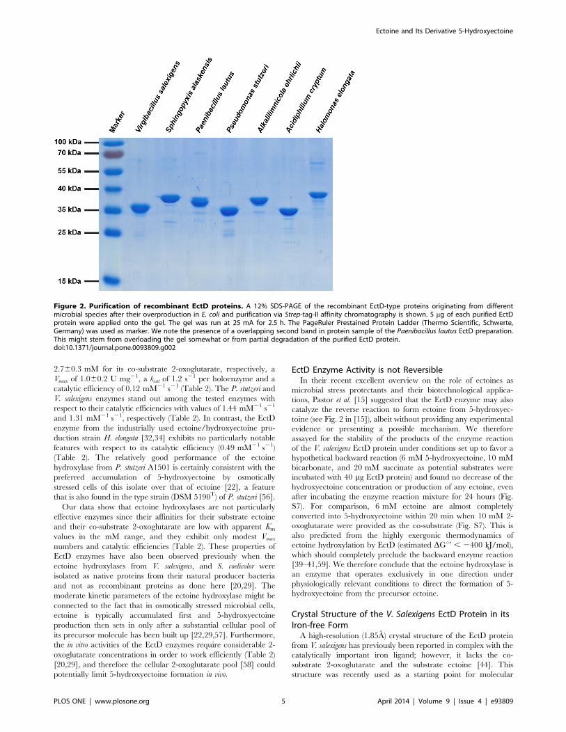
consist of 332 amino acids (Table 1). The migration of some of the
purified recombinant EctD proteins on a 12% SDS-polyacryl-
amide gel (Fig. 2) deviates somewhat from their calculated
molecular mass (Table 1), a property that might be connected
with the particular amino acid composition of individual EctD
proteins.
Since the presence of a correctly complexed iron ligand is
critical for EctD-mediated enzyme catalysis [20,43,44], we
determined the iron-content of each of these recombinant proteins
and found between 0.87 and 0.96 mole iron per mol of EctD
protein. Hence, these recombinant EctD proteins should all be
functional. An initial assessment of their enzymatic activities under
the same assay conditions as used previously for the ectoine
hydroxylases from V. salexigens and S. coelicolor [20,29] demonstrat-
ed that this was indeed the case.
Biochemical Properties of the Ectoine HydroxylasesWe determined for each of the EctD enzymes its temperature
and pH optimum and measured the influence of various salts
(KCl, NaCl, K-glutamate, NH4Cl) on the catalytic efficiency. The
data from this set of experiments are summarized in Table 1 and
are documented in detail for the S. alaskensis enzyme in Fig. 3. The
data for all other enzymes are summarized in Fig. S1 to Fig. S5.
Overall, the basic biochemical parameters of the six newly studied
EctD enzymes and the re-analyzed EctD protein from V. salexigens
[20] were all quite similar (Table 1), regardless of the environ-
mental parameters that were prevalent in the habitats of those
microorganisms from which they originate. However, differences
were noted with respect to their resistance to the inhibiting action
of increased salt concentrations (Table 1).
In studying the biochemical properties of the ectoine biosyn-
thetic enzymes from H. elongata, Ono et al. [17] reported that the
in vitro activity of these proteins was strongly dependent on high
concentrations of NaCl (0.4–0.5 M), a type of salt that is unlikely
to be accumulated to such high levels in vivo by osmotically stressed
H. elongata cells, since sodium ions are toxic for bacterial cells. We
did not find any strong stimulating effect of high NaCl
concentrations on any of the ectoine hydroxylases we studied
here (Table 1), including that of H. elongata (Fig. S1). On the
contrary, high concentrations of NaCl typically inhibited the
enzyme activities of the EctD variants (Fig. 3 and Fig. S1 to Fig.
S5). However, notable stimulating effects [about two- to three-fold
(Fig. 3 and Fig. S1 to Fig. S5)] on EctD enzyme activities were
recorded with KCl or K-glutamate solutions.
We assessed the quaternary structure of the six newly studied
EctD proteins by gel filtration. An example of this analysis is
shown in Fig. S6 for the S. alaskensis EctD protein. The protein
eluted between 72 to 83 ml (maximum: 77. 5 ml) from the size
exclusion chromatography column and thereby corresponds to a
protein of about 70.4 kDa. Since the calculated molecular mass of
the S. alaskensis EctD protein monomer with the attached Strep-tag-
II affinity peptide (nine amino acids) is 35.29 kDa, the ectoine
hydroxylase is apparently a homodimer. The same conclusion was
derived for all other analyzed EctD proteins (data not shown),
including that from V. salexigens, which has previously been
suggested to be a monomer [20].
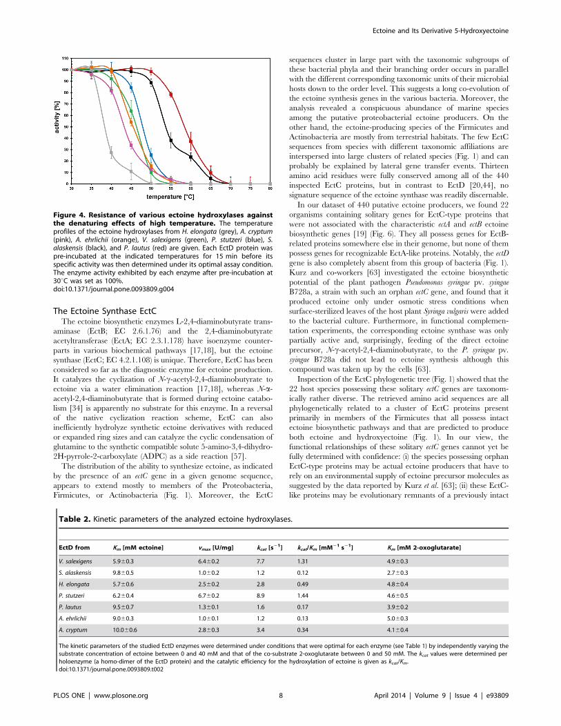
Temperature Stability of the Ectoine Hydroxylases: The S.Alaskensis and P. Lautus Enzymes Stand Out
The studied EctD enzymes have similar temperature optima but
differ in the range of temperatures in which they operate naturally
(Table 1). To investigate this further, we studied their temperature
stability. For these experiments, we pre-incubated 100 mg of each
enzyme in 100 ml TES-buffer (pH 7–8) for 15 min at a given
temperature and then measured its activity under assay and
temperature conditions that had been optimized for each
individual EctD protein (Table 1). The ectoine hydroxylase from
H. elongata turned out to be the most temperature labile protein,
whereas those from S. alaskensis and P. lautus proved to be quite
temperature resistant; all other enzymes possessed intermediate
degrees of temperature stability (Fig. 4). The strong temperature
resistance of the P. lautus EctD protein does not come as a surprise
since this Paenibacillus species was isolated from a hot spring with
water temperatures ranging between 42–90uC [54]. The consid-
erable heat tolerance of the S. alaskensis EctD enzyme is more of a
surprise since this bacterium is well adapted to permanently cold
(4–10uC) marine environments although it can grow at higher
temperatures [52].
Kinetic Parameters of Ectoine HydroxylasesAfter having optimized the parameters of the enzyme activity
assays for each of the six purified ectoine hydroxylases (Table 1),
we determined their apparent kinetic parameters for the co-
substrate 2-oxoglutarate and the substrate ectoine (Table 2). This
assessment showed that the studied ectoine hydroxylases all possess
similar kinetic parameters. For instance, the S. alaskensis enzyme
had an apparent Km of 9.860.5 mM for its substrate ectoine and of
Ectoine and Its Derivative 5-Hydroxyectoine
PLOS ONE | www.plosone.org 4 April 2014 | Volume 9 | Issue 4 | e93809
2.760.3 mM for its co-substrate 2-oxoglutarate, respectively, a
Vmax of 1.060.2 U mg21, a kcat of 1.2 s21 per holoenzyme and a
catalytic efficiency of 0.12 mM21 s21 (Table 2). The P. stutzeri and
V. salexigens enzymes stand out among the tested enzymes with
respect to their catalytic efficiencies with values of 1.44 mM21 s21
and 1.31 mM21 s21, respectively (Table 2). In contrast, the EctD
enzyme from the industrially used ectoine/hydroxyectoine pro-
duction strain H. elongata [32,34] exhibits no particularly notable
features with respect to its catalytic efficiency (0.49 mM21 s21)
(Table 2). The relatively good performance of the ectoine
hydroxylase from P. stutzeri A1501 is certainly consistent with the
preferred accumulation of 5-hydroxyectoine by osmotically
stressed cells of this isolate over that of ectoine [22], a feature
that is also found in the type strain (DSM 5190T) of P. stutzeri [56].
Our data show that ectoine hydroxylases are not particularly
effective enzymes since their affinities for their substrate ectoine
and their co-substrate 2-oxoglutarate are low with apparent Km
values in the mM range, and they exhibit only modest Vmaxnumbers and catalytic efficiencies (Table 2). These properties of
EctD enzymes have also been observed previously when the
ectoine hydroxylases from V. salexigens, and S. coelicolor were
isolated as native proteins from their natural producer bacteria
and not as recombinant proteins as done here [20,29]. The
moderate kinetic parameters of the ectoine hydroxylase might be
connected to the fact that in osmotically stressed microbial cells,
ectoine is typically accumulated first and 5-hydroxyectoine
production then sets in only after a substantial cellular pool of
its precursor molecule has been built up [22,29,57]. Furthermore,
the in vitro activities of the EctD enzymes require considerable 2-
oxoglutarate concentrations in order to work efficiently (Table 2)
[20,29], and therefore the cellular 2-oxoglutarate pool [58] could
potentially limit 5-hydroxyectoine formation in vivo.
EctD Enzyme Activity is not ReversibleIn their recent excellent overview on the role of ectoines as
microbial stress protectants and their biotechnological applica-
tions, Pastor et al. [15] suggested that the EctD enzyme may also
catalyze the reverse reaction to form ectoine from 5-hydroxyec-
toine (see Fig. 2 in [15]), albeit without providing any experimental
evidence or presenting a possible mechanism. We therefore
assayed for the stability of the products of the enzyme reaction
of the V. salexigens EctD protein under conditions set up to favor a
hypothetical backward reaction (6 mM 5-hydroxyectoine, 10 mM
bicarbonate, and 20 mM succinate as potential substrates were
incubated with 40 mg EctD protein) and found no decrease of the
hydroxyectoine concentration or production of any ectoine, even
after incubating the enzyme reaction mixture for 24 hours (Fig.
S7). For comparison, 6 mM ectoine are almost completely
converted into 5-hydroxyectoine within 20 min when 10 mM 2-
oxoglutarate were provided as the co-substrate (Fig. S7). This is
also predicted from the highly exergonic thermodynamics of
ectoine hydroxylation by EctD (estimated DGu’ , 2400 kJ/mol),
which should completely preclude the backward enzyme reaction
[39–41,59]. We therefore conclude that the ectoine hydroxylase is
an enzyme that operates exclusively in one direction under
physiologically relevant conditions to direct the formation of 5-
hydroxyectoine from the precursor ectoine.
Crystal Structure of the V. Salexigens EctD Protein in itsIron-free Form
A high-resolution (1.85A) crystal structure of the EctD protein
from V. salexigens has previously been reported in complex with the
catalytically important iron ligand; however, it lacks the co-
substrate 2-oxoglutarate and the substrate ectoine [44]. This
structure was recently used as a starting point for molecular
Figure 2. Purification of recombinant EctD proteins. A 12% SDS-PAGE of the recombinant EctD-type proteins originating from differentmicrobial species after their overproduction in E. coli and purification via Strep-tag-II affinity chromatography is shown. 5 mg of each purified EctDprotein were applied onto the gel. The gel was run at 25 mA for 2.5 h. The PageRuler Prestained Protein Ladder (Thermo Scientific, Schwerte,Germany) was used as marker. We note the presence of a overlapping second band in protein sample of the Paenibacillus lautus EctD preparation.This might stem from overloading the gel somewhat or from partial degradation of the purified EctD protein.doi:10.1371/journal.pone.0093809.g002
Ectoine and Its Derivative 5-Hydroxyectoine
PLOS ONE | www.plosone.org 5 April 2014 | Volume 9 | Issue 4 | e93809
dynamics simulations and site-directed mutagenesis experiments to
glean information about the coordination of the ligands within the
EctD active site [43]. We continued our efforts to obtain an EctD
crystal structure containing all ligands and therefore pushed the
recombinant production of the V. salexigens EctD protein in E. coli
to very high levels in order to supply the large quantities of protein
needed for the crystallization trials. In this way, we increased the
amounts of the purified V. salexigens recombinant EctD enzyme
from about 20–25 mg per liter of culture (the ectoine hydroxylase
source for biochemical studies) to 200–300 mg per liter of culture.
However, after analyzing the iron content of this strongly
overproduced EctD enzyme preparation, it became apparent that
most of the isolated proteins did not contain an iron molecule; the
iron content of the EctD protein solution dropped to 0.1–0.2 mole
iron per 1 mol of EctD, rendering the enzyme largely inactive.
Upon addition of Fe2+ ions prior to the enzyme activity
measurements, we observed that the activity returned to levels
observed before [20,43], indicating that the missing iron catalyst in
the purified EctD protein can be restored after the protein has
adopted its native cupin barrel fold [44].
These observations prompted us to explore whether the V.
salexigens EctD protein adopts a similar conformation in its iron-
free and iron-bound forms, or whether the incorporation of the
iron ligand leads to substantial structural changes. We grew EctD
crystals and collected a 1.9 A X-ray dataset. The cell constants and
the space-group (Table S1) were identical to the structure of the
iron-bound EctD, suggesting that the apo-EctD protein crystalized
in a manner similar to that found in the iron bound form [44].
After solving the new crystal structure of the EctD protein, it
became apparent that the iron ligand was lacking, as evidenced by
the missing pronounced electron density that is present in the iron-
bound EctD crystal structure [44]. Otherwise, the apo- and the
iron-bound forms are almost identical, as indicated by the RMSD
value of 0.34 A over 280 Ca atoms. An overlay of both EctD
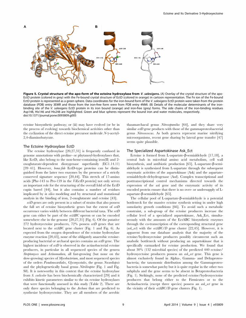
crystal structures is shown in Fig. 5A.
In the V. salexigens EctD protein, the iron ligand is bound via
interaction with two histidine side-chains, His-146 and His-248,
and the side-chain of Asp-148 (Fig. 5A and B) [44]. Together these
residues form a conserved H6D/E…H motif, the so-called 2-His-
1-carboxylate facial triad. [39–41,59]. A comparison of the iron-
binding residues in the apo- and iron-bound structures of the
VsEctD protein shows that they exhibit the same architecture,
except that the iron ligand is present in one structure and absent in
the other (Fig. 5B). Interestingly, in the apo-structure of EctD, two
water molecules populate the iron-binding site formed by the 2-
His-1-carboxylate facial triad. This keeps the side chains of the
His-146, His-248 and Asp-148 in an orientation very similar to
that observed in the iron-bound EctD crystal structure (Fig. 5B).
Hence, the EctD apo-protein exists in a form that is pre-set to
incorporate the iron catalyst [43].
Phylogenetic Distribution of the EctC and EctD GenesPrevious studies have indicated that the ability to produce
ectoine and hydroxyectoine is widely distributed in the microbial
world but is absent from eukarya [15,34,44,60]. We updated and
extended this information on a genome-wide scale in the following
way: (i) first, we visualized the relationship among the 440
retrieved EctC sequences via the iTOL tool [61] to analyze their
taxonomic association with members of the Bacteria and Archaea; (ii)
we then projected the information on the presence of the ectoine
hydroxylase in a given microbial species onto this phylogenetic
tree of the EctC protein to reveal the extent and taxonomic
distribution of EctD protein among putative ectoine producers; (iii)
we inspected the genome context of each of the 440 microbial
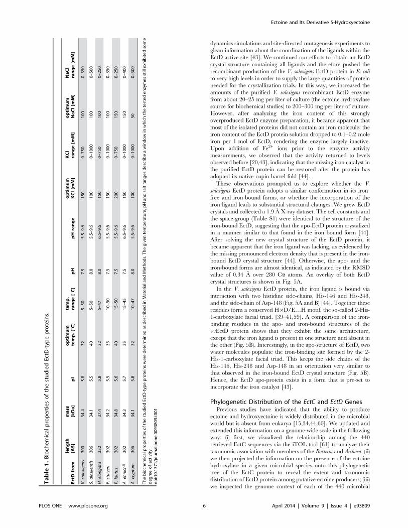
Table
1.Biochemical
propertiesofthestudiedEctD-typ
eproteins.
EctD
from
length
[AS]
mass
[kDa]
pI
optimum
temp.[uC]
temp.
range[uC]
pH
pH
range
optimum
KCl[m
M]
KCl
range[m
M]
optimum
NaCl[m
M]
NaCl
range[m
M]
V.salexigens
300
34.4
5.8
32
5–50
7.5
5.5–9.6
150
0–750
100
0–350
S.alaskensis
306
34.1
5.5
40
5–50
8.0
5.5–9.6
100
0–1000
100
0–500
H.elongata
332
37.4
5.8
32
5–47
8.0
6.5–9.6
150
0–750
100
0–250
P.stutzeri
302
34.2
5.5
35
10–50
7.5
5.5–9.6
150
0–1000
100
0–350
P.lautus
302
34.8
5.6
40
15–50
7.5
5.5–9.6
200
0–750
150
0–250
A.ehrlichii
302
34.3
5.7
35
15–45
7.5
6.5–9.6
150
0–1000
150
0–400
A.cryptum
306
34.1
5.8
32
10–47
8.0
5.5–9.6
100
0–1000
50
0–300
ThebiochemicalpropertiesofthestudiedEctD-typ
eproteinswere
determ
inedas
describedin
Materialan
dMethods.Thegiventemperature,p
Han
dsaltrangesdescribeawindowin
whichthetestedenzymesstillexh
ibitedsome
degreeofactivity.
doi:10.1371/journal.pone.0093809.t001
Ectoine and Its Derivative 5-Hydroxyectoine
PLOS ONE | www.plosone.org 6 April 2014 | Volume 9 | Issue 4 | e93809
species that possessed ectC and from this bioinformatics approach
retrieved the genetic organization of the ect biosynthetic gene
cluster; (iv) furthermore, we assessed the co-localization of the ect
genes with genes that have been functionally associated with
ectoine/hydroxyectoine biosynthesis, the gene for a specialized
aspartokinase Ask_Ect [22,45,60], and that of the transcriptional
regulator EctR [24,25].
In the first step of this in silico analysis, we aligned the retrieved
440 EctC sequences using the ClustalW [62] algorithm and found
amino acid sequence identities that ranged between 88% and 27%
with reference to the V. salexigens EctC protein [20]. The
corresponding numbers for the degree of identity of the 272 EctD
proteins range between 79% and 37% with reference to the V.
salexigens EctD protein. The visualization of the taxonomic
distribution of the EctC and EctD proteins with the iTOL-
software package [61] revealed putative bacterial and archaeal
ectoine producers in 17 phyla (Fig. 1). Fifteen of these phyla are
taxonomically associated with the domain of the Bacteria and two
with the domain of the Archaea. The taxonomic distribution of the
putative hydroxyectoine producers was more restricted: ectoine
hydroxylase genes are found only in nine phyla (Fig. S8). In the
following, we further consider the ectoine synthase, the ectoine
hydroxylase, the specialized aspartokinase Ask_Ect, and the
transcriptional regulator EctR.
Figure 3. Biochemical properties of the EctD enzyme from S. alaskensis. The enzyme activity of the ectoine hydroxylase from S. alaskensis isshown with respect to (A) the temperature optimum, (B) the pH optimum and the influence of different salts: (C) potassium chloride, (D) sodiumchloride, (E) potassium glutamate and (F) ammonium chloride.doi:10.1371/journal.pone.0093809.g003
Ectoine and Its Derivative 5-Hydroxyectoine
PLOS ONE | www.plosone.org 7 April 2014 | Volume 9 | Issue 4 | e93809
The Ectoine Synthase EctCThe ectoine biosynthetic enzymes L-2,4-diaminobutyrate trans-
aminase (EctB; EC 2.6.1.76) and the 2,4-diaminobutyrate
acetyltransferase (EctA; EC 2.3.1.178) have isoenzyme counter-
parts in various biochemical pathways [17,18], but the ectoine
synthase (EctC; EC 4.2.1.108) is unique. Therefore, EctC has been
considered so far as the diagnostic enzyme for ectoine production.
It catalyzes the cyclization of N-c-acetyl-2,4-diaminobutyrate to
ectoine via a water elimination reaction [17,18], whereas N-a-
acetyl-2,4-diaminobutyrate that is formed during ectoine catabo-
lism [34] is apparently no substrate for this enzyme. In a reversal
of the native cyclization reaction scheme, EctC can also
inefficiently hydrolyze synthetic ectoine derivatives with reduced
or expanded ring sizes and can catalyze the cyclic condensation of
glutamine to the synthetic compatible solute 5-amino-3,4-dihydro-
2H-pyrrole-2-carboxylate (ADPC) as a side reaction [57].
The distribution of the ability to synthesize ectoine, as indicated
by the presence of an ectC gene in a given genome sequence,
appears to extend mostly to members of the Proteobacteria,
Firmicutes, or Actinobacteria (Fig. 1). Moreover, the EctC
sequences cluster in large part with the taxonomic subgroups of
these bacterial phyla and their branching order occurs in parallel
with the different corresponding taxonomic units of their microbial
hosts down to the order level. This suggests a long co-evolution of
the ectoine synthesis genes in the various bacteria. Moreover, the
analysis revealed a conspicuous abundance of marine species
among the putative proteobacterial ectoine producers. On the
other hand, the ectoine-producing species of the Firmicutes and
Actinobacteria are mostly from terrestrial habitats. The few EctC
sequences from species with different taxonomic affiliations are
interspersed into large clusters of related species (Fig. 1) and can
probably be explained by lateral gene transfer events. Thirteen
amino acid residues were fully conserved among all of the 440
inspected EctC proteins, but in contrast to EctD [20,44], no
signature sequence of the ectoine synthase was readily discernable.
In our dataset of 440 putative ectoine producers, we found 22
organisms containing solitary genes for EctC-type proteins that
were not associated with the characteristic ectA and ectB ectoine
biosynthetic genes [19] (Fig. 6). They all possess genes for EctB-
related proteins somewhere else in their genome, but none of them
possess genes for recognizable EctA-like proteins. Notably, the ectD
gene is also completely absent from this group of bacteria (Fig. 1).
Kurz and co-workers [63] investigated the ectoine biosynthetic
potential of the plant pathogen Pseudomonas syringae pv. syingae
B728a, a strain with such an orphan ectC gene, and found that it
produced ectoine only under osmotic stress conditions when
surface-sterilized leaves of the host plant Syringa vulgaris were added
to the bacterial culture. Furthermore, in functional complemen-
tation experiments, the corresponding ectoine synthase was only
partially active and, surprisingly, feeding of the direct ectoine
precursor, N-c-acetyl-2,4-diaminobutyrate, to the P. syringae pv.
syingae B728a did not lead to ectoine synthesis although this
compound was taken up by the cells [63].
Inspection of the EctC phylogenetic tree (Fig. 1) showed that the
22 host species possessing these solitary ectC genes are taxonom-
ically rather diverse. The retrieved amino acid sequences are all
phylogenetically related to a cluster of EctC proteins present
primarily in members of the Firmicutes that all possess intact
ectoine biosynthetic pathways and that are predicted to produce
both ectoine and hydroxyectoine (Fig. 1). In our view, the
functional relationships of these solitary ectC genes cannot yet be
fully determined with confidence: (i) the species possessing orphan
EctC-type proteins may be actual ectoine producers that have to
rely on an environmental supply of ectoine precursor molecules as
suggested by the data reported by Kurz et al. [63]; (ii) these EctC-
like proteins may be evolutionary remnants of a previously intact
Figure 4. Resistance of various ectoine hydroxylases againstthe denaturing effects of high temperature. The temperatureprofiles of the ectoine hydroxylases from H. elongata (grey), A. cryptum(pink), A. ehrlichii (orange), V. salexigens (green), P. stutzeri (blue), S.alaskensis (black), and P. lautus (red) are given. Each EctD protein waspre-incubated at the indicated temperatures for 15 min before itsspecific activity was then determined under its optimal assay condition.The enzyme activity exhibited by each enzyme after pre-incubation at30uC was set as 100%.doi:10.1371/journal.pone.0093809.g004
Table 2. Kinetic parameters of the analyzed ectoine hydroxylases.
EctD from Km [mM ectoine] vmax [U/mg] kcat [s21] kcat/Km [mM21 s21] Km [mM 2-oxoglutarate]
V. salexigens 5.960.3 6.460.2 7.7 1.31 4.960.3
S. alaskensis 9.860.5 1.060.2 1.2 0.12 2.760.3
H. elongata 5.760.6 2.560.2 2.8 0.49 4.860.4
P. stutzeri 6.260.4 6.760.2 8.9 1.44 4.660.5
P. lautus 9.560.7 1.360.1 1.6 0.17 3.960.2
A. ehrlichii 9.060.3 1.060.1 1.2 0.13 5.060.3
A. cryptum 10.060.6 2.860.3 3.4 0.34 4.160.4
The kinetic parameters of the studied EctD enzymes were determined under conditions that were optimal for each enzyme (see Table 1) by independently varying thesubstrate concentration of ectoine between 0 and 40 mM and that of the co-substrate 2-oxoglutarate between 0 and 50 mM. The kcat values were determined perholoenzyme (a homo-dimer of the EctD protein) and the catalytic efficiency for the hydroxylation of ectoine is given as kcat/Km.doi:10.1371/journal.pone.0093809.t002
Ectoine and Its Derivative 5-Hydroxyectoine
PLOS ONE | www.plosone.org 8 April 2014 | Volume 9 | Issue 4 | e93809
ectoine biosynthetic pathway; or (iii) may have evolved (or be in
the process of evolving) towards biochemical activities other than
the cyclization of the direct ectoine precursor molecule N-c-acetyl-
2,4-diaminobutyrate.
The Ectoine Hydroxylase EctDThe ectoine hydroxylase [20,27,31] is frequently confused in
genome annotations with proline- or phytanoyl-hydroxylases that,
like EctD, also belong to the non-heme-containing iron(II) and 2-
oxoglutarate-dependent dioxygenase superfamily (EC1.14.11)
[39–41]. However, bona-fide EctD-type proteins can be distin-
guished from the latter two enzymes by the presence of a strictly
conserved signature sequence [20,44]. This stretch of 17-amino
acids [Phe-143 to Pro-159 in the VsEctD protein] not only serves
an important role for the structuring of the overall fold of the EctD
cupin barrel [44], but it also contains a number of residues
implicated by in silico modeling and by structural and mutational
analysis in the binding of iron, 2-oxoglutarate and ectoine [43].
ectD genes are only present in a subset of strains that also possess
the full set of ectoine biosynthetic genes but the extent of ectD
occurrence varies widely between different bacterial taxa. The ectD
gene can either be part of the ectABC operon or can be encoded
somewhere else in the genome [20,27,31] (Fig. 6). Of the putative
272 hydroxyectoine producers, 72% possess ectD genes that are
located next to the ectABC gene cluster (Fig. 1 and Fig. 6). As
expected from the oxygen dependence of the ectoine hydroxylase
enzyme reaction [20,43], none of the obligately anaerobic ectoine-
producing bacterial or archaeal species contains an ectD gene. The
highest incidence of ectD is observed in the actinobacterial ectoine
producers, in particular in all sequenced species of the genera
Streptomyces and Actinomadura, all fast-growing (but none on the
slow-growing) species of Mycobacterium, and most sequenced species
of the orders Pseudonocardiales, Glycomycetales, the genus Nocardiopsis
and the phylogenetically basal genus Nitriliruptor (Fig. 1 and Fig.
S8). It is noteworthy in this context that the ectoine hydroxylase
from S. coelicolor has been biochemically characterized [29] and it
exhibits kinetic parameters similar to the six ectoine hydroxylases
that were functionally assessed in this study (Table 2). There are
only three species belonging to the Archaea that are predicted to
synthesize hydroxyectoine. They are all members of the aerobic
thaumarchaeal genus Nitrosopumilus [64], and they share very
similar ectD gene products with those of the gammaproteobacterial
genus Nitrosococcus. As both genera represent marine nitrifying
microorganisms, recent gene sharing by lateral gene transfer [47]
seems quite plausible.
The Specialized Aspartokinase Ask_EctEctoine is formed from L-aspartate-b-semialdehyde [17,18], a
central hub in microbial amino acid metabolism, cell wall
biosynthesis, and antibiotic production [65]. L-aspartate-b-semi-
aldehyde is synthesized from L-aspartate through the subsequent
enzymatic activities of the aspartokinase (Ask) and the aspartate-
semialdehyde-dehydrogenase (Asd). Complex transcriptional and
posttranscriptional control mechanisms directed towards the
expression of the ask gene and the enzymatic activity of its
encoded protein ensure that there is no over- or undersupply of L-
aspartate-b-semialdehyde [65].
The cellular pool of L-aspartate-b-semialdehyde is a potential
bottleneck for the massive ectoine synthesis setting in under high
osmolarity growth conditions [66]. To avoid such a metabolic
constraint, a sub-group of the ectoine producers increase the
cellular level of a specialized aspartokinase, Ask_Ect, simulta-
neously with the amounts of the EctABC biosynthetic enzymes
through the co-transcription of the corresponding structural gene
(ask_ect) with the ectABC/D gene cluster [22,45]. However, it is
apparent from our database analysis that the majority of the
ectoine/hydroxyectoine producers possibly circumvent such an
anabolic bottleneck without producing an aspartokinase that is
specifically earmarked for ectoine production. We found that
about 30% (132 microbial species) of the predicted 440 ectoine/
hydroxyectoine producers possess an ask_ect gene. This gene is
almost exclusively found in Alpha-, Gamma- and Deltaproteo-
bacteria; the taxonomic distribution among the Gammaproteo-
bacteria is somewhat patchy but it is quite regular in the other two
subphyla and the gene seems to be absent in Betaproteobacteria
(Fig. 1). Strikingly, none of the predicted ectoine/hydroxyectoine
producers that belong either to the Firmicutes or to the
Actinobacteria (except three species) possess an ask_ect gene in
the vicinity of their ectABC/D gene clusters (Fig. 1).
Figure 5. Crystal structure of the apo-form of the ectoine hydroxylase from V. salexigens. (A) Overlay of the crystal structure of the apo-EctD protein (colored in grey) with the Fe-bound crystal structure of EctD (colored in orange) in cartoon representation. The Fe ion of the Fe-boundEctD protein is represented as a green sphere. Data coordinates for the iron-bound form of the V. salexigens EctD protein were taken from the proteindatabase (PDB) entry 3EMR and those from the iron-free form were from PDB entry 4NMI. (B) Details of the molecular determinants of the iron-binding site of the V. salexigens EctD protein in its iron bound (orange) and iron-free (grey) forms. The side chains of the iron-binding residuesAsp148, His146 and His248 are highlighted. Green and blue spheres represent the bound iron and water molecules, respectively.doi:10.1371/journal.pone.0093809.g005
Ectoine and Its Derivative 5-Hydroxyectoine
PLOS ONE | www.plosone.org 9 April 2014 | Volume 9 | Issue 4 | e93809
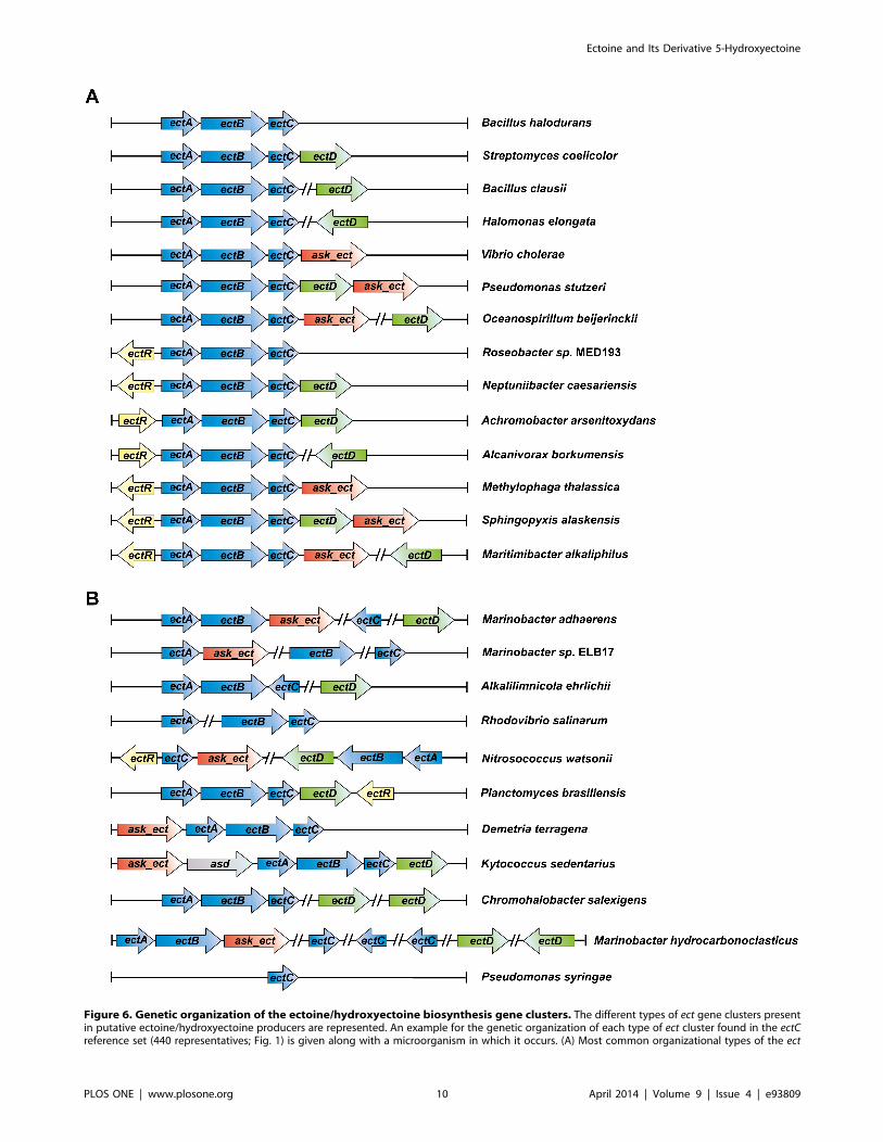
Figure 6. Genetic organization of the ectoine/hydroxyectoine biosynthesis gene clusters. The different types of ect gene clusters presentin putative ectoine/hydroxyectoine producers are represented. An example for the genetic organization of each type of ect cluster found in the ectCreference set (440 representatives; Fig. 1) is given along with a microorganism in which it occurs. (A) Most common organizational types of the ect
Ectoine and Its Derivative 5-Hydroxyectoine
PLOS ONE | www.plosone.org 10 April 2014 | Volume 9 | Issue 4 | e93809
Aspartokinases are ubiquitously found in microorganisms and
several aspartokinases with distinct regulatory features are often
present in the same bacterial cell [65]. The latter is true for the
ectoine/hydroxyectoine producer Pseudomonas stutzeri A1501,
where a comparative biochemical analysis of the specialized
aspartokinase Ask_Ect and the anabolic aspartokinase LysC
revealed distinct feedback inhibition profiles by metabolites [22].
The Transcriptional Regulator EctRThe functional association of the ectR gene with ectoine
biosynthesis was first demonstrated in the halotolerant methano-
trophic Gammaproteobacterium Methylomicrobium alcaliphilum,
where EctR serves as a repressor of ectABC-ask_ect gene transcrip-
tion [25]. Notably, the elevated transcription of the ectoine
biosynthetic genes in an ectR mutant of M. alcaliphilum remains
osmotically inducible [25]. It is currently not known which
environmental or cellular cues dictate the binding to or the release
of EctR from its operator sequence. In M. alcaliphilum, the EctR
operator overlaps the 210 sequence of the ect promoter and EctR
might also regulate the expression of its own structural gene [25];
however, this latter regulatory feature does not always seem to
exist [24]. The EctR repressor protein is a member of the widely
distributed group of MarR transcriptional regulators but forms a
distinct sub-group within this superfamily [24]. Of the 440
putative ectoine/hydroxyectoine producers, 24% possess ectR-type
genes (107 microbial species) (Fig. 1) whose transcriptional
direction is frequently oriented divergently from that of the ect
gene cluster (Fig. 6). EctR is found almost exclusively among the
Proteobacteria; all Alpha- and Betaproteobacteria that are
predicted to synthesize ectoines possess an ectR gene, whereas its
distribution among the Gammaproteobacteria is more irregular
(Fig. 1).
It is worth noting that in Vibrio cholerae an EctR-related MarR-
type transcriptional regulator (CosR) has been described that
negatively controls ectoine biosynthetic and compatible solute
uptake genes in response to the ionic strength of the growth
medium. CosR from V. cholerae and EctR from M. alcaliphilum
exhibit 51% amino acid sequence identity; however, unlike ectR,
the cosR gene is not located in the vicinity of the ectoine gene
cluster present in V. cholerae [67]. Ectoine biosynthesis, but not that
of hydroxyectoine, is widespread among V. cholerae strains [68] and
other Vibrio species [69], microorganisms that primarily live in
marine habitats and estuarine ecosystems. For instance, genome
sequence of 139 V. cholerae strains have been deposited in the
databases and each of these strains is predicted to synthesize
ectoine (data not shown).
Genetic Organization of the Ectoine and HydroxyectoineStructural Genes
After the initial discovery of the ectABC gene cluster for the
synthesis of ectoine in Marinococcus halophilus [19], transcriptional
profiling of the corresponding genes in several Gram-negative and
Gram-positive bacteria showed that they were transcribed as
operons inducible by osmotic or temperature stress
[11,20,21,23,26,27]. In some ectoine producers, the ectABC genes
are expressed from a single promoter [11,20], whereas in others a
more complex pattern of regulation of this gene cluster has been
reported [21,24,34]. Evidence for the presence of a putatively
nitrogen-responsive Sig-54 type promoter driving the separate
expression of ectC has been presented in the case of H. elongata [34].
We inspected the genetic organization of the ect genes in the 440
microbial species that we regarded as putative ectoine producers
from our database analysis (Fig. 1). In the vast majority (85%), the
ectABC genes were located next to each other, strongly suggesting
that their transcriptional organization is centrally based on an
evolutionarily highly conserved operon structure (Fig. 6A). The
basic ectABC gene cluster is frequently associated either with ectD,
ask_ect, or ectR, and various genetic configurations of these ect-
associated genes can be found in microbial genomes (Fig. 6A). The
ectD gene may either be part of the ectABC operon or form a
separate transcription unit somewhere else in the genome.
Our database analysis shows that the genetic organization of the
ect gene cluster is well preserved in groups of microorganisms that
are widely separated taxonomically (Fig. 6A). Nevertheless, there is
a substantial sub-group (about 15%) of the putative ectoine/
hydroxyectoine producers where the ectoine/hydroxyectoine
biosynthetic genes and the functionally associated ask_ect and ectR
genes are not organized in the well-defined gene clusters found in
85% of our reference data set. In this group of bacteria, the order
of the various ect genes have either been scrambled or they have
been separated from each other on the chromosome (Fig. 6B). It is
currently unclear whether this non-canonical gene organization
has any consequences for transcriptional induction or the level of
ectoine/hydroxyectoine production in response to osmotic or
temperature stress since, to the best of our knowledge, none of the
putative ectoine/hydroxyectoine producers with deviating gene
organizations have been functionally studied.
In a few of the hydroxyectoine producers (e.g., Arthrobacter castelli
DSM 16402, Marinobacter aquaeolei VT8, Rhodococcus opacus B4,
Rhodococcus sp. RHA1, Chromohalobacter salexigens DSM 3043), two
copies of the ectD gene are found (Fig. 6B). Studies with C. salexigens
DSM 3043 have shown that only one of these ectoine hydroxylases
is responsible for the production of the majority of the
hydroxyectoine found in this highly salt-tolerant bacterium [70].
In the genomes of other microorganisms, several ectC-type genes
are found (e.g.; there are three ectC genes in Marinobacter aquaeolei).
However, nothing is known whether these genes are all expressed
and what (if any) the functional consequences of multiple, closely
related EctC proteins within the same bacterium might be.
Another interesting finding of our database analysis is the
identification of microorganisms that possess either two (e.g.,
Phaeobacter arcticus DSM 23566, Vibrio cholerae 0395, Streptomyces
flavogriseus ATCC 33331) or even three (e.g., Streptomyces clavuligerus
ATCC 27064) full copies of the ectoine/hydroxyectoine biosyn-
thetic gene cluster. A pairwise comparison of the amino acid
sequence of the various Ect proteins within a given species
indicates that the increase in the ect gene copy number has likely
arisen via gene duplication events since the corresponding proteins
are all closely related to each other (data not shown). Whether
these extra copies of the ectoine/hydroxyectoine biosynthetic
genes are actually functionally expressed and whether the bacteria
with the additional ect gene copies produce more ectoine than
those with only one copy of the corresponding genes remains an
interesting question for future studies.
Concluding RemarksOur comprehensive database analysis of finished microbial
genomes revealed at the time of the search (July 2013) the presence
gene clusters. (B) Representatives of the organization of the ectoine/hydroxyectoine biosynthetic genes that deviate from the otherwise commonlyfound genetic organization.doi:10.1371/journal.pone.0093809.g006
Ectoine and Its Derivative 5-Hydroxyectoine
PLOS ONE | www.plosone.org 11 April 2014 | Volume 9 | Issue 4 | e93809
of ectoine biosynthetic genes [19] in about 7% of the represented
microorganisms, and about two thirds of these are predicted to
produce hydroxyectoine [20,27,31] as well. Since hydroxyectoine
often possess function-preserving and stress-relieving properties
superior to those of ectoine [29,35–38], one wonders why not all
ectoine producers synthesize 5-hydroxyectoine since this can
readily be accomplished from ectoine in a single step [20]. Part of
the answer to this question becomes apparent when one considers
the physiological requirements and the oxygen dependence of the
ectoine/hydroxyectoine producer microorganisms. The EctD-
catalyzed formation of 5-hydroxyectoine is an O2-dependent
enzyme reaction [20,43] and consequently none of the predicted
hydroxyectoine producers is an obligate anaerobe, whereas both
aerobic and anaerobic microorganisms can produce ectoine
(Fig. 1).
Our data view the potential ectoine and hydroxyectoine
producers within a wider taxonomic context (Fig. 1 and Fig. S8).
With a few notable exceptions that revealed ectoine/hydroxyec-
toine biosynthetic genes in five members of the Archaea
(Methanosaeta and Nitrosopumilus species), ectoine and hydroxyec-
toine producers are taxonomically affiliated with the domain of the
Bacteria. We assessed the genetic organization of the ectoine/
hydroxyectoine biosynthetic genes (Fig. 6) and those of proteins
that are functionally associated with ectoine production, the
specialized aspartokinase Ask_Ect [22,45] and the transcriptional
regulator EctR [25] (Fig. 1). By analyzing the occurrence of these
proteins on a genome-wide scale and by viewing them in a
taxonomic context, we derived the currently most comprehensive
in silico analysis of the production potential for the stress
protectants and chemical chaperones ectoine and 5-hydroxyec-
toine in microorganisms (Fig. 1 and 6; Fig. S8). This dataset can
therefore serve as a solid benchmark for future assessments as
microbial genome and metagenomic sequence analysis continues
to progress in a rapid pace.
Our analysis of the genetic organization of the ectABC/D
biosynthetic genes revealed a robust arrangement into an operon-
like structure in taxonomically widely separated microorganisms
(Fig. 6). This assessment does not only provide clues for their
potential transcriptional organization, but also gives hints about
which of these gene clusters might be useful as building blocks for
synthetic ectoine/hydroxyectoine production in heterologous host
systems [22,56,71–74]. For instance, we surmise that the ectoine/
hydroxyectoine biosynthetic genes from Kytococcus sedentarius [75]
might be effectively exploited as a synthetic ‘‘bio-brick’’ for this
purpose. In this microorganism, the genes for both enzymes
(Ask_Ect and Asd) required for the synthesis of the direct ectoine
precursor, L-aspartate-b-semialdehyde [17,18,65], seem to be co-
transcribed with the ectABCD operon (Fig. 6B). Co-expression of
the ask_ect-asd-ectABCD gene cluster should help to avoid the build-
up of potential bottlenecks during heterologous ectoine/hydro-
xyectoine production for biotechnological and medical purposes
[15,32,33].
We placed special emphasis in our study on the further
biochemical [20,29] and structural analysis [44] of the ectoine
hydroxylase. In terms of the EctD crystal structure, our new data
reveal that the apo- and iron-liganded forms are virtually identical
(Fig. 5A). Hence, the ectoine hydroxylase is pre-set in a
configuration ready to accept the iron molecule (Fig. 5B) and
the binding of the iron catalyst does not trigger large conforma-
tional changes. Together with the EctD proteins from V. salexigens
and S. coelicolor that were previously studied biochemically [20,29],
the six ectoine hydroxylases examined here define the salient
biochemical features (Table 1 and 2) of this group of closely related
enzymes (Fig. S8). The ectoine hydroxylases analyzed so far all
possess similar kinetic parameters and catalytic efficiencies
(Table 1) [20,29] but differ in their tolerance towards high
temperature (Fig. 4) and in the influence of various salts on their
enzyme activity (Table 2). It is hoped that the properties of some of
the newly characterized EctD proteins will be suitable for further
crystallographic studies so that a crystal structure of the ectoine
hydroxylase with all its ligands (or its reaction product 5-
hydroxyectoine) can be obtained in the future.
Materials and Methods
ChemicalsEctoine and 5-hydroxyectoine were kind gifts from Dr. Thomas
Lentzen and Dr. Irina Bagyan (bitop AG, Witten, Germany). 2-
oxoglutarate (disodium salt) was obtained from Sigma-Aldrich (St.
Louis, MO, USA). Anhydrotetracycline-hydrochloride (AHT),
desthiobiotin, and Strep-Tactin Superflow chromatography ma-
terial were purchased from IBA GmbH (Gottingen, Germany). X-
Gal was obtained from AppliChem (Darmstadt, Germany), and
the antibiotics kanamycin and ampicillin were purchased from
Serva Electrophoresis GmbH (Heidelberg, Germany) and Carl
Roth GmbH (Karlsruhe, Germany).
Bacteria, Media and Growth ConditionsThe Escherichia coli strain DH5a (Invitrogen, Karlsruhe,
Germany) was used as host for recombinant plasmids and as
overproduction strain for EctD-proteins; it was maintained
routinely on LB agar plates and liquid media [76]. When it
contained recombinant plasmids, either ampicillin (100 mg ml21)
or kanamycin (50 mg ml21) was added to the growth medium to
select for the presence of the plasmids. When appropriate, X-gal
was included in agar plates to screen for the insertion of the
desired DNA fragments into the cloning vector pENTRY-IBA20
(IBA, Gottingen, Gemany). For the overproduction of EctD-type
proteins, minimal medium A (MMA) [76] was used that was
supplemented with 0.5% (w/v) glucose as the carbon source, 0.5%
(w/v) casaminoacids, 1 mM MgSO4, and 3 mM thiamine.
Recombinant DNA Techniques and Construction ofPlasmids
All recombinant DNA techniques followed routine procedures.
To construct expression plasmids carrying either the H. elongata or
the S. alaskensis ectD gene with a C-terminal Strep-tag-II affinity
peptide, we amplified these ectD genes from chromosomal DNA
with PCR using custom synthesized DNA primers. A BsaI
restriction site was introduced at both ends of the amplified
DNA fragments allowing the directed insertion of the PCR
products into the expression vector pASK-IBA3 (IBA, Gottingen,
Gemany) via BsaI restriction and ligation reactions. The generated
plasmids were pMP32 (ectD from H. elongata) and pMP40 (ectD
from S. alaskensis). Expression plasmids carrying the P. stutzeri, P.
lautus, A. ehrlichii or A. cryptum ectD gene with a C-terminal Strep-tag-
II affinity peptide were constructed using the IBA Stargate cloning
system (IBA, Gottingen, Gemany). The ectD gene from P. stutzeri
was amplified from chromosomal DNA via PCR using custom
synthesized primers that carried synthetically added LguI DNA
restriction sites at their ends; this PCR fragment was cloned into
the donor vector pENTRY-IBA20 via LguI restriction and
concurrent ligation thereby yielding plasmid pMP34. DNA
sequences from P. lautus, A. ehrlichii and A. cryptum genes were
retrieved from the database and this information was used for
codon-optimized synthesis of ectD genes (GeneScript, Piscataway,
USA). An LguI restriction site was added to both ends of these
genes, and they were inserted into the pENTRY-IBA20 donor
Ectoine and Its Derivative 5-Hydroxyectoine
PLOS ONE | www.plosone.org 12 April 2014 | Volume 9 | Issue 4 | e93809
vector via LguI restriction and concurrent ligation. This generated
plasmids pMP36 (ectD from P. lautus), pMP37 (ectD from A. ehrlichii),
and pMP38 (ectD from A. cryptum). The synthetically manufactured
ectD genes optimized for the expression in E.coli by GeneScript
were deposited into the NCBI database with accession numbers
JN019032 (P. lautus ectD), JN019031 (A. ehrlichii ectD) and JN019030
(A. cryptum ectD), respectively. To clone the ectD genes present on
pMP34, pMP36, pMP37 and pMP38 into the pASG-IBA3
expression vector, Esp3I restriction and concurrent ligation of
these plasmids and the expression vector pASG-IBA3 were carried
out. In this way, in each of the recombinant ectD genes a short
DNA fragment encoding the Strep-tag-II affinity peptide was added
at their 39-ends. The resulting plasmids were pMP41 (ectD from P.
stutzeri), pMP43 (ectD from P. lautus), pMP44 (ectD from A. ehrlichii)
and pMP48 (ectD from A. cryptum). The correct nucleotide sequence
of all constructed plasmids was ascertained by DNA sequence
analysis, which was carried out by Eurofins MWG Operon
(Ebersberg, Germany).
Overproduction and Purification of Recombinant EctDEnzymes
In each of the constructed recombinant plasmids, the ectD gene
is expressed from the tet promoter under the control of the AHT
inducible TetR repressor (encoded by the tetR gene present on the
expression vector). Overproduction of the different ectoine
hydroxylases was performed in a chemically defined medium
containing glucose as the carbon source essentially as previously
described [43,44]. Briefly, cells of the E. coli strain DH5aharboring an appropriate plasmid were grown to an OD578 of
about 0.7 at 37uC, the inducer AHT was then added to the culture
to a final concentration of 0.2 mg mL21, and the growth
temperature was then reduced to 35uC; growth of the cultures
was continued for two hours. The cells were harvested by
centrifugation (10 min, 5000 rpm, 4uC) and stored at 220uC until
further used. A Strep-Tactin Superflow column was used to purify
the recombinant EctD enzymes by affinity chromatography as
detailed previously [20,43,44]. The purified EctD proteins were
shock-frozen in liquid nitrogen and stored at 280uC until they
were further used in HPLC-based enzyme activity assays. These
EctD preparations typically contained between 0.87 and 0.96 mole
iron per mol of EctD protein.
To provide large amounts of EctD protein for the crystallization
trials, the above described overexpression protocol was varied
somewhat. Cells of E. coli DH5a harboring a recombinant plasmid
carrying an ectD gene were grown at 37uC to an OD578 of about
0.5 in a flask set on an aerial shaker (180 rpm). The cultivation
temperature was then reduced to 30uC, and the shaker speed were
decreased to 100 rpm. The cells were then grown to an OD578 of
about 0.7, after which the inducer (AHT) of the TetR repressor
was added to the cultures at a final concentration of 0.2 mg mL21.
Cultures were grown for additional 2 hours and then harvested by
centrifugation. By this modified overexpression protocol, the
amount of purified EctD protein was increased from an average of
20–25 mg L21 obtained by the initial protocol to 200–300 mg
L21. The purity of the EctD was assessed by SDS-PAGE (12%
polyacrylamide) and concentrated by ultra-filtration on spin
columns (Sartorius Stedim Biotech GmBH, Gottingen, Germany)
to about 10 mg ml21 prior to the crystallization experiments.
Gel filtration chromatography was performed to determine the
size of each individual purified EctD protein by loading 1 ml of
each protein solution [5 mg ml21] onto a HiLoad 16/600
Superdex 200 pg column (GE Healthcare Europe GmbH,
Freiburg, Germany) connected to an AKTA pure 25 L system
(GE Healthcare Europe GmbH, Freiburg, Germany). The column
was equilibrated and run in a 20 mM TES-buffer containing 150
NaCl. The evaluation of the column run was carried out with the
Unicorn 6.3 software package (GE Healthcare Europe GmbH,
Freiburg, Germany). A protein solution [3 mg ml21] of carbonic
anhydrase (from bovine erythrocytes) (29 kDa), albumin (from
bovine serum) (66 kDa), and alcohol dehydrogenase (from
Saccharomyces cerevisiae) (150 kDa) was used as standard (Gel
Filtration Markers Kit; Sigma-Aldrich, St. Louis, MO, USA).
Ectoine Hydroxylase Activity AssaysAfter affinity purification on Strep-Tactin Superflow material,
the iron contents of the recombinantly produced EctD proteins
were determined as described [77]. The hydroxylation of ectoine
by EctD-type enzymes was measured by an HPLC-based enzyme
assay [20]. In general, 30-ml reaction volumes containing 10 mM
TES (pH 7.5), 1 mM FeSO4, 10 mM 2-oxoglutarate, 6 mM
ectoine, and various amounts of the purified EctD enzymes were
incubated aerobically in an Eppendorf thermo-mixer (Hamburg,
Germany) (set to 700 rpm) at 32uC for 20 min. The enzyme
reaction was stopped by adding 30-ml acetonitrile (100%) to the
reaction mixture, immediately followed by centrifugation (10 min,
4uC, 320006g) to remove the denatured EctD protein. The
conversion of ectoine to 5-hydroxyectoine was assessed by loading
20 ml of the reaction mixture supernatant onto a GROM-SIL
Amino-1PR column (125 mm by 4 mm; 3 mm particle size
(GROM, Rottenburg-Hailfingen, Germany) attached to a UV-
visible detector system (LINEAR UVIS 205; SYKAM, Fursten-
feldbruck, Germany) in an HPLC system (SYKAM). The
absorbance of ectoine and 5-hydroxyectoine was monitored at
210 nm [11,20], and the amount of 5-hydroxyectoine formed was
quantitated using the ChromStar 7.0 software package (SYKAM,
Furstenfeldbruck, Germany).
To determine the biochemical properties of the different EctD-
type proteins, the above-described standard enzyme assay was
modified with respect to the incubation temperature, the buffer
and pH conditions, and the salt content of the assay solution. To
determine the kinetic parameters of the studied ectoine hydrox-
ylases, each of the different EctD enzymes was assayed at its
optimal conditions (Table 1) with varied concentrations of either
ectoine (between 0 and 80 mM) or 2-oxoglutarate (between 0 and
50 mM). To assess the ability of the EctD protein to perform the
reverse enzyme reaction (forming ectoine from 5-hydroxyectoine),
samples of the purified V. salexigens EctD protein were incubated
under the assay conditions described above, except that various
concentrations of 5-hydroxyectoine (from 6 mM to 100 mM)
instead of ectoine, succinate (from 5 to 40 mM) instead of 2-
oxoglutarate, and bicarbonate (between 5 mM to 20 mM) were
used. These reaction samples were incubated (either with or
without shaking in a thermo-mixer) for various time periods (from
20 min to 24 hours), and processed as described above. The
products of the enzyme reactions were then analyzed by HPLC
[20].
Database Searches, Alignments of Amino AcidSequences, and Construction of Phylogenetic Trees ofEctC- and EctD-type Proteins
The bioinformatics tools available at the DOE Joint Genome
Institute website (http://www.jgi.doe.gov) [46] were used to retrieve
EctC- and EctD-type protein sequences from finished microbial
genomes (search date: 07/31/2013). For these database searches,
the amino acid sequences of the EctC (accession number:
AAY29688) and EctD (accession number: AAY29689) proteins
from V. salexigens [20] were used as the query sequence using the
Ectoine and Its Derivative 5-Hydroxyectoine
PLOS ONE | www.plosone.org 13 April 2014 | Volume 9 | Issue 4 | e93809
BLAST program [78]. The retrieved EctC and EctD protein
sequences were aligned and compared using ClustalW [62]. Based
on these alignments, phylogenetic trees were calculated using the
iTOL-software package (http://itol.embl.de/) [61] to visualize the
distribution of EctC and EctD proteins among members of the
Bacteria and Archaea. The genetic organization of the ectABC/(ectD)
gene cluster and its flanking sequences were analyzed using the
online tool available from the DOE Joint Genome Institute website
[46].
Crystallization of the V. Salexigens EctD Protein in its Iron-free Form
Crystallization trials were performed using the sitting-drop
vapor diffusion method at 20uC. A homogenous protein solution
of the affinity-purified EctD protein (in 20 mM TES pH 7.5,
80 mM NaCl) was concentrated to 10 mg/ml prior to crystalli-
zation experiments. EctD crystals were grown by mixing 1.5 ml
protein solution with 1.5 ml reservoir solution containing 100 mM
MES pH 5.0 and 1.2 M ammonium sulfate; the EctD crystals
grew within 6–12 days to their final size of around
806906100 mm3. Crystals were cryoprotected by carefully adding
1 ml 100% glycerol to the crystallization drop before freezing the
crystals in liquid nitrogen.
Data Collection, Refinement and CrystallographicAnalysis of the EctD Protein
EctD crystals diffracted X-rays to a minimum resolution of 1.85
A for the apo-EctD. The dataset was collected at the ID23-EH2
beamline at the ESRF (Grenoble, France) and processed with
XDS [79]. The crystal structure of the iron-bound V. salexigens
EctD protein (PDB code: 3EMR) [44] was used as a template to
obtain initial phases using PHASER [80]. The structure was
further refined using REFMAC5 [81] and manually adjusted
using COOT [82]. Dataset and refinement statistics for the apo-
EctD crystal structure are listed in Table S1 and were analyzed
with Procheck [83]. The crystallographic information for the V.
salexigens apo-EctD protein was deposited in the Protein Data Base
(PDB) [84] with the PDB accession code 4NMI. Figures of protein
molecules derived from crystal structures were prepared using the
PyMol software suit (www.pymol.org).
Supporting Information
Figure S1 Biochemical properties of the EctD enzyme from
Halomonas elongata. The enzyme activity of the ectoine hydroxylase
from H. elongata is shown with respect to (A) the temperature
optimum, (B) the pH optimum, and the influence of different salts:
(C) potassium chloride, (D) sodium chloride, (E) potassium
glutamate, and (F) ammonium chloride.
(TIF)
Figure S2 Biochemical properties of the EctD enzyme from
Pseudomonas stutzeri. The enzyme activity of the ectoine hydroxylase
from P. stutzeri is shown with respect to (A) the temperature
optimum, (B) the pH optimum, and the influence of different salts:
(C) potassium chloride, (D) sodium chloride, (E) potassium
glutamate, and (F) ammonium chloride.
(TIF)
Figure S3 Biochemical properties of the EctD enzyme from
Paenibacillus lautus. The enzyme activity of the ectoine hydroxylase
from P. lautus is shown with respect to (A) the temperature
optimum, (B) the pH optimum, and the influence of different salts:
(C) potassium chloride, (D) sodium chloride, (E) potassium
glutamate, and (F) ammonium chloride.
(TIF)
Figure S4 Biochemical properties of the EctD enzyme from
Alkalilimnicola ehrlichii. The enzyme activity of the ectoine
hydroxylase from A. ehrlichii is shown with respect to (A) the
temperature optimum, (B) the pH optimum, and the influence of
different salts: (C) potassium chloride, (D) sodium chloride, (E)
potassium glutamate, and (F) ammonium chloride.
(TIF)
Figure S5 Biochemical properties of the EctD enzyme from
Acidiphilium cryptum. The enzyme activity of the ectoine hydroxylase
from A. cryptum is shown with respect to (A) the temperature
optimum, (B) the pH optimum, and the influence of different salts:
(C) potassium chloride, (D) sodium chloride, (E) potassium
glutamate, and (F) ammonium chloride.
(TIF)
Figure S6 Gel filtration analysis of the Sphingopyxis alaskensis EctD
protein. The S. alaskensis EctD protein was purified by affinity
chromatography and its quaternary structure was then assessed by
gel filtration analysis on a HiLoad 16/600 Superdex 200 pg
column. The column was equilibrated and run in a 20 mM TES-
buffer containing 150 NaCl. A protein solution [3 mg/ml] of
carbonic anhydrase (from bovine erythrocytes) (29 kDa), albumin
(from bovine serum) (66 kDa), and alcohol dehydrogenase (from
Saccharomyces cerevisiae) (150 kDa) was used as a standard. The
calculated molecular mass of the S. alaskensis EctD protein with the
attached Strep-tag-II affinity peptide (nine amino acids) is
35.29 kDa; the molecular mass calculated from the column run
was 70.38 kDa. Arrows indicate the elution of the standard
proteins from the gel filtration column. mAU: milli absorption
units.
(TIF)
Figure S7 Enzyme activity of the ectoine hydroxylase is not
reversible. The forward and backward enzyme reactions of the
EctD protein from S. alaskensis were tested, and the formation of
ectoine and hydroxyectoine was monitored by HPLC analysis. (a)
Chromatograms from HPLC measurements monitored at 210 nm
of a mixture of commercially available ectoine and 5-hydro-
xyectoine standards. (b) HPLC tracing of the EctD-catalyzed
enzyme reaction mixture that initially contained 6 mM ectoine;
the enzyme assay was run for 20 min. (c) HPLC tracing of the
EctD-catalyzed ‘‘reverse’’ enzyme reaction mixture that initially
contained 6 mM hydroxyectoine; the enzyme assay was run for
24 h.
(TIF)
Figure S8 Phylogenetic tree of EctD-type proteins. The
phylogenetic tree of ectoine hydroxylases shown is based on the
alignment of EctD amino acid sequences identified by a BLAST
search at the JGI Web-server, and that were then aligned using
ClustalW. The phylogenetic distribution of the aligned EctD
proteins was assessed via the iTOL Web-server. Evolutionary
distances are not given. The color code indicates the distribution of
EctD among members of the Bacteria and Archaea.
(TIF)
Table S1 Data collection and refinement statistics for the crystal
structure of the EctD protein from V. salexigens in its iron-free form.
(DOC)
Acknowledgments
We thank Jochen Sohn for excellent technical assistance and Georg
Lentzen and Irina Bagyan (Bitop AG, Witten, Germany) for their kind gifts
of ectoine and 5-hydroxyectoine. We greatly appreciate the expert help of
Ectoine and Its Derivative 5-Hydroxyectoine
PLOS ONE | www.plosone.org 14 April 2014 | Volume 9 | Issue 4 | e93809
Vickie Koogle in the language editing of our manuscript and thank Lutz
Schmitt for helpful discussions. E.B. greatly valued the hospitality and kind
support of Tom Silhavy during a sabbatical at the Department of
Molecular Biology of Princeton University (Princeton, NJ, USA). N.W. and
E.B. are very grateful to Rolf Thauer for his continued support.
Author Contributions
Conceived and designed the experiments: EB SHJS. Performed the
experiments: NW AH MP. Analyzed the data: NW AH MP JH SHJS EB.
Wrote the paper: SHJS JH EB.
References
1. Bremer E, Kramer R (2000) Coping with osmotic challenges: osmoregulation
through accumulation and release of compatible solutes. In: Storz G, Hengge-
Aronis R, editors. Bacterial Stress Responses: ASM Press. 79–97.
2. Wood JM, Bremer E, Csonka LN, Kramer R, Poolman B, et al. (2001)
Osmosensing and osmoregulatory compatible solute accumulation by bacteria.
Comp Biochem Physiol Part A Mol Integr Physiol 130: 437–460.
3. da Costa MS, Santos H, Galinski EA (1998) An overview of the role and
diversity of compatible solutes in Bacteria and Archaea. Adv Biochem Eng
Biotechnol 61: 117–153.
4. Kempf B, Bremer E (1998) Uptake and synthesis of compatible solutes as
microbial stress responses to high osmolality environments. Arch Microbiol 170:
319–330.
5. Roebler M, Muller V (2001) Osmoadaptation in bacteria and archaea: common
principles and differences. Env Microbiol Rep 3: 743–754.
6. Wood JM (2011) Bacterial osmoregulation: a paradigm for the study of cellular
homeostasis. Annu Rev Microbiol 65: 215–238.
7. Street TO, Bolen DW, Rose GD (2006) A molecular mechanism for osmolyte-
induced protein stability. Proc Natl Acad Sci USA 103: 13997–14002.
8. Cayley S, Record MT Jr (2003) Roles of cytoplasmic osmolytes, water, and
crowding in the response of Escherichia coli to osmotic stress: biophysical basis of
osmoprotection by glycine betaine. Biochemistry 42: 12596–12609.
9. Brown AD (1976) Microbial water stress. Bacteriol Rev 40: 803–846.
10. Hoffmann T, Wensing A, Brosius M, Steil L, Volker U, et al. (2013) Osmotic
control of opuA expression in Bacillus subtilis and its modulation in response to
intracellular glycine betaine and proline pools. J Bacteriol 195: 510–522.
11. Kuhlmann AU, Bremer E (2002) Osmotically regulated synthesis of the
compatible solute ectoine in Bacillus pasteurii and related Bacillus spp. Appl Env
Microbiol 68: 772–783.
12. Cayley S, Lewis BA, Record MT Jr (1992) Origins of the osmoprotective
properties of betaine and proline in Escherichia coli K-12. J Bacteriol 174: 1586–
1595.
13. Inbar L, Lapidot A (1988) The structure and biosynthesis of new tetrahy-
dropyrimidine derivatives in actinomycin D producer Streptomyces parvulus. Use of13C- and 15N-labeled L-glutamate and 13C and 15N NMR spectroscopy. J Biol
Chem 263: 16014–16022.
14. Galinski EA, Pfeiffer HP, Truper HG (1985) 1,4,5,6-Tetrahydro-2-methyl-4-
pyrimidinecarboxylic acid. A novel cyclic amino acid from halophilic
phototrophic bacteria of the genus Ectothiorhodospira. Eur J Biochem 149: 135–
139.
15. Pastor JM, Salvador M, Argandona M, Bernal V, Reina-Bueno M, et al. (2010)
Ectoines in cell stress protection: uses and biotechnological production.
Biotechnol Adv 28: 782–801.
16. Galinski EA, Truper HG (1994) Microbial behaviour in salt-stressed ecosystems.
FEMS Microbiol Rev 15: 95–108.
17. Ono H, Sawada K, Khunajakr N, Tao T, Yamamoto M, et al. (1999)
Characterization of biosynthetic enzymes for ectoine as a compatible solute in a
moderately halophilic eubacterium, Halomonas elongata. J Bacteriol 181: 91–99.
18. Peters P, Galinski EA, Truper HG (1990) The biosyntheis of ectoine. FEMS
Microbiol Lett 71: 157–162.
19. Louis P, Galinski EA (1997) Characterization of genes for the biosynthesis of the
compatible solute ectoine from Marinococcus halophilus and osmoregulated
expression in Escherichia coli. Microbiology 143: 1141–1149.
20. Bursy J, Pierik AJ, Pica N, Bremer E (2007) Osmotically induced synthesis of the
compatible solute hydroxyectoine is mediated by an evolutionarily conserved
ectoine hydroxylase. J Biol Chem 282: 31147–31155.
21. Calderon MI, Vargas C, Rojo F, Iglesias-Guerra F, Csonka LN, et al. (2004)
Complex regulation of the synthesis of the compatible solute ectoine in the
halophilic bacterium Chromohalobacter salexigens DSM 3043T. Microbiology 150:
3051–3063.
22. Stoveken N, Pittelkow M, Sinner T, Jensen RA, Heider J, et al. (2011) A
specialized aspartokinase enhances the biosynthesis of the osmoprotectants
ectoine and hydroxyectoine in Pseudomonas stutzeri A1501. J Bacteriol 193: 4456–
4468.
23. Saum SH, Muller V (2008) Regulation of osmoadaptation in the moderate
halophile Halobacillus halophilus: chloride, glutamate and switching osmolyte
strategies. Saline Systems 4: 4.
24. Mustakhimov II, Reshetnikov AS, Fedorov DN, Khmelenina VN, Trotsenko
YA (2012) Role of EctR as transcriptional regulator of ectoine biosynthesis genes
in Methylophaga thalassica. Biochemistry (Mosc) 77: 857–863.
25. Mustakhimov II, Reshetnikov AS, Glukhov AS, Khmelenina VN, Kalyuzhnaya
MG, et al. (2010) Identification and characterization of EctR1, a new
transcriptional regulator of the ectoine biosynthesis genes in the halotolerant
methanotroph Methylomicrobium alcaliphilum 20Z. J Bacteriol 192: 410–417.
26. Kuhlmann AU, Bursy J, Gimpel S, Hoffmann T, Bremer E (2008) Synthesis of
the compatible solute ectoine in Virgibacillus pantothenticus is triggered by high
salinity and low growth temperature. Appl Environ Microbiol 74: 4560–4563.
27. Garcia-Estepa R, Argandona M, Reina-Bueno M, Capote N, Iglesias-Guerra F,
et al. (2006) The ectD gene, which is involved in the synthesis of the compatible
solute hydroxyectoine, is essential for thermoprotection of the halophilic
bacterium Chromohalobacter salexigens. J Bacteriol 188: 3774–3784.
28. Kuhlmann AU, Hoffmann T, Bursy J, Jebbar M, Bremer E (2011) Ectoine and
hydroxyectoine as protectants against osmotic and cold stress: uptake through
the SigB-controlled betaine-choline- carnitine transporter-type carrier EctTfrom Virgibacillus pantothenticus. J Bacteriol 193: 4699–4708.
29. Bursy J, Kuhlmann AU, Pittelkow M, Hartmann H, Jebbar M, et al. (2008)
Synthesis and uptake of the compatible solutes ectoine and 5-hydroxyectoine by
Streptomyces coelicolor A3(2) in response to salt and heat stresses. Appl EnvironMicrobiol 74: 7286–7296.
30. Inbar L, Frolow F, Lapidot A (1993) The conformation of new tetrahydropyr-
imidine derivatives in solution and in the crystal. Eur J Biochem 214: 897–906.
31. Prabhu J, Schauwecker F, Grammel N, Keller U, Bernhard M (2004) Functional
expression of the ectoine hydroxylase gene (thpD) from Streptomyces chrysomallus in
Halomonas elongata. Appl Environ Microbiol 74: 3130–3132.
32. Lentzen G, Schwarz T (2006) Extremolytes: natural compounds from
extremophiles for versatile applications. Appl Microbiol Biotechnol 72: 623–634.
33. Graf R, Anzali S, Buenger J, Pfluecker F, Driller H (2008) The multifunctionalrole of ectoine as a natural cell protectant. Clin Dermatol 26: 326–333.
34. Schwibbert K, Marin-Sanguino A, Bagyan I, Heidrich G, Lentzen G, et al.
(2011) A blueprint of ectoine metabolism from the genome of the industrial
producer Halomonas elongata DSM 2581 T. Environ Microbiol 13: 1973–1994.
35. Borges N, Ramos A, Raven ND, Sharp RJ, Santos H (2002) Comparative study
of the thermostabilizing properties of mannosylglycerate and other compatible
solutes on model enzymes. Extremophiles 6: 209–216.
36. Lippert K, Galinski EA (1992) Enzyme stabilization by ectoine-type compatible
solutes: protection against heating, freezing and drying. Appl Microbial
Biotechnol 37: 61–65.
37. Kurz M (2008) Compatible solute influence on nucleic acids: many questions but
few answers. Saline Systems 4: 6.
38. Van-Thuoc D, Hashim SO, Hatti-Kaul R, Mamo G (2013) Ectoine-mediatedprotection of enzyme from the effect of pH and temperature stress: a study using
Bacillus halodurans xylanase as a model. Appl Microbiol Biotechnol 97: 6271–
6278.
39. Hausinger RP (2004) FeII/alpha-ketoglutarate-dependent hydroxylases andrelated enzymes. Crit Rev Biochem Mol Biol 39: 21–68.
40. Aik W, McDonough MA, Thalhammer A, Chowdhury R, Schofield CJ (2012)
Role of the jelly-roll fold in substrate binding by 2-oxoglutarate oxygenases. CurOpin Struct Biol 22: 691–700.
41. Hangasky JA, Taabazuing CY, Valliere MA, Knapp MJ (2013) Imposing
function down a (cupin)-barrel: secondary structure and metal stereochemistry in
the alphaKG-dependent oxygenases. Metallomics 5: 287–301.
42. Grzyska PK, Appelman EH, Hausinger RP, Proshlyakov DA (2010) Insight into
the mechanism of an iron dioxygenase by resolution of steps following the
FeIV = HO species. Proc Natl Acad Sci USA 107: 3982–3987.
43. Widderich N, Pittelkow M, Hoppner A, Mulnaes D, Buckel W, et al. (2013)
Molecular dynamics simulations and structure-guided mutagenesis provide
insight into the architecture of the catalytic core of the ectoine hydroxylase. J MolBiol 426: 586–600.
44. Reuter K, Pittelkow M, Bursy J, Heine A, Craan T, et al. (2010) Synthesis of 5-
hydroxyectoine from ectoine: crystal structure of the non-heme iron(II) and 2-
oxoglutarate-dependent dioxygenase EctD. PloS one 5: e10647.
45. Reshetnikov AS, Khmelenina VN, Trotsenko YA (2006) Characterization of the
ectoine biosynthesis genes of haloalkalotolerant obligate methanotroph ‘‘Methy-
lomicrobium alcaliphilum 20Z’’. Arch Microbiol 184: 286–297.
46. Nordberg H, Cantor M, Dusheyko S, Hua S, Poliakov A, et al. (2013) The
genome portal of the Department of Energy Joint Genome Institute: 2014
updates. Nucleic Acids Res 42: D26–D31.
47. Polz MF, Alm EJ, Hanage WP (2013) Horizontal gene transfer and the evolution
of bacterial and archaeal population structure. Trends Genet 29: 170–175.
48. Vreeland RH, Lichtfiled CD, Martin EL, Elliot E (1980) Halomonas elongata, anew genus and species of extremely salt-tolerant bacteria. Int J Syst Bacteriol 30:
485–495.
49. Kusel K, Dorsch T, Acker G, Stackebrandt E (1999) Microbial reduction of
Fe(III) in acidic sediments: isolation of Acidiphilium cryptum JF-5 capable ofcoupling the reduction of Fe(III) to the oxidation of glucose. Appl Environ
Microbiol 65: 3633–3640.
50. Bilgin AA, Silverstein J, Jenkins JD (2004) Iron respiration by Acidiphilium cryptum
at pH 5. FEMS Microbiol Ecol 49: 137–143.
Ectoine and Its Derivative 5-Hydroxyectoine
PLOS ONE | www.plosone.org 15 April 2014 | Volume 9 | Issue 4 | e93809
51. Hoeft SE, Blum JS, Stolz JF, Tabita FR, Witte B, et al. (2007) Alkalilimnicola
ehrlichii sp. nov., a novel, arsenite-oxidizing haloalkaliphilic gammaproteobacter-
ium capable of chemoautotrophic or heterotrophic growth with nitrate or
oxygen as the electron acceptor. Int J Syst Evol Microbiol 57: 504–512.
52. Ting L, Williams TJ, Cowley MJ, Lauro FM, Guilhaus M, et al. (2010) Cold
adaptation in the marine bacterium, Sphingopyxis alaskensis, assessed using
quantitative proteomics. Environ Microbiol 12: 2658–2676.
53. Vancanneyt M, Schut F, Snauwaert C, Goris J, Swings J, et al. (2001)
Sphingomonas alaskensis sp. nov., a dominant bacterium from a marine oligotrophic
environment. Int J Syst Evol Microbiol 51: 73–79.
54. Mead DA, Lucas S, Copeland A, Lapidus A, Cheng JF, et al. (2012) Complete
genome sequence of Paenibacillus strain Y4.12MC10, a novel Paenibacillus lautus
strain isolated from Obsidian Hot Spring in Yellowstone National Park. Stand
Genomic Sci 6: 381–400.
55. Yan Y, Yang J, Dou Y, Chen M, Ping S, et al. (2008) Nitrogen fixation island
and rhizosphere competence traits in the genome of root-associated Pseudomonas
stutzeri A1501. Proc Natl Acad Sci USA 105: 7564–7569.
56. Seip B, Galinski EA, Kurz M (2011) Natural and engineered hydroxyectoine
production based on the Pseudomonas stutzeri ectABCD-ask gene cluster. Appl
Environ Microbiol 77: 1368–1374.
57. Witt EM, Davies NW, Galinski EA (2011) Unexpected property of ectoine
synthase and its application for synthesis of the engineered compatible solute
ADPC. Appl Microbiol Biotechnol 91: 113–122.
58. Yan D, Lenz P, Hwa T (2011) Overcoming fluctuation and leakage problems in
the quantification of intracellular 2-oxoglutarate levels in Escherichia coli. Appl
Environ Microbiol 77: 6763–6771.
59. Straganz GD, Nidetzky B (2006) Variations of the 2-His-1-carboxylate theme in
mononuclear non-heme FeII oxygenases. Chembiochem 10: 1536–1548.
60. Reshetnikov AS, Khmelenina VN, Mustakhimov, II, Kalyuzhnaya M, Lidstrom
M, et al. (2011) Diversity and phylogeny of the ectoine biosynthesis genes in
aerobic, moderately halophilic methylotrophic bacteria. Extremophiles 15: 653–
663.
61. Letunic I, Bork P (2011) Interactive Tree Of Life v2: online annotation and
display of phylogenetic trees made easy. Nucleic Acids Res 39: W475–478.
62. Thompson JD, Plewniak F, Thierry JC, Poch O (2000) DbClustal: rapid and
reliable global multiple alignments of protein sequences detected by database
searches. Nucleic Acids Res 28: 2919–2926.
63. Kurz M, Burch AY, Seip B, Lindow SE, Gross H (2010) Genome-driven
investigation of compatible solute biosynthesis pathways of Pseudomonas syringae
pv. syringae and their contribution to water stress tolerance. Appl Environ
Microbiol 76: 5452–5462.
64. Spang A, Hatzenpichler R, Brochier-Armanet C, Rattei T, Tischler P, et al.
(2010) Distinct gene set in two different lineages of ammonia-oxidizing archaea
supports the phylum Thaumarchaeota. Trends Microbiol 18: 331–340.
65. Lo CC, Bonner CA, Xie G, D’Souza M, Jensen RA (2009) Cohesion group
approach for evolutionary analysis of aspartokinase, an enzyme that feeds a
branched network of many biochemical pathways. Microbiol Mol Biol Rev 73:
594–651.
66. Bestvater T, Louis P, Galinski EA (2008) Heterologous ectoine production in
Escherichia coli: by-passing the metabolic bottle-neck. Saline Systems 4, 12.67. Shikuma NJ, Davis KR, Fong JNC, Yildiz FH (2013) The transcriptional
regulator, CosR, controls compatible solute biosynthesis and transport, motility
and biofilm formation in Vibrio cholerae. Environ Microbiol 15: 1387–1399.68. Pflughoeft KJ, Kierek K, Watnick PI (2003) Role of ectoine in Vibrio cholerae
osmoadaptation. Appl Environ Microbiol 69: 5919–5927.69. Ongagna-Yhombi SY, Boyd EF (2013) Biosynthesis of the osmoprotectant
ectoine, but not glycine betaine, is critical for survival of osmotically stressed
Vibrio parahaemolyticus cells. Appl Environ Microbiol 79: 5038–5049.70. Vargas C, Argandona M, Reina-Bueno M, Rodriguez-Moya J, Fernandez-
Aunion C, et al. (2008) Unravelling the adaptation responses to osmotic andtemperature stress in Chromohalobacter salexigens, a bacterium with broad salinity
tolerance. Saline Systems 4: 14.71. Schubert T, Maskow T, Benndorf D, Harms H, Breuer U (2007) Continuous
synthesis and excretion of the compatible solute ectoine by a transgenic,
nonhalophilic bacterium. Appl Environ Microbiol 73: 3343–3347.72. Rodriguez-Moya J, Argandona M, Iglesias-Guerra F, Nieto JJ, Vargas C (2013)
Temperature- and salinity-decoupled overproduction of hydroxyectoine byChromohalobacter salexigens. Appl Environ Microbiol 79: 1018–1023.
73. Eilert E, Kranz A, Hollenberg CP, Piontek M, Suckow M (2013) Synthesis and
release of the bacterial compatible solute 5-hydroxyectoine in Hansenula
polymorpha. J Biotechnol 167: 85–93.
74. Becker J, Schafer R, Kohlstedt M, Harder BJ, Borchert NS, et al. (2013) Systemsmetabolic engineering of Corynebacterium glutamicum for production of the
chemical chaperone ectoine. Microb Cell Fact 12: 110.75. Sims D, Brettin T, Detter JC, Han C, Lapidus A, et al. (2009) Complete genome
sequence of Kytococcus sedentarius type strain (541). Stand Genomic Sci 1: 12–20.
76. Miller JH (1972) Experiments in molecular genetics. Cold Spring Harbor NewYork: Cold Spring Harbor Laboratory.
77. Lovenberg W, Buchanan BB, Rabinowitz JC (1963) Studies on the chemicalnature of Clostridial ferredoxin. J Biol Chem 238: 3899–3913.
78. Altschul SF, Gish W, Miller W, Myers EW, Lipman DJ (1990) Basic local
alignment search tool. J Mol Biol 215: 403–410.79. Kabsch W (1993) Automatic processing of rotatation diffraction data from
crystalls of initially unknown symetry and cell constants. J Appl Crystallogr 26:795–800.
80. McCoy AJ, Grosse-Kunstleve RW, Adams PD, Winn MD, Storoni LC, et al.(2007) Phaser crystallographic software. J Appl Crystallogr 40: 658–674.
81. Murshudov GN, Vagin AA, Dodson EJ (1997) Refinement of macromolecular
structures by the maximum-likelihood method. Acta Crystallogr D Biol Crystal-logr 53: 240–255.
82. Emsley P, Cowtan K (2004) Coot: model-building tools for molecular graphics.Acta Crystallogr D Biol Crystallogr 60: 2126–2132.
83. Laskowski RA, MacArthur MW, Moss DS, Thornton JM (1993) PROCHECK:
a program to check the stereochemical quality of protein structures. J ApplCrystallogr 26: 283–291.
84. Berman HM, Westbrook J, Feng Z, Gilliland G, Bhat TN, et al. (2000) Theprotein data bank. Nucleic Acids Res 28: 235–342.
Ectoine and Its Derivative 5-Hydroxyectoine
PLOS ONE | www.plosone.org 16 April 2014 | Volume 9 | Issue 4 | e93809
![Biphenyl 4-Hydroxylases Involved in Aucuparin Biosynthesis ...Biphenyl 4-Hydroxylases Involved in Aucuparin Biosynthesis in Rowan and Apple Are Cytochrome P450 736A Proteins1[OPEN]](https://img.pdfslide.us/doc/110x75/604367eb187f3f61c47fbd55/biphenyl-4-hydroxylases-involved-in-aucuparin-biosynthesis-biphenyl-4-hydroxylases.jpg)