Embed Size (px)
Citation preview
haematologica | 2021; 106(2) 351
Received: July 3, 2020.
Accepted: October 29, 2020.
Pre-published: January 7, 2021.
©2021 Ferrata Storti Foundation
Material published in Haematologica is covered by copyright.All rights are reserved to the Ferrata Storti Foundation. Use ofpublished material is allowed under the following terms andconditions: https://creativecommons.org/licenses/by-nc/4.0/legalcode. Copies of published material are allowed for personal or inter-nal use. Sharing published material for non-commercial pur-poses is subject to the following conditions: https://creativecommons.org/licenses/by-nc/4.0/legalcode,sect. 3. Reproducing and sharing published material for com-mercial purposes is not allowed without permission in writingfrom the publisher.
Correspondence: FRANCESCO [email protected]
Haematologica 2021Volume 106(2):351-362
REVIEW ARTICLE
https://doi.org/10.3324/haematol.2020.248542
Ferrata Storti Foundation
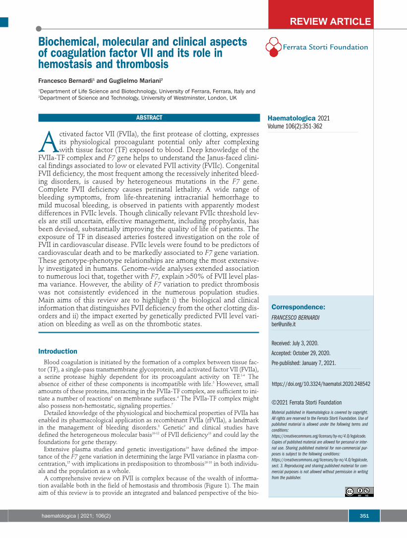
Activated factor VII (FVIIa), the first protease of clotting, expressesits physiological procoagulant potential only after complexingwith tissue factor (TF) exposed to blood. Deep knowledge of the
FVIIa-TF complex and F7 gene helps to understand the Janus-faced clini-cal findings associated to low or elevated FVII activity (FVIIc). CongenitalFVII deficiency, the most frequent among the recessively inherited bleed-ing disorders, is caused by heterogeneous mutations in the F7 gene.Complete FVII deficiency causes perinatal lethality. A wide range ofbleeding symptoms, from life-threatening intracranial hemorrhage tomild mucosal bleeding, is observed in patients with apparently modestdifferences in FVIIc levels. Though clinically relevant FVIIc threshold lev-els are still uncertain, effective management, including prophylaxis, hasbeen devised, substantially improving the quality of life of patients. Theexposure of TF in diseased arteries fostered investigation on the role ofFVII in cardiovascular disease. FVIIc levels were found to be predictors ofcardiovascular death and to be markedly associated to F7 gene variation.These genotype-phenotype relationships are among the most extensive-ly investigated in humans. Genome-wide analyses extended associationto numerous loci that, together with F7, explain >50% of FVII level plas-ma variance. However, the ability of F7 variation to predict thrombosiswas not consistently evidenced in the numerous population studies.Main aims of this review are to highlight i) the biological and clinicalinformation that distinguishes FVII deficiency from the other clotting dis-orders and ii) the impact exerted by genetically predicted FVII level vari-ation on bleeding as well as on the thrombotic states.
Biochemical, molecular and clinical aspectsof coagulation factor VII and its role in hemostasis and thrombosisFrancesco Bernardi1 and Guglielmo Mariani2
1Department of Life Science and Biotechnology, University of Ferrara, Ferrara, Italy and2Department of Science and Technology, University of Westminster, London, UK
ABSTRACT
IntroductionBlood coagulation is initiated by the formation of a complex between tissue fac-
tor (TF), a single-pass transmembrane glycoprotein, and activated factor VII (FVIIa),a serine protease highly dependent for its procoagulant activity on TF.1-4 Theabsence of either of these components is incompatible with life.5 However, smallamounts of these proteins, interacting in the FVIIa-TF complex, are sufficient to ini-tiate a number of reactions6 on membrane surfaces.4 The FVIIa-TF complex mightalso possess non-hemostatic, signaling properties.7
Detailed knowledge of the physiological and biochemical properties of FVIIa hasenabled its pharmacological application as recombinant FVIIa (rFVIIa), a landmarkin the management of bleeding disorders.8 Genetic9 and clinical studies havedefined the heterogeneous molecular basis10-12 of FVII deficiency13 and could lay thefoundations for gene therapy.Extensive plasma studies and genetic investigations14 have defined the impor-
tance of the F7 gene variation in determining the large FVII variance in plasma con-centration,15 with implications in predisposition to thrombosis16-18 in both individu-als and the population as a whole. A comprehensive review on FVII is complex because of the wealth of informa-
tion available both in the field of hemostasis and thrombosis (Figure 1). The mainaim of this review is to provide an integrated and balanced perspective of the bio-
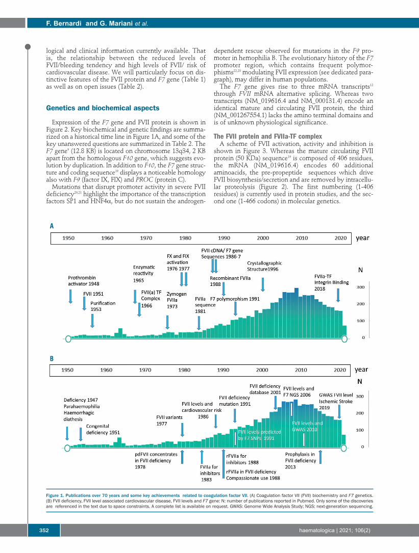
logical and clinical information currently available. Thatis, the relationship between the reduced levels ofFVII/bleeding tendency and high levels of FVII/ risk ofcardiovascular disease. We will particularly focus on dis-tinctive features of the FVII protein and F7 gene (Table 1)as well as on open issues (Table 2).
Genetics and biochemical aspects
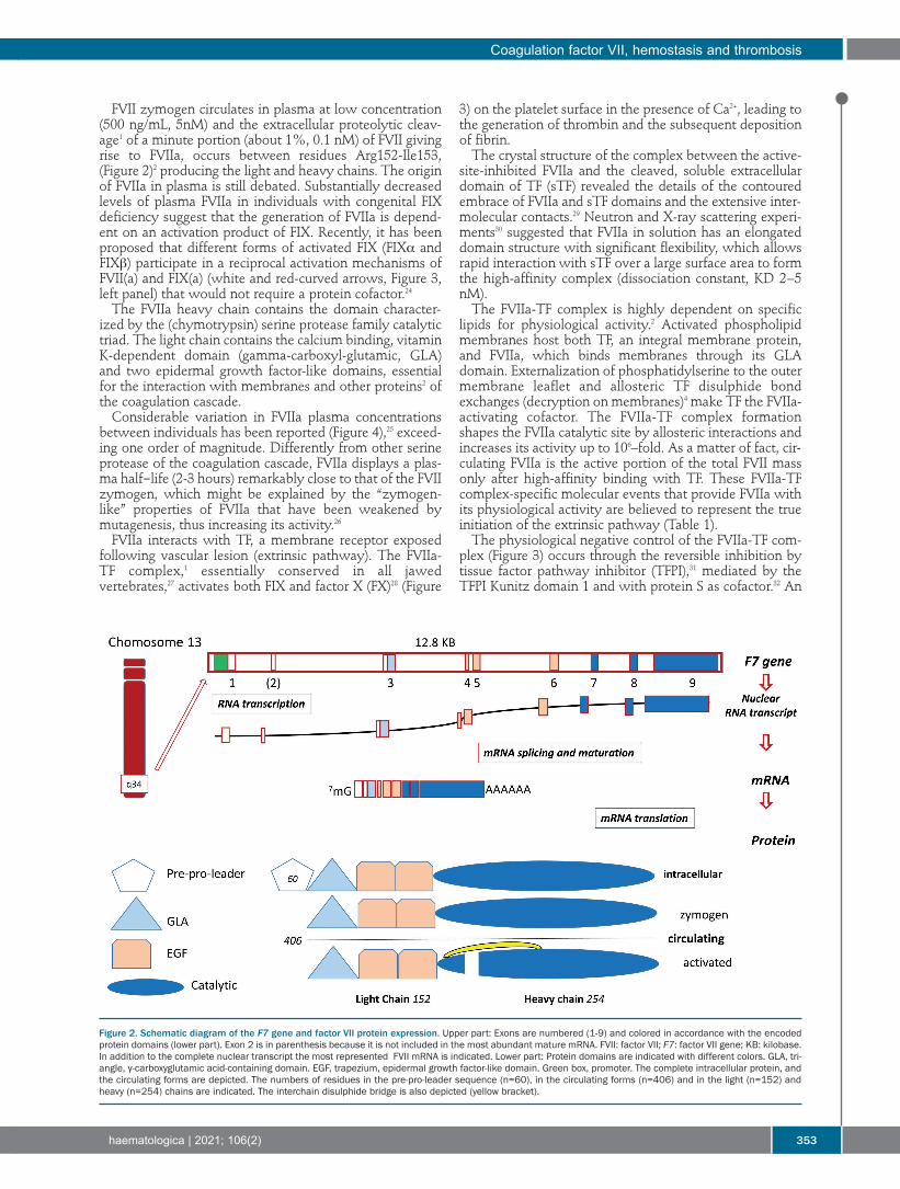
Expression of the F7 gene and FVII protein is shown inFigure 2. Key biochemical and genetic findings are summa-rized on a historical time line in Figure 1A, and some of thekey unanswered questions are summarized in Table 2. TheF7 gene9 (12.8 KB) is located on chromosome 13q34, 2 KBapart from the homologous F10 gene, which suggests evo-lution by duplication. In addition to F10, the F7 gene struc-ture and coding sequence19 displays a noticeable homologyalso with F9 (factor IX, FIX) and PROC (protein C). Mutations that disrupt promoter activity in severe FVII
deficiency20,21 highlight the importance of the transcriptionfactors SP1 and HNF4α, but do not sustain the androgen-
dependent rescue observed for mutations in the F9 pro-moter in hemophilia B. The evolutionary history of the F7promoter region, which contains frequent polymor-phisms22,23 modulating FVII expression (see dedicated para-graph), may differ in human populations. The F7 gene gives rise to three mRNA transcripts12
through FVII mRNA alternative splicing. Whereas twotranscripts (NM_019616.4 and NM_000131.4) encode anidentical mature and circulating FVII protein, the third(NM_001267554.1) lacks the amino terminal domains andis of unknown physiological significance.
The FVII protein and FVIIa-TF complex A scheme of FVII activation, activity and inhibition is
shown in Figure 3. Whereas the mature circulating FVIIprotein (50 KDa) sequence19 is composed of 406 residues,the mRNA (NM_019616.4) encodes 60 additionalaminoacids, the pre-propeptide sequences which driveFVII biosynthesis/secretion and are removed by intracellu-lar proteolysis (Figure 2). The first numbering (1-406residues) is currently used in protein studies, and the sec-ond one (1-466 codons) in molecular genetics.
F. Bernardi and G. Mariani et al.
352 haematologica | 2021; 106(2)
Figure 1. Publications over 70 years and some key achievements related to coagulation factor VII. (A) Coagulation factor VII (FVII) biochemistry and F7 genetics.(B) FVII deficiency, FVII level associated cardiovascular disease, FVII levels and F7 gene: N: number of publications reported in Pubmed. Only some of the discoveriesare referenced in the text due to space constraints. A complete list is available on request. GWAS: Genome Wide Analysis Study; NGS: next-generation sequencing.
A
B
FVII zymogen circulates in plasma at low concentration(500 ng/mL, 5nM) and the extracellular proteolytic cleav-age1 of a minute portion (about 1%, 0.1 nM) of FVII givingrise to FVIIa, occurs between residues Arg152-Ile153,(Figure 2)2 producing the light and heavy chains. The originof FVIIa in plasma is still debated. Substantially decreasedlevels of plasma FVIIa in individuals with congenital FIXdeficiency suggest that the generation of FVIIa is depend-ent on an activation product of FIX. Recently, it has beenproposed that different forms of activated FIX (FIXα andFIXb) participate in a reciprocal activation mechanisms ofFVII(a) and FIX(a) (white and red-curved arrows, Figure 3,left panel) that would not require a protein cofactor.24The FVIIa heavy chain contains the domain character-
ized by the (chymotrypsin) serine protease family catalytictriad. The light chain contains the calcium binding, vitaminK-dependent domain (gamma-carboxyl-glutamic, GLA)and two epidermal growth factor-like domains, essentialfor the interaction with membranes and other proteins2 ofthe coagulation cascade.Considerable variation in FVIIa plasma concentrations
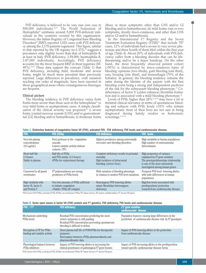
between individuals has been reported (Figure 4),25 exceed-ing one order of magnitude. Differently from other serineprotease of the coagulation cascade, FVIIa displays a plas-ma half-life (2-3 hours) remarkably close to that of the FVIIzymogen, which might be explained by the “zymogen-like” properties of FVIIa that have been weakened bymutagenesis, thus increasing its activity.26FVIIa interacts with TF, a membrane receptor exposed
following vascular lesion (extrinsic pathway). The FVIIa-TF complex,1 essentially conserved in all jawedvertebrates,27 activates both FIX and factor X (FX)28 (Figure
3) on the platelet surface in the presence of Ca2+, leading tothe generation of thrombin and the subsequent depositionof fibrin. The crystal structure of the complex between the active-
site-inhibited FVIIa and the cleaved, soluble extracellulardomain of TF (sTF) revealed the details of the contouredembrace of FVIIa and sTF domains and the extensive inter-molecular contacts.29 Neutron and X-ray scattering experi-ments30 suggested that FVIIa in solution has an elongateddomain structure with significant flexibility, which allowsrapid interaction with sTF over a large surface area to formthe high-affinity complex (dissociation constant, KD 2−5nM).The FVIIa-TF complex is highly dependent on specific
lipids for physiological activity.2 Activated phospholipidmembranes host both TF, an integral membrane protein,and FVIIa, which binds membranes through its GLAdomain. Externalization of phosphatidylserine to the outermembrane leaflet and allosteric TF disulphide bondexchanges (decryption on membranes)4 make TF the FVIIa-activating cofactor. The FVIIa-TF complex formationshapes the FVIIa catalytic site by allosteric interactions andincreases its activity up to 106–fold. As a matter of fact, cir-culating FVIIa is the active portion of the total FVII massonly after high-affinity binding with TF. These FVIIa-TFcomplex-specific molecular events that provide FVIIa withits physiological activity are believed to represent the trueinitiation of the extrinsic pathway (Table 1). The physiological negative control of the FVIIa-TF com-
plex (Figure 3) occurs through the reversible inhibition bytissue factor pathway inhibitor (TFPI),31 mediated by theTFPI Kunitz domain 1 and with protein S as cofactor.32 An
Coagulation factor VII, hemostasis and thrombosis
haematologica | 2021; 106(2) 353
Figure 2. Schematic diagram of the F7 gene and factor VII protein expression. Upper part: Exons are numbered (1-9) and colored in accordance with the encodedprotein domains (lower part). Exon 2 is in parenthesis because it is not included in the most abundant mature mRNA. FVII: factor VII; F7: factor VII gene; KB: kilobase.In addition to the complete nuclear transcript the most represented FVII mRNA is indicated. Lower part: Protein domains are indicated with different colors. GLA, tri-angle, γ-carboxyglutamic acid-containing domain. EGF, trapezium, epidermal growth factor-like domain. Green box, promoter. The complete intracellular protein, andthe circulating forms are depicted. The numbers of residues in the pre-pro-leader sequence (n=60), in the circulating forms (n=406) and in the light (n=152) andheavy (n=254) chains are indicated. The interchain disulphide bridge is also depicted (yellow bracket).
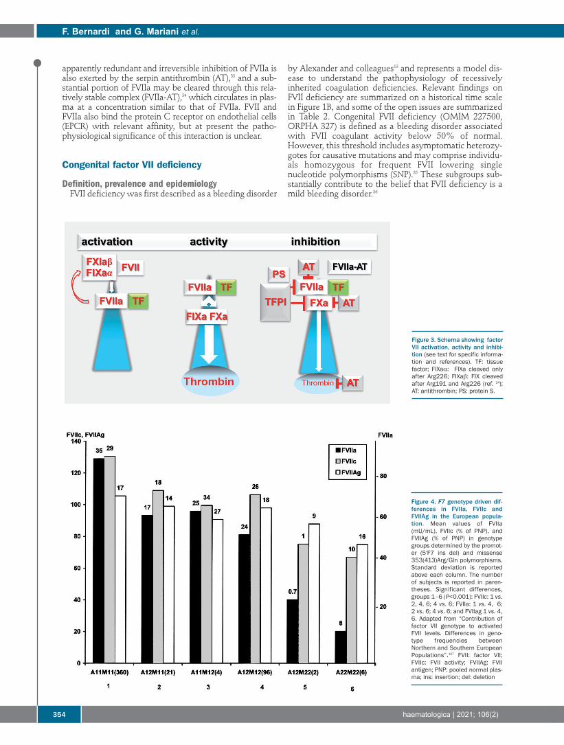
apparently redundant and irreversible inhibition of FVIIa isalso exerted by the serpin antithrombin (AT),33 and a sub-stantial portion of FVIIa may be cleared through this rela-tively stable complex (FVIIa-AT),34 which circulates in plas-ma at a concentration similar to that of FVIIa. FVII andFVIIa also bind the protein C receptor on endothelial cells(EPCR) with relevant affinity, but at present the patho-physiological significance of this interaction is unclear.
Congenital factor VII deficiency
Definition, prevalence and epidemiology FVII deficiency was first described as a bleeding disorder
by Alexander and colleagues13 and represents a model dis-ease to understand the pathophysiology of recessivelyinherited coagulation deficiencies. Relevant findings onFVII deficiency are summarized on a historical time scalein Figure 1B, and some of the open issues are summarizedin Table 2. Congenital FVII deficiency (OMIM 227500,ORPHA 327) is defined as a bleeding disorder associatedwith FVII coagulant activity below 50% of normal.However, this threshold includes asymptomatic heterozy-gotes for causative mutations and may comprise individu-als homozygous for frequent FVII lowering singlenucleotide polymorphisms (SNP).35 These subgroups sub-stantially contribute to the belief that FVII deficiency is amild bleeding disorder.36
F. Bernardi and G. Mariani et al.
354 haematologica | 2021; 106(2)
Figure 4. F7 genotype driven dif-ferences in FVIIa, FVIIc andFVIIAg in the European popula-tion. Mean values of FVIIa(mU/mL), FVIIc (% of PNP), andFVIIAg (% of PNP) in genotypegroups determined by the promot-er (5′F7 ins del) and missense353(413)Arg/Gln polymorphisms.Standard deviation is reportedabove each column. The numberof subjects is reported in paren-theses. Significant differences,groups 1–6 (P<0.001): FVIIc: 1 vs.2, 4, 6; 4 vs. 6; FVIIa: 1 vs. 4, 6;2 vs. 6; 4 vs. 6; and FVIIag 1 vs. 4,6. Adapted from “Contribution offactor VII genotype to activatedFVII levels. Differences in geno-type frequencies betweenNorthern and Southern EuropeanPopulations”.107 FVII: factor VII;FVIIc: FVII activity; FVIIAg: FVIIantigen; PNP: pooled normal plas-ma; ins: insertion; del: deletion
Figure 3. Schema showing factorVII activation, activity and inhibi-tion (see text for specific informa-tion and references). TF: tissuefactor; FIXaα: FIXa cleaved onlyafter Arg226; FIXab: FIX cleavedafter Arg191 and Arg226 (ref. 24);AT: antithrombin; PS: protein S.
FVII deficiency is believed to be very rare (one case in500,000 individuals).36,37 The World Federation ofHemophilia38 estimates around 5,000 FVII-deficient indi-viduals in the countries covered by this organization.However, the Italian Registry of Congenital Rare BleedingDisorders39 (RBD) reported 1,023 cases with FVII deficien-cy among the 2,178 patients registered. This figure, similarto that reported by the UK registry (n=1.373),38 suggests aprevalence only slightly lower than that of hemophilia B,at least in Italy (FVII deficiency, 1:59,000; haemophilia B,1:67,000 individuals). Accordingly, FVII deficiencyaccounts for the most frequent RBD in most registries (36-49%).37,39 These data support the concept (Table 1) thatFVII deficiency, including mild, moderate and severeforms, might be much more prevalent than previouslyreported. Large differences in prevalence, with variationreaching one order of magnitude, have been reported inthose geographical areas where consanguineous marriagesare frequent.
Clinical pictureThe bleeding tendency in FVII deficiency varies from
forms more severe than those seen in the hemophilias40 tovery mild forms or asymptomatic cases. A simple classifi-cation of the clinical severity was proposed:10 i) severeforms (central nervous system [CNS] and/or gastrointesti-nal [GI] bleeding and/or hemarthrosis; ii) moderate forms
(three or more symptoms other than CNS and/or GIbleeding and/or hemarthrosis); iii) mild forms (one or twosymptoms, mostly muco-cutaneous, and other than CNSand/or GI and/or hemarthrosis).In the International F7 Registry and the Seven
Treatment Evaluation Registry (STER)41 that include 787cases, 12% of individuals had a severe to very severe phe-notype and three fourth of them bled within the first yearof age (Table 4). About 20% of individuals with FVII defi-ciency suffer from a bleeding disorder that can be life-threatening and/or be a major handicap. On the otherhand, the most frequently observed patient cohort(>50%) is characterized by muco-cutaneous types ofbleeding: epistaxis (two thirds), gum bleeding (one third),easy bruising (one third), and menorrhagia (70% of thefemales). In general, the bleeding tendency remains thesame during the lifetime of the patient with the firstbleeding event being a strong and independent predictorof the risk for the subsequent bleeding phenotype.41 Co-inheritance of factor V Leiden enhances thrombin forma-tion and is associated with a mild bleeding phenotype.42Levels of FVIIc higher than 26%11,41,43 may have a nil or
minimal clinical relevance in terms of spontaneous bleed-ing and subjects with FVIIc levels <10% who remainasymptomatic most of their lives are not rare in beingdiagnosed during family studies or hemostaticscreenings.10,11,35
Coagulation factor VII, hemostasis and thrombosis
haematologica | 2021; 106(2) 355
Table 1. Distinctive features of coagulation factor VII (FVII), activated FVII , FVII deficiency, FVII levels and cardiovascular disease.FVII FVII (FVIIa) FVII deficiency FVII levels
Very low plasma First protease in the coagulation Highest prevalence among autosomally High variability in human populationsconcentrations cascade. recessive rare bleeding disorders High number of environmental (0.5 mg/mL) Lowest catalytic activity without determinants cofactor (TF).Short half-life Half-life of FVIIa Complete deficiency results in perinatal High proportion of variance2-4 hours and FVII similar (2-3 hours) mortality explained by F7 gene variationStable in plasma rFVIIa for replacement therapy High incidence of intracranial The genotype/phenotype relationship bleeding (severe form) is one of the most extensively investigated among human genesConserved in all jawed F7 polymorphisms are strong Wide variation of bleeding phenotype Frequent FVII level- lowering alleles vertebrates predictors of FVIIa levels in relation to modest FVII level variations with wide differences in human populationsHigh similarity with Very low amounts of FVIIa sufficient Homozygous FVII-lowering alleles High/low levels associated withfactor IX, factor X to initiate coagulation mimic Mendelian heterozygous predisposition/ protectionand Protein C «Stable» FVIIa-AT complex deficiency towards/from cardiovascular diseaseFVII: factor VII; FVIIa: activated FVII; rFVIIa: recombinant FVIIa; TF: tissue factor; AT: serpin antithrombin; F7: factor VII gene.
Table 2. Some open issues in factor VII (FVII) protein and F7 genetics, FVII deficiency, FVII levels and cardiovascular disease.FVII / F7 FVII deficiency F7 gene variation cardiovascular disease
Mechanisms underlying Residual FVII concentration predicting the most Population features causing large differences in the FVIIa levels severe symptoms is still puzzling. prediction of cardiovascular disease risk by F7 genotypes Residual FVII concentration preventing spontaneous bleeding is difficult to define.Decryption of TF for FVIIa Short plasma half life of FVII/rFVIIa for therapeutic Impact of FVII-lowering alleles in the protection binding and catalytic activity purposes. from cardiovascular disease Discrepancy between rFVIIa pharmacokinetic and pharmacodynamic dataPhysiological balance between Impact of FVII-lowering alleles in increasing the Impact of FVII-increasing alleles in the predisposition FVIIa inhibitors clinical penetrance of pathological F7 gene lesions toward specific cardiovascular disease formsFVII: factor VII; FVIIa: activated FVII; rFVIIa: recombinant FVIIa; TF: tissue factor; F7: factor VII gene.
However, the therapeutic thresholds conferring protec-tion from traumatic or post-operative bleeds are still amatter of debate.44,45 Surgical bleeding has been reportedin 15-24% of cases42,45 with FVIIc <7%.42 Bleeds in the firstpost-operative day46 are particularly frequent if the bleed-ing disorder was not diagnosed before, and are mostlyreported during orthopedic procedures,46 a major hemo-static challenge. Presenting symptoms as post-circumci-sion bleeds and hemorrhage from the umbilical stumpfrequently herald a severe disease form.
DiagnosisThe spectrum of bleeding symptoms is wide, involves
both males and females, can start after birth and mayoccur at any age. The most important predictors of bleed-ing risk, familial and personal histories, can be silent asobserved in diseases with recessive inheritance. The rele-vance of the gynecological and obstetric histories are dis-cussed in a dedicated paragraph.The laboratory workup47 performed after a bleeding
episode or during a family screening includes routinescreening assays (prothrombin time [PT], activated partialthromboplastin time [aPTT]), followed by FVIIc measure-ment, which is necessary to confirm the diagnosis.Evaluation of FVIIc is determined with a one-stage assayadding diluted plasma samples to FVII-deficient plasmaand thromboplastin as the source of TF. The accuracy ofFVIIc assays is related to the sensitivity of the thrombo-plastin reagent and to the quality of the FVII-deficientplasma and of the calibrators. The most suitable activa-tors are the human placenta-derived and the recombinantpreparations,47 that are also recommended to monitor thetreatment with rFVIIa.25 New chromogenic or fluorogenicassays for FVIIa are available for research purposes and tomonitor treatment with rFVIIa.48The sensitivity of the FVIIc assay to levels below 2%
may be poor, with negative implications for the accuratedefinition of the relationship between FVII levels andbleeding phenotypes (Table 1), as well as between geno-type and phenotype. A low assay sensitivity may explainsome inconsistencies such as the paradoxical observationof asymptomatic cases with low FVII levels. Furthermore,severe bleeding is sporadically observed in individualswith moderately reduced levels of FVII. The combinationof the low assay sensitivity with the minimal amount ofFVIIa-TF complex needed to trigger coagulation does notpermit one to efficiently differentiate clinical phenotypes.As a consequence, in FVII deficiency a variety of bleedingphenotypes are observed in the presence of apparentlymodest differences in FVIIc levels (Table 1). In summary,the clotting tests do not always predict the degree ofbleeding tendency in the single individual.The presence of the FVII protein (FVII antigen [FVIIag])
can be determined by enzyme-linked immunosorbentassays or immune-turbidimetric assays, using monoclon-al epitope-specific antibodies.47 The FVIIag assay47 doesnot predict the bleeding tendency, but does allow one todistinguish between type I (quantitative defects, withdecrease of both FVIIc and FVIIAg), and type II defects(qualitative defects, with low FVIIc and normal orreduced levels of FVIIAg), and may help to clarify the dif-ferent mutational mechanisms.
Molecular genetics The key findings are summarized on a historical time
scale in Figure 1B, and the main findings in individualswith FVII deficiency are shown in Table 3. The function-al characterization of protein variants in plasma was car-ried out in seminal studies49 well before the genetic char-acterization of FVII deficiencies. The first F7 gene muta-tions in FVII deficiency were reported in the UK50 andItalian populations, and more than 1,000 genetic diag-noses in several countries are reported in theInternational Registry on FVII deficiency (IRF7)10 and theGreifswald Registry.11 Mutations and residual FVII levelshave been presented in databases.51 The recent EHADdatabase12 (http://www.umd.be/F7/W_F7/index.html)includes 221 unique variants identified in 728 individu-als, of great help for the evaluation of F7 mutationsfound during genetic counseling and diagnosis, includingprenatal diagnosis.5F7mutations have been detected by sequencing coding,
splicing and in promoter regions in the vast majority ofpatients. High throughput genomic sequencing will helpin the case no mutations are found.52 Cases with severesymptoms are virtually all homozygous or doubly het-erozygous for F7 mutations. Symptomatic mutationsimpairing vitamin K metabolism, and thus posttransla-tional modification of GLA residues, have been found tocause combined vitamin K-dependent clotting factor defi-ciency that includes an abnormal FVII biosynthesis andlow FVIIc levels. As reported in registries11 and databases,12 single
nucleotide variants are the most common type of genedefect (around 90%), and include a large proportion(between two thirds and three quarters) of missensemutations. Numerous missense mutations have been found in the
homozygous condition, related to geographical, ethnicand mutational pattern features. Clusters of homozygotesoften indicate genetic isolates or ethnic customs favoringconsanguinity (see below). Since they have been well presented in databases,12 we
shall briefly comment on a few of them. The severe Gln100(160)Arg53,54 and the moderate/mild
Ala294(354)Val55,56 are prevalent in Northern Europe. Theframeshift mutation in the last carboxyl terminal codons(Pro404(464)Hfs) is very prevalent in cis with theAla294(354)Val missense change and causes a 28 residues-elongated FVII molecule. This moderate to severe andcombined variant is frequent in Central Europe and espe-cially in Slovakia.10 The mild Gly331(391)Ser mutation isprevalent in the gulf of Naples, Italy, as well as in othercountries.57 Other mild/asymptomatic changes, theArg304(364)Gln50, Cys310(370)Phe/Ser andThr359(419)Met substitutions, have been found in severalpopulations, suggesting recurrent nucleotide changes at aCpG site. The frequent Arg304(364)Gln is also character-ized by discrepant FVII activity findings depending on thethromboplastin reagent.49Splicing mutations are not rare,52 and the homozygous
splicing mutation IVS7+5 g>a, relatively frequent in Italy,has been used for cellular58,59 and animal60 models of genetherapy. F7 deletions are also not rare and together with duplica-
tions, insertions and indel rearrangements represent about10% of all genetic lesions.12 However, large and extendeddeletions have not been described in homozygous individ-uals. Differently from hemophilia A and B, individualswith complete FVII deficiency might die perinatally or
F. Bernardi and G. Mariani et al.
356 haematologica | 2021; 106(2)
shortly after birth due to severe hemorrhage.5 CompleteFVII deficiency causes perinatal mortality in “knockout”mice, in which the presence of a trace amount of FVII(0.7%) seems sufficient for survival with symptoms.61Several cases with heterozygous combined FVII and FXdeficiency, due to large deletions within the terminal endof chromosome 13, have been reported.62Recombinant expression after site-directed mutagenesis
has been performed for a number of mutations detected inpatients, which permits one to investigate the molecularbases of the deficiency. Recombinant studies of the rareand potentially null homozygous nonsense mutationssupport the notion that gain-of-function and translationalreadthrough over premature stop codons may preventtruly null conditions and may contribute to the unexpect-edly variable bleeding phenotype in individuals homozy-gous for F7 nonsense changes.63 Whereas life-threateningsymptoms have been associated with the p.Ser52(112)X,moderate bleeding was observed in p.Cys82 (132)Xhomozygotes. The Arg402(462)X change permits thesecretion of a small amount of protein with gain-of-func-tion features, causing a mild phenotype paradoxicallyassociated with a “null” mutation.64Chimeric fluorescent FVII molecules have also been
constructed to investigate intracellular processing of vari-ants, and chemical chaperones have been used to stimu-
late their secretion.54 The FVII protein has been purifiedand biochemically characterized for very few variants,among these the frequent Arg304(364)Gln50 andAla294(354)Val55 changes, as well as the severeGln160Arg33 and the activation sequence Val154(214)Gly65
changes. These laborious experiments provided strongsupport for the understanding of the mild or severe coag-ulation phenotypes in vivo and for FVII altered expressionand properties.The homozygous conditions for polymorphisms pre-
dicting lower FVII levels might not be pathogenic butmimic Mendelian heterozygous FVII deficiency35 (Table 1),which frequently leads to the request for clotting defectdiagnosis in pre-surgical screenings, and generates confu-sion in estimating the incidence of inherited FVII deficien-cy (see dedicated section). In light of their effects on low-ering FVII plasma levels, these variants might contribute topathogenicity of co-inherited variants.52 As a matter of factthe frequencies of the FVII-lowering alleles were signifi-cantly higher51 in the FVII-deficient individuals tested thanin controls, suggesting that the presence of these allelesmay have increased, through increased clinical signifi-cance (Table 2), the likelihood of identifying “causative”F7 gene lesions. Genotyping of these frequent variantswould be especially useful for the interpretation of mildand moderate FVII deficiency.
Coagulation factor VII, hemostasis and thrombosis
haematologica | 2021; 106(2) 357
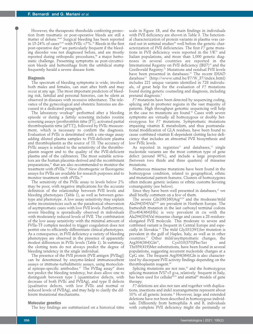
Table 3. Overview of molecular genetics findings in factor VII deficiency.Molecular genetics of FVII deficiency
Genetic diagnosis Sanger sequencing of exons and splicing junctions in >1,000 individuals with FVII deficiency Next-generation sequencing in a few individuals Phenotypic assays do not define specific genetic defects* No highly frequent gene lesions Large heterogeneity of genetic causes (n>300 different mutations) Geographical/ethnic clustering of identical-by-descent mutations (n>15) Several recurrent mutations in different populationsMutation zygosity and Severe deficiencies caused by compound heterozygous/homozygous mutationsdisease severity Increased prevalence of homozygotes in genetic isolates/consanguineous marriages Otherwise asymptomatic heterozygous variants associated with frequent FVII-lowering single nucleotide polymorphism in mild deficienciesMutation type frequency Missense>>Small deletions>Splicing>Nonsense>Large deletions (not reported in homozygosity)Recombinant expression Numerous variants expressed after site directed mutagenesis A few purified protein variants Translational read through over premature termination codons Gain-of-function premature termination codon*The asymptomatic Arg364Gln is detectable by using different thromboplastins59. Small deletions include also insertions and indels. FVII: factor VII.
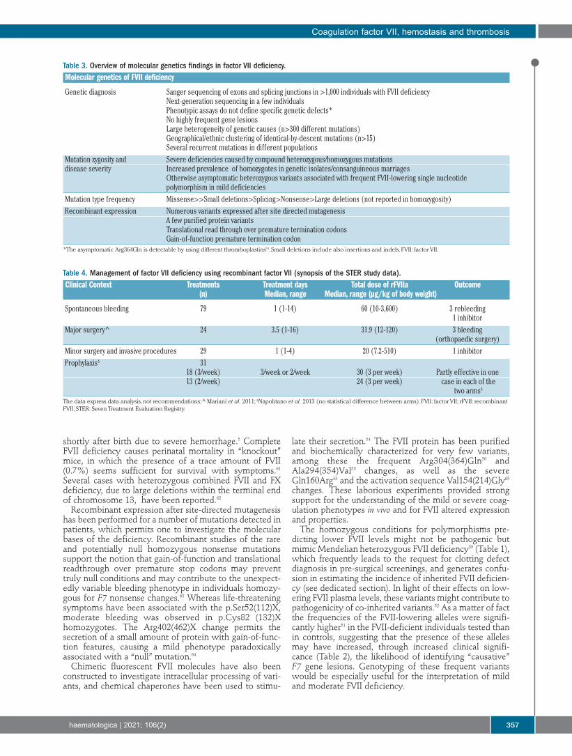
Table 4. Management of factor VII deficiency using recombinant factor VII (synopsis of the STER study data).Clinical Context Treatments Treatment days Total dose of rFVIIa Outcome (n) Median, range Median, range (µg/kg of body weight)
Spontaneous bleeding 79 1 (1-14) 60 (10-3,600) 3 rebleeding 1 inhibitorMajor surgery^ 24 3.5 (1-16) 31.9 (12-120) 3 bleeding (orthopaedic surgery)Minor surgery and invasive procedures 29 1 (1-4) 20 (7.2-510) 1 inhibitorProphylaxis§ 31 18 (3/week) 3/week or 2/week 30 (3 per week) Partly effective in one 13 (2/week) 24 (3 per week) case in each of the two arms§
The data express data analysis, not recommendations; ^ Mariani et al. 2011; §Napolitano et al. 2013 (no statistical difference between arms). FVII: factor VII; rFVII: recombinantFVII; STER: Seven Treatment Evaluation Registry.
Replacement therapy and prophylaxis Replacement therapy (RT) options are determined by:
the rarity of the disorder; the availability and supply ofproducts and the economic and geographical factors.These include: i) recombinant FVIIa (rFVIIa), ii) plasma-derived FVII (pdFVII), iii) fresh frozen plasma (FFP) andprothrombin complex concentrates (PCC). In the STERprospective trial (comprising 312 RT) most therapieswere carried out with rFVIIa (78%), the remaining withFFP (10%), pdFVII concentrates (10%%) and PCC (2%). Based on the modest catalytic activity of FVIIa in the
absence of TF, the therapeutic/prophylactic use of FVIIaand rFVIIa (initially proposed for bleeding diathesis otherthan FVII deficiency) represents a therapeutic milestone8
and distinguishes replacement therapy in FVII deficiencyfrom that in the other coagulation defects (Table 1).Although in FVII deficiency rFVIIa is employed (Figure1A) at doses (Table 3) much lower than those needed forpatients with FVIII inhibitors, supraphysiological FVIIaconcentrations are also produced in plasma of FVII defi-cient subjects. It is still a matter of debate whether rFVIIa acts
through the binding with TF, provided by microparticlesshed into the circulation following diverse stimuli, orbinds at high concentration66 to anionic phospholipidsexposed on activated platelets, thus directly activatingFX to FXa. FXa would in turn generate thrombin, bypass-ing the tenase complex. It is tempting to speculate thatthe physiological FVIIa-TF function (Figure 3) prevails atthe rFVIIa doses used in FVII deficiency and in the pres-ence of normal FVIII levels. rFVIIa easily diffuses into theextravascular spaces where it could be retained forextended time periods. As supported by pharmacokinet-ic studies,67 rFVIIa prolongs its pharmacological effects atlow concentration, in accordance with the hypothesis ofphysiological binding to TF. rFVIIa has a very good safety-to-efficacy ratio.46 One-
day therapy with ‘intermediate’ doses of rFVIIa can beeffective and safe for the treatment of most of sponta-neous bleeds as well as for minor surgery and invasiveprocedures (Table 3). Replacement with rFVIIa is alsoeffective to prevent bleeding in major surgical proce-dures.46The large volume of the infusions and the limited
availability make pd-FVII concentrates less appealing.Currently, the average pd-FVII dosages used are 15–20IU/kg for mucosal bleeding, and 30–40 IU/kg for severeor life-threatening hemorrhages. Although FFP is easily available in developing coun-
tries, its effectiveness is limited owing to the high risk offluid overload and the consequent need for slow infu-sions. In the case of mild mucosal bleeds (epistaxis, mild
menorrhagia) tranexamic acid and hormones are current-ly considered. Anti-fibrinolytic agents are contraindicated in hema-
turia, and may trigger thrombosis in association to PCC. Prophylaxis is warranted for patients with the most
severe bleeding picture and should be prescribed fromchildhood or soon after the first bleeding event.45,68Effective prophylaxis schedules have been reported forrFVIIa68,69 and pdFVII concentrates.70
Treatment complications Inhibitors to FVII are a rare (1-2%)71 complication that
occurs mainly in severely deficient patients, particularly inchildren younger than 1 year on prophylaxis. We observedonly high responders (>5 BU Bethesda Unit)71, and in thepresence of a high-titer inhibitor to FVII treatmentbecomes a problem.71At variance with the homologous deficiency of FIX (F9,
HB), complete homozygous gene deletions that predis-pose to FIX inhibitors have never been detected in FVIIdeficiency (Table 1),5,12 and FVII inhibitors have never beenshown to be complicated by allergic reactions. Paradoxically, a dozen of cases with FVII deficiency and
thrombosis have been reported. Surgical interventionsand/or replacement therapies had a close temporal rela-tionship with thrombotic episodes, but apparently spon-taneous events were also reported.68,72 Different replace-ment therapies were associated with the thromboticevents: PCC (three cases), rFVIIa (three cases), pdFVII (twocases), FFP (one case), no replacement (three cases).72 Thissuggests that FVII deficiency does not seem to offer pro-tection from strong thrombosis risk factors such as sur-gery/high dose radiotherapy.
Women with factor VII deficiency Autosomally transmitted RBD occur as frequently in
women as in men, but women may experience morebleeding than men because gynecological and obstetricchallenges to hemostasis add an important burden to thebackground bleeding related to the hemostatic defect.Menstruation and ovulation are associated with anincreased risk of bleeding, as are pregnancy and delivery.73While in normal and heterozygous women FVII plasmalevels rise during pregnancy, no such increase is observedin women with a severe homozygous deficiency. As aconsequence, symptomatic women with FVII deficiencymay continue to bleed during pregnancy and postpartum.In a large study (234 women with FVII deficiency),74 men-orrhagia during the reproductive age occurred in half ofthe cases, and in 12% represented the first bleed. Further,frequent gynecological problems, such as uterine fibroids,were diagnosed earlier because of the hemostatic defect.Although FVIIc was an important predictor of gynecolog-ical bleeding, other determinants including endocrinepathologies may also play a role.For severe menorrhagia, management has been moved
from hemostatic agents like tranexamic acid, oral contra-ceptives and intra-uterine devices, to replacement therapyand prophylaxis with effective single- or multiple-doseschedules.68,73,74 This will hopefully change clinical practicethat, until recently, included surgical approaches such asendometrial ablation or hysterectomy.
Therapeutics in development The first attempts to improve rFVIIa activity through
mutagenesis, or its half-life in plasma by glycoPEGylation,have increased FVII antigenicity. Longer-acting FVIIa hasbeen tested in different recombinant preparations, whichare currently in clinical trials (reviewed in Menegatti etal.)75: i) addition of a c-terminal peptide76 potentially suit-able for subcutaneous administration that led to pro-longed pharmacodynamics effects, ii) an Fc receptor-fusedrFVIIa that displayed a 5-fold longer plasma half-life andiii) an albumin-fused rFVIIa molecules that showedimproved features compared to rFVIIa (2- to 3-fold longerhalf-life and 4- to 8-fold lower clearance) in patients withcongenital FVII deficiency.77 Recently, an engineered albu-
F. Bernardi and G. Mariani et al.
358 haematologica | 2021; 106(2)
min-fused rFVIIa molecule showed enhanced transcellulartransport upon intranasal delivery and extended plasmahalf-life in transgenic mice.78The development of non-substitutive therapy for the
hemophilias, such as the use of anti-antithrombin RNAinterference or of aptamer and monoclonal antibodiesdirected against TFPI, have raised expectations forimprovement of the quality of life in individuals with FVIIdeficiency.75Based on adeno-associated viral-mediated expression of
FVII79, gene therapy has produced sustained correction ofsevere FVII deficiency in dogs. In addition, gene therapyusing FVIIa, inserted into the same viral vector, could alsobe used to treat congenital FVII deficiency with lower vec-tor doses, which increases the chance of efficient and longterm expression of the transgene. Small engineered RNAhave also been used as an approach of personalized genetherapy to correct a human FVII splicing mutation inmice.59
Factor VII levels, F7 genotypes and cardiovascu-lar disease
Several findings have created interest and still lead to anintense investigation on the role of FVII in cardiovasculardisease (Figure 1B). Exposure of TF to blood in (coronary)artery disease, and especially plaque rupture, may favorFVIIa-TF complex formation. Further, lipids are importantcomponents of atherosclerosis and determinants of FVIIactivation and activity as well.79,80 Additional linksbetween high FVII levels and risk for thrombosis havebeen suggested81 by finding that the TF-FVIIa-Xa complexactivates FVIII before coagulation amplification and thatheparanase increases generation of FXa by the FVIIa-TF-complex.82 Furthermore, the FVIIa-TF complex is believedto mediate non-hemostatic functions in diverse biologicalprocesses, such as angiogenesis, inflammation, atheroscle-rosis and vascular and cardiac remodeling, by activatingsignaling pathways (reviewed in D'Alessandro et al.)84through FVIIa-integrin binding and PAR2 cleavage.85The F7 promoter may respond to a number of metabolic
components, and FVIIc levels are associated with severalenvironmental factors linked to atherosclerosis i.e., bodymass index, dietary fat intake, plasma lipids and particu-larly triglyceride concentration. Age- and sex-related variations in FVII levels add com-
plexity to the investigation of clinical correlates. FVII lev-els were found to increase with age and to be significantlylower in women than men at younger ages.Postmenopausal women displayed the highest levels,except when undergoing hormone replacement therapy.86This conundrum of environmental factors interacts withthe F7 gene variation.
Factor VII levels and F7/genome wide genotypes FVII levels show ample variation in the normal popula-
tion and have a substantial heritable component, also inrelation to polymorphisms15 (Figure 4). Missense,87 repeatnumber variation,58 insertion/deletion22 and SNP23 werefound to be associated with FVIIc, FVIIa and FVIIag levels(Figure 4) through epigenetic, transcription, biosynthesis-or stability-mediated mechanisms. Whereas twin studies88estimated about 60% of genetic-associated level of vari-ance in plasma, the F7 locus variation accounted for up to
40% of variance. Genotype effects, perhaps stronger onFVIIa than on FVIIag (Figure 4),15 might modulate theresponse to environmental stimuli and the sex-dependentregulation.89 Serum phospholipids were found to be strongand F7 genotype-associated FVIIa determinants.90The number of F7 single SNP was substantially
increased by highthroughput F7 gene sequencing, whichenabled very informative F7 population studies.91Genome-wide association studies (GWAS) confirmed14,92
the strong association between FVII levels and F7 genevariation.93 Importantly, GWAS have detected a numberof genomic regions associated with FVII levels (GCKR,ADH4, MS4A6A, PROCR, APOA5, HNF4A, REEP3-JMJD1C, JAZF1-AS1, MLXIPL and XXYLT1),14,92 that couldexplain an additional one fifth of the FVII variance in plas-ma levels and are also, in part, associated with lipidswhich in turn modulate FVII activity. These gene-basedassociation scan initiatives have been established in sever-al populations.94 Overall, this picture defines one of themost extensively investigated relationship between geno-types and multiple quantitative phenotypes (FVIIc, FVIIagand FVIIa) (Table 1).
Levels, genotypes and cardiovascular disease The Northwick Park Heart Study investigators were the
first to report that high FVII levels were predictors ofdeath due to coronary disease,16 and a number of studiesconfirmed this observation in different populations byalso evaluating FVIIa levels.95Recent investigations on the level of the FVIIa-AT com-
plex, which may reflect levels of FVIIa as well its interac-tion with TF, indicated that higher complex concentra-tions were associated with increased mortality in theCardiovascular Health Study.96 In patients with stablecoronary artery disease (Verona Heart Study) higher FVIIa-AT complex levels were associated with increased cardio-vascular mortality and increased thrombin and FXa gener-ation,97 particularly in the coagulation initiation phase.98However, in small groups of patients with unstable angi-na, acute myocardial infarction99 or post-infarction80 FVIIalevels were not higher than in controls. High plasmaFVIIag levels were associated with failure of thrombolytictherapy in patients with myocardial infarction.100 Elevatedlevels of FVII have not been consistently associated withvenous thromboembolism.101The influence of F7 genotypes on the hemostatic bal-
ance and on the susceptibility to cardiovascular diseasehas been extensively investigated in several large cohortsof patients in relation to both myocardial infarction andstroke. F7 polymorphisms with an opposite effect onFVIIa levels may positively or negatively modulate the riskof MI in males with advanced coronary artery disease,18and some FVII genotypes may protect against myocardialinfarction17 by affecting transcription levels and reducingprotein functional activity. The modulation of stroke riskin atrial fibrillation by F7 genotypes may follow a similarscheme102 and recently, in a meta-analysis of severalGWAS14 variations in FVII-related genes and FVIIc levelswere associated with a risk of the incidence of ischemicstroke in the general population. The physiological effectsof FVII lowering alleles may represent a natural model foranticoagulation; in fact F7 polymorphisms were shown toplay a role in determining the initial response to war-farin103 and influencing the risk of thrombosis in patientswith essential thrombocythemia104.
Coagulation factor VII, hemostasis and thrombosis
haematologica | 2021; 106(2) 359
Large haplotype studies confirmed that the F7 genestrongly influences FVII levels, but associations with coro-nary artery disease and FVII level were inconsistent.105Variable genetic, environmental and atherogenic risk fac-tors in different populations may explain this discrepancy(Table 2). The frequencies of F7 genotypes predictinglower FVII levels are higher in Caucasians and much lowerin Chinese and Malays populations. On the other hand,the distribution of these F7 genotypes covaries across pop-ulations in Europe with the rate of myocardial infarctionmortality, being higher in countries at low risk.106The most recent meta-analysis of large studies14 appears
to be consistent with positive and potentially clinicallyimportant causal effects of FVIIc levels, both on coronaryartery disease and venous thromboembolism. However,several open issues remain (Table 2) and the usefulness ofFVIIc and FVIIa evaluation in patients with cardiovascularrisks is still unclear.
Conclusive remarksIn summary, in the 70 years since FVII investigation
started, research has been particularly intense (Figure 1),and provided excellent examples at the basic science and
translational levels. The multidisciplinary approacheshave spanned structural biology, biochemistry, recombi-nant protein biotechnology, molecular and populationgenetics, pharmacology, clinics and epidemiology, andencompassed various areas of hematology, hemostasisand thrombosis. The large number of studies on FVII have not only
served to substantially improve our knowledge on FVIIbiology and related phenotypes and to improve the quali-ty of life of the individuals with FVII deficiency, but alsoto train three generations of scientists in different fields.
DisclosuresPfizer Research Grant
ContributionsFB and GM wrote the Manuscript
Acknowledgments The authors express their gratitude to Prof. Peter Lydyard
(emeritus at UCL London, UK and associate at GeorgiaUniversity, Tbilisi Georgia) for his very helpful text revision andDr Barbara Lunghi for her help in the selection of references.
F. Bernardi and G. Mariani et al.
360 haematologica | 2021; 106(2)
References
1.Nemerson Y, Esnouf MP. Activation of aproteolytic system by a membrane lipopro-tein: mechanism of action of tissue factor.Proc Natl Acad Sci U S A. 1973;70(2):310-314.
2.Gajsiewicz JM, Morrissey JH. Structure-function relationship of the interactionbetween tissue factor and factor VIIa.Semin Thromb Hemost. 2015;41(7):682-690.
3.McVey JH. The role of the tissue factorpathway in haemostasis and beyond. CurrOpin Hematol. 2016;23(5):453-461.
4. Ansari SA, Pendurthi UR, Rao LVM. Role ofcell surface lipids and thiol-disulphideexchange pathways in regulating theencryption and decryption of Tissue Factor.Thromb Haemost. 2019;119(6):860-870.
5.McVey JH, Boswell EJ, Takamiya O, et al.Exclusion of the first EGF domain of factorVII by a splice site mutation causes lethalfactor VII deficiency. Blood 1998;92(3):920-926.
6. Lawson JH, Kalafatis M, Stram S, MannKG. A model for the tissue factor pathwayto thrombin. I. An empirical study. BiolChem. 1994;269(37):23357-23366.
7. Zelaya H, Rothmeier AS and Ruf W. Tissuefactor at the crossroad of coagulation andcell signaling. J Thromb Haemost.2018;16(10):1941-1952.
8.Hedner U, Glazer S, Pingel K, et al.Successful use of recombinant factor VIIa ina patient with severe hemophilia A duringsynovectomy. Lancet. 1988;2(8621):1193.
9.O'Hara PJ, Grant FJ, Haldeman BA, et al.Nucleotide sequence of the gene coding forhuman factor VII, a vitamin K-dependentprotein participating in blood coagulation.Proc Natl Acad Sci U S A. 1987;84(15):5158-5162.
10.Mariani G, Herrmann FH, Dolce A, et al.International Factor VII Deficiency StudyGroup. Clinical phenotypes and factor VIIgenotype in congenital factor VII deficien-cy. Thromb Haemost. 2005;93(3):481-487.
11.Herrmann FH, Wulff K, Auerswald G, et al.Greifswald Factor FVII Deficiency StudyGroup. Factor VII deficiency: clinical mani-festation of 717 subjects from Europe andLatin America with mutations in the factor7 gene. Haemophilia. 2009;15(1):267-280.
12.Giansily-Blaizot M, Rallapalli PM, PerkinsSJ, et al. The EAHAD blood coagulationfactor VII variant database. Hum Mutat.2020;41(7):1209-1219.
13.Alexander B, Goldstein R, Landwehr G,Cook CD. Congenital SPCA deficiency: ahitherto unrecognized coagulation defectwith hemorrhage rectified by serum andserum fractions. J Clin Invest. 1951;30(6):596-608.
14. de Vries PS, Sabater-Lleal M, Huffman JE, etal. A genome-wide association study iden-tifies new loci for factor VII and implicatesfactor VII in ischemic stroke etiology.Blood. 2019;133(9):967-977.
15. Bernardi F, Marchetti G, Pinotti M, et al.Factor VII gene polymorphisms contributeabout one third of the factor VII level vari-ation in plasma. Arterioscler Thromb VascBiol. 1996;16(1):72-76.
16.Meade TW, Mellows S, Brozovic M, et al.Haemostatic function and ischaemic heartdisease: principal results of the NorthwickPark Heart Study. Lancet. 1986;2(8506):533-537.
17. Iacoviello L, Di Castelnuovo A, De Knijff P,et al. Polymorphisms in the coagulationfactor VII gene and the risk of myocardialinfarction. N Engl J Med. 1998;338(2):79-85.
18.Girelli D, Russo C, Ferraresi P, et al.Polymorphisms in the factor VII gene andthe risk of myocardial infarction in patientswith coronary artery disease. N Engl J Med.2000;343(11):774-780.
19.Hagen FS, Gray CL, O'Hara P, et al.Characterization of a cDNA coding forhuman factor VII. Proc Natl Acad Sci U S A.1986;83(8):2412-2416.
20.Arbini AA, Pollak ES, Bayleran JK, HighKA, Bauer KA. Severe factor VII deficiencydue to a mutation disrupting a hepatocytenuclear factor 4 binding site in the factor
VII promoter. Blood. 1997;89(1):176-82. 21. Barbon E, Pignani S, Branchini A, Bernardi
F, Pinotti M, Bovolenta M. An engineeredtale-transcription factor rescues transcrip-tion of factor VII impaired by promotermutations and enhances its endogenousexpression in hepatocytes. Sci Rep.2016;6:28304.
22.Marchetti G, Patracchini P, Papacchini M,Ferrati M, Bernardi F. A polymorphism inthe 5' region of coagulation factor VII gene(F7) caused by an inserted decanucleotide.Hum Genet. 1993;90(5):575-576.
23. van't Hooft FM, Silveira A, Tornvall P, et al.Two common functional polymorphismsin the promoter region of the coagulationfactor VII gene determining plasma factorVII activity and mass concentration. Blood.1999;93(10):3432-3441.
24.Misenheimer TM, Kumfer KT, Bates BE,Nettesheim ER, Schwartz BS. A candidateactivation pathway for coagulation factorVII. Biochem J. 2019;476(19):2909-2926.
25.Morrisey JH, Macik BG, NeuenschwanderPF, Comp PC. Qantitation of activated fac-tor VII levels in plasma using a tissue factormutant selectively deficient in promotingfactor VII activation. Blood. 1993;81(3):734-744.
26. Sorensen AB, Tuneew I, Anders SvenssonL, et al. Beating tissue factor at its owngame: Design and properties of a solubletissue factor-independent coagulation fac-tor VIIa. J Biol Chem. 2020;295(2):517-528.
27.Davidson CJ, Hirt RP, Lal K, et al. Molecularevolution of the vertebrate blood coagula-tion network. Thromb Haemost. 2003;89(3):420-428.
28.Osterud B, Rapaport SI. Activation of factorIX by the reaction product of tissue factorand factor VII: additional pathway for initi-ating blood coagulation. Proc Natl Acad SciU S A. 1977;74(12):5260-5264.
29. Banner DW, D'Arcy A, Chene C, et al. Thecrystal structure of the complex of bloodcoagulation factor VIIa with soluble tissuefactor. Nature 1996;380(6569):41-46.
30. Rode-Mosbaek C, Nolan D, Persson E,Svergun DI, Bukrinsky JT, Vestergaard B.
Extensive small-angle X-ray scattering stud-ies of blood coagulation factor VIIa revealinterdomain flexibility Biochemistry.2010;49(45):9739-9745.
31.Girard TJ, Warren LA, Novotny WF, BejcekBE, Miletich JP, Broze GJ Jr. Identification ofthe 1.4 kb and 4.0 kb messages for thelipoprotein associated coagulation inhibitorand expression of the encoded protein.Thromb Res. 1989;55(1):37-50.
32.Hackeng TM, Rosing J. Protein S as cofac-tor for TFPI. Arterioscler Thromb Vasc Biol.2009;29(12):2015-2020.
33. Broze GJ Jr, Majerus PW. Purification andproperties of human coagulation factor VII.J Biol Chem. 1980;255(4):1242-1247.
34.Agersø H, Brophy DF, Pelzer H, et al.Recombinant human factor VIIa (rFVIIa)cleared principally by antithrombin follow-ing intravenous administration in hemo-philia patients. J Thromb Haemost.2011;9(2):333-338.
35. Bernardi F, Castaman G, Pinotti M, et al.Mutation pattern in clinically asympto-matic coagulation factor VII deficiency.Hum Mutat. 1996;8(2):108-115.
36.Gallani D, Wheeler AP, Neff AT. Rare coag-ulation factor deficiencies. In Hoffman etal. Hematology, Basic Principles andPractice. Chapter 137:2034-50. Elsevier,Philadelphia (PA) USA, 2018.
37. Palla R, Peyvandi F, Shapiro A. Rare bleed-ing disorders: diagnosis and treatment.Blood. 2015;125(13):2052-2061.
38.World Federation of Hemophilia. Report onthe Annual Global Survey 2017.
39.Abbonizio F, Hassan JH, Riccioni R, ArcieriR, Giampaolo A. National Registry of con-genital bleeding disorders (AICE). Report2017. Istituto Superiore di Sanità, RapportiIstisan. 2019,III:54.
40. Bernardi F, Dolce A, Pinotti M, et al.International Factor VII Deficiency StudyGroup. Major differences in bleedingsymptoms between factor VII deficiencyand hemophilia B. J Thromb Haemost.2009;7(5):774-779.
41.Di Minno MND, Dolce A, Mariani G onbehalf of the STER Study Group. Bleedingsymptoms at disease presentation and pre-diction of ensuing bleeding in inheritedFVII deficiency. Thromb Haemost.2013;109(6):1051-1059.
42.Castoldi E, Govers-Riemslag JW, Pinotti M,et al. Coinheritance of factor V (FV) Leidenenhances thrombin formation and is associ-ated with a mild bleeding phenotype inpatients homozygous for the FVII9726þ5G>A (FVII Lazio) mutation. Blood.2003;102(12):4014-4020.
43. Benlakhl F, Mura T, Schved JK, Giansily-Blaizot M on behalf of the French StudyGroup of FVII Deficiency. A retrospectiveanalysis of 157 surgical procedures per-formed without replacement therapy in 83unrelated factor VII-deficient patients. JThromb Haemost. 2011;152(6):340-346.
44.Giansily-Blaizot M, Verdier R, Biron-Adréani C, et al. Analysis of biological phe-notypes from 42 patients with inheritedfactor VII deficiency: can biological testspredict the bleeding risk? Haematologica.2004;89(6):704-709.
45. Siboni SM, Biguzzi E, Mistretta C,Garagiola L, Peyvandi F. Long-term prophy-laxis in severe FVII deficiency.Haemophilia. 2015;21(6):812-819.
46.Mariani G, Dolce A, Batorova A et al.Recombinant, activated factor VII for sur-gery in factor VII deficiency: a prospectiveevaluation – the surgical STER. Br JHaematol. 2011;152(3):340-346.
47. Sevenet PO, Kaczor DA, Depasse F. FactorVII deficiency: from basics to clinical labo-ratory diagnosis and patient management.Clin Appl Thromb Hemost. 2017;23(7):703-710.
48.Cid AR, Lorenzo JI, Haya S, Montoro JM,Casana P, Aznar JA. A comparison of FVII:cand FVIIa assays for the monitoring ofrecombinant factor VIIa treatment.Haemophilia. 2001,7(1):39-41.
49.Girolami A, Fabris F, Dal Bo Zanon R,Ghiotto G, Burul A. Factor VII Padua: acongenital coagulation disorder due to anabnormal factor VII with a peculiar activa-tion pattern. J Lab Clin Med. 1978;91(3):387-395.
50.O'Brien DP, Gale KM, Anderson JS, et al.Purification and characterization of factorVII 304-Gln: a variant molecule withreduced activity isolated from a clinicallyunaffected male. Blood. 1991;78(1):132-140.
51.McVey JH, Boswell E, Mumford AD,Kemball-Cook G, Tuddenham EG. FactorVII deficiency and the FVII mutation data-base. Hum Mutat. 2001;17(1):3-17.
52. Ferraresi P, Balestra D, Guittard C, et al.Next-generation sequencing and recombi-nant expression characterized aberrantsplicing mechanisms and provided correc-tion strategies in factor VII deficiency.Haematologica. 2020;105(3):829-837.
53. Kemball-Cook G, Johnson DJ, Takamiya O,Banner DW, McVey JH, Tuddenham EG.Coagulation factor VII Gln100 --> Arg.Amino acid substitution at the epidermalgrowth factor 2-protease domain interfaceresults in severely reduced tissue factorbinding and procoagulant function. J BiolChem. 1998;273(14):8516-8521.
54.Andersen E, Chollet ME, Baroni M, et al.The effect of the chemical chaperone 4-phenylbutyrate on secretion and activity ofthe p.Q160R missense variant of coagula-tion factor FVII. Cell Biosci. 2019;9:69.
55. Toso R, Pinotti M, High KA, Pollak ES,Bernardi F. A frequent human coagulationFactor VII mutation (A294V, c152) in loop140s affects the interaction with activators,tissue factor and substrates. Biochem J.2002;363(Pt 2):411-416.
56.Herrmann FH, Wulff K, Strey R, SiegemundA, Astermark J, Schulman S, InternationalGreifswald Registry of FVII deficiency.Variability of clinical manifestation of fac-tor VII-deficiency in homozygous and het-erozygous subjects of the european F7 genemutation A294V. Haematologica. 2008;93(8):1273-1275.
57. Etro D, Pinotti M, Wulff K, et al. TheGly331Ser mutation in factor VII in Europeand the Middle East. Haematologica.2003;88(12):1434-1436.
58. Pinotti M, Toso R, Redaelli R, Berrettini M,Marchetti G, Bernardi F. Molecular mecha-nisms of FVII deficiency: expression ofmutations clustered in the IVS7 donorsplice site of factor VII gene. Blood.1998;92(5):1646-1651.
59. Pinotti M, Rizzotto L, Balestra D, et al. U1-snRNA-mediated rescue of mRNA process-ing in severe factor VII deficiency. Blood.2008;111(5):2681-2684.
60. Balestra D, Faella A, Margaritis P, et al. Anengineered U1 small nuclear RNA rescuessplicing defective coagulation F7 geneexpression in mice. J Thromb Haemost.2014;12(2):177-185.
61. Rosen ED, Xu H, Liang Z, Martin JA,Suckow M, Castellino FJ. Generation ofgenetically-altered mice producing verylow levels of coagulation factor VII.
Thromb Haemost. 2005;94(3):493-497. 62. Pavlova A, Preisler B, Driesen J, et al.
Congenital combined deficiency of coagu-lation factors VII and X--different geneticmechanisms. Haemophilia. 2015;21(3):386-391.
63. Branchini A, Ferrarese M, Lombardi S, MariR, Bernardi F, Pinotti M. Differential func-tional readthrough over homozygous non-sense mutations contributes to the bleedingphenotype in coagulation factor VII defi-ciency. J Thromb Haemost. 2016;14(10):1994-2000.
64. Branchini A, Rizzotto L, Mariani G, et al.Natural and engineered carboxy-terminalvariants: decreased secretion and gain-of-function result in asymptomatic coagula-tion factor VII deficiency. Haematologica.2012;97(5):705-709.
65. Toso R, Bernardi F, Tidd T, et al. Factor VIImutant V154G models a zymogen-likeform of factor VIIa. Biochem J. 2003;369(Pt3):563-571.
66.Augustsson C, Persson E. In vitro evidenceof a tissue factor-independent mode ofaction of recombinant factor VIIa in hemo-philia. Blood. 2104;124(20):3172-3174.
67.Morfini M, Batorova A, Mariani G, et al.Pharmacokinetic properties of recombinantFVIIa in inherited FVII deficiency accountfor a large distribution at steady state and aprolonged pharmakodinamic effect.Thromb Haemost. 2014;112(2):424-425.
68.Napolitano M, Giansily-Blaizot M, DolceA, et al. Prophylaxis in congenital factor VIIdeficiency: indications, efficacy and safety.Results from the Seven TreatmentEvaluation Registry (STER).Haematologica. 2013;98(4):538-544.
69. Kuperman AA, Barg AA, Fructhman Y, etal. Primary prophylaxis for children withsevere congenital FVII deficiency. Clinicaland laboratory assessment. Blood CellsMol Dis. 2017;67:86-90.
70.Mariani G, Mannucci PM, MazzucconiMG, Capitanio A. Treatment of congenitalfactor VII deficiency with a new concen-trate. Thromb Haemost. 1978;39(3):675-682.
71.Mariani G, Konkle AB, Kessler CM.Inhibitors in hemophilias. In Hoffman et al.Hematology, Basic Principles and Practice.5th edition. Chapter 136:2023-33. Elsevier,Philadelphia (PA) USA, 2018.
72.Mariani G, Herrmann FH, Shulman S, et al.Thrombosis in inherited FVII deficiency. JThromb Haemost. 2003;1(10):2153-2158.
73. Peyvandi F, Garagiola I, Menegatti M.Gynecological and obstetrical manifesta-tions of inherited bleeding disorders inwomen. J Thromb Haemost. 2011;9(Suppl1):S236-245.
74.Napolitano M, Di Minno MN, Batorova A,et al. Women with congenital factor VIIdeficiency: clinical phenotype and treat-ment options from two international stud-ies. Haemophilia. 2016;22(5):752-759.
75.Menegatti M, Peyvandi F. Treatment of rarefactor deficiencies other than hemophilia.Blood. 2019;133(5):415-424.
76. Bar-Ilan A, Livnat T, Hoffmann M, et al. Invitro characterization of MOD-5014, anovel longacting carboxy-terminal peptide(CTP)- modified activated FVII.Haemophilia. 2018;24(3):477-486.
77. Laros-van Gorkom B, André Holme P,Joch C, et al. Pharmacokinetics and phar-macodynamics of a recombinant fusionprotein linking activated coagulation factorVII with human albumin (rVIIa-FP) inpatients with congenital FVII deficiency.Hematology. 2020;25(1):17-25.
Coagulation factor VII, hemostasis and thrombosis
haematologica | 2021; 106(2) 361
78. Bern M, Nilsen J, Ferrarese M, et al. Anengineered human albumin enhances half-life and transmucosal delivery when fusedto protein-based biologics. Sci Transl Med.2020;12(565):eabb0580.
79.Marcos-Contreras OA, Smith SM, BellingerDA, et al. Sustained correction of FVII defi-ciency in dogs using AAV-mediated expres-sion of zymogen FVII. Blood. 2016;127(5):565-571.
80.Miller GJ. Dietary fatty acids and thehaemostatic system. Atherosclerosis. 2005;179(2):213-227.
81.Moor E, Silveira A, van't Hooft F, et al.Coagulation factor VII mass and activity inyoung men with myocardial infarction at ayoung age. Role of plasma lipoproteins andfactor VII genotype. Arterioscler ThrombVasc Biol. 1995;15(5):655-664.
82. Kamikubo Y, Mendolicchio GL, ZampolliA, et al. Selective factor VIII activation bythe tissue factor–factor VIIa–factor Xa com-plex Blood. 2017; 130(14):1661-1670.
83.Nadir Y, Brenner B, Fux L, Shafat I, Attias J,Vlodavsky I. Heparanase enhances the gen-eration of activated factor X in the presenceof tissue factor and activated factor VII.Haematologica. 2010;95(11):1927-1934.
84.D'Alessandro E, Posma JJN, Spronk HMH,Ten Cate H. Tissue Factor (:Factor VIIa) inthe heart and vasculature: more than anenvelope. Thromb Res. 2018;168:130-137.
85. Rothmeier AS, Liu E, Chakrabarty S, et al.Identification of the integrin-binding siteon coagulation factor VIIa required forproangiogenic PAR2 signaling. Blood.2018;131(6):674-685.
86. Scarabin PY, Vissac AM, Kirzin JM, et al.Population correlates of coagulation factorVII. Importance of age, sex, andmenopausal status as determinants of acti-vated factor VII. Arterioscler Thromb VascBiol. 1996;16(9):1170-1176.
87.Green F, Kelleher C, Wilkes H, Temple A,Meade T, Humphries S. A common geneticpolymorphism associated with lower coag-ulation factor VII levels in healthy individu-als. Arterioscler Thromb. 1991;11(3):540-546.
88. de Lange M, Snieder H, Ariens RA, SpectorTD, Grant PJ. The genetics of haemostasis:a twin study. Lancet. 2001;357(9250):101-105.
89. Eriksson-Berg M, Deguchi H, Hawe E, et al.Influence of factor VII gene polymorphisms
and environmental factors on plasma coag-ulation factor VII concentrations in middle-aged women with and without manifestcoronary heart disease. Thromb Haemost.2005;93(2):351-358.
90.Mariani G, Bernardi F, Bertina R, et al.Serum phospholipids are the main environ-mental determinants of activated factor VIIin the most common FVII genotype.European Union Concerted Action"Clotart". Haematologica. 1999;84(7):620-626.
91. Sabater-Lleal M, Almasy L, Martínez-Marchán E, et al. Genetic architecture ofthe F7 gene in a Spanish population: impli-cation for mapping complex diseases andfor functional assays. Clin Genet.2006;69(5):420-428.
92. Smith NL, Chen MH, Dehghan A, et al.Novel associations of multiple genetic lociwith plasma levels of factor VII, factor VIII,and von Willebrand Factor: The CHARGE(Cohorts for Heart and Aging Research inGenome Epidemiology) Consortium.Circulation. 2010;121(12):1382-1392.
93. Tang W, Schwienbacher C, Lopez LM, etal. Genetic associations for activated partialthromboplastin time and prothrombintime, their gene expression profiles, andrisk of coronary artery disease meta-analy-sis. Am J Hum Genet. 2012;91(1):152-162.
94. Taylor KC, Lange LA, Zabaneh D, et al. Agene-centric association scan for coagula-tion factor VII levels in european andAfrican Americans: the candidate geneassociation resource (CARe) consortium.Hum Mol Genet. 2011;20(17):3525-3534.
95. Kario K, Miyata T, Sakata T, Matsuo T,Kato H. Fluorogenic assay of activated fac-tor VII. Plasma factor VIIa levels in relationto arterial cardiovascular diseases inJapanese. Arterioscler Thromb. 1994;14(2):265-274.
96.Olson NC, Raffield LM, Lange LA, et al.Associations of activated coagulation factorVII and factor VIIa-antithrombin levelswith genome-wide polymorphisms andcardiovascular disease risk. J ThrombHaemost. 2018;16(1):19-30.
97.Martinelli N, Girelli D, Baroni M, et al.Activated factor VII-antithrombin complexpredicts mortality in patients with stablecoronary artery disease: a cohort study. JThromb Haemost. 2016;14(4):655-666.
98. Baroni M, Martinelli N, Lunghi B, et al.
Aptamer-modified FXa generation assaysto investigate hypercoagulability in plasmafrom patients with ischemic heart disease.Thromb Res. 2020;189:140-146.
99.Merlini PA, Ardissino D, Oltrona L,Broccolino M, Coppola R, Mannucci PM.Heightened thrombin formation but nor-mal plasma levels of activated factor VII inpatients with acute coronary syndromes.Arterioscler Thromb Vasc Biol. 1995;15(10):1675-1679.
100.Holm J, Tödt T, Berntorp E, Erhardt L.Failure of thrombolytic therapy in patientswith myocardial infarction is associatedwith high plasma levels of factor VII anti-gen. Thromb Haemost. 1998;79(5):928-931.
101.Folsom AR, Cushman M, Heckbert SR,Ohira T, Rasmussen-Torvik L, Tsai MY.Factor VII coagulant activity, factor VII -670A/C and -402G/A polymorphisms, andrisk of venous thromboembolism. JThromb Haemost. 2007;5(8):1674-1678.
102.Roldán V, Marín F, González-Conejero R, etal. Factor VII -323 decanucleotide D/I poly-morphism in atrial fibrillation: implicationsfor the prothrombotic state and stroke risk.Ann Med. 2008;40(7):553-559.
103.D'Ambrosio RL, D'Andrea G, Cappucci F,et al. Polymorphisms in factor II and factorVII genes modulate oral anticoagulationwith warfarin. Haematologica. 2004;89(12):1510-1516.
104.Buxhofer-Ausch V, Olcaydu D, GisslingerB, et al. Decanucleotide insertion polymor-phism of F7 significantly influences the riskof thrombosis in patients with essentialthrombocythemia. Eur J Haematol. 2014;93(2):103-111.
105.Ken-Dror G, Drenos F, Humphries SE, et al.Haplotype and genotype effects of the F7gene on circulating factor VII, coagulationactivation markers and incident coronaryheart disease in UK men. J ThrombHaemost. 2010;8(11):2394-2403.
106.Donati MB, Zito F, Castelnuovo AD,Iacoviello L. Genes, coagulation and cardio-vascular risk. J Hum Hypertens. 2000;14(6):369-372.
107.Bernardi F, Arcieri P, Bertina RM, et al.Contribution of factor VII genotype to acti-vated FVII levels. Differences in genotypefrequencies between northern and south-ern European populations. ArteriosclerThromb Vasc Biol. 1997;17(11):2548-2553.
F. Bernardi and G. Mariani et al.
362 haematologica | 2021; 106(2)