Embed Size (px)
Citation preview
R
Eo
PPa
b
c
d
e
a
ARRAA
KLMp2
1
rtactmmbif
R
P
h1
Biochemical Engineering Journal 118 (2017) 25–33
Contents lists available at ScienceDirect
Biochemical Engineering Journal
jo ur nal home page: www.elsev ier .com/ locate /be j
egular article
ffect of lactate and pH on mouse pluripotent stem cells: Importancef media analysis
riyanka Gupta a,b,c,1, Kerry Hourigan b,d, Sameer Jadhav c, Jayesh Bellare c,aul Verma b,e,∗
IITB Monash Research Academy, Mumbai, IndiaLaboratory for Biomedical Engineering, Monash University, Melbourne, Victoria, AustraliaDepartment of Chemical Engineering, Indian Institute of Technology Bombay, Mumbai, IndiaDepartment of Mechanical and Aerospace Engineering, Monash University, Melbourne, Victoria, AustraliaSouth Australian Research and Development Institute, Rosedale, South Australia, Australia
r t i c l e i n f o
rticle history:eceived 30 July 2016eceived in revised form 15 October 2016ccepted 6 November 2016vailable online 8 November 2016
eywords:actateouse pluripotent stem cell
a b s t r a c t
Pluripotent stem cells have generated a great deal of excitement concerning their use for clinical therapyand regenerative medicine. Similar to other mammalian cells, the growth of these cells is dependent onvarious culture conditions. The presence of lactic acid as a metabolic by-product has been reported to bedetrimental for mammalian cells in most cases. However, the use of lactate as a source of energy has alsobeen reported for certain cell types. The current study was carried out to examine the effect of medialactate concentration and extracellular pH change, two important parameters that occur due to lacticacid production, on mouse pluripotent stem cells, both embryonic and induced pluripotent stem cells.We examined both feeder dependent and feeder independent embryonic stem cell lines for a compre-
HD expansion
hensive observation. It was noted that increase or decrease in pH affected cell proliferation, viability andpluripotency of all three cell lines. The effect of lactate was less obvious. Supplementation with lactatedecreased cell proliferation and cell number. However, from the data obtained, we hypothesize that thesurviving cells were able to adapt to the change in environment and utilize lactate as an energy source.There was no significant effect of lactate on the pluripotency of the cells.
© 2016 Elsevier B.V. All rights reserved.
. Introduction
Pluripotent stem cells have the unique properties of extensiveeplication in an undifferentiated state while retaining the abilityo differentiate into derivatives of all three germ layers on receivingppropriate stimuli. By virtue of these properties, pluripotent stemells have been deemed to be of immense importance in the fields ofissue engineering, regenerative medicine, drug discovery, disease
odelling, etc. The field of stem cell biology has undergone enor-ous advancements over the past few years with extensive studies
eing carried out for the various intrinsic and extrinsic parametersnvolved. Biocompatible materials have been used to replace theeeder cells as attachment matrices [1–5]. Various growth factors,
∗ Corresponding author at: South Australian Research and Development Institute,osedale, South Australia, Australia.
E-mail address: [email protected] (P. Verma).1 Current Prometheus, Skeletal Biology and Engineering Research Group,
rometheus, KU Leuven, Belgium.
ttp://dx.doi.org/10.1016/j.bej.2016.11.005369-703X/© 2016 Elsevier B.V. All rights reserved.
mouse embryonic fibroblast (MEF) conditioned media and spe-cialised media have been used for the expansion and pluripotencymaintenance of the cells [6–11]. Efforts have also been made toscale up pluripotent stem cell (PSC) cultures using different typesof bioreactors with or without the use of 3 dimensional constructs[12–18]. The effects of various culture parameters like metabolites,pH, dissolved oxygen, etc., have also been studied [19–21].
Lactic acid, a by-product of glucose metabolism, has beenreported to be one of the potential growth inhibitors for mam-malian cells as early as 1958 [22]. It is known to have significanteffect on growth, proliferation, metabolism, antibody productionand even differentiation of various mammalian cell lines. The detri-mental effect of lactic acid accumulation can be due to the presenceof lactate ions or the accompanying change in pH and osmotic pres-sure. To date, various studies have shown the effect of all three
on different cells. It is well established that the influence of lac-tic acid on different cell lines is diverse and is dependent on theirtolerance level as well as their response to the resultant changein culture conditions. Addition of sodium lactate externally to the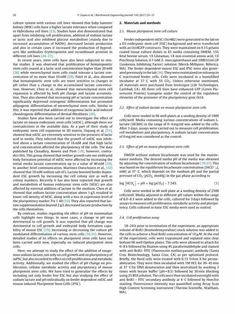
2 gineer
cktaoiaud
is[ctptetsatc
lcesaiapdbiIsdcaaslttt
cddbmdbc
nmalpism
6 P. Gupta et al. / Biochemical En
ulture system with various cell lines showed that baby hamsteridney (BHK) cells have a higher lactate tolerance when comparedo Hybridoma cell lines [23]. Studies have also demonstrated thatpart from inhibiting cell proliferation, addition of sodium lactater lactic acid also inhibited glucose metabolism (mainly due to
ncreased accumulation of NADH), decreased lactate productionnd also in certain cases it increased the production of byprod-cts like antibodies Erythropoietin and recombinant proteins inifferent cell lines [24–27].
In recent years, stem cells have also been subjected to sim-lar studies. It was observed that proliferation of hematopoietictem cells ceased at a lactic acid concentration higher than 20 mM28] while mesenchymal stem cells could tolerate a lactate con-entration of no more than 10 mM [29]. Patel et al., also showedhat hematopoietic stem cells are more sensitive to changes inH rather than a change in the accumulated lactate concentra-ion. However, Chen et al., showed that mesenchymal stem cellxpansion is affected by both pH change and lactate accumula-ion. They also showed that increasing pH or lactate concentrationignificantly depressed osteogenic differentiation but promoteddipogenic differentiation of mesenchymal stem cells. Similar tohis, it was reported that addition of exogenous lactic acid inducedhondrogenic differentiation of dermal fibroblasts [30].
Studies have also been carried out to investigate the effect ofactate on mouse embryonic stem cells (mESC), although there areontradictions in the available data. As a part of their study ofmbryonic stem cell expansion in 3D matrix, Ouyang et al. [31],howed that mESC are extremely sensitive to the presence of lacticcid in media. They inferred that the growth of mESC was inhib-ted above a lactate concentration of 16 mM and that high lacticcid concentration affected the pluripotency of the cells. The dataublished by Chaudhry, Bowen and Piret [19], however, contra-ict this. This group showed that neither growth rate nor embryoidody formation potential of mESC were affected by increasing the
nitial media lactate concentration up to a value of 40 mM [19].n another brief communication, Martinez-Outschoorn et al., [32]howed that 10 mM sodium salt of L-Lactate boosted feeder depen-ent ESC growth by increasing the cell colony size as well asolony numbers. Recently it has also been reported that growthnd metabolism of human embryonic stem cells (hESC) are alsoffected by external addition of lactate to the medium. Chen et al.howed that sodium lactate concentration above 1 g/L resulted inower cell density of hESC, along with a loss in expression level ofhe pluripotency marker Tra 1-60 [33]. They also reported that lac-ate supplementation beyond 2 g/L decreased lactate production byhe cells themselves.
By contrast, studies regarding the effect of pH on mammalianells highlight two things. In most cases, a change in pH wasetrimental to cell growth. It was reported that pH change wasetrimental to cell growth and embryoid body formation capa-ility of mouse ESC [19]. Increasing or decreasing the culture pHodulated differentiation of various stem cells [34,35]. However,
etailed studies of its effects on pluripotent stem cells have noteen carried until now, especially on induced pluripotent stemells.
Here, we attempt to study the effect of the addition of exoge-ous sodium lactate, not only on cell growth and on pluripotency ofPSC, but also recorded its effect on cell proliferation and metabolic
ctivity. Additionally, we studied the effect of pH change on pro-iferation, cellular metabolic activity and pluripotency of mouseluripotent stem cells. We have tried to generalise the effects by
ncluding not only feeder free ESC but also studying the effect of
odium lactate and pH individually on feeder dependent mESC andouse induced Pluripotent Stem Cells (iPSC).ing Journal 118 (2017) 25–33
2. Materials and methods
2.1. Mouse pluripotent stem cell culture
Feeder independent mESC (Oct4B2) were generated in the lab ona D3 embryonic stem cell (ESC) background and were transfectedwith an Oct4GFP constructs. They were maintained on 0.1% gelatincoated tissue culture dishes in ES media containing DMEM, 15%fetal bovine serum, 1X Glutamax, 1X non-essential amino acid, 1XPen/Strep Solution, 0.1 mM 2- mercaptoethanol and 1000 U/ml LIF(Leukemia Inhibiting Factor) solution (Merck-Millipore, Billerica,MA). The feeder dependent mouse ESC and iPSC were also gener-ated previously in the lab [36]. They were maintained on mitomycinC inactivated feeder cells. Cells were incubated in a humidifiedincubator at 37 ◦C with 5% CO2. Unless otherwise mentioned,all materials were purchased from Invitrogen (Life Technologies,Carlsbad, CA). All three cell lines have enhanced GFP (Green Flu-orescent Protein) transgene under the control of the regulatoryelements of the promoter of the pluripotency gene Oct4.
2.2. Effect of sodium lactate on mouse pluripotent stem cells
Cells were seeded in 96 well plates at a seeding density of 1000cells/well. Media containing various concentrations of sodium L-lactate (SIGMA) in the range of 0–4 mg/ml was added to the cells.After 3 days, assays were carried out to measure cell proliferation,cell metabolism and pluripotency. A sodium lactate concentrationof 0 mg/ml was considered as the control.
2.3. Effect of pH on mouse pluripotent stem cells
DMEM without sodium bicarbonate was used for the mainte-nance medium. The desired media pH of the media was obtainedby adjusting the concentration of sodium bicarbonate [19,37]. Thiswas based on the equilibrium bicarbonate concentration ([HCO−
3],mM) at 37 ◦C, which depends on the medium pH and the partialpressure of CO2 (pCO2, mmHg) in the gas phase according to
log[HCO−
3
]= pH + log [pCO2] − 7.543. (1)
Cells were seeded in 48 well plate at a seeding density of 2500cells/well. Media adjusted to different pH values within the rangeof 6.0–8.5 were added to the cells, cultured for 3 days followed byassays to measure cell proliferation, metabolic activity and pluripo-tency. Cells cultured in basic ESC media were used as control.
2.4. Cell proliferation assay
18–24 h prior to termination of the experiment, an appropriatevolume of BrdU (Bromodeoxyuridine) stock solution was added tothe cells to achieve a final BrdU concentration of 10 �M. At the endof the experiment, cells were trypsinized and replated onto clearbottom 96 well Optilux plates. The cells were allowed to attach for6–8 h followed by fixation using 4% paraformaldehyde and stainedwith anti BrdU- FITC (Fluorescein isothiocyanate) antibody (SantaCruz Biotechnology, Santa Cruz, CA) as per optimized protocol.Briefly, the fixed cells were treated with 0.1% Triton X for perme-abilization. They were then incubated with 1M HCL for 30–45 minat 37 ◦C for DNA denaturation and then neutralized by washing 3times with borate buffer (pH = 8.5) followed by 30 min blockingusing 2% BSA solution. The cells were then incubated overnight with
anti BrdU – FITC secondary antibody at 4 ◦C followed by Hoechststaining. Fluorescence intensity was quantified using Array ScanHigh Content Screening instrument (Thermo Scientific, Waltham,MA).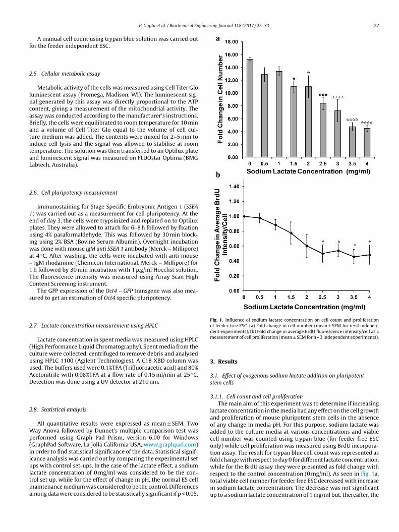
gineering Journal 118 (2017) 25–33 27
f
2
lncaBatitaL
2
1epuiwa–1TC
s
2
(cuuAD
2
Wp(iiultma
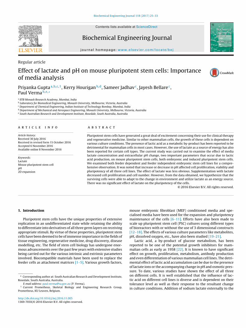
Fig. 1. Influence of sodium lactate concentration on cell count and proliferationof feeder free ESC. (a) Fold change in cell number (mean ± SEM for n = 4 indepen-dent experiments), (b) Fold change in average BrdU fluorescence intensity/cell as a
P. Gupta et al. / Biochemical En
A manual cell count using trypan blue solution was carried outor the feeder independent ESC.
.5. Cellular metabolic assay
Metabolic activity of the cells was measured using Cell Titer Glouminescent assay (Promega, Madison, WI). The luminescent sig-al generated by this assay was directly proportional to the ATPontent, giving a measurement of the mitochondrial activity. Thessay was conducted according to the manufacturer’s instructions.riefly, the cells were equilibrated to room temperature for 10 minnd a volume of Cell Titer Glo equal to the volume of cell cul-ure medium was added. The contents were mixed for 2–5 min tonduce cell lysis and the signal was allowed to stabilise at roomemperature. The solution was then transferred to an Optilux platend luminescent signal was measured on FLUOstar Optima (BMGabtech, Australia).
.6. Cell pluripotency measurement
Immunostaining for Stage Specific Embryonic Antigen 1 (SSEA) was carried out as a measurement for cell pluripotency. At thend of day 3, the cells were trypsinized and replated on to Optiluxlates. They were allowed to attach for 6–8 h followed by fixationsing 4% paraformaldehyde. This was followed by 30 min block-
ng using 2% BSA (Bovine Serum Albumin). Overnight incubationas done with mouse IgM anti SSEA 1 antibody (Merck – Millipore)
t 4 ◦C. After washing, the cells were incubated with anti mouse IgM rhodamine (Chemicon International, Merck – Millipore) for h followed by 30 min incubation with 1 �g/ml Hoechst solution.he fluorescence intensity was measured using Array Scan Highontent Screening instrument.
The GFP expression of the Oct4 – GFP transgene was also mea-ured to get an estimation of Oct4 specific pluripotency.
.7. Lactate concentration measurement using HPLC
Lactate concentration in spent media was measured using HPLCHigh Performance Liquid Chromatography). Spent media from theulture were collected, centrifuged to remove debris and analysedsing HPLC 1100 (Agilent Technologies). A C18 XBD column wassed. The buffers used were 0.1%TFA (Trifluoroacetic acid) and 80%cetonitrile with 0.08%TFA at a flow rate of 0.15 ml/min at 25 ◦C.etection was done using a UV detector at 210 nm.
.8. Statistical analysis
All quantitative results were expressed as mean ± SEM. Twoay Anova followed by Dunnet’s multiple comparison test was
erformed using Graph Pad Prism, version 6.00 for WindowsGraphPad Software, La Jolla California USA, www.graphpad.com)n order to find statistical significance of the data. Statistical signif-cance analysis was carried out by comparing the experimental setps with control set-ups. In the case of the lactate effect, a sodium
actate concentration of 0 mg/ml was considered to be the con-rol set up, while for the effect of change in pH, the normal ES cell
aintenance medium was considered to be the control. Differencesmong data were considered to be statistically significant if p < 0.05.
measurement of cell proliferation (mean ± SEM for n = 3 independent experiments).
3. Results
3.1. Effect of exogenous sodium lactate addition on pluripotentstem cells
3.1.1. Cell count and cell proliferationThe main aim of this experiment was to determine if increasing
lactate concentration in the media had any effect on the cell growthand proliferation of mouse pluripotent stem cells in the absenceof any change in media pH. For this purpose, sodium lactate wasadded to the culture media at various concentrations and viablecell number was counted using trypan blue (for feeder free ESConly) while cell proliferation was measured using BrdU incorpora-tion assay. The result for trypan blue cell count was represented asfold change with respect to day 0 for different lactate concentration,while for the BrdU assay they were presented as fold change withrespect to the control concentration (0 mg/ml). As seen in Fig. 1a,
total viable cell number for feeder free ESC decreased with increasein sodium lactate concentration. The decrease was not significantup to a sodium lactate concentration of 1 mg/ml but, thereafter, the
28 P. Gupta et al. / Biochemical Engineering Journal 118 (2017) 25–33
Fim
vs
ssplc
iTsdtamfo
3
ltcaamel
ccwai
3
dac
ig. 2. Effect of sodium lactate on cell proliferation of feeder dependent ES andPSC measured by fold change in BrdU fluorescence intensity/cell. Data represent
ean ± SEM for n = 3 independent experiments.
iable cell count continued to decrease significantly with increasingodium lactate concentration.
BrdU incorporation assay also supported the above data. Fig. 1bhows the fold change in average BrdU-FITC fluorescence inten-ity/cell. Although not significant at the lower concentrations, cellroliferation showed a decreasing trend with increasing sodium
actate concentration. The decrease became significant once theoncentration reached a value of 2.5 mg/ml.
Fig. 2 shows that cell proliferation for feeder dependent ES andPSC followed a trend similar to that of the feeder independent ESC.he decrease in cell proliferation for the ESC was not statisticallyignificant across the entire range of sodium lactate. For iPSC, theecrease in cell proliferation became significant from a sodium lac-ate concentration of 3.5 mg/ml. Comparison of Figs. 1b and 2 shows
significant decrease in cell proliferation for feeder independentESC at a lower concentration of sodium lactate compared with
eeder dependent mESC and miPC. It is possible that the presencef feeder cells acts as a protection for the cells.
.1.2. Cellular metabolic activityMetabolic activity of the cells was estimated by measuring the
uminescent signal of Cell Titer Glo assay. As mentioned previously,he results were denoted as fold change with respect to the controloncentration (0 mg/ml). Usually the cellular metabolic activity is
direct measurement of cell viability and proliferation and shows similar trend. Interestingly, in this case it was observed that theetabolic activity assay showed almost uniform values across the
ntire range of tested sodium lactate concentration for all three cellines (Fig. 3).
This scenario was possible if the metabolic activity of the viableells increased corresponding to an increase in sodium lactate con-entration in the medium. Therefore, we hypothesized that the cellsere able to alter their metabolic pathway to use the externally
dded lactate as a source of energy along with glucose, thus increas-ng their metabolic activity and resulting in these contrasting data.
.1.3. Lactate concentration estimation by HPLC
Based on the cell proliferation and cellular metabolic activityata, we hypothesized that the cells may be utilizing exogenouslydded lactate as a source of energy along with, or in place of, glu-ose. In order to test our hypothesis to some extent, HPLC of the
Fig. 3. Fold change in cellular metabolic activity of (a) feeder free ESC, (b) feederdependent ES and iPSC measured by Cell Titer Glo assay. Data represent mean ± SEMof n = 3 independent experiments.
spent media was carried out to estimate the lactate concentration.It was expected that if the hypothesis were to be supported, HPLCanalysis would show a decrease in concentration of lactate in thespent media with externally added lactate over time.
Fig. 4 shows the result for HPLC analysis of spent media toassess the change in lactate concentration. The fold change wasmeasured with respect to the initial lactate concentrations in eachcase. As expected, when there was no external addition of sodiumlactate, lactate concentration of the spent media increased withtime confirming the fact that lactate is indeed produced by thecells. However, we observed that when external sodium lactatewas added to the culture, the lactate concentration proceeded todecrease with time. The decrease was significant at and above con-centrations of 2 mg/ml and 1 mg/ml for feeder independent andfeeder dependent cell lines, respectively. These data support tosome extent our hypothesis of lactate consumption by the pluripo-tent cells although further analysis is indeed required.
3.1.4. Cell pluripotencyApart from immunostaining of the surface marker SSEA 1,
pluripotency was also estimated by measuring the fluorescenceintensity of the GFP reporter gene controlled by Oct4 promoter.
Average fluorescence intensity/cell was measured and the resultswere shown as fold change with respect to the control concen-tration (0 mg/ml). For feeder independent ESC, although there isa slight decrease in the SSEA 1 intensity with increasing lactate
P. Gupta et al. / Biochemical Engineer
Fig. 4. Fold change in measurement of lactate concentration in spent media from(a) feeder free ESC, (b) feeder dependent ESC and (c) feeder dependent iPSC by HPLC.Data represent mean ± SEM of n = 3 independent experiments.
ing Journal 118 (2017) 25–33 29
(Fig. 5a), there is no significant change in pluripotency with increas-ing sodium lactate concentration. There was also no measurablechange in the GFP intensity (Fig. 5b).
Similarly, Oct4-GFP fluorescence intensity and SSEA 1 immunos-taining fluorescence intensity/cell were also measured for feederdependent cell lines. As observed in Fig. 5(c,d), although there wasvariation in the fluorescence intensity fold change for both Oct4-GFP and SSEA 1 immunostaining, there was no specific trend or sig-nificant change in cell pluripotency with increasing sodium lactateconcentration.
3.2. Effect of pH on mouse pluripotent stem cells
Change in extracellular culture media pH was found to havesignificant effect on cell proliferation, cellular metabolism and cellpluripotency of mouse pluripotent stem cells. All data are repre-sented as fold change compared with regular ESC media.
3.2.1. Cell proliferationBoth acidic and alkaline media pH affected the proliferation of
mouse pluripotent stem cells irrespective of the absence or pres-ence of feeder layer. As seen in Fig. 6, cell proliferation decreasedsignificantly if the pH decreased or increased beyond 7-7.5. Thefeeder independent cells seem to require a more precise control ofpH, since it showed a significant decrease in proliferation even at apH of 7.0.
3.2.2. Cellular metabolic activityThe cellular metabolic activity (Fig. 7) for the pluripotent stem
cells was reduced with both decrease or increase in pH and followeda trend similar to cell proliferation. The decline was significant forall three cell lines.
3.2.3. Cell pluripotencyUnlike the effect of lactate concentration, change in media pH
significantly affected the cell pluripotency (Fig. 8). Decrease inpluripotency for the feeder free ESC was significant due to mediaacidity in comparison to alkalinity of media. However, pluripotencyof the feeder dependent cells was equally affected by increase ordecrease in media pH; in particular, the iPSC looked to be especiallysensitive.
4. Discussion
Lactic acid has been reported to be one of the major waste prod-ucts in mammalian cell culture, produced predominantly duringglycolysis. The influence of lactic acid on cells can be explainedmainly by change in pH or effect of lactate ions. To date, a numberof papers have reported the detrimental effects of both pH changeand lactate ion accumulation on mammalian cells. However, veryfew studies are available that elucidate the effect of lactate onpluripotent stem cells. In addition, the current literature has vari-ous discrepancies within. In this paper, we have reported the effectof exogenously added lactate on cell proliferation, pluripotency andcellular metabolic activity on three different mouse pluripotentstem cell lines to have a more general trend.
No significant change in pluripotency of mouse pluripotentcells was observed on addition of exogenous sodium lactate. Thesedata support previously published literature on mouse ESC, whichdemonstrates that the presence of lactate does not affect pluripo-tency of mouse ESC [32]. On the other hand, similar to Ouyanget al.’s observation, total cell number for feeder free mouse ESC
decreased with increase in lactate concentration [31]. Chen et al.have reported similar findings for human ESC too, wherein theyobserved a decrease in cell number as well as pluripotency ofhuman ESC on supplementation of medium with sodium lactate
30 P. Gupta et al. / Biochemical Engineering Journal 118 (2017) 25–33
F in SSi an ± S
[pbsdcnipiomtdlsef
cwot
ig. 5. Effect of sodium lactate concentration on pluripotency. (a,c) Fold changentensity/cell. (a,b) feeder free cells, (c,d) feeder dependent cells. Data represent me
33]. The difference regarding the effect of sodium lactate onluripotency between our observation and Chen et al.’s data cane attributed to the fact that human cells are known to be moreensitive than mouse cells to a change in growth environment. Theecrease in cell number became significant from a sodium lactateoncentration of 1.5 mg/ml and the significance of the change in cellumber increased with increasing lactate concentration. Extend-
ng the scope of our work beyond this, we observed that the cellroliferation rate for feeder free mouse ESC also decreased with
ncreasing sodium lactate concentration and became significantnce the sodium lactate concentration reached 2.5 mg/ml. Experi-entation with feeder dependent mouse ES and iPSC to determine
he effect of lactate on their proliferation rate showed a similarecreasing trend in cell proliferation rate. The decrease in cell pro-
iferation for iPSC showed statistical significance only when theodium lactate concentration reached a value of 3.5 mg/ml. It isntirely possible that the feeder cells were in some way responsibleor protecting the cells from the detrimental effect of lactate.
In most cases, the cellular metabolic activity is proportional to
ell proliferation and cell number. However, we observed that thereas no measurable change in the cellular metabolic activity for anyf the cell lines. These data were in contrast to the cell prolifera-ion results. Based on the cell proliferation and cellular metabolic
EA 1 fluorescence intensity/cell (b,d) Fold change in Oct4 promoter driven GFPEM for n = 3 independent experiments.
activity data, we hypothesized that either addition of sodium lac-tate to the medium decreased lactate production by the cellsthemselves or the cells may have been able to utilize exogenouslyadded lactate as a source of energy along with glucose.
In order to support our hypothesis to some extent, we carried outHPLC analysis of the spent media from cell culture with sodium lac-tate concentration between 1 and 4 mg/ml. We found that, when noadditional sodium lactate was added to the media, the lactate con-centration increased over time, suggesting that lactate was beingproduced by the cells as a metabolic by product. However, whensodium lactate was added to the culture, we observed a decreasein the fold change in measured lactate concentration over time.Similar observations were made by Chen et al. on hESC whereinthey reported a decrease in lactate production by the cells uponsupplementation of the media with sodium lactate [33]. Furtherstudies are, however, required to elucidate fully this interestingphenomenon and to understand if the externally added sodiumlactate is merely quenching the production of lactate by the cellsor if they are using it as an energy source.
Although relatively less studied, alteration of cell metabolismdue to externally added lactate and utilization of lactate as a pre-ferred energy source have been reported earlier for a number of celllines. Recombinant CHO cells have been shown to have increased

P. Gupta et al. / Biochemical Engineering Journal 118 (2017) 25–33 31
Ffi
etgcarpaotasfac[
biTcgd
Fig. 7. Effect of pH on cellular metabolic activity of mouse pluripotent stem cells(a) Feeder free ESC, (b) feeder dependent ES and iPSC. Data represent mean ± SEM
ig. 6. Effect of pH on cell proliferation of mouse pluripotent stem cells (a) Feederree ESC, (b) feeder dependent ES and iPSC. Data represent mean ± SEM of n = 3ndependent experiments.
rythropoietin production on addition of exogenous sodium lac-ate, although there was a decrease in specific cell growth andlucose consumption [26]. The group suggested that the high con-entration of sodium lactate led to oxidation of lactate to pyruvatet a high rate by the LDH (lactate dehydrogenase) enzyme. Thisesulted in an accumulation of NADH and high concentration ofyruvate. The energy thus generated, however, was used for aminocid and protein synthesis. Zagari et al., [38] have also carriedut studies on lactate metabolism by CHO cells. They reportedhat consumption of lactate by the CHO cells was associated withn increased mitochondrial activity and oxygen consumption. Theame group also identified the malate – aspartate shuttle as a keyactor in lactate consumption by CHO cells [39]. The use of lactates a preferred source of energy has also been reported for germells along with the ability of lactate to inhibit germ cell apoptosis40–42].
Interestingly, a positive effect of lactate on mouse ESC has alsoeen reported. It was shown that media enriched with lactate
ncrease colony number and size for feeder dependent ESC [32].
hey suggested that lactate was converted to Acetyl CoA, whichan be used either in mitochondrial metabolism or in increasingene expression via histone acetylation. Our HPLC data that showecrease in lactate concentration are consistent with these data.of n = 3 independent experiments.
Findings on the effect of change in extracellular pH on mousepluripotent stem cells were congruent to the available literature.Cell proliferation, cellular metabolic activity as well as pluripotencyof the mouse pluripotent stem cells were significantly decreasedwith any change in the media pH towards acidity or alkalinity.
To conclude, we have shown here the effect of exogenouslyadded lactate and change in media pH on mouse pluripotent stemcell culture. Pluripotent stem cells are extremely sensitive to anychange in media pH, which affects the cells’ growth, viability, pro-liferation and pluripotency. We have also shown that althoughpluripotency is not affected, cell proliferation is negatively affectedby increase in lactate concentration. Further studies are also neededto fully understand the effect of these interesting parameters. Eg, itis possible that addition sodium lactate to the media and/or changein media pH, changes the intracellular pH of the cells, which in turncan have interesting effects on cell behaviour. Effect of osmolalityis also an important factor and needs to be studied. Long term stud-ies for these parameters would also add interesting information inthis area. In conclusion, these studies suggest that careful consider-
ation of media pH and media composition is of utmost importancefor proper growth and maintenance of pluripotent stem cells.
32 P. Gupta et al. / Biochemical Engineering Journal 118 (2017) 25–33
F ESC, (e
A
GrAisB
R
[
[
ig. 8. Effect of pH on pluripotency of mouse pluripotent stem cells (a, c) Feeder freexperiments.
cknowledgements
The authors would like to thank Dr. Trevor Wilson, Medicalenomics Facility, Monash Health and Technology Precinct (cur-
ently, Hudson Institute of Medical Research), for his help in therray Scan analysis and Shane Reeve, Biomedical Proteomics Facil-
ty, Monash University, for her help in HPLC analysis. This work wasupported by the Australia India Strategic Research Fund (GrantF050038).
eferences
[1] H. Hakala, K. Rajala, M. Ojala, S. Panula, S. Areva, M. Kellomaki, R. Suuronen, H.Skottman, Comparison of biomaterials and extracellular matrices as a cultureplatform for multiple, independently derived human embryonic stem celllines, Tissue Eng. Part A 15 (2009) 1775–1785.
[2] Y.J. Li, E.H. Chung, R.T. Rodriguez, M.T. Firpo, K.E. Healy, Hydrogels as artificialmatrices for human embryonic stem cell self-renewal, J. Biomed. Mater. Res.A 79 (2006) 1–5.
[3] Z. Li, M. Leung, R. Hopper, R. Ellenbogen, M. Zhang, Feeder-free self-renewal
of human embryonic stem cells in 3D porous natural polymer scaffolds,Biomaterials 31 (2010) 404–412.[4] C. Xu, M.S. Inokuma, J. Denham, K. Golds, P. Kundu, J.D. Gold, M.K. Carpenter,Feeder-free growth of undifferentiated human embryonic stem cells, Nat.Biotechnol. 19 (2001) 971–974.
b, d) feeder dependent ES and iPSC. Data represent mean ± SEM of n = 3 independent
[5] M. Amit, C. Shariki, V. Margulets, J. Itskovitz-Eldor, Feeder layer- andserum-free culture of human embryonic stem cells, Biol. Reprod. 70 (2004)837–845.
[6] V. Akopian, P.W. Andrews, S. Beil, N. Benvenisty, J. Brehm, M. Christie, A. Ford,V. Fox, P.J. Gokhale, L. Healy, F. Holm, O. Hovatta, B.B. Knowles, T.E. Ludwig,R.D. McKay, T. Miyazaki, N. Nakatsuji, S.K. Oh, M.F. Pera, J. Rossant, G.N.Stacey, H. Suemori, Comparison of defined culture systems for feeder cell freepropagation of human embryonic stem cells, In vitro cellular &developmental biology, Animal 46 (2010) 247–258.
[7] M. Amit, M.K. Carpenter, M.S. Inokuma, C.P. Chiu, C.P. Harris, M.A. Waknitz, J.Itskovitz-Eldor, J.A. Thomson, Clonally derived human embryonic stem celllines maintain pluripotency and proliferative potential for prolonged periodsof culture, Dev. Biol. 227 (2000) 271–278.
[8] D.A. Claassen, M.M. Desler, A. Rizzino, ROCK inhibition enhances the recoveryand growth of cryopreserved human embryonic stem cells and humaninduced pluripotent stem cells, Mol. Reprod. Dev. 76 (2009) 722–732.
[9] A.G. Smith, J.K. Heath, D.D. Donaldson, G.G. Wong, J. Moreau, M. Stahl, D.Rogers, Inhibition of pluripotential embryonic stem cell differentiation bypurified polypeptides, Nature 336 (1988) 688–690.
10] L. Wang, L. Li, P. Menendez, C. Cerdan, M. Bhatia, Human embryonic stem cellsmaintained in the absence of mouse embryonic fibroblasts or conditionedmedia are capable of hematopoietic development, Blood 105 (2005)4598–4603.
11] R.L. Williams, D.J. Hilton, S. Pease, T.A. Willson, C.L. Stewart, D.P. Gearing, E.F.
Wagner, D. Metcalf, N.A. Nicola, N.M. Gough, Myeloid leukaemia inhibitoryfactor maintains the developmental potential of embryonic stem cells, Nature336 (1988) 684–687.
gineer
[
[
[
[
[
[
[
[
[
[
[
[
[
[
[
[
[
[
[
[
[
[
[
[
[
[
[
[
[
[41] K. Erkkilä, H. Aito, K. Aalto, V. Pentikäinen, L. Dunkel, Lactate inhibits germcell apoptosis in the human testis, Mol. Hum. Reprod. 8 (2002) 109–117.
P. Gupta et al. / Biochemical En
12] A.M. Fernandes, T.G. Fernandes, M.M. Diogo, C.L. da Silva, D. Henrique, J.M.Cabral, Mouse embryonic stem cell expansion in a microcarrier-based stirredculture system, J. Biotechnol. 132 (2007) 227–236.
13] E.Y. Fok, P.W. Zandstra, Shear-controlled single-step mouse embryonic stemcell expansion and embryoid body-based differentiation, Stem Cells 23 (2005)1333–1342.
14] S. Levenberg, N.F. Huang, E. Lavik, A.B. Rogers, J. Itskovitz-Eldor, R. Langer,Differentiation of human embryonic stem cells on three-dimensional polymerscaffolds, Proc. Natl. Acad. Sci. U. S. A. 100 (2003) 12741–12746.
15] H. Liu, S.F. Collins, L.J. Suggs, Three-dimensional culture for expansion anddifferentiation of mouse embryonic stem cells, Biomaterials 27 (2006)6004–6014.
16] P.A. Marinho, D.T. Vareschini, I.C. Gomes, S. Paulsen Bda, D.R. Furtado, R.Castilho Ldos, S.K. Rehen, Xeno-free production of human embryonic stemcells in stirred microcarrier systems using a novelanimal/human-component-free medium, Tissue Eng. Part C Methods 19(2013) 146–155.
17] B.W. Phillips, R. Horne, T.S. Lay, W.L. Rust, T.T. Teck, J.M. Crook, Attachmentand growth of human embryonic stem cells on microcarriers, J. Biotechnol.138 (2008) 24–32.
18] M.P. Storm, C.B. Orchard, H.K. Bone, J.B. Chaudhuri, M.J. Welham,Three-dimensional culture systems for the expansion of pluripotentembryonic stem cells, Biotechnol. Bioeng. 107 (2010) 683–695.
19] M.A. Chaudhry, B.D. Bowen, J.M. Piret, Culture pH and osmolality influenceproliferation and embryoid body yields of murine embryonic stem cells,Biochem. Eng. J. 45 (2009) 126–135.
20] H.J. Lim, J. Han, D.H. Woo, S.E. Kim, S.K. Kim, H.G. Kang, J.H. Kim, Biochemicaland morphological effects of hypoxic environment on human embryonicstem cells in long-term culture and differentiating embryoid bodies, Mol.Cells 31 (2011) 123–132.
21] J.R. Millman, J.H. Tan, C.K. Colton, The effects of low oxygen on self-renewaland differentiation of embryonic stem cells, Curr. Opin. Organ Transplant. 14(2009) 694–700.
22] H. Eagle, S. Barban, M. Levy, H.O. Schulze, The utilization of carbohydrates byhuman cell cultures, J. Biol. Chem. 233 (1958) 551–558.
23] T. Hassell, S. Gleave, M. Butler, Growth inhibition in animal cell culture. Theeffect of lactate and ammonia, Appl. Biochem. Biotechnol. 30 (1991) 29–41.
24] M.S. Lao, D. Toth, Effects of ammonium and lactate on growth andmetabolism of a recombinant Chinese hamster ovary cell culture, Biotechnol.Progr. 13 (1997) 688–691.
25] S.S. Ozturk, M.R. Riley, B.O. Palsson, Effects of ammonia and lactate onhybridoma growth, metabolism, and antibody production, Biotechnol. Bioeng.39 (1992) 418–431.
26] Y. Choi, D. Lee, I. Kim, H. Kim, H. Park, T. Choe, I.-H. Kim, Enhancement of
erythropoietin production in recombinant Chinese hamster ovary cells bysodium lactate addition, Biotechnol. Bioprocess Eng. 12 (2007) 60–72.27] T. Omasa, K. Higashiyama, S. Shioya, K. Suga, Effects of lactate concentrationon hybridoma culture in lactate-controlled fed-batch operation, Biotechnol.Bioeng. 39 (1992) 556–564.
[
ing Journal 118 (2017) 25–33 33
28] S.D. Patel, E.T. Papoutsakis, J.N. Winter, W.M. Miller, The lactate issuerevisited: novel feeding protocols to examine inhibition of cell proliferationand glucose metabolism in hematopoietic cell cultures, Biotechnol. Progr. 16(2000) 885–892.
29] T. Chen, Y. Zhou, W.S. Tan, Influence of lactic acid on the proliferation,metabolism, and differentiation of rabbit mesenchymal stem cells, Cell Biol.Toxicol. 25 (2009) 573–586.
30] S.B. Nicoll, A. Wedrychowska, N.R. Smith, R.S. Bhatnagar, Modulation ofproteoglycan and collagen profiles in human dermal fibroblasts by highdensity micromass culture and treatment with lactic acid suggests change toa chondrogenic phenotype, Connect. Tissue Res. 42 (2001) 59–69.
31] A. Ouyang, R. Ng, S.T. Yang, Long-term culturing of undifferentiatedembryonic stem cells in conditioned media and three-dimensional fibrousmatrices without extracellular matrix coating, Stem Cells 25 (2007) 447–454.
32] U.E. Martinez-Outschoorn, M. Prisco, A. Ertel, A. Tsirigos, Z. Lin, S. Pavlides, C.Wang, N. Flomenberg, E.S. Knudsen, A. Howell, R.G. Pestell, F. Sotgia, M.P.Lisanti, Ketones and lactate increase cancer cell stemness, driving recurrence,metastasis and poor clinical outcome in breast cancer: achieving personalizedmedicine via Metabolo-Genomics, Cell Cycle 10 (2011) 1271–1286.
33] X. Chen, A. Chen, T.L. Woo, A.B. Choo, S. Reuveny, S.K. Oh, Investigations intothe metabolism of two-dimensional colony and suspended microcarriercultures of human embryonic stem cells in serum-free media, Stem Cells Dev.19 (2010) 1781–1792.
34] T.A. McAdams, W.M. Miller, E.T. Papoutsakis, pH is a potent modulator oferythroid differentiation, Br. J. Haematol. 103 (1998) 317–325.
35] D.L. Hevehan, E.T. Papoutsakis, W.M. Miller, Physiologically significant effectsof pH and oxygen tension on granulopoiesis, Exp. Hematol. 28 (2000)267–275.
36] P.A. Tat, H. Sumer, K.L. Jones, K. Upton, P.J. Verma, The efficient generation ofinduced pluripotent stem (iPS) cells from adult mouse adipose tissue-derivedand neural stem cells, Cell Transplant. 19 (2010) 525–536.
37] A.E. Schmelzer, V.M. deZengotita, W.M. Miller, Considerations for osmolalitymeasurement under elevated pCO(2): comparison of vapor pressure andfreezing point osmometry, Biotechnol. Bioeng. 67 (2000) 189–196.
38] F. Zagari, M. Jordan, M. Stettler, H. Broly, F.M. Wurm, Lactate metabolism shiftin CHO cell culture: the role of mitochondrial oxidative activity, NewBiotechnol. 30 (2013) 238–245.
39] F. Zagari, M. Stettler, L. Baldi, H. Broly, F.M. Wurm, M. Jordan, High expressionof the aspartate–glutamate carrier Aralar1 favors lactate consumption in CHOcell culture, Pharm. Bioprocess. 1 (2013) 19–27.
40] M. Mita, P.F. Hall, Metabolism of round spermatids from rats: lactate as thepreferred substrate, Biol. Reprod. 26 (1982) 445–455.
42] J.A. Grootegoed, R. Jansen, H.J. Van der Molen, The role of glucose, pyruvateand lactate in ATP production by rat spermatocytes and spermatids, Biochim.Biophys. Acta 767 (1984) 248–256.


![COPY : BCJ-QMS-0082 H : 8 30 H 10] 5 BCJ-SAR ISO 9001 …hazkari.co.jp/eco/img/iso9001me.pdf · bcj-sar iso 9001 jab cm018 . copy h. bcj-sar iso 9001 jab oms cm018 in in & : jis q](https://img.pdfslide.us/doc/110x75/5b4d1c877f8b9ac9758b51a4/copy-bcj-qms-0082-h-8-30-h-10-5-bcj-sar-iso-9001-bcj-sar-iso-9001-jab-cm018.jpg)