Embed Size (px)
Citation preview
Biochemical EndocrinologyBCH 560

Endocrinology is the capacity of specialized tissues to function in integral fashion as components of intact organism which is made possible in large by two control mechanism.
Nervous system – Neurotransmittersi.e.: Acetylcholine, Dopamine, Serotonin, Gamma Aminobutyric Acid (GABA), Glutamate, Epinephrine and Norepinephrine, Endorphins
Endocrine system – Hormonesi.e.: ADH, ACTH, Prolactin, Calcitonin,Parathyroid, Renin, Insulin, Glucagon, CCK, Resistin, Leptin…etc
Endocrine versus Nervous Systems
Major communication systems in the body Integrate stimuli and responses to changes in
external and internal environment Both are crucial to coordinated functions of
highly differentiated cells, tissues and organs Unlike the nervous system, the endocrine
system is anatomically discontinuous.
Cont…
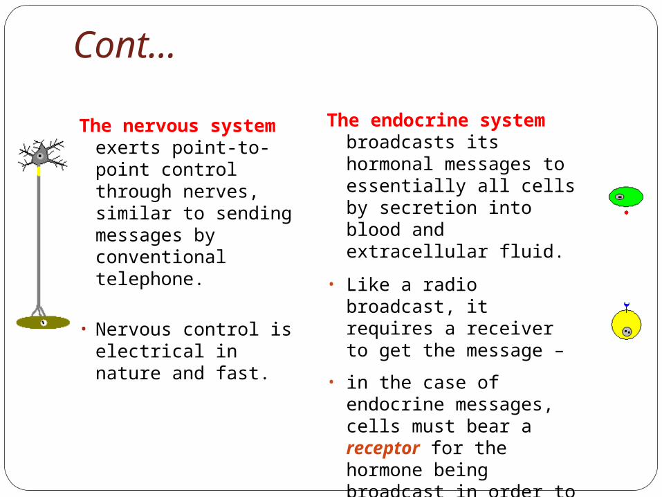
The nervous system exerts point-to-point control through nerves, similar to sending messages by conventional telephone.
• Nervous control is electrical in nature and fast.
The endocrine system broadcasts its hormonal messages to essentially all cells by secretion into blood and extracellular fluid.
• Like a radio broadcast, it requires a receiver to get the message –
• in the case of endocrine messages, cells must bear a receptor for the hormone being broadcast in order to respond.

Endocrinology traditionally defined as the action of hormones and the organs in which the hormones are formed.
It is About:1.The study of anatomy and physiological
function of the major endocrine organs.2.The secretory products of these organs. 3.The mechanism of hormone action.4.The clinical manifestation of hormone
action
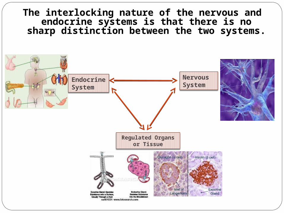
The interlocking nature of the nervous and endocrine systems is that there is no sharp
distinction between the two systems.
NervousSystem
EndocrineSystem
Regulated Organsor Tissue

Endocrine Gland
Their products are secreted directly and internally into the blood stream which do not utilize ducts. These ductless glands
were termed Endocrine.
Cont…

Types of Hormones Products of the ductless gland
(Endocrine Hormones)
Work at a relatively short range (Telecrine)
Work at a nearby hormones (Paracrine)- cells secret
substances that diffuse into the extracellular
fluid and affect neighboring cells.
Endocrine and Telecrine glands or specialized cells release hormones into the circulating blood that influence the function of cells at another location in the body.
At present, there are about 50 known hormones such as protein, small peptide and steroids.

Mechanism of Action of Hormone
Hormone are chemical messengers synthesized by organism that initiate
biological responses by binding with high affinity and specificity to target
cell receptors within the same individual.They are:Endogenous substance High affinity and specificity of binding to specific
receptors on target cellsInitiates biological response

Amine HormoneDerivatives of tyrosineCatecholamines (epinephrine, dopamine) Catecholamines are both
neurohormones and neurotransmitters. These include epinephrine, and norepinephrine Epinephrine and norepinephrine are produced by the adrenal
medulla both are water soluble Secreted like peptide hormones
Thyroid Hormone (dipeptides) are basically a "double" tyrosine with the critical incorporation of 3 or 4 iodine atoms. Thyroid hormone is produced by the thyroid gland and is lipid
soluble Thyroid hormones are produced by modification of a tyrosine residue
contained in thyroglobulin, post-translationally modified to bind iodine, then proteolytically cleaved and released as T4 and T3. T3 and T4 then bind to thyroxin binding globulin for transport in the blood
Tryptophan derivative Melatonin
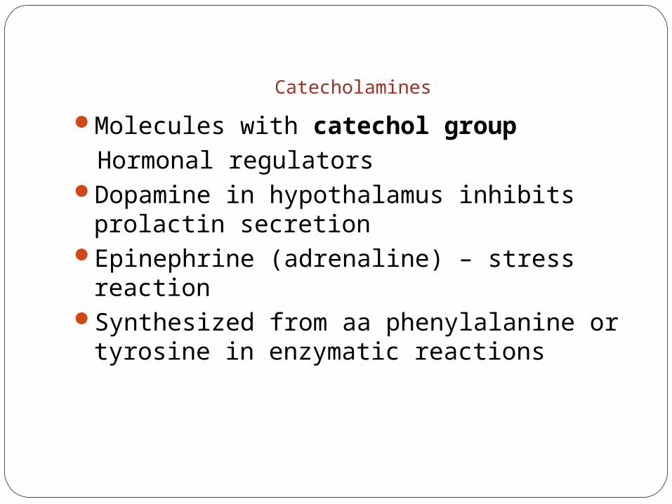
Catecholamines
Molecules with catechol group Hormonal regulators
Dopamine in hypothalamus inhibits prolactin secretion
Epinephrine (adrenaline) – stress reactionSynthesized from aa phenylalanine or
tyrosine in enzymatic reactions
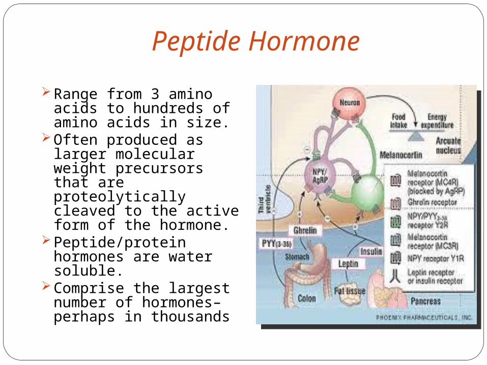
Peptide Hormone
Range from 3 amino acids to hundreds of amino acids in size.
Often produced as larger molecular weight precursors that are proteolytically cleaved to the active form of the hormone.
Peptide/protein hormones are water soluble.
Comprise the largest number of hormones– perhaps in thousands

By one or two genese.i.: Insulin from single gene Glycoprotein hormone from two precursors.
Initial ribosomal product called Preprohormones prohormone hormone
mRNA on ribosomal membrane Translation of mRNA results in an AA’s sequence at
NH2 terminus of nascent polypeptide.Cleavage occurs at sequence
lys – Arg or Arg – Aig in Golgi complex -Transport it to Rough Endoplasmic Reticulum -An energy-requiring process facilitated by activity
of cellular microfilaments
Synthesis (peptide Hormone)
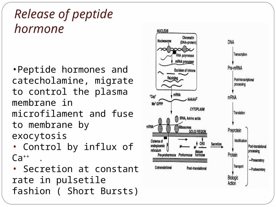
•Peptide hormones and catecholamine, migrate to control the plasma membrane in microfilament and fuse to membrane by exocytosis• Control by influx of Ca++ .• Secretion at constant rate in pulsetile fashion ( Short Bursts)
Release of peptide hormone

StorageAfter synthesis in rough
endoplasmic reticulumPacked in membrane
vesicles to form granules in the Golgi complex as prohormone.
Glands for peptide hormone contain up to one day supply of hormone
Glands for steroids hormone contain longer time

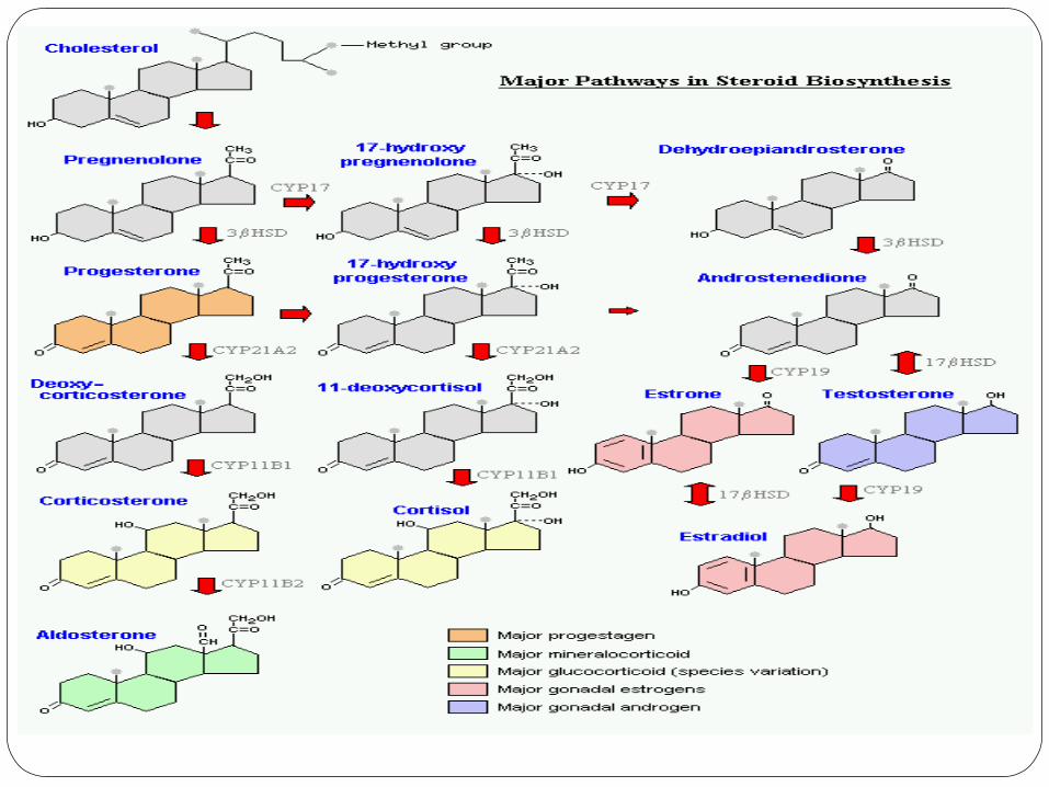
Steroid HormoneAll steroid hormones are derived from
cholesterol and differ only in the ring structure and side chains attached to it.
All steroid hormones are lipid soluble
Glucocorticoids; cortisol is the major representative in most mammals
Mineralocorticoids; aldosterone being most prominent
Androgens such as testosterone
Estrogens, including estradiol and estrone
Progestogens (also known a progestins) such as progesterone
Types of steroid hormones

Facts about steroid hormoneAre not packaged, but synthesized and immediately
releasedAre all derived from the same parent compound: CholesterolEnzymes which produce steroid hormones from cholesterol
are located in mitochondria and smooth ERSteroids are lipid soluble and thus are freely permeable to
membranes so are not stored in cellsSteroid hormones are not water soluble so have to be
carried in the blood complexed to specific binding globulins. Corticosteroid binding globulin carries cortisolSex steroid binding globulin carries testosterone and
estradiol In some cases a steroid is secreted by one cell and is
converted to the active steroid by the target cell: an example is androgen which secreted by the gonad and converted into estrogen in the brain
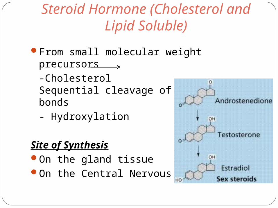
Steroid Hormone (Cholesterol and Lipid Soluble)
From small molecular weight precursors-Cholesterol Sequential cleavage of carbon-carbon bonds- Hydroxylation
Site of SynthesisOn the gland tissueOn the Central Nervous System
TransportLipid soluble hormones require transport proteins albumin and transthyretin (prealbumin) specific transport molecules (thyroxine-binding globulin) only unbound hormone can enter the cell. Steroid and thyroid hormones are 99% attached to
special transport proteins i.e. Binding Carrier
Circulation in Blood From seconds (epinephrine) to hours (insulin), to days
(reproductive hormone) Typical resting concentration very low Under stimulated condition:
Peptide hormone: 5-100 foldsCatecholamine: 5-100 foldsSteroids: 5-1000 folds

cholesterol
Extracellularlipoprotein
Cholesterolpool
LH
ATP
cAMPPKA+
Pregnenolone
Progesterone
Androstenedione
TESTOSTERONE
3HSD
P450c17
17HSD
acetate
Feedback Relationship
Distinguishing characteristics of Endocrine System –Feedback control production.
To maintain homeostatic balance for body fluid and rate of various metabolic process.
Example: 1. Increase of parathyroid hormone sensed
by (Ca++) level↑ (Ca++) (-) Feedback↓ (Ca++) (-) Feedback
2.(Complex)- InteractionPituitary- Thyroid hormoneAdrenal – Gonads Hormone

Type of FeedbackCation ( Ca++ on PTH) Metabolites (Glucose on insulin and increase
glucagon)Hormone (Somatostatin on insulin and
glucagon)Osmolality (Vassopressin, renin, aldosterone)Feedback is useful in the assessment of
pathological states:Insulin level-Glucose levelTSH levels- Serum Thyroxine


Function of Hormones
Hormonal function involves four Broad domain
Reproduction – Regulate reproductive system
Maintenance of internal environment Growth and development Energy production, utilization and storage
Cont…
Other Function of HormonesRegulate gametogenesisControl dimorphic, anatomical function and
behavioral developmentRegulate stability of body fluid and electrolytes,
heart rate, acid base balance, body temperature bone mass, muscle and fat.
Mediator for substrate flux, conversion of calories to energy.
Mediator in catabolism – glucagon of glycogen breakdown AA’s and FA’s to glucose
Help regulate circadian rhythms ( Sleep/wake periods)
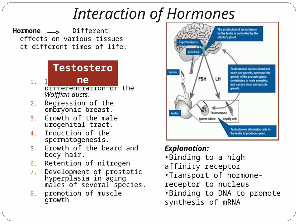
Interaction of HormonesHormone Different
effects on various tissues at different times of life.
1. Induction of male differentiation of the Wolffian ducts.
2. Regression of the embryonic breast.
3. Growth of the male urogenital tract.
4. Induction of the spermatogenesis.
5. Growth of the beard and body hair.
6. Retention of nitrogen7. Development of prostatic
hyperplasia in aging males of several species.
8. promotion of muscle growth
Testosterone
Testosterone
Explanation:•Binding to a high affinity receptor•Transport of hormone-receptor to nucleus•Binding to DNA to promote synthesis of mRNA
Cont…
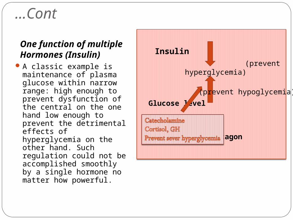
One function of multiple Hormones (Insulin)
A classic example is maintenance of plasma glucose within narrow range: high enough to prevent dysfunction of the central on the one hand low enough to prevent the detrimental effects of hyperglycemia on the other hand. Such regulation could not be accomplished smoothly by a single hormone no matter how powerful.
Insulin (prevent
hyperglycemia)
Glucose level
Glucagon
(prevent hypoglycemia)
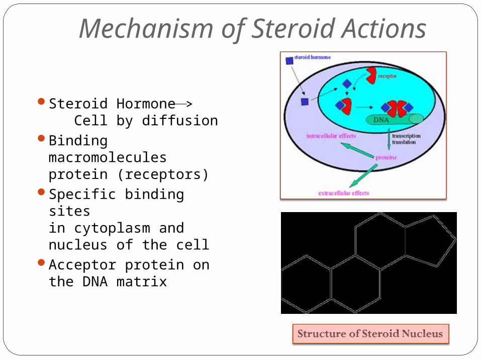
Mechanism of Steroid Actions
Steroid Hormone Cell by diffusion
Binding macromolecules protein (receptors)
Specific binding sitesin cytoplasm and nucleus of the cell
Acceptor protein on the DNA matrix
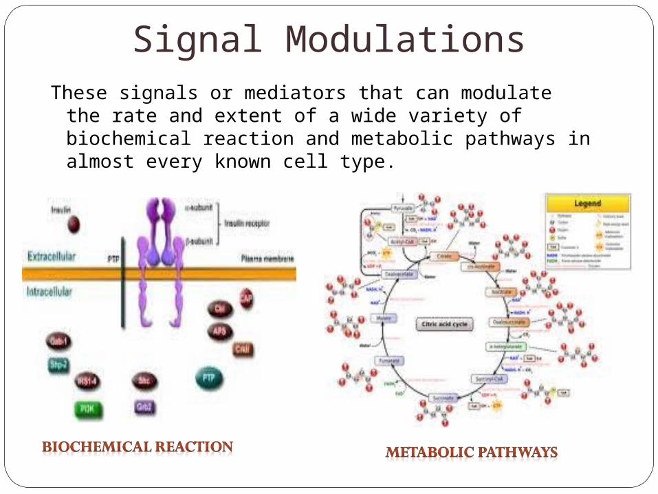
Signal ModulationsThese signals or mediators that can modulate the rate
and extent of a wide variety of biochemical reaction and metabolic pathways in almost every known cell type.
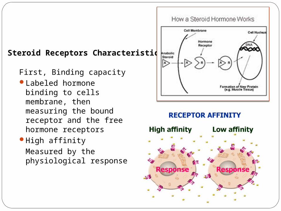
Steroid Receptors Characteristic
First, Binding capacityLabeled hormone
binding to cells membrane, then measuring the bound receptor and the free hormone receptors
High affinity Measured by the physiological response
Specificity Biological ResponseReceptor sites have
capacity for recognition to the primer hormone rather for other agonistic or antagonistic.
Certain tissue are specific for certain hormone e.g. Sex Steroids for (uterus, vagina)
Hormone receptor binding precedes tissue response

Control of Hormone Binding
By Site Activation Functional ActivationPhospohorylation of
active site by ATP and protein kinase i.e.,Glucocorticoid receptor
Dephosphorylation by phosphatase i.e., estrogen receptors causes the loss of estrogen binding activity
Hormone regulation theeffective receptor titer: Down rregulation –
represent a redubtion inhormone-binding activity. i.e.,progesterone receptor number decreaseswithin 1 hour after progesterone administration.
Augment receptor titer Estrogen and estradiol
administration causes increase in receptor level
Induced fil.

Functional Activity of Receptors
Presents in a small amount in cells. (0.001%-0.1% of total soluble proteins)
Structurally differ form receptor to another
High affinity of DNA from hormone
Estrogen Receptor binds to DNA after hormone-receptor complex formation.
Progesterone receptorA subunit of receptor possesses the DNA binding.
Glucocorticoids Receptors should be saturated with the hormone
Feature of Steroid Receptors
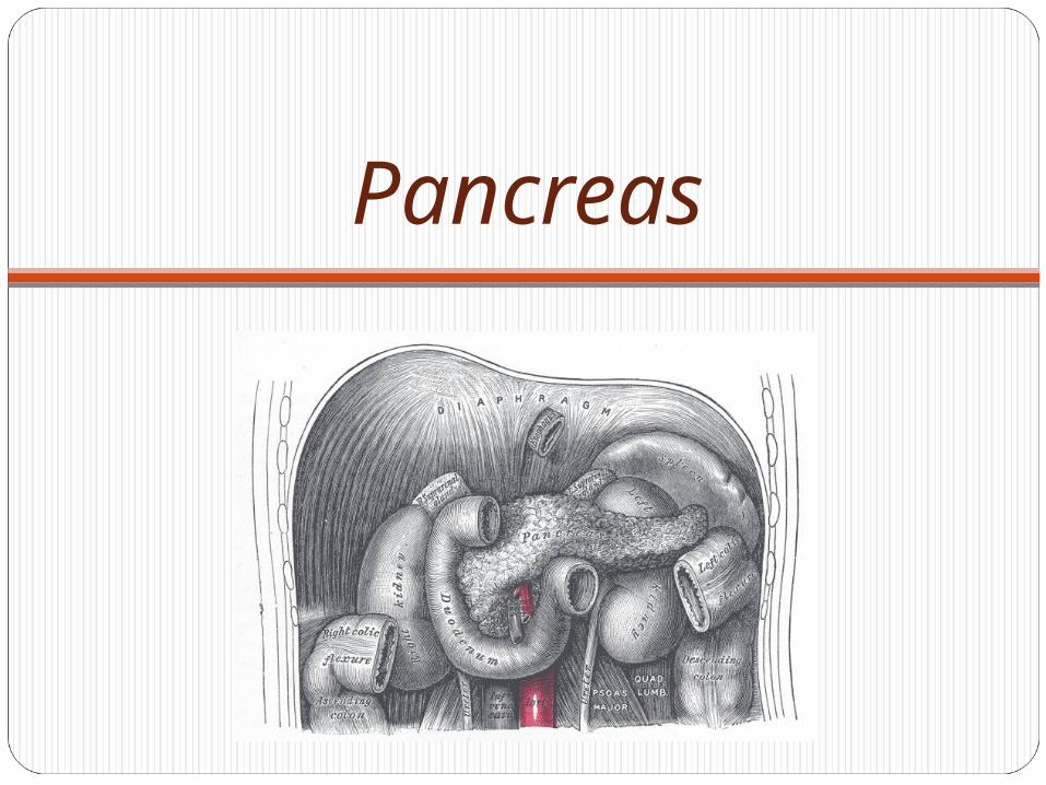
Pancreas

Brief HistoryHerophilus, Greek surgeon first described
pancreas.Wirsung discovered the pancreatic duct in
1642 now called duct of Wirsung.Pancreas as a secretory gland was
investigated by Graaf in 1671.R. Fitz established pancreatitis as a disease
in 1889.Dr. Whipple performed the first pancreatico-
duodenectomy in 1935 and refined it in 1940 now called Whipple procedure.

Anatomy of Pancreas
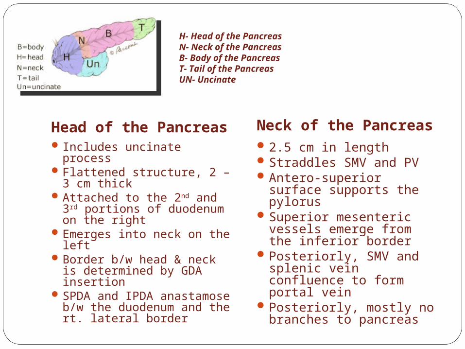
H- Head of the PancreasN- Neck of the PancreasB- Body of the PancreasT- Tail of the PancreasUN- Uncinate
Head of the Pancreas Neck of the Pancreas Includes uncinate processFlattened structure, 2 – 3
cm thickAttached to the 2nd and 3rd
portions of duodenum on the right
Emerges into neck on the left
Border b/w head & neck is determined by GDA insertion
SPDA and IPDA anastamose b/w the duodenum and the rt. lateral border
2.5 cm in lengthStraddles SMV and PVAntero-superior
surface supports the pylorus
Superior mesenteric vessels emerge from the inferior border
Posteriorly, SMV and splenic vein confluence to form portal vein
Posteriorly, mostly no branches to pancreas

Body of Pancreas Tail of Pancreas
Elongated, long structureAnterior surface,
separated from stomach by lesser sac
Posterior surface, related to aorta, lt. adrenal gland, lt. renal vessels and upper 1/3rd of lt. kidney
Splenic vein runs embedded in the post. Surface
Inferior surface is covered by tran. mesocolon
Narrow, short segmentLies at the level of the
12th thoracic vertebraEnds within the splenic
hilumLies in the
splenophrenic ligamentAnteriorly, related to
splenic flexure of colonMay be injured during
splenectomy (fistula)
H- Head of the PancreasN- Neck of the PancreasB- Body of the PancreasT- Tail of the PancreasUN- Uncinate
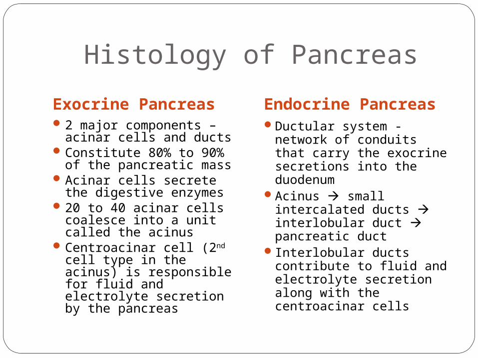
Histology of Pancreas
Exocrine Pancreas Endocrine Pancreas2 major components –
acinar cells and ductsConstitute 80% to 90%
of the pancreatic massAcinar cells secrete the
digestive enzymes20 to 40 acinar cells
coalesce into a unit called the acinus
Centroacinar cell (2nd cell type in the acinus) is responsible for fluid and electrolyte secretion by the pancreas
Ductular system - network of conduits that carry the exocrine secretions into the duodenum
Acinus small intercalated ducts interlobular duct pancreatic duct
Interlobular ducts contribute to fluid and electrolyte secretion along with the centroacinar cells