Embed Size (px)
Citation preview

ABSTRACT
Back ground:Gold nanoparticles (GNPs) offer a great promise inbiomedicine. Currently, there is no data available regarding the toxicity ofGNPs .ROS and free radical production is one of the primary mechanismsof nanoparticle toxicity.The present in vivo study investigated thetoxicological effects of size-sorted GNPs along four different time intervalsat the cellular levels that include liver, spleen and blood cells throughbiophysical and biochemical studies, these studies were confirmed byhistopathological studiesAnimals and Methods:Adult SpragueDewally male rats weighting (100-120g) received intraperitoneal injection of colloidal gold nanoparticles ofdifferent sizes(10,50,100nm) that were repeated day by day .Animals werescarified after (14,21,30 and 40) blood samples ,liver and spleen werecollected. The parameters studied included: measuring of whole bloodviscosity , RBCs aggregation parameters, hepatic lipid peroxidation, Superoxide dismutase (SOD), glutathione (GSH), glutathione disulfide (GSSG),GSH⁄GSSG ratio, glutathione redox potential (∆EG) ,and tumor necrosisfactor‑alpha (TNF‑α) were measured in both liver homogenate and plasma of allstudied and control groups.Histopathological examination of the liver andspleen were done .Results:Significant increases were observed in all studied groupscompared to controls in RBCs aggregation whole blood viscosities andliver lipidperoxidation, while significant decreases were noticed in hepaticSOD, GSH concentrations , GSH⁄GSSG ratio, glutathione redox potential(∆EG), and significant increase in hepatic and plasma TNF‑α concentrationof all studied groups after the four time intervals included in the presentexperiment. In all the studied parameters, the percentage of the increaseor decrease was time dependentand was higher in 50nm group than 10nmand 100nm groups.Thehistopathological findings confirmed thebiochemical alterations and revealed that,there were various degrees ofhepatic tissue injury in the studied groups after the four time intervals ofthe experiment, characterized by mild to heavy inflammation, as well asnecrosis and apoptosis The spleen sections of GNPs-administrated ratsrevealed no significant differences in histology after 14 and 21 day of GNPsadministrationConclusion:In vivo cytotoxicity of size-sorted GNPs is the induction ofoxidative stress and immune response. The toxicological effects of GNPsare time dependent. Different sizes of GNPs could be endocytosed byhepatic and splenic cells and have large toxic effect on hepatic tissue(apoptosis and necrosis) while its effect on splenic tissue is limited and couldbe observed after long-term repeated administration
KEYWORDS: Gold nanoparticle, Sizes, Rats, Oxidative stress,Antioxidant enzymes, cell death
1.0 INTRODUCTION
Nanotechnology can simply be defined as the technology atthe scale of one-billionth of a meter. It is the design,characterization, synthesis and application of materials,structures, devices and systems by controlling shape andsize at nanometer scale.⁽¹,²⁾
Gold nanoparticles (GNPs) have gained increasing interestdue to their special features, such as unusual optical andelectronic properties, high stability and biological
compatibility, controllable morphology and size dispersion,and easy surface functionalization⁽³,⁴⁾.
Gold and gold compounds are mainly applied as a drug forthe treatment of rheumatic diseases including psoriasis,palindromic rheumatism, juvenile arthritis and discoidlupus erythematous.⁽ ⁵,⁶⁾
Nanoparticles could have many adverse effects at thecellular level by interacting with vital cell components suchas the membrane, mitochondria, or nucleus. Adverseoutcomes could include organelle or DNA damage, oxidativestress, apoptosis (programmed cell death), mutagenesis, andprotein up/ down regulation. ⁽⁷,‑⁸⁾
Various in-vitro and in-vivo studies show that free radicalformation can be triggered by nanoparticles.⁽⁹⁾Nanoparticles can be taken up actively (phagocytosis) bycertain cells (macrophages) and initiate ROS formation.Passive cellular uptake of particles has also beendocumented. ⁽¹⁰, ¹¹⁾
Nanoparticles tend to form aggregations/agglomerations.It is unclear whether they can produce elevated ROS levelsin this configuration. ROS can also develop directly on thesurface of the particles, although this depends on particlestructure. The overproduction or chronic production of ROScan cause inflammatory reactions, tissue changes and DNA,protein and lipid damage. Nanoparticles also causemechanical damage within the cells and thus triggeroxidative stress. ⁽¹², ¹³⁾
Oxidative damage to cellular DNA can lead to mutations andmay, therefore, play an important role in the initiation andprogression of multistage carcinogenesis. ⁽¹⁴⁾Nevertheless,high ROS concentrations induce apoptotic cell death invarious cell types, suggesting that ROS contribute to celldeath whenever they are generated in the context of theapoptotic process. ⁽¹⁵, ¹⁶⁾
Lipid peroxidation ( LPO) changes molecule characteristics,making it more hydrophilic;this can alter structure andfunction of the membrane .The LPO products such asaldehydes (malondialdehyde and hydroxynonenal), arecapable of forming crosslinks with lipids, proteins andnucleic acids, thereby causing damage to themacromolecules, which are essential components ofbiological tissues.⁽¹⁷⁾
Science Journal of Medicine and Clinical TrialsISSN:2276-7487http://www.sjpub.org/sjmct.html© Author(s) 2014. CC Attribution 3.0 License.
Published ByScience Journal Publication
International Open Access Publisher
Research Article
BIOCHEMICAL AND HISTOPATHOLOGICAL STUDY OF TOXICITY OF DIFFERENT SIZES OFSPHERICAL GOLD NANOPARTICLES ON NORMAL RATS - IN VIVO STUDY
Bothaina F Mahmoud ¹, Iman A Sharaf¹, Amani H Kazem², Heba S Ramadan³, Ahmed F Taha¹
Department of Biochemistry¹ Department of Pathology² Department of Medical BioPhysics³Medical Research Institute University of Alexandria
Volume 2014, Article ID sjmct-104, 15 Pages, 2014. doi: 10.7237/sjmct/104
Accepted 5�� March, 2014
Corresponding Author: Iman A SharafDepartment of Biochemistry, Medical Research Institute University of Alexandria Email address: [email protected]

Science Journal of Medicine and Clinical Trials( ISSN:2276-7487) page 2
,
Antioxidants can neutralize the free radicals throughhomeostatic activity of the cells. The main enzymatic"scavengers" responsible for the prevention of ROSformation and oxidation are superoxide dismutase, catalaseand glutathione peroxidase.⁽¹⁸⁾Superoxide dismutaseenzyme converts superoxide radical anion into hydrogenperoxide (H2O2). Hydrogen peroxide is a weak oxidantattacking mainly thiols. ⁽¹⁹⁾
Glutathione (GSH) is among the most important antioxidantsin cells, being used in enzymatic reactions to eliminateperoxides and in nonenzymatic reactions to maintainascorbate and α‑tocopherol in their reduced and functionalforms. In these reactions, GSH is converted to its disulfideform, GSSG. The most widely used indicator of the redoxstate of the GSH pool is the ratioof reduced glutathione (GSH)to oxidized glutathione (GSSG). ⁽²⁰⁾
Glutathione oxidation precedes nuclear DNA fragmentation.These signs of oxidative stress are caused, at least in part,by an increase in peroxide production by mitochondria fromapoptotic cells. ⁽ ²¹⁾
Nanoparticles could also be identified as foreign by theimmune cells, causing the cells to react against either surfaceor core components to mount an inflammatory response,which involve secretion of signaling molecules (known ascytokines such as TNF‑α) to attract more cells to destroy theforeign substances.⁽¹⁰⁾
2.0 Study Objectives
The present study aimed toevaluate from the biochemical,biophysical and histopathological points of view thetoxicological effects of different sizes of colloidal goldnanoparticles administrated intrapritoneally to normal adultmale rats.
3.0 MATERIALS AND METHODS
3.1 Experimental animals and their groups:
The experiments were carried out on 140 male Sprague-Dewally rats of mean weight 100-120g (purchased fromFaculty of Medicine, Alexandria University, animal house).Each four animals were housed in a cage in a wellventilatedroom (25 light: dark cycle at the animal house. The animalswere acclimated to the environment for at least two weeksbefore onset of the experiments. The design of the study wasin accordance with the ethical guide lines prescribed by theMedical Research Institute.
After 1 week of acclimatization, the animals were randomlydivided into 3 main experimental groups (n=40/gp)
Groups (I,II,III) were injected intrapritoneally with asuspension of gold nanoparticles of about 10nm, 50nm,100nm at a dose of 1ml (1mM)/200gm (animal weight)respectively and group IV consists of twenty normal malerats that were injected intrapritoneally with saline only andserve as control group.
Animals scarified after 14, 21, 30 and 40 days of repeatedadministration- day by day-of size sorted GNPs. At time ofscarification, animals were anaesthetized by light ether.
Blood samples were collected from dorsal vein on EDTAcoated tubes for determination of red blood cells aggregationand plasma TNF‑α.
3.2 Tissue samples
As fast as possible, a biopsy of liver was excised from animalsand blood was removed by perfusing the tissue with a coldphosphate buffer saline (pH=7.4, 0.1M). The biopsy wasweighed and homogenized in phosphate buffer saline(pH=7.4, 0.1M) to make up 1 to 5 W⁄ V final preparation.The whole homogenate was centrifuged at 1600 rpm for 20min at 5 oC, the supernatant was immediately stored at -20oC for further use.
Also, another biopsy of liver and spleen tissues was dissectedand preserved in 10% formalin for preparation ofmicroscopic sections and exploring morphological changes.
3.3 Biophysical studies
3.3.1 Preparation of gold nanoparticles:⁽²²⁾
Gold nanoparticles were prepared using citrate ions whichacts as a reducing and a capping agent. This formation ofgold nanoparticles can be observed by a change in colorsince small nanoparticles of gold are red. The presence ofthis colloidal suspension can be detected by the reflectionof a laser beam from the particles.
3.3.2 Characterization of Gold Nanoparticles ⁽²³‑²⁵⁾
3.3.2.1 Particle size distribution
The particle size distribution of the gold nanoparticles wasdetermined by laser light scattering on a Beckman CoulterParticle Size Analyzer (N5 submicron particle size ananalyzer, Japan).
The shape of prepared nanoparticles was determined byTransmission Electron Microscope (Jeol, JSM-6360LA,Japan) after mounting them on carbon coater copper grideand stained with uranyl acetate (SPI-ModuleTM sputtercoater, Japan).
3.3.3 Aggregation shape parameter (ASP):
After the Scion program counted each groups and calculatedthe area and perimeter of each count, the data was enteredinto an Excel program sheet in order to calculate the ASP foreach count, which was calculated using the followingformula ⁽²⁶⁾:
ASP = 4 π A / P2
Where:
How to Cite this Article: Bothaina F Mahmoud, Iman A Sharaf, Amani H Kazem, Heba S Ramadan, Ahmed F Taha,"Biochemical and Histopathological Studyof Toxicity of Different Sizes of Spherical Gold Nanoparticles on Normal Rats - in Vivo Study",Science Journal of Medicine and Clinical Trials,Volume 2014,Article ID sjmct-104, 15 Pages, 2014. doi: 10.7237/sjmct/104

page 3 Science Journal of Medicine and Clinical Trials( ISSN:2276-7487)
A is the projected area of the aggregate,P is the perimeter of the project
3.3.4 Determination of whole blood viscosity ⁽²⁷⁾
A wells‑Brookfield Cone ⁄ Plate LVDV-II+ viscometer witha CP-40 cone (Brook field laboratories, Japan) coupled to arefrigerated recirculating fluid path to control thetemperature of the sample (Cole Parmer, Vernon Hills, IL)was used to measure the viscosity over a range of differentshear rates
3.4 Biochemical studies
3.4.1 Determination of lipid peroxidation:
Malondialdehyde in whole liver homogenate wasdetermined according to the method of Draper and Hadley.(28) The sample under test was heated with thiobarbituricacid (TBA) at low pH. The resulting pink chromogen has amaximal absorbance at 532nm
3.4.2 Determination of superoxide dismutase (SOD)activity:
was done by pyrogallol method of Marklund andMarklund.⁽²⁹⁾. The method depends on the spontaneousautoxidation of pyrogallol at alkaline pH, resulting in theproduction of superoxide anion radicals (O·2¯), which in turnenhance autoxidation of pyrogallol. Autoxidation ismanifested as an increase in absorbance at 420 nm.
3.4.3 Determination of glutathione and glutathionedisulfide in liver homogenate:
was done by the enzymatic method described by Griffith etal⁽³⁰⁾ The method depends on the oxidation of GSH by 5,5-dithiobis-(2-nitrobenzoic acid) (DTNB) to yield GSSG and5-thio-2-nitrobenzoic acid (TNB). Oxidized GSSG is reducedenzymatically by the action of glutathione reductase andNADPH to regenerate GSH which reacts again. The rate ofTNB formation is monitored at 412 nm and is proportionalto the sum of GSH and GSSG (tGSH) present in the sample:.
The GSSG content is determined by the same assay as totalglutathione, but the reduced glutathione is bound by 2-vinylpyridine.The GSSG content in the samples weredetermined from a standard curve . Results weresubsequently expressed as nmol /mg protein.
Reduced glutathione was obtained by subtracting the valuesof oxidized glutathione (GSSG) from the values of totalglutathione (tGSH):.
rGSH= tGSH— GSSG
The most widely used indicator of the redox state of the cellsis the ratio of reduced glutathione (rGSH) to oxidizedglutathione (GSSG). ⁽²⁰⁾
3.4.4 Calculation of redox potential ⁽³¹⁾
Redox state is a term that describes the ratio of the oxidizedand reduced form of a specific redox couple. The redox state
of a redox couple is defined by the half-cell reductionpotential and the reducing capacity of that couple.The reducing capacity would be estimated by determiningthe concentration of the reduced species in the redox couple,the reduction potential can be estimated with the Nernstequation, which is written as:
ΔE= ΔEo ‑ [59.1/n] log Q
Where:
ΔE :is the reduction potential or electromotive force
ΔEo: is the electromotive force under standard conditions.
n: Is the number of electrons in the reaction.
Q: is the mass action expression for the redox equation;
3.5 Determination of total protein in the sample by themethod of Lowry et al.⁽ ³²⁾.
3.5.1 Quantitative detection of rat plasma and liverTNF‑α by ELISA kit ⁽³³⁾
A colored product is formed in proportion to the amount ofrat TNF‑a present in the sample or the standard. Thereaction is determined by addition of acid and absorbanceis measured at 450 nm. A standard curve is prepared from7 rats TNF‑a standard dilutions and rat plasma TNF‑a sampleconcentration determined
3.6 Histopathological study
3.6.1 Light microscopic examination
Liver and spleen samples were excised from rats of eachgroup and preserved in a 10% formalin solution forhistopathological study. The fixed tissues were embeddedin paraffin; sections 3‑5 μm thick were obtained,deparaffinized, dehydrated in ethanol (50-100%), andcleared with xylene. The extent of GNPs-inducedcytotoxicity, including inflammation, cell apoptosis, cellnecrosis, steatosis and others was evaluated by assessingthe morphological changes in liver and spleen sectionsstained with hematoxylin and eosin (H&E) under lightmicroscope.⁽ ³⁴⁾
3.7 Statistical analysis of the data⁽³⁵⁾
Data were analyzed using SPSS software package version18.0 (SPSS, Chicago, IL, USA). Test of normality was appliedon the data by using Kolmogorov-Smirnov test, Shapiro-Wilktest, 7 and also D'Agstino. Quantitative data were expressedusing range, mean, standard deviation and median.
Quantitative data were analyzed using F-test (ANOVA) tocompare the three categories of outcome. Non-normallydistributed quantitative data was were analyzed using MannWhitney test for comparing two groups while for more thantwo groups Kruskal Wallis test was applied. Pearsoncoefficient was used to analyze correlation between any twovariables. The p value was assumed to be significant at 0.05.4.0 RESULT
How to Cite this Article: Bothaina F Mahmoud, Iman A Sharaf, Amani H Kazem, Heba S Ramadan, Ahmed F Taha,"Biochemical and Histopathological Studyof Toxicity of Different Sizes of Spherical Gold Nanoparticles on Normal Rats - in Vivo Study",Science Journal of Medicine and Clinical Trials,Volume 2014,Article ID sjmct-104, 15 Pages, 2014. doi: 10.7237/sjmct/104

Science Journal of Medicine and Clinical Trials( ISSN:2276-7487) Page 4
4.1 Biophysical studies:
4.1.1 Preparation and characterization of size-sortedGNP
Particle size distribution that was carried out by particle sizeanalyzer showed preparation of gold nanoparticles withmean particle sizes of 10.4nm, 53.3nm and 103.1nm withbaseline error of 3.29%, 1.34% and 0.22% respectively at
diffraction angle of 11.1 °. These results are presented infFigures 1-3 respectively.
The findings obtained from transmission electronmicroscopy examination revealed the presence ofcompletely spherical GNPs with smooth surfaces that havesizes in the range of 10nm, 50nm and 100nm. These resultsare shown in Figures 4 –6 respectively.
Fig. (1): Particle size distribution of prepared gold nanoparticles showing mean particlesize of 10.4nm at diffraction angle of 11.1°.
Fig. (2): Particle size distribution of prepared gold nanoparticles showing mean particlesize of 53.3nm at diffraction angle of 11.1°.
How to Cite this Article: Bothaina F Mahmoud, Iman A Sharaf, Amani H Kazem, Heba S Ramadan, Ahmed F Taha,"Biochemical and Histopathological Studyof Toxicity of Different Sizes of Spherical Gold Nanoparticles on Normal Rats - in Vivo Study",Science Journal of Medicine and Clinical Trials,Volume 2014,Article ID sjmct-104, 15 Pages, 2014. doi: 10.7237/sjmct/104

page 5 Science Journal of Medicine and Clinical Trials( ISSN:2276-7487)
Fig. (3): Particle size distribution of prepared gold nanoparticles showing mean particle sizeof 103.1 nm at diffraction angle of 11.1°.
Fig. (4): TEM of spherical GNPs with an average size of 10nm. (Mag. 35000x)
Fig. (5): TEM of spherical GNPs with an average size of 50nm (Mag. 50000x)
How to Cite this Article: Bothaina F Mahmoud, Iman A Sharaf, Amani H Kazem, Heba S Ramadan, Ahmed F Taha,"Biochemical and Histopathological Studyof Toxicity of Different Sizes of Spherical Gold Nanoparticles on Normal Rats - in Vivo Study",Science Journal of Medicine and Clinical Trials,Volume 2014,Article ID sjmct-104, 15 Pages, 2014. doi: 10.7237/sjmct/104

Science Journal of Medicine and Clinical Trials( ISSN:2276-7487) Page 6
Fig. (6): TEM of spherical GNPs with an average size of 100nm. (Mag. 55000x)
Statistical analysis of aggregation shape parameter (ASP)and blood viscosity values of studied and control groups arepresented in (Table 1).
After 14 days of repeated administration of different sizesof gold nanoparticles, the results showed that 100nm groupexhibited a non-significant (p > 0.05) increase in ASP byabout 3.26% compared to control group. While, 50nm groupand 10nm group exhibited a significant (p . 0.05) increasein ASP by about 11.31%, 7.99 % respectively.
The data obtained also showed that, after 21, 30 and 40 daysof repeated administration of size-sorted gold nanoparticles,there were marked significant (p. 0.05) increase in ASP inall studied groups that included 100nm, 50nm and 10nmgroups.
The absolute viscosity of blood was measured for bothcontrols and studied groups over a wide range of shear ratesat all times intervals included in this experiment (Table 1).
The results showed that, in all studied groups, non-significant (p>0.05) increase in blood viscosity was shownafter 14 days of administration of different sizes of goldnanoparticles, while , after 21, 30 and 40 days of repeatedadministration of size-sorted gold nanoparticles, the studiedgroups showed marked significant (p. 0.05) increase inblood viscosity compared to controls The presented datarevealed that, in each time interval, the percentage of theincrease in ASP values and whole blood viscosity werehigher in 50nm group than in both 10nm and 100nm groupsand were directly proportional with the time ofadministration.
Biophysicalparameters
days Ofsacrification Control 100 nm 50 nm 10 nm ��p
ASP
14 0.63 ± 0.03 0.65 ± 0.02 0.70@# ± 0.03 0.68@ ± 0.03 0.002*21 0.65 ± 0.02 0.72@ ± 0.02 0.79@# ± 0.02 0.74@$ ± 0.04 <0.00 1*30 0.62 ± 0.02 0.75@ ± 0.05 0.81@# ± 0.02 0.76@$± 0.02 <0.00 1*40 0.65 ± 0.03 0.76@ ± 0.03 0.83@# ± 0.03 0.77@$ ± 0.02 <0.00 1*
cP
14 10.16±0.1 5 10.16±0.13 10.20±0.21 10.22±0.15 0.93821 10.34±0.1 3 11.22@±0.5 7 12.36@#±0.6 7 11.86@±0.46 0.002*30 10.40±0.1 2 11.34@±0.5 4 12.78@#±0.3 8 12.04@#$±0.4 5 0.001*40 10.38±0.1 9 11.48@±0.5 8 13.38@#±0.8 1 12.82@#±0.4 0 0.001*
(Values expressed asMean±SD)
Table 1: Statistical analysis of aggregation shape parameters (ASP) and whole blood viscosity (cP) of studiedgroups compared to control group in each time interval
��p: p value for Kruskal Wallis test@: p value for Mann Whitney test between control and each other groups#: p value for Mann Whitney test between 100 nm group and each other groups$: p value for Mann Whitney test between 50 nm and 10 nm group*: Statistically significant at p ≤ 0.05
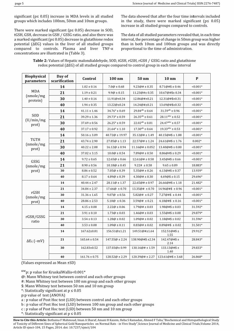
4.2 Biochemical studies:
Hepatic malondialdehyde levels, SOD, tGSH, rGSH, rGSH ⁄GSSG ratio and glutathione redox potential (ΔEG) in controls
and different studied groups are illustrated in (Table 2.).
The data obtained showed that after the four time intervalsincluded in the present experiment, there were marked
How to Cite this Article: Bothaina F Mahmoud, Iman A Sharaf, Amani H Kazem, Heba S Ramadan, Ahmed F Taha,"Biochemical and Histopathological Studyof Toxicity of Different Sizes of Spherical Gold Nanoparticles on Normal Rats - in Vivo Study",Science Journal of Medicine and Clinical Trials,Volume 2014,Article ID sjmct-104, 15 Pages, 2014. doi: 10.7237/sjmct/104
page 5 Science Journal of Medicine and Clinical Trials( ISSN:2276-7487)
significant (p≤ 0.05) increase in MDA levels in all studiedgroups which includes 100nm, 50nm and 10nm groups.
There were marked significant (p≤ 0.05) decrease in SOD,tGSH, GSH, decrease in GSH ⁄ GSSG ratio, and also there wasa marked significant (p≤ 0.05) decrease in glutathione redoxpotential (ΔEG) values in the liver of all studied groupscompared to controls. Plasma and liver TNF‑αconcentrations are illustrated in (Table 3).
The data showed that after the four time intervals includedin the study, there were marked significant (p≤ 0.05)increase in all studied groups compared to controls.
The data of all studied parameters revealed that, in each timeinterval, the percentage of change in 50nm group was higherthan in both 10nm and 100nm groups and was directlyproportional to the time of administration.
Biophysicalparameters
Day ofscarification Control 100 nm 50 nm 10 nm P
MDA(nmole/mg
protein)
14 1.02 ± 0.16 7.0@ ± 0.68 9.23@# ± 0.35 8.71@#$ ± 0.46 <0.001*
21 1.19 ± 0.21 9.9@ ± 0.15 11.23@#± 0.35 10.67@#$± 0.34 <0.001*
30 1.40 ± 0.16 11.91@±0.34 12.86@#±0.21 12.31@#$±0.31 <0.001*
40 1.94 ± 0.35 13.22@±0.24 14.24@#±0.21 13.69@#$±0.32 <0.001*
SOD(U⁄min⁄mg
prot)
14 41.11 ± 1.46 34.76� ± 0.69 29.84�� ± 0.64 31.59�� ± 0.96 <0.001*
21 39.29 ± 1.36 29.73� ± 0.59 26.35�� ± 0.61 28.11��� ± 0.52 <0.001*
30 37.69 ± 0.56 26.25� ± 0.59 22.02�� ± 0.81 24.47��� ± 0.57 <0.001*
40 37.17 ± 0.92 21.64� ± 1.10 17.30�� ± 0.64 19.33��� ± 0.53 <0.001*
TGTH(nmole⁄mg
prot)
14 50.16 ± 3.09 40.72@ ± 19.97 35.12@# ± 1.49 40.15@#$ ± 1.88 <0.001*
21 43.74 ± 2.90 27.85@ ± 1.13 22.57@# ± 1.24 24.61@#$ ± 1.76 0.002*
30 40.22 ± 2.08 16.12@ ± 0.94 11.16@# ± 0.052 13.40@#$ ± 0.80 <0.001*
40 37.02 ± 3.15 10.0@ ± 0.54 7.89@# ± 0.50 8.86@#$ ± 0.29 <0.001*
GSSG(nmole⁄mg
prot)
14 9.72 ± 0.65 12.65@ ± 0.66 12.61@# ± 0.58 3.45@#$ ± 0.66 <0.001*
21 8.90 ± 0.56 10.18@ ± 0.45 9.22# ± 0.58 9.65 ± 0.89 18.883*
30 8.86 ± 0.52 7.05@ ± 0.39 5.35@# ± 0.26 6.13@#$ ± 0.37 13.939*
40 8.17 ± 0.64 4.89@ ± 0.39 4.30@# ± 0.30 4.69@$ ± 0.15 29.696*
rGSH(nmole⁄mg
prot)
14 40.44 ± 2.47 28.11@ ± 1.37 22.65@# ± 0.97 26.66@#$ ± 1.18 21.482*
21 34.84 ± 2.37 17.66@ ± 0.70 13.35@# ± 0.70 14.96@#$ ± 0.96 <0.001*
30 31.36 ± 1.65 9.07@ ± 0.56 5.82@# ± 0.27 7.27@#$ ± 0.44 <0.001*
40 28.86 ± 2.53 5.10@ ± 0.36 3.59@# ± 0.21 4.18@#$ ± 0.16 <0.001*
rGSH⁄GSSGratio
14 4.15 ± 0.08 2.22@ ± 0.06 1.79@# ± 0.03 1.98@#$ ± 0.03 31.592*
21 3.91 ± 0.10 1.73@ ± 0.03 1.46@# ± 0.03 1.55@#$ ± 0.08 29.879*
30 3.54 ± 0.13 1.28@ ± 0.02 1.09@# ± 0.02 1.18@#$ ± 0.02 31.596*
40 3.53 ± 0.08 1.09@ ± 0.11 0.83@# ± 0.02 0.89@#$ ± 0.02 31.501*
∆EG (–mV)
14 167.62±0.81 156.51@±1.21 149.51@#±1.64 152.51@#$ ±1.55
29.912*
21 165.64 ± 0.54 147.55@ ± 2.24 138.90@#$ ±2.34 142.47@#$ ±2.14
28.843*
30 162.83±0.52 137.03@± 0.99 130.16@# ± 1.59 133.13@#$ ±1.40
29.833*
40 161.76 ± 0.75 128.52@ ± 2.29 120.39@# ± 2.27 123.61@#$ ± 3.68 26.868*
Table 2: Values of Hepatic malondialdehyde, SOD, tGSH, rGSH, rGSH ⁄ GSSG ratio and glutathioneredox potential (ΔEG) of all studied groups compared to control group in each time interval
(Values expressed as Mean ±SD)
��p: p value for KruskalWallis<0.001*@: Mann Whitney test between control and each other groups#: Mann Whitney test between 100 nm group and each other groups$: Mann Whitney test between 50 nm and 10 nm group*: Statistically significant at p ≤ 0.05p:p value of test (ANOVA)a : p value of Post Hoc test (LSD) between control and each other groupsb : p value of Post Hoc test (LSD) between 100 nm group and each other groupsc : p value of Post Hoc test (LSD) between 50 nm and 10 nm group*: Statistically significant at p ≤ 0.05
How to Cite this Article: Bothaina F Mahmoud, Iman A Sharaf, Amani H Kazem, Heba S Ramadan, Ahmed F Taha,"Biochemical and Histopathological Studyof Toxicity of Different Sizes of Spherical Gold Nanoparticles on Normal Rats - in Vivo Study",Science Journal of Medicine and Clinical Trials,Volume 2014,Article ID sjmct-104, 15 Pages, 2014. doi: 10.7237/sjmct/104

Science Journal of Medicine and Clinical Trials( ISSN:2276-7487) Page 6
Biochemicalparameters
Day ofscarification Control 100 nm 50 nm 10 nm KWp
PlasmaTNF‑ αValues
(pg/ml)
14 day 41.40 ±3.97 78.10@#$ ± 2.77 91.10# ± 2.92 84.0 ± 3.33 0.010*21 day 43.0 ± 3.81 95.30@ ± 3.37 109.20@# ±3.3 104.20@#$±3.29 <0.001*30 day 455.80 ±5.0 112.80@ ± 3.71 125.20@# ± 3.55 120.60@#$ ±3.50 <0.001*40day 48.60 ±4.39 131.20@ ± 3.61 143.20@# ± 1.81 38.30@#$ ±1.42 <0.001*
HepaticTNF‑ αValues
(pg/ml)
14 day 164.60± 4.3 381.40@ ± 2.80 404.40@# ± 3.03 394.10@#$ ±1.60 <0.001*21 day 168.8 ± 4.0 409.0@ ± 1.56 431.60@# ± 2.99 423.50@#$ ± 2.22 <0.001*30 day 173.20 ±4.0 439.50@ ± 2.27 460.60@# ± 2.50 452.50@#$ ± 1.08 <0.001*40 day 176.60 ±4.8 468.50@ ± 2.68 487.90@#±3.28 478.70@#$ ± 3.40 <0.001*
Table 3 : TNF‑ α values in plasma and liver samples of controls and different study groups
(Values expressed as mean ±SD)
KWp: p value for Kruskal@: p value for Mann Whitney test between control and each other groups#: p value for Mann Whitney test between 100 nm group and each other groups$: p value for Mann Whitney test between 50 nm and 10 nm group*: Statistically significant at p ≤ 0.05
4.3 Correlation between glutathione redox potentialand hepatic TNF‑α in the 50nm group
redox potential and hepatic TNF‑a after 21, 30 and 40 days,respectively, of repeated administration of 50nm GNPs . (Fig.7, 8, and 9).
4.4 Histopathological study
The histopathology of liver and spleen sections of all studiedand control groups after the four time intervals of theexperiment are presented in Figures (10—-17). The hepatictissue sections of studied animals revealed various degreesof hepatic tissue injury after the four time intervals of theexperiment, characterized by mild to heavy portal andlobular inflammation, hepatocellular degeneration, and fattychanges (steatosis).
The histopathological examination revealed that there were
two types of liver cell death, apoptosis (or programmed celldeath) and necrosis (or sudden cell death).
Also, it was observed that necrotic foci were often associatedwith fibrosis. On the other hand, the splenic tissue sectionsof GNPs-administrated rats revealed no significantdifferences in histology after 14 day and 21 day of GNPsadministration.
Some morphological changes that include congested redpulp, reactive white pulp and wide reactive marginal zonewere observed after 30day and 40 day of GNPsadministration.
It was noticed that the recorded abnormal morphologicalchanges in both the hepatic and splenic tissue sectionsincreased with increasing time of administration. In eachtime interval, the recorded destructive and degenerativechanges were maximum at 50nm GNPs administrated rats.
Fig. (7): Correlation between glutathione redox potential and hepatic TNF‑α after 21 day of repeatedadministration of 50nm GNPs
How to Cite this Article: Bothaina F Mahmoud, Iman A Sharaf, Amani H Kazem, Heba S Ramadan, Ahmed F Taha,"Biochemical and Histopathological Studyof Toxicity of Different Sizes of Spherical Gold Nanoparticles on Normal Rats - in Vivo Study",Science Journal of Medicine and Clinical Trials,Volume 2014,Article ID sjmct-104, 15 Pages, 2014. doi: 10.7237/sjmct/104

Science Journal of Medicine and Clinical Trials( ISSN:2276-7487) Page 6
Fig. (8): Correlation between glutathione redox potential and hepatic TNF‑α after 30 day of repeatedadministration of 50nm GNPs
Fig. (9): Correlation between glutathione redox potential and hepatic TNF‑α after 40 day of repeatedadministration of 50nm GNPs
Fig(10): Hepatic tissue section showing normal liver architecture. (X400)
How to Cite this Article: Bothaina F Mahmoud, Iman A Sharaf, Amani H Kazem, Heba S Ramadan, Ahmed F Taha,"Biochemical and Histopathological Studyof Toxicity of Different Sizes of Spherical Gold Nanoparticles on Normal Rats - in Vivo Study",Science Journal of Medicine and Clinical Trials,Volume 2014,Article ID sjmct-104, 15 Pages, 2014. doi: 10.7237/sjmct/104

Science Journal of Medicine and Clinical Trials( ISSN:2276-7487) Page 6
Fig(11): H&E stained splenic tissue section showing normal splenic architecture (normalwhite and red pulp). (x200)
Fig(12): H&E stained hepatic tissue section showing minimal portal inflammation andcongestion with apoptotic hepatocytes (arrows). (X400)(100nm after 21day)
Fig(13): H&E stained hepatic tissue section showing intralobular inflammatory infiltrate with lyticnecrosis and Councilman apoptotic bodies (arrows).(X400)(50mn after21 day)
How to Cite this Article: Bothaina F Mahmoud, Iman A Sharaf, Amani H Kazem, Heba S Ramadan, Ahmed F Taha,"Biochemical and Histopathological Studyof Toxicity of Different Sizes of Spherical Gold Nanoparticles on Normal Rats - in Vivo Study",Science Journal of Medicine and Clinical Trials,Volume 2014,Article ID sjmct-104, 15 Pages, 2014. doi: 10.7237/sjmct/104

Science Journal of Medicine and Clinical Trials( ISSN:2276-7487) Page 6
Fig(14): H&E stained hepatic tissue section showing heavy portal inflammation and an apoptotichepatocyte (arrow)(100nm after 30 min)
Fig(15):H&E stained hepatic tissue section showing early portal fibrosis with short septae (arrows),feathery degeneration of hepatocytes. (X400 )(50nm after30 day)
Fig(16): H&E stained splenic tissue section showing reactive white pulp with wide marginal zone.The central arteriole has thick hyalinized wall (arrow). (X400) (50nm after40 day)
How to Cite this Article: Bothaina F Mahmoud, Iman A Sharaf, Amani H Kazem, Heba S Ramadan, Ahmed F Taha,"Biochemical and Histopathological Studyof Toxicity of Different Sizes of Spherical Gold Nanoparticles on Normal Rats - in Vivo Study",Science Journal of Medicine and Clinical Trials,Volume 2014,Article ID sjmct-104, 15 Pages, 2014. doi: 10.7237/sjmct/104

Science Journal of Medicine and Clinical Trials( ISSN:2276-7487) Page 6
Fig(17): H&E stained hepatic tissue section showing moderate portal inflammation, with necrotichepatocytes (arrows). (X400) (100nm after 40 days)
5.0 DISCUSSION
Nanotechnology has recently emerged as a promisingapproach for treatment and diagnosis of a variety ofdiseases.( 36) In order to use GNPs in drug delivery,diagnosis, and treatment, it is essential to characterize thetoxicity associated with repeated administration of thesemolecules. In the present study, the toxicological effects ofdifferent sizes of colloidal gold nanoparticles were evaluatedby assessment of oxidative stress , antioxidants and liver andsplenic cell death (apoptosis or necrosis).
In the present study the size and morphology of GNPs weredetermined by particle size analyzer (PSA) and transmissionelectron microscopy (TEM) ⁽³⁷⁾. The results revealed thepresence of completely spherical GNPs with smooth surfacesand have sizes of 10nm, 50nm and 100nm.
The aggregation shape parameter (ASP) was significantlyhigher in studied groups compared to the control group, andthe percentage of the increase was directly proportional tothe time of administration. In each time interval, the ASPvalues in the 50nm group were higher than in both the 10nmand 100nm groups.
This study presents aggregation shape parameter (ASP) as aquantitative determination of RBC aggregate morphology,derived from the numerical process of digitized image of RBCaggregates. This provides a useful reference for measuringdeviations of RBC aggregate morphology: a rouleauxstructure characterizes normal aggregates, while theformation of RBC clusters characterizes disease states.⁽ ³⁸⁾
Starting from 21st day, whole blood viscosity wassignificantly higher in studied groups compared to controlgroup. The results are in agreement with several studies whowhich reported that hyperviscosity syndrome can result fromgross increase in plasma viscosity, red cell aggregation,elevated hematocrit, and from increased number ofcirculating rigid red cells (e.g. sickling disorder) or white cells(hyperleucocytosis).⁽³⁹‑⁴¹⁾
It is known that the inflammatory response is associated withan acute phase reaction characterized by appearance of anincreased leukocyte count, accelerated erythrocytesedimentation rate(ESR), hyperfibrinogenemia, hyper-gammaglobulinemia, increased synthesis of C-reactiveprotein (CRP) and other acute phase proteins. The stimulusfor production is likely to be inflammatory cytokines such asinterleukin-1, interleukin-6 and tumor necrosis factor (TNF).There is a concept that the acute phase proteins are involvedin increased red cell aggregation and sedimentation⁽⁴²,⁴³⁾.
In the present study, the toxicity of GNPs has beeninvestigated at the cellular level. The levels ofmalondialdehyde (MDA) in liver homogenate weresignificantly higher in all studied groups compared to thecontrol group. Activity of superoxide dismutase (SOD) andlevels of reduced glutathione (GSH), and ratio of reducedglutathione to oxidized glutathione (GSH⁄GSSG) in liverhomogenate were significantly lower in all studied groupscompared to the control which may confirm the presence ofoxidative stress.
Nanomaterial toxicity can occur through several differentmechanisms in the body. The main molecular mechanism ofin vivo nanotoxicity is the induction of oxidative stress by freeradical formation.⁽ ⁴⁴⁾ Oxidative stress may have a role in theinduction or the enhancement of inflammation throughupregulation of redox sensitive transcription factors (e.g.Nuclear Factor‑κB).( 45) Slow clearance and tissueaccumulation (storage) of potential free radical producingnanomaterials as well as prevalence of numerous phagocyticcells in the organs of the reticuloendothelial system (RES)makes organs such as the liver and spleen main targets ofoxidative stress.⁽ ⁴⁶⁾
Once generated, free radicals can react with all cellularmacromolecules, including lipids and proteins. Proteinoxidation, particularly of enzymes, can lead to impairment oftheir function.⁽ ⁴⁷⁾ Lipid peroxidation is a free radical chainreaction, which arises from the oxidative conversion of
How to Cite this Article: Bothaina F Mahmoud, Iman A Sharaf, Amani H Kazem, Heba S Ramadan, Ahmed F Taha,"Biochemical and Histopathological Studyof Toxicity of Different Sizes of Spherical Gold Nanoparticles on Normal Rats - in Vivo Study",Science Journal of Medicine and Clinical Trials,Volume 2014,Article ID sjmct-104, 15 Pages, 2014. doi: 10.7237/sjmct/104

Science Journal of Medicine and Clinical Trials( ISSN:2276-7487) Page 6
polyunsaturated fatty acids by O H∙ to lipid peroxides, whichin turn can damage biological membranes.(48) MDA level iswidely utilized as a marker of lipid peroxidation in states ofelevated oxidative stress. Possible mechanisms involved inthe elevated lipid peroxidation in hepatic toxicity have beenproposed.
First, cellular membranes in the liver are rich inpolyunsaturated fatty acids, which are especially sensitive tofree radical attack. Second, the liver contains a significantamount of iron ions, which stimulate free radicalgeneration.⁽⁴⁹⁾
GSH is a free radical scavenger and a proton donor forglutathione peroxidase. It was reported that depletion of GSH,decreasing GSH to GSSG ratio and decreased glutathioneredox potential (ΔEG) are markers of for oxidative stress.⁽ ⁵⁰,⁵¹⁾ Our present results were confirmed in accordance withthose of Siddiqi et al ⁽⁵²⁾ who demonstrated that GNPs causegeneration of oxidative stress and an impairment of theantioxidant enzyme glutathione peroxidase in rat brain. GNPsalso cause generation of 8-hydroxydeoxyguanosine (8OHdG),caspase-3 and heat shock protein70 (Hsp70), and which maylead to inflammation and DNA damage/cell death.
It was demonstrated that the total GNPs content in cellsincreased in a time-dependent manner without reaching aplateau in the first 24 h. GNPs were shown to damage thecytoskeleton organization, with the most prominent effectseen for GNPs with size 5 nm.⁽⁵³⁾
The results revealed that, the levels of TNF‑α in plasma andliver homogenate were significantly higher in all studiedgroups compared to the control group . These results are inagreement with the other studies which found that, TNF‑αlevels were elevated in rats with liver injury. Hepatocytescarry receptors that respond even to low TNF‑αconcentration. Thus, hepatocytes are naturally sensitive tothe cytokines. The interaction of TNF‑α and TNF‑α receptorsinitiate chemical processes in the cell that lead to apoptosis.⁽⁵⁴,⁵⁵⁾
Increased levels of TNF‑α in plasma and liver homogenatemay be also due to identification of GNPs as foreign by theimmune cells, causing the cells to react against either surfaceor core components to mount an inflammatory response,which involve secretion of signaling molecules to attractmore cells to destroy the foreign substances.⁽⁵⁴⁾
Histopathological studies of the hepatic tissue sections ofstudied animals revealed various degrees of hepatic tissueinjury after the four time intervals of the experiment,characterized by mild to heavy portal and lobularinflammation, hepatocellular degeneration, fatty changes(steatosis) , apoptosis and necrotic foci which were oftenassociated with fibrosis. On the other hand, the splenic tissuesections of GNPs-administrated rats revealed no significantdifferences in histology after 14 and 21 days of GNPsadministration. While, some morphological changes thatinclude congested red pulp, reactive white pulp and widereactive marginal zone were observed after 30 and 40 daysof GNPs administration.
It was noticed that the recorded abnormal morphologicalchanges in both the hepatic and splenic tissue sections
increased with increasing time of administration. In eachtime interval, the recorded destructive and degenerativechanges were maximum at 50nm GNPs administrated rats.Cho et al. ⁽⁵⁶⁾ studied the in vivo toxic effects of 13 nm sizePEG-coated GNPs on mice. The nanoparticles were seen toinduce acute inflammation and apoptosis in the liver. Theyaccumulated in the liver and spleen for up to 7 days afterinjection and had long blood circulation times. In addition,transmission electron microscopically examinations revealedthat numerous cytoplasmic vesicles and lysosomes of liverKupffer cells and spleen macrophages contained the PEG-coated gold nanoparticles. Because PEG-coated GNPs arewidely used in biomedical applications these effects haveobvious clinical implications.
It is thought that nanoparticles should have finalhydrodynamic diameters ≤ 5.5 nm to be excreted from therat body by the renal route. ⁽⁵⁷⁾ Since the majority of thestudied gold nanoparticles are larger than this renal filtrationcutoff, in the few studies that have been performed, the goldnanoparticles (NPs) were not excreted in urine; instead theywere found to be eliminated from the blood by thereticuloendothelial system (RES) and thus to accumulate inthe spleen and liver.⁽⁵⁸,⁵⁹⁾
The percentages of the change in all studied parameters weredirectly proportional to the time of administration. In eachtime interval, the percentages of the decrease in 50nm groupwere higher than in both 10nm group and 100nm groupThese results are in agreement with the finding of Hyllier andAlbertch,⁽⁶⁰⁾ who showed that administrated GNPs appearedin various tissues in mice and that the amount of absorptionand distribution in the blood was in correlationed with thesize of the particles.
In most studies, systemically administrated NPs wereprimarily taken up by liver and spleen in a large quantity andsmall amounts distributed in the lung, kidney, heart, andbrain after single administration. However, little is knownabout biodistribution, accumulation and toxicity of GNPsafter repeated administration.⁽⁶¹‑⁶³⁾Accumulation ofnanomaterials in the liver and spleen after being taken up bythe reticuloendothelial system could lead to hepatic andsplenic toxicity.⁽ ⁶⁴⁾
In the present histopathological findings, two processesplayed a role in hepatocyte destruction— apoptosis andnecrosis. When a cell undergoes apoptosis, the entire cell,including the nucleus, separates into numerous fragments(i.e., apoptotic bodies).
Simultaneously, the genetic material (i.e., DNA) of apoptoticcells breaks into a characteristic pattern of pieces of varyingsizes. During the breakup of the cell, the cell continues toproduce proteins and adenosine triphosphate (ATP), amolecule that is required for most of the cell’s energy-consuming metabolic processes and which is essential forcell functioning. As a result, each apoptotic body, which issurrounded by a piece of cell membrane, contains intactfunctional cell components.⁽⁶⁵,,⁶⁶⁾
6.0 CONCLUSION
In conclusion, the results of in vivo cytotoxicity of size-sortedGNPs are the induction of oxidative stress and immune
How to Cite this Article: Bothaina F Mahmoud, Iman A Sharaf, Amani H Kazem, Heba S Ramadan, Ahmed F Taha,"Biochemical and Histopathological Studyof Toxicity of Different Sizes of Spherical Gold Nanoparticles on Normal Rats - in Vivo Study",Science Journal of Medicine and Clinical Trials,Volume 2014,Article ID sjmct-104, 15 Pages, 2014. doi: 10.7237/sjmct/104

Science Journal of Medicine and Clinical Trials( ISSN:2276-7487) Page 6
response. The toxicological effects of GNPs are size and timedependent. Different sizes of GNPs could be endocytosed byhepatic and splenic cells and have large toxic effect onhepatic tissue( apoptosis and necrosis) while its effect onsplenic tissue is limited and could be observed after long-term of repeated administration.
7.0 RECOMMENDATIONS
According to the results of our study we recommend thefollowing:
● Avoiding direct exposure to GNPs during theirpreparation .
● Investigation of the toxicological influence of differentsizes and other different properties of GNPs on differenttissues such as kidney, brain and lungs.
● Administration of antioxidants before treatment withGNPs in a trial to minimize the toxicological effects ofGNPs.
8.0 ACKNOWLEDGEMENTS
The authors are grateful to the animal house of the MedicalResearch Institute, Alexandria University, Egypt forproviding us with the animals used in this study.
9.0 REFERENCES
1. G.K.Stylios, P.V.Giannoudis, T. Wan. Applications ofnanotechnology in medical practice. Injury. 36, 6(2005).
2. V.Balzani, Nanoscience and nanotechnology: A personal viewof a chemist. Small.1, 278(2005).
3. R.Sperling, P. Gil, F. Zhang , M.Zanella, W.Parak. Biologicalapplications of gold nanoparticles. Chem. Soc. Rev. 37,1896(2008).
4. M.Grzelczak, J. Perez‑Juste, P.Mulvaney, L. Liz‑Marzan. ShapeControl in Gold Nanoparticle Synthesis. Chem. Soc. Rev.37,1783(2008).
5. D.T.Felson, J.J. Anderson, R.F.Meenan. The comparative efficacyand toxicity of second-line drugs in rheumatoid arthritis.Results of two meta-analyses. Arthritis Rheum. 33,1449(1990)
6. I.C. Shaw. Gold-based therapeutic agents. Chem Rev. 99,2589(1999).
7. K.Unfried, C. Albrecht, L.O. Klotz, A.Von Mikecz, S,Grether-Beck, R.P..FSchins. Cellular responses to nanoparticles: targetstructures and mechanisms. Nanotoxicology. 1,52(2007).
8. Y. Pan, A.Leifert, D.Ruau, S.Neuss, J.Bornemann et al. Goldnanoparticles of diameter 1.4 nm trigger necrosis by oxidativestress and mitochondrial damage. Small.5, 2067 (2009).
9. P. Ionita, M. Conte, B. C. Gilbert,V.Chechik Gold nanoparticle-initiated free radical oxidations and halogen abstraction.Org.Biomol. Chem. 5, 3504(2007)
10. D. Brown. Calcium and ROS-mediated activation oftranscription factors and TNF-alpha cytokine gene expressionin macrophages exposed to ultrafine particles, Am J PhysiolLung Cell Mol Physiol.286,344 (2004).
11. L.Risom. Acute hypoxia and reoxygenation-induced DNAoxidation in human mononuclear blood cells. Mutat Res.625,125(2007).
12. C.Sioutas, R.J.Delfino, M. Singh. Exposure assessment foratmospheric ultrafine particles (UFPs) and implications inepidemiologic research, Environ Health Perspect.113,947(2005).
13. V. Stone, M.Tuinman, J.E.Vamvakopoulos, J. Shaw, D.Brownetal. Increased calcium influx in a monocytic cell line onexposure to ultrafine carbon black. EurRespir J. 15, 297(2000)
14. G.Waris, H.Ahsan. Reactive oxygen species: role in thedevelopment of cancer Carcinog. 5, 14 (2006).
15. A. Slater, I. Nobel, S.Orrenius. The role of intracellular oxidantsin apoptosis. Biochem. Biophys. Acta.1271,59(1995)
16. Dumont, S.P.Hehner, T.G.Hofmann etal. Hydrogen peroxideinduced-apoptosis is CD95- independent, requires the releaseof mitochondria-derived reactive oxygen species and theactivation of NF-kB. Oncogene. 18,747 (1999)
17. E.C. Opera. Oxidative stress. Dis. Mon.52, 183(2006)
18. J.Mcdermott. Antioxidant nutrients: current dietaryrecommendations and research update. J Am Pharm assoc.40,785(2000)
19. J.A.Tainer, E.D.Getzoff, J.S. Richardson, D.C. Richardson.Structure and mechanism of copper, zinc superoxidedismutase. Nature.306 , 284(1983)
20. A.Pompella. The changing faces of glutathione, a cellularprotagonist. Biochemical Pharmacology. 66, 1499( 2003)
21. T. Toshiyuki, T.Takahito. Carnosic acid and carnosol inhibitadipocyte differentiation in mouse 3T3-L1 cells throughinduction of phase2 enzymes and activation of glutathionemetabolism. Biochem. Biophys. Res. Commun. 382, 549(2009).
22. A.D. McFarland et al. Color My Nanoworld. J. Chem. Educ.81,544A(2004).
23. S.Peng, Y. Lee, C.Wang, H. Yin, S. Dai, S. Sun.A Facile Synthesisof Monodisperse Au Nanoparticles and Their Catalysis of COOxidation. Nano Res.1, 229 (2008).
24. N. Nguyen, V. Le, D. Chu, C.Sai, T.Cao et al. Synthesis and opticalproperties of colloidal gold nanoparticles. J.Phys.187,12(2009).
25. H.Yazid, R. Adnan, S.Abdulhamid, M.Farrukh. Synthesis andcharacterization of gold nanoparticles supported on zinc oxidevia the deposition-precipitation method. Turk JChem..34,639(2010).
26. M.Elbelbese. Biophysical behavior of red blood cellsaggregation under different shear rates. PhD. Thesis submittedto Biophysics Department, Medical Research Institute,Alexandria. Egypt.12, 44(2005).
27. M. David, B. Shelly, S. Mark, T. Albert. Hematocrit, Volumeexposure, Temperature, and Shear Rate Effects on Bloodviscosity. Anesth Analg. 91,539(2000).
28. H.H. Draper, M. Hadley. Malondialdehyde determination asindex of lipid peroxidation. Methods Enzy mol.186, 421(1990).
29. S. G.Marklund. Involvement of superoxide anion radical inautoxidation of pyrogallol and a convenient assay for SOD. Eur.J. Biochem.47, 469(1974).
30. O.W. Griffith. Determination of glutathione and glutathionedisulfide using glutathione reductase and 2-vinyl pyridine.Anal Biochem.106,207 (1980).
How to Cite this Article: Bothaina F Mahmoud, Iman A Sharaf, Amani H Kazem, Heba S Ramadan, Ahmed F Taha,"Biochemical and Histopathological Studyof Toxicity of Different Sizes of Spherical Gold Nanoparticles on Normal Rats - in Vivo Study",Science Journal of Medicine and Clinical Trials,Volume 2014,Article ID sjmct-104, 15 Pages, 2014. doi: 10.7237/sjmct/104

Science Journal of Medicine and Clinical Trials( ISSN:2276-7487) Page 6
31. F. Q. Schafer, G FBuettner. Redox environment of the cell asviewed through the redox state of the glutathione disulfide .glutathione couple. Free Radic Biol Med.30,1191.(2001).
32. O.H. Lowry, N.J.Rosebrough, A.L. Farr, R.J. Randall. Proteinmeasurements with Folin-phenol reagent. J. Biol. Chem.193,265(1951).
33. J.I.Y. Chung, E.N.Benveniste. Cloning and sequence analysis ofthe rat tumor necrosis factor –encoding genes. Gene.132,227(1993).
34. J.D. Bancroft, A.Stevans, D.R.Tuener. (1996): Theory andPractice of Histopathological Techniques", 4th ed., ChurchillLivingstone, Edinburgh, London, Melbourne, New York
35. M.J.Nursis. Statistical package for social sciences for the IBMPC/ XT: statistical guide, ver. 11.5.0. Chicago, Illinoise: SPSS Inc.;2002.
36. S.D. Caruthers, SAWickline, GMLanza. Nanotechnologicalapplications in medicine. Curr. Opin. Biotechnol. 18,26(2007).
37. A.D.McFarland and C.L. Haynes. Color My Nanoworld. J. Chem.Educ. 81,544A(2004).
38. S. Chen. Monitoring of erythrocyte aggregate morphologyunder flow by computerized image analysis.Biorrheology.32,487(1995)
39. J. Fahey, W Barth, A. Solomon. Serum hyperviscositysyndrome.J.A.M.A. 192,464(1965).
40. M.K.Home. Sickle cell anemia as a rheologic disease. Amer. J.Med..70,288 (1981).
41. M.Lichtman, J. Row. Hyperleukocytic leukemias: Rheological,clinical and therapeutic considerations blood. 60, 279(1982).
42. S.E. Epstein, Y.F. Zhou, J. Zhu. Infection and atherosclerosis:emerging mechanistic paradigms. Circulation.100,20 ( 1999).
43. X.Weng, G. Cloutier, R. Beaulieu, et al. Influence of acute-phaseproteins on erythrocyte aggregation. Am JPhysiol..271,2346(1996).
44. S.Lanone, J.Boczkowski. Biomedical applications and potentialhealth risks of nanomaterials: molecular mechanisms. CurrMolMed. 6,651(2006).
45. I.Rahman. Regulation of nuclear factorκB, activator protein-1,and glutathione levels by tumor necrosis factor‑α anddexamethasone in alveolar epithelial cells. BiochemPharmacol.60,1041(2000).
46. I. Rahman, S.K.Biswas, L.A.Jimenez, M. Torres, H.J.Forman.Glutathione, stress responses, and redox signaling in lunginflammation. Antioxid Redox Signal.7, 42(2005)
47. A. Cherubini, C.Polidori, C. Benedetti, S.Ercolani, U.Senin,P.Mecocci. Association Between Ischemic Stroke and IncreasedOxidative Stress. Stroke.28,2231(2002).
48. A.Yildirim, M.Gumus, S.Dalga, Y.N.Sahin, F.Akcay.Dehydroepiandrosterone improves hepatic antioxidantsystems after renal ischemia-reperfusion injury in rabbits. AnnClin Lab Sci. 33, 459(2003).
49. S.M.H.Sadrzadeh, A.A.Nanji, .PL. Price. The oral iron chelator,1,2-dimethyl-3- hydroxypyrid-4-one reduces hepatic free iron,lipid peroxidation and fat accumulation in chronically ethanol-fed rats. Journal of Pharmacology and ExperimentalTherapies.269,632 (1994).
50. J.B. Schulz, J.Lindenau, J.Seyfried, J.Dichgans. Glutathione,oxidative stress and neurodegeneration.Eur J Biochem . 267,4904 (2000)
51. D.S. Warner, H, Sheng IBatinic-Haberle. Oxidants, antioxidantsand the ischemic brain.J Exp Biol. 207, 3221(2004).
52. J.N.Siddiqi,A.K. M.Abdelhalim,A. K. El-Ansary,S.A.Alhomida,W.Y.Ong. Identification of potential biomarkers of goldnanoparticle toxicity in rat brains. JNeuroinflammation.9,123(2012).
53. C. P.Gioria, P.Garcıa, F.Nativo, D.Franchin, J. Gilliland, F.Ponti.In'Toxicology Letters' .11,22(2012).
54. C.J.McClain, D.A. Cohen, C.A.Dinarello, J.G. Cannon, S.I.Shedlofskyet al. Cytokines and alcoholic liverdisease.Seminars in Liver Disease. 13,170.(1993).
55. J.Ghos, J. Das, P .Manna, P.C.Sil. Acetaminophen induced renalinjury via oxidative stress andTNF-alpha production:Therapeutic potential of arjunolic acid. Toxicology.268, 8(2010).
56. W.S. Cho, M. Cho, J.Jeong, M.Choi, H.Y.Cho, B.S.Han, S.H. Kim,H.O. Kim, Y.T.Limc, B.H. Chung, J.Jeong. Acute toxicity andpharmacokinetics of 13 nm-sized PEG-coated goldnanoparticles. Toxicol Appl Pharmacol. 236,16(2009).
57. H.S. Choi, J.P. Zimmer, E. Tanaka, J.V.Frangioni, I. M.Bawendetal. Renal clearance of quantum dots. NatBiotechnol.25,1165(2007)
58. W.H. De Jong, W.I.Hagens, P.Krystek, R.MCBurge, A.J.A.M.Sips,R.E.Geertsma. Particle sizedependent organ distribution ofgold nanoparticles after intravenous administration.Biomaterials.29,1912(2008).
59. G.Von MaltzahnJi-Ho ParkA.Agrawal, N. K.Bandaru, S. K. DasM.J. Sailor,S. N. Bhatia.Computationally guided photothermaltumor therapy using long circulating gold nanorod antennas.Cancer Res.69,3892(2009)
60. J.F.Hillyer, R.M.Albrecht. Gastrointestinal persorption andtissue distribution of differently sized colloidal goldnanoparticles. J. Pharm. Sci. 90,1927(2001).
61. G.Sonavane, K.Tomoda, K. Makino.Biodistribution of colloidalgold nanoparticles after intravenous administration: effect ofparticle size. Colloids Surf. B Biointerfaces.66 ,274(2008)
62. W.H. De Jong, W.I.Hagens, P.Krystek, M.C. Burger, A.J.Sips et al.Particle size-dependent organ distribution of goldnanoparticles after intravenous administration. Biomaterials. 29,1912(2008)
63. M.Semmler-Behnke, W.G.Kreyling, J.Lipka, S.Fertsch, A.Wenk,S.Takenaka et al. Biodistribution of 1.4- and 18-nm goldparticles in rats, Small .4,2108(2008)
64. Y.S. Chen, Y.C. Hung, I.Liau, G.S. Huang. Assessment of the invivo toxicity of gold nanoparticles.Nanoscale Res Lett.4,858(2009)
65. B.I.Jugdutt, H.A.Idikio. Apoptosis and oncosis in acute coronarysyndromes: Assessment and implications. Mol Cell Biochem.270, 177( 2005)
66. B.G. Rosser, G.J.Gores. Liver cell necrosis: Cellular mechanismsand clinical implications. Gastroenterology. 108,252(1995)
How to Cite this Article: Bothaina F Mahmoud, Iman A Sharaf, Amani H Kazem, Heba S Ramadan, Ahmed F Taha,"Biochemical and Histopathological Studyof Toxicity of Different Sizes of Spherical Gold Nanoparticles on Normal Rats - in Vivo Study",Science Journal of Medicine and Clinical Trials,Volume 2014,Article ID sjmct-104, 15 Pages, 2014. doi: 10.7237/sjmct/104


![The Locally Denatured State of Glutathione S- Transferase AI-I ...psb.stanford.edu/psb-online/proceedings/psb99/nieslanik.pdf3.2 X-ray structure of the binary complex [GST. GSH]. In](https://img.pdfslide.us/doc/110x75/5fa3a4de2322d0570176cfae/the-locally-denatured-state-of-glutathione-s-transferase-ai-i-psb-32-x-ray.jpg)


![Review Article Role of Glutathione in Cancer Progression ...downloads.hindawi.com/journals/omcl/2013/972913.pdf · GCL and glutathione S-transferases [ ]. 2. GSH Biosynthesis Glutathione](https://img.pdfslide.us/doc/110x75/5edbd12aad6a402d666637cd/review-article-role-of-glutathione-in-cancer-progression-gcl-and-glutathione.jpg)