Embed Size (px)
Citation preview
8/18/2019 Biochem 1995
http://slidepdf.com/reader/full/biochem-1995 1/7
Subscriber access provided by BIRLA INST OF TECH
Biochemistry is published by the American Chemical Society. 1155 Sixteenth StreetN.W., Washington, DC 20036
Interaction of Polycyclic Aromatic Hydrocarbons
and Flavones with Cytochromes P450 in theEndoplasmic Reticulum: Effect on CO Binding KineticsAditya P. Koley, Richard C. Robinson, Allen Markowitz, and Fred K. Friedman
Biochemistry , 1995, 34 (6), 1942-1947• DOI: 10.1021/bi00006a015 • Publication Date (Web): 01 May 2002
Downloaded from http://pubs.acs.org on February 10, 2009
More About This Article
The permalink http://dx.doi.org/10.1021/bi00006a015 provides access to:
• Links to articles and content related to this article• Copyright permission to reproduce figures and/or text from this article
8/18/2019 Biochem 1995
http://slidepdf.com/reader/full/biochem-1995 2/7
1942
Bio chem is t ry 1 9 9 5 ,3 4 ,
1942-1947
Interaction of Polycyclic Aromatic Hydrocarbons and Flavones with Cytochromes
P450
in the Endoplasmic Reticulum: Effect on CO Binding Kinetics
Aditya P. Koley,' Richard
C.
Robinson,$ Allen Markowitz,o and Fred
K.
Friedman*,$
Laboratory
of
Molecular Carcinogenesis, National Cancer Institute, and Biomedical Instrumentation and Engineering Program,
National Institutes
of
Health, Bethesda, Maryland 20892
Received August 29, 1994; Revised Manuscript Received October 20 1994@
ABSTRACT: Th e flash photolysis technique was used
to
examine the kinetics of CO binding to cytochromes
P450 in rat liver microsomes. The effect of polycyclic aromatic hydrocarbons (PA Hs) and flavones was
used to distinguish the kinetic behavior of the PAH-metabolizing P450 1Al from that of the remaining
multiple microsom al P450s. Applying this approach to microsomes from
3-methylcholanthrene-treated
rats showed that although all tested PAHs accelerated CO binding to P450 1A1, the extent varied markedly
for different PA Hs. Th e tricyclic PAH s phenanthrene and anthracene enhanced CO binding by 37- and
49-fold, respectively, while several tetracyclic and pentacyclic PAHs increased the rate by 3
-
6-fold.
The results indicate that PAHs exert a dual effect on the rate of CO binding to P450 1Al: a general
enhancement via widening of the CO access channel and a reduction that is dependent on PAH size.
Although 5,6-benzoflavone increased the rate of CO binding to P450 1Al by 3.5-fold, it additionally
decelerated binding to a constitutive P450 by 15-fold. This flavone thus exerts markedly different effects
on two P450s within the same microsomal sample.
In
contrast, the sole effect of 7,8-benzoflavone was
acceleration of CO binding to P450 1 A l by 18-fold. Th e divergent effects of these isomeric flavones,
which only differ in positioning of an aromatic ring, illustrate the sensitivity of CO binding to substrate
structure. Th e varying effects of these PAHs and flavones on CO binding kinetics show that they
differentially m odulate P450 conform ation and access of ligands to the P450 hem e and demon strate that
binding of carcinogens to a specific target P450 can be evaluated in its native microsomal milieu.
The cytochromes P450 are a family of hemeprotein
enzymes that catalyze the oxidation of a wide variety of
lipophilic compound s. These include xenobiotics such as
drugs and carcinogens as well as endogenous compounds
such as steroids and prostaglandins (Lu West, 1980; Ortiz
de Mon tellano et al., 1986; Ryan Levin, 1990). Th e
different forms of P4501 exhibit unique catalytic activity
profiles toward various substrates. In particular, the 3-meth -
ylcholanthrene (MC) inducible 1 Al form, which is the major
P450 in the livers of rats treated with MC (Thom as et al.,
1981; Dannan et al., 1983), has been extensively studied.
This P450 efficiently metabolizes polycyclic aromatic hy-
drocarbons (PAHs) (Ryan et al., 1982) to a variety of
produ cts, including activated metabolites that covalently bond
to cellular macromolecules and m ay initiate carcinogenesis
(Conney , 1982 , and references cited therein).
The carcino genicities of num erous PAHs have been
evaluated and related to their structures (Arcos Argus,
1974; Wislocki Lu, 1988; Harvey, 1991). The interaction
of different PAHs with P450s and the role of PAH structure
in P45 0-mediated activation remains an important question
in the field of PAH-induced carcinogenesis. Regio- and
stereospecific relationships between the P450 1A l heme and
PAH binding sites have been inferred from PAH m etabolite
*
Address correspondence o
NIH,
Bldg. 37, Room 3E-24, Bethesda,
MD 20892. Telephone: 301-496-6365; FAX: 301-496-8419.
Laboratory of Molecular Carcinogenesis.
Biomedical Instrumentation and Engineering Program.
Abstract published in Advance ACS Abstracts, January 15, 1995.
I
Abbreviations: P450, cytochrome P450; MC, 3-methylchol-
anthrene; MC-microsomes, microsomes from MC-treated rats; PAH,
polycyclic aromatic hydrocarbon.
profiles (Jerina et al., 1982 ; van Bladeren et al., 198 4) and
PAH binding studies (Imai, 1982a). However, details of
this
interaction are unknown because, in contrast to P450cam
whose three d imension al structure and mode of substrate and
ligand binding has been well-characterized (Raag Poulos,
1991; Raag et al., 1993 , and references cited therein), similar
information is unavailable for mammalian P450s.
The catalytic mechanism of P450s involves substrate
binding, reduction of ferric heme to the ferro us state, oxyge n
binding to heme iron follow ed by its activation, and oxidation
of the substrate (White Coon, 1980; Guengerich
MacDonald, 1990). Since oxygen binding is a crucial step
in the catalytic cycle, it is important to elucidate the
mechanism of ligand binding to heme. CO has been used
as an alternative ligand probe to oxygen since it uniquely
yields a photodissociable complex with P450, a property
which allows for study ing the kinetics of C O binding to P450
by flash photolysis (Koley et al., 199 4, and references cited
therein). This method entails disruption of the photolabile
heme-CO bond by a laser flash and monitoring recombina-
tion of CO by the heme absorbance change at 450 nm. The
kinetics are sensitive to a variety of factors that influence
the rate of CO diffusion through the protein matrix to the
heme and provide a valuable probe of P450 conformation
and dynamics.
In order to gain fu rther insight into the nature of the PAH-
P450 interaction and its impact on P450 conform ation and
ligand binding, we exam ined the effects of selected PAHs
on the kinetics of CO binding to P450 1Al. However, in
contrast to previous studies which evaluated the effects of
PAHs on the kinetics of purified P450 s (Imai et al., 1982;
This article not subject to
U.S.
opyright. Published 1995 by the A merican Chemical Society
8/18/2019 Biochem 1995
http://slidepdf.com/reader/full/biochem-1995 3/7
CO B inding to M icrosomal P450
Shimizu et al., 199 1), we utilized rat liver microsomes which
more closely approximate the natural environment of the
P450 in the endoplasm ic reticulum. A recently developed
kinetic difference method (Koley et al., 1994) was applied
to distinguish the kinetics of P450 1Al from other micro-
somal P450s. This approach revealed that various PAH s as
well as two structurally related flavones differentially
mod ulate the rate of ligand binding and thus alter protein-
assisted positioning of substrate, ligand, and heme in the
P450 active site. Binding of the ligand oxygen, which is
essential for P450-m ediated activation of PAHs, m ay thus
also be regulated in a PAH-dependent manner.
MATERIALS AND METHODS
Rat Liver Microsomes.
Male Sprague-Dawley rats (8-9
weeks old) were injected intraperitoneally daily with 3-
methylcho lanthrene (MC ) (40 mg/kg of body weight for 3
days) to induce P450 1A l. Liver microsomes were prepared
by differential centrifugation and were suspended in 0.25
M sucrose and stored at
-80
C. The microsomal P450
content was spectrally determined by the CO difference
method (Omura Sato, 1964), and the protein concentration
was determined by the BCA protein assay (Pierce) using
bovine serum albumin as a standard.
Flash P hotolysis.
Reactions were carried out using 0.39
mg/mL microsomes and 20 p M C O, at 23
C
n 0.1
M
sodium phosphate (pH 7 3 , 20% glycerol (w/v) . When
present, PAHs and flavones were added (from a 1 0mM stock
solution in DM SO) to yield a final concentration of 10p M,
and the mixture was incubated for 20 min befo re adding CO.
Further details were previously described (Koley et al., 1994).
The instrum entation for photodissociation of the P4 50-CO
comp lex and monitoring of reassociation kinetics at 450 nm
was previously described (M arkowitz et al., 19 92).
Data Ana lysis.
Since m icrosomes contain a m ultiplicity
of P45 0s, classical multiexpon ential analysis of CO b inding
kinetics yields parameters for m ixtures of kinetically un re-
solvable P450s rather than individual P450s.
To
overcome
this problem, we developed a kinetic difference method
(Koley et al., 1994 ) in which the influence of a substrate or
other effector for a specific P450 is used to d efine the kinetic
behavior of that P450 in microsom es. Using this approach,
kinetic parameters for individual P450s can thus be obtained
by least-squares fitting of the data to
m; m = Cal e (-k , *)
Ca '
(1)
where
AA;
and AA, re the absorban ce changes observed at
time t for the reactions in the presence and absence of
substrate; a,' and a , are the ab sorbance changes, and
k,'
and
k,
are the pseudo-first-order rate constants for the effector-
specific P450s in the presence and absence of substrate,
respectively. Data were processed and analyzed with RS/1
software (BBN Software Products, Cambridge, MA).
Accessible surface areas of PAH s were determined using
Quanta 3.0 so ftware (Molecu lar Simulations, Waltham, MA)
on a Silicon Graphics 4D/70G workstation. Structures were
energy minimized with 100 steps of the steepest descents
algorithm, and areas were calculated using a 3.0-A probe.
RESULTS
AND DISCUSSION
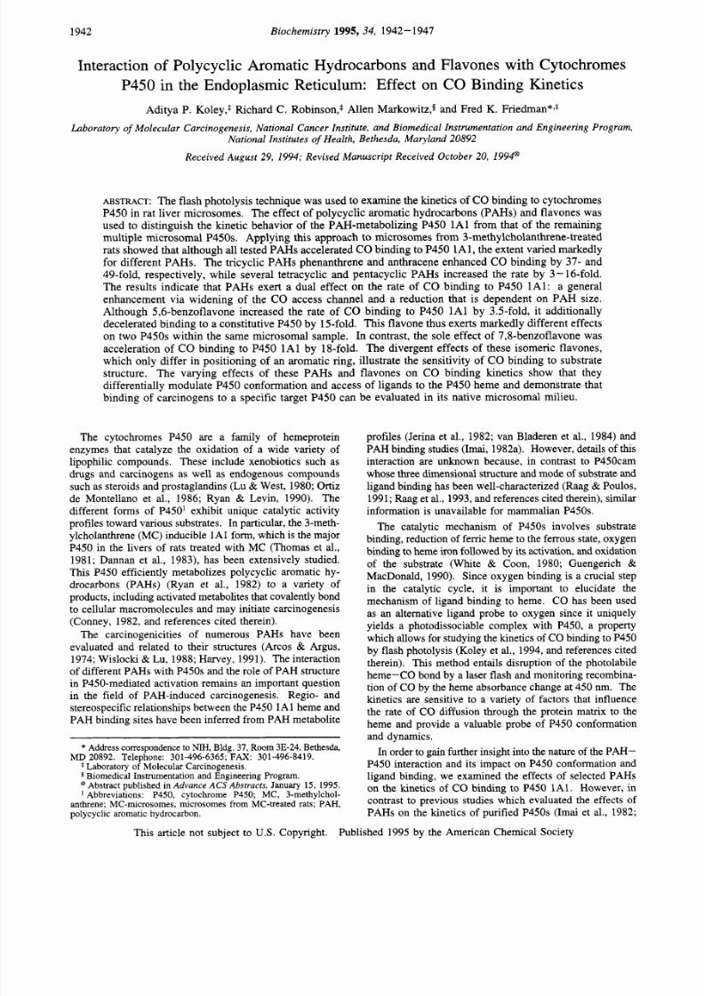
We examined the effect of representative PAHs of
different sizes and shapes (Figure 1) on the kinetics of CO
Biochemistry, Vol
34, NO. 6, 1995
1943
Anthracene Phenanthrene
1.2-Benzanthracene 2,3-Benzanthracene Pyrene
Benzo[e]pyrene Benzo[a]pyrene
Di~ n z[a9 c1 an th racen e
5,6-Benzoflavone 7,8-Benzoflavone
FIGURE : Polycyclic aromatic hydrocarbons and flavones evaluated
for
their effect on the CO binding kinetics of MC-microsomes.
0.030
8
c
g 0.020
8
s
0.010
0.000 1 1
0.00
0.20 0.40
0.60
0.80 1.00
tlme (sec)
FIGURE: Effect of phenanthrene and pyrene on binding of CO
to
P450s in MC-microsomes. Progress curves (a) in the absence and
presence of (b) phenanthrene and (c) pyrene, respectively.
The
CO concentration was
20
pM; PAH concentration was 10 pM;
microsomal concentration was 0.39 mg/mL in
0.1 M
sodium
phosphate buffer (pH 7.5) containing 20% (w/v) glycerol; tem-
perature, 23 C.
binding to MC -microsom es. A typical CO binding curve is
shown in Figu re 2 along with cu rves obtained in the presence
of phenanthrene or pyrene. While both PAHs accelerated
CO binding, examination of the early part of the reaction
curve (up to =0.1 s) clearly shows that phen anthrene was
more effective than pyrene. The remaining PAHs likewise
accelerated
CO
bind ing and yielded distinct reaction profiles
(data not shown).
Interpretation of CO binding data for
microsom es is not straightforward since the contribution of
mu ltiple P450s to the overa ll reaction com plicates extraction
of kinetic information for a p articular PAH -specific P450.
We therefore applied a recently d eveloped kinetic difference
method (Koley et al., 1994) to our data.
This
approach
evaluates the difference between the kinetic profiles obtained
in the presence and absen ce of the P AH and thus effectively
8/18/2019 Biochem 1995
http://slidepdf.com/reader/full/biochem-1995 4/7
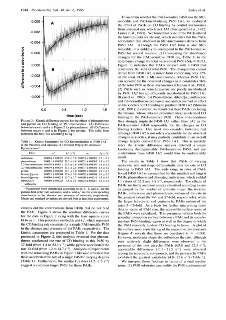
1944
Rioch rmi . r t y ,
Vot. 34 No. 6. I995
Kolcv et al.
6.006 bl
o.OO0
-
6 002
6.004
6.006
1
I
r
0.0
0.5 1
o
1.5
cancelc out the contributions from
P4Sk
that do not hind
thc PAH. Fipurc 3 chowc the rccultant d i f fm n cc cuwcc
for the data in F i p r c
2
along with thc least quarcc cuwc
f i t to q
.
This prwedurc yicldcd k l
and
Ai . which wpmcnt
the CObin din g mtc conctantc for a cinplc PAH-specific P450
in the ahccncc and prcccncc of the
PAH.
rccpwt ivc ly. Thc
kinct ic panmeters arc prcccntmi
in
Table
1 .
For the data
prcwntcd in Fipurc
2. th ic
analvcic
revealed that
phcnan-
thrcnc accelcnted the n t c of CO binding to
th i s
P450 hv
37-fold
(from
1.4 to S2.3
c - l )
while pyrcnc rrccclentcd thc
rate
1
?-fold (from 1.2 to 14.7 s
1.
Analywc o f cxpcnmentc
with the remaining PAHc in Fipurc 1 l ikcwicc rcvealcd that
thcce acce lm tcd the rate
of a
sinplc
P45O
o varying
cicgms
Table 1).
Funhennore.
the similar
A.1
values
1.2 -
.6
s
I
suppect a common target
P450
fo r the e PAHc.
To
ascertain whcthcr the PAH -ccncitivc
P450
wac thc IMC-
inducihlc anti PAM-mctaholit inp P450 1A I w o e cvaluatcd
thc effect of
PAHI
on
C O
hindinp
hy
control micmcomcc
from untrcattxi
n ts ,
which 1,xk I A I (Gticnpcrich ct a .. IW2:
1,ustcr ct
al.. 1W.1,.
found that n o w
ot
thc PAHs altered
thc kincticc data not shown,. wh ich indicatcc that
the PAH-
accclcntcxi n t c ohccncd in MC-micnwomcc dcr ivcc f rom
P450
A I .
Although thc P450
A 2
form i\
alco
MC-
inducihlc. i t i e unl ikc lv to comepond to thc PAH-ccncit ivc
P450
or
ccvcnl
m e :
f
1
Comparinp thc ahcorhancc
chmpcc for thc PAH-wncit ivc P450 01. T;ihlc to the
ahwrhancc chanpc for total micmcom al P490
Mn
0.033,
I+giirc 1 i n h x c c that PAHc in tcnc t w i th a P450 that
c o n w t u t e ICr-4Oq o f total P450. Thc ch an ge thus cannot
dcnvc f rom
P450
A2 . a minor
form
compming on ly I Z C i
o f the total
P450
n .MC-microcomcc.
whcrcaI P450 1 / 4 1
can account for the ohwwmi chanpcc ac i t conctitutcc
569
of thc total P450 n h e w m i c m o m c c
(Dannan
et ;I .. 9831.
(2)
PAHs
such as hcn7ola)pyrcnc arc
pmrly
mctah1i;rcd
by
P450
1A2
hut
arc cf f ic icnt lv mctahol i tcd hv P4CO
It11
(Ryan ct
al.. 1982 . (3 ,
I'hcnanthrcnc. dihcn7[o,c-)anthnccnc
ami
7.8-kn7oflavonc (icrc;ivxl. ami anthnccnc h7d no cffcct
on the kinct icc o f COhindinp to purificd
P450
A2 (Sh imi iu
ct al.. 1W l
; in
contract. u'c tcriind
that
t h AHs ( iwl i id inp
the flavone.
w hov l data
arc pw wn red 1;itcr)
accclcntcd
CO
hindinp to thc PAH-scncit ivc
P450.
Thccc concidcntionc
thus srmngly implicate
P450
IAI nthcr than
It12 as
the
PAH-ccncttivc
P4CO
rccpncihlc for the chanpcc in
C O
hindin g kincticc.
One
must nlco concider. hcrtvcvcr. that
although
P4CO I A2 i s
not
colclv
mpo ncihlc for thc
o Kcn.cd
ch;ingc\ in kincticc.
i t
mav pa rtially contnhutc
t o
an ovcnll
chmgc
largely dcr i vcd
f rom P450
A I .
T h i c i c unl ikc ly
cincc
thc
kinctic diffcrcncc analycic dctcctcd a sinplc
kinct icallv ciictinpui\hahlc PA H-w ncit iv c P450. and ;iny
contnhution from P450
1A2 would t huc
hc undctectahlv
small.
The rccultc
in
Tahlc
1
chow
that PAHc of varying
molccular c i i c and ch a p di ffcrcnt ially al ter the nt e of CO
binding to P450
I
AI. Thc uidc n n p c
in
mice o f PAH-
hound
P450 1
A
I
i c cxcmpl i f icd hv thc cmallcct and larpcct
PAHI.
ph t h r cnc
and
dikwla.cjanthnctnc. which
y i c l M
k l valucs
o f
52.3
anti 4.x . ccpcctivcly. Thc c f fcc t I of
PAHc
arc
ftmtly and moct cimp lv
cl;iccific<l
.xconlinp
to
ci te
ac paiipcd hy thc n u m k r
of
;immatic nnpc: thc
rr icycl ic
PAlfs.
;inthnccnc and phcnanthrcnc. cnh;inccd thc n t c to
thc prcatcst extent thy
4
and 37-fold. rccpcctivclv). while
thc larpcr tctncyclic and pcntacvclic
P N f c
cnhanccd thc
ntcc 3-16-foId.
Ae
a
haw
for
funhcr
intcrprctinp thcw
&ita
in
t c n n c of P A H
cim.
thc acccesihlc w r f a c c arcas of
thc P AH c wcrc calculatmi.
T h i c
panm ctcr rcflcctc
hoth
thc
prcn t ia l intcmction cutf;icc k r w w n a PAH and i t c complc-
mcntary P450hinciinp: cpion as
well as
thc d c g m t o wh ich
thc PAH ctcncally hindcrs C O binding
t o
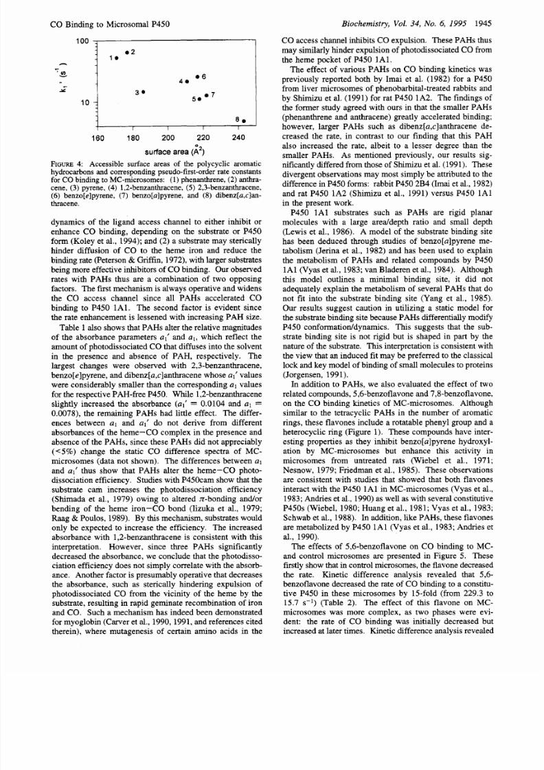
hcnrc.
A plot
of
thc
curf,,7cc arcac
v c n i c thc log o f the
rc\pcctivc
mtc com tantc
(Fipurc
4 )
rcvcalc that thcw
arc
conclatcd f t =
-0.831.
Hou.cvcr. molc ciilar shape alco influcnccc thc ntc: although
only rclativclv slipht diffcrcnccc u crc ohwnc<i in thc
prcwncc
of
thc
t w o
t r icycl ic PAHc
( 0 3 . X
;ind
52.1 c
b
;ipprcciahlc diffcrcnccc
(
I I
1-2
1.5
e
'1
wcrc
ohwrvcd
among thc tctcicyclic co mp un dc. and the pcntacvclic
PAHc
cxhihitcd the pwatect vanabilitv
(4.8-25.0 1
t'l'ahlc
I ) .
N c interpret thcw findings in tcrmc ot a dual mccha-
nicm: I P4CO ctiktmtcc can modify thc
P4.Y)
conformation/
8/18/2019 Biochem 1995
http://slidepdf.com/reader/full/biochem-1995 5/7
CO B inding to M icrosomal P450
Biochemistry, Vol.
34, No.
6,
1995
1945
CO access channel inhibits CO expulsion. These PAHs thus
may similarly hinder expulsion of photodissociated C O from
the heme pocket of P450 1Al.
The effect of various PAHs on CO binding kinetics was
previously reported both by Imai et al. (1982) for a P450
from liver m icrosome s of pheno barbital-treated rabbits and
by Sh imizu et al. (1991) for rat P450 1A2. The findings of
the forme r study agreed with ours in that the sm aller PAHs
(phenan threne and an thracene) greatly accelerated binding;
however, larger PAHs such as dibenz[a,c]anthracene de-
creased the rate, in contrast to our finding that this PAH
also increased the rate, albeit to a lesser degree than the
smaller PAH s. As men tioned previously, our results sig-
nificantly differed from those of Shimizu et al. (1991). These
divergent observations may most simply be attributed to the
difference in P450 forms: rabbit P450 2B4 (Imai et al., 198 2)
and rat P450 1A2 (Shimizu et al., 1991) versus P450 1A l
in the present work.
P450 1Al substrates such as PAHs are rigid planar
molecules with a large areddepth ratio and small depth
(Lewis et al., 1986). A mod el of the substrate binding site
has been deduced through studies of benzo[a]pyrene me-
tabolism (Jerina et al., 1 982) and has been used to ex plain
the metabolism of PAHs and related compounds by P450
1A l (Vyas et al., 1983; van Bladeren et al., 1984). Although
this model outlines a minimal binding site, it did not
adequately explain the m etabolism of several PAHs that do
not fit into the substrate binding site (Yang et al., 1985).
Our results suggest caution in utilizing a static model for
the substrate binding site becaus e PAHs differentially modify
P450 conform atioddynam ics. This suggests that the sub-
strate binding site is not rigid but is shaped in part by the
natu re of the substrate.
This
interpretation is consisten t with
the view that an induced fit may be preferred to the classical
lock and key model of binding of small molecules to proteins
(Jorgensen, 1991).
In addition to PAH s, we also evalua ted the effect of two
related com pound s, 5,6-benzo flavone and 7,8-benzof lavone,
on the
CO
binding kinetics of MC -microso mes. Althoug h
similar to the tetracyclic PAHs in the number of aromatic
rings, these flavones include a rotatable phenyl group and a
heterocyclic ring (F igure 1). These comp ounds have inter-
esting properties as they inhibit benzo[a]pyrene hydroxyl-
ation by MC-microsomes but enhance this activity in
microsomes from untreated rats (Wiebel et al., 1971;
Nesnow, 19 79; Friedm an et al., 1985). These observations
are consistent with studies that showed that both flavones
interact with the P450 1A l in M C-microsomes (Vyas et al.,
1983; Andries et al., 199 0) as well as with several constitutive
P450s (W iebel, 1980; Huan g et al., 1981; Vyas et al., 1983;
Schwab et al., 1988). In addition, like PAHs, these flavones
are metabolized by P45 0 1A l (Vya s et al., 1983; Andries et
al., 1990).
The effects of 5,6-b enzoflavon e on CO binding to MC-
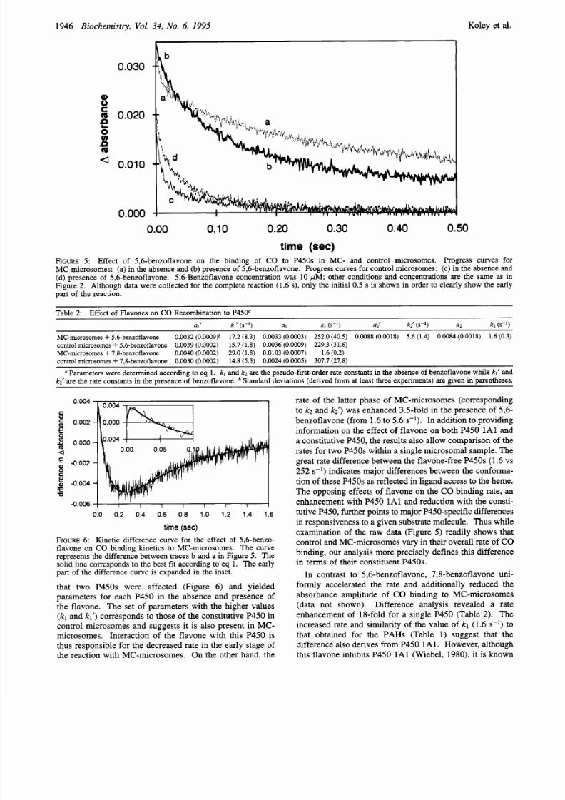
and control microsomes are presented in Figure 5. These
firstly show that in control m icrosomes, the flavone decreased
the rate. Kinetic difference analysis revealed that 5,6-
benzoflavone decreased the rate of CO binding to a constitu-
tive P450 in these microso mes by 15-fold (from 229 .3 to
15.7
s-’)
(Table 2). The effect of this flavone on MC-
microsomes was more complex, as two phases were evi-
dent: the rate of CO binding was initially decreased but
incre ased at later times. Kinetic difference analysis reve aled
100
4 i
06
v )
4.
i 3 . 5 0 . 7
.-
10
8 .
I
1
I
I
160 180 200 220 240
surface area
2)
FIGURE
:
Accessible surface areas of the polycyclic aromatic
hydrocarbons and corresponding pseudo-fust-order rate constants
for
CO
binding
to
MC-microsomes:
1)
phenanthrene,
(2)
anthra-
cene, 3) pyrene, (4) 1,2-benzanthracene, 5) 2,3-benzanthracene,
6)
benzo[e]pyrene,
(7)
benzo[a]pyrene, and (8) dibenz[a,c]an-
thracene.
dynamics of the ligand access channel to either inhibit or
enhance CO binding, depending on the substrate or P450
form (Koley et al., 19 94); and (2) a substrate may sterically
hinder diffusion of CO to the heme iron and reduce the
binding rate (Peterson Griffin, 1972), with larger substrates
being more effective inhibitors of CO binding. Our observed
rates with PAHs thus are a combination of two opposing
factors. Th e first mechanism is always operative and widens
the CO access channel since all PAHs accelerated CO
binding to P450 1Al. The second factor is evident since
the rate enhancement is lessened with increasing PAH size.
Tab le 1 also shows that PAHs alter the relative magnitudes
of the absorbance parameters al’ nd al, hich reflect the
amount of p hotodissociated CO that diffuses into the solvent
in the presence and absence of PAH, respectively. The
largest changes were observed with 2,3-benzanthracene,
benzo[e]p yrene, and dibenz[a,c]anthracen ewhose al’ values
were considerably smaller than the correspondin g
a1
values
for the respective PAH-free P450. While 1 ,Zbenzan thracene
slightly increased the absorbance (al’
=
0.0104 and a1
=
0.0078), the remaining PAHs had little effect. The differ-
ences between a1 and al’ do not derive from different
absorban ces of the heme-CO comp lex in the presence and
absence of the PAHs, since these PAHs did not appreciably
(<5%) change the static CO difference spectra of MC-
microsom es (data not shown). The differences between a1
and
al’
thus show that PAHs alter the heme-CO photo-
dissociation efficiency. Studies with P450c am show that the
substrate cam increases the photodissociation efficiency
(Shimada et al., 1979) owing to altered n-bonding andor
bending of the heme iron-CO bond (Iizuka et al., 1979;
Raag Poulos, 1989). By
this
mechanism, substrates would
only be expected to increase the efficiency. The increased
absorban ce with 1,2-benzan thracene is consistent with this
interpretation. How ever, since three PAHs significantly
decreased the absorb ance, we conclu de that the photodisso -
ciation efficiency does not simply correlate with the absorb -
ance. Another factor is presumably operative that decreases
the absorbance, such as sterically hindering expulsion of
photod issociated CO fro m the vicinity of the heme by the
substrate, resulting in rapid g eminate recomb ination of iron
and CO. Such a mechan ism has indeed been demonstrated
for myo globin (Carver et al., 19 90, 1991, and references cited
therein), where mutagenesis of certain amino acids in the
8/18/2019 Biochem 1995
http://slidepdf.com/reader/full/biochem-1995 6/7
1946
Biochemistry,
Vol. 34, No. 6, 1995 Koley et al.
0.030
a 0.010
0.000
0.00 0.10 0.20 0.30 0.40 0.50
t ime
(sec)
FIGURE :
Effect
of
5,6-benzoflavone on
the
binding of CO to
P450s
in MC- and control microsomes. Progress curves for
MC-microsomes: (a) in the absence and (b ) presence
of
5,6-benzoflavone. Progress curves for control microsomes:
(c)
in the absence
and
(d)
presence of 5,6-benzoflavone. 5,6-Benzoflavone concentration was 10 pM other conditions and concentrations are the same
as in
Figure
2.
Although data were collected for the complete reaction (1.6
s),
only the
initial 0.5 s
is shown in order
to
clearly show
the
early
part
of the reaction.
Table
2:
Effect
of Flavones
on
CO
Recombination
to
P450
~~
al
kl
(S-') a1
ki
(s- )
a i k{
SKI)
a2
kz ( s - l )
MC-microsomes 5,6-benzoflavone 0.0032 0.0009)* 17.2 8.3) 0.0033 0.0003) 252.0 40.5) 0.0088 0.0018) 5.6 1.4) 0.0084 0.0018) 1.6 0.3)
control microsomes 5,6-benzoflavone 0.0039 0.0002)
15.7 1.8) 0.0036
(O.OOO9)
229.3 31.6)
MC-microsomes 7,8-benzoflavone 0.0040 0.0002)
29.0 1.8) 0.0103 0.0007) 1.6 0.2)
control microsomes 7.8-benzoflavone 0.0030 0.0002)
14.8 5.3) 0.0024 0.0005) 307.7 27.8)
Parameters were determined according to
eq
1 kl and k z are the pseudo-first-orderrate constants in the absence
of benzoflavone
while kl and
k i are the rate constants in the presence of benzoflavone. Standard deviations derived from at least three
experiments)
are given in parentheses.
00 0 2 04 0 6 0 8 10 1 2 1.4 16
time (sec)
FIGURE
: Kinetic
difference
curve
for the effect
of
5,6-benzo-
flavone on CO binding kinetics to MC-microsomes. The curve
represents the difference between traces
b
and a
in
Figure 5 . The
solid line corresponds to the best fit according to eq 1. The early
part
of
the difference
curve is
expanded in the inset.
that two P450s were affected (Figure 6) and yielded
parameters for each P450 in the absence and presence of
the flavone. The set of parameters with the higher values
(kl
and
kl )
correspon ds to those of the constitutive P450 in
control microsomes and suggests it is also present in MC-
microsom es. Interaction of the flavone with this P450 is
thus respon sible for the d ecreased rate in the early stage of
the reaction with MC -microsom es. On the other hand, the
rate of the latter phase of MC-microsomes (corresponding
to k~ and k i ) was enhance d 3.5-fold in the presence of 5,6-
benzoflavo ne (from 1.6 to 5.6 s-l . In addition to providing
information on the effect of flavone on both P450 1Al and
a constitutive P450, the results also allow comp arison of the
rates for two P450s w ithin a single microsom al sample. The
great rate difference between the flavone-free P450s (1.6 vs
252 s-l indicates major differences between the confo rma-
tion of these P450s as reflected in ligand acces s to the hem e.
The op posing effects of flavone on the CO binding rate, an
enhancem ent with P45 0 1 A l and reduction with the consti-
tutive P450, further points to m ajor P450-specific differences
in responsiveness o a given substrate molecule. Thus while
examination of the raw data (Figure
5 )
readily show s that
control and M C-m icrosom es vary in their overall rate
of
CO
binding, our analysis more precisely defines this difference
in terms of their constituent P450s.
In contrast to 5,6-benzoflav one, 7,8-benzo flavone uni-
formly accelerated the rate and additionally reduced the
absorbance amplitude of CO binding to MC-microsomes
(data not shown). Difference analysis revealed a rate
enhancem ent of 18-fold for a single P450 (Tab le 2). The
increased rate and similarity of the value of
kl
(1.6 s-') to
that obtained for the PAHs (Table 1) suggest that the
difference also derives from P450 1 A l. How ever, although
this flavone inhibits P450 1A l (Wieb el, 1980), it is known
8/18/2019 Biochem 1995
http://slidepdf.com/reader/full/biochem-1995 7/7
CO B inding to Microsomal P450
to also interact with constitutive P450 s, including P450 3A
(Schwab et al., 198 8). W e thus assessed its effect on control
microso mes from untreated rats and found it decreased the
CO binding rate of a single P450 by 21-fold (Table 2). Since
the results with M C-m icrosom es showed that the sole effect
of this flavone was rate enhancem ent for a single P450, the
7,8-benzoflavone-sensitive
450 in control microsomes is
absent or undetectable in MC-m icrosom es. The different
effects of 5,6- and 7,8-benzoflavo ne on P450 1A l (rate
enhancements
of
3.5- and 18-fold, respectively) and their
different selectivities for constitutive P450s thus illustrate
the sensitivity of ligand bind ing to substrate structure.
The MC-inducible P450 1A l form efficiently metabolizes
PAHs relative to other P450s (Ryan et al., 1982) and is
primarily responsible for PAH activation (Gozukara et al.,
1982). How ever details of the PAH interaction with this
hemeprotein remain unclear since the three-dimensional
structure of a mammalian P450 has not been determined.
The effect of a PAH substrate on the CO binding kinetics
offers a unique app roach to probe the interaction of substrate
with P450 heme and protein, since it is sensitive to both
structure and dynamics. The results indicate that PAHs exert
a dual effect both by altering protein confo rmatioddy namics
to widen the CO access channel to enhance binding and by
sterically hindering binding. The divergen t effects of two
structurally related flavones on P450 1 A l and a constitutive
P450 further demonstrate the sensitivity of P450 ligand
binding to substrate structure.
REFERENCES
Andries,
M.
J., Lucier, G. W., Goldstein, J., Thompson, C. L.
(1990) Mol. P h a m c o l . 37, 990-995.
Arcos,
J.
C., Argus, M. F. (1974) in Chemical Induction of
Cancer, Structural Bases and Biological Mechanisms Wolf, G.,
Ed.) Vol. IIA, pp 15-65, Academic Press, New York.
Carver, T. E., Rohlfs, R. J., Olson, J.
S .
Gibson, Q. H., Blackmore,
R. S . Springer, B. A., Sligar, S . G. (1990) J . Bi d. Chem.
265, 20007-20020.
Carver, T. E., Olson,J. S . Smerdon,S . J., Krzywda,
S .
Wilkinson,
A. J., Gibson, Q .
H.,
Blackmore, R. S . Dezz Ropp,
J.,
Sligar,
S .
G. (1991)
Biochemistry
30,4697-4705.
Dannan , G. A., Guengerich, F. P., Kaminsky, L. S., Aust,
S .
D.
(1983) J . Biol. Chem. 258, 1282-1288.
Friedman , F. K., Wiebel, F. J., Gelboin, H. V. (1985) Phamta-
cology
31, 194-202.
Gozukara, E. M., Guengerich, F. P., Miller,
H.,
Gelboin,
H.
V.
(1982)
Carcinogenesis
3, 129-133.
Guengerich, F. P., MacDonald T. L. (1990) FASEB J. 4,2453-
2459.
Guengerich, F. P., Ghazi, A. D., Wright,
S .
T., Martin, M. V.,
Kaminsky, L.
S .
(1982) Biochemistry 21, 6019-6030.
Harvey, R. G. (1991) in
Polycyclic Aromatic Hydrocarbons:
Chemistry and Carcinogenicity,
pp 26-95, Cambridge University
Press, Cambridge.
Huang, M. T., Johnson, E. F., Muller-Eberhard, A., Koop, D. R.,
Coon, M. J., Conney, A. H. (1981)
J . Biol. Chem.
256,10897-
10901.
Iizuka, T., Shimada, H., Ueno, R., Ishimura, Y. (1979) in
Cytochrome Oxidase (Chance , B., King, T. E., O kunuki, K.,
Biochemistry, Vol. 34, No.
6,
1995
1947
Orii, Y.,
Eds.)
pp 9-20, Elsevier, Amsterdam.
Imai
Y. (1982)
J . Biochem.
92, 77-88.
Imai Y., Iizuka, T., Ishimura, Y. (1982) J. Biochem. 92, 67-
75.
Jerina, D. M., Michaud, D. P., Feldm ann, R. S . Armstrong, R. N.,
Vyas, K. P., Thakker, D. R., Yagi, H., Ryan, D. E., Thomas, P.
E., Levin, W. (1982) in
Microsomes, Drug Oxidations and
Drug Toxicity
(Sato , R., Kato, R., Eds.) pp 195-201, Japan
Scientific Societies Press, Tokyo.
Jorgensen, W. L . (1991)
Science
254, 954-955.
Koley, A. P., Robinson, R. C., Markowitz,
A , ,
Friedman F. K.
Lewis, D. F. V., Ioannides, C., Parke, D. V. (1986)
Biochem.
Lu, A. Y.
H.,
West,
S .
B. (1980)
Pharmacol. Rev.
31, 277-
Luster, M. I., Lawson, L. D., Linko, P., Goldstein, J. A. (1983)
Markowitz, A., Robinson, R. C., Omata, Y., Friedman, F. K.
Nesnow, S . A. (1979) J . Med. Chem. 22, 1244-1247.
Omura, T., Sato, R. (1964)
J . Biol. Chem.
239, 2370-2385.
Ortiz de Montellano, P. R., Ed. (1986) Cytochrome P-450
Structure, Mechanism and Biochemistry,
Plenum Press, New
York.
Peterson, J. A., Griffin, B. W. (1972)
Arch. Biochem. Biophys.
Raag, R., Poulos, T. L. (1989)
Biochemistry
28, 7586-7592.
Raag, R., Poulos, T. L. (1991) Biochemistry 30, 2674-2684.
Raag, R., Li, H., Jones, B. C., Poulos, T. L. (1993) Biochemistry
Ryan, D. E., Levin, W. (1990) Pharmacol. Ther. 45, 153-239.
Ryan, D., Thomas, P. E., Reik, L. M., Levin, W. (1982)
Xenobiotica
12, 727-744.
Shimada, H., Iizuka, T., Ueno, R., Ishimura, Y. (1979)
FEBS
Lett. 98, 290-294.
Shimizu,
T., Ito, O., Hatano, M., Fujii-Kuriyam a, Y. (1991)
Biochemistry 30, 4659-4662.
Thomas, P. E., Reik, L. M., Ryan, D. E., Levin, W. (1981)
J .
Biol. Chem.
256, 1044-1052.
van Bladeren, P. J., Vyas, K. P., Sayer, J. M., Ryan, D. E., Thomas,
P. E., Levin, W., Jerina, D.
M.
(1984) J. Biol. Chem. 259,
8966-8973.
Vyas, K. P., Sh ibata, T., H ighet, R. J., Yeh,
H.
J., Thomas, P. E.,
Ryan, D. E., Levin, W., Jerina, D. M. (1983)
J . Biol. Chem.
258, 5649-5659.
White, R. E., Coon, M.
J.
(1980)
Annu. Rev. Biochem.
49,315-
356.
Yang,
S .
K., Mushtaq, M., Chiu, Pei-Lu (1985) in
Polycyclic
Hydrocarbons and Carcinogenesis
(Harvey R. G., Ed.) ACS
Symposium Series 283, pp 19-34, Washington, DC, American
Chemical Society.
Wiebel, F. J. (1980) in
Carcinogenesis: Mod ers
of
Chemical
Carcinogenesis
(Slaga, T. J., Ed.) Vol. 5,pp 57-84, Raven Press,
New York.
Wiebel, F. J., Leutz, J. C., Diamond, L., Gelboin, H. V. (1971)
Arch. Biochem. Biophys. 144, 78-86.
Wislocki, P. G., Lu,
Y.
H. (1988)
in
Polycyclic Aromatic
Hydrocarbon Carcinogenesis: Structure-Activity Relationships
(Yang, S . K., Silverman , B. D., Eds.) Vol I, pp 1-30, CRC
Press, Boca Raton, FL.
BI9420323
(1994)
Biochemistry
33,
2484-2489.
Pharmacol. 35, 2179-2185.
295.
Mol. Phamtacol. 23, 252-257.
(1992)Anal. Instrum. 20, 213-221.
151,427-433.
32, 4571-4578.


![Biochem [Enzymes]](https://img.pdfslide.us/doc/110x75/55cf8d225503462b1392585f/biochem-enzymes.jpg)