Embed Size (px)
Citation preview
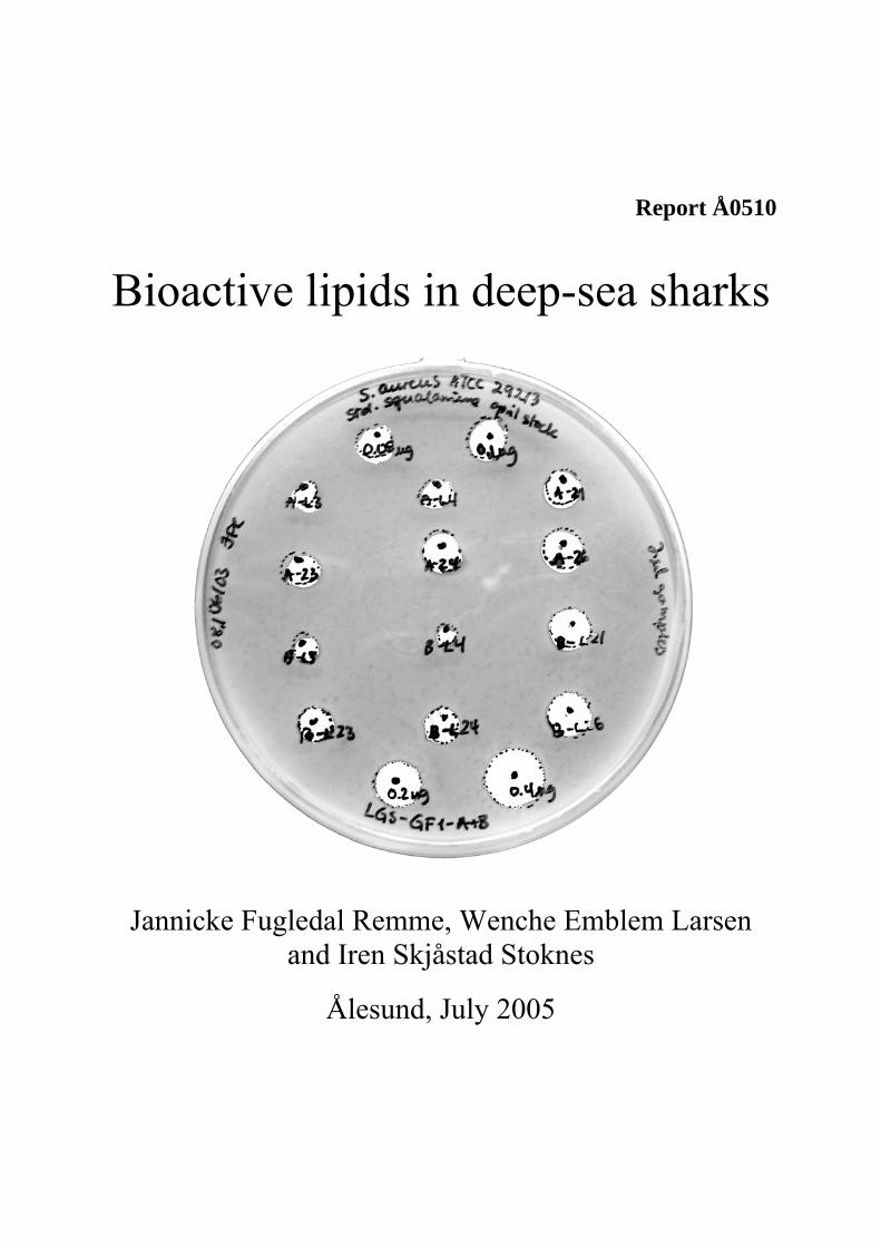
Report Å0510
Bioactive lipids in deep-sea sharks
Jannicke Fugledal Remme, Wenche Emblem Larsen and Iren Skjåstad Stoknes
Ålesund, July 2005


Index
Møre Research -6- Å0510
INDEX
PREFACE.............................................................................................................................................................. 5 1 INTRODUCTION ......................................................................................................................................... 7
1.1 BIOACTIVE LIPIDS ................................................................................................................................. 7 1.1.1 Long chain polyunsaturated fatty acids ..................................................................................... 7 1.1.2 Phospholipids.............................................................................................................................. 10 1.1.5 Squalene ...................................................................................................................................... 11 1.1.3 Squalamine ................................................................................................................................. 12 1.1.4 Other aminosterols..................................................................................................................... 13
1.2 LIPID METABOLISM IN ELASMOBRANCH FISHES................................................................................. 14 2 MATERIALS AND METHODS ................................................................................................................ 15
2.1 MATERIALS ........................................................................................................................................ 15 2.2 METHODS ........................................................................................................................................... 15
2.2.1 Water content ............................................................................................................................. 15 2.2.2 Lipid content............................................................................................................................... 15 2.2.3 Polar and nonpolar lipid content .............................................................................................. 15 2.2.4 Fatty acid composition............................................................................................................... 15 2.2.5 Squalene ...................................................................................................................................... 16 2.2.6 Extraction of aminosterols ........................................................................................................ 16 2.2.7 Extraction of bioactive lipids..................................................................................................... 17 2.2.8 Antimicrobial activity ................................................................................................................ 17 2.2.9 Statistical analysis ...................................................................................................................... 17
3 RESULTS.................................................................................................................................................... 18 3.1 WATER CONTENT................................................................................................................................ 18 3.2 LIPID CONTENT ................................................................................................................................... 18 3.3 POLAR AND NONPOLAR FAT................................................................................................................ 19 3.4 FATTY ACID COMPOSITION.................................................................................................................. 20
3.4.1 Total fat....................................................................................................................................... 20 3.4.2 The distribution of saturated, monounsaturated and polyunsaturated fatty acids.............. 26 3.4.3 Polar and nonpolar fatty acid composition .............................................................................. 27
3.5 SQUALENE .......................................................................................................................................... 32 3.6 ANTIMICROBIAL ACTIVITY.................................................................................................................. 32
3.6.1 Aminosterols ............................................................................................................................... 32 3.6.2 Activity in water and lipid extracts from lipid extraction ...................................................... 33
4 DISCUSSION ............................................................................................................................................. 34 4.1 WATER CONTENT................................................................................................................................ 34 4.2 LIPID CONTENT ................................................................................................................................... 34 4.3 FATTY ACID COMPOSITION.................................................................................................................. 34 4.4 SQUALENE .......................................................................................................................................... 37 4.5 ANTIMICROBIAL ACTIVITY.................................................................................................................. 38
5 CONCLUSION ........................................................................................................................................... 39 6 REFERENCES ............................................................................................................................................ 40
Introduction
Møre Research -7- Å0510
1 INTRODUCTION The oceans cover more than 71% of the surface of the earth and comprise approximately half of the total global biodiversity, for which estimates a range between 3 and 500 x 106 different species (Haug et al, 2002). The marine environment is therefore a rich source for discovering novel lead compounds for the development of new antibacterial drugs. A variety of low molecular weight antibiotics have been isolated from diverse animals (Moore et al, 1993). These agents, which include peptides, lipids and alkaloids, exhibit antibiotic effect against environmental microbes and are thought to play a role in innate immunology. Among these compounds, a broad spectrum steroidal antibiotic, squalamine, has been isolated from tissues of the dogfish shark (Squalus acanthias). The proportion of sharks caught in the deep-sea fishing industry can vary (Bakes and Nichols, 1995). The amount of sharks landed varies due to factors such as seasonal variation and reproductive status. In the past, when sharks were only a minor bycatch, they were either discarded or used as fishmeal. An international market for shark liver oils has existed for some time, but more efficient utilization of shark byproducts must be considered. Møre Research has focused on deep-sea species since 1990, by assisting the fishing industry in the process of establishing fisheries of new species and increase the utilization (Hareide and Thomsen, 1997; Kjerstad et al, 1999; Kjerstad et al, 2000; Kjerstad and Hellevik, 2000; Kjerstad and Fossen, 2001; Kjerstad et al, 2003; Remme et al, 2003; Stoknes and Fjørtoft, 1998, Stoknes et al, 2004; Økland et al, 2005; Remme et al, 2005). Deep sea species are living below 400 meters. Research and development has taken place both regarding resource biology, fishing technology, catch handling, utilization of byproducts, product development and marketing. These species have previously been discarded because of problems with production, marketing and selling. With marine biotechnology it is possible to increase the utilisation of these species and their byproducts. The objective of this work was to search for bioactive lipids in internal organs from deep-sea sharks. 1.1 Bioactive lipids 1.1.1 Long chain polyunsaturated fatty acids The fish oils constitute an important source of omega-3 polyunsaturated fatty acids (PUFA), mainly the eicosapentaenoic acid (EPA) and the docosahexaenoic acid (DHA) (Navarro-Garcia et al, 2004). The omega-3 PUFA provides several benefits to the human health. They are essential for development and function of certain organs and for several biochemical and physiological responses of the organism. The brain is one of the organs where omega-3 PUFAs are essential. The tissue of this organ is particularly rich in DHA, showing a close correlation between the consumption of this acid and its deposition in the cellular membrane. DHA take part in the brain development and retina formation of the child during pregnancy. Dietary long chain PUFAs affect processes including growth, neurological development, lean and fat mass accretion, reproduction, innate and acquired immunity, infectious pathologies of virus , bacteria and parasites, and the incidence and severity of virtually all chronic and degenerative diseases including cancer, atherosclerosis, stroke, arthritis, diabetes, osteoporosis and neurogenerative, inflammatory and skin diseases (Berger et al, 2002). Microarray studies
Introduction
Møre Research -8- Å0510
indicate that fish oil LC-PUFA likely increase fatty acid (FA) β-oxidation, and gluconeogenesis, and decrease FA synthesis.
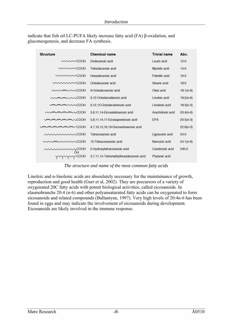
The structure and name of the most common fatty acids
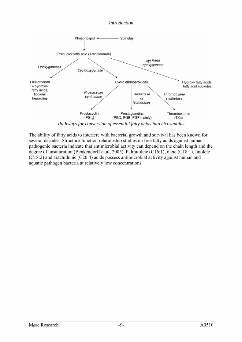
Linoleic and α-linolenic acids are absoulutely necessary for the maintainance of growth, reproduction and good health (Gurr et al, 2002). They are precursors of a variety of oxygenated 20C fatty acids with potent biological activities, called eicosanoids. In elasmobranchs 20:4 (n-6) and other polyunsaturated fatty acids can be oxygenated to form eicosanoids and related compounds (Ballantyne, 1997). Very high levels of 20:4n-6 has been found in eggs and may indicate the involvement of eicosanoids during development. Eicosanoids are likely involved in the immune response.
Introduction
Møre Research -9- Å0510
Pathways for conversion of essential fatty acids into eicosanoids
The ability of fatty acids to interfere with bacterial growth and survival has been known for several decades. Structure-function relationship studies on free fatty acids against human pathogenic bacteria indicate that antimicrobial activity can depend on the chain length and the degree of unsaturation (Benkendorff et al, 2005). Palmitoleic (C16:1), oleic (C18:1), linoleic (C18:2) and arachidonic (C20:4) acids possess antimicrobial activity against human and aquatic pathogen bacteria at relatively low concentrations.
Introduction
Møre Research -10- Å0510
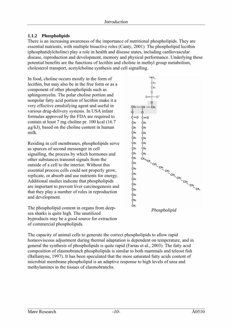
1.1.2 Phospholipids There is an increasing awareness of the importance of nutritional phospholipids. They are essential nutrients, with multiple bioactive roles (Canty, 2001). The phospholipid lecithin (phosphatidylcholine) play a role in health and disease states, including cardiovascular disease, reproduction and development, memory and physical performance. Underlying these potential benefits are the functions of lecithin and choline in methyl group metabolism, cholesterol transport, acetylcholine synthesis and cell signalling. In food, choline occurs mostly in the form of lecithin, but may also be in the free form or as a component of other phospholipids such as sphingomyelin. The polar choline portion and nonpolar fatty acid portion of lecithin make it a very effective emulsifying agent and useful in various drug-delivery systems. In USA infant formulas approved by the FDA are required to contain at least 7 mg choline pr. 100 kcal (16.7 µg/kJ), based on the choline content in human milk. Residing in cell membranes, phospholipids serve as spurces of second messenger in cell signalling, the process by which hormones and other substances transmit signals from the outside of a cell to the interior. Without this essential process cells could not properly grow, replicate, or absorb and use nutrients for energy. Additional studies indicate that phospholipids are important to prevent liver carcinogenesis and that they play a number of roles in reproduction and development. The phospholipid content in organs from deep-sea sharks is quite high. The unutilized byproducts may be a good source for extraction of commercial phospholipids.
Phospholipid
The capacity of animal cells to generate the correct phospholipids to allow rapid homeoviscous adjustment during thermal adaptation is dependent on temperature, and in general the synthesis of phospholipids is quite rapid (Farias et al., 2003). The fatty acid composition of elasmobranch phospholipids is similar to both mammals and teleost fish (Ballantyne, 1997). It has been speculated that the more saturated fatty acids content of microbial membrane phospholipid is an adaptive response to high levels of urea and methylamines in the tissues of elasmobranchs.
Introduction
Møre Research -11- Å0510
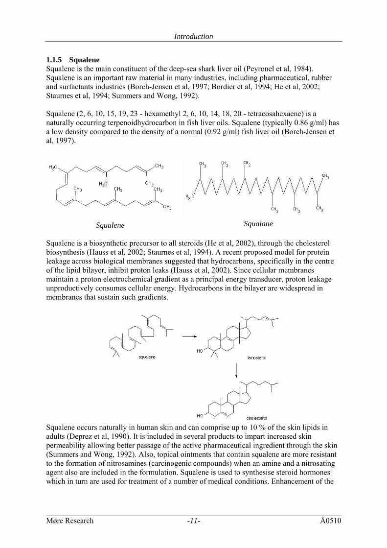
1.1.5 Squalene Squalene is the main constituent of the deep-sea shark liver oil (Peyronel et al, 1984). Squalene is an important raw material in many industries, including pharmaceutical, rubber and surfactants industries (Borch-Jensen et al, 1997; Bordier et al, 1994; He et al, 2002; Staurnes et al, 1994; Summers and Wong, 1992). Squalene (2, 6, 10, 15, 19, 23 - hexamethyl 2, 6, 10, 14, 18, 20 - tetracosahexaene) is a naturally occurring terpenoidhydrocarbon in fish liver oils. Squalene (typically 0.86 g/ml) has a low density compared to the density of a normal (0.92 g/ml) fish liver oil (Borch-Jensen et al, 1997).
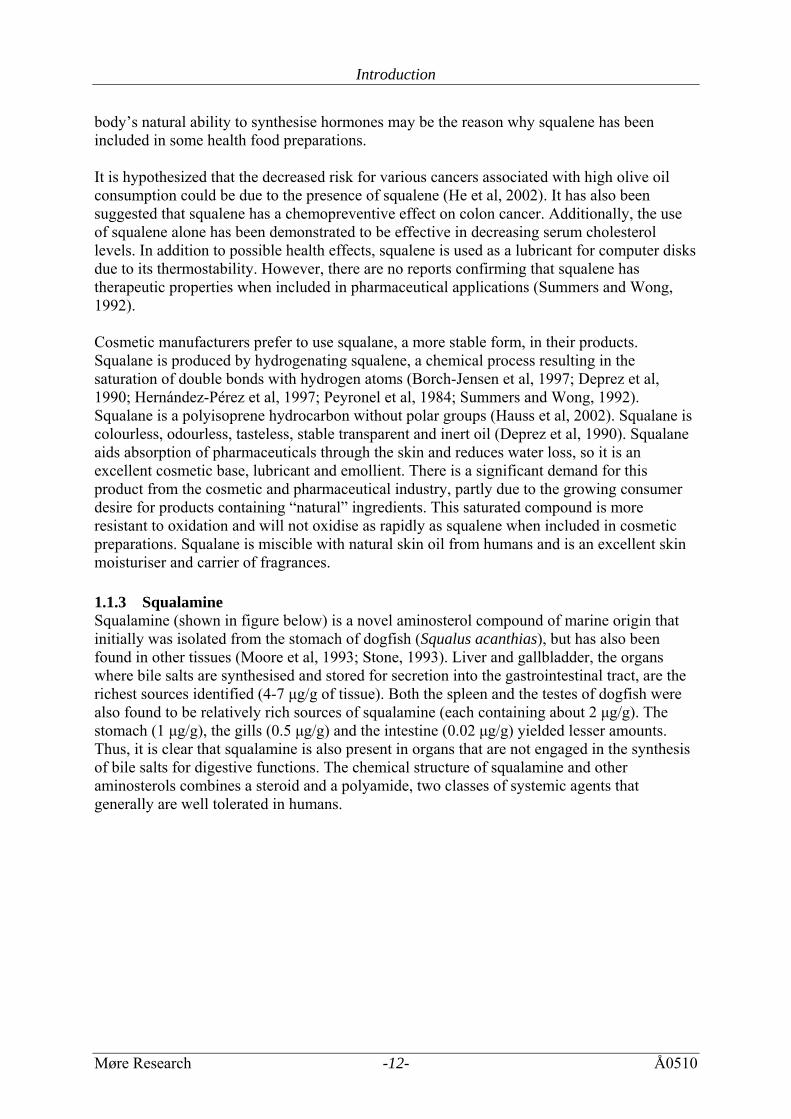
Squalene Squalane Squalene is a biosynthetic precursor to all steroids (He et al, 2002), through the cholesterol biosynthesis (Hauss et al, 2002; Staurnes et al, 1994). A recent proposed model for protein leakage across biological membranes suggested that hydrocarbons, specifically in the centre of the lipid bilayer, inhibit proton leaks (Hauss et al, 2002). Since cellular membranes maintain a proton electrochemical gradient as a principal energy transducer, proton leakage unproductively consumes cellular energy. Hydrocarbons in the bilayer are widespread in membranes that sustain such gradients.
Squalene occurs naturally in human skin and can comprise up to 10 % of the skin lipids in adults (Deprez et al, 1990). It is included in several products to impart increased skin permeability allowing better passage of the active pharmaceutical ingredient through the skin (Summers and Wong, 1992). Also, topical ointments that contain squalene are more resistant to the formation of nitrosamines (carcinogenic compounds) when an amine and a nitrosating agent also are included in the formulation. Squalene is used to synthesise steroid hormones which in turn are used for treatment of a number of medical conditions. Enhancement of the
Introduction
Møre Research -12- Å0510
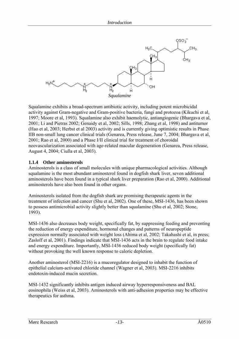
body’s natural ability to synthesise hormones may be the reason why squalene has been included in some health food preparations. It is hypothesized that the decreased risk for various cancers associated with high olive oil consumption could be due to the presence of squalene (He et al, 2002). It has also been suggested that squalene has a chemopreventive effect on colon cancer. Additionally, the use of squalene alone has been demonstrated to be effective in decreasing serum cholesterol levels. In addition to possible health effects, squalene is used as a lubricant for computer disks due to its thermostability. However, there are no reports confirming that squalene has therapeutic properties when included in pharmaceutical applications (Summers and Wong, 1992). Cosmetic manufacturers prefer to use squalane, a more stable form, in their products. Squalane is produced by hydrogenating squalene, a chemical process resulting in the saturation of double bonds with hydrogen atoms (Borch-Jensen et al, 1997; Deprez et al, 1990; Hernández-Pérez et al, 1997; Peyronel et al, 1984; Summers and Wong, 1992). Squalane is a polyisoprene hydrocarbon without polar groups (Hauss et al, 2002). Squalane is colourless, odourless, tasteless, stable transparent and inert oil (Deprez et al, 1990). Squalane aids absorption of pharmaceuticals through the skin and reduces water loss, so it is an excellent cosmetic base, lubricant and emollient. There is a significant demand for this product from the cosmetic and pharmaceutical industry, partly due to the growing consumer desire for products containing “natural” ingredients. This saturated compound is more resistant to oxidation and will not oxidise as rapidly as squalene when included in cosmetic preparations. Squalane is miscible with natural skin oil from humans and is an excellent skin moisturiser and carrier of fragrances. 1.1.3 Squalamine Squalamine (shown in figure below) is a novel aminosterol compound of marine origin that initially was isolated from the stomach of dogfish (Squalus acanthias), but has also been found in other tissues (Moore et al, 1993; Stone, 1993). Liver and gallbladder, the organs where bile salts are synthesised and stored for secretion into the gastrointestinal tract, are the richest sources identified (4-7 μg/g of tissue). Both the spleen and the testes of dogfish were also found to be relatively rich sources of squalamine (each containing about 2 μg/g). The stomach (1 μg/g), the gills (0.5 μg/g) and the intestine (0.02 μg/g) yielded lesser amounts. Thus, it is clear that squalamine is also present in organs that are not engaged in the synthesis of bile salts for digestive functions. The chemical structure of squalamine and other aminosterols combines a steroid and a polyamide, two classes of systemic agents that generally are well tolerated in humans.
Introduction
Møre Research -13- Å0510
Squalamine
Squalamine exhibits a broad-spectrum antibiotic activity, including potent microbicidal activity against Gram-negative and Gram-positive bacteria, fungi and protozoa (Kikuchi et al, 1997; Moore et al, 1993). Squalamine also exhibit haemolytic, antiangiogenic (Bhargava et al, 2001; Li and Pietras 2002; Genaidy et al, 2002; Sills, 1998; Zhang et al, 1998) and antitumor (Hao et al, 2003; Herbst et al 2003) activity and is currently giving optimistic results in Phase IIB non-small lung cancer clinical trials (Genarea, Press release, June 7, 2004; Bhargava et al, 2001; Rao et al, 2000) and a Phase I/II clinical trial for treatment of choroidal neovascularization associated with age-related macular degeneration (Genarea, Press release, August 4, 2004; Ciulla et al, 2003). 1.1.4 Other aminosterols Aminosterols is a class of small molecules with unique pharmacological activities. Although squalamine is the most abundant aminosterol found in dogfish shark liver, seven additional aminosterols have been found in a typical shark liver preparation (Rao et al, 2000). Additional aminosterols have also been found in other organs. Aminosterols isolated from the dogfish shark are promising therapeutic agents in the treatment of infection and cancer (Shu et al, 2002). One of these, MSI-1436, has been shown to possess antimicrobial activity slightly better than squalamine (Shu et al, 2002; Stone, 1993). MSI-1436 also decreases body weight, specifically fat, by suppressing feeding and preventing the reduction of energy expenditure, hormonal changes and patterns of neuropeptide expression normally associated with weight loss (Ahima et al, 2002; Takahashi et al, in press; Zasloff et al, 2001). Findings indicate that MSI-1436 acts in the brain to regulate food intake and energy expenditure. Importantly, MSI-1436 reduced body weight (specifically fat) without provoking the well known response to caloric depletion. Another aminosterol (MSI-2216) is a mucoregulator designed to inhabit the function of epithelial calcium-activated chloride channel (Wagner et al, 2003). MSI-2216 inhibits endotoxin-induced mucin secretion. MSI-1432 significantly inhibits antigen induced airway hyperresponsiveness and BAL eosinophila (Weiss et al, 2003). Aminosterols with anti-adhesion properties may be effective therapeutics for asthma.
Introduction
Møre Research -14- Å0510
1.2 Lipid Metabolism in Elasmobranch Fishes Most elasmobranchs are carnivorous, with protein and lipid as the main energy sources (Ballantyne, 1997). The metabolic rate of most elasmobranchs examined to date has suggested a lower metabolism than that of the more advanced teleost fishes under similar conditions. The ability of elasmobranchs to oxidize fatty acids or fatty alcohols is largely confined to liver and, to a lesser extent, the kidney. The liver serves as the main lipid storage site. Lipids comprise a substantial proportion of the liver wet weight in most elasmobranch. The hepatosomatic index (liver weight divided by body weight) of elasmobranchs is much higher then that of other fishes. There are several types of lipids stored in the liver, with triglycerides, alkyldiacylglycerols (ADAG), wax esters and hydrocarbons such as squalene and pristane variously occurring as the major lipids. Structurally, the triacylglycerols, ADAG and wax esters are analogous with two or three acyl chains linked to each other (wax esters) or to glycerol backbones (Ballantyne, 1997). These three classes of storage lipids are likely mobilized as energy sources. Squalene may be more metabolically inert. Although three of these lipid classes have obvious metabolic roles as energy sources, they also have a buoyancy function. The importance of the various lipids as mediators of buoyancy varies with lifestyle and habitant. The specific gravity of squalene and wax esters is 0.86 g/ml, whereas ADAGs are more dense (0.91 g/ml) and triacylglycerols are the densest at 0.93 g/ml. Adjustments in the proportions of lipids appear to be made to maintain buoyancy. Fatty acids are incorporated preferentially into liver lipids in the following order: Fatty acids>triacylglycerols>phospholipids>ADAG in the liver of S. achantias (Ballantyne, 1997). Fatty alchohols are incorporated into lipids in the order nonesterified glycol ethers>fatty acids>triacylglycerols>phospholipids. There is a high rate of conversion of fatty alcohols to fatty acids. The predominant fatty acids of triacylglycerols are also saturated and monounsaturated in liver of S. achanthias and other cold water elasmobranchs. This contrasts the high proportion of polyunsaturated fatty acids found in triacylglycerols of other cold water marine organisms. Of more practical interest is the fact that elasmobranches have the lowest incidence of neoplasia of any vertebrate group, and although the basis for this remains to be resolved, it is of considerable interest (Ballantyne, 1997). Elasmobranchs are osmoconformers, using a combination of solutes to maintain the extracellular and intracellular osmolarity close to that of the environment. What is unusual in this is that about 40 % of the osmolarity is due to urea with concentrations ranging as high as 680 mM. Such levels pose potential threats to the structure and function of proteins and other cellular components. The ability to produce and retain such high concentrations of urea has shaped the metabolism of elasmobranches, making them different from most other aquatic vertebrates.
Methods
Møre Research -15- Å0510
2 MATERIALS AND METHODS All the species studied are regarded as deep-sea species (ICES are using 400 m as a definition of what might be considered as deep-sea). 2.1 Materials The sharks, Portuguese dogfish (Centroscymnus coeloleps), leafscale gulper shark (Centrophorus squamosus) and black dogfish (Centrocyllium fabricii), were captured on long lines at Hatton Bank, in the North Atlantic, in May/June, 2000 and April-July 2002, at depths around 800-1500 meters. The fishes were slaughtered, bleed and frozen at -30°C until processing. Before processing at land, they where thawed in cold running water over night. The tissue samples were collected from about 20 individuals from each species. The heart, spleen, rectalgland, stomach, intestine, kidney, liver and pancreas were removed. The different tissue samples from the 20 individuals were pooled, except from the liver samples. 2.2 Methods 2.2.1 Water content The water content was determined in samples frozen at -60 °C. The samples (about 50 grams) were weighed and freeze-dried using a freeze dryer from Christ (Osterode am Hartz, Germany) for at least 96 hours. The samples were weighed periodically and the drying was continued until a stable weight was obtained. The water content was found gravimetrically. Three parallels were processed, and the results are presented as mean value with standard deviation. 2.2.2 Lipid content The total lipid content was determined according to Bligh and Dyer (1959), with modifications. Samples of 5.0 gram were homogenised and lipids were extracted in 25 ml of a chloroform:methanol:water (2:2:1) solution.The lipid phase was transferred to vials and dried in a airhood cabinet overnight. The samples were then placed over phosphoric anhydride in a vacuum desiccator until the weight was stable. Three parallels were conducted, and the results are presented as mean value with standard deviation. 2.2.3 Polar and nonpolar lipid content Lipid was extracted from freezedried samples using a supercritical fluid extraction (SFE) system from ISCO Inc. (Lincoln, NE, USA) equipped with two pumps. The amount of lipid in the samples was determined gravimetrically. The conditions used for the extraction of nonpolar lipids were: extraction medium (pure CO2), extraction chamber (pressure 8000 psi, temperature 100°C), restrictor (75°C), flow (2mL/min) and extraction time (45 minutes). Two parallels were conducted, and the results are presented as mean value with standard deviation. Extraction of polar lipid was performed on dried samples after the non polar fat had been extracted using the following conditions: extraction medium (CO2 + 10% methanol), extraction chamber (pressure 8000 psi, temperature 100°C), restrictor (75°C), flow (2mL/min) and extraction time (120 minutes). One parallel was processed. 2.2.4 Fatty acid composition The polar and nonpolar lipid extracts were derivated using 2% sodium ethylate in ethanol. The total lipid fraction was derivatized using a 2 % sodium methylate in ethanol. The fatty acid ethyl esters (polar and nonpolar lipids) and fatty acid methyl esters (total lipid) were
Methods
Møre Research -16- Å0510
extracted by addition of isooctane. The sample was washed twice with water and dried with Na2SO4. The derivatives were then analyzed on a gas chromatograph equipped with flame ionization detector (GC-FID) from Perkin Elmer Inc (Boston, MA, USA). Separations of fatty acid in the polar and nonpolar fractions were performed on a 100 m long capillary column (0.25 mm i.d.) with 0.25 μm film thickness (Cp-sil 88) from Varian (Palo Alto, CA, USA) under the following conditions: 80°C (hold for 2 minutes), then 45°C/min until 130°C, followed by 1°C/min until 220°C (hold 10 minutes). One parallel was processed. Separation of fatty acid in the total lipid fraction was performed on 25 m long capillary WCOT fused silica column (0.25 mm i.d.) (Cp-sil 5cb) from Varian (Palo Alto, CA, USA) under the following conditions: 80°C (hold for 2 minutes), then 20°C/min until 160°C, followed by 5°C/min until 220°C (hold 2 minutes).The injection and detector temperature was 255°C and 280°C, respectively. Three parallels were processed, and the results are presented as mean value with standard deviation. The identification of the fatty acids was performed by comparing retention times with a standard mixture of fatty acids. All quantifications presented in the report are based upon relative peak areas. Results are expressed as a percent of the total fatty acid area. 2.2.5 Squalene The content of squalene was analysed during the analysis of the fatty acid composition. Three parallels were processed, and the results are presented as mean value with standard deviation. 2.2.6 Extraction of aminosterols Aminosterols were extracted, according to a method by Shinnar et al (2003), with modifications. Crude homogenate (CH) was made by homogenisation of tissue samples of 20 grams in 60 ml of homogenization buffer (0.05 M Na2PO4, 0.25 M sucrose at pH 7). For the liver samples the fat layer on top was transferred to a new tube and used for extraction of aminosterol from grey fat. Aminosterol from crude homogenate was extracted by transferring 1 ml to a new tube and adding 5 ml 60 % CH3CN, 0.1 % TFA in dH2O. The samples were vortexed, shaked and centrifuged at 6000 rpm (4629g) for 15 min at 4°C. The cytosol was transferred to a new tube and vacuum centrifuged. The samples were resuspended in 1 ml dH2O, before 5 ml folch solution (2:1 chloroform:methanol) was added. The samples were shaked overnight, at 4◦C. The next day, the samples were vortexed and centrifuged at 5000 rpm (3215g) for 15 min at 4°C. The upper layer was transferred to a new tube, and vacuum centrifuged till they were dry. The samples were resuspended in 1 ml dH2O and vortexed till they were dissolved. High Performance Extraction Disk Cartridges C18-CD (octadceyl) 4mm/1 ml tubes were used. The cartridges were primed by addition of: 2 x 500 µl 100 % CH3OH (the solution is pushed through for every new point), 2 x 500 µl 67 % CH3OH, 2 x 500 µl 5 % CH3OH and 2 x 500 µl 5 % CH3CN, 0,1 % TFA. The samples were added to the cartridges. The cartridges were washed twice with 200 µl 5 % CH3CN, 0.1 % TFA. Squalamine was eluted from the cartridges with 500 µl 100 % CH3CN. The samples were dried in the vacuum centrifuge. The samples were resuspended in 22 µl dH2O and neutralized by addition of dH2O and 2 M Tris pH 8. Tris buffer was added until the pH was in the range of 5.5-6.0.
Methods
Møre Research -17- Å0510
2.2.7 Extraction of bioactive lipids During the extraction of lipids, the water/methanol phase was dried and resuspended in 1 ml of dH2O. These samples were further purified by the use of High Performance Extraction Disk Cartridges C18-CD (octadceyl) 4mm/1 ml tubes. The cartridges were primed by addition of: 2 x 500 µl 100 % CH3OH (the solution is pushed through for every new point), 2 x 500 µl 67 % CH3OH, 2 x 500 µl 5 % CH3OH and 2 x 500 µl 5 % CH3CN, 0,1 % TFA. The samples were added to the cartridges. The cartridges were washed twice with 200 µl 5 % CH3CN, 0.1 % TFA. Squalamine was eluted from the cartridges with 500 µl 100 % CH3CN. The samples were dried in the vacuum centrifuge. The samples resuspended in 22 µl dH2O and neutralized by addition of water and 2 M Tris pH 8. Tris buffer was added to the pH was in the range of 5.5-6.0. The lipid fraction was resuspended in DMSO to a concentration of 1g/ml, and tested for antimicrobial activity. 2.2.8 Antimicrobial activity The antimicrobial activity in the samples were tested against Staphylococcus aureus ATCC 29213, Escherichia coli ATCC 25922, Enteriobacter cloacae ATCC 23355, Streptococcus pyogenes ATCC 9610 and Candida albicans ATCC 90028. The microorganisms were cultured at 37º C over night, bacterias in Mueller Hinton agar and yeast in Shadomy agar. Colonies (6-10) were suspended in to 5 ml sterile saline and diluted 1:100 (Tepe et al 2003; Almås 2005; NEO-Sensitabs 2004). The microorganisms were flow-inoculated into the surface and the surplus was removed. The agar plates were placed in room temperature for approximately 20 minutes to dry before 2 μl of extract solutions where added in droplets at the top of the agar. In addition to the extract solutions every plate were supplied with standard series. Three different types of standards were used, ampicillin against S.aurus, chloramfenicol against E.coli, E.cloacae and S.pyrogenes and amphotericin B against C.albicans. After incubation for 2 hours in 37º C the agar plates were turned up side down before they were incubated over night. This prevents droplets from condensation water. After incubation all plates were photographed and examined for zones of growth inhibition. The activity was measured from the zones of growth inhibition. No zones if growth inhibition indicated no antimicrobial activity (0). Weak but not clear zones indicated weak activity (1), whereas a combination of weak unclear and clear zones in three parallel samples indicated a moderate activity (2). Three clear zones indicated strong activity (3). 2.2.9 Statistical analysis The mean and standard deviation for each component analysed were calculated and reported from two to three parallels. Data were also subjected to t-tests (Microsoft excel) to determine significant differences between means. There is a significant difference when the value calculated is P<0.05.
Results
Møre Research -18- Å0510
3 RESULTS 3.1 Water content The content of water was determined in heart, intestine, kidney, pancreas, rectalgland, spleen and stomach from the deep sea sharks Portuguese dogfish, leafscale gulper shark and black dogfish, and the results are shown in Table 1. The water content varies from 76.8 % to 84.5 %, with a mean value of 81.5 ± 2.0% for leafscale gulper shark, 80.7 ± 2.7% for Portuguese dogfish and 82.0 ± 1.6% for black dogfish. There are no significant differences in the water content between species and intestines. The water content is quite similar between all the species and intestines examined. Table 1: Water content (%) in tissues from Portuguese dogfish, leafscale gulper shark
and black dogfish (n=2). Portuguese dogfish Leafscale gulper
shark Black dogfish
Heart 82.7 ± 4.14 84.2 ± 4.21 82.6 ± 4.13 Intestine 82.5 ± 4.13 80.1 ± 4.01 84.5 ± 4.23 Kidney 84.2 ± 4.21 78.0 ± 3.90 80.7 ± 4.04 Pancreas 80.1 ± 4.01 76.8 ± 3.84 82.5 ± 4.13 Rectalgland 81.6 ± 4.08 82.7 ± 4.14 80.1 ± 4.01 Spleen 78.0 ± 3.90 83.0 ± 4.15 81.1 ± 4.06 Stomach 81.4 ± 4.07 80.0 ± 4.00 81.2 ± 4.06 3.2 Lipid content The lipid content was examined in pancreas, heart, kidney, stomach, spleen and liver from Portuguese dogfish, leafscale gulper shark and black dogfish, and the results are shown in Table 2. The mean lipid content in viscera, when liver is removed, from Portuguese dogfish, leafscale gulper shark and black dogfish was 3.22 ± 1.83%, 2.38 ± 0.73% and 2.64 ± 0.45%, respectively. Table 2: Lipid content (%) in tissues from Portuguese dogfish, leafscale gulper shark
and black dogfish (n=3). Portuguese dogfish Leafscale gulper shark Black dogfish Heart 5.41 ± 3.20 1.90 ± 0.25 2.07 ± 0.55 Kidney 4.78 ± 0.35 2.85 ± 0.90 2.89 ± 1.06 Pancreas 2.89 ± 0.29 3.01 ± 0.16 2.35 ± 0.05 Spleen 1.78 ± 0.05 2.01 ± 0.13 2.67 ± 0.45 Stomach 1.23 ± 0.49 1.22 ± 0.08 3.23 ± 0.37 Liver 37.37 ± 3.11 49.89 ± 11.79 35.38 ± 1.96 Rectalgland 3.28 ± 0.81 The lipid content in the viscera was quite similar, with an average content of 2.75 ± 0.35%, 3.13 ± 1.97%, 3.51 ± 1.10%, 1.89 ± 0.46%, 2.67 ± 0.46% for pancreas, heart, kidney, stomach and spleen, respectively. The lipid content in liver was 35-37 % in Portuguese dogfish and black dogfish, and 50 % in leafscale gulper shark.
Results
Møre Research -19- Å0510
3.3 Polar and nonpolar fat The content of polar (mainly phospholipids) and non polar fat (mainly triglycerides), was determined in dry weight samples from seven internal organs in the deep-sea sharks mentioned above. The results (lipid content from dried material) are listed in table 3. The mean content of polar fat is 9.2 ± 2.1% in leafscale gulper shark, 10.2 ± 1.2% in Portuguese dogfish and 10.8 ± 2.6% in black dogfish. The content of polar fat was highest in pancreas and stomach, 11.7 ± 3.0% and 11.4 ± 0.5%, respectively. There were no significant differences between the polar fat content between the species and the intestines. The rectalgland and spleen had the lowest content, at 8.8 ±1.9% and 8.1 ± 1.6%, respectively. The mean content of non polar fat is 10.7 ± 2.8 % in leafscale gulper shark, 9.3 ± 2.3 % in Portuguese dogfish and 11.6 ± 4.6 % in black dogfish. There were no significant differences between the non polar fat content between the species and the intestines. The content of polar fat was highest in the heart, at 11.2 ± 3.1 % and lowest in the intestine, at 8.3 ± 3.2 %. Table 3: The content of polar (%) (n=1) and nonpolar (%) (n=2) lipids in tissues from
Portuguese dogfish, leafscale gulper shark and black dogfish, calculated from dried material.
Portuguese dogfish
Polar Non polar Leafscale gulper shark Polar Non polar
Black dogfish Polar Non polar
Heart 11.8 9.3 ± 0.1 10.0 14.8 ± 0.1 8.5 8.7 ± 1.0 Intestine 10.9 6.1 ± 0.6 7.4 6.9 ± 1.0 10.0 12.5 ± 0.7 Kidney 8.6 8.9 ± 1.2 10.8 11.1 ± 0.6 13.0 12.7 ± 0.4 Pancreas 9.1 10.7 ± 1.0 11.0 13.1 ± 1.1 15.0 9.1 ± 0.6 Rectalgland 11.0 13.1 ± 0.1 7.5 11.4 ± 2.7 8.0 7.8 ± 0.4 Spleen 9.1 9.4 ± 0.4 6.3 8.7 ± 0.1 9.0 9.0 ± 0.0 Stomach 11.0 7.3 ± 0.4 11.3 8.8 ± 1.1 12.0 18.5 ± 4.9 The content of polar and nonpolar lipids in the tissues compared to total weight is shown in table 4. Table 4: The content of polar (%) and nonpolar (%) lipids in tissues from Portuguese
dogfish, leafscale gulper shark and black dogfish, calculated from total weight. Portuguese dogfish
Polar Non polar Leafscale gulper shark Polar Non polar
Black dogfish Polar Non polar
Heart 2.04 1.61 ± 0.02 1.58 2.34 ± 0.02 1.48 1.51 ± 0.17 Intestine 1.91 1.07 ± 0.10 1.47 1.37 ± 0.20 1.55 1.94 ± 0.11 Kidney 1.36 1.40 ± 0.19 2.38 2.44 ± 0.13 2.51 2.45 ± 0.08 Pancreas 1.81 2.13 ± 0.20 2.55 3.04 ± 0.26 2.48 1.49 ± 0.11 Rectalgland 2.02 2.41 ± 0.03 1.30 1.97 ± 0.47 1.59 1.54 ± 0.07 Spleen 2.00 2.07 ± 0.09 1.07 1.48 ± 0.02 1.70 1.70 ± 0.00 Stomach 2.05 1.36 ± 0.08 2.26 1.76 ± 0.23 2.26 3.48 ± 0.93
Results
Møre Research -20- Å0510
3.4 Fatty acid composition 3.4.1 Total fat The fatty acid composition was examined in the tissues mentioned above, from Portuguese dogfish (table 5), leafscale gulper shark (table 6) and black dogfish (table 7). The saturated fraction was 26.0 ± 3.9% in Portuguese dogfish, 34.0 ± 8.6% in black dogfish and 33.8 ± 4.9% in leafscale gulper shark. The major fatty acid within this group was palmic acid (C16:0) in all species and tissues examined. The monounsaturated fraction was 40.9 ± 10.9% in Portuguese dogfish, 38.2 ± 5.2% in black dogfish and 34.6 ± 9.0% in leafscale gulper shark. The major fatty acid in this group was C18:1 (n-9), but high amounts of C18:1 (n-7) were also detected. The content of polyunsaturated fatty acids (PUFA) was 34.3 ± 10.7% in Portuguese dogfish, 33.8 ± 11.8% in black dogfish and 36.1 ± 8.9% in leafscale gulper shark. Docosahexaenoic acid (DHA, C22:6) was the major PUFA in Portuguese dogfish (11.9 ± 4.8%) and leafscale gulper shark (12.9 ± 6.2%), which also had high contents of C20:4 (8.3 ± 6.2% and 10.2 ± 6.1%, respectively) and C20:2 (9.5 ± 3.9% and 7.2 ± 4.1%, respectively). In black dogfish the major PUFA was C20:2 (11.1 ± 5.6), with high content of C22:6 (8.9 ± 7.1%) and C20:4 (7.9 ± 6.6%). Table 5: The fatty acid composition (%) in tissues from Portuguese dogfish.
Fatty acid
Liver Kidney Stomach Spleen Heart Pancreas
C14:1 0.10±0.00 0.08±0.01 0.13±0.08 0.08±0.05 0.20±0.09 0.13±0.03C14:0 1.46±0.18 0.75±0.06 1.30±0.41 0.80±0.24 1.16±0.45 1.04±0.16C16:1 6.83±0.10 4.10±0.15 4.62±1.33 3.86±1.92 6.28±1.88 4.73±0.41C16:0 15.06±0.31 19.65±0.27 21.30±3.05 20.76±3.47 19.86±1.20 22.35±2.3C18:3 1.38±0.04 2.22±0.08 1.94±0.18 1.98±0.35 2.46±1.58 1.65±0.46C18:1 n-9 34.36±0.63 20.65±0.17 24.33±6.26 17.41±3.75 37.20±1.05 20.99±3.73C18:1 n-7 5.68±0.28 7.87±0.22 6.92±1.56 7.15±0.70 6.40±1.08 5.78±0.41C18:0 1.82±0.10 5.67±0.09 6.92±1.69 6.20±1.37 4.44±0.58 4.29±0.25C20:4 0.73±0.14 11.42±0.59 10.00±0.18 16.12±1.35 0.79±0.32 10.60±1.44C20:5 0.83±0.07 5.55±0.44 1.83±2.40 6.42±0.81 0.35±0.18 4.98±0.75C20:3 0.37±0.01 0.34±0.00 0.48±0.11 0.37±0.05 0.44±0.11 0.39±0.06C20:2 16.25±0.24 4.76±0.17 9.44±1.45 7.47±1.69 10.86±0.72 7.98±1.70C20:0 0.18±0.00 0.05±0.40 0.15±0.06 0.11±0.06 0.18±0.06 0.16±0.10C22:6 8.48±0.61 11.99±0.18 11.62±1.24 17.19±2.43 2.87±2.21 13.25±1.22C22:1 4.00±0.07 1.32±0.42 1.41±1.10 1.17±0.15 2.97±1.69 1.23±0.39C22:0 0.02±0.04 0.00±0.00 0.00±0.00 0.00±0.00 0.11±0.09 0.00±0.00C24:1 2.44±0.10 0.57±0.05 1.05±0.46 0.43±0.07 2.47±1.60 0.47±0.41
Results
Møre Research -21- Å0510
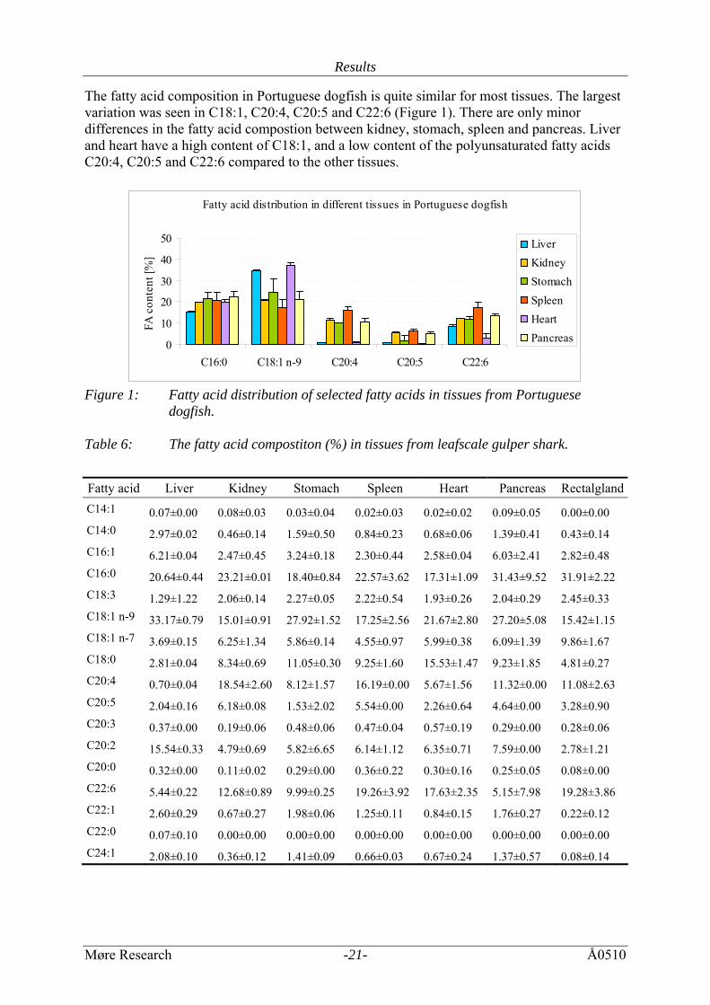
The fatty acid composition in Portuguese dogfish is quite similar for most tissues. The largest variation was seen in C18:1, C20:4, C20:5 and C22:6 (Figure 1). There are only minor differences in the fatty acid compostion between kidney, stomach, spleen and pancreas. Liver and heart have a high content of C18:1, and a low content of the polyunsaturated fatty acids C20:4, C20:5 and C22:6 compared to the other tissues.
Fatty acid distribution in different tissues in Portuguese dogfish
0
10
20
30
40
50
C16:0 C18:1 n-9 C20:4 C20:5 C22:6
FA c
onte
nt [%
]
LiverKidneyStomachSpleenHeartPancreas
Figure 1: Fatty acid distribution of selected fatty acids in tissues from Portuguese
dogfish. Table 6: The fatty acid compostiton (%) in tissues from leafscale gulper shark.
Fatty acid Liver Kidney Stomach Spleen Heart Pancreas RectalglandC14:1 0.07±0.00 0.08±0.03 0.03±0.04 0.02±0.03 0.02±0.02 0.09±0.05 0.00±0.00 C14:0 2.97±0.02 0.46±0.14 1.59±0.50 0.84±0.23 0.68±0.06 1.39±0.41 0.43±0.14 C16:1 6.21±0.04 2.47±0.45 3.24±0.18 2.30±0.44 2.58±0.04 6.03±2.41 2.82±0.48 C16:0 20.64±0.44 23.21±0.01 18.40±0.84 22.57±3.62 17.31±1.09 31.43±9.52 31.91±2.22 C18:3 1.29±1.22 2.06±0.14 2.27±0.05 2.22±0.54 1.93±0.26 2.04±0.29 2.45±0.33 C18:1 n-9 33.17±0.79 15.01±0.91 27.92±1.52 17.25±2.56 21.67±2.80 27.20±5.08 15.42±1.15 C18:1 n-7 3.69±0.15 6.25±1.34 5.86±0.14 4.55±0.97 5.99±0.38 6.09±1.39 9.86±1.67 C18:0 2.81±0.04 8.34±0.69 11.05±0.30 9.25±1.60 15.53±1.47 9.23±1.85 4.81±0.27 C20:4 0.70±0.04 18.54±2.60 8.12±1.57 16.19±0.00 5.67±1.56 11.32±0.00 11.08±2.63 C20:5 2.04±0.16 6.18±0.08 1.53±2.02 5.54±0.00 2.26±0.64 4.64±0.00 3.28±0.90 C20:3 0.37±0.00 0.19±0.06 0.48±0.06 0.47±0.04 0.57±0.19 0.29±0.00 0.28±0.06 C20:2 15.54±0.33 4.79±0.69 5.82±6.65 6.14±1.12 6.35±0.71 7.59±0.00 2.78±1.21 C20:0 0.32±0.00 0.11±0.02 0.29±0.00 0.36±0.22 0.30±0.16 0.25±0.05 0.08±0.00 C22:6 5.44±0.22 12.68±0.89 9.99±0.25 19.26±3.92 17.63±2.35 5.15±7.98 19.28±3.86 C22:1 2.60±0.29 0.67±0.27 1.98±0.06 1.25±0.11 0.84±0.15 1.76±0.27 0.22±0.12 C22:0 0.07±0.10 0.00±0.00 0.00±0.00 0.00±0.00 0.00±0.00 0.00±0.00 0.00±0.00 C24:1 2.08±0.10 0.36±0.12 1.41±0.09 0.66±0.03 0.67±0.24 1.37±0.57 0.08±0.14
Results
Møre Research -22- Å0510
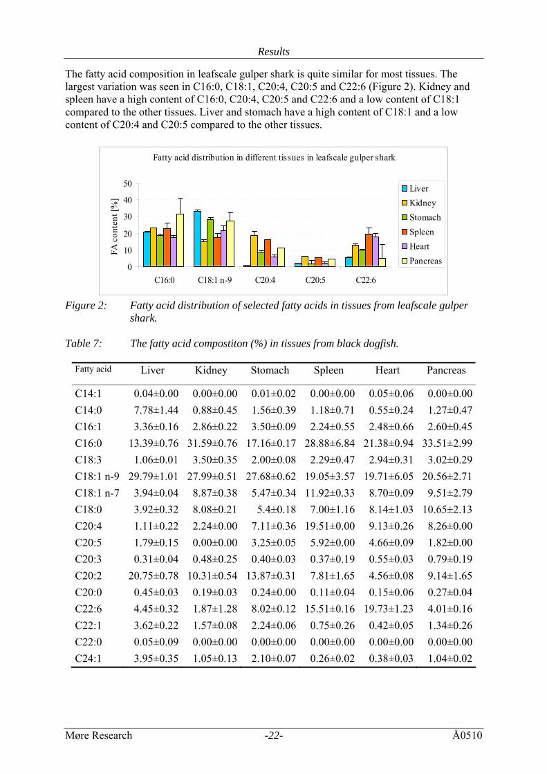
The fatty acid composition in leafscale gulper shark is quite similar for most tissues. The largest variation was seen in C16:0, C18:1, C20:4, C20:5 and C22:6 (Figure 2). Kidney and spleen have a high content of C16:0, C20:4, C20:5 and C22:6 and a low content of C18:1 compared to the other tissues. Liver and stomach have a high content of C18:1 and a low content of C20:4 and C20:5 compared to the other tissues.
Fatty acid distribution in different tissues in leafscale gulper shark
0
10
20
30
40
50
C16:0 C18:1 n-9 C20:4 C20:5 C22:6
FA c
onte
nt [%
]
LiverKidneyStomachSpleenHeartPancreas
Figure 2: Fatty acid distribution of selected fatty acids in tissues from leafscale gulper
shark. Table 7: The fatty acid compostiton (%) in tissues from black dogfish.
Fatty acid
Liver Kidney Stomach Spleen Heart Pancreas
C14:1 0.04±0.00 0.00±0.00 0.01±0.02 0.00±0.00 0.05±0.06 0.00±0.00C14:0 7.78±1.44 0.88±0.45 1.56±0.39 1.18±0.71 0.55±0.24 1.27±0.47C16:1 3.36±0.16 2.86±0.22 3.50±0.09 2.24±0.55 2.48±0.66 2.60±0.45C16:0 13.39±0.76 31.59±0.76 17.16±0.17 28.88±6.84 21.38±0.94 33.51±2.99C18:3 1.06±0.01 3.50±0.35 2.00±0.08 2.29±0.47 2.94±0.31 3.02±0.29C18:1 n-9 29.79±1.01 27.99±0.51 27.68±0.62 19.05±3.57 19.71±6.05 20.56±2.71C18:1 n-7 3.94±0.04 8.87±0.38 5.47±0.34 11.92±0.33 8.70±0.09 9.51±2.79C18:0 3.92±0.32 8.08±0.21 5.4±0.18 7.00±1.16 8.14±1.03 10.65±2.13C20:4 1.11±0.22 2.24±0.00 7.11±0.36 19.51±0.00 9.13±0.26 8.26±0.00C20:5 1.79±0.15 0.00±0.00 3.25±0.05 5.92±0.00 4.66±0.09 1.82±0.00C20:3 0.31±0.04 0.48±0.25 0.40±0.03 0.37±0.19 0.55±0.03 0.79±0.19C20:2 20.75±0.78 10.31±0.54 13.87±0.31 7.81±1.65 4.56±0.08 9.14±1.65C20:0 0.45±0.03 0.19±0.03 0.24±0.00 0.11±0.04 0.15±0.06 0.27±0.04C22:6 4.45±0.32 1.87±1.28 8.02±0.12 15.51±0.16 19.73±1.23 4.01±0.16C22:1 3.62±0.22 1.57±0.08 2.24±0.06 0.75±0.26 0.42±0.05 1.34±0.26C22:0 0.05±0.09 0.00±0.00 0.00±0.00 0.00±0.00 0.00±0.00 0.00±0.00C24:1 3.95±0.35 1.05±0.13 2.10±0.07 0.26±0.02 0.38±0.03 1.04±0.02
Results
Møre Research -23- Å0510
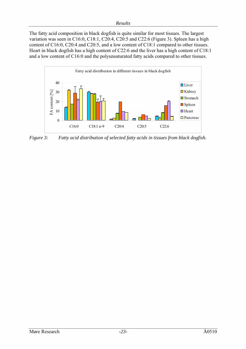
The fatty acid composition in black dogfish is quite similar for most tissues. The largest variation was seen in C16:0, C18:1, C20:4, C20:5 and C22:6 (Figure 3). Spleen has a high content of C16:0, C20:4 and C20:5, and a low content of C18:1 compared to other tissues. Heart in black dogfish has a high content of C22:6 and the liver has a high content of C18:1 and a low content of C16:0 and the polyunsaturated fatty acids compared to other tissues.
Fatty acid distribution in different tissues in black dogfish
0
10
20
30
40
C16:0 C18:1 n-9 C20:4 C20:5 C22:6
FA c
onte
nt [%
]
LiverKidneyStomachSpleenHeartPancreas
Figure 3: Fatty acid distribution of selected fatty acids in tissues from black dogfish.
Results
Møre Research -24- Å0510
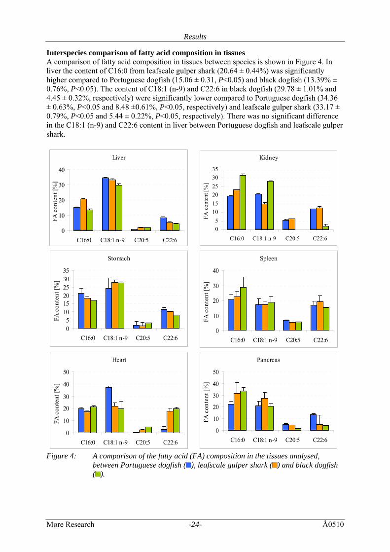
Interspecies comparison of fatty acid composition in tissues A comparison of fatty acid composition in tissues between species is shown in Figure 4. In liver the content of C16:0 from leafscale gulper shark (20.64 ± 0.44%) was significantly higher compared to Portuguese dogfish (15.06 ± 0.31, P<0.05) and black dogfish (13.39% ± 0.76%, P<0.05). The content of C18:1 (n-9) and C22:6 in black dogfish (29.78 ± 1.01% and 4.45 ± 0.32%, respectively) were significantly lower compared to Portuguese dogfish (34.36 ± 0.63%, P<0.05 and 8.48 ±0.61%, P<0.05, respectively) and leafscale gulper shark (33.17 ± 0.79%, P<0.05 and 5.44 ± 0.22%, P<0.05, respectively). There was no significant difference in the C18:1 (n-9) and C22:6 content in liver between Portuguese dogfish and leafscale gulper shark.
Liver
0
10
20
30
40
C16:0 C18:1 n-9 C20:5 C22:6
FA c
onte
nt [%
]
Kidney
05
101520253035
C16:0 C18:1 n-9 C20:5 C22:6
FA c
onte
nt [%
]
Stomach
05
101520253035
C16:0 C18:1 n-9 C20:5 C22:6
FA c
onte
nt [%
]
Spleen
0
10
20
30
40
C16:0 C18:1 n-9 C20:5 C22:6
FA c
onte
nt [%
]
Heart
0
10
20
30
40
50
C16:0 C18:1 n-9 C20:5 C22:6
FA c
onte
nt [%
]
Pancreas
0
10
20
30
40
50
C16:0 C18:1 n-9 C20:5 C22:6
FA c
onte
nt [%
]
Figure 4: A comparison of the fatty acid (FA) composition in the tissues analysed,
between Portuguese dogfish ( ), leafscale gulper shark ( ) and black dogfish ( ).
Results
Møre Research -25- Å0510
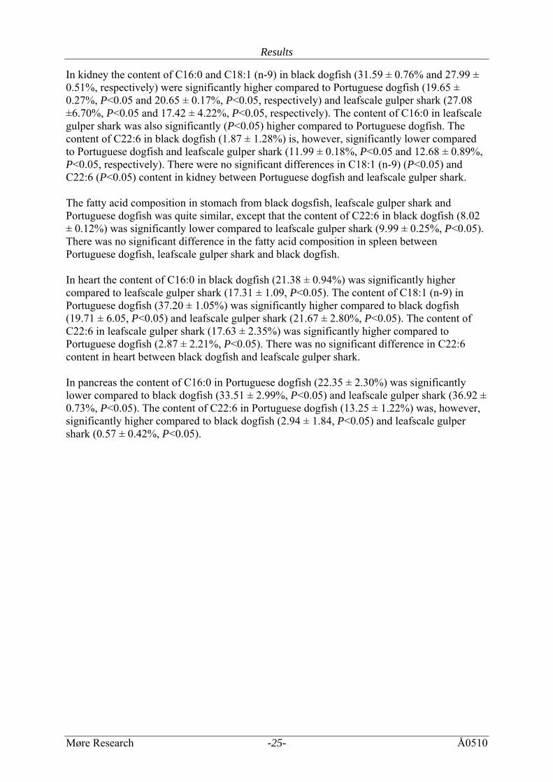
In kidney the content of C16:0 and C18:1 (n-9) in black dogfish (31.59 ± 0.76% and 27.99 ± 0.51%, respectively) were significantly higher compared to Portuguese dogfish (19.65 ± 0.27%, P<0.05 and 20.65 ± 0.17%, P<0.05, respectively) and leafscale gulper shark (27.08 ±6.70%, P<0.05 and 17.42 ± 4.22%, P<0.05, respectively). The content of C16:0 in leafscale gulper shark was also significantly (P<0.05) higher compared to Portuguese dogfish. The content of C22:6 in black dogfish (1.87 ± 1.28%) is, however, significantly lower compared to Portuguese dogfish and leafscale gulper shark (11.99 ± 0.18%, P<0.05 and 12.68 ± 0.89%, P<0.05, respectively). There were no significant differences in C18:1 (n-9) (P<0.05) and C22:6 (P<0.05) content in kidney between Portuguese dogfish and leafscale gulper shark. The fatty acid composition in stomach from black dogsfish, leafscale gulper shark and Portuguese dogfish was quite similar, except that the content of C22:6 in black dogfish (8.02 ± 0.12%) was significantly lower compared to leafscale gulper shark (9.99 ± 0.25%, P<0.05). There was no significant difference in the fatty acid composition in spleen between Portuguese dogfish, leafscale gulper shark and black dogfish. In heart the content of C16:0 in black dogfish (21.38 ± 0.94%) was significantly higher compared to leafscale gulper shark (17.31 ± 1.09, P<0.05). The content of C18:1 (n-9) in Portuguese dogfish (37.20 ± 1.05%) was significantly higher compared to black dogfish (19.71 ± 6.05, P<0.05) and leafscale gulper shark (21.67 ± 2.80%, P<0.05). The content of C22:6 in leafscale gulper shark (17.63 ± 2.35%) was significantly higher compared to Portuguese dogfish (2.87 ± 2.21%, P<0.05). There was no significant difference in C22:6 content in heart between black dogfish and leafscale gulper shark. In pancreas the content of C16:0 in Portuguese dogfish (22.35 ± 2.30%) was significantly lower compared to black dogfish (33.51 ± 2.99%, P<0.05) and leafscale gulper shark (36.92 ± 0.73%, P<0.05). The content of C22:6 in Portuguese dogfish (13.25 ± 1.22%) was, however, significantly higher compared to black dogfish (2.94 ± 1.84, P<0.05) and leafscale gulper shark (0.57 ± 0.42%, P<0.05).
Results
Møre Research -26- Å0510
3.4.2 The distribution of saturated, monounsaturated and polyunsaturated fatty acids The distribution of saturated (SFA), monounsaturated (MUFA) and polyunsaturated (PUFA) fatty acids in viscera from Portuguese dogfish, black dogfish and leafscale gulper shark was quite similar (Figure 5), except the amount of saturated fatty acids that were lower in Portuguese dogfish compared to the other species. The content of SFA, MUFA and PUFA in Portuguese dogfish was 25.97 ± 3.90%, 40.90 ± 10.86% and 34.30 ± 10.68%, respectively. The content of SFA in Portuguese dogfish was significantly lower than the content of MUFA (P<0.05). The distribution of SFA, MUFA and PUFA in leafscale gulper shark was 33.80 ± 4.87%, 34.55 ± 9.01% and 36.06 ± 8.88%, respectively. The content of SFA in leafscale gulper shark was significantly lower compared to Portuguese dogfish (P<0.05). The content of SFA, MUFA and PUFA in black dogfish was 33.96 ± 8.61%, 38.18 ± 5.19% and 33.76 ± 11.60%, respectively. The MUFA content was high in livers from all species, and the PUFA content was high in all spleen tested. In the other organs the SFA, MUFA and PUFA were evenly distributed.
Dis tribution of saturated, monounsaturated and polyunsaturated fatty acids in viscera from deep-sea sharks
0
10
20
30
40
50
60
∑SFA ∑MUFA ∑PUFA
Fatty
aci
d co
nten
t [%
]
Portuguese dogfishLeafscale gulper sharkBlack dogfish
Figure 5: Distribution of saturated, monounsaturated and polyunsaturated fatty acids in
viscera from Portuguese dogfish, leafscale gulper shark and black dogfish.
Results
Møre Research -27- Å0510
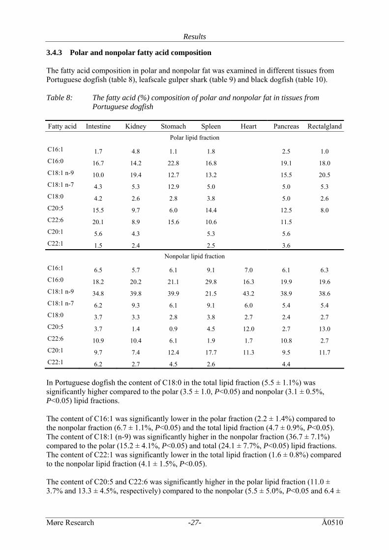
3.4.3 Polar and nonpolar fatty acid composition The fatty acid composition in polar and nonpolar fat was examined in different tissues from Portuguese dogfish (table 8), leafscale gulper shark (table 9) and black dogfish (table 10). Table 8: The fatty acid (%) composition of polar and nonpolar fat in tissues from
Portuguese dogfish Fatty acid Intestine Kidney Stomach Spleen Heart Pancreas Rectalgland
Polar lipid fraction
C16:1 1.7 4.8 1.1 1.8 2.5 1.0 C16:0 16.7 14.2 22.8 16.8 19.1 18.0 C18:1 n-9 10.0 19.4 12.7 13.2 15.5 20.5 C18:1 n-7 4.3 5.3 12.9 5.0 5.0 5.3 C18:0 4.2 2.6 2.8 3.8 5.0 2.6 C20:5 15.5 9.7 6.0 14.4 12.5 8.0 C22:6 20.1 8.9 15.6 10.6 11.5 C20:1 5.6 4.3 5.3 5.6 C22:1 1.5 2.4 2.5 3.6
Nonpolar lipid fraction
C16:1 6.5 5.7 6.1 9.1 7.0 6.1 6.3 C16:0 18.2 20.2 21.1 29.8 16.3 19.9 19.6 C18:1 n-9 34.8 39.8 39.9 21.5 43.2 38.9 38.6 C18:1 n-7 6.2 9.3 6.1 9.1 6.0 5.4 5.4 C18:0 3.7 3.3 2.8 3.8 2.7 2.4 2.7 C20:5 3.7 1.4 0.9 4.5 12.0 2.7 13.0 C22:6 10.9 10.4 6.1 1.9 1.7 10.8 2.7 C20:1 9.7 7.4 12.4 17.7 11.3 9.5 11.7 C22:1 6.2 2.7 4.5 2.6 4.4 In Portuguese dogfish the content of C18:0 in the total lipid fraction (5.5 ± 1.1%) was significantly higher compared to the polar (3.5 ± 1.0, P<0.05) and nonpolar (3.1 ± 0.5%, P<0.05) lipid fractions. The content of C16:1 was significantly lower in the polar fraction (2.2 ± 1.4%) compared to the nonpolar fraction (6.7 ± 1.1%, P<0.05) and the total lipid fraction (4.7 ± 0.9%, P<0.05). The content of C18:1 (n-9) was significantly higher in the nonpolar fraction (36.7 ± 7.1%) compared to the polar (15.2 ± 4.1%, P<0.05) and total (24.1 ± 7.7%, P<0.05) lipid fractions. The content of C22:1 was significantly lower in the total lipid fraction (1.6 ± 0.8%) compared to the nonpolar lipid fraction (4.1 ± 1.5%, P<0.05). The content of C20:5 and C22:6 was significantly higher in the polar lipid fraction (11.0 ± 3.7% and 13.3 ± 4.5%, respectively) compared to the nonpolar (5.5 ± 5.0%, P<0.05 and 6.4 ±
Results
Møre Research -28- Å0510
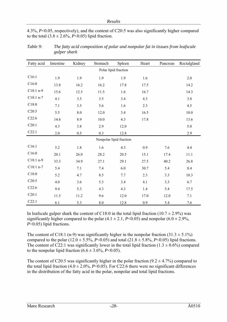
4.3%, P<0.05, respectively), and the content of C20:5 was also significantly higher compared to the total (3.8 ± 2.6%, P<0.05) lipid fraction. Table 9: The fatty acid composition of polar and nonpolar fat in tissues from leafscale
gulper shark Fatty acid Intestine Kidney Stomach Spleen Heart Pancreas Rectalgland
Polar lipid fraction
C16:1 1.9 1.9 1.9 1.9 1.6 2.0 C16:0 13.8 16.2 16.2 17.8 17.5 14.2 C18:1 n-9 15.6 12.5 11.5 1.6 16.7 14.3 C18:1 n-7 4.1 3.5 3.5 3.4 4.3 3.8 C18:0 7.1 3.5 5.6 1.6 2.3 4.5 C20:5 5.5 8.0 12.0 3.4 16.5 10.0 C22:6 14.6 8.9 10.0 4.3 17.8 13.6 C20:1 4.3 3.8 2.9 12.0 5.0 C22:1 2.0 0.5 0.3 12.8 2.9
Nonpolar lipid fraction
C16:1 5.2 1.8 1.6 4.3 0.9 7.6 4.4 C16:0 20.1 26.0 28.2 20.5 15.1 17.4 11.1 C18:1 n-9 33.3 34.9 27.1 29.1 27.5 40.2 26.8 C18:1 n-7 5.4 7.1 7.4 6.0 30.7 5.4 8.4 C18:0 5.2 4.7 8.5 7.7 2.3 3.3 10.3 C20:5 4.0 3.6 5.3 3.4 4.1 3.3 6.7 C22:6 9.4 5.3 4.3 4.3 1.4 5.4 17.5 C20:1 11.5 11.2 9.6 12.0 17.0 12.0 7.1 C22:1 6.1 5.3 8.0 12.8 0.9 5.4 7.6 In leafscale gulper shark the content of C18:0 in the total lipid fraction (10.7 ± 2.9%) was significantly higher compared to the polar (4.1 ± 2.1, P<0.05) and nonpolar (6.0 ± 2.9%, P<0.05) lipid fractions. The content of C18:1 (n-9) was significantly higher in the nonpolar fraction (31.3 ± 5.1%) compared to the polar (12.0 ± 5.5%, P<0.05) and total (21.8 ± 5.8%, P<0.05) lipid fractions. The content of C22:1 was significantly lower in the total lipid fraction (1.3 ± 0.6%) compared to the nonpolar lipid fraction (6.6 ± 3.6%, P<0.05). The content of C20:5 was significantly higher in the polar fraction (9.2 ± 4.7%) compared to the total lipid fraction (4.0 ± 2.0%, P<0.05). For C22:6 there were no significant differences in the distribution of the fatty acid in the polar, nonpolar and total lipid fractions.
Results
Møre Research -29- Å0510
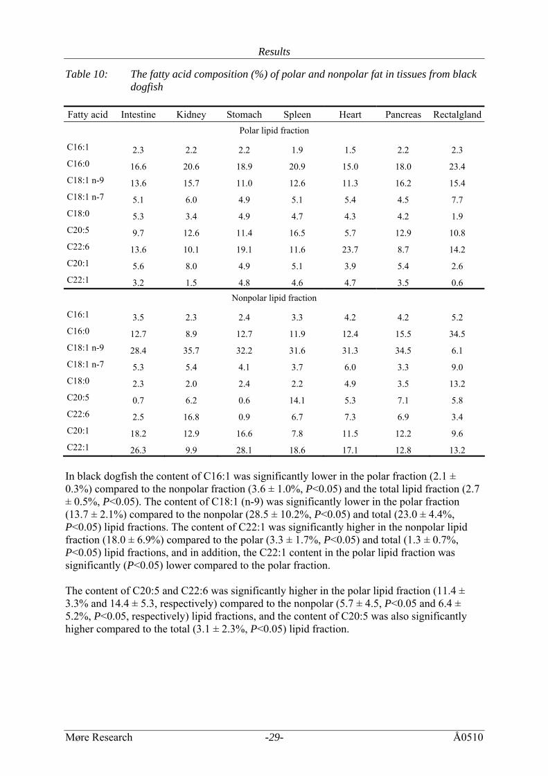
Table 10: The fatty acid composition (%) of polar and nonpolar fat in tissues from black dogfish
Fatty acid Intestine Kidney Stomach Spleen Heart Pancreas Rectalgland
Polar lipid fraction
C16:1 2.3 2.2 2.2 1.9 1.5 2.2 2.3 C16:0 16.6 20.6 18.9 20.9 15.0 18.0 23.4 C18:1 n-9 13.6 15.7 11.0 12.6 11.3 16.2 15.4 C18:1 n-7 5.1 6.0 4.9 5.1 5.4 4.5 7.7 C18:0 5.3 3.4 4.9 4.7 4.3 4.2 1.9 C20:5 9.7 12.6 11.4 16.5 5.7 12.9 10.8 C22:6 13.6 10.1 19.1 11.6 23.7 8.7 14.2 C20:1 5.6 8.0 4.9 5.1 3.9 5.4 2.6 C22:1 3.2 1.5 4.8 4.6 4.7 3.5 0.6
Nonpolar lipid fraction
C16:1 3.5 2.3 2.4 3.3 4.2 4.2 5.2 C16:0 12.7 8.9 12.7 11.9 12.4 15.5 34.5 C18:1 n-9 28.4 35.7 32.2 31.6 31.3 34.5 6.1 C18:1 n-7 5.3 5.4 4.1 3.7 6.0 3.3 9.0 C18:0 2.3 2.0 2.4 2.2 4.9 3.5 13.2 C20:5 0.7 6.2 0.6 14.1 5.3 7.1 5.8 C22:6 2.5 16.8 0.9 6.7 7.3 6.9 3.4 C20:1 18.2 12.9 16.6 7.8 11.5 12.2 9.6 C22:1 26.3 9.9 28.1 18.6 17.1 12.8 13.2 In black dogfish the content of C16:1 was significantly lower in the polar fraction (2.1 ± 0.3%) compared to the nonpolar fraction (3.6 ± 1.0%, P<0.05) and the total lipid fraction (2.7 ± 0.5%, P<0.05). The content of C18:1 (n-9) was significantly lower in the polar fraction (13.7 ± 2.1%) compared to the nonpolar (28.5 ± 10.2%, P<0.05) and total (23.0 ± 4.4%, P<0.05) lipid fractions. The content of C22:1 was significantly higher in the nonpolar lipid fraction (18.0 ± 6.9%) compared to the polar (3.3 ± 1.7%, P<0.05) and total (1.3 ± 0.7%, P<0.05) lipid fractions, and in addition, the C22:1 content in the polar lipid fraction was significantly (P<0.05) lower compared to the polar fraction. The content of C20:5 and C22:6 was significantly higher in the polar lipid fraction (11.4 ± 3.3% and 14.4 ± 5.3, respectively) compared to the nonpolar (5.7 ± 4.5, P<0.05 and 6.4 ± 5.2%, P<0.05, respectively) lipid fractions, and the content of C20:5 was also significantly higher compared to the total (3.1 ± 2.3%, P<0.05) lipid fraction.
Results
Møre Research -30- Å0510
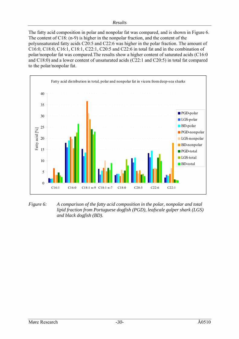
The fatty acid composition in polar and nonpolar fat was compared, and is shown in Figure 6. The content of C18: (n-9) is higher in the nonpolar fraction, and the content of the polyunsaturated fatty acids C20:5 and C22:6 was higher in the polar fraction. The amount of C16:0, C18:0, C16:1, C18:1, C22:1, C20:5 and C22:6 in total fat and in the combination of polar/nonpolar fat was compared.The results show a higher content of saturated acids (C16:0 and C18:0) and a lower content of unsaturated acids (C22:1 and C20:5) in total fat compared to the polar/nonpolar fat.
Fatty acid distribution in total, polar and nonpolar fat in vicera from deep-sea sharks
0
5
10
15
20
25
30
35
40
C16:1 C16:0 C18:1 n-9 C18:1 n-7 C18:0 C20:5 C22:6 C22:1
Fatty
aci
d [%
]
PGD-polarLGS-polarBD-polarPGD-nonpolarLGS-nonpolarBD-nonpolarPGD-totalLGS-totalBD-total
Figure 6: A comparison of the fatty acid composition in the polar, nonpolar and total
lipid fraction from Portuguese dogfish (PGD), leafscale gulper shark (LGS) and black dogfish (BD).
Results
Møre Research -31- Å0510
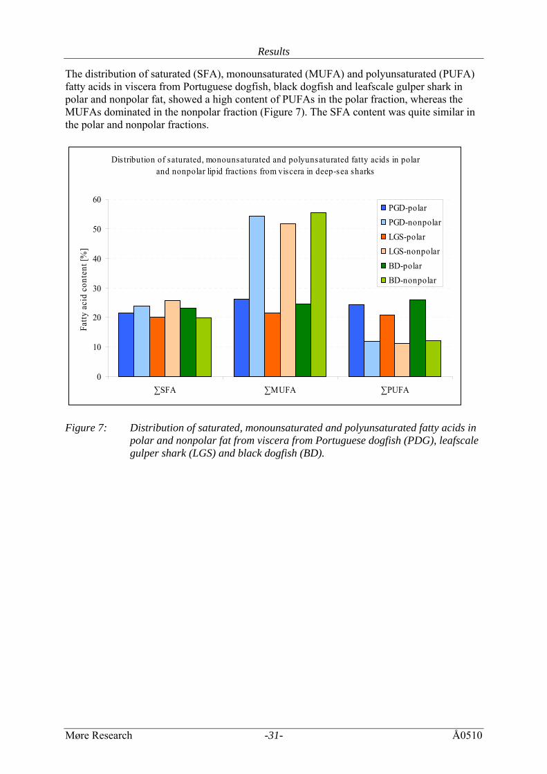
The distribution of saturated (SFA), monounsaturated (MUFA) and polyunsaturated (PUFA) fatty acids in viscera from Portuguese dogfish, black dogfish and leafscale gulper shark in polar and nonpolar fat, showed a high content of PUFAs in the polar fraction, whereas the MUFAs dominated in the nonpolar fraction (Figure 7). The SFA content was quite similar in the polar and nonpolar fractions.
Dis tribution of saturated, monounsaturated and polyunsaturated fatty acids in polar and nonpolar lipid fractions from viscera in deep-sea sharks
0
10
20
30
40
50
60
∑SFA ∑MUFA ∑PUFA
Fatty
aci
d co
nten
t [%
]
PGD-polarPGD-nonpolarLGS-polarLGS-nonpolarBD-polarBD-nonpolar
Figure 7: Distribution of saturated, monounsaturated and polyunsaturated fatty acids in
polar and nonpolar fat from viscera from Portuguese dogfish (PDG), leafscale gulper shark (LGS) and black dogfish (BD).
Results
Møre Research -32- Å0510
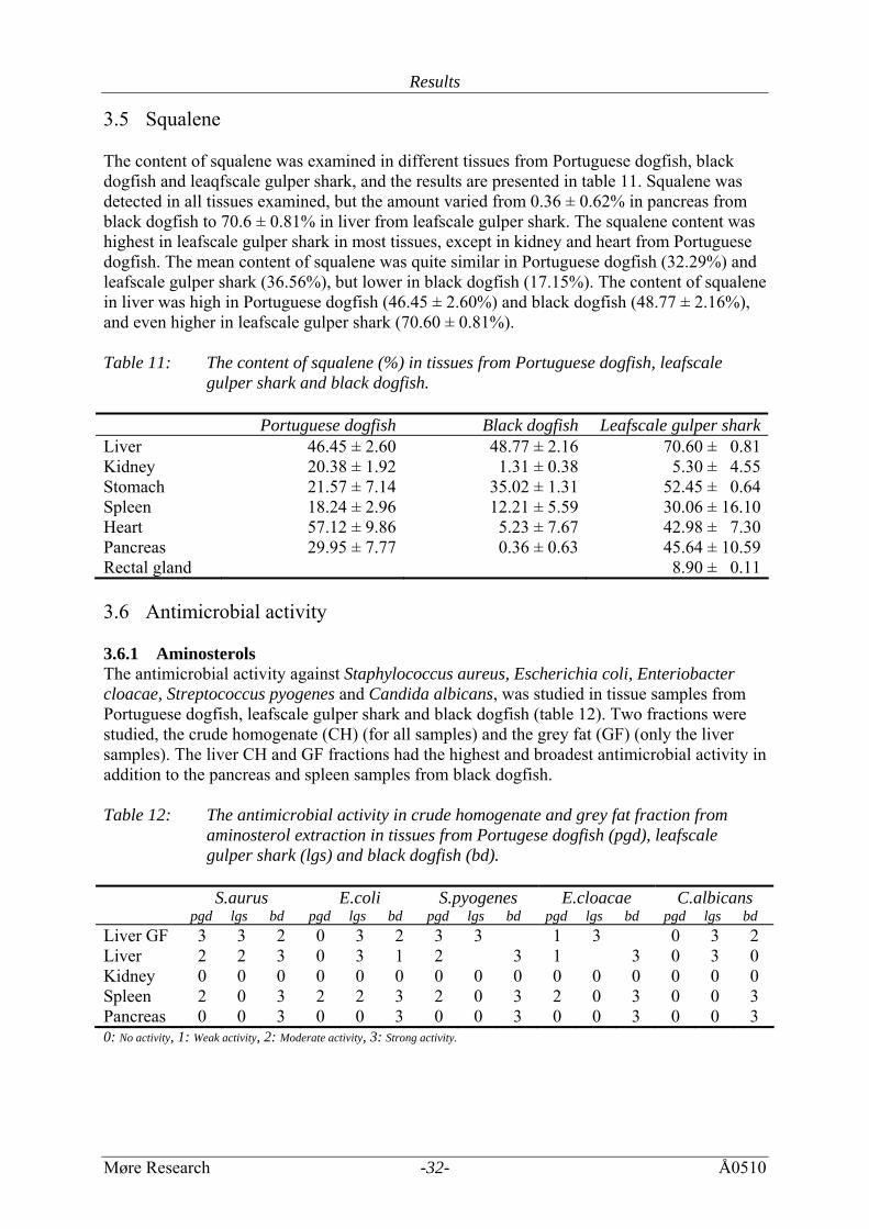
3.5 Squalene The content of squalene was examined in different tissues from Portuguese dogfish, black dogfish and leaqfscale gulper shark, and the results are presented in table 11. Squalene was detected in all tissues examined, but the amount varied from 0.36 ± 0.62% in pancreas from black dogfish to 70.6 ± 0.81% in liver from leafscale gulper shark. The squalene content was highest in leafscale gulper shark in most tissues, except in kidney and heart from Portuguese dogfish. The mean content of squalene was quite similar in Portuguese dogfish (32.29%) and leafscale gulper shark (36.56%), but lower in black dogfish (17.15%). The content of squalene in liver was high in Portuguese dogfish (46.45 ± 2.60%) and black dogfish (48.77 ± 2.16%), and even higher in leafscale gulper shark (70.60 ± 0.81%). Table 11: The content of squalene (%) in tissues from Portuguese dogfish, leafscale
gulper shark and black dogfish. Portuguese dogfish Black dogfish Leafscale gulper sharkLiver 46.45 ± 2.60 48.77 ± 2.16 70.60 ± 0.81Kidney 20.38 ± 1.92 1.31 ± 0.38 5.30 ± 4.55Stomach 21.57 ± 7.14 35.02 ± 1.31 52.45 ± 0.64Spleen 18.24 ± 2.96 12.21 ± 5.59 30.06 ± 16.10 Heart 57.12 ± 9.86 5.23 ± 7.67 42.98 ± 7.30Pancreas 29.95 ± 7.77 0.36 ± 0.63 45.64 ± 10.59Rectal gland 8.90 ± 0.11 3.6 Antimicrobial activity 3.6.1 Aminosterols The antimicrobial activity against Staphylococcus aureus, Escherichia coli, Enteriobacter cloacae, Streptococcus pyogenes and Candida albicans, was studied in tissue samples from Portuguese dogfish, leafscale gulper shark and black dogfish (table 12). Two fractions were studied, the crude homogenate (CH) (for all samples) and the grey fat (GF) (only the liver samples). The liver CH and GF fractions had the highest and broadest antimicrobial activity in addition to the pancreas and spleen samples from black dogfish. Table 12: The antimicrobial activity in crude homogenate and grey fat fraction from
aminosterol extraction in tissues from Portugese dogfish (pgd), leafscale gulper shark (lgs) and black dogfish (bd).
S.aurus
pgd lgs bd E.coli
pgd lgs bd S.pyogenes
pgd lgs bd E.cloacae
pgd lgs bd C.albicans
pgd lgs bd Liver GF 3 3 2 0 3 2 3 3 1 3 0 3 2 Liver 2 2 3 0 3 1 2 3 1 3 0 3 0 Kidney 0 0 0 0 0 0 0 0 0 0 0 0 0 0 0 Spleen 2 0 3 2 2 3 2 0 3 2 0 3 0 0 3 Pancreas 0 0 3 0 0 3 0 0 3 0 0 3 0 0 3 0: No activity, 1: Weak activity, 2: Moderate activity, 3: Strong activity.
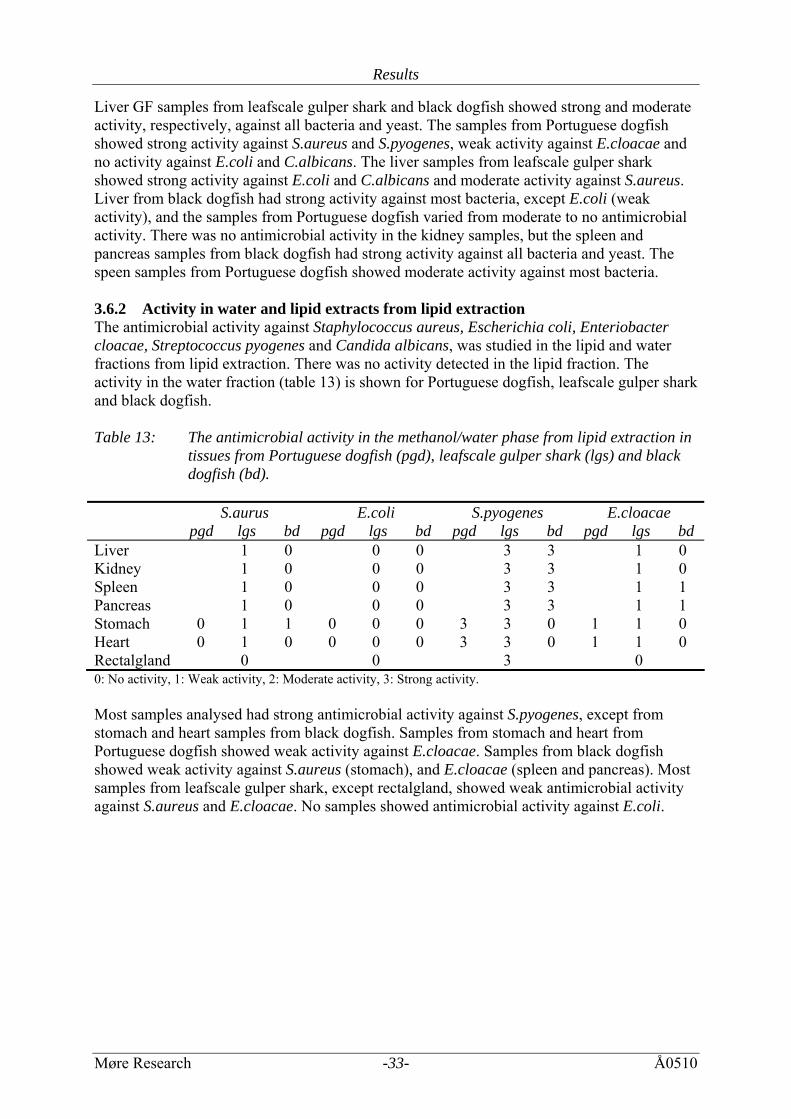
Results
Møre Research -33- Å0510
Liver GF samples from leafscale gulper shark and black dogfish showed strong and moderate activity, respectively, against all bacteria and yeast. The samples from Portuguese dogfish showed strong activity against S.aureus and S.pyogenes, weak activity against E.cloacae and no activity against E.coli and C.albicans. The liver samples from leafscale gulper shark showed strong activity against E.coli and C.albicans and moderate activity against S.aureus. Liver from black dogfish had strong activity against most bacteria, except E.coli (weak activity), and the samples from Portuguese dogfish varied from moderate to no antimicrobial activity. There was no antimicrobial activity in the kidney samples, but the spleen and pancreas samples from black dogfish had strong activity against all bacteria and yeast. The speen samples from Portuguese dogfish showed moderate activity against most bacteria. 3.6.2 Activity in water and lipid extracts from lipid extraction The antimicrobial activity against Staphylococcus aureus, Escherichia coli, Enteriobacter cloacae, Streptococcus pyogenes and Candida albicans, was studied in the lipid and water fractions from lipid extraction. There was no activity detected in the lipid fraction. The activity in the water fraction (table 13) is shown for Portuguese dogfish, leafscale gulper shark and black dogfish. Table 13: The antimicrobial activity in the methanol/water phase from lipid extraction in
tissues from Portuguese dogfish (pgd), leafscale gulper shark (lgs) and black dogfish (bd).
S.aurus
pgd lgs bd E.coli
pgd lgs bd S.pyogenes
pgd lgs bd E.cloacae
pgd lgs bd Liver 1 0 0 0 3 3 1 0 Kidney 1 0 0 0 3 3 1 0 Spleen 1 0 0 0 3 3 1 1 Pancreas 1 0 0 0 3 3 1 1 Stomach 0 1 1 0 0 0 3 3 0 1 1 0 Heart 0 1 0 0 0 0 3 3 0 1 1 0 Rectalgland 0 0 3 0 0: No activity, 1: Weak activity, 2: Moderate activity, 3: Strong activity. Most samples analysed had strong antimicrobial activity against S.pyogenes, except from stomach and heart samples from black dogfish. Samples from stomach and heart from Portuguese dogfish showed weak activity against E.cloacae. Samples from black dogfish showed weak activity against S.aureus (stomach), and E.cloacae (spleen and pancreas). Most samples from leafscale gulper shark, except rectalgland, showed weak antimicrobial activity against S.aureus and E.cloacae. No samples showed antimicrobial activity against E.coli.
Discussion
Møre Research -34- Å0510
4 DISCUSSION Over the last decades, the interest in fish lipids has increased dramatically. Fish oils are now regarded as an excellent source for polyunsaturated fatty acids, PUFA (Navarro-Garcia et al, 2000). Two of these acids, eicosapentaenoic EPA and docosahexaenoic DHA have been reported to be important in preventing or reducing heart disease, inflammatory disorders and in the case of DHA, as a nutritional supplement for the brain and retina development in babies. 4.1 Water content The water content in heart, intestine, kidney, pancreas, rectalgland, spleen and stomach from Portuguese dogfish, leafscale gulper shark and black dogfish was between 76.8% and 84 %, which is in agreement with the water content in muscle (79.9% ± 0.17, 82.3% ± 0.18% and 84.1% ± 0.09%, respectively) (Økland et al, 2005), but lower than the water content in eyes (89.6% ± 0.2%, 90.1% ± 0.2% and 88.2% ± 0.3%, respectively) in the same species. The content of water in eggs (55.8% ± 0.01%, 44.3% ± 0.1% and 58.0% ± 0.1%, respectively) (Remme et al, 2005) differed highly from the rest of the tissues examined. 4.2 Lipid content The total lipid content in heart, kidney, pancreas, rectalgland, spleen and stomach from Portuguese dogfish, leafscale gulper shark and black dogfish was between 1.22% and 5.41%. The lipid content in the liver from the sample species was 37%, 50% and 35%, respectively. The content of lipids in the tissues mentioned is in agreement with the lipid content in brain (3.8% ± 0.1%, 4.1% ± 0.1% and 3.2% ± 0.0%, respectively) (Stoknes et al, 2004), lower in muscle (0.95% ± 0.07%, 0.99% ± 0.05% and 0.70% ± 0.02%, respectively) (Økland et al, 2005) and eyes (0.3% ± 0.0, 0.3% ± 0.0 and 0.3% ± 0.0, respectively) (Stoknes et al, 2004), and higher in eggs (22.58% ± 5.0%, 30.2% ± 5.0% and 21.2% ± 5.0%, respectively) (Remme et al, 2005) in the same species. The polar and nonpolar lipid content in heart, kidney, pancreas, rectalgland, spleen and stomach from Portuguese dogfish, leafscale gulper shark and black dogfish varied from 1.07% to 2.55% and 1.07% to 3.04%, respectively. The content in stomach from black dogfish was measured to 3.48% ± 0.93%. These results may be higher than in Portuguese dogfish and leafscale gulper shark (1.36% and 1.76%, respectively) due to stomach content with high lipid content. The mean content of polar and nonpolar lipids is quite similar for all analysed shark species. There are some variations in the polar/nonpolar lipid distribution in the different tissues, but there are no significant differences. 4.3 Fatty acid composition The fatty acid composition in the different tissues from Portuguese dogfish, leafscale gulper shark and black dogfish was relatively similar to other marine organisms, with high contents of C16:0, C18:1 and C22:6 (Saito et al, 1997; Üstün et al, 1996; Watanabe et al, 1995). The fatty acid composition in all tissues was dominated by the monounsaturated fatty acid C18:1 (n-9) (25.8% ± 8.1% in Portuguese dogfish, 22.5% ± 7.1% in leafscale gulper shark and 24.1% ± 4.9% in black dogfish). It has previously been reported that organisms from the
Discussion
Møre Research -35- Å0510
deep-sea are enriched (up to 50% of total FA) in C18:1 fatty acids compared to specimens from either shallow warm or cold water, suggesting that this may be an adaptive response to high pressure in deep waters (Hazel and Williams, 1990). All tissues also had a high content of the saturated fatty acid C16:0 (19.8 ± 2.5% in Portuguese dogfish, 23.6 ± 5.9% in leafscale gulper shark and 24.3 ± 8.2% in black dogfish). In seawater fish, EPA and DHA are often the major PUFAs (Bell and Tocher, 1989). The samples studied had a high content of the polyunsaturated fatty acid C22:6 (DHA) (10.9 ± 4.8% in Portuguese dogfish, 12.8 ± 6.2% in leafscale gulper shark and 8.9 ± 7.1% in black dogfish). In accordance with a previous study (Dunstan et al, 1988) this study revealed that the tissues from these cartilaginous species contain low levels of EPA compared to bony fish. Dunstan et al (1988) previously reported mean values of EPA in flesh from chondrichthyes of 3.5%-4.9%, and 8.6%-11.7% and 18 % for bony fish and cephalopods, respectively. The fatty acid composition of the polar and nonpolar fraction differed regarding the content of MUFAs and PUFAs. The content of saturated fatty acids was, however, quite similar. The nonpolar fraction had a high content of MUFAs (mean: 53.7%), but a lower content of PUFAs (mean: 24.1%). The polar fraction, however, had a high content of PUFAs (mean: 23.7%). The content of MUFAs in the polar fraction was low (mean: 11.6%). The high level of unsaturated fatty acids in phospholipids (polare lipids) during cold acclimation as a mechanism for concerving membrane fluidity appear to be a common feature of membrane systems (Farias et al., 2003). Studies have also reported a high content of PUFAs in polar lipid and a high content of MUFAs in non polar lipids from marine species (Farias et al., 2003; Peng et al., 2003; Shirai et al., 2003). The fatty acid profile compared to other tissues in the same species The fatty acid profiles from the kidney, stomach, spleen, heart and pancreas from Portuguese dogfish, leafscale gulper shark and black dogfish are mostly in agreement with the fatty acid profiles previously reported for muscle (Økland et al, 2005), eggs (Remme et al, 2005) and brain (Stoknes et al, 2004) (table 14). The dominant fatty acids were C16:0, C18:1 and C22:6 in all tissues. Table 14: Fatty acid (%) composition in egg, muscle and brain tissues from Portuguese
dogfish, leafscale gulper shark and black dogfish, previously reported, and the mean from value in tissues from our study.
Portuguese dogfish
Eggs1 Muscle2 Brain3 Tissues4 Leafscale gulper shark
Eggs1 Muscle2 Brain3 Tissues4 Black dogfish
Eggs1 Muscle2 Brain3 Tissues4 C16:0 18.4 11.5 12.2 20.8 18.5 12.0 10.7 22.6 16.8 12.5 12.8 26.5 C18:0 1.7 3.1 7.7 5.5 2.5 7.7 9.2 10.7 1.8 4.9 8.0 7.9 C16:1 4.9 4.4 9.9 4.7 5.4 0.9 8.4 3.3 4.1 0.7 10.4 2.7 C18:1 31.1 22.5 22.7 30.9 39.1 19.1 20.0 27.5 27.3 20.5 24.9 31.9 C22:1 6.4 3.8 3.6 1.6 4.2 1.9 1.6 1.3 11.5 0.8 0.9 1.3 C20:5 2.6 3.0 4.8 3.8 2.8 2.5 5.5 4.0 5.1 4.7 4.8 3.1 C22:6 15.1 33.2 22.8 11.4 14.5 39.7 27.0 12.9 12.0 38.7 21.8 9.8 1 Remme et al, 2005; 2 Økland et al, 2005; 3 Stoknes et al, 2004; 4 Tissues are the mean value from kidney, stomach, spleen, heart and pancreas. The content of C16:0 is high in kidney, stomach, spleen, heart and pancreas (20.8% in Portuguese dogfish, 22.6% in leafscale gulper shark and 26.5% in black dogfish) compared to
Discussion
Møre Research -36- Å0510
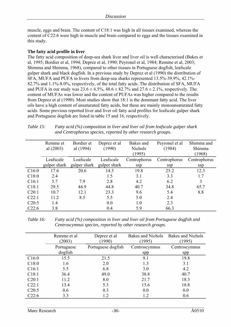
muscle, eggs and brain. The content of C18:1 was high in all tissues examined, whereas the content of C22:6 were high in muscle and brain compared to eggs and the tissues examined in this study. The fatty acid profile in liver The fatty acid composition of deep-sea shark liver and liver oil is well characterised (Bakes et al, 1995; Bordier et al, 1994; Deprez et al, 1990; Peyronel et al, 1984; Remme et al, 2003; Shimma and Shimma, 1968), compared to other tissues in Portuguese dogfish, leafscale gulper shark and black dogfish. In a previous study by Deprez et al (1990) the distribution of SFA, MUFA and PUFA in livers from deep-sea sharks represented 13.5%-39.9%, 42.1%-82.7% and 1.1%-8.0%, respectively, of the total fatty acids. The distribution of SFA, MUFA and PUFA in our study was 23.6 ± 4.5%, 48.6 ± 82.7% and 27.6 ± 2.1%, respectively. The content of MUFAs was lower and the content of PUFAs was higher compared to the results from Deprez et al (1990). Most studies show that 18:1 is the dominant fatty acid. The liver oils have a high content of unsaturated fatty acids, but these are mainly monounsaturated fatty acids. Some previous reported liver and liver oil fatty acid profiles for leafscale gulper shark and Portuguese dogfish are listed in table 15 and 16, respectively. Table 15: Fatty acid (%) composition in liver and liver oil from leafscale gulper shark
and Centrophorus species, reported by other research groups. Remme et
al (2003) Bordier et al (1994)
Deprez et al (1990)
Bakes and Nichols (1995)
Peyronel et al (1984)
Shimma and Shimma (1968)
Leafscale gulper shark
Leafscale gulper shark
Leafscale gulper shark
Centrophorus ssp
Centrophorus ssp
Centrophorus ssp
C16:0 17.6 20.6 14.5 19.8 25.2 12.3 C18:0 2.4 1.5 3.1 3.3 1.7 C16:1 5.7 7.9 2.8 4.2 6.2 3 C18:1 29.5 44.9 44.8 40.7 34.8 65.7 C20:1 10.7 12.1 23.3 9.6 5.4 8.8 C22:1 11.2 8.5 5.5 5.0 2.4 C20:5 1.4 0.0 1.0 2.3 C22:6 3.8 0.4 5.9 66.3 Table 16: Fatty acid (%) composition in liver and liver oil from Portuguese dogfish and
Centroscymnus species, reported by other research groups. Remme et al
(2003) Deprez et al
(1990) Bakes and Nichols
(1995) Bakes and Nichols
(1995) Portuguese
dogfish Portuguese dogfish Centroscymnus
spp Centroscymnus
spp C16:0 15.5 21.5 9.1 19.8 C18:0 1.6 2.0 1.3 3.1 C16:1 5.5 6.8 3.0 4.2 C18:1 36.4 49.0 38.8 40.7 C20:1 11.2 8.0 21.7 18.3 C22:1 13.4 5.3 15.6 18.8 C20:5 0.6 0.3 0.0 0.0 C22:6 3.3 1.2 1.2 0.6
Discussion
Møre Research -37- Å0510
In the present study the fatty acid composition in the whole liver was examined. There are some differences in the fatty acid composition, especially in the amount of C16:0, C18:1 and C22:6. The content of C18:1 varies among species and individuals, but is generally high in all deep-sea sharks, with a content of 30 - 49 %. The content of the other monounsaturated fatty acids, C20:1 and C22:1, varies from 2.4 % to 23.3%, but is generally found at concentrations of about 10 %. The content of the polyunsaturated fatty acids C20:5 and C22:6 in liver from deep sea shark is low, compared to the content of monounsaturated fatty acids. The content of C20:5 varies from 0% to 2.3% and the content of C22:6 varies from 0.4% to 5.9%, except from one study (Peyronel el at, 1984) which reported the content of C22:6 to be 66.3%. 4.4 Squalene Deep-sea sharks livers generally have high levels of squalene (Deprez et al, 1990). The liver is the principal site of lipid storage, and the high content of low density squalene is believed to provide hydrostatic lift and enable the sharks to maintain neutral buoyancy. The squalene content of the liver oil has been studied by many research groups. The squalene content in liver from Portuguese dogfish (46.45 ± 2.60%) in the present study was in agreement with previous studies, reporting squalene content at 35% and 48%, (Remme et al, 2003), 52 % (Deprez et al, 1990), 33.3 ± 6.7% (Batista and Nunes, 1992), 51.63 ± 6.88% (Hernandes-Perez et al, 1997) and 51% (Sargent et al, 1973). The squalene content in liver from leafscale gulper shark (70.60 ± 0.81%) in the present study was in agreement with previous studies, reporting squalene content at 66% and 76%, (Remme et al, 2003), 67.9 ± 0.2% (Batista and Nunes, 1992), 70% (Hernandes-Perez et al, 1997), 70 ± 10% (Wetherbee and Nichols, 2000) and 73% (Salmonowicz and Krawczak-Krogulecka, 1981). It is, however, in conflict with result from Deprez (1990), which found the squalene content in liver oil from leafscale gulper shark to be 1 %. The squalene content in liver from black dogfish (48.77 ± 2.16%) in the present study was higher compared to previous measured squalene content in liver samples from black dogfish (18.3 %) (Remme et al, 2003). It seems that there is little difference in the squalene content in liver oil and in liver tissue samples. The content of squalene is higher in leafscale gulper shark compared to Portuguese dogfish and black dogfish. The individual variability of these species is large and the differences observed in these studies may be due to a variety of factors, such as age, sex, geographic location and seasonal variation (Hayashi and Takagi, 1981). Differences in oil composition within species have also previously been documented. Because squalene has a key role in sterol biosynthesis, it is possible that differences in squalene concentrations reflect differential regulation of steroid hormone levels (Wetherbee and Nichols, 2000). Because there is very little information on the squalene content in other tissues, the content of squalene was examined in kidney, stomach, spleen, heart, and pancreas from Portuguese dogfish, leafscale gulper shark and black dogfish as well. In addition, the content of squalene in the rectalgland of leafscale gulper shark was measured. Squalene was detected, at various concentrations, in all tissues examined. The squalene content varied from 18.24 ± 2.96% in spleen to 57.12 ± 9.86% in heart in Portuguese dogfish, from 5.30 ± 4.55% in kidney to 52.45
Discussion
Møre Research -38- Å0510
± 0.64% in stomach in leafscale gulper shark and from 0.36 ± 0.63% in pancreas to 35.02 ± 1.31% in stomach in black dogfish. The content of squalene was generally lower in black dogfish compared to Portuguese dogfish and leafscale gulper shark, but the difference was not significant (P=0.08). Squalene content above 40 % was also found in stomach (52.45 ± 0.64%), heart (42.98 ± 7.30%) and pancreas (45.64 ± 10.59%) from leafscale gulper shark and in heart (57.12 ± 9.86%) from Portuguese dogfish. 4.5 Antimicrobial activity The antimicrobial activity in the liver, spleen, pancreas and kidney from Portuguese dogfish, lefscale gulper shark and black dogfish was examined. Most of the liver samples showed broad antimicrobial activity, but there were some differences in the activities between the samples. Portuguese dogfish, leafscale gulper shark and black dogfish are from the same order, Squaliformes, as piked dogfish (Squalus acanthias), in which squalamine first was found. In this project grey fat and crude homogenate was purified according to the method used to purify squalamine. Several previous studies have shown that squalamine has a potent antimicrobial activity against Gram-negative bacteria (Escherichia coli, Pseudomonas aeroginosa), Gram-positive bacteria (Staphylococcus aureus, Enterococcus faecalis) and fungi (Candida albicans, Paramecium caudatum) (Choucair et al 2004, Moore et al 1993, Selinsky et al 1998, Wehrli et al 1993). In addition, squalamine is fungicidal and induces osmotic lysis of protozoa (Moore et al 1993, Wehrli et al 1993). This study shows that the liver samples had a different distribution in antimicrobial activity compared with squalamine. Squalamine from spiked dogfish had strong activity against bacteria and yeast, but the liver samples from Portuguese dogfish, leafscale gulper shark and black dogfish varied from no activity to strong activity against both bacteria and yeast. The difference in the activity may be a result of differences in the purification procedures and the degree of purification, but it may also indicate the presence of other antimicrobial aminosterols. Although squalamine is the most abundant aminosterol found in piked dogfish liver, seven additional aminosterols have been found (Rao et al, 2000). Samples from kidney, spleen and pancreas were also examined in this study. Spleen and pancreas samples from black dogfish possessed a strong antimicrobial activity against both bacteria and yeast, whereas the spleen sample from Portuguese dogfish showed moderate acitivity against bacteria, but no activity against yeast. Squalamine has been detected in spleen from piked dogfish (Moore et al, 1993), but additional aminosterols have also been found in other organs. Aminosterols is a class of small molecules that have shown unique pharmacological activities. They are promising therapeutic agents in the treatment of infection and cancer (Shu et al, 2002; Stone, 1993). Additional biological activities include decreasing body weight, specifically fat by MSI-1436 (Ahima et al, 2002; Takahashi et al, in press; Zasloff et al, 2001), MSI-2216 inhibits endotoxin-induced mucin secretion (Wagner et al, 2003) and MSI-1432 significantly inhibits antigen induced airway hyperresponsiveness and BAL eosinophila (Weiss et al, 2003). It may be possible that the antimicrobial activity detected is due to other aminosterols, or novel compounds.
Conclusion
Møre Research -39- Å0510
5 CONCLUSION The fatty acid composition in tissues (liver, kidney, stomach, intestine, spleen, heart, pancreas and rectalgland) from Portuguese dogfish, leafscale gulper shark and black dogfish is similar to other marine organisms, with a dominance of C16:0, C18:1 and C22:6. All tissues had a high content of unsaturated fatty acids. The polar lipid fraction had a high content of polyunsaturated fatty acids, whereas the nonpolar lipid fraction was dominated by the monounsaturated fatty acids. All tissues examined (liver, kidney, stomach, spleen, heart, pancreas and rectalgland) contained squalene. Samples from stomach, heart, rectalgland, kidney, liver, spleen and pancreas from Portuguese dogfish, leafscale gulper shark and black dogfish showed antimicrobial activity against the bacteria S.aurus, E.coli, S.pyogenes and E.cloacae and the yeast C.albicans. The finding of antimicrobial activity in several tissues from the analysed species is interesting and gives a good basis for further purification and identification of bioactive components From this work it can be concluded that there is a potential for better commercial utilization of deep sea sharks. A high content of phospholipids, polyunsaturated fatty acids and squalene is a good basis for extraction and further utilization of bioactive lipids.
Reference
Møre Research -40- Å0510
6 REFERENCES Ahima R, Patel H, Takahashi N, Qi Y, Hileman S and Zasloff M. (2002). Appetite Suppression and Weight Reduction by a centrally active Aminosterol. Diabetes 51, 2099-2104. Almås S. (2005). Laboratoriehefte for mikrobiologi ved høgskolen i Ålesund. HiÅ arbeidskompendium, 2005. Bakes, M.J. and Nichols, P.D. (1995). Lipid, fatty acid and squalene composition of liver oil from six species of deep-sea sharks collected in southern Australian waters. Comp. Biochem. Physiol. 110B, 267-275. Ballantyne, J.S. (1997). Jaws: The inside story. The metabolism of Elasmobranch fishes. Comp. Biochem. Physiol 118B, 703-742. Batista, I. and Nunes, M.L. (1992). Characterisation of liver oils. Fisheries Resarch 14, 329-334. Bell, M.V. and Tocher, R.T. (1989) Molecular species composition of the major phospholipids in brain and retina from rainbow trout (Salmo gairdneri). Biochem. J. 265, 909-915 Benkendorff, K., Davis, A.R., Rogers, C.N. and Bremner, J.B. (2005). Free fatty acids and sterols in the benthic spawn of aquatic molluscs, and their associated antimicrobial properties. Journal of Experimental Marine Biology and Ecology 316 (1):29-44. Berger, A., Mutch, D.M., German, J.B. and Roberts, M.A. (2002). Dietary effects of arachidonate-rich fungal oil and fish oil on murine hepatic and hippocampal gene expression. Lipids in health and disease 1. Bhargava P, Marshall JL, Dahut W, Rizvi N, Trocky N, Williams JI, Hait H, Song S, Holroyd KJ and Hawkins MJ (2001). A Phase I Pharmacokinetic Study of Squalamine, a Novel Antiangiogenic Agent, in Patients with Advanced Cancers. Clinical Cancer Research 7, 3912-3919. Bligh EG and Dyer WJ (1959). A rapid method of total lipid extraction and purification. Can. J. Biochem. Physiol. 37, 911-917. Borch-Jensen C, Magnussen MP and Mollerup J (1997). Capillary Supercritical Fluid Chromatographic Analysis of Shark Liver Oils. JAOCS 74, 497-503. Bordier CG, Sellier N, Foucault AP and Le Goffic F (1994). Characterization and Purification of Fatty Acid Metyl Esters from the Liver Oil of the Deep-sea Shark (Centrophorus squamosus) by Gas Chromatography-Mass Spectrometry and Counter current Chromatography. Chromatographia 39, Canty, D.J (2001). Lecithin and Choline: New roles for old nutrients. Handbook of Neutraceuticals and Functional Foods, Chapter 26. Ciulla, T.A., Criswell, M.H., Danis, R.P., Williams, J.I., Mclane, M.P. and Holroyd, K.J. (2003). Squalamine lactate reduces choroidal neovascularization in laser-injury model in the rat. Retina 23, 808-814. Choucair, B., Dherbomez, M., Roussakis, C. and El kihel, L. (2004). Synthesis of 7a and 7b-spermidinylcholesterol, squalamine analogues. Bioorganic & Medical Chemistry Letters 14, 4213-4216. Deprez, P.P., Volkmann, J.K. and Davenport, S.R. (1990). Squalene Content and neutral Lipid Composition of Livers from Deep- sea Sharks Caught in Tasmanian Waters. Aust. J. Mar. Freshwater Res. 41, 375-387. Dunstan, G.A., Sinclair, A.J., O'Dea, K. and Naughton, J.M. (1988) The Lipid Content and Fatty Acid Composition of Various Marine Species from Southren Australian Coastal Waters. Comp. Biochem. Physiol. 91B, 165-169 Farias, A., Bell, J.G., Uriarte, I. and Sargent, J.R. (2003) Polyunsaturated fatty acids in total lipid and phospholipids of chilean scallop Argopecten purpuratus (L.) larvae: effects of diet and temperature. Aquaculture 228 (1-4):289-305. Fjørtoft KL and Kjerstad M (2001). Marknadsutvikling for djuphavsartar i samband med Hatton Bank toktet 1999. Møreforsking rapport Å0107 (in norwegian).
Reference
Møre Research -41- Å0510
Genaidy, M., Kazi, A.A., Peyman, G.A., Passos-Machado, E., Farahat, H.G., Williams, J.I., Holroyd, K.J. and Blake, D.A. (2002). Effect of squalamine on iris neovascularization in monkeys. Retina 22, 772-778. Genaera Corporation, Press release August 4, 2003: Genaera Reports Squalamine improves vision in age related macular degeneration. Genaera Corporation, Press release June 7, 2004: Genaera presents updated phase II lung cancer clinical trial results at ASCO. Gurr, M.I., Harwood, J.L., Frain, K.N. (2002). Lipid Biochemistry, an introduction. 5th edition. Blackwell science. ISBN 0-632-05409-3 Hao, D., Hammons, L.A., Eckhardt, S.G., Patnaik, A., Takimoto, C.H., Schwartz, G.H., Goetz, A.D., Tolcher, A.W., McCreery, H.A., Mamun, K., Williams, J.I., Holroyd, K.J. and Rowinsky, E.K. (2003). A phase I and Pharmacokinetic study of squalamine, an aminosterol anhiogenesis inhibitor. Clinical Cancer Research 9, 2465-2471. Hareide NR og Thomsen B, 1997. Felles fiskebestander - Nye Ressurser. Dypvannsfiske i internasjonalt farvann. Møreforsking rapport nr. Å9722. Haug, T., Kjuul, A.K., Styrvold, O.B., Sandsdalen, E., Olsen, O.M. and Stensvag, K. (2002). Antibacterial activity in Strongylocentrotus droebachiensis (Echinoidea), Cucumaria frondosa (Holothuroidea), and Asterias rubens (Asteroidea). Journal of Invertebrate Pathology 81 (2):94-102. Hauss T, Dante S, Dencher N and Haines T (2002). Squalane is in the midplane of the lipid bilayer: implications for it's function as a proton permeability barrier. Biochimica et biophysica Acta 1556, 149-154. Hayashi, K. and Takagi, T. (1981). Distribution of squalene and diacyl glyceryl ethers in the different tissues of deep sea shark, Dalatias lichia. Bulletin of Japanese Society of Scientific Fisheries 47, 281-288. Hazel JR and Williams E. (1990). The role of alternations in membrane lipid composition in enabeling physiological adaption of organisms to their physical environment. Prog. Lipid Res. 29, 167-227. He H, Cai Y, Sun M and Corke H (2002). Extraction and purification of squalene from Amaranthus Grain. J. Agric. Food Chem. 50, 368-372. Herbst, R.S., Hammond, L.A., Carbone, D.P., Tran, H.T., Holroyd, K.J., Desai, A., Williams, J.I., Bekele, B.N., Hait, H., Allgood, V., Solomon, S. and Schiller, J.H. (2003). A Phase I/II Trial of continuous five-day infusion of squalamine lactate (MSI-1256F) plus carboplatin and paclitaxel in patients with advanced non-small lung cancer. Clinical Cancer Research 9, 4108-4115. Hernández-Pérez, M., Gallego, R.M.M., Alayón, P.J.P. and Hernandéz, A.B. (1997). Squalene content in livers from deep-sea sharks caught in Canary Island waters. Mer. Freshwater Res. 48, 573-576. Kikuchi K, Bernard EM, Sadownik A, Regen SL and Armstrong D. (1997). Antimicrobial Activities of Squalamine Mimics. Antimicrobial Agents and Chemotherapy 41, 1433-1438. Kjerstad M og Fossen I, 2001. Fangstbehandling og marknadsutvikling for djuphavsartar. Tilknytta forskningstokt på Hatton Bank 2000. Møreforsking Ålesund, rapport Å0109. Kjerstad M, Stoknes IS, Hellevik AH, Willemsen HM, 2000. Fangstbehandling og totalutnytting av djuphavshai. Møreforsking Ålesund, rapport Å0014. Kjerstad M og Hellevik AH, 2000. Fangstbehandling, ombordproduksjon og markedstesting av dyphavsarter. Tilknyttet forskningstokt på Hatton Bank 1999. Møreforsking rapport nr Å0008. Kjerstad M, Rønneberg JE, Stoknes IS, 1999. Fangstbehandling, ombordproduksjon og markedstesting av dyphavsarter. Tilknyttet forskningstokt på Hatton Bank 1998. Møreforsking rapport nr. Å9912.
Reference
Møre Research -42- Å0510
Kjerstad, M., Fossen, I. & Willemsen, H.M. (2003). Utilization of Deep-sea Sharks at Hatton Bank in the North Atlantic. J. Northw. Atl. Fish. Sci. 31, 333-338. Li, D., Williams, J. and Pietras, R. (2002). Squalamine and cisplain block angiogenesis and growth of human ovarian cancer cells with or without HER-2 gene overexpression. Oncogene 21, 2805-2814. Moore KS, Wehrli S, Roder H, Rogers M, Forrest JN, McCrimmon D and Zasloff M. (1993). Squalamine: An aminosterol antibiotic from the shark. Proc. Natl. Acad. Sci. USA 90, 1354-1358. Navarro-Garcia, G., Pacheco-Aguilar, R., Vallejo-Cordova, B., Ramirez-Suarez, J.C. and Bolanos, A. (2000). Lipid Composition of the Liver Oil of Shark Species from the Caribbean and Gulf of California Waters. Journal of Food Composition and Analysis 13 (5):791-798. Navarro-Garcia, G., Pacheco-Aguilar, R., Bringas-Alvarado, L. and Ortega-Garcia, J. (2004). Characterization of the lipid composition and natural antioxidants in the liver oil of Dasyatis brevis and Gymnura marmorata rays. Food Chemistry 87 (1):89-96. NEO-Sensitabs 2004: Susceptibility testing, user guide. 17th Ed. 2004 Peng, J., Larondelle, Y., Pham, D., Ackman, R.G. and Rollin, X. (2003) Polyunsaturated fatty acid profiles of whole body phospholipids and triacylglycerols in anadromous and landlocked Atlantic salmon (Salmo salar L.) fry. Comparative Biochemistry and Physiology Part B: Biochemistry and Molecular Biology 134 (2):335-348. Peyronel, D., Artaud, J., Iatrides, M.C., Rancurel, P. and Chevalier, J.L. (1984). Fatty Acid and Squalene Composition of Mediterranean Centrophorus SPP Egg and Liver Oil in Relation to Age. LIPIDS 19, 643-648. Rao MN, Shinnar AE, Noecker LA, Chao TL, Feibush B, Snyder B, Sharkansky I, Sarkahian A, Zhang X, Jones SR, Kinney WA and Zasloff M. (2000). Aminosterols from the Dogfish shark Squalus acanthias. J. Nat. Prod. 63, 631-635. Remme JF, Synnes M, Kjerstad M, Økland, HMW, Emblem W, Hellevik AH, Stoknes IS (2003). Utilization of selected deep sea species. Chemical characterisation, raw material properties, value added products and novel compounds. Møreforsking rapport Å0323. Remme, J.F., Synnes, M. and Stoknes, I.S. (2005). Chemical characterisation of eggs from deep-sea sharks. Comparative Biochemistry and Physiology Part B: Biochemistry and Molecular Biology 141 (2):140-146. Saito H, Ishihara K and Murase T (1997). The Fatty Acid Composition in Tuna (Bonito, Euthynnus pelamis) Caught at Three Different Localities from Tropics to Temperate. J. Sci. Food. Agric 73, 53-59. Salmonowicz, J. and Krawczak-Krogulecka, W. (1981). Possibilities for Complex Use Liver Oil of Some Species of Sharks. Zeszyty Problemowe Postepow Nauk Rolniczych 211, 207-210. Sargent, J.R., Gatten, R.R. and McIntosh, R. (1973). The distribution of neutral lipids in shark tissues. J. mar. biol. Ass. U.K. 53, 649-656. Selinsky, B.S., Zhou, Z., Fotjik, K.G., Jones, S.R., Dollahon, N.R. and Shinnar, A.E. (1998). The aminosterol antibiotic squalamine permeabilizes large unimellar phosholipid vesicles. Biochimica et Biophysica Acta 1370, 218-234. Shimma H and Shimma Y (1968). Studies on egg oil of deep-sea sharks of Suruga Bay. Bulletin of the Japanese Society of Scientific Fisheries 34, 1015-1021. Shinnar AE, Musich C, Shu J, Willemsen HM, Stoknes IS, Kjerstad M. 2003. Biosynthesis of squalamine in various shark species. Society for Integrative and Comparative Biology, Annual Meeting. Shirai, N., Higuchi, T. and Suzuki, H. (2006) Analysis of lipid classes and the fatty acid composition of the salted fish roe food products, Ikura, Tarako, Tobiko and Kazunoko. Food Chemistry 94 (1):61-67.
Reference
Møre Research -43- Å0510
Shu Y, Jones S, Kinney W and Selinsky B. (2002). The synthesis of spermine analogs of the shark aminosterol squalamine. Steroids 67, 291-304. Sills AK et al. (1998). Squalamine inhibits angiogenesis and solid tumor growth in vivo and perturbs embryotic vasculature. Cancer research 58, 2784-2792. Staurnes M, Rainuzzo JR, Sigholt T and Jørgensen L (1994). Acclimation of Atlantic cod (Gadus morhua) to cold water: stress response, osmoregulation, gill lipid composition and gill Na-K-ATPase activity. Comp. Biochem. Physiol 109A, 413-421. Stoknes, I.S., Økland, H.M.W., Falch, E. and Synnes, M. (2004). Fatty acid and lipid class composition in eyes and brain from teleosts and elasmobranchs. Comparative Biochemistry and Physiology, Part B 138, 183-191. Stoknes IS og Fjørtoft KL, 1998. Markedsundersøkelser og prøveproduksjon av dyphavsarter. Møreforsking rapport nr. Å9816. Stone R. (1993). Déjà Vu Guides the Way to New Antimicrobial Steroid. Science 259, 1125. Summers G and Wong R (1992). Cosmetic Products from Semi-refined Shark Liver Oil. INFOFISH International 2, 55-58. Takahashi, N., Qi, Y., Patel, H.R. and Ahima, R.S. A novel aminosterol reverses diabetes and fatty liver disease in obese mice. Journal of Hepatology In press Tepe, B. Daferera, D. Sokmen, A. Sokmen, M. and Mischos Polissiou (2003). Antimicrobial and antioxidant activities of essential oil and various extracts of Salvia tomentosa Miller (Lamiaceae). Food Chemistry 90 (2005)333-340. Üstün G, Akova A and Dandik L (1996). Oil Content and Fatty Acid Composition of Commercially Important Turkish Fish Species. Journal of American Oil Chemists' Society 73, 389-391. Wagner JG, Harkema JR, Levitt RC and Holroyd KJ. Attenuation of Endotoxin-induced mucus hypersecretion and airway inflammation by MSI-2216, a calcium-activated chloride channel inhibitor. www.genaera.com/abstracts/2003_ats_05.html Watanabe T, Murase T and Saito H (1995). Specificity of fatty acid composition of higly migratory fish. A comparison of dicosahexaenoic acid content in total lipids extracted in various organs of bonito (Euthynnus pelamis). Comp. Biochem. Physiol. 111B, 691-695. Wehrli, S.L., Moore, K.S., Roder, H., Durell, S. and Zasloff, M. (1993). Structure of the novel sterodial antibiotic squalamine determined by two-dimensional NMR spectroscopy. STEROIDS 58, 370-378. Weiss C, Tomer Y, Jones SR, Noecker LA, Comegys M, Holroyd KJ, Levitt RC and McLane MP. Aminosterols, a novel class of compounds, inhibit airway hyperresponsiveness and BAL eosinopholia in murine asthma model. www.genaera.com/abstracts/2003_ats_03.html Wetherbee, B. and Nichols, P. (2000). Lipid composition of the liver oil of deep-sea sharks from the Chatham Rise, New Zealand. Comparative Biochemistry and Physiology - Part B 125, 511-521. Zasloff M, Williams JI, Chen Q, Anderson M, Maeder T, Holroyd K, Jones S, Kinney W, Cheshire K and McLane M. (2001). A spermidine-coupled cholesterol metabolite from the shark with potent appetite suppressant and antidiabetic properties. International Journal of Obesity 25, 689-697 Zhang X, Rao MN, Jones SR, Shao B, Feibush P, McGuigan M, Tzodikov N, Feibush, B, Sharkansky I, Snyder B, Mallis LM, Sarkahian A, Wilder S, Turse JE, Kinney WA, Kjærsgaard HJ and Michalak RS (1998). Synthesis of Squalamine Utilizing a Readily Accessible Spermidine Equivalent. J. Org. Chem. 63, 8599-8603.
Reference
Møre Research -44- Å0510
Økland, H.M.W., Stoknes, I.S., Remme, J.F., Kjerstad, M. and Synnes, M. (2005). Proximate composition, fatty acid and lipid class composition of the muscle from deep-sea teleosts and elasmobranchs. Comparative Biochemistry and Physiology Part B: 140: 437-443.