Embed Size (px)
Citation preview

8/14/2019 Bio Mechanics of Intratunnel Anterior
http://slidepdf.com/reader/full/bio-mechanics-of-intratunnel-anterior 1/20
Biomechanics of Intratunnel AnteriorCruciate Ligament Graft Fixation
Neal C. Chen, MDa , b,c , Jef f C. Brand, Jr, MDd,*,Charles H. Brown, Jr, MDe
a Combined Harvard Orthopaedic Residency Program, Boston, MA, USA bHand and Upper Extremity Service, Massachusetts General Hospital,55 Fruit Street, Boston, MA 02114, USA c Sports Medicine and Shoulder Service, Hospital for Special Surgery,
535 East 70th Street, New York, NY 10021, USA dAlexandria Orthopaedics and Sports Medicine Asssociates, 1500 Irving, Alexandria,MN 56308, USA eAbu Dhabi Knee and Sports Medicine Centre, 6th Floor, Saif Tower, Electra Street,P.O. Box 43330, Abu Dhabi, United Arab Emirates
In 1983 Lambert [1] first introduced the technique of intratunnel anterior cru-ciate ligament (ACL) graft fixation by securing a vascularized bone-patellartendon-bone ACL graft with 6.5-mm AO cancellous screws. Kurosaka and
colleagues [2] demonstrated that fixation of a 10-mm bone-patellar tendon-boneACL graft in human cadaveric knees with a custom-designed headless 9.0-mmfully threaded interference screw had better strength and stiffness than fixationwith a 6.5-mm AO cancellous screw, staple fixation, or tying sutures over a but-ton. Because of the many biomechanical studies demonstrating superior initialfixation properties and clinical outcomes studies demonstrating a high rate of success, interference screw fixation of bone-patellar tendon-bone grafts nowis considered the standard against which all ACL graft-fixation techniquesare compared [3,4]. Based on the success of interference screw fixation of
bone-patellar tendon-bone ACL grafts, Pinczewski [5] in 1996 introduced theuse of blunt, threaded metal interference screws to fix four-strand hamstring tendon ACL grafts, and in 1997 Fu [6] described quadrupled hamstring tendongrafts (QHTGs) for ACL reconstruction secured with a bioabsorbable interfer-ence screw.
Rigid initial graft fixation minimizes elongation and prevents failure at thegraft-attachment sites, maintaining knee ligament stability during cyclical load-ing of the knee before biologic fixation of the ACL graft. The advantages of early
joint motion, early weight bearing, and closed-chain exercises following ACL
*Corresponding author. E-mail address: [email protected] (J.C. Brand, Jr).
0278-5919/07/$ – see front matter ª 2007 Elsevier Inc. All rights reserved.doi:10.1016/j.csm.2007.06.009 sportsmed.theclinics.com
Clin Sports Med 26 (2007) 695–714
CLINICS IN SPORTS MEDICINE

8/14/2019 Bio Mechanics of Intratunnel Anterior
http://slidepdf.com/reader/full/bio-mechanics-of-intratunnel-anterior 2/20
reconstruction have been well documented. These activities place greaterdemands on initial ACL graft fixation. One of the still unanswered questionsregarding ACL graft fixation is, ‘‘How strong and stiff do the initial graft-fixationmethods need to be to allow use of an accelerated ACL rehabilitation program?’’
In the late 1960s Morrison [7,8], using force plate and gait analysis, estimatedthat the forces experienced by the ACL during activities of daily living rangedfrom 27 newtons (N) to 445 N. Noyes and colleagues [9] estimated that theACL is loaded to approximately 454 N during activities of daily living. In vitromechanical studies have demonstrated that the initial strength and stiffness of
bone-patellar tendon-bone grafts and QHTGs far exceed the estimated loadson the ACL [9–11]. The forces placed on the ACL with rehabilitation exercisesperformed in the early postoperative period or during activities of daily living are unknown. The initial tensile properties of all current ACL graft-fixationmethods are inferior to those of the ACL grafts themselves [12]. Therefore,the mechanical fixation of the ACL graft in the bone tunnels is the weaklink in the early postoperative period.
This article discusses some of the limitations of in vitro biomechanical stud-ies and reviews variables that influence the tensile properties of intratunnelfixation methods for bone-tendon-bone and soft tissue grafts.
LIMITATIONS OF BIOMECHANICAL STUDIESAlthough in vitro biomechanical studies most commonly are used to evaluateinitial ACL graft-fixation properties [13,14], these investigations have inherentlimitations. First, the differing research models and biomechanical testing protocols make it difficult to compare the results of one study with those of another. Ideally, young human specimens are used for biomechanical testing;
but the material properties of cortical and cancellous bone and of tendonsand ligaments can vary greatly from specimen to specimen. Because of thelack of availability of human cadaveric specimens in the age range of patientstypically undergoing ACL reconstruction, specimens from older donors often
are used, or the same specimen is tested multiple times. Brown and colleagues[15] evaluated the initial fixation strength of bone-patellar tendon-bone graftsfixed with metal interference screws in the distal femur of bovine cadavers,of young human cadavers (mean age, 41 years; range. 33–52 years), and of elderly human cadavers (mean age, 73 years; range, 68–81 years). Therewas no significant difference in the failure load of the bovine (799 Æ 261 N)and young human specimens (655 Æ 186 N); however, the failure load of the elderly human specimens (382 Æ 118 N) was significantly lower thanthat of the young human and bovine specimens. The authors concluded that
elderly human cadavers are not appropriate models for ACL reconstructionfixation studies. Beynnon and Amis [13] have suggested testing male specimens
below 65 years of age and female specimens below 50 years of age to minimizethis problem. Performing multiple tests in the same specimen introduces carry-over effects that may affect the fixation properties of subsequent techniquestested after the first fixation method has been tested.
696 CHEN, BRAND, & BROWN

8/14/2019 Bio Mechanics of Intratunnel Anterior
http://slidepdf.com/reader/full/bio-mechanics-of-intratunnel-anterior 3/20
Animal models have the advantages of eliminating the potential variabilityintroduced because of the large differences in bone mineral density (BMD)that exists in human specimens, and their availability eliminates the need toperform multiple tests using the same specimen. Because human and animalspecimens differ in BMD and in the tensile properties of bone, the results of
biomechanical tests performed using animal models cannot be compareddirectly with studies performed using human specimens. Aerssens andcolleagues [16] have shown that human female femoral specimens (age range,30–60 years) demonstrate lower BMD and failure stress than specimens fromdogs, pigs, cows, or sheep. In this study the pig femur came closest to matching the BMD and failure stress of the human femur. Nagarkatti and colleagues [17]found significantly greater load to failure with both central quadriceps tendonand QHTG in porcine tibia tunnels (mean BMD, 1.42 g/cm2) than in cadavers(mean age, 71 years; mean BMD, 0.30 g/cm2) with an 8-mm bioabsorbable in-terference screw in a single load-to-failure test. Nurmi and colleagues [18] founda significant difference in bone mineral density of porcine tibias (210 Æ 45 mg/cm3) compared with those from young women (129 Æ 30 mg/cm3) and young men (134 Æ 34 mg/cm3). Because of the higher BMD and tensile properties of animal specimens, biomechanical tests performed in animal models tend tooverestimate initial fixation properties (Table 1) [19–21]. This overestimationis particularly true for devices such as interference screws and cross-pins that
rely on cancellous bone for fixation strength.The in vitro biomechanical studies fail to account for the progressive healing
of the ACL graft to the bone tunnel walls, which shifts the weak link from theACL graft fixation–bone tunnel interface to the bone–ligament interface andeventually to the intra-articular part of the ACL graft [22]. Although the healing response does not affect graft-fixation properties in the early postoperativeperiod, bony or soft tissue healing in the bone tunnels alters graft-fixation prop-erties over time. There are few studies documenting the time frame for healing to occur at the ACL graft-fixation sites. Based on the studies of Clancy and
colleagues [22] and Walton [23], the bone blocks of bone-tendon-bone graftsseem to heal to the bone tunnel wall by 6 weeks. In a dog model, Rodeo andcolleagues [24] demonstrated the formation of Sharpey’s fibers connecting theperiphery of a soft tissue graft to the bone tunnel wall at 6 weeks. Mechanicalfixation was not achieved until 12 weeks, however. In a sheep model with trans-verse femoral fixation, bone plug fixation was stronger than graft strength at 1month after implantation, whereas soft tissue tendon incorporation was weakerthan the graft until 2 months [25]. Soft tissue grafts take longer than bone-ten-don-bone grafts to re-establish mechanical strength at the graft–tunnel interface.
Two types of biomechanical tests are used commonly to evaluate the me-chanical behavior of ACL ligament-fixation techniques [13,14]. Single-cycleload-to-failure tests, the most prevalent, attempt to simulate the response of the graft-fixation technique to a sudden mechanical overload event such asa slip or fall. The load–displacement curve can be analyzed to determine theultimate failure load, yield load, linear stiffness, and displacement at failure.
697BIOMECHANICS OF INTRATUNNEL ACL GRAFT FIXATION

8/14/2019 Bio Mechanics of Intratunnel Anterior
http://slidepdf.com/reader/full/bio-mechanics-of-intratunnel-anterior 4/20
Table 1Biomechanical studies with similar methodology that tested bone-patella tendon-bone grafts in a bone tunnel fix
Constructb Substratec Failure (N)d F
9 Â 20-mm metal interference screw,endoscopic on the femur [19]
Bovine 1198 (93) B
7Â
20-mm metal interference screw,endoscopic on the femur [19] Bovine 1161 (93) B7 Â 25-mm bioabsorbable interference
screw, endoscopic [20]Bovine 1151 (472) B
7 Â 25-mm bioabsorbable interferencescrew, outside-in [20]
Bovine 1017 (409) B
7 Â 20-mm metal interference screw,outside-in [21]
Bovine 768.4 (163.3) A
9 Â 20-mm metal interference screw,outside-in [21]
Bovine 728.2 (252.6) A
7 Â 20-mm metal interference screw,
endoscopic in either 0 or 10
of divergence [38]
Porcine 607 (46) N
7 Â 25-mm metal interference screw,endoscopic [3]
Human (mean age, 69.5 years) 588 (282) B
9 Â 20-mm bioabsorbable interferencescrew, outside-in [46]
Bovine 564.5 (272.3) B
9 Â 25-mm metal interference screw [3] Human (mean age, 69.5 years) 423 (175) PInterference screw, endoscopic [30] Human (mean age, 79 years) 256 (130) BInterference screw, outside-in [30] Human (mean age, 79 years) 235 (124) BaThis table demonstrates that the maximum failure loads of bovine and porcine specimens exceed those of many human specimbConstruct describes the type of interference screw and the manner of insertion.c
Substrate is the type of bone that was used in the investigation.dFailure is the maximum load at failure in Newtons (N).eFailure mode describes the type of failure observed at the maximum failure load. The studies are listed in order of descendingf Interligamentous or midsubstance failures of the graft are rare in human studies because the maximum load at failure of the graft
8/14/2019 Bio Mechanics of Intratunnel Anterior
http://slidepdf.com/reader/full/bio-mechanics-of-intratunnel-anterior 5/20
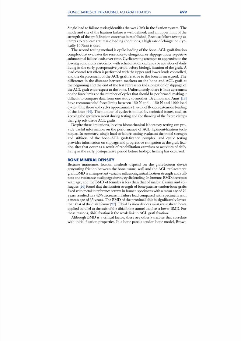
Single load-to-failure testing identifies the weak link in the fixation system. Themode and site of the fixation failure is well defined, and an upper limit of thestrength of the graft-fixation construct is established. Because failure testing at-tempts to replicate traumatic loading conditions, a high rate of elongation (typ-ically 100%/s) is used.
The second testing method is cyclic loading of the bone–ACL graft-fixationcomplex that evaluates the resistance to elongation or slippage under repetitivesubmaximal failure loads over time. Cyclic testing attempts to approximate theloading conditions associated with rehabilitation exercises or activities of dailyliving in the early postoperative period before biologic fixation of the graft. A load-control test often is performed with the upper and lower loads controlled,and the displacement of the ACL graft relative to the bone is measured. Thedifference in the distance between markers on the bone and ACL graft atthe beginning and the end of the test represents the elongation or slippage of the ACL graft with respect to the bone. Unfortunately, there is little agreementon the force limits or the number of cycles that should be performed, making itdifficult to compare data from one study to another. Beynnon and Amis [13]have recommended force limits between 150 N and À150 N and 1000 loadcycles. One thousand cycles approximates 1 week of flexion-extension loading of the knee [14]. The number of cycles is limited by technical issues, such askeeping the specimen moist during testing and the thawing of the freeze clamps
that grip soft tissue ACL grafts.Despite these limitations, in vitro biomechanical laboratory testing can pro-
vide useful information on the performance of ACL ligament-fixation tech-niques. In summary, single load-to-failure testing evaluates the initial strengthand stiffness of the bone–ACL graft-fixation complex, and cyclic testing provides information on slippage and progressive elongation at the graft fixa-tion sites that occur as a result of rehabilitation exercises or activities of dailyliving in the early postoperative period before biologic healing has occurred.
BONE MINERAL DENSITY Because intratunnel fixation methods depend on the graft-fixation devicegenerating friction between the bone tunnel wall and the ACL replacementgraft, BMD is an important variable influencing initial fixation strength and stiff-ness and resistance to slippage during cyclic loading. In humans BMD decreaseswith age, and the BMD of females is less than that of males. Cassim and col-leagues [26] found that the fixation strength of bone-patellar tendon-bone graftsfixed with metal interference screws in human specimens with a mean age of 79years resulted in a 42% decrease in failure load compared with specimens with
a mean age of 35 years. The BMD of the proximal tibia is significantly lowerthan that of the distal femur [27]. Tibial fixation devices must resist shear forcesapplied parallel to the axis of the tibial bone tunnel that has a lower BMD. Forthese reasons, tibial fixation is the weak link in ACL graft fixation.
Although BMD is a critical factor, there are other variables that correlatewith initial fixation properties. In a bone-patella tendon-bone model, Brown
699BIOMECHANICS OF INTRATUNNEL ACL GRAFT FIXATION

8/14/2019 Bio Mechanics of Intratunnel Anterior
http://slidepdf.com/reader/full/bio-mechanics-of-intratunnel-anterior 6/20
and colleagues [15] found that insertion torque, an indirect measure of BMD,was correlated linearly with pull-out force but with weak significance. Using elderly human cadaveric knees, Brand and colleagues [27] found that BMDmeasured using dual-energy X-ray absorptiometry and screw insertion torqueexplained 77% of the ultimate failure load observed in QHTG fixed with
bioabsorbable interference screws in the distal femur and proximal tibia of human specimens. The R2 value for the relationship between ultimate failureload and BMD was 0.65, indicating that BMD explained 65% of the ultimatefailure load. This study found that a BMD of 0.6 g/cm2 or higher resulted in
better initial fixation properties. Using the proximal tibia of human cadavericspecimens (mean age, 40 Æ 11 years; range, 17–54 years) and doubled tibialistendons fixed with a tapered bioabsorbable screw, Jarvinen and colleagues [28]found that insertion torque was linearly correlated to fixation strength (R2 ¼
0.54) and in their study was the variable most strongly predictive of fixationstrength. Unfortunately, despite the correlation, insertion torque was a poorpredictor of cyclic loading failure or single load-to-failure. The remainder of this article is aimed toward explicating the role and importance of secondaryfactors that influence the biomechanical properties of intratunnel graft fixation.
BONE-PATELLA TENDON-BONE FIXATIONThe fixation properties of interference screw fixation of bone-tendon-bonegrafts depend on the generation of friction generated by compression of the
bone block into the bone tunnel wall and engagement of the interference screwthreads. As illustrated in Fig. 1, factors that influence the initial tensile proper-ties of interference screw fixation of bone-tendon-bone ACL grafts include
1. Screw diameter2. Gap size3. Screw length4. Screw divergence
There is overlap between the effects of screw diameter and gap size on initialfixation properties. Kohn and Rose [29], using human cadaveric knees (meanage, 30 years), reported that tibial fixation using 9-mm screws was significantlystronger than tibial fixation using 7-mm screws. Based on their findings, theyrecommended against the using 7-mm screws for tibial fixation. With elderlyhuman cadaveric specimens, two groups found no significant difference inthe fixation strength of bone-patellar tendon-bone grafts fixed in the distalfemur using retrograde or endoscopically inserted 7-mm screws and 9-mmscrews inserted using a rear-entry or an antegrade technique [15,30]. The influ-
ence of screw diameter on initial fixation properties is probably most relevantwhen there is a significant size discrepancy between the bone block and the
bone tunnel wall; this difference often is referred to as ‘‘gap size.’’After studying various fixation methods, Kurosaka and colleagues [2]
hypothesized that the gap size between the bone block and bone tunnel wasa critical factor in interference screw fixation. Butler and colleagues [31], in
700 CHEN, BRAND, & BROWN

8/14/2019 Bio Mechanics of Intratunnel Anterior
http://slidepdf.com/reader/full/bio-mechanics-of-intratunnel-anterior 7/20
a porcine study, found that, with a gap size of 3 to 4 mm, increasing the screwdiameter size from 7 mm to 9 mm significantly increased the load at whichfailure occurred. With a similar porcine experimental model, Reznik andcolleagues [32] demonstrated that gap size significantly influenced the ultimate
failure load of bone-tendon-bone grafts fixed in 10-mm bone tunnels with 7-mmscrews. When the gap between the bone block and bone tunnel wall was 4 mmor more, increasing the screw diameter to 9 mm increased the failure load by97%. When the gap was larger than 4 mm and a 9-mm screw was used, how-ever, the results were inferior to using a 7-mm screw with a gap of less than4 mm. It was demonstrated in an elderly human model that a cylindrical
bone plug improves fixation by 19.9% (845. 8 N versus 691.7 N) comparedwith a bone plug with a trapezoidal cross section. Although gap size was notmeasured, it will be smaller with a cylindrical bone plug than with a trapezoidal
bone plug [33]. In what may be the best investigation of the topic using humanand bovine tissue, the authors found that, although screw diameter or gap sizealone was not significant, interference (defined as the screw’s outer threaddiameter minus the tunnel–bone block gap) was significantly related to loadat failure in the pooled specimens and in the bovine model [15]. A numberof authors have suggested using larger screws as the gap size increases [34].
Fig. 1. Factors that influence the initial tensile properties of interference screw fixation of bone-tendon-bone ACL grafts. (A) Screw diameter: effect of a larger screw on bone plug.(B) Gap size: increase in gap size decreases strength of construct. (C ) Screw length: lateralview of graft with longer (left panel ) and shorter (right panel ) interference screws. In bone-ten-
don-bone grafts screw length does not affect screw pullout significantly. (D ) Screw divergence:anteroposterior view of the knee showing screw divergence from the tunnel. (E ) Screw diver-gence, lateral view.
701BIOMECHANICS OF INTRATUNNEL ACL GRAFT FIXATION

8/14/2019 Bio Mechanics of Intratunnel Anterior
http://slidepdf.com/reader/full/bio-mechanics-of-intratunnel-anterior 8/20
Screw length probably does not have a large influence on the initial fixationproperties of bone-patella tendon-bone grafts fixed with interference screws.Brown and colleagues [30] found no significant difference in fixation strength
between 7 Â 20-mm and 7 Â 30-mm screws or between 9 Â 20-mm and9 Â 30-mm screws fixed in the distal femur of human specimens. Black andcolleagues [35] compared 9 Â 12.5-mm, 9 Â 15-mm, and 9 Â 20-mm interfer-ence screws in a porcine tibia model. No significant differences in insertiontorque, failure load, stiffness, or displacement to failure was found among the different-length screws. Pomeroy and colleagues [36] also found no signif-icant effect of screw length on fixation strength for a given screw diameter.Fixation strength is not improved by using an interference screw longer thanthe bone plug.
Divergence of the interference screw from the bone block and the axis of the bone tunnel can occur with both rear-entry and endoscopic techniques. Theincidence of screw divergence is more common with the endoscopic technique(femur > tibia) [37]. Using a porcine model, Jomha and colleagues [38] reportedno significant difference in femoral fixation strength with endoscopicallyinserted interference screws with divergence up to 10, but there was a signifi-cant decrease with screw divergence of 20 or greater. Pierz and colleagues [39],using porcine tibias, demonstrated that interference screws inserted to simulatea rear-entry femoral fixation technique or fixation of a tibial bone block resulted
in a significant decrease in fixation strength with divergence between 0 and15 compared with 15 to 30 of divergence. Interference screws inserted tosimulate an endoscopic technique resulted in a significant decrease in fixationstrength only at 30 of screw divergence. These authors concluded that optimalinterference screw fixation occurs when the screw is placed parallel to the bone
block and bone tunnel. Because of the creation of a wedge effect, screw diver-gence has less effect on endoscopically inserted femoral screws. Based on clin-ical studies, screw divergence of less than 30 does not seem to havea significant effect on the clinical outcome [40]. Because of the in-line direction
of pull, however, minor degrees of divergence have a greater effect on thefixation strength of femoral screws inserted through a rear-entry techniqueand tibial fixation screws.
Metal interference screws can distort MRI images, lacerate the graft during insertion, and complicate revision ACL surgery. Bioabsorbable interferencescrews have been proposed as a method to eliminate potential complications[41]. Several biomechanical studies have compared the initial fixation strengthof bioabsorbable interference screws and conventional metal interferencescrews in animal and human cadaveric models. These studies showed that
most bioabsorbable interference screws provide fixation strength similar tothat of metal interference screws and concluded that the use of these screwsmay allow an accelerated postoperative rehabilitation program [42–48].
Concerns with bioabsorbable interference screws have focused largely on theissues of screw breakage and biocompatibility. Screw breakage has beenaddressed largely by designing screws and screwdrivers that allow the insertion
702 CHEN, BRAND, & BROWN

8/14/2019 Bio Mechanics of Intratunnel Anterior
http://slidepdf.com/reader/full/bio-mechanics-of-intratunnel-anterior 9/20
torque to be distributed along the entire length of the screw and by decreasing the insertion torque by notching the bone tunnel wall. To prevent screw break-age, it is important that the screwdriver be engaged fully during insertion of thescrew.
In summary, based on a review of the literature, gap size is probably themost important factor influencing the initial fixation properties of interferencescrew fixation of bone-tendon-bone grafts. Gap size also is the one factor thatcan be measured easily intraoperatively and controlled by the surgeon.Improvements in initial graft fixation can be achieved by increasing the diam-eter of the screw to compensate for the gap size. Increasing screw length seemsto offer minimal improvements in initial graft fixation properties.
Guidelines and Recommendations for Intratunnel Fixationof Bone-Tendon-Bone GraftsFemoral fixation: two-incision technique The authors recommend using metal screws 7 and 8 mm in diameter witha length of 20 to 25 mm. Bioabsorbable screws can be used, but the higherinsertion torque generated by the insertion of the screw against the hard cortexof distal femur may result in a higher incidence of screw breakage comparedwith bioabsorbable screws inserted using an endoscopic technique. When thegap between the bone block and bone tunnel wall is greater than 4 mm,
suture/post or plastic button fixation should be considered in addition to intra-tunnel fixation.
Femoral fixation: endoscopic technique The authors recommend using metal or bioabsorbable screws 8 or 9 mm indiameter, with a length of 20 to 25 mm. For bioabsorbable screws, one shouldreview and use the manufacturer’s guidelines regarding tapping or notching the
bone tunnel wall to minimize the risk of screw breakage. The authors preferusing the EndoButton CL (Closed Loop; Smith and Nephew, Andover, Mas-
sachusetts) to avoid graft–tunnel mismatch in long grafts when the gap size isgreater than 4 mm and when blowout of the posterior wall of the femoraltunnel occurs.
Tibial fixation The authors recommend using screws 8 or 9 mm in diameter with a length of 20 to 25 mm. For gap sizes greater than 4 mm, one should consider suture/postor button as hybrid fixation. In soft bone or when low insertion torque isencountered, one should consider backing up the interference screw fixation
by tying sutures around a fixation post.
SOFT TISSUE GRAFTSInterference screw fixation of soft tissue grafts depends on many of the samefactors as fixation for bone-patella tendon-bone grafts; but the relative impor-tance of each of these factors differs [49]. As in bone-tendon-bone grafts, theinitial fixation properties depend on the fixation device generating friction
703BIOMECHANICS OF INTRATUNNEL ACL GRAFT FIXATION

8/14/2019 Bio Mechanics of Intratunnel Anterior
http://slidepdf.com/reader/full/bio-mechanics-of-intratunnel-anterior 10/20
between the soft tissue graft and the bone tunnel wall. Friction is generated bycompression of the soft tissue graft against the bone tunnel wall. Because thesoft tissue graft is more compressible than the bone blocks of bone-tendon-
bone grafts, a screw of a given diameter generates compression between thescrew and bone tunnel wall. The amount of friction contributed by engagementof the screw threads in the bone tunnel wall and soft tissue graft also is signif-icantly lower because of the lack of engagement of the screw threads into thesoft tissue graft. As illustrated in Fig. 2, factors that may contribute to the initialfixation properties of soft tissue grafts with interference screws are
1. Screw geometry (length and diameter)2. Tendon fit
3. Tunnel impaction or dilation4. Screw placement (concentric versus eccentric)
Unlike interference screw fixation of bone-tendon-bone grafts, screw lengthseems to have a greater effect on the initial fixation properties of soft tissuegrafts fixed with interference screws. Because of the lower BMD of the proxi-mal tibia, screw length has a greater influence on tibial fixation properties [27].Screw length may have a more significant effect on the fixation properties of soft tissue grafts because the area over which friction is generated between
Fig. 2. Factors that contribute to the initial fixation properties of soft tissue grafts with interfer-ence screws. (A) Screw geometry (length and diameter): length and diameter of screw canaffect construct strength. (B) Tendon fit: more precise fit to 0.5 mm allows improved strengthwith screw insertion. (C ) Tunnel impaction or dilation: impaction drilling or serial dilationdoes not seem to significantly improve graft mechanics. (D ) Screw placement (concentricversus eccentric): concentric or eccentric screw placement does not affect construct strength.
704 CHEN, BRAND, & BROWN

8/14/2019 Bio Mechanics of Intratunnel Anterior
http://slidepdf.com/reader/full/bio-mechanics-of-intratunnel-anterior 11/20
the bone tunnel wall and soft tissue graft is determined by the screw length,rather than by the length of a bone block, which is typically 20 to 25 mm.In a bovine proximal tibia model, Weiler and colleagues [50] found that23-mm screws had lower pull-out strengths than 28-mm screws with equivalentdiameters. This study also found that increasing screw length had a greaterinfluence on failure load than increasing the screw diameter. Using humantibias (age range, 24–45 years), Selby and colleagues [51] demonstrated signif-icantly higher ultimate failure loads for screws 35 mm long than for screws28 mm long. Placing the screw so that it engaged the cortex of the tibia allowedsignificantly less slippage than screw insertion that engaged only cancellous
bone [52]. Based on their findings, the authors recommend that the screwhead be placed to engage the tibial cortex.
Few studies have examined the influence of screw diameter on the initialfixation properties of soft tissue ACL grafts fixed with interference screws.Using human hamstring tendons grafts and bovine proximal tibias, Weilerand colleagues [50] found that increasing the diameter of a 23-mm length bio-absorbable interference screw from 7 mm to 8 mm increased the mean pull-outforce from 367 N to 479 N.
The fit of the soft tissue graft in the bone tunnel seems to have a significantinfluence on the initial fixation properties of interference screw fixation of softtissue grafts. Using a human cadaveric model, Steenlage and colleagues [53]
demonstrated that QHTGs fixed in the distal femur with a bioabsorbable screwhad a significantly higher ultimate failure load if the bone tunnel was sizedwithin 0.5 mm of the graft diameter rather than within 1 mm of the graftdiameter.
Because BMD has such a significant effect on the initial tensile properties of interference screw fixation of soft tissue grafts, compaction drilling or bone tun-nel dilation has been proposed as a method of creating increased bone densityalong the bone tunnel walls. It has been speculated that this approach willimprove initial fixation properties. Using human male cadaveric knees,
Rittmeister and colleagues [54] found that serial dilation failed to improvethe initial fixation strength of QHTGs fixed in the tibia with metal interferencesscrews. Nurmi and colleagues [55] investigated the effects of compaction dril-ling versus conventional or extraction drilling on the initial fixation strengthof QHTGs fixed with bioabsorbable screws in the proximal tibia of humanspecimens (mean age, 41 Æ 11 years; range, 17–49 years). The biomechanicaltesting protocol consisted of cyclic loading followed by a single load-to-failuretest. They found no significant difference in initial stiffness or displacement
between the two drilling methods during cyclic testing or in the single load-
to-failure test and detected no significant differences between the two drilling methods in yield load, displacement at yield load, or stiffness.
In a second biomechanical study, Nurmi and colleagues [56] investigated theeffect of tunnel compaction by serial dilators compared with conventional dril-ling on the initial fixation strength of doubled anterior tibial tendons fixed inthe proximal tibia of human specimens (mean age, 40 Æ 11 years; range,
705BIOMECHANICS OF INTRATUNNEL ACL GRAFT FIXATION

8/14/2019 Bio Mechanics of Intratunnel Anterior
http://slidepdf.com/reader/full/bio-mechanics-of-intratunnel-anterior 12/20
17–54 years) using bioabsorbable interference screws. The specimens weretested under cyclic loading followed by a single load-to-failure test. Althoughno significant difference in stiffness or displacement between the two tech-niques was demonstrated during cyclic testing, the number of failures during cyclic loading of the extraction drilling group was twice that of the seriallydilated group. In the subsequent single load-to-failure test, there was no signif-icant difference in failure load or stiffness between the two groups. A limitationof this study was the size of the tibial bone tunnel was not matched to the size of the soft tissue grafts: a bone tunnel 10 mm in diameter was created in allspecimens.
The only study to demonstrate a beneficial effect of tunnel dilation on thefixation strength of soft tissue ACL grafts was performed by Cain and col-leagues [57] using paired human cadaveric knees (average age, 42 years; range,29–47 years). QHTGs were fixed with bioabsorbable screws in 0.5-mm size-matched femoral and tibial tunnels. The tibial tunnel was created using smoothtunnel dilators in one knee of the pair, and extraction drilling was used in theopposite knee of the pair. The femur-hamstring ACL graft-tibia complex wastested to failure using anterior tibial translation with the knee positioned in20 of flexion, as previously described by Steiner and colleagues [3]. Thismethod of testing attempts to mimic the Lachman test. All specimens failed
by the graft pulling out of the tibial tunnel, but the ultimate failure load was
significantly higher for the dilated tibial tunnels. Testing methodology makesit difficult to compare the results of this study with earlier studies; however,
based on the literature, the benefits of compaction drilling or serial dilationprobably do not justify the extra cost and operating time.
Although there is general agreement that interference screws should beinserted on the cancellous side of bone-tendon-bone grafts, controversy existsregarding placement of tibial interference screws used to fix multiple-strandedhamstring tendon grafts. Soft tissue grafts may be fixed by inserting the screwon the side (eccentrically) or down the center (concentrically) of the graft
strands. Concentric screw placement maximizes contact between the graftstrands and the bone tunnel wall, providing a greater surface area for healing.Simonian and colleagues [58] were unable to detect a significant difference ininitial fixation properties between eccentric versus concentric interferencescrew position against a model of human hamstring tendon grafts fixed ina polyurethane foam. Shino and Pflaster [59] investigated the effect of eccentricversus concentric screw placement on the initial fixation properties of QHTGsfixed in the proximal tibia of paired human cadaveric knees (average age,51 years; range, 49–54 years). There were no significant differences in stiffness,
yield load, ultimate failure load, or slippage between the two screw positions.As with bone-patellar tendon-bone grafts, QHTGs in elderly human bone
tunnels, bioabsorbable interference screws provide initial biomechanical fixa-tion properties similar to those of a metal interference screw [60]. In thelaboratory, two separate investigations have noted an association betweena metal interference screw applied against a soft tissue graft and graft laceration
706 CHEN, BRAND, & BROWN

8/14/2019 Bio Mechanics of Intratunnel Anterior
http://slidepdf.com/reader/full/bio-mechanics-of-intratunnel-anterior 13/20
or damage. Brand and colleagues [60], using human QHTG in elderly cadaver bone, noted a greater rate of graft laceration in the group in which a metalscrew was used than in the group in which a bioabsorbable screw of compara-
ble dimensions was used. Another group compared the effect of a singleinsertion of a metal screw versus the use two separate bioabsorbable screws,a poly-D,L-lactide and a poly-D,L-lactide with tricalcium phosphate. Afterthe screws had been inserted, the soft tissue grafts were tested to failure. Themetal screw damaged the tendon more extensively, resulting in a significantlysmaller load at failure and stiffness than seen in the tendons damaged by
bioabsorbable screws [61].
Guidelines and Recommendations for Intratunnel Fixation of Soft TissueGraftsUnlike bone-tendon-bone grafts, in which the bone tunnel size and dimensionsof the bone blocks are standardized, there are large variations in the diameterand length of soft tissue grafts. These variations make it difficult to arrive atdefinitive recommendations regarding selection of interference screw fixation.Nevertheless, the authors’ interpretation of the literature has led to the follow-ing conclusions:
1. Because of the lower BMD, and because the line of applied force is parallel
to the axis of the tibial tunnel, tibial fixation is weaker, less stiff, and morelikely to slip under cyclic loading than fixation using the femoral fixation site.
2. Screw length has a more significant effect on the initial fixation properties of interference screw fixation of soft tissue ACL grafts than of bone-tendon-boneACL grafts.
3. In biomechanical testing longer screws result in higher ultimate failure loadsand stiffness and less slippage.
4. Fixation properties are improved by having the screw head engage the tibialcortex.
5. The effect of screw diameter on initial fixation properties is unclear, making it
difficult to establish clear guidelines for screw sizing.6. Matching the size of the bone tunnel to within 0.5 mm of the measured size
of the graft may improve initial fixation properties.7. Compaction drilling or serial dilation does not seem to improve initial fixa-
tion properties significantly.
ALTERNATIVE INTRATUNNEL TIBIAL FIXATION TECHNIQUESThe stimulus for the development of alternative intratunnel tibial fixation tech-niques for soft tissue ACL grafts arose from the desire to decrease slippage and
the high rate of fixation failure reported with interference screws under cyclicloading conditions; to eliminate or reduce the need for supplemental tibial fix-ation; and to improve soft tissue-to-bone healing at the graft-fixation sites[49,52,62]. The IntraFix (DePuy Mitek, Norwood, Massachusetts) was de-signed to capture individually each of the four strands of a soft tissue graftin a separate compartment using a plastic sheath and to achieve direct
707BIOMECHANICS OF INTRATUNNEL ACL GRAFT FIXATION

8/14/2019 Bio Mechanics of Intratunnel Anterior
http://slidepdf.com/reader/full/bio-mechanics-of-intratunnel-anterior 14/20
compression of each of the graft strands against the bone tunnel wall by theinsertion of a tapered screw into the central chamber of the plastic sheath[62]. In a porcine tibia model using human hamstring tendon grafts, Kousa and colleagues [63] demonstrated that the IntraFix had the highest load failureload (1309 Æ 302 N) and stiffness (267 Æ 36 N/mm) and the least amount of slippage (1.5 mm) after cyclic loading when compared with two cortical fixationtechniques and three other interference screw fixation techniques. When usedto fix a QHTG in a human tibial tunnel, the IntraFix device had a load at fail-ure (796 Æ 193 N versus 647 Æ 269 N) and stiffness (49 Æ 21.9 N/mm versus64.5 Æ 22 N/mm) similar to that of a bioabsorbable interference screw 35 mmin length [64].
The GTS System (Graft Tunnel Solution; Smith & Nephew Endoscopy,Andover, Massachusetts) is a intratunnel tibial fixation technique that positionsa poly-L-lactic, tapered, fine-pitch screw concentrically within the four-strandsoft tissue graft [65]. The screw features a tapered design and shorter threaddistance, which enhances compression of the soft tissue graft in cancellous
bone. The graft sleeve is a three-lumen, woven, nonabsorbable polypropylenemesh graft sleeve that organizes the four-strand soft tissue graft in the tibial tun-nel. The graft sleeve prevents graft twisting during screw insertion, which helpsmaintain equal tension in the four graft strands, maximizes bone–tendon con-tact that enhances healing, and provides better compression of each ligament
strand against the bone tunnel wall while protecting the graft strands fromscrew damage. Cyclic testing followed by single load-to-failure testing of thegraft sleeve, tapered screw, and IntraFix has been performed using human dou-
bled gracilis and semitendinosus tendon grafts in the proximal tibia of calf bone(2 years or younger) with BMD similar to that of the proximal tibia in young humans. There was no significant difference in slippage, ultimate failure load,or stiffness between the two devices.
HYBRID FIXATIONAlthough intratunnel fixation of soft tissue grafts has improved with longer tib-ial screws and precise sizing of grafts to tunnel diameters, concerns persist thatinitial fixation strength that is less than the strength of the native ACL boneconstruct allows graft-tunnel motion that may contribute to knee laxity. In par-ticular, older women and other patients who have lower BMC are candidatesfor hybrid fixation that combines improved structural properties with the bio-mechanical and biologic advantages of joint-line interference-fit fixation to aug-ment intratunnel devices [66]. The EndoPearl (Linvatec, Largo, Forida) linkedwith #5 Ethibond sutures (Ethicon, Somerville, New Jersey) against the tip of
a bioabsorbable interference screw significantly improved femoral fixationwhen compared with a bioabsorbable interference screw in a femoral tunnelof a calf model as measured by maximum load-to-failure (658.9 Æ 118.1 N ver-sus 385.9 Æ 185.6 N) and stiffness (41.7 Æ 11 N/mm versus 25.7 Æ 8.5 N/mm)[67]. Hammond and colleagues [66] evaluated femoral fixation of quadrupledflexor tendons in porcine bone using a bioabsorbable screw, EndoButton
708 CHEN, BRAND, & BROWN

8/14/2019 Bio Mechanics of Intratunnel Anterior
http://slidepdf.com/reader/full/bio-mechanics-of-intratunnel-anterior 15/20
CL, and hybrid fixation using an EndoButton CL and bioabsorbable screw.The hybrid fixation group demonstrated greater yield and ultimate failureloads and greater stiffness under displacement-controlled cyclic loads. In a sim-ilar study, Oh and colleagues [68] demonstrated that the addition of an inter-ference screw to suspensory fixation using an EndoButton CL increasedultimate failure load and stiffness and decreased slippage. Although hybridfixation has improved initial graft-fixation tensile properties in biomechanicalstudies, there have been no clinical studies demonstrating that it results inimproved clinical results.
CLINICAL STUDIESMa and colleagues [69] compared fixation techniques with patients recon-structed with QHTG in a prospective, nonrandomized study. Fifteen patientsin each group were fixed with either a bioabsorbable interference screw inthe femoral or tibial bone tunnel or femoral fixation with an EndoButtonand tibial fixation with a screw post. There were no significant differences inInternational Knee Documentation Committee (IKDC) knee scores (85 Æ 11versus 81 Æ 17) or side-to-side KT differences (3.2 Æ 2.6 mm versus 2.4 Æ1.8 mm). There was more tunnel widening (as measured in the femoral tunnelin the sagittal plane) in the group of patients in which the bioabsorbable screw
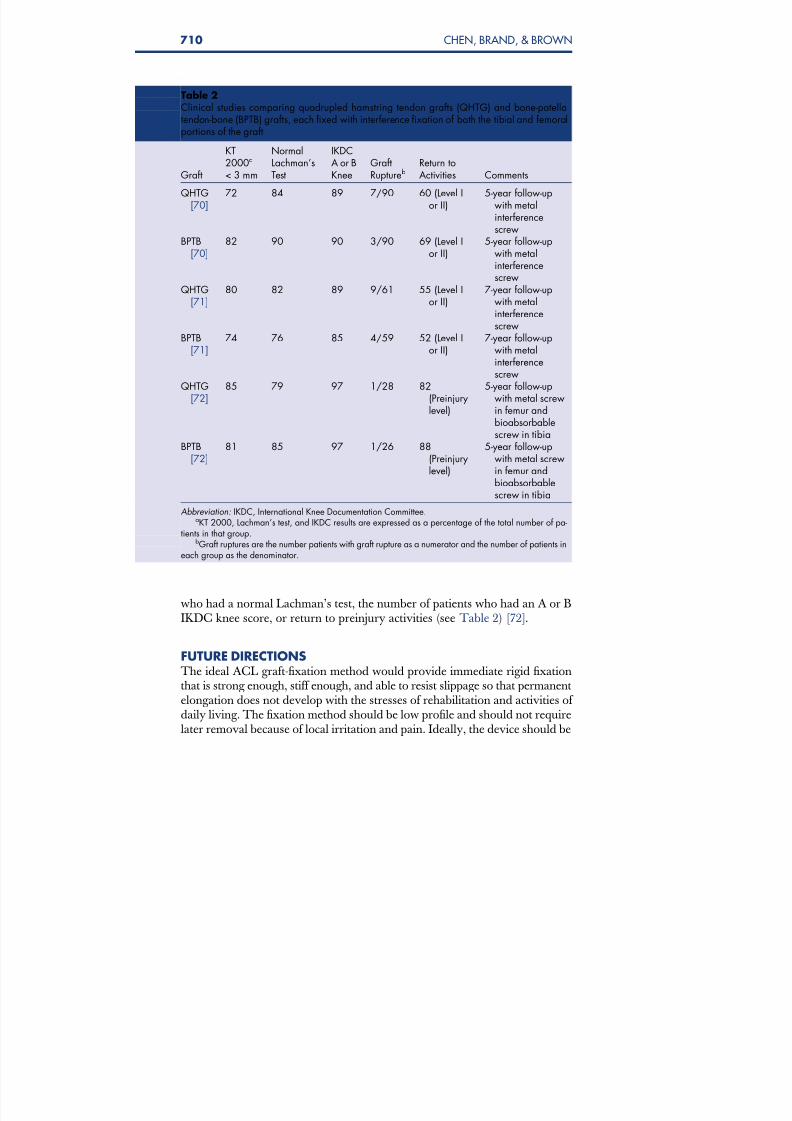
was used than in the group of patients fixed with extracortical fixation. Allother measurements of tunnel width were not significantly different betweengroups [69]. A prospective, nonrandomized clinical trial of patients who hadQHTGs and bone-patellar tendon-bone grafts fixed with metal interferencescrews with a 5-year follow-up found only that the bone-patellar tendon-bonegroup had a greater rate of arthritis. There were no significant differences inKT 2000 maximum manual testing, the percentage of patients who had a nor-mal Lachman’s test, the number of patients who had an A or B IKDC kneescore, or the return to level I or II activities (Table 2) [70]. The same group
of patients was followed up again at a mean of 7 years postoperatively. Therewas a greater rate of arthritis on radiographic review by the IKDC criteria inthe bone-patellar tendon-bone group (45%) than in the QHTG group (14%).The number of patients in the bone-patellar tendon-bone group with loss of extension increased significantly between the 5-year and 7-year follow-ups,
but there was not a significant difference between the bone-patellar tendon- bone and QHTG groups in the number of patients who had extension loss(see Table 2) [71]. A prospective, randomized investigation from Slovenia com-pared 64 patients randomly assigned to receive bone-patellar tendon-bone
grafts or QHTGs. Interference fixation was provided with a metal screw inthe femoral socket and a bioabsorbable screw of similar dimensions in the tibialtunnel. These investigators also found a greater rate of arthritis in the bone-pa-tellar tendon-bone group: 50% had at least grade B arthritis by the IKDCscoring sheet, compared with 17% in the QHTG group. There were nodifferences in KT 2000 maximum manual testing, the percentage of patients
709BIOMECHANICS OF INTRATUNNEL ACL GRAFT FIXATION
8/14/2019 Bio Mechanics of Intratunnel Anterior
http://slidepdf.com/reader/full/bio-mechanics-of-intratunnel-anterior 16/20
who had a normal Lachman’s test, the number of patients who had an A or BIKDC knee score, or return to preinjury activities (see Table 2) [72].
FUTURE DIRECTIONSThe ideal ACL graft-fixation method would provide immediate rigid fixationthat is strong enough, stiff enough, and able to resist slippage so that permanentelongation does not develop with the stresses of rehabilitation and activities of daily living. The fixation method should be low profile and should not requirelater removal because of local irritation and pain. Ideally, the device should be
Table 2Clinical studies comparing quadrupled hamstring tendon grafts (QHTG) and bone-patellatendon-bone (BPTB) grafts, each fixed with interference fixation of both the tibial and femoral
portions of the graft
Graft
KT2000a
< 3 mm
NormalLachman’sTest
IKDCA or BKnee
GraftRuptureb
Return toActivities Comments
QHTG[70]
72 84 89 7/90 60 (Level Ior II)
5-year follow-upwith metalinterferencescrew
BPTB[70]
82 90 90 3/90 69 (Level Ior II)
5-year follow-upwith metalinterferencescrew
QHTG[71]
80 82 89 9/61 55 (Level Ior II)
7-year follow-upwith metalinterferencescrew
BPTB[71]
74 76 85 4/59 52 (Level Ior II)
7-year follow-upwith metalinterferencescrew
QHTG[72] 85 79 97 1/28 82(Preinjurylevel)
5-year follow-upwith metal screwin femur andbioabsorbablescrew in tibia
BPTB[72]
81 85 97 1/26 88(Preinjurylevel)
5-year follow-upwith metal screwin femur andbioabsorbablescrew in tibia
Abbreviation: IKDC, International Knee Documentation Committee.a
KT 2000, Lachman’s test, and IKDC results are expressed as a percentage of the total number of pa-tients in that group.
bGraft ruptures are the number patients with graft rupture as a numerator and the number of patients ineach group as the denominator.
710 CHEN, BRAND, & BROWN

8/14/2019 Bio Mechanics of Intratunnel Anterior
http://slidepdf.com/reader/full/bio-mechanics-of-intratunnel-anterior 17/20
replaced by cancellous bone and result in the development of a normal histo-logic ligament-to-bone attachment site.
Future improvements in intratunnel ACL graft fixation will depend on betterunderstanding of the in vivo forces experienced by the ACL with rehabilitationexercises and activities in the early postoperative period and of the biology of fixation-site healing. Osteoconductive or osteoinductive materials that will stim-ulate the development of normal osseous tissue are under developmentcurrently. Bone cement that will provide immediate rigid fixation and eventu-ally be replaced by bone may be developed. On-going basic science research isdirected at promoting and accelerating healing of soft tissue to bone. Ultra-sound, bone morphogenetic proteins, and biologic growth factors currentlyare being investigated as possible methods to promote and accelerate tendon-to-bone healing.
References[1] Lambert K. Vascularized patellar tendon graft with rigid internal fixation for anterior cruciate
ligament insufficiency. Clin Orthop Relat Res 1983;172:85–9.[2] Kurosaka M, Yoshiya S, Andrish J. A biomechanical comparison of different surgical tech-
niques of graft fixation of anterior cruciate ligament reconstruction. Am J Sports Med1987;15(3):225–9.
[3] Steiner M, Hecker A, Brown C. Anterior cruciate ligament graft fixation: comparison of ham-
string and patellar tendon grafts. Am J Sports Med 1994;22(2):240–7.[4] Bach B, Tradonsky S, Bojchuk J, et al. Arthroscopically assisted anterior cruciate ligament
reconstruction using patellar tendon autograft. Five-to nine-year follow-up evaluation. Am J Sports Med 1998;26(1):20–9.
[5] Pinczewski L. Endoscopic ACL reconstruction utilizing a quadrupled hamstring tendonautograft with direct RCI interference screw fixation. Presented at the Lecture/LaboratorySession of RCI Screw, Smith & Nephew Donjoy. Columbus (GA), February, 1996.
[6] Fu F. Using bioabsorbable interference screw fixation for hamstring ACL-reconstruction.Orthropaedics Today 1997;16:36–7.
[7] Morrison J. Function of the knee joint in various activities. BiomedEng 1969;4(12):573–80.[8] Morrison J. The mechanics of the knee joint in relation to normal walking. J Biomech
1970;3:51–61.[9] Noyes F, Butler D, Grood E, et al. Biomechanical analysis of human ligament grafts used in
knee-ligament repairs and reconstructions. J Bone Joint Surg 1984;66(3):344–52.[10] Cooper D, Deng X, Burstein A, et al. The strength of the central third patellar tendon graft:
a biomechanical study. Am J Sports Med 1993;21(6):818–24.[11] Hamner D, Brown C, Steiner M, et al. Hamstring tendon grafts for reconstruction of the
anterior cruciate ligament: biomechanical evaluation of the use of multiple strands andtensioning techniques. J Bone Joint Surg Am 1999;81(4):549–57.
[12] Brand J, Weiler A, Caborn D, et al. Graft fixation in cruciate ligament reconstruction.Current concepts. Am J Sports Med 2000;28(5):764–74.
[13] Beynnon B, Amis A. In vitro testing protocols for cruciate ligaments and ligament reconstruc-tions. Knee Surg Sports Traumatol Arthrosc 1998;6(Suppl 1):S70–6.
[14] Weiss J, Paulos L. Mechanical testing of ligament fixation devices. Techniques in Orthopae-dics 1999;14:14–21.
[15] Brown G, Pena F, Grontvedt T, et al. Fixation strength of interference screw fixation inbovine, young human, and elderly human cadaver knees: influence of insertion torque, tun-nel-bone block gap, and interference. Knee Surg Sports Traumatol Arthrosc 1996;3(4):238–44.
711BIOMECHANICS OF INTRATUNNEL ACL GRAFT FIXATION

8/14/2019 Bio Mechanics of Intratunnel Anterior
http://slidepdf.com/reader/full/bio-mechanics-of-intratunnel-anterior 18/20

8/14/2019 Bio Mechanics of Intratunnel Anterior
http://slidepdf.com/reader/full/bio-mechanics-of-intratunnel-anterior 19/20
[37] Lemos M, Albert J, Simon T, et al. Radiographic analysis of femoral interference screw place-ment during ACL reconstruction: endoscopic versus open technique. Arthroscopy1993;9(2):154–8.
[38] Jomha N, Raso V, Leung P. Effect of varying angles on the pullout strength of interferencescrew fixation. Arthroscopy 1993;9(5):580–3.[39] Pierz K, Baltz M, Fulkerson J. The effect of Kurosaka screw divergence on the holding
strength of bone-tendon-bone grafts. Am J Sports Med 1995;23(3):332–5.[40] Dworsky B, Jewell B, Bach B. Interference screw divergence in endoscopic anterior cruciate
ligament reconstruction. Arthroscopy 1996;12(1):45–9.[41] Barber F, Elrod B, McGuire D, et al. Preliminary results of an absorbable interference screw.
Arthroscopy 1995;11(5):537–48.[42] Weiler A, Windhagen H, Raschke M, et al. Biodegradable interference screw fixation
exhibits pull-out force and stiffness similar to titanium screws. Am J Sports Med 1998;26(1):119–28.
[43] Kousa P, Jarvinen T, Kannus P, et al. Initial fixation strength of bioabsorbable and titaniuminterference screws in anterior cruciate ligament reconstruction: biomechanical evaluationby single cycle and cyclic loading. Am J Sports Med 2001;29(4):420–5.
[44] Johnson L, vanDyk G. Metal and biodegradable interference screws: comparison of failurestrength. Arthroscopy 1996;12(4):452–6.
[45] Caborn D, Urban W, Johnson D, et al. Biomechanical comparison between bioscrew andtitanium alloy interference screws for bone-patellar tendon-bone graft fixation in anteriorcruciate ligament reconstruction. Arthroscopy 1997;13(2):229–32.
[46] Abate J, Fadale P, Hulstyn M, et al. Initial fixation strength of polylactic acid interferencescrews in anterior cruciate ligament reconstruction. Arthroscopy 1998;14(3):278–84.
[47] Pena F, Grontvedt T, Brown G, et al. Comparison of failure strength between metallic and
absorbable interference screws. Influence of insertion torque, tunnel-bone block gap,bone mineral density, and interference. Am J Sports Med 1996;24(3):329–34.
[48] Rupp S, Krauss P, Fritsch E. Fixation strength of a biodegradable interference screw anda press-fit technique in anterior cruciate ligament reconstruction with a BPTB graft. Arthros-copy 1997;13(1):61–5.
[49] Brand J, Caborn D, Johnson D. Biomechanics of soft tissue interference screw fixation foranterior cruciate ligament reconstruction. Orthopedics 2003;26(4):432–9.
[50] Weiler A, Hoffman R, Siepe C, et al. The influence of screw geometry on hamstring tendoninterference fit fixation. Am J Sports Med 2000;28(3):356–9.
[51] Selby J, Johnson D, Hester P, et al. Effect of screw length on bioabsorbable interferencescrew fixation in a tibial bone tunnel. Am J Sports Med 2000;29(5):614–9.
[52] Harvey A, Thomas N, Amis A. The effect of screw length and position on fixation of four-stranded hamstring grafts for anterior cruciate ligament reconstruction. Knee2003;10(1):97–102.
[53] Steenlage E, Brand J, Johnson D, et al. Correlation of bone tunnel diameter with quadrupledhamstring strength using a biodegradable interference screw. Arthroscopy 2002;18(8):901–7.
[54] Rittmeister M, Noble P, Bocell J, et al. Interactive effects of tunnel dilation on the mechanicalproperties of hamstring grafts fixed in the tibia with interference screws. Knee Surg SportsTraumatol Arthrosc 2001;9(5):267–71.
[55] Nurmi J, Kannus P, Sievanen H, et al. Compaction drilling does not increase the initial fixa-tion strength of the hamstring tendon graft in anterior cruciate ligament reconstruction in acadaver model. Am J Sports Med 2003;31(3):353–8.
[56] Nurmi J, Kannus P, Sievanen H, et al. Interference screw fixation of soft tissue grafts in ante-rior cruciate ligament reconstruction: part 1. Effect of tunnel compaction by serial dilatorsversus extraction drilling on the initial fixation strength. Am J Sports Med 2004;32(2):411–7.
713BIOMECHANICS OF INTRATUNNEL ACL GRAFT FIXATION

8/14/2019 Bio Mechanics of Intratunnel Anterior
http://slidepdf.com/reader/full/bio-mechanics-of-intratunnel-anterior 20/20
[57] Cain E, Phillips B, Charlebois S, et al. Effect of tibial tunnel dilation on pullout strength of semitendinosus-gracilis graft in anterior cruciate ligament reconstruction. Orthopedics2005;28(8):779–83.
[58] Simonian P, Sussman P, Baldini T. Interference screw position and hamstring graft locationfor ACL reconstruction. Presented at the 17th Annual Meeting of Arthroscopy Associationof North America. Orlando (FL), April 30–May 3, 1998.
[59] Shino K, Pflaster D. Comparison of eccentric and concentric screw placement for hamstringgraft fixation in the tibial tunnel. Knee Surg Sports Traumatol Arthrosc 2000;8(2):73–5.
[60] Brand J, Nyland J, Caborn D, et al. Soft-tissue interference fixation: bioabsorbable screwversus metal screw. Arthroscopy 2005;21(8):911–6.
[61] Zantop T, Weimann A, Schmidtko R, et al. Graft laceration and pullout strength of soft-tissueanterior cruciate ligament reconstruction: in vitrostudycomparing titanium, poly-D,L-lactide,and poly-D,L-lactide-tricalcium phosphate screws. Arthroscopy 2006;22(11):1204–10.
[62] Magen H, Howell S, Hull M. Structural properties of six tibial fixation methods for anterior
cruciate ligament soft tissue grafts. Am J Sports Med 1999;27(1):35–43.[63] Kousa P, Jarvinen T, Vihavainen M, et al. The fixation strength of six hamstring tendon graftfixation devices in anterior cruciate ligament reconstruction. Part II: tibial site. Am J SportsMed 2003;31(2):182–8.
[64] Caborn D, Brand J, Nyland J, et al. A biomechanical comparison of initial soft tissue fixationdevices: the Intrafix versus a tapered 35-mm bioabsorbable interference screw. Am J SportsMed 2004;32(4):956–61.
[65] Brown C, Darwich N. Anterior cruciate ligament reconstruction using autogenous doubledgracilis and semitendinosus tendons with GTS sleeve and tapered screw fixation. Tech-niques in Orthopaedics 2005;20:290–6.
[66] Hammond G, Armstrong K, McGarryM, et al. Hybridfixation improves structural properties
of a free tendon anterior cruciate ligament reconstruction. Arthroscopy 2006;22(7):781–6.[67] Weiler A, Richter M, Schmidmaier G, et al. The EndoPearl device increases fixation strength
and eliminates construct slippage of hamstring tendon grafts with interference screw fixa-tion. Arthroscopy 2001;17(4):353–9.
[68] Oh Y, Namkoong S, Strauss E, et al. Hybrid femoral fixation of soft-tissue grafts in anteriorcruciate ligament reconstruction using EndoButton CL and bioabsorbable interferencescrews: a biomechanical study. Arthroscopy 2006;22(11):1218–24.
[69] Ma C, Francis K, Towers J, et al. Hamstring anterior cruciate ligament reconstruction: A com-parison of bioabsorbable interference screw and EndoButton-post fixation. Arthroscopy2004;20(2):122–8.
[70] Pinczewski L, Deehan D, Salmon L, et al. A five-year comparison of patellar tendon versus
four-strand hamstring tendon autograft for arthroscopic reconstruction of the anterior cruci-ate ligament. Am J Sports Med 2002;30(4):523–36.
[71] Roe J, Pinczewski L, Russell V, et al. A 7-year follow-up of patellar tendon and hamstring ten-don grafts for arthroscopic anterior cruciate ligament reconstruction. Am J Sports Med2005;33:1337–45.
[72] Sajovic M, Vengust V, Komadina R, et al. A prospective, randomized comparison of semite-ndinosus and gracilis tendon versus patellar tendon autografts for anterior cruciate ligamentreconstruction. Am J Sports Med 2006;34(12):1933–40.
714 CHEN, BRAND, & BROWN