Embed Size (px)
Citation preview

Carlos J. Garcia, MD The Regenerative Spine and Joint Institute
Bluetail Regenerative
Bio-Cellular Disc
Interventions
Disclosures
NONE
“Always a student, frequently a mentor and never an expert”.
Disclaimers
Bio-Cellular Disc Interventions (BDI) are not designed as stand alone therapies
Some of the BDI may considered investigational and proper consents should be obtained
Consult with your State Medical Board and the FDA for regulatory guidance
BDI should only be performed by practitioners with significant expertise in interventional spine techniques
BDI should only be considered when reasonable conservative therapies have failed or conservative treatment is not feasible
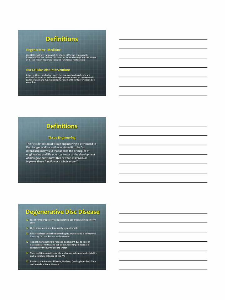
Definitions
Regenerative Medicine
Multi Disciplinary approach in which different therapeutic interventions are utilized, in order to induce biologic enhancement of tissue repair, regeneration and functional restoration.
Bio-Cellular Disc Interventions
Interventions in which growth factors, scaffolds and cells are utilized, in order to induce biologic enhancement of tissue repair, regeneration and functional restoration of the intervertebral disc complex.
Definitions
Tissue Engineering
The first definition of tissue engineering is attributed to Drs. Langer and Vacanti who stated it to be "an interdisciplinary field that applies the principles of engineering and life sciences towards the development of biological substitutes that restore, maintain, or improve tissue function or a whole organ".
Degenerative Disc Disease Is a chronic progressive degenerative condition with no known
cure
High prevalence and frequently symptomatic
It is associated with the normal aging process and is influenced by many factors, known and unknown
The hallmark change is reduced disc height due to loss of extracellular matrix and cell death, resulting in decrease capacity of the IVD to absorb water
The condition can deteriorate and cause pain, motion instability and ultimately collapse of the IVD
It affects the Annulus Fibrosis, Nucleus, Cartilaginous End Plate and Vertebral Bone Marrow
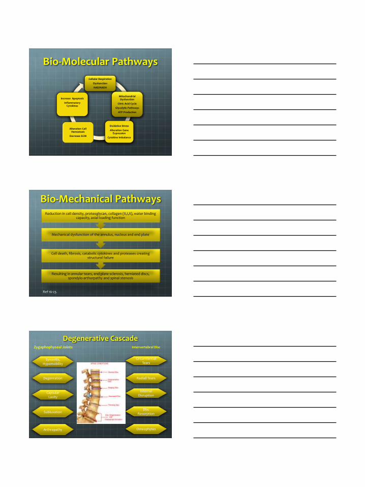
Bio-Molecular Pathways
Cellular Respiration
Dysfunction
NAD/NADH
Mitochondrial Dysfunction
Citric Acid Cycle
Glycolytic Pathways
ATP Production
Oxidative Stress
Alteration Gene Expression
Cytokine Imbalance
Alteration Cell Hemostasis
Decrease ECM
Increase Apoptosis
Inflammatory Cytokines
Bio-Mechanical Pathways
Resulting in annular tears, end plate sclerosis, herniated discs, spondylo-arthorpathy and spinal stenosis
Cell death, fibrosis, catabolic cytokines and proteases creating structural failure
Mechanical dysfunction of the annulus, nucleus and end plate
Reduction in cell density, proteoglycan, collagen (II,I,X), water binding capacity, axial loading function
Ref 16-23.
Degenerative Cascade
Synovitis, Hypomobility
Degenration
Capsular Laxity
Subluxation
Arthropathy
Zygaphophyseal Joints
Disc Resorption
Internal Disruption
Radiall Tears
Circumfential Tears
Osteophytes
Intervertebral Disc
Symptomatic DDD Treatment Challenges
Multifactorial Etiology
Presence of systemic pro-inflammatory conditions modulating pain pathways: Obesity, RA, SLE, DM, Diets
Lack of specific biologic markers
Lack of correlation significant between diagnostic imaging and pain
Non-specific symptoms and non-specific physical findings
Diagnostic test such as provocative discography and MRI, considered the “gold standards” have concerns with specificity and sensitivity
Lack of consensus
Lack of an Integrated Regenerative Approach
Pain can not be imaged . Maybe.
Dr. McCoy’s Lab
Bio Computer Scanner
Bio Bed Bones
Degenerative Disc Disease
STAGING
RSJI STAGING SYSTEM: Evaluation tool designed to standardized evaluation of symptomatic DDD from a regenerative perspective.
It is utilized to assist in biologic tool selection and intervention prognosis
Utilizes four imaging classifications : MRI, Pffirmann (MRI), Modic (MRI, X rays), Dynamic Video Discography
Co-Morbidity factors assessment that affect the overall cell function
Dynamic Video Discography
Utilizes real time video recording during discography
Allows to evaluation of contrast flow analysis based on volume , not pressure
Provides critical information regarding volumetric capacity of the IVD
Better assessment of annular tear dynamics
Essential in designing tissue engineering constructs
Modic Classification Type I
T1: Hypo-intense signal
T2: Hyper-intense signal
Bone marrow edema and inflammation
Type II
T1: Hyper-intense
T2: Iso or Hyper-intense
Often represents normal red haematopoietic
bone marrow conversion into yellow fatty
marrow as a result of marrow ischemia
Type III
T1: Hypointense
T2 :Hypointense
Often represents sub-chondral bone sclerosis
Modic MT, Steinberg PM, Ross JS, et al. Degenerative disk disease: assessment of changes in vertebral body marrow with MR imaging. Radiology 1988;166:193–99
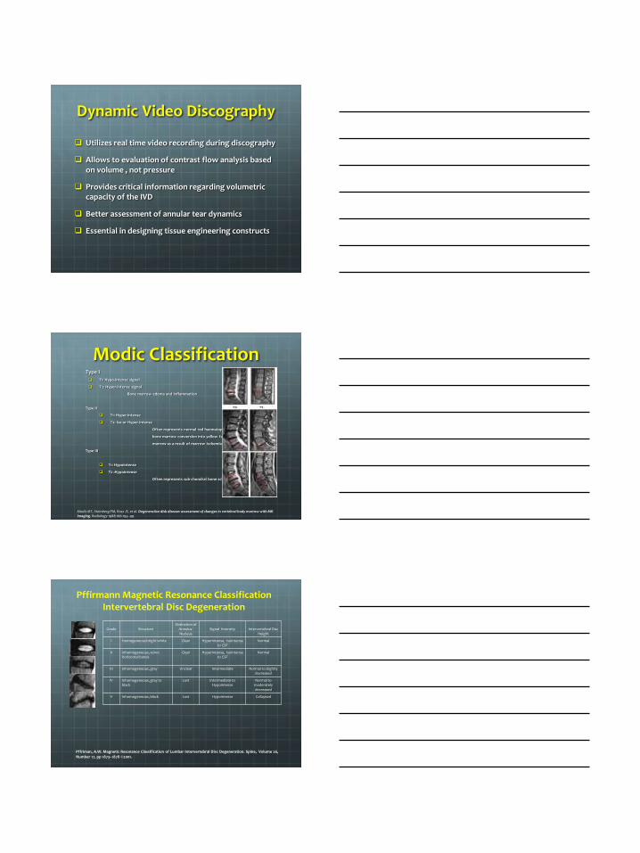
Pffirmann Magnetic Resonance Classification Intervertebral Disc Degeneration
Pffriman, A.W. Magnetic Resonance Classification of Lumbar Intervertebral Disc Degeneration. Spine, Volume 26, Number 17, pp 1873–1878 ©2001.
Grade
Structure
Distinction of Annulus-Nucleus
Signal Intensity
Intervertebral Disc
Height
I Homogeneous bright white Clear Hyperintense, Isointense to CSF
Normal
II Inhomogeneous, w/w0 horizontal bands
Clear
Hyperintense, Isointense to CSF
Normal
III Inhomogeneous, gray Unclear Intermediate Normal to slightly decreased
IV Inhomogeneous, gray to black
Lost Intermediate to Hypointense
Normal to moderately decreased
V Inhomogeneous, black
Lost Hypointense Collapsed
CO-MORBIDITY FACTORS
Translational Instability
Poor Core Biomechanics
Advanced Auto Immune Diseases
Nutritional Depletion
Severe Systemic Diseases
Morbid Obesity
Substance Abuse
Psychological Dysfunction
Prior Back Surgery
Chronic Infections
CO-MORBIDITY FACTORS
Greater than 2 symptomatic discs
Chronic Neuropathic Pain
Smoking History
Non-ambulatory
Failed Spinal Cord Stimulation
Failed Intra Thecal Therapies
Disability Seeking Behavior
Hormonal Deficiencies
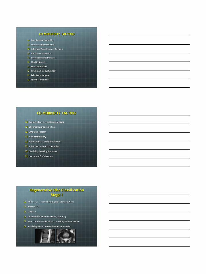
Regenerative Disc Classification Stage I
DHR 0 -25% Herniation: 0-3mm Stenosis- None
Pfirman : I,II
Modic O
Discography: Pain-Concordant, Grade 1-3
Pain: Location- Mainly back Intensity :Mild-Moderate
Instability: None Co-Morbidities: None-Mild
Regenerative Disc Classification Stage II
DHR 0 -25% Herniation: 0-6mm Stenosis- None
Pfirman : III
Modic O-I
Discography: Pain-Concordant, Grade 1-5
Pain: Location: Back> leg Intensity :Mild-Moderate
Instability: None, Facet Hypertrophy – Hypo-Hyper Mobility
Co-Morbidities: None-Moderate
Regenerative Disc Classification Stage III
DHR 25-50% Herniation: >6mm Stenosis- Mild-Moderate
Pfirman : III-IV
Modic I-III
Discography: Pain-Concordant, Grade 1-5
Pain: Location: Leg=Back Intensity :Moderate
Instability: Spondylolisthesis I, Co-Morbidities: Mild-Severe
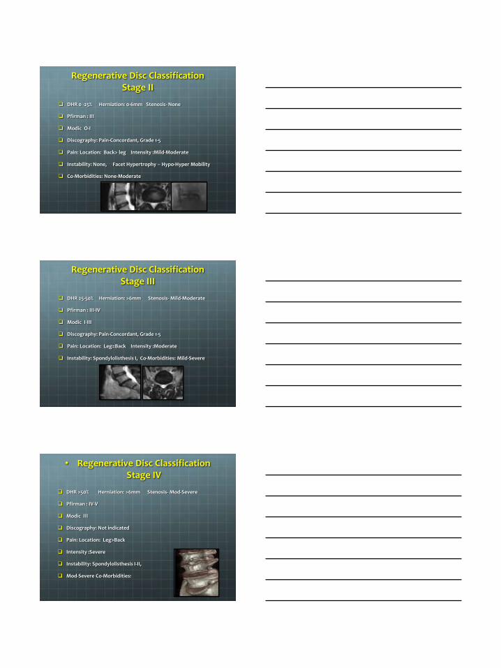
• Regenerative Disc Classification Stage IV
DHR >50% Herniation: >6mm Stenosis- Mod-Severe
Pfirman : IV-V
Modic III
Discography: Not indicated
Pain: Location: Leg>Back
Intensity :Severe
Instability: Spondylolisthesis I-II,
Mod-Severe Co-Morbidities:
The Regenerative Approach
Look upstream
Integrated Regenerative Approach Degenerative Disc Disease
All Stages Bio-Mechanical
Core strengthening
Weight reduction
Exercise
PT, Chiropractic, Acupuncture, Low level laser, Yoga, Bio feedback, TENS
Nutritional Optimization
Anti-inflammatory Diet
Supplements
Hormonal Optimization
Cortisol, Testosterone, Progesterone, Thyroid, Estrogen
Primary End Point •Safety •Pain Relief
Secondary End Point •Functional Restoration (Regenerate, Repair, Restore)
Tertiary End Point •Prevention of Further Degeneration •Prevention of Adjacent Level Degeneration •Restoration of Disc Height
Bio-cellular Therapies for IVD
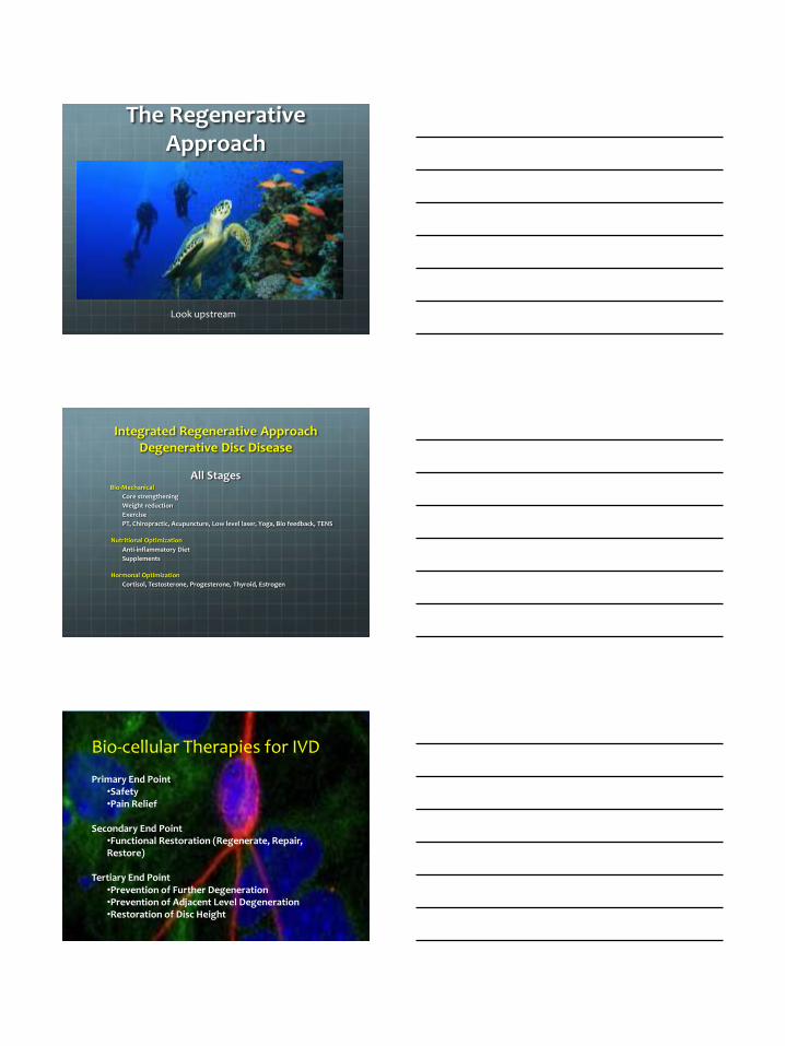
Bio-Regenerative Disc Index Prognostic Model
Stage I: Very Good – Excellent
Stage II: Good – Very Good Stage III: Fair – Good
Stage IV: Poor *At this time, there is not enough clinical data to validate this prognostic model.
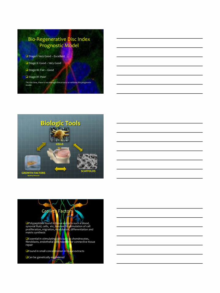
Biologic Tools
CELLS
GROWTH FACTORS Signaling Molecules
SCAFFOLDS
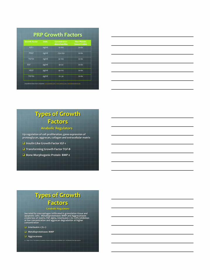
Polypeptides found in tissue extracts such a blood, synovial fluid, cells, etc, involved in stimulation of cell proliferation, migration, modulation, differentiation and matrix synthesis
Essential in stimulating cells such as chondrocytes, fibroblasts, endothelial cells needed for connective tissue repair
Found in small concentration in tissue extracts
Can be genetically engineered
Growth Factors

Mechanism of Action
Autologous
Synthetic
Allogeneic
Growth Factors (Stimulating Molecules)
PRP
Imbedded in Scaffolds, Amniotic
Tissue
GDF-5,BMP-2, Peptides (IGF-1)
Growth Factor Units Concentration above baseline
Final Platelet Concentration
IGF-1 ng/ml 1x-10x
2x-6x
PDGF ng/ml .75x-25x
2x-6x
TGF-b1 ng/ml 3x-10x
2x-6x
EGF pg/ml 4x-5x
2x-6x
VEGF pg/ml 4x-11x
2x-6x
TGF-b2 pg/ml 2x-.3x
2x-6x
Unpublished data from companies: www.biomet.com, www.arthrex.com, www.harvesttech.com
Types of Growth Factors
Anabolic Regulators
Up regulation of cell proliferation, gene expression of proteoglycan, aggrecan, collagen and extracellular matrix
Insulin Like Growth Factor IGF-1
Transforming Growth Factor TGF-B
Bone Morphogenic Protein BMP-2
Types of Growth Factors
Catabolic Regulators
Secreted by macrophages infiltrated in granulation tissue and apoptotic cells. Metalloproteinases MMP and Aggrecanasee create loss of matrix.TNF alpha, Interleukin-1 (IL-1):PG inhibition at low concentration and aggrecan degradation at higher concentration
Interleukin-1 (IL-1)
Metalloproteinases MMP
Aggracanases
24. Dingle JT,et al . The sensitivity of synthesis of human cartilage matrix to inhibition by IL-1. Cell Biochem Funct 1991; 9:99-102.

Endocrine: Affect multiple distant tissues , usually carried in the blood.
Paracrine: Cells are the source of growth factors, usually close contact.
Autocrine: Cell produces growth factors which act on the same cell.
Growth Factors Mechanism of Action
Dr. Kuebler Dr. Scarpone
Scaffolds Scaffolds provide a provisional matrix in a three-dimensional
microenvironment, to localize cells for cellular and molecular
interactions appropriate for cell survival and differentiation.
Natural or Synthetic
Low immunogenicity
Bio-degradable and Bio-compatible
Optimal Mechanical and Architectural Properties
Non toxic
Preferably hydrophilic
Kuo CK, Li WJ, Mauck RL et al (2006) Cartilage tissue engineering: its potential and uses. Curr Opin Rheumatol 18:64–73
Autologous
Synthetic
Allogeneic
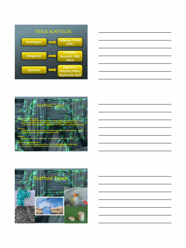
TISSUE SCAFFOLDS
Adipose, Fibrin (PPP)
Collagen, Amniotic BM,
Fibrin
Alginates, Polymer, Nano, Hyaluronic Acid
Hyaluronic Acid/ Hydrogels A normal constituent of both connective tissue matrix and synovial fluid, is a large mucopolysaccharids produced by synoviocytes and chondrocytes, respectively. Large molecular weight with cross-linking are preferable (Synvisc). Fibrin Gel Fibrin Glue contains large amounts of fibrinogen and thrombin.Ideal for growth factor delivery. Autologous processed from PRP .
Collagen Sponges, micronized , allogeneic , most effective with cell therapy.
Human Amniotic Membrane De-cellurized basement membrane from the innermost placental layer, ` composed of a thick basement membrane, avascular stroma.
Scaffold Types
Scaffold Types
Cell Based Therapies Treatment in which stem cells are induced to differentiate into
the specific cell type required to repair damaged or destroyed cells or tissues
Produce significant growth factors to induced stimulate endogenous stem cell population
Successful cell to cell interaction with the Niche
Survive in the recipient after transplant
Engraft into the surrounding tissue after transplant
Produce enough trophic growth factors to achieve desired effect
Avoid harming the recipient in any way
Cell Based Therapy Coheim, a German pathologist almost 130 years ago proposed the existence of non-hematopoietic stem cells. He suggested that bone marrow cells can assist in the repair of process of various peripheral tissues. In 1970, Friedenstein discovered that bone marrow contained fibroblastoid cells with clonogenic potential in vitro capable of forming colonies CFU-F. He was able to regenerate heterotopic bone tissue in different transplants. Thus providing evidence of cell renewal potential of these cells. Friedstein, AJ, et al. The development of fibroblast colonies in monolayer cultures of guinea-pig bone marrow and spleen cells. Cell Tissue Kinet. 1970; 3(4):393-403.
Autologous
Point of Care
Cell-Based Therapies
BMAC (Bone Marrow Aspirate Concentrate)
ADSVF (Adipose Derived Stromal Vascular Fraction)
Ex-Vivo Expansion
BMMSC (Bone Marrow Mesenchymal Stem Cells)
ADMSC (Adipose Derived Mesenchymal Stem Cells)
UCMSC (Umbilical Cord Mesenchymal Stem Cells)
OTHER TISSUES ( MUSCLE, SYNOVIUM, MENSTRUAL BLOOD, ETC)

Cell Based Therapy
Bone Marrow Aspirate Concentrate (BMAC) Non hematopoietic fraction composed of endothelial progenitor cells (EPC), mesenchymal stem cells (MSC), very small embryonic like cell (VSEL), platelets, lymphocytes, growth factors and other extracellular matrix factors.
Mesenchymal stem cell population less than .01-.002% of total nucleated cells.
Current clinical trials : critical limb ischemia trials, cardiac (ischemia and heart failure), stroke, diabetes (I,II), autoimmune, etc.
Cell Based Therapy
Adipose Derived Stromal Vascular Fraction (ADSVF) Endothelial progenitor cells (EPC), mesenchymal stem cells (MSC), very small embryonic like cell (VSEL), lymphocytes, pericytes, growth factors and other extracellular matrix factors.
Mesenchymal stem cell population 5-10% of total nucleated cells.
Current clinical trials: graft vs. host, tissue reconstruction, cosmetic surgery, orthopedics, spinal cord injury, RA, SLE, etc.
Niche and Homing
Cell behavior is induced by encoding proteins and
cytokines secreted by the local tissue or niche.
SDF-1 molecules bind with the local surface receptors and integrins of the MSC outer membrane (CXCR4)
Cell are then induced to differentiate into specific phenotype or proliferate.
Niche signaling pathways are critical for regenerative therapies to be successful.
Stage I
Stage III
Stage II
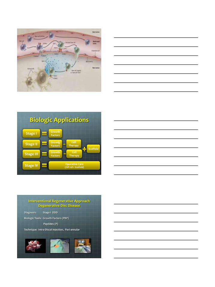
Biologic Applications
Growth Factors
Growth Factors
Growth Factors
Cell Therapy
Cell Therapy
Scaffold
Stage IV Operative Care (SVF+GF+ Scaffold)
a
Interventional Regenerative Approach Degenerative Disc Disease
Diagnosis: Stage I DDD
Biologic Tools: Growth Factors (PRP)
Peptides (P)
Technique: Intra-Discal Injection, Peri-annular
Interventional Regenerative Approach Degenerative Disc Disease
Diagnosis: Stage II DDD
Biologic Tools: BMAC (CBT) + HA (S) +-Peptides (GF)
Amniotic (CBT) + HA (S) +- Peptides (GF)
ADSVF (CBT) + AF (S) PRP +-Peptides (GF)
Technique: Intra-Discal Injection, Lipoaspirate or Bone Marrow Aspirate
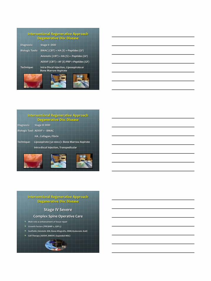
Interventional Regenerative Approach Degenerative Disc Disease
Diagnosis: Stage III DDD
Biologic Tool: ADSVF + - BMAC
HA , Collagen, Fibrin
Technique: Lipoaspirate (50-100cc)+ Bone Marrow Aspirate
Intra-discal Injection, Transpedicular
Interventional Regenerative Approach Degenerative Disc Disease
Stage IV Severe
Complex Spine Operative Care
Main role as enhancement of tissue repair
Growth Factors (PRP,BMP-2, GDF-5)
Scaffolds (Amniotic BM, Bone-Allografts, DBM,Hyaluronic Acid)
Cell Therapy (ADSVF, BMSVF, Expanded MSC)

Treatment of Lumbar Degenerative Disc Disease with
Adipose-Derived Stromal Vascular Fraction, Point of Care
A 48 y/o female patient with intractable low back and left lower
extremity radicular symptoms
Duration 6 years, not responding to conservative care and interventional pain management therapeutics
Patient obtained prior neurosurgical consultation recommending an inter-body fusion with instrumentation or a total disc arthroplasty at L-5
Garcia, C. Treatment of Lumbar Degenerative Disc Disease with Adipose-Derived Stromal Vascular Fraction,Point of Care. Abstract. IFATS, Oct 2012, Quebec.
Treatment of Lumbar Degenerative Disc Disease with
Adipose-Derived Stromal Vascular Fraction, Point of Care
Lumbar MRI consistent with a L-5 central disc herniation (3mm), significant disc desiccation, end plate changes and a “black disc”
Garcia, C. Abstract. IFATS, Oct 2012, Quebec.
Biologic Construct
Adipose Derived SVF
• Standard procedures were used for SVF isolation from autologous lipo-aspirate 50cc .
• Cells processed via a lecithin based emulsification
• 93% viability
• 80 % CD 34-, CD 45-, CD 29+, CD90+, CD 105+
• Estimated MSC injected 5 x 10⁶
PRP - Platelet Rich Plasma (20%)
Fragmented Collagen (porcine)
Results
Lumbar MRI at 6 and 12 months post procedure was positive for an increased in the T2 weighted signal at the L5 nucleus, consistent with ECM deposition
Preservation of disc height at the treated level (L5) and the adjacent level (L4)
No ectopic bone formation
Visual Analog Scale
0
1
2
3
4
5
6
7
8
Pre Month 1 Month 3 Month 6 Month 9 Month12
VAS
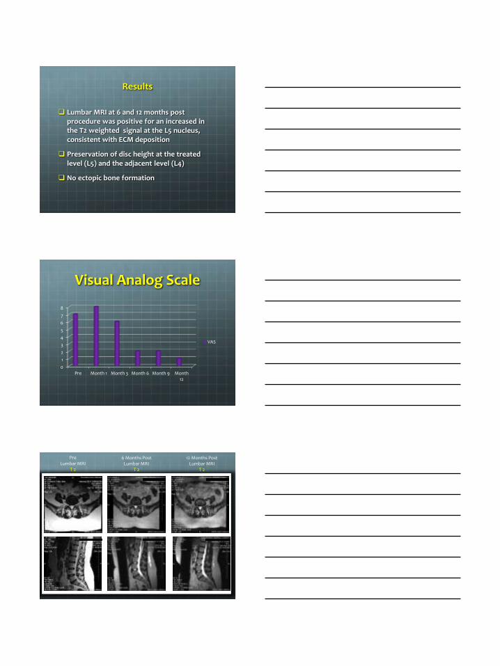
Pre Lumbar MRI
T 2
6 Months Post Lumbar MRI
T 2
12 Months Post Lumbar MRI
T 2
v

MRI Results
Lumbar MRI T 2 weighted
Pre 12 Months Post
MRI Results
Lumbar MRI T 2 weighted
Pre 12 Months Post
Conclusion
A biologic construct with autologous AD-SVF at point of care was successful in treating a patient with advanced symptomatic Lumbar DDD
ECM production in the nucleus pulposus at 6 and 12 months post-transplantation was confirmed by increased T2 Lumbar MRI
Prevention of further disc degeneration at the treated L-5 and adjacent L-4 disc was achieved
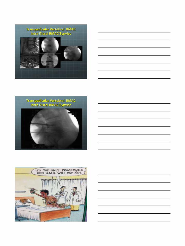
Transpedicular Vertebral BMAC Intra-Discal BMAC/Synvisc
Transpedicular Vertebral BMAC Intra-Discal BMAC/Synvisc

Thank You
Thank You