Embed Size (px)
Citation preview

7/30/2019 Bio 25 Lab - Cat Muscles
http://slidepdf.com/reader/full/bio-25-lab-cat-muscles 1/26
Muscles of the Abdominal Wall
• y. green – internal oblique
with transversus (under) – O: lumbodorsal fascia; border of
pelvic girdle
– I: linea alba
– A: compresses abdomen
• yellow – external oblique – O: lumbodorsal fascia; posterior ribs
– I: linea alba
– A: constricts abdomen
7/30/2019 Bio 25 Lab - Cat Muscles
http://slidepdf.com/reader/full/bio-25-lab-cat-muscles 2/26
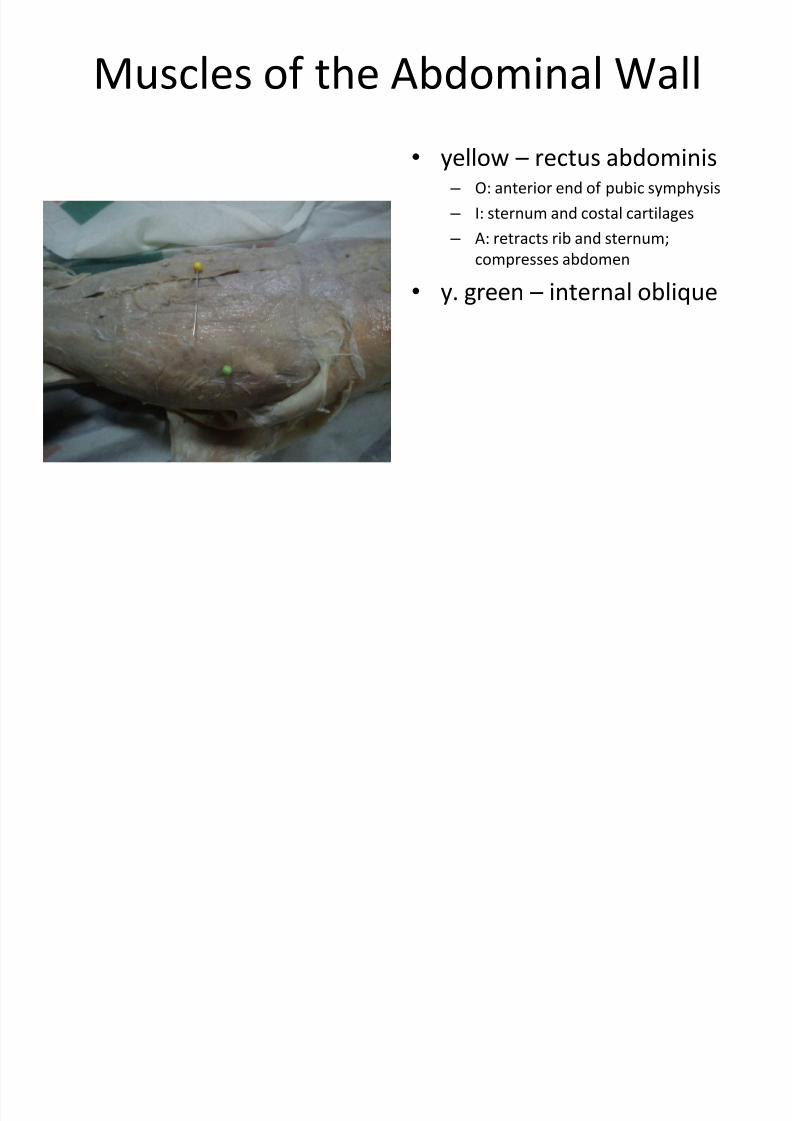
Muscles of the Abdominal Wall
• yellow – rectus abdominis – O: anterior end of pubic symphysis
– I: sternum and costal cartilages
– A: retracts rib and sternum;
compresses abdomen
• y. green – internal oblique
7/30/2019 Bio 25 Lab - Cat Muscles
http://slidepdf.com/reader/full/bio-25-lab-cat-muscles 3/26
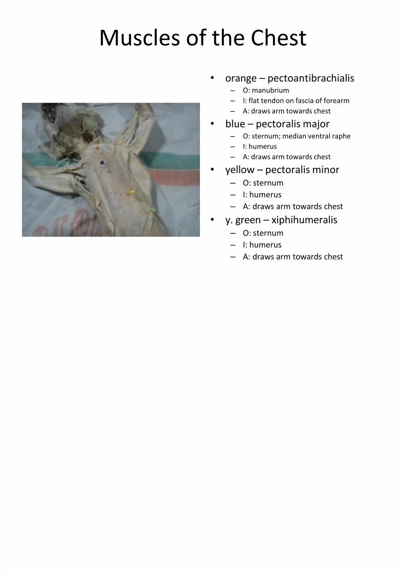
Muscles of the Chest
• orange – pectoantibrachialis – O: manubrium
– I: flat tendon on fascia of forearm
– A: draws arm towards chest
• blue – pectoralis major – O: sternum; median ventral raphe
– I: humerus
– A: draws arm towards chest
• yellow – pectoralis minor – O: sternum
– I: humerus
– A: draws arm towards chest
• y. green – xiphihumeralis – O: sternum
– I: humerus
– A: draws arm towards chest
7/30/2019 Bio 25 Lab - Cat Muscles
http://slidepdf.com/reader/full/bio-25-lab-cat-muscles 4/26
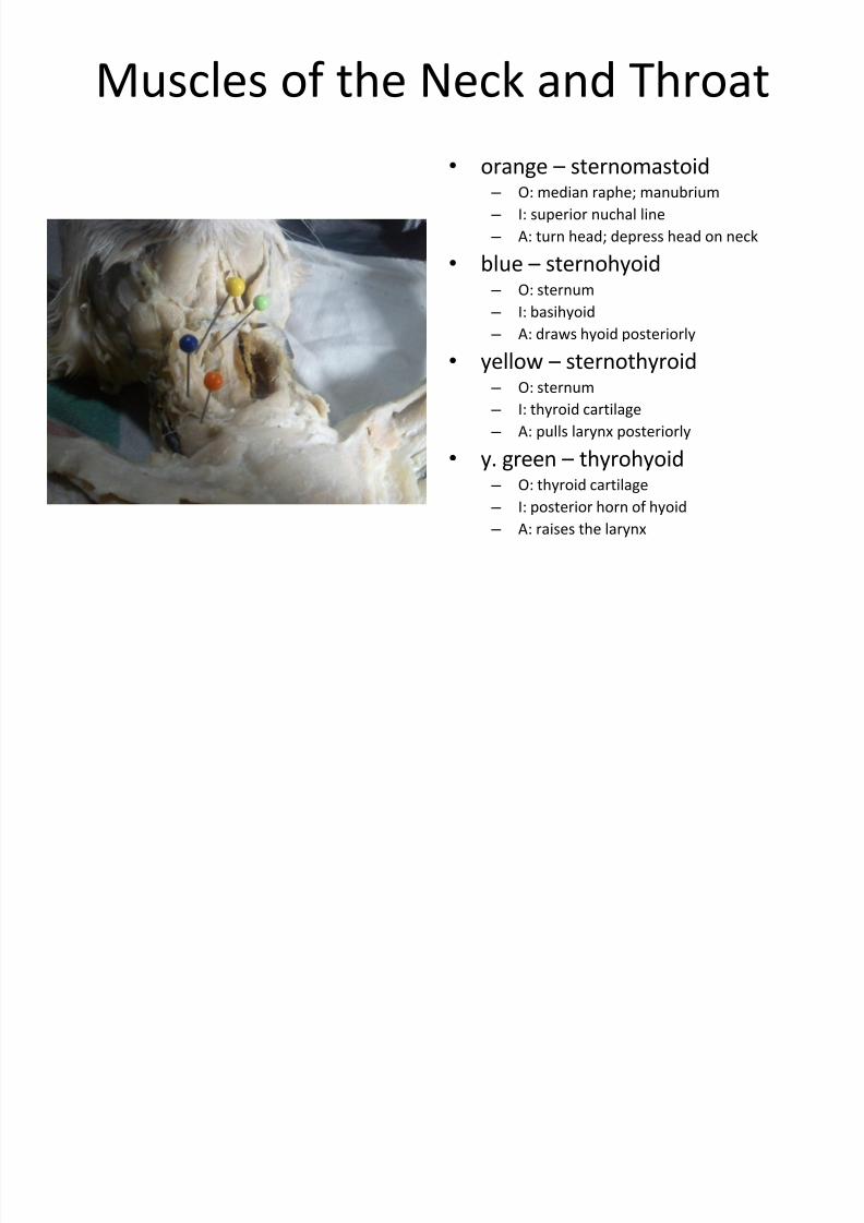
Muscles of the Neck and Throat
• orange – sternomastoid – O: median raphe; manubrium
– I: superior nuchal line
– A: turn head; depress head on neck
• blue – sternohyoid – O: sternum
– I: basihyoid
– A: draws hyoid posteriorly
• yellow – sternothyroid – O: sternum
– I: thyroid cartilage
– A: pulls larynx posteriorly
• y. green –
thyrohyoid – O: thyroid cartilage
– I: posterior horn of hyoid
– A: raises the larynx

7/30/2019 Bio 25 Lab - Cat Muscles
http://slidepdf.com/reader/full/bio-25-lab-cat-muscles 5/26
Muscles of the Neck and Throat
• yellow – clavobrachialis – O: clavicle; fibers of clavotrapezius
– I: ulna
– A: flexes forearm
•
green –
masseter – O: zygomatic arch
– I: mandible
– A: elevates lower jaw
• blue – digastric
– O: jugular and mastoid processes – I: mandible
– A: depresses lower jaw
7/30/2019 Bio 25 Lab - Cat Muscles
http://slidepdf.com/reader/full/bio-25-lab-cat-muscles 6/26
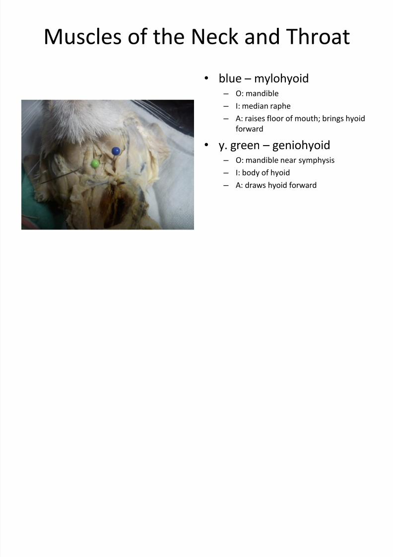
Muscles of the Neck and Throat
• blue – mylohyoid – O: mandible
– I: median raphe
– A: raises floor of mouth; brings hyoid
forward
• y. green – geniohyoid – O: mandible near symphysis
– I: body of hyoid
– A: draws hyoid forward
7/30/2019 Bio 25 Lab - Cat Muscles
http://slidepdf.com/reader/full/bio-25-lab-cat-muscles 7/26
7/30/2019 Bio 25 Lab - Cat Muscles
http://slidepdf.com/reader/full/bio-25-lab-cat-muscles 8/26
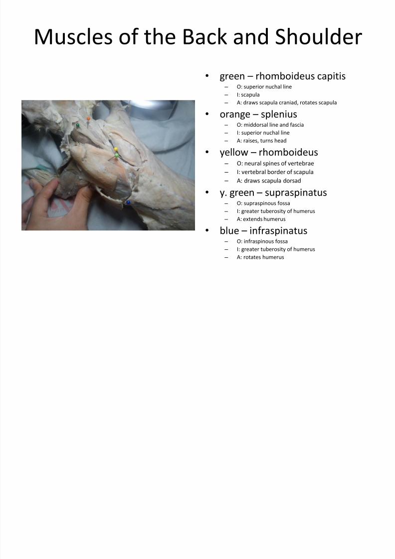
Muscles of the Back and Shoulder
• green – rhomboideus capitis – O: superior nuchal line
– I: scapula
– A: draws scapula craniad, rotates scapula
• orange – splenius – O: middorsal line and fascia
– I: superior nuchal line
– A: raises, turns head
• yellow – rhomboideus – O: neural spines of vertebrae
– I: vertebral border of scapula
– A: draws scapula dorsad
• y. green – supraspinatus – O: supraspinous fossa
– I: greater tuberosity of humerus
– A: extends humerus
• blue – infraspinatus – O: infraspinous fossa
– I: greater tuberosity of humerus
– A: rotates humerus
7/30/2019 Bio 25 Lab - Cat Muscles
http://slidepdf.com/reader/full/bio-25-lab-cat-muscles 9/26
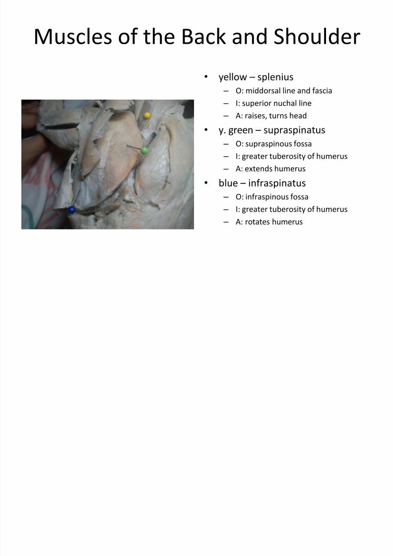
Muscles of the Back and Shoulder
• yellow – splenius
– O: middorsal line and fascia
– I: superior nuchal line
– A: raises, turns head
• y. green – supraspinatus
– O: supraspinous fossa
– I: greater tuberosity of humerus
– A: extends humerus
• blue – infraspinatus
– O: infraspinous fossa
– I: greater tuberosity of humerus
– A: rotates humerus
7/30/2019 Bio 25 Lab - Cat Muscles
http://slidepdf.com/reader/full/bio-25-lab-cat-muscles 10/26
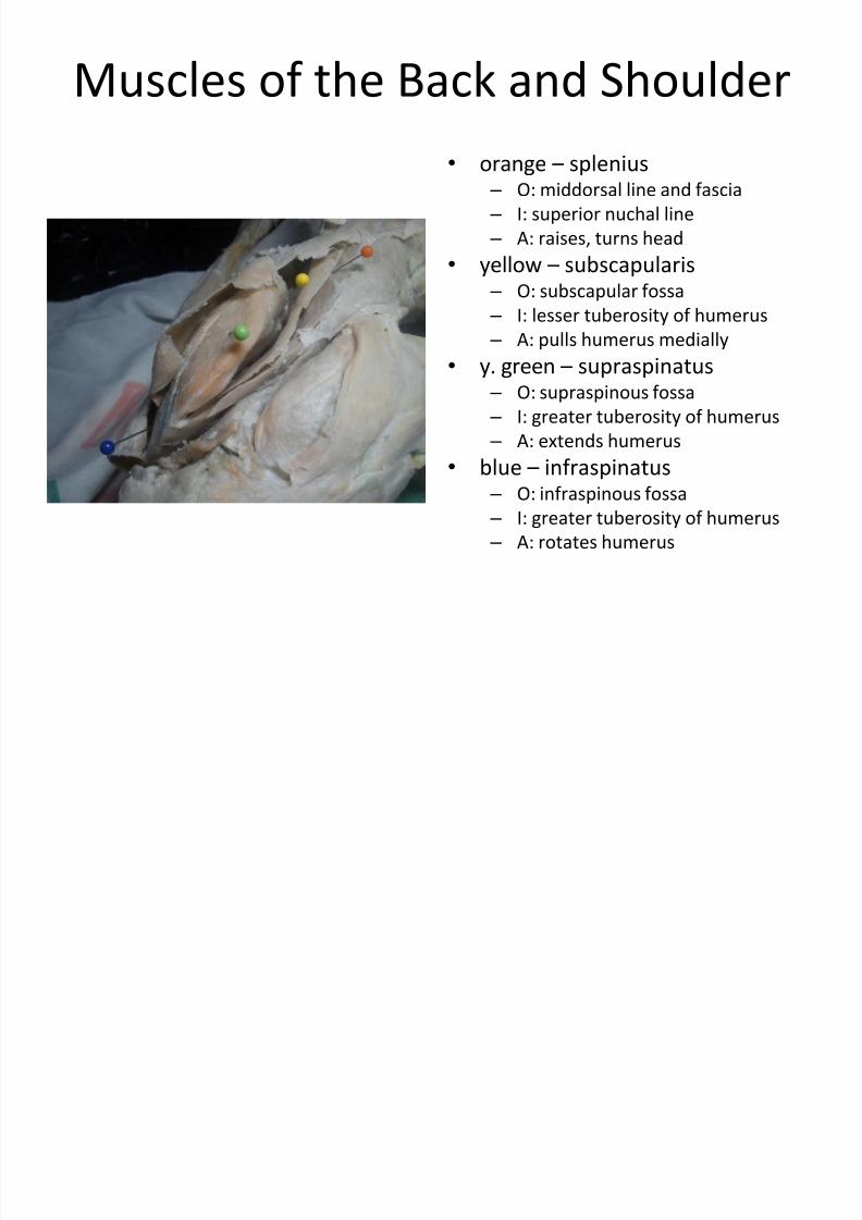
Muscles of the Back and Shoulder
• orange –
splenius – O: middorsal line and fascia
– I: superior nuchal line
– A: raises, turns head
• yellow – subscapularis – O: subscapular fossa
– I: lesser tuberosity of humerus
– A: pulls humerus medially
• y. green – supraspinatus – O: supraspinous fossa
– I: greater tuberosity of humerus
– A: extends humerus
• blue – infraspinatus – O: infraspinous fossa
– I: greater tuberosity of humerus
– A: rotates humerus
7/30/2019 Bio 25 Lab - Cat Muscles
http://slidepdf.com/reader/full/bio-25-lab-cat-muscles 11/26
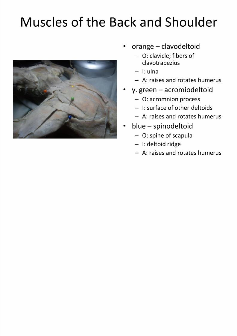
Muscles of the Back and Shoulder
• orange – clavodeltoid
– O: clavicle; fibers of clavotrapezius
– I: ulna
–
A: raises and rotates humerus• y. green – acromiodeltoid
– O: acromnion process
– I: surface of other deltoids
– A: raises and rotates humerus
• blue –
spinodeltoid – O: spine of scapula
– I: deltoid ridge
– A: raises and rotates humerus

7/30/2019 Bio 25 Lab - Cat Muscles
http://slidepdf.com/reader/full/bio-25-lab-cat-muscles 12/26
Muscles of the Back and Shoulder (Dorso-lateral View)
• y. green – serratus ventralis
– O: slips from first 9~10 ribs;
transverse process of last 5
cervical vertebrae
– I: scapula
– A: draws scapula craniad,
ventrad, against thoracic wall
• yellow – serratus dorsalis
– O: aponeurosis from medial line
–
I: last ribs – A: draws ribs forward
7/30/2019 Bio 25 Lab - Cat Muscles
http://slidepdf.com/reader/full/bio-25-lab-cat-muscles 13/26
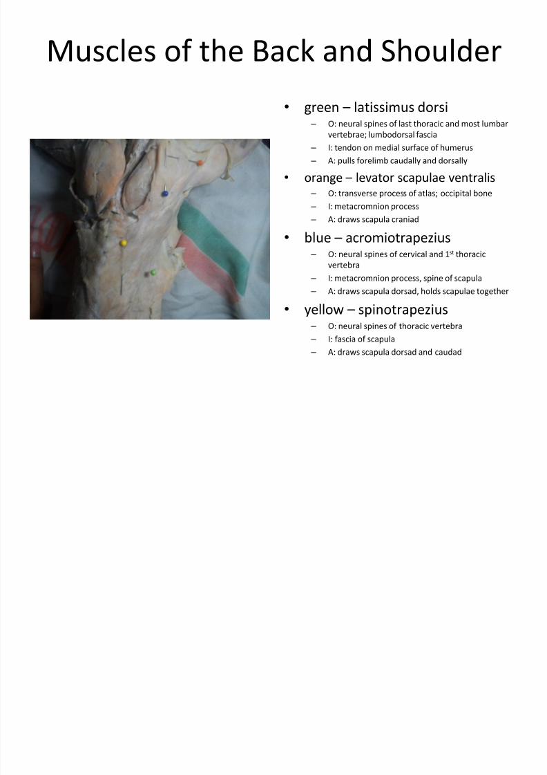
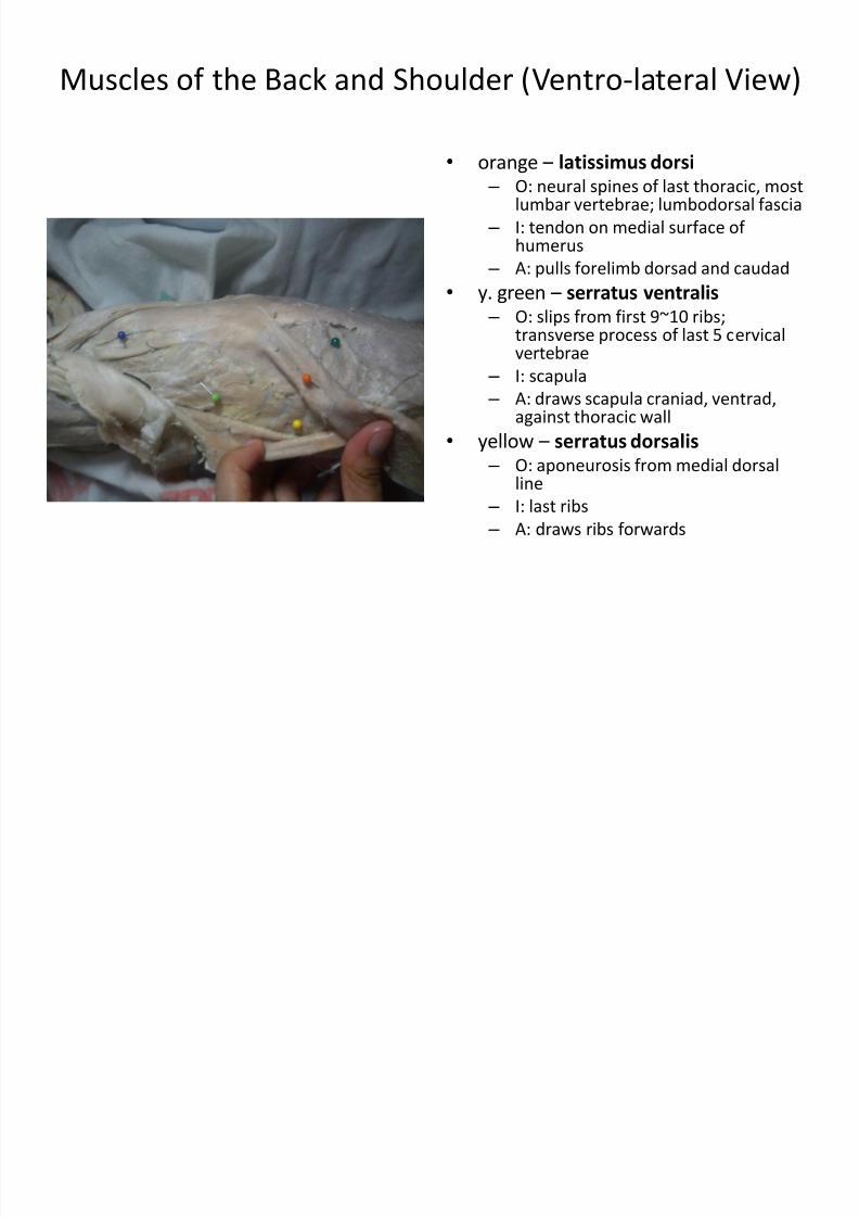
Muscles of the Back and Shoulder (Ventro-lateral View)
• orange –
latissimus dorsi
– O: neural spines of last thoracic, mostlumbar vertebrae; lumbodorsal fascia
– I: tendon on medial surface of humerus
– A: pulls forelimb dorsad and caudad
•
y. green –
serratus ventralis – O: slips from first 9~10 ribs;
transverse process of last 5 cervicalvertebrae
– I: scapula
– A: draws scapula craniad, ventrad,against thoracic wall
•
yellow –
serratus dorsalis – O: aponeurosis from medial dorsal
line
– I: last ribs
– A: draws ribs forwards
7/30/2019 Bio 25 Lab - Cat Muscles
http://slidepdf.com/reader/full/bio-25-lab-cat-muscles 14/26
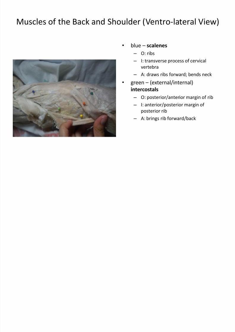
Muscles of the Back and Shoulder (Ventro-lateral View)
• blue – scalenes
– O: ribs
– I: transverse process of cervical
vertebra
– A: draws ribs forward; bends neck
• green –
(external/internal)intercostals
– O: posterior/anterior margin of rib
– I: anterior/posterior margin of
posterior rib
– A: brings rib forward/back
7/30/2019 Bio 25 Lab - Cat Muscles
http://slidepdf.com/reader/full/bio-25-lab-cat-muscles 15/26
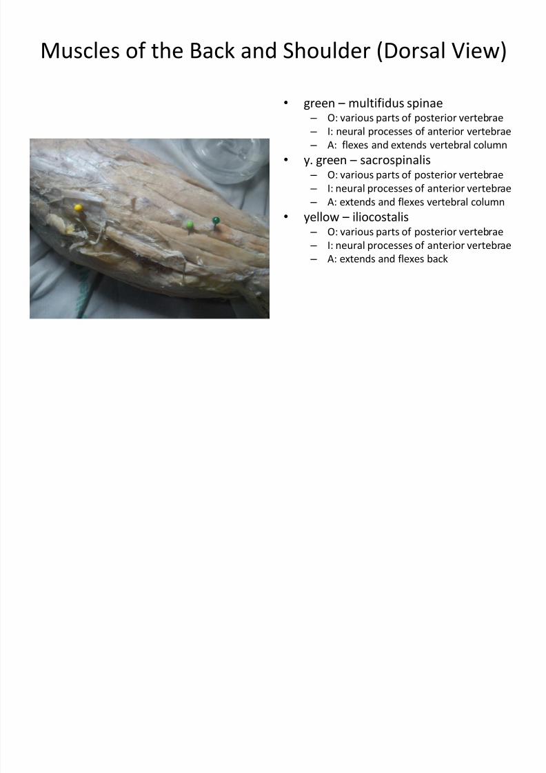
Muscles of the Back and Shoulder (Dorsal View)
• green –
multifidus spinae – O: various parts of posterior vertebrae
– I: neural processes of anterior vertebrae
– A: flexes and extends vertebral column
• y. green – sacrospinalis – O: various parts of posterior vertebrae
–
I: neural processes of anterior vertebrae – A: extends and flexes vertebral column
• yellow – iliocostalis – O: various parts of posterior vertebrae
– I: neural processes of anterior vertebrae
– A: extends and flexes back
7/30/2019 Bio 25 Lab - Cat Muscles
http://slidepdf.com/reader/full/bio-25-lab-cat-muscles 16/26
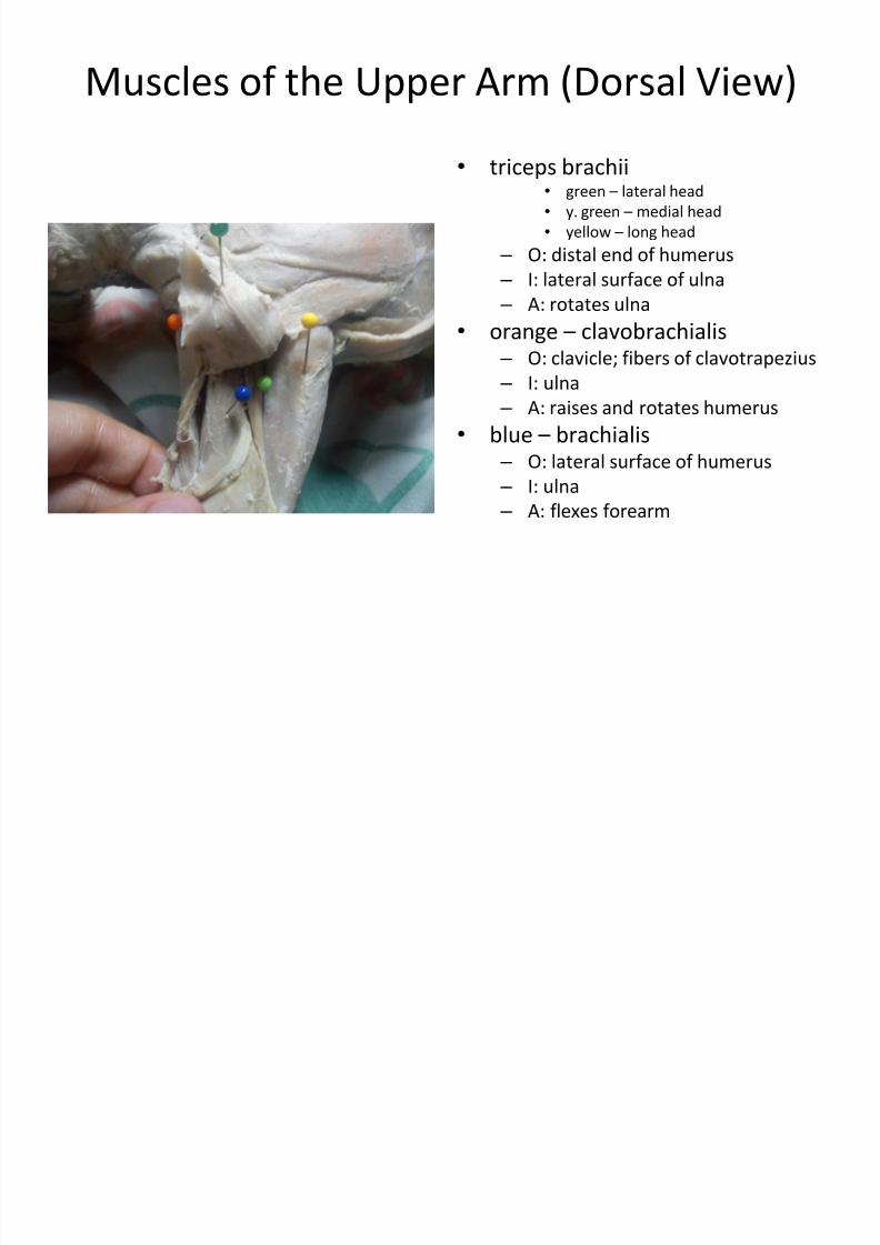
Muscles of the Upper Arm (Dorsal View)
• triceps brachii• green – lateral head
• y. green – medial head
• yellow – long head
– O: distal end of humerus
– I: lateral surface of ulna
–
A: rotates ulna• orange – clavobrachialis
– O: clavicle; fibers of clavotrapezius
– I: ulna
– A: raises and rotates humerus
• blue – brachialis
– O: lateral surface of humerus – I: ulna
– A: flexes forearm
7/30/2019 Bio 25 Lab - Cat Muscles
http://slidepdf.com/reader/full/bio-25-lab-cat-muscles 17/26
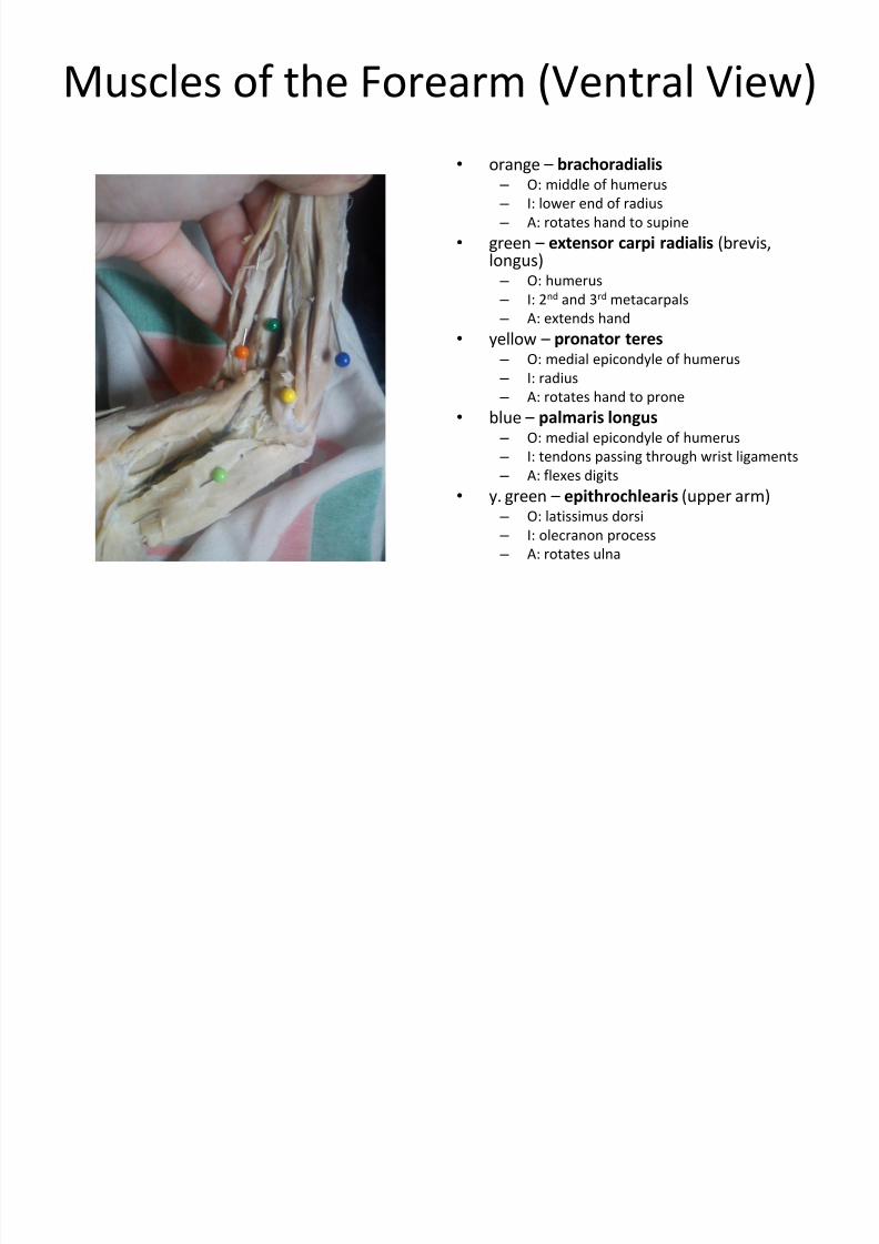
Muscles of the Forearm (Ventral View)
• orange –
brachoradialis
– O: middle of humerus
– I: lower end of radius
– A: rotates hand to supine
• green – extensor carpi radialis (brevis,longus) – O: humerus
– I: 2nd and 3rd metacarpals
– A: extends hand
• yellow – pronator teres
– O: medial epicondyle of humerus
– I: radius
– A: rotates hand to prone
• blue – palmaris longus
– O: medial epicondyle of humerus
–
I: tendons passing through wrist ligaments – A: flexes digits
• y. green – epithrochlearis (upper arm) – O: latissimus dorsi
– I: olecranon process
– A: rotates ulna
7/30/2019 Bio 25 Lab - Cat Muscles
http://slidepdf.com/reader/full/bio-25-lab-cat-muscles 18/26
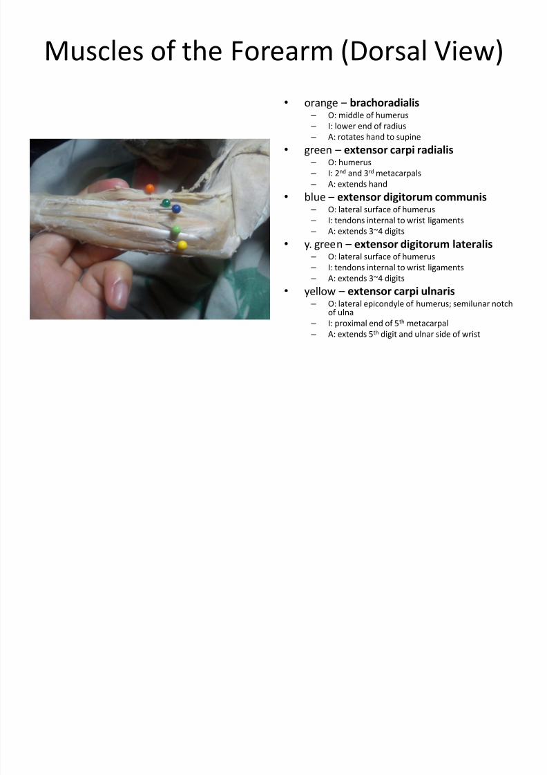
Muscles of the Forearm (Dorsal View)
• orange –
brachoradialis – O: middle of humerus
– I: lower end of radius
– A: rotates hand to supine
• green – extensor carpi radialis – O: humerus
– I: 2nd and 3rd metacarpals
– A: extends hand
• blue –
extensor digitorum communis – O: lateral surface of humerus
– I: tendons internal to wrist ligaments
– A: extends 3~4 digits
• y. green – extensor digitorum lateralis – O: lateral surface of humerus
– I: tendons internal to wrist ligaments
– A: extends 3~4 digits
• yellow –
extensor carpi ulnaris – O: lateral epicondyle of humerus; semilunar notch
of ulna
– I: proximal end of 5th metacarpal
– A: extends 5th digit and ulnar side of wrist
7/30/2019 Bio 25 Lab - Cat Muscles
http://slidepdf.com/reader/full/bio-25-lab-cat-muscles 19/26
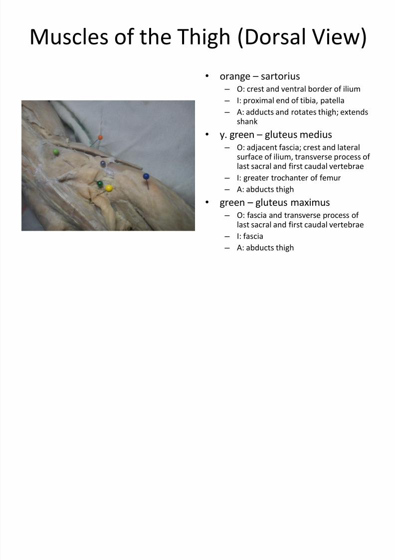
Muscles of the Thigh (Dorsal View)
• orange –
sartorius – O: crest and ventral border of ilium
– I: proximal end of tibia, patella
– A: adducts and rotates thigh; extendsshank
•
y. green –
gluteus medius – O: adjacent fascia; crest and lateral
surface of ilium, transverse process of last sacral and first caudal vertebrae
– I: greater trochanter of femur
– A: abducts thigh
• green – gluteus maximus – O: fascia and transverse process of
last sacral and first caudal vertebrae
– I: fascia
– A: abducts thigh
7/30/2019 Bio 25 Lab - Cat Muscles
http://slidepdf.com/reader/full/bio-25-lab-cat-muscles 20/26
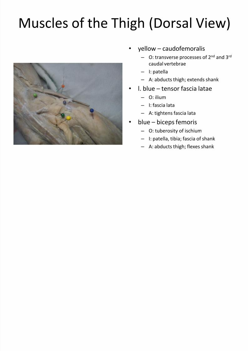
Muscles of the Thigh (Dorsal View)
• yellow – caudofemoralis
– O: transverse processes of 2nd and 3rd
caudal vertebrae
– I: patella
– A: abducts thigh; extends shank
• l. blue –
tensor fascia latae – O: ilium
– I: fascia lata
– A: tightens fascia lata
• blue – biceps femoris
– O: tuberosity of ischium – I: patella, tibia; fascia of shank
– A: abducts thigh; flexes shank
7/30/2019 Bio 25 Lab - Cat Muscles
http://slidepdf.com/reader/full/bio-25-lab-cat-muscles 21/26
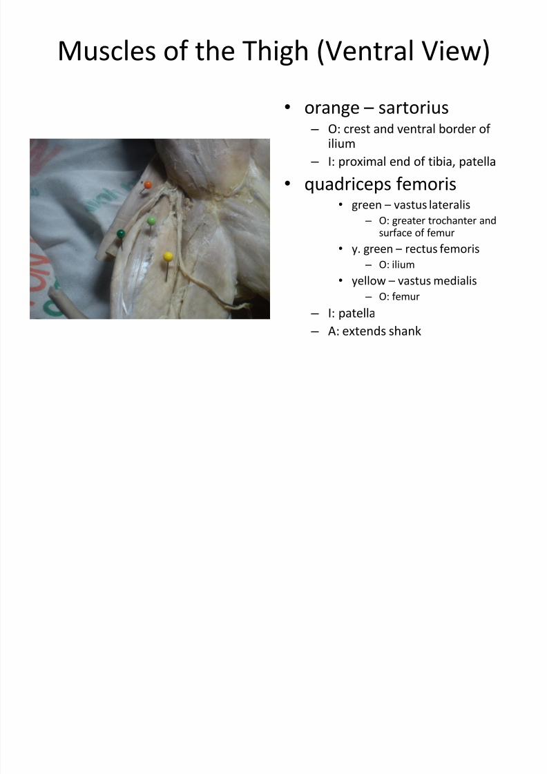
Muscles of the Thigh (Ventral View)
• orange –
sartorius – O: crest and ventral border of
ilium
– I: proximal end of tibia, patella
•
quadriceps femoris• green – vastus lateralis
– O: greater trochanter andsurface of femur
• y. green – rectus femoris
– O: ilium
• yellow – vastus medialis
– O: femur
– I: patella
– A: extends shank

7/30/2019 Bio 25 Lab - Cat Muscles
http://slidepdf.com/reader/full/bio-25-lab-cat-muscles 22/26
Muscles of the Thigh (Ventral View)
• blue – gracilis
– O: ischial and pubic symphysis
– I: aponeurosis passing tibia
– A: adducts leg
• l. blue – adductor longus
– O: pubis
– I: femur
– A: adducts thigh
• orange – adductor femoris
– O: pubis
– I: femur
– A: adducts thigh
7/30/2019 Bio 25 Lab - Cat Muscles
http://slidepdf.com/reader/full/bio-25-lab-cat-muscles 23/26
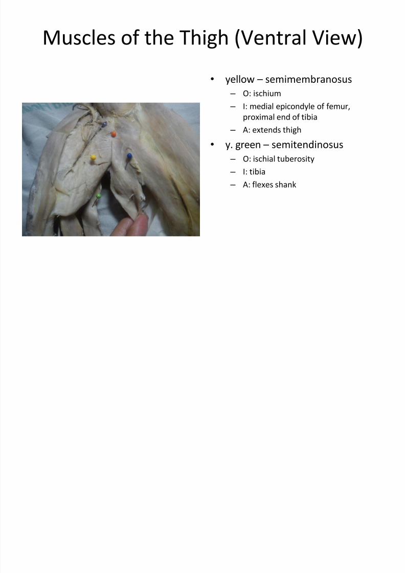
Muscles of the Thigh (Ventral View)
• yellow – semimembranosus
– O: ischium
– I: medial epicondyle of femur,
proximal end of tibia
– A: extends thigh
• y. green –
semitendinosus – O: ischial tuberosity
– I: tibia
– A: flexes shank
7/30/2019 Bio 25 Lab - Cat Muscles
http://slidepdf.com/reader/full/bio-25-lab-cat-muscles 24/26
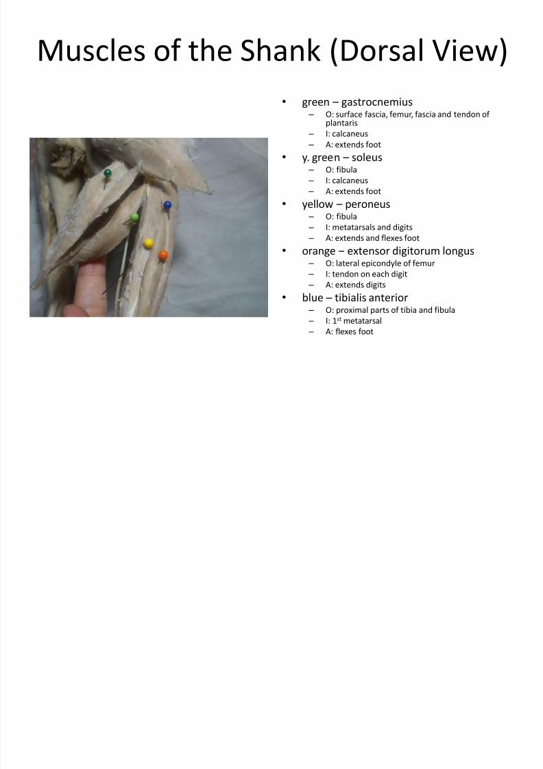
Muscles of the Shank (Dorsal View)
• green –
gastrocnemius – O: surface fascia, femur, fascia and tendon of
plantaris
– I: calcaneus
– A: extends foot
• y. green – soleus – O: fibula
– I: calcaneus
– A: extends foot• yellow – peroneus
– O: fibula
– I: metatarsals and digits
– A: extends and flexes foot
• orange – extensor digitorum longus – O: lateral epicondyle of femur
– I: tendon on each digit
–
A: extends digits• blue – tibialis anterior
– O: proximal parts of tibia and fibula
– I: 1st metatarsal
– A: flexes foot
7/30/2019 Bio 25 Lab - Cat Muscles
http://slidepdf.com/reader/full/bio-25-lab-cat-muscles 25/26
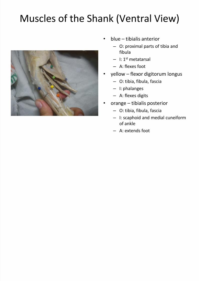
Muscles of the Shank (Ventral View)
• blue – tibialis anterior
– O: proximal parts of tibia and
fibula
– I: 1st metatarsal
– A: flexes foot
• yellow – flexor digitorum longus
– O: tibia, fibula, fascia
– I: phalanges
– A: flexes digits
•
orange –
tibialis posterior – O: tibia, fibula, fascia
– I: scaphoid and medial cuneiform
of ankle
– A: extends foot

7/30/2019 Bio 25 Lab - Cat Muscles
http://slidepdf.com/reader/full/bio-25-lab-cat-muscles 26/26
Muscles of the Shank (Ventral View)
• y. green – soleus
– O: fibula
– I: calcaneus
– A: extends foot
•
l. blue –
plantaris – O: patella and femur
– I: calcaneus
– A: flexes digits
• green – gastrocnemius
– O: surface fascia, femur, tendonand fascia of plantaris
– I: calcaneus
– A: extends foot