Embed Size (px)
Citation preview

Binding of Myelin Basic Protein Peptides to Human HistocompatibilityLeukocyte Antigen Class 11 Moleculesand Their Recognition by T Cells from Multiple Sclerosis PatientsAntonietta Valli,* Alessandro Sette,t Ludwig Kappos,I Carla Oseroff,t John Sidney,tGuido Miescher,I Martina Hochberger,11 Ekkehard D. Albert,11 and Luciano Adorini**Preclinical Research, Sandoz Pharma Ltd., CH-4002 Basel, Switzerland; tCytel Corp., San Diego, California 92121; IDepartment ofNeurology and Department of Research, University of Basel, Kantonsspital, CH-4031 Basel, Switzerland; and lLaboratory ofImmunogenetics, University of Munich, D-8000 Munich, Federal Republic of Germany
Abstract
Multiple sclerosis (MS) is an autoimmune disease in whichmyelin proteins have been implicated as autoantigens recog-nized by pathogenic autoreactive T cells. To study the relation-ship between human myelin basic protein (hMBP) and HLAalleles associated to MSsusceptibility, such as DRB1*1501,the binding of synthetic peptides spanning the entire hMBPsequence to 10 purified HLA-DR molecules was determined.All the hMBPpeptides tested showed binding affinity for atleast one of the DRmolecules analyzed, but three hMBPpep-tides, included in sequences 13-32,84-103, and 144-163 werefound capable of binding to three or more DRmolecules. ThehMBPpeptide 84-103 was the most degenerate in binding, inthat it bound to 9 out of 10 DRmolecules tested. Interestingly,it bound with highest affinity to DRB1*1501 molecules. Tocorrelate the binding pattern of hMBPpeptides to HLA classII molecules with their recognition by T cells, 61 hMBP-spe-cific T cell lines (TCL) were established from the peripheralblood of 20 MSpatients, who were homozygous, heterozygous,or negative for DRB1*1501. Analysis of hMBPepitopes recog-nized by these TCL and their HLA restriction demonstrated avery good correlation between binding data and T cell prolifera-tion to hMBPpeptides. Although virtually all hMBPpeptidestested could be recognized by at least one TCL from MSpa-tients, three immunodominant T cell epitopes were apparentamong the TCL examined, corresponding exactly to the hMBPpeptides capable of binding to several DRmolecules. No majordifference could be detected in the recognition of immunodomi-nant hMBPpeptides by TCL from DRB1*1501 positive ornegative MSpatients. These results have implications for therole of hMBPas relevant autoantigen, and of DRB1*1501 assusceptibility allele in MS. (J. Clin. Invest. 1993. 91:616-628.) Key words: immunodominance - human histocompatibil-
Dr. Valli is currently at Cattedra di Malattie Infettive, Universita diParma, Via Gramsci 14,1-43 100 Parma, Italy; Dr. Adorini is currentlyat Roche Milano Ricerche, Via Olgettina 58, 1-20132 Milan, Italy.
Address reprint requests to Dr. Luciano Adorini at his permanentaddress: Preclinical Research 386-106, Sandoz Pharma, Ltd., CH-4002Basel, Switzerland.
Receivedfor publication 6 July 1992 and in revisedform 17 Sep-tember 1992.
ity leukocyte antigen class II molecules - multiple sclerosis-myelin basic protein - T cell epitopes
Introduction
Multiple sclerosis (MS)' is a chronic, inflammatory disease ofthe human central nervous system (CNS) characterized by de-myelination and by focal infiltrates of macrophages, plasmacells, and T cells in the CNS(1). Although the cause of MSisunknown, a T cell-dependent autoimmune process, involvingautoreactive T cells specific for a myelin protein, has been pos-tulated (reviewed in reference 2) and the demonstration of invivo-activated T cells specific for human myelin basic protein(hMBP) in the blood of MSpatients has implicated hMBPas acandidate autoantigen in the pathogenesis of MS(3).
The possible role of hMBPin MShas been further sup-ported by the observation that experimental allergic encephalo-myelitis (EAE), an animal model for MS, can be induced byimmunization with MBPor MBPpeptides in adjuvant (4, 5),as well as by passive transfer of MBP-specific CD4+ T cells(6-8). Encephalitogenic T cells in mice and rats are specific forimmunodominant MBPepitopes presented by specific MHCclass II molecules; for example, in BIO.PL mice encephalito-genic T cells recognize the amino-terminal MBPpeptide 1-9presented by I-AU molecules (9). The variable regions of the Tcell receptor (TCR) in MBP-specific T cells have been found oflimited heterogeneity, with a preferential expression of VB 8.2and Va 2 or Va 4, both in mice and rats (10). Thus, in thesespecies, MHCclass II and TCRgene products associated torecognition of encephalitogenic MBPepitopes by T cells havebeen identified. Recently, however, it has been demonstratedthat EAEcan be induced by other autoantigens, such as proteo-lipid protein ( 11), raising the possibility that a multiplicity ofmyelin antigens maybe capable oftriggering autoimmune reac-tions resulting in the clinical manifestations characteristicof EAE.
A significant association between major histocompatibilitycomplex (MHC) class II gene products and disease has beenobserved in MSpatients. The higher concordance rate for MSin monozygotic compared to dizygotic twins indicates a geneti-cally determined susceptibility to the disease (12). Because thefrequency of the HLA class II allele DRB1*1501, and of theassociated DQB1*0602, is significantly increased in Northern
1. Abbreviations used in this paper: APC, antigen-presenting cells;EAE, experimental allergic encephalomyelitis; EDSS, expanded disabil-ity status scale; hMBP, human myelin basic protein; MS, multiple scle-rosis; TCL, T cell line; TCR, T cell receptor.
616 Valli et al.
J. Clin. Invest.© The American Society for Clinical Investigation, Inc.0021-9738/93/02/0616/13 $2.00Volume 91, February 1993, 616-628

European MSpatients compared to controls, DRB1 * 1501, orgene(s) in linkage disequilibrium with it, could represent asusceptibility gene for MS( 13, 14). Although the DRBl * 1501haplotype is associated with the highest relative risk of MS,other HLA class II alleles are overrepresented among certainethnic groups of MSpatients: DR4 in Southern Italians ( 15)and Arabs (16) and DR13 in Japanese (17) and Mexicans( 18). In contrast to the well-established association betweenHLAgenes and MS, the association of TCRgenes, either in thegermline ( 19, 20) or in the hMBP-specific peripheral T cellrepertoire (21-25), with MSis still unclear.
To assess the possible role of hMBPas autoantigen in MS,several groups have studied the epitope specificity of hMBP-reactive T cells in MSpatients and controls (26-32). Autoreac-tive T cells, mostly CD4' and DR-restricted, specific for sev-eral hMBPpeptides have been obtained from both MSpatientsand healthy controls. Although, collectively, major differenceswere not apparent in the T cell reactivity to hMBPbetween MSpatients and controls, it has been suggested that recognition ofimmunodominant hMBPepitopes in individuals expressing anMS-associated HLA class II allele could be important in thepathogenesis of the disease. This would be expected if hMBPisthe autoantigen in MS, and if the role of the MS-associatedclass II alleles is to present hMBPpeptides to autoreactive Tcells.
To test this hypothesis, the binding of a panel of hMBPpeptides to several purified DRmolecules, including those as-sociated to increased frequency of MS, was determined. In ad-dition, T cell lines (TCL) specific for hMBPwere establishedfrom MSpatients positive or negative for the MS-associatedallele HLA-DRB1* 1501.
As demonstrated in the present study, some hMBPpeptidesbind with highest affinity to HLAclass II molecules encoded byalleles associated to increased frequency of MS, thus providinga possible molecular explanation for the association betweenclass II genes and MS, and at the same time implicating thehMBPsequence 84-103 as a possible candidate for the autoan-tigenic determinant. The finding that this peptide binds, withlower affinity, to several other DRmolecules, may also providean explanation for the incomplete association between MSandclass II genes.
However, the observation that hMBP-specific T cells fromeither DRB1 * 1501 -positive or -negative MSpatients recognizeequally well this and other hMBPpeptides cautions against asimplistic interpretation of the binding data, and illustrates thecomplexity and heterogeneity of T cell responses againsthMBP.
Methods
Antigens. hMBPwas prepared as described (33). Purity of hMBPprep-arations, as assessed by silver staining after gel electrophoresis, wasroutinely found to be > 90%. Peptides were synthetized on an AppliedBiosystems Inc. (Foster City, CA) 430A peptide synthetizer. After re-moval of the a-amino-tert-butyloxycarbonyl protecting group, thephenylacetamidomethyl resin peptide was coupled with a fourfold ex-cess of preformed symmetrical anhydride (hydroxybenzyltriazole es-ters for arginine, histidine, asparagine, and glutamine) for I h in di-methylformamide. For arginine, asparagine, glutamine, and histidineresidues, the coupling step was repeated to obtain a high coupling effi-ciency. After synthesis was completed, the peptide was cleaved fromthe resin and the protecting groups removed by treatment with hydro-
gen fluoride in the presence of appropriate scavengers. The peptideswere then purified by reversed phase high-performance liquid chroma-tography (HPLC). Peptide purity was substantiated by amino acidsequence and/or composition analysis. They were routinely > 95%pure after HPLC.
Affinity purification of DR molecules. The following Epstein-Barr virus (EBV)-transformed homozygous cell lines-LG2(DRB1*0101), 3107 (DRB5*0101), MAT(DRBI*0301), PRIESS(DRBI*0404), BIN40 (DRBI*0404), SWEIG (DRBI*1101),PITOUT (DRBI*0701): or transfected fibroblasts-L466.1(DRBI*1501), L416.3 (DRB5*0101), L242.5 (DRBI*1601), L255.1(DRB5*0201 )-were used as a source of HLA class II molecules. DR-transfected fibroblast were a kind gift of Dr. R. W. Karr (University ofIowa, Iowa City, IA). The HLAnomenclature is according to reference34. Cell lines were cultured in RPMI 1640 medium supplemented with10% heat-inactivated fetal calf serum (FCS) or horse serum (HazeltonBiologics, Inc., Lenexa, KS). Cells were lysed at a concentration of 108cells/ml in 50 mMTris-HCI, pH 8.5, containing 2%Renex, 150 mMNaCI, 5 mMEDTA, and 2 mMphenylmethyl sulfonyl fluoride(PMSF). The lysates were cleared of nuclei and debris by centrifuga-tion at 10,000 g for 20 min.
DRmolecules were purified essentially as described (35, 36) usingthe mab LB3.1 (37) covalently coupled to protein A-Sepharose CL-4B.Aliquots of cell lysates equivalent to - 10 g of cells were passed se-quentially through the following columns: Sepharose CL-4B (10 ml),protein A-Sepharose (5 ml), W6/32-protein A-Sepharose (10 ml),and LB3.1-protein A-Sepharose (15 ml) using a flow rate of 15 ml/h.The columns were washed with 10 column volumes of 10 mMTris-HC1, pH 8.0, 0.1 %Renex (5 ml/h); 2 column volumes of PBS, and 2column volumes of PBS- 1%octylglucoside. DRmolecules were elutedfrom the LB3.1 column with 0.05 Mdiethylamine, in 0.15 MNaCIcontaining 1%octylglucoside (pH 11.5), immediately neutralized with2 M glycine, pH 2.0, and concentrated by ultrafiltration through aAmicon YM-30 membrane (Amicon Corp., Danvers, MA). Proteincontent was evaluated by a BCAprotein assay (Pierce Chemical Co.,Rockford, IL) and confirmed by SDS-PAGE.
Binding of hMBPpeptides to purified HLA class II molecules. Puri-fied DRmolecules (5-500 nM) were incubated with 5 nM125I-radiola-beled peptides for 48 h in PBS containing 5% dimethylsulfoxide(DMSO) in the presence of a protease inhibitor mixture. Purified pep-tides were iodinated using the chloramine-T method. The final concen-trations of peptide inhibitors were: 1 mMPMSF, 1.3 mM1.10 phenan-troline, 73 ,M pepstatin A, 8 mMEDTA, 6 mMN-ethylmaleimide,and 200 MMN-Ap-tosyl-L-lysine chloromethyl ketone (TLCK). Finaldetergent concentration in the incubation mixture was 0.05% NP-40.Assays were performed at pH 7.0 with the exception of DRB1*0301,which was performed at pH 4.5, and DRBI*1601, which was per-formed at pH 5. The pH was adjusted as previously described (38). TheDR-peptide complexes were separated from free peptides by gel filtra-tion on Sephadex G50 columns as previously described (39, 40) orTSK2000 (7.8 mmX 15 cm) eluted at 1.2 ml/min in PBS, pH 6.5,containing 0.5% NP-40 and 0.1% NaN3. Because the large size of theradiolabeled peptide used for the DRBI * 1501 binding assay makesseparation of bound from unbound peaks more difficult under theseconditions, all DRBI*1 501 assays were performed using a TSK2000(7.8 mmX 30 cm) column eluted at 0.6 ml/min. Column eluates were
passed through a model 170 radioisotope detector (Beckman Instru-ments, Inc., Palo Alto, CA), and radioactivity was plotted and inte-grated with a model 3396A integrator (Hewlett-Packard Co., PaloAlto, CA). The fraction of peptide bound was determined as previouslydescribed (40). The radiolabeled peptides used were: HA Y307-319for DRB1*0101, DRB5*0201, and DRB1*1601; hMBP Y78-101for DRBl* 1501; MT 65 kD Y3-1 3 with Y7 substituted with Ffor DRB1*0301; a non-natural peptide with the sequence YARFQS-QTTLKQKT for DRB1*0401 and DRB1*0404; TT 830-843 forDRB5*0101, DRB1* 101, and DRB1*0701. In that the LB3.1 mAbused for DRpurification is chain-specific, B 1 molecules were not sepa-
Myelin Basic Protein Peptides Binding to HLA-DR Molecules 617

rated from B3 molecules. The specificity of the binding assay for DRBlmolecules is obvious in the case of DRBl*0101 where no B3 chain isexpressed, and has been demonstrated for DRBl*0301 (40),DRBl*0401, and DRBl*0404 (A. Sette, unpublished observations),DRB1 ** 0 1 (40), and DRB1 *0701 (4 1). The problem of DRBchainspecificity in assays for peptide binding to DRBl*1 501, DRB5*0l01,DRBl*1601, and DRB5*0201 is circumvented by the use of trans-fected fibroblasts. In preliminary experiments, each of the DRprepara-tions was titered in the presence of fixed amounts of radiolabeled pep-tides to determine the concentration of DRmolecules necessary tobind 10-20% of the total radioactivity. All inhibition assays were per-formed using these DRconcentrations and inhibitory peptides weretypically tested at concentrations ranging from 120 Mg/ml to 1.2 ng/ml. The data were then plotted and the dose yielding 50% inhibitiondetermined. Each peptide was tested in two to four independent experi-ments and results are presented as arithmetic mean of binding capacityexpressed in nanomolar concentration.
Patients. 20 patients with clinically definite MSaccording to theSchumacher criteria (42) were included in this study. They were all freeof immunosuppressive treatment at the time of blood sampling andhad not received corticosteroids or adrenocorticotropin (ACTH) dur-ing the previous six months. Their disability status was assessed by thesame neurologist (Dr. Kappos) and quantified by Kurtzke's expandeddisability status scale (EDSS, 43). Disease activity at the time of firstblood sampling was also assessed by the same rater according to thefollowing criteria: Active disease ( 1 ) was defined as ongoing deteriora-tion or relapse of the disease during the preceding 2 wk. Clinicallyinactive (0) were considered all patients without any deterioration orrelapse during the past 12 mo. Patients who did not meet these criteriawere considered probably active (2). In addition to the clinical assess-ment, magnetic resonance imaging (44, 45) was performed within 2mobefore the first blood sampling by a 2.OT Siemens Magnetom (Er-langen, FRG) according to the recommendations of the EuropeanConcerted Action on MS(46). Patients were divided according to thepresence ( 1 ) or absence (0) of gadolinium enhancing lesions (45, 47).Informed consent was obtained from MSpatients according to therequirements of the Declaration of Helsinki.
HLA typing. Typing for the polymorphism of HLA class II lociDRB1, DQA1, and DQBI was performed using a standard techniquefor oligonucleotide typing of polymerase chain reaction-amplifiedDNA. Primers and oligonucleotides as well as hybridization and wash-ing conditions were those recommended by the 1th International His-tocompatibility Workshop (48). Nonradioactive labelingofoligonucle-otides and chemiluminescence detection was performed according toNevinny-Stickel et al. (49).
Establishment of MBP-specific TCL. Peripheral blood mononu-clear cells (PBL) from MSpatients, separated by Ficoll-Hypaque den-sity gradient centrifugation, were resuspended in culture medium at106 cells/ml and 200 Ml was cultured in round-bottomed wells of mi-crotiter plates (Nunc, Copenhagen, DK) together with 30 ,g/mlhMBP. Culture medium was RPMI 1640 (Gibco, Basel, CH) supple-mented with 25 mMHepes, 2 mML-glutamine, 50,gM 2-mercaptoeth-anol, 50 ,g/ml gentamicin (Sigma Chemical Co., St. Louis, MO)and10% heat-inactivated human AB positive serum. After 6 d of incuba-tion at 37°C in a humidified atmosphere of 6%CO2 in air, 100 Ml ofculture medium was replaced by fresh medium containing 20 U/mlhuman recombinant IL-2 (Hoffmann-La Roche, Basel). After 3-4 dthe cultures were inspected microscopically for growth and positivecultures were split in complete medium with or without interleukin 2(IL-2). After an additional 3-4 d, the latter cultures were washed twiceand split into two adjacent microtiter wells. Irradiated (3,000 rad) au-tologous PBL ( 10' cells per well) were added to both wells and hMBP(30 Mg/ml) to one of them. hMBP-specific proliferation was measuredby tritiated thymidine incorporation for the last 6 h of a 3-d cultureperiod. At day 15, hMBP-specific cell lines grown in medium contain-ing IL-2 were restimulated with autologous irradiated PBL (106/ml)and hMBP(30 Mg/ml) in medium containing IL-2. Thereafter, thesecultures were restimulated in the same way every 7-10 d.
T cell proliferation assays. TCL were washed twice in RPMI and 5X 10⁴ cells were incubated with 105 irradiated autologous PBLper well.Alternatively, mitomycin c-treated homologous EBV-transformed Bcells (2 X 10⁴ cells per well) were used as antigen-presenting cells(APC). EBV-transformed B cells (EBV-B) were incubated (107/ml)with 50 ,ug/ml mitomycin c (Sigma Chemical Co.) for 45 min at 370Cand then washed five times. Triplicate cultures were set up withoutantigen, with 30 Mug/ml hMBPor with 1 Mg/ml of hMBPsyntheticpeptides. Each TCL was always tested with the entire panel of 16 syn-thetic hMBPpeptides. After 3 d of culture, I MCi per well tritiatedthymidine (Amersham, Bucks, UK; 40 Ci/ mmol) was added, the cellsharvested 6 h later and thymidine incorporation measured by scintilla-tion spectrometry.
Determination ofHLA restriction. Monoclonal antibodies recogniz-ing monomorphic determinants of DR(Dl-12), DQI (BT3/4), DQ2(XIII358/4), DQ3(XIV466), and DP(B7/21 ) molecules were a giftfrom Dr. R. S. Accolla, University of Verona, Italy (50-52). APCwereincubated with monoclonal antibodies (final dilution of ascitic fluid1:1,000) for 2 h before addition of T cells and antigen. Cultures werethen carried out as described above. A TCL was considered restrictedby a given HLA class II isotype when inhibition by the correspondingmonoclonal antibody was > 50% of the control response. In mostcases, inhibition was between 80% and 100%. The HLA-DR allele re-stricting the response was determined by stimulating TCLwith a panelof homozygous EBV-B cells obtained from the European Collection ofAnimal Cell Cultures (Porton, UK). Mitomycin c-treated EBV-B cellswere pulsed with antigen for 4 h, then washed twice before additionof TCL.
Surface phenotype of T cell lines. The CD4and CD8phenotype ofTCL was determined by cytofluorografic analysis. TCL were stainedwith anti-Leu-3 fluorescein isothiocyanate (FITC) (CD4) plus anti-Leu-8 PE (CD8), (Simultest, Becton, Dickinson & Co., MountainView, CA) and analyzed by a FACScan flow cytometer.
Results
Binding of hMBPpeptides to HLA class II molecules. Wewished to examine the correlation between DRmolecules asso-ciated with MSand their capacity to bind hMBPpeptides. Inaddition, we wished to compare the capacity of hMBPpeptidesto bind to DRmolecules with their antigenicity in vitro forhMBP-specific TCL derived from MSpatients. Therefore, apanel of overlapping peptides spanning through the entirehMBPmolecule was synthetized and tested for binding to sev-eral different HLA class II molecules (Tables I and II). Thebinding affinity of hMBPpeptides to six different DRmole-cules is presented in Table I. As expected, different hMBPpep-tides bind, with different affinities, to distinct DRmolecules.For example, three nonoverlapping peptides (84-103, 134-153, 153-170) bound with high affinity to DRB1*0101,whereas no high-affinity binders were detected for DRB1*0301molecules. Overall, as documented in Tables I and II, 8 / 16(50%) hMBPpeptides bound with high or medium affinity(KD 5-500 nM) to at least one DRmolecule. Similar resultswere obtained when the binding capacity of hMBPpeptides todifferent DR2allelic products and isotypes was measured (Ta-ble II). Although all hMBPpeptides examined showed somedegree of binding affinity for at least one of the DRmoleculestested, the three peptides corresponding to sequences 13-32,84-103, and 144-163 were capable of binding with relativelyhigh affinity (KD < 500 nM) to several HLAclass II molecules.This degenerate binding capacity was particularly evident forpeptide 84-103, able to bind to 8 out of 10 HLAclass II mole-cules tested with a KD of 100 nM or less. Very high affinitieswere measured in the binding of hMBPpeptide 84-103 to DR
618 Valli et al.

Table I. Binding of hMBPPeptides to Different DRBJ Molecules
Sequence Position DRBI*0101 DRB1*0301 DRBI*0401 DRBI*0404 DRBI*1 101 DRBI*0701
nM
Ac-ASQKRPSQRHGSKYLATAST Ac-1-20 5,000 2,273KYLATASTMDHARHGFLPRH 13-32 13,000 100 87 5,000 157HARHGFLPRHRDTGILDSIG 23-42 - 4,000RDTGILDSIGRFFGGDRGAP 33-52 2,153 169RFFGGDRGAPKRGSGKDSHH 43-62 5,000GSGKDSHHPARTAHYGSLPQ 55-74 _RTAHYGSLPQKSHGRTQDEN 65-84 3,333QKSHGRTQDENPVVHFFKNI 74-93 1,667 900 1,515 12,500NPVVHFFKNIVTPRTPPPSQ 84-103 13 7 36 17 66VTPRTPPPSQGKGRGLSLSR 94-113 10,000 - 5,625 16,667 10,000GKGRGLSLSRFSWGAEGQRP 104-123 833 726 2,632 1,667 4,167FSWGAEGQRPGFGYGGRASD 114-133 2,045GFGYGGRASDYKSAHKGFKG 124-143 13,000 - 15,000 139 -YKSAHKGFKGVDAQGTLSKI 134-153 70 5,000 2,000 107VDAQGTLSKIFKLGGRDSRS 144-163 25 4,545 9,000 314 18 1,136IFKLGGRDSRSGSPMARR 153-170 25 10,000
Dash indicates binding > 20,000 nM.
molecules associated with increased MS susceptibility The allele DRB1 * 1501 (formerly denominated DR2Dw2), in(DRB 1 * 1501 and DRB1 *0401). Interestingly, this peptide linkage disequilibrium with DQB1 *0602, occurs with signifi-could also bind, although with lower affinities, to DR2 mole- cantly increased frequency in MSpatients of Northern Euro-cules encoded by alleles, such as DRBI *1601 and DRB5*020 1, pean ancestry (14). Among the 20 MSpatients we have stud-not associated with increased frequency of MS. Conversely, ied, 3 were DRB1 * 1501 homozygous, 6 heterozygous, and 11peptide 13-32 did not bind to DRB1*1501 molecules and 144- negative. Therefore, the DRB1* 1501 allele was present in 45%163 bound with very low affinity. of the MSpatients we analyzed, in agreement with other stud-
HLADRand DQalleles ofMSpatients. The salient clinical ies of HLA-MS association (13, 14).features of the MSpatients analyzed are summarized in Table Establishment of hMBP-specific TCL from MSpatients.III. The HLA DRand DQalleles of MSpatients from whom hMBP-specific TCL were obtained from 20 out of 28 (71%)hMBP-specific TCL were established are shown in Table IV. MSpatients tested. Out of the 61 TCL recognizing at least one
Table I. Binding of hMBPPeptides to Different DR2Alleles and Isotypes
Sequence Position DRBI*1501 DRB5*0101 DRBI*1601 DRB5*0201
nM
Ac-ASQKRPSQRHGSKYLATAST Ac-1-20 545 9,100KYLATASTMDHARHGFLPRH 13-32 - 10,000 4,200HARHGFLPRHRDTGILDSIG 23-42RDTGILDSIGRFFGGDRGAP 33-52 11,000 -RFFGGDRGAPKRGSGKDSHH 43-62 7,300 10,000GSGKDSHHPARTAHYGSLPQ 55-74 6,400RTAHYGSLPQKSHGRTQDEN 65-84 2,900 -QKSHGRTQDENPVVHFFKNI 74-93 43 1,850 341 125NPVVHFFKNIVTPRTPPPSQ 84-103 5 156 21 98VTPRTPPPSQGKGRGLSLSR 94-113 -GKGRGLSLSRFSWGAEGQRP 104-123 8,600 6,600 2,100 6,200FSWGAEGQRPGFGYGGRASD 114-133 3,800GFGYGGRASDYKSAHKGFKG 124-143 2,000 136 16,000YKSAHKGFKGVDAQGTLSKI 134-153 1,300 5,500 4,100VDAQGTLSKIFKLGGRDSRS 144-163 7,700 6,600 388 1,500IFKLGGRDSRSGSPMARR 153-170 1,600 3,300 15,000 4,700
Dash indicates binding > 20,000 nM.
Myelin Basic Protein Peptides Binding to HLA-DR Molecules 619

Table III. Clinical Characteristics of MSPatients Included in The Study
Disease activity
Disease Disease Gd ClinicalPatient Age Sex duration course* EDSSt enhancements assessment"
yr yr
BV 23 F 2 2 4.0 0 0BC 44 F 11 3 6.0 0 1SE 38 F 17 4 6.0 1 1BJ 66 M 41 4 6.0 0 20I 55 F 11 3 3.5 0 0SJ 43 M 9 2 3.0 0 0HU 44 F 1 1 1.5 1 0TB 57 M 18 2 2.5 0 0GA 39 F 12 2 3.5 0 0LA 63 F 39 2 2.5 0 0SI 54 F 7 4 2.5 0 0KH 48 M 16 4 6.0 0 1DV 45 F 1 2 2.0 1 0SP 55 M 21 3 4.5 0 2NA 35 F 7 4 4.0 1 2SM 48 F 20 2 5.5 0 0DM 37 M 17 1 1.5 0 0SV 40 F 12 3 5.0 1 1WB 58 F 8 4 6.0 1 1MG 55 F 1 2 3.5 1 2
* Disease course: (1) relapsing-remitting with complete remissions, (2) relapsing-remitting with residual neurological deficit, (3) relapsing withprogression between relapses, (4) secondary chronic progressive (disease initiated as relapsing-remitting and continued as chronic progressive).t Disability is shown in grades of Kurtzke's EDSS. I Disease activity evaluated by Gadolinium (Gd) - enhanced MRI: (1) enhancing lesions,(0) no enhacing lesions. 1l Disease activity evaluated by clinical assessment: (1) active, (2) probably active, (0) inactive (see text).
of the hMBP peptides tested, 26 (43%) were isolated fromDRB*1501o-positive MS patients and 35 (57%) fromDRBI*1501-negative MSpatients (Table IV). ATCLwas con-sidered to be hMBP-specific when the stimulation index in thepresence of 30 ,gg/ml hMBPwas > 2 in at least two indepen-dent proliferation assays. The T cell proliferative response toMBPwas very variable among different TCL, with stimulationindexes ranging from 2 to 1,100 (Table IV). In general, TCLwere stable and maintained a similar level of proliferation tohMBPover a period of several months. No relationship wasobserved between expression of the allele DRBI * 1501 by theMSpatient and degree of responsiveness to hMBPin individ-ual TCL. 15 DR-restricted TCL specific for hMBPpeptideswere tested for surface expression of CD4and CD8 molecules:5 TCL were mostly composed (> 70%) of CD4' T cells,whereas 10 TCL contained, in addition to single-positiveCD4' cells, a substantial proportion (46-92%) of double-posi-tive CD4+/CD8+ T cells (data not shown).
The majority of hMBP-specific TCLfrom MSpatients arerestricted by DRalleles. The HLA class II molecule presentingthe hMBPpeptides to hMBP-specific T cells was characterized,in terms of isotype, for 45 TCL (Table IV). The HLA class IIisotype restricting the anti-hMBP T cell response was assessedby inhibition of T cell proliferation by anti-DR, anti-DQ, andanti-DP monoclonal antibodies specific for HLAclass II mono-morphic determinants. Representative examples of blockingby anti-class II monoclonal antibodies are shown in Fig. 1. All
the 45 TCL tested were found to be restricted by a single HLAclass II isotype. The majority of hMBP-specific TCL (38/45,85%) were DR restricted. Few TCL (3/45, 6%) recognizedhMBPpeptides presented by DQmolecules. Interestingly, DQ-restricted TCL were only obtained from MSpatients homozy-gous for DQAl*0102/DQBI*0602. Some TCL (4/45, 9%)were restricted by DPmolecules.
hMBPepitopes recognized by hMBP-specific TCLfrom MSpatients. 61 hMBP-specific TCL established from 20 MSpa-tients responded to one or more of the hMBPpeptides tested.hMBPepitope(s) were recognized by - 75% of the hMBP-specific TCL tested. Most T cell lines were specific for a singlesequence of hMBP, but in some cases T cell proliferation tononoverlapping hMBPpeptides was observed. The prolifera-tive response to hMBPand to synthetic hMBPpeptides of fourrepresentative TCL is shown in Fig. 2. The hMBPT cell epi-topes recognized by all TCL tested, as well as the degree ofresponsiveness expressed as stimulation index, is presented inTable IV. T cell proliferation to hMBPand to hMBPpeptideswas dose dependent, and was usually induced by lower molarconcentrations of the appropriate synthetic peptide, as com-pared to hMBP(Fig. 3).
The 61 hMBP-specific TCL tested responded, overall, to 78hMBPpeptides. The hMBPpeptides recognized by TCL, irre-spective of their HLA restriction, are presented in Fig. 4. Of the16 hMBPpeptides tested, 14 were recognized by at least onehMBP-specific TCL. Only two hMBPpeptides, 23-42 and 74-
620 Valli et al.

Table IV Epitope Specificity and HLA Class II Restriction of hMBP-speciJfic TCLfrom MSPatients
Line hMBPPatient DRBI* DRB3/4 DQA1* DQBI* no. (SI) Peptide Si Restriction
BV 1302, 07 w52, w53BC 1501, 07 w53
SE 1051,BJ 0101, 0401
01 1101, w52
SJ 1501,
HU 1501, 0401 w53
TB 1501,-
GA 1101,0801 w52
LA 0404,1101 w52,w53
SI 1101, 1001 w52
KH 1301, 07 w52,w53
DV 0301, 0803 w52
SP 1501, 1301 w52
NA 1501, 1301 w52
SM 1501, 1101 w52
C
C
0
0
0102, 0201 0604,0201 11 553 84-1030102, 0201 0602,0201 27 65 84-103
41 133 84-10344 214 43-62
0102, 0602,- 3 114 55-740101, 0301 0501,0302 3 3 144-163
9 5 84-10310 299 144-163
153-1700501,- 0301, 15 14 144-163
19 170 33-5284-103
144-1630102, 0602, 1 434 13-32
28 9 104-1230102, 0301 0602, 0302 2 5 13-32
9 10 13-320102, 0602, 2 2 13-32
7 2 84-1039 3 84-103
12 7 104-1230401,0501 0301,0402 2 500 144-163
7 120 33-5210 180 13-3222 3 124-143
0301, 0501 0302, 0301 1 135 84-1038 12 84-103
144-1639 90 84-103
13 133 84-10314 6 13-3221 279 84-10333 140 144-163
0101, 0501 0501, 0301 2 319 33-524 30 84-1036 13 124-1439 110 84-103
134-15314 98 84-103
134-15317 15 84-103
0103, 0401 0201, 0402 1 12 84-10320 60 144-16321 18 144-163
)501, 0301 0201, 0601 2 80 1-2024 330 65-8426 1100 124-14329 110 94-113
)103,0102 0602, 0603 9 980 144-16310 230 144-16338 19 33-5239 10 134-153
153-170)102, 0103 0602,0603 15 110 84-103
144-16318 12 144-163
1102, 0501 0602,0301 3 40 13-32
685 DRB1*130269 DR
210 DR64 DR
1763
15294 DRB1*0101
7511732582
455 DQ4 DQ362 DQ2 DR3 DR3
560 DR20 DR12 DR10 DR65 DR12 DRB1*1 1011410 DR
1503 DR
350 DRB1*1 101190 DR27 DRB1*1101
6 DRB1*110125 DR17 DR
12033 DR791017 DP37 DR22 DR20 DP
35065 DR55
800 DRB1*130115 DRB1*130110 DRB1*1501 (DRB5*0101)6 DR9
70 DR12038 DP76
Myelin Basic Protein Peptides Binding to HLA-DR Molecules 621
-1 - - - - -I - - - -
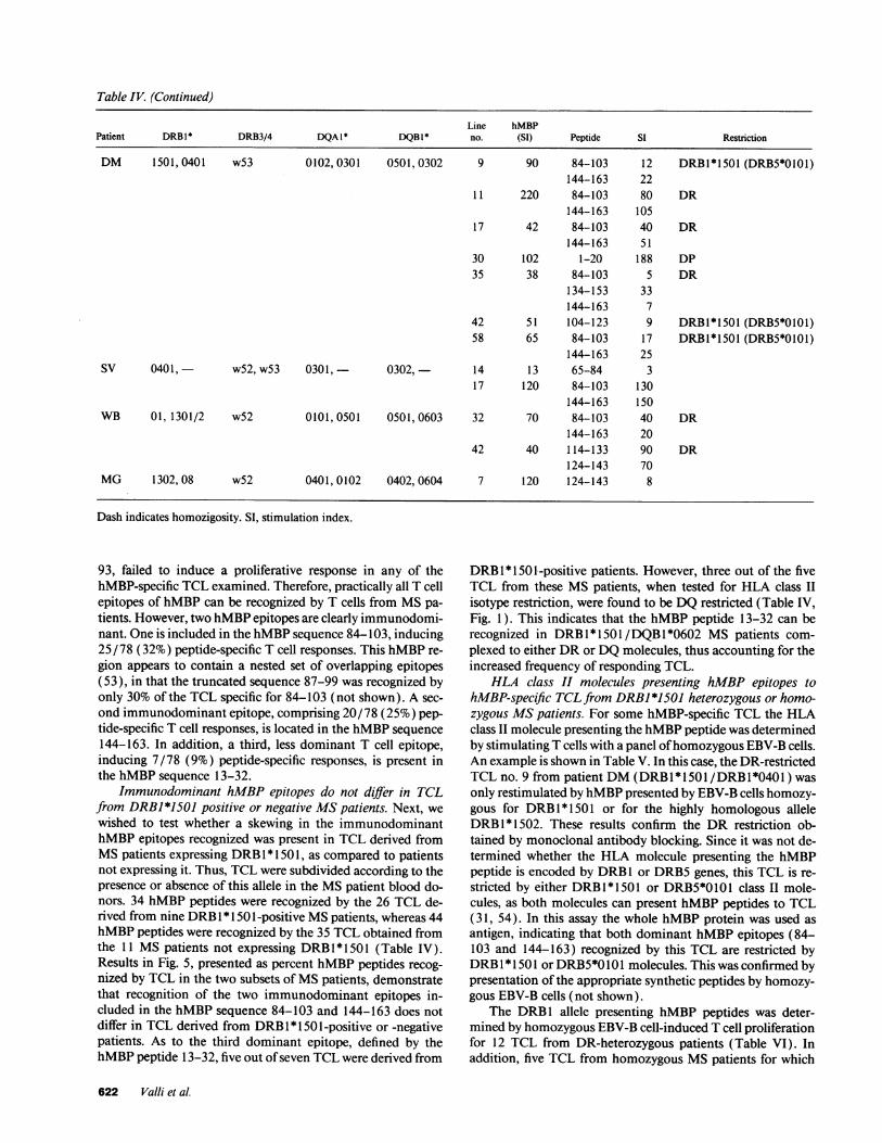
Table IV. (Continued)
Line hMBPPatient DRB1* DRB3/4 DQA1 * DQBI* no. (SI) Peptide SI Restriction
DM 1501, 0401 w53 0102, 0301 0501,0302 9 90 84-103 12 DRB1*1501 (DRB5*0101)144-163 22
11 220 84-103 80 DR144-163 105
17 42 84-103 40 DR144-163 51
30 102 1-20 188 DP35 38 84-103 5 DR
134-153 33144-163 7
42 51 104-123 9 DRB1*1501 (DRB5*0101)58 65 84-103 17 DRB1*1501 (DRB5*0101)
144-163 25SV 0401, w52, w53 0301,- 0302,- 14 13 65-84 3
17 120 84-103 130144-163 150
WB 01, 1301/2 w52 0101, 0501 0501,0603 32 70 84-103 40 DR144-163 20
42 40 114-133 90 DR124-143 70
MG 1302, 08 w52 0401, 0102 0402, 0604 7 120 124-143 8
Dash indicates homozigosity. SI, stimulation index.
93, failed to induce a proliferative response in any of thehMBP-specific TCL examined. Therefore, practically all T cellepitopes of hMBPcan be recognized by T cells from MSpa-tients. However, two hMBPepitopes are clearly immunodomi-nant. One is included in the hMBPsequence 84-103, inducing25/78 (32%) peptide-specific T cell responses. This hMBPre-gion appears to contain a nested set of overlapping epitopes(53), in that the truncated sequence 87-99 was recognized byonly 30% of the TCL specific for 84-103 (not shown). A sec-ond immunodominant epitope, comprising 20/78 (25%) pep-tide-specific T cell responses, is located in the hMBPsequence144-163. In addition, a third, less dominant T cell epitope,inducing 7/78 (9%) peptide-specific responses, is present inthe hMBPsequence 13-32.
Immunodominant hMBPepitopes do not differ in TCLfrom DRBI *1501 positive or negative MSpatients. Next, wewished to test whether a skewing in the immunodominanthMBPepitopes recognized was present in TCL derived fromMSpatients expressing DRB1 * 1501, as compared to patientsnot expressing it. Thus, TCL were subdivided according to thepresence or absence of this allele in the MSpatient blood do-nors. 34 hMBPpeptides were recognized by the 26 TCL de-rived from nine DRB1 * 1501 -positive MSpatients, whereas 44hMBPpeptides were recognized by the 35 TCL obtained fromthe 11 MSpatients not expressing DRB1 * 1501 (Table IV).Results in Fig. 5, presented as percent hMBPpeptides recog-nized by TCL in the two subsets of MSpatients, demonstratethat recognition of the two immunodominant epitopes in-cluded in the hMBPsequence 84-103 and 144-163 does notdiffer in TCL derived from DRB1 * 1501-positive or -negativepatients. As to the third dominant epitope, defined by thehMBPpeptide 13-32, five out of seven TCLwere derived from
DRB1 * 1501-positive patients. However, three out of the fiveTCL from these MSpatients, when tested for HLA class IIisotype restriction, were found to be DQrestricted (Table IV,Fig. 1). This indicates that the hMBPpeptide 13-32 can berecognized in DRB1 * 1501 /DQB 1 *0602 MS patients com-plexed to either DRor DQmolecules, thus accounting for theincreased frequency of responding TCL.
HLA class II molecules presenting hMBP epitopes tohMBP-specific TCLfrom DRBJ*1 501 heterozygous or homo-zygous MSpatients. For some hMBP-specific TCL the HLAclass II molecule presenting the hMBPpeptide was determinedby stimulating T cells with a panel of homozygous EBV-B cells.An example is shown in Table V. In this case, the DR-restrictedTCL no. 9 from patient DM(DRB I * 1501 /DRB 1 *0401) wasonly restimulated by hMBPpresented by EBV-B cells homozy-gous for DRB1 * 1501 or for the highly homologous alleleDRB1 * 1502. These results confirm the DR restriction ob-tained by monoclonal antibody blocking. Since it was not de-termined whether the HLA molecule presenting the hMBPpeptide is encoded by DRB1 or DRB5genes, this TCL is re-stricted by either DRB1 * 1501 or DRB5*0101 class II mole-cules, as both molecules can present hMBPpeptides to TCL(31, 54). In this assay the whole hMBPprotein was used asantigen, indicating that both dominant hMBPepitopes (84-103 and 144-163) recognized by this TCL are restricted byDRB1* 1501 or DRB5*0101 molecules. This was confirmed bypresentation of the appropriate synthetic peptides by homozy-gous EBV-B cells (not shown).
The DRB1 allele presenting hMBP peptides was deter-mined by homozygous EBV-B cell-induced T cell proliferationfor 12 TCL from DR-heterozygous patients (Table VI). Inaddition, five TCL from homozygous MSpatients for which
622 Valli et al.

cpm x 10^3
no ag
NA line 15
no ag ME
SJ line 1
cpmx 10^3
25 -
20 -
15 -
10-
5
0 _no ag
NA line 18
3VPX &Xnti-DR anti-DMlenMtant-DQ ani-MBP anti-DR anti-DO1 enti{)Q2 anti-DO3 entl{DP
anti-DOl antf-002 anti-D03 anti-DP
Figure 1. Characterization of HLAclass II isotypic molecules present-ing naturally processed hMBPpeptides to hMBP-specific TCL. TheHLA class II isotype restricting the anti-hMBP T cell response wasassessed by inhibition of T cell proliferation by anti-DR, -DQ, and-DP monoclonal antibodies specific for HLA class II monomorphicdeterminants, as detailed in Methods. A description of these TCL isincluded in Table IV.
class II isotype restriction by antibody blocking was available,are also included. These results demonstrate that the dominantepitopes 84-103 and 144-163 can be presented by differentclass II molecules, as expected from their degenerate binding
capacity (Tables I and II). In particular, the peptide 84-103can be presented to TCL by DRBl * 1302, DRB1 * 1101, andDRB1 * 1501 (or DRB5*0101) molecules. The peptide 144-163 can be presented by at least four DR molecules:DRB1*0101, DRBl*l 101, DRBl*1301, and DRB1*1501 (orDRB5*01 01), further demonstrating the functional relevanceof binding data.
These results highlight three points relevant to recognitionof hMBPepitopes by TCL from DRB1 * 1501 -positive MSpa-tients. First, APCfrom MSpatients carrying the DRB1 * 1501haplotype can present to TCL at least six different hMBPpep-tides. Second, these peptides are not all presented byDRBI*1501 (or DRB5*0101 ) molecules. Third, in some casesthe same peptide, e.g., 104-123, can be presented by either DRor DQmolecules.
Discussion
Based on experimental models of MS, hMBPis considered alikely candidate for the self antigen recognized by autoreactive,pathogenic human T cells capable of mediating MS(2, 55).
In the present study we have systematically analyzed thebinding of overlapping peptides encompassing the entirehMBPmolecule to several HLA class II molecules, and corre-lated their binding capacity to the recognition of hMBPpep-tides by HLA class II-restricted TCL. In particular, we havefocused on the binding of hMBPpeptides to HLAclass II mole-cules associated or not to increased susceptibility to MS, and onthe analysis of hMBP-specific TCL from MSpatients express-ing or not these susceptibility alleles. Although the emergingpattern is complex, some points appear to be quite clear.
Several hMBPpeptides bind with high affinity to one ortwo of the HLA class II molecules tested. In addition, threehMBPdeterminants, included in sequences 13-32, 84-103,and 144-163 exhibit degenerate binding, since they are able toform complexes with several of the HLA class II moleculesexamined. Degenerate binding of peptides to HLAclass II mol-ecules is not unprecedented because it has been described forpeptides derived from malaria circumsporozoite protein (56),tetanus toxoid (57), and influenza hemagglutinin (41, 58, 59).Analysis of a large unbiased sample of naturally occurring se-quences has revealed that degenerate binding is detectable onlyfor a minority of peptides capable of binding to HLA class IImolecules (39), and subsequent studies have illustrated thatdegenerate DRbinding is characteristic of peptides capable ofbinding with high affinity to at least one DRmolecule (59). Toour knowledge, the present study shows the first example ofdegenerate binding to HLA class II molecules of peptides de-rived from a candidate autoantigen possibly involved in a hu-man autoimmune disease.
A striking parallel exists between binding of hMBPpeptidesto HLA class II molecules and hMBPepitopes recognized byhMBP-specific TCL from MSpatients. First, the peripheral Tcell repertoire of MSpatients, as predicted from binding data,includes T cells able to recognize almost any hMBPpeptide.Second, the three hMBPpeptides capable of degenerate bind-ing (13-32, 84-103, and 144-163) correspond precisely to thethree immunodominant epitopes we have identified in hMBP-specific T cell lines from MSpatients. These results confirmprevious studies indicating the immunodominance of hMBPpeptides 84-103 and 144-163 (27, 29-32), and suggesting theexistence of a third T cell epitope in the amino-terminal region
Myelin Basic Protein Peptides Binding to HLA-DR Molecules 623
D=

ONNNCYl WIV mm C00 Coco0ONCOV 10 0 0
110_-CN1
01
W r-
, N t 0 0OD O-N 03 V 9
7 I
0.C0mC c
- - - --___ ~irCYiiCYC Iq Iq 0 I3V3 Timc mo
~~~~~~G Yc 00.V
10N-
0N
0T-
x
-E
o El0
.10
CO,6
-J
0
Cj
0
0
Q
COa
m2zr
00N
0to
0O
xW-O X
Ea0
-o10
0)
6
0C
J
-o0
Q
0
a
m
-C
Li-i -T--o-i-i -, -T- -i o- -T 1- .LTI0 e YCYNeNN c o oc o oC~C)'0 00O
coN N0 W
r- w 0 11-CN0 v0 0 f- aCOlNC'IIIIIN C','t1 Vt'--- 0- 0- 011- 116 eT* TTTT
mm~~~~~~~~~~~~~~~~~~~~~~~O~-Nov
*2 -N-0 Slb 1 0.0CON01I
V-T-v-T-4r-q-T
p~~qpJ.ww~qp~~qpii IONNNC)2st03C0000 00ff.0 CYNCY cm IVIV0R 03 coo-N cc0 CDCL
X ~~~~~---___fl aI IlI I II eO
'Im omo v(10(')Cr
I
O) -4
10~~~
0to o 3_
CO U
o < ; E
-
60 0
X o 0
.V-
- 1 CU
0
CU
a.
x ._
CD
_CY
OCU
0.
0~~~
A U)
C
oo
~ 4
0-
o_ ._ ._
L)
C_*_ C)
>e =0.
Zs m o
624 Valli et al.
IC)to
6c
z01c
0-o
0aC(IQ,co
1~
6c
0C>j
0-o
0a4-
-C
I
.L'Ir-ff-

cpm x 1O' 3
30- Figure 3. Dose-depen-dent T cell proliferation
25- to hMBPand to thehMBPpeptide 144-163.The TCL no. 10 from
20 patient BJ (5 x 10⁴ cellsper well) was cultured
15 - with the indicated con-centrations of hMBP
10- (e) or hMBPpeptide144-163 (+) in the
5- 1 / presence of mitomycinc-treated autologousEBV-transformed B
0- I cells(2 xl104perwell).0.018 0.05 0.17 0.5 1.5 Results are expressed
[antigen] pM as in Fig. 2.
of hMBP (30). In particular, the immunodominant hMBPepitopes corresponding to sequences 84-103 and 144-163were previously identified, using overlapping hMBPpeptides,by Ota et al. (29). Pette et al. (31) assigned at least three inde-pendent T cell epitopes within the thrombic hMBPfragment131-170, and defined a fourth one using the synthetic peptide80-99. Martin et al. (30) identified two immunodominant re-gions in the hMBPsequences 87-106 and 154-172. In addi-tion, Martin et al. (53) have shown that the dominant peptidecorresponding to the sequence 84-103 contains a nested set ofepitopes presented to T cells by different HLA class II mole-cules, thereby implying degenerate binding of this hMBPse-quence.
Immunodominance of T cell epitopes is certainly in-fluenced by many mechanisms (60), but the present data indi-cate that, at the population level, degeneracy of binding playsan important role in determining immunodominance of se-lected T cell epitopes. The immunodominant T cell epitopeincluded in the hMBPsequence 84-103 corresponds to the
hMBPpeptide
Ac-1-2013 - 3223 - 4233 - 5243 - 6255 - 7465-8474 - 93
84- 10394 - 113
104 -123114 - 133124- 143134 -153144- 163153-170-::
0 5 10 15 20 25 30 35
percent of TCL recognizing hMBPpeptides
Figure 4. hMBPepitopes recognized by hMBP-specific TCL from MSpatients. Results are expressed as percentage of TCL responding toa given hMBPpeptide (n = 78).
peptide expressing the most degenerate binding pattern, since itbinds to 8 out of the 10 DRmolecules tested. In particular, thispeptide binds with highest affinity, in the low nM range, toclass II molecules encoded by DRB1* 1501 and DRB1*0401alleles. Because these alleles are associated to increased fre-quency of MSin Northern ( 14) and Southern ( 15) Europeanpopulations, respectively, the results would suggest that highaffinity binding of this hMBPpeptide may be relevant to thepathogenesis of MS. Conversely, the other major immunodom-inant peptide, 144-163, binds very weakly to DRB1*1501 andDRB1*0401 molecules.
A point of debate is the binding affinity of autoantigenicpeptides for the class II molecules associated to disease suscepti-bility. According to one theory, autoreactive pathogenic T cellclones could recognize low affinity binders, and for this reasonhave escaped thymic deletion during negative selection (61 ).The opposite view considers autoantigenic peptides as high af-finity binders, and therefore highly antigenic. When eventssuch as molecular mimicry (62) or regulatory imbalances (63)lead to breakdown of tolerance, their antigenic potential can beexpressed. The hMBPpeptide centered on the sequence 84-103 is a high-affinity binder to the DRmolecules associatedwith increased frequency of MS. In analogy with binding ofMBPpeptides to mouse class II molecules (64), this resultwould suggest that high affinity binding may be necessary for aself peptide to become a dominant determinant recognized byautoreactive T cells.
Considering the binding data, and especially degeneracy ofbinding, it is therefore perhaps not surprising to find that thehMBPpeptides 84-103 and 144-163 represent dominant epi-topes recognized by about 30% of hMBP-specific TCL. In thatonly 84-103 binds with high affinity to DRmolecules asso-ciated with increased frequency of MS, it would be tempting toinfer that this epitope may be relevant to disease inductionand/or progression. However, both immunodominant hMBPepitopes are recognized with the same frequency by TCL fromDRB1*1501-positive or -negative MSpatients. Moreover, inthe three MS patients homozygous for DRB1*1501 manyhMBPpeptides are recognized by TCL in addition to 84-103,and in at least three of these TCL the response is restricted by
Table V. HLA Class II Restriction of TCL DMNo. 9
EBV-B Nocells DRB1* DRB3/4 antigen hMBP
cpm
Autologous 1501,0401 w53 3,557 31,593MGAR 1501, 1,410 27,165E4181324 1502, 582 16,281KASO1 1601, 351 575RML 1602, 3,155 2,340PRIESS 0401, w53 138 83PE117 0404, w53 255 159BSM 0401, w53 351 443MOU 0701, w53 1,140 577
The indicated EBV-transformed B cell lines, after treatment with mi-tomycin c, were pulsed with 30 ,g/ml hMBP, washed and incubated(2 X 10⁴ cells per well) with TCL no. 9 from patient DM. Results arepresented as in Fig. 2.
Myelin Basic Protein Peptides Binding to HLA-DR Molecules 625
A BhMBPpeptide
Ac - 1 - 2013 - 3223 - 4233 - 6243 - 6256 - 7465 - 8474 - 93
84 - 10394 - 113
104 - 123114 - 133124 - 143134 - 153144 - 163
163 - 170
0 10
hMBPpeptideAc - 1 - 20
13 - 3223 - 4233 - 5243 - 6256 - 7465 - 8474 - 93
84 - 10394 - 113
104 - 123114 - 133124 - 143134 - 153144 - 163153 - 170
20 30 40 0 10 20 30 40
Figure 5. hMBPepitopes recognized by hMBP-specific TCL from DRBI * 1501-positive or -negative MSpatients. Results are expressed as per-centage of TCL responding to a given hMBPpeptide. (A) Peptides (n = 34) recognized by TCL (n = 26) from DRB1 * 1501 -positive MSpatients(n = 9). (B) Peptides (n = 44) recognized by TCL (n = 35) from DRBl*1501-negative MSpatients (n = 1 1).
DQrather than DRmolecules. Furthermore, in MSpatientsheterozygous for DRB1 * 1501 hMBP-specific TCL can also berestricted by the other DRallele.
This complexity has probably multiple causes. First, mosthMBPpeptides bind to some HLAclass II molecules, account-ing for recognition, at the population level, of practically allhMBPepitopes by hMBP-specific TCL. In this respect, hMBPmay be considered as a continuum of T cell epitopes, making itdifficult to identify those who may be associated with the acti-vation of pathogenic autoreactive T cells. This problem is em-
phasized by the observation that multiple hMBPepitopes are
recognized by hMBP-specific TCL derived from either normal
donors or MSpatients (26-32), presumably reflecting lack ofthymic negative selection by this sequestered antigen.
Second, at least three hMBPpeptides exhibit degeneratebinding, accounting for the immunodominance of T cell epi-topes corresponding to these sequences. This is likely to be amajor reason for the blurred relationship between hMBPepi-topes recognized by TCL and HLA class II molecules restrict-ing the response. Thus, even if the hMBPpeptide centered on
the sequence 84-103 binds with highest affinity toDRBI * 1501, it is equally well recognized by TCL fromDRB1 * 1501-positive or -negative MS patients. This wouldsuggest that if hMBPis the autoantigen in MSand 84-103 a
Table VI. HLA Class II Molecules Presenting hMBPEpitopes to hMBP-specific TCL
hMBPPatient DRBI* DQAI* DQBI* Line no. peptide Restriction
BV 1302, 07 0102, 0201 0604,0201 11 84-103 DRB1*1302BJ 0101, 0401 0101, 0301 0501, 0302 10 144-163 DRB1*0101
153-170LA 0401, 1101 0301,0501 0302,0301 8 84-103 DRB1*1101
144-16321 84-103 DRB1*1 101
SI 1101, 10 0101, 0501 0501, 0301 2 33-52 DRB1*11014 84-103 DRB1*I 101
SP 1501, 1301 0103, 0102 0602, 9 144-163 DRB1*130110 144-163 DRB1*130138 33-52 DRBI*1501 (DRB5*0101)
DM 1501,0401 0102,0301 0501,0302 9 84-103 DRB1*1501 (DRB5*0101)144-163
42 104-123 DRB1*1501 (DRB5*0101)58 84-103 DRB1*1501 (DRB5*0101)
144-163SJ 1501,- 0102, 0602, 1 13-32 DQA1*0102/DQB 1 *0602
28 104-123 DQA1*0102/DQB1*0602TB 1501,- 0102,- 0602,- 2 13-32 DQAI*0102/DQB1*0602
7 84-103 DRB1*1501 (DRB5*0101)9 84-103 DRBI*1501 (DRB5*0101)
626 Valli et al.
- l - ,
-1I I - - I
I - I -I I

relevant epitope, the real susceptibility gene(s), although insignificant linkage disequilibrium with DRBl*1501, lie else-where in the HLA region. In this respect, interesting candidatesfor disease susceptibility genes could be represented by the poly-morphic genes encoding proteins associated to peptide trans-port (Tap- 1, Tap-2) and to the proteasome (Lmp-2, Lmp-7),since polymorphism in Lmp genes may result in the produc-tion of different peptides in different individuals, and polymor-phism in Tap genes may influence peptide loading of HLAmolecules (65).
Third, our evaluation of hMBP epitopes recognized byTCL from MSpatients was conducted, as in any study of thistype, on PBLand therefore the T cells infiltrating the inflamma-tion site in the CNSwere not directly assessed.
Fourth, MSwas already clinically evident in all the patientstested at least 1 yr before establishment of hMBP-specific TCL,and it is expected that in the course of autoimmune diseases Tcells not involved in disease induction may also become re-cruited (66). This obviously also applies to nonpathogenic Tcells specific for hMBPepitopes.
In conclusion, the fact that hMBP84-103 binds preferen-tially to the HLA class II molecules encoded by alleles asso-ciated to increased MSfrequency could be consistent with itsproposed role in the pathogenesis of MS(29, 67). Because theother major dominant peptide, 144-163, binds very weakly toDRmolecules encoded by MSsusceptibility alleles, we favorthe hypothesis that this epitope is not involved in the pathogen-esis of MS(29). The finding that peptide 84-103 also binds toseveral DRmolecules not associated with disease may explainwhy many MSpatients do not carry the DRB1*1501 suscepti-bility allele. In fact, recognition of hMBPpeptides by TCLfrom MSpatients does not appear to be directly linked to theputative HLA susceptibility alleles, because very similar hMBPimmunodominant epitopes are recognized by TCL fromDRB1 * 150 1-positive or -negative MSpatients. It would be in-teresting to know whether selective immunotherapies of estab-lished MSbased on hMBPas autoantigen (2) may influencethe course of disease, although, considering all this complexity,the approach may prove more problematic than originally an-ticipated.
Acknowledgments
Wethank Dr. R. W. Karr for transfected fibroblasts; Dr. R. S. Accollafor monoclonal antibodies; Prof. J. Ulrich for human brain specimens;C. Linington, P. Hiestand, and H. Zihlmann for hMBPpurification;and A. Schoenberger, M. Baschonga, G. Zuercher, and M. Weber fortechnical help. The authors thank all MSpatients participating in thisstudy. Dr. Kappos and Miescher are supported by the Swiss MultipleSclerosis Society.
References
1. Allen, 1. V. 1991. Pathology of Multiple Sclerosis. In McAlpine's MultipleSclerosis. W. B. Matthews, E. D. Acheson, J. R. Batchelor, and R. 0. Weller,editors. Churchill Livingstone, Edinburgh. 341-387.
2. Wucherpfennig, K. W., H. L. Weiner, and D. A. Hafler. 1991. T-cell recog-nition of myelin basic protein. Immunol. Today. 12:277-282.
3. Allegretta, M., J. A. Nicklas, S. Sriram, and R. J. Albertini. 1990. T cellsresponsive to myelin basic protein in patients with multiple sclerosis. Science(Wash. DC). 247:718-721.
4. McFarlin, D. E., S. E. Blank, R. F. Kibler, S. McKneally, and R. Shapira.1973. Experimental allergic encephalomyelitis in the rat: response to encephalito-genic proteins and peptides. Science (Wash. DC) . 179:478-480.
5. Paterson, P. Y., and R. H. Swanborg. 1988. Demyelinating diseases of thecentral and peripheral nervous systems. In Immunological Diseases. M. Samter,D. J. Talmage, M. M. Frank, K. F. Austen, and H. N. Claman, editors. LittleBrown & Co., Boston. 1877-1915.
6. Pettinelli, C. B., and D. E. McFarlin. 1981. Adoptive transfer of experimen-tal allergic encephalomyelitis in SJL/J mice after in vitro activation of lymphnode cells by myelin basic protein: requirement for Lyt +2- lymphocytes. J. Im-munol. 127:1420-1423.
7. Ben-Nun, A., H. Wekerle, and I. R. Cohen. 1981. The rapid isolation ofclonable antigen-specific T lymphocyte lines capable of mediating autoimmuneencephalitis. Eur. J. Immunol. 11:195-199.
8. Zamvil, S. S., P. Nelson, J. Trotter, D. Mitchell, R. Knobler, R. Fritz, and L.Steinman. 1985. T-cell clones specific for myelin basic protein induce chronicrelapsing paralysis and demyelination. Nature (Lond.). 317:355-358.
9. Zamvil, S. S., D. J. Mitchell, A. C. Moore, K. Kitamura, L. Steinman, andJ. B. Rothbard. 1986. T cell epitope of the autoantigen myelin basic protein thatinduces encephalomyelitis. Nature (Lond.). 324:258-260.
10. Acha-Orbea, H., L. Steinman, and H. 0. McDevitt. 1989. T cell receptorsin murine autoimmune diseases. Annu. Rev. Immunol. 7:371-405.
11. Tuohy, V. K., Z. Lu, R. A. Sobel, R. A. Laursen, and M. B. Lees. 1989.Identification of an encephalitogenic determinant of myelin proteolipid proteinfor SJL mice. J. Immunol. 142:1523-1528.
12. Ebers, G. C., D. E. Bulman, A. D. Sadovnick, D. W. Paty, S. Warren, W.Hader, T. J. Murray, P. Seland, P. Duquette, T. Grey, et al. 1986. A population-based study of multiple sclerosis in twins. N. Engl. J. Med. 315:1638-1641.
13. Tiwari, J. L., and P. I. Terasaki. 1985. HLA and Disease Associations.Springer-Verlag, Inc., NewYork. 152 pp.
14. Francis, D. A., A. J. Thompson, P. Brookes, N. Davey, R. I. Lechler, W. I.McDonald, and J. R. Batchelor. 1991. Multiple sclerosis and HLA: is the suscepti-bility gene really HLA-DR or -DQ? Hum. Immunol. 32:119-124.
15. Marrosu, M. G., F. Muntoni, M. R. Murru, G. Spinicci, M. P. Pischedda,F. Goddi, P. Cassu, and M. Pirastu. 1988. Sardinian multiple sclerosis is asso-ciated with HLA-DR4: a serologic and molecular analysis. Neurology. 348:1749-1753.
16. Yaqub, B. A., and A. K. Daif. 1988. Multiple sclerosis in Saudi Arabia.Neurology. 328:621-625.
17. Naito, S., Y. Kuroiwa, T. Itoyama, T. Tsubachi, A. Horikawa, T. Sazasu-chi, S. Noguchi, S. Othsuchi, H. Tokuomi, T. Miyatake, et al. 1978. HLA andJapanese MS. Tissue Antigens. 12:19-24.
18. Gorodezky, C., R. Najera, B. E. Rangel, L. E. Castro, J. Flores, G. Velas-quez, J. Granados, and J. Sotelo. 1986. Immunogenetic profile of multiple sclero-sis in Mexicans. Hum. Immunol. 16:364-374.
19. Sherritt, M. A., J. Oskenberg, N. Kerlero de Rosbo, and C. C. A. Bernard.1991. Influence of HLA-DR2, HLA-DPw4, and T cell receptor a chain genes onthe susceptibility to multiple sclerosis. Int. Immunol. 4:177-181.
20. Hillert, J., C. Leng, and 0. Olerup. 1992. T cell receptor chain germlinegene polymorphism in multiple sclerosis. Neurology. 42:80-84.
21. Oskenberg, J. R., S. Stuart, A. Begovich, R. B. Bell, H. A. Erlich, L.Steinman, and C. C. A. Bernard. 1990. Limited heterogeneity of rearranged T cellreceptor Va transcripts in brains of multiple sclerosis patients. Nature (Lond.).345:344-346.
22. Wucherpfennig, K. W., K. Ota, N. Endo, J. G. Seidman, A. Rosenzweig,H. L. Weiner, and D. A. Hafler. 1990. Shared human T cell receptor Vj# usage toimmunodominant regions of myelin basic protein. Science (Wash. DC).248: 1016-1019.
23. Ben-Nun, A., R. S. Liblau, L. Cohen, D. Lehmann, E. Tournier-Lasserve,A. Rosenzweig, Z. Jingwu, J. C. M. Raus, and M.-A. Bach. 1991. Restricted T cellreceptor V, gene usage by myelin basic protein-specific T cell clones in multiplesclerosis: predominant genes vary in individuals. Proc. Natl. Acad. Sci. USA.88:2466-2470.
24. Kotzin, B. L., S. Karuturi, Y. K. Chou, J. Lafferty, J. M. Forrester, M.Better, G. E. Nedwin, H. Offner, and A. A. Vandenbark. 1991. Preferential T cellreceptor a-chain variable gene use in myelin basic protein-reactive T cell clonesfrom patients with multiple sclerosis. Proc. Natl. Acad. Sci. USA. 88:9161-9165.
25. Giegerich, G., M. Pette, E. Meinl, J. T. Epplen, H. Wekerle, and A.Hinkkanen. 1992. Diversity of T cell receptor a and , chain genes expressed byhuman T cells specific for similar myelin basic protein peptide/major histocom-patibility complexes. Eur. J. Immunol. 22:753-758.
26. Richert, J., C. A. Reuben-Burnside, G. E. Deibler, and M. W. Kies. 1988.Peptide specificities of myelin basic protein-reactive human T cell clones. Neurol-ogy. 38:739-745.
27. Baxevanis, C. N., G. J. Reclos, C. Servis, E. Anastasopulos, P. Arsenis, A.Katsiyannis, N. Matikas, J. D. Lambris, and M. Papamichail. 1989. Peptidesfrom myelin basic protein stimulate T lymphocytes from patients with multiplesclerosis. J. Neuroimmunol. 22:23-30.
28. Chou, Y. K., M. Vainiene, R. Whithman, D. Bourdette, C. H.-J. Chou, G.Hashim, H. Offner, and A. A. Vandenbark. 1989. Response of human T lympho-cyte lines to myelin basic protein: Association of dominant epitopes with HLAclass II restriction molecules. J. Neurosci. Res. 23:207-216.
29. Ota, K., M. Matsui, E. L. Milford, G. A. Mackin, H. L. Weiner, and D. A.
Myelin Basic Protein Peptides Binding to HLA-DR Molecules 627

Hailer. 1990. T cell recognition of an immunodominant myelin basic proteinepitope in multiple sclerosis. Nature (Lond.). 346:183-187.
30. Martin, R., D. Jaraquemada, M. Flerlage, J. Richert, J. Whitaker, E. 0.Long, D. E. McFarlin, and H. F. McFarland. 1990. Fine specificity and HLArestriction of myelin basic protein-specific cytotoxic T cell lines from multiplesclerosis patients and healthy individuals. J. Immunol. 145:540-548.
31. Pette, M., K. Fujita, D. Wilkinson, D. M. Altmann, J. Trowsdale, G.Giegerich, A. Hinkkanen,J. T. Epplen, L. Kappos, and H. Wekerle. 1990. Myelinautoreactivity in multiple sclerosis: recognition of myelin basic protein in thecontext of HLA-DR2 products by T lymphocytes of multiple sclerosis patientsand healthy donors. Proc. Natl. Acad. Sci. USA. 87:7968-7972.
32. Liblau, R., E. Tournier-Lasserve, J. Maciazek, G. Dumas, 0. Siffert, G.Hashim, and M.-A. Bach. 1991. T cell response to myelin basic protein epitopesin multiple sclerosis patients and healthy subjects. Eur. J. Immunol. 21:1391-1395.
33. Eylar, E. H., P. J. Knisknern, and J. J. Jackson. 1979. Myelin basic pro-teins. Methods Enzymol. 32B:323-354.
34. Bodmer, J. C., S. G. E. Marsh, E. D. Albert, W. F. Bodmer, B. Dupont,H. A. Herlich, B. Mach, W. R. Mayr, P. Parham, T. Sasazuki, etal. 1991. Nomen-clature for factors of the HLA system, 1990. Hum. Immunol. 31:186-194.
35. Gorga, J. C., V. Horejsi, D. R. Johnson, R. Raghupathy, and J. L. Stro-minger. 1987. Purification and characterization of class II histocompatibility an-tigens from a homozygous human B cell line. J. Biol. Chem. 262:16087-1609 1.
36. O'Sullivan, D., J. Sidney, M.-F. Del Guercio, S. M. Colon, and A. Sette.1991. Truncation analysis of several DR binding epitopes. J. Immunol.146:1240-1246.
37. Gorga, J. C., P. J. Kudsen, J. A. Foran, J. L. Strominger, and S. J. Bura-koff. 1986. Immunochemically purified DRantigens in liposomes stimulate xen-ogenic cytolytic T cells in secondary in vitro cultures. Cell. Immunol. 103:160-173.
38. Sette, A., S. Southwood, D. O'Sullivan, F. C. A. Gaeta, J. Sidney, andH. M. Grey. 1992. Effect of pH on MHCclass II-peptide interactions. J. Im-munol. 148:844-851.
39. O'Sullivan, D., J. Sidney, E. Appella, L. Walker, L. Phillips, S. M. Colon,C. Miles, R. W. Chesnut, and A. Sette. 1990. Characterization of the specificity ofpeptide binding to four DRhaplotypes. J. Immunol. 145:1799-1808.
40. Sette, A., J. Sidney, M. Albertson, C. Miles, S. M. Colon, T. Pedrazzini,A. G. Lamont, and H. M. Grey. 1990. A novel approach to the generation of highaffinity class II binding peptides. J. Immunol. 145:1809-1813.
41. Krieger, J. I., R. W. Karr, H. M. Grey, W.-Y. Yu, D. O'Sullivan, L.Batovsky, Z.-L. Zheng, S. M. Colon, F. C. A. Gaeta, J. Sidney, et al. 1991. Singleamino acid changes in DRand antigen define residues critical for peptide-MHCbinding and T cell recognition. J. Immunol. 146:2331-2339.
42. Schumacher, G. A., G. W. Beebe, R. F. Kibler, L. T. Kurland, J. F.Kurtzke, F. McDowell, B. Nagler, W. A. Sibley, W. W. Tourtellotte, and T. L.Welmon. 1965. Problems of experimental trials in therapy of multiple sclerosis:report by the panel on the evaluation of experimental trials of therapy in multiplesclerosis. Ann. N. Y. Acad. Sci. 122:552-562.
43. Kurtzke, J. F. 1983. Rating neurologic impairment in multiple sclerosis:an expanded disability status scale (EDSS). Neurology. 33:1444-1452.
44. McDonald, W. I., and D. Barnes. 1989. Lessons from magnetic resonanceimaging in multiple sclerosis. Trends Neurosci. 12:376-386.
45. Kappos, L., R. Gold, E. Hofmann, W. Keil, and W. Clauss. 1990. Multiplesclerosis: diagnostic criteria and the role of contrast-enhanced MRI. In ContrastMedia in MRI. G. Bydder, R. Felix, E. Buecheler, B. P. Drayer, H. P. Niendorf,M. Takahashi, and K.-J. Wolf, editors. Medicom Busum. 127-132.
46. Miller, D. H., F. Barkoff, J. Berry, L. Kappos, G. Scotti, and A. J. Thomp-son. 1991. Magnetic resonance imaging in monitoring the treatment of multiplesclerosis: Commission of European Communities (CEC) guidelines. J. Neurol.Neurosurg. Psychiatry. 54:638-644.
47. Grossman, R. I., F. Gonzales-Scarano, S. W. Atlas, S. Galetta, and D. H.Silberberg. 1986. Multiple sclerosis: gadolinium enhancement in MRimaging.Radiology. 161:721-726.
48. Sasazuki, T., editor. HLA-1991. Oxford University Press, Oxford, UK. Inpress.
49. Nevinny-Stickel, C., M. Hinzpeter, A. Andreas, and E. D. Albert. 1991.Non-radioactive oligotyping for HLA-DRI -DRw10 using polymerase chain reac-tion, digoxygenin-labelled oligonucleotides and chemiluminescence detection.Eur. J. Immunogenet. 18:323-329.
50. Accolla, R. S., N. Gross, S. Carrel, and G. Corte. 1981. Distinct forms ofboth a and P subunits are present in the human Ia molecular pool. Proc. Nati.Acad. Sci. USA. 78:4549-4553.
51. Corte, G., F. Calabi, G. Damiani, A. Bargellesi, R. Tosi, and R. Sorren-tino. 1981. Human Ia molecules carrying DCl determinants differ in both a andP subunits from Ia molecules carrying DR determinants. Nature (Lond.).292:357-359.
52. Watson, A. J., R. DeMars, 1. S. Trowbridge, and F. H. Bach. 1983. Detec-tion of a novel human class II HLA antigen. Nature (Lond.). 304:358-360.
53. Martin, R., U. Utz, J. E. Coligan, J. R. Richert, M. Flerlage, E. Robinson,R. Stone, W. E. Biddison, D. E. McFarlin, and H. F. McFarland. 1992. Diversityin fine specificity and T cell receptor usage of the human CD4' cytotoxic T cellresponse specific for the immunodominant myelin basic protein peptide 87-106.J. Immunol. 148:1359-1366.
54. Jaraquemada, D., R. Martin, S. Rosen-Bronson, M. Flerlage, H. F.McFarland, and E. 0. Long. 1990. HLA-DR2a is the dominant restriction mole-cule for the cytotoxic T cell response to myelin basic protein in DR2Dw2individ-uals. J. Immunol. 145:2880-2885.
55. Alvord, E. C. 1984. Is myelin basic protein the right antigen and experi-mental allergic encephalomyelitis the right model for multiple sclerosis? In Experi-mental Allergic Encephalomyelitis: A Useful Model for Multiple Sclerosis. E. C.Alvord, M. W. Kies, and A. J. Suckling, editors. Alan R. Liss, Inc., NewYork.503-508.
56. Sinigaglia, F., M. Guttinger, J. Kilgus, D. M. Doran, H. Matile, H. Et-linger, A. Treciak, D. Gillessen, and J. R. L. Pink. 1988. A malaria T cell epitoperecognized in association with most mouse and human MHCclass II molecules.Nature (Lond.). 336:778-780.
57. Panina-Bordignon, P., A. Tan, A. Termijtelen, S. Demotz, G. Corradin,and A. Lanzavecchia. 1989. Universally immunogenic T cell epitopes: promiscu-ous binding to human MHCclass II and promiscuous recognition by T cells. Eur.J. Immunol. 19:2237-2242.
58. Busch, R., G. Strang, K. Howland, and J. B. Rothbard. 1990. Degeneratebinding of immunogenic peptides to HLA-DR proteins on B cell surfaces. Int.Immunol. 19:2237-2245.
59. O'Sullivan, D., T. Arrhenius, J. Sidney, M.-F. Del Guercio, M. Albertson,M. Wall, C. Oseroff, S. Southwood, S. M. Colon, F. C. A. Gaeta, et al. 1991. Onthe interaction of promiscous antigenic peptides with different DRalleles. Identi-fication of common structural motifs. J. Immunol. 147:2663-2669.
60. Adorini, L., E. Appella, G. Doria, and Z. Nagy. 1988. Mechanisms in-fluencing the immunodominance of T cell determinants. J. Exp. Med.168:2091-2104.
61. Gammon,G., and E. E. Sercarz. 1989. Howsome T cells escape toleranceinduction. Nature (Lond.). 342:183-185.
62. Oldstone, M. B. 1989. Molecular mimicry as a mechanism for the causeand a probe uncovering etiologic agent(s) of autoimmune diseases. Curr. Top.Microbiol. Immunol. 145:127-139.
63. Nepom, G. T. 1991. MHCclass II molecules and autoimmunity. Annu.Rev. Immunol. 9:493-520.
64. Wall, M., S. Southwood, J. Sidney, C. Oseroff, M.-F. Del Guercio, A.Lamont, S. M. Colon, T. Arrhenius, F. C. A. Gaeta, and A. Sette. 1992. Highaffinity for class II molecules as a necessary but not sufficient characteristic ofencephalitogenic determinants. Int. Immunol. 4:773-777.
65. Monaco, J. J. 1992. A molecular model of MHCclass I-restricted antigenprocessing. Immunol. Today. 13:173-178.
66. Sinha, A. A., M. T. Lopez, and H. 0. McDevitt. 1990. Autoimmunediseases: The failure of self tolerance. Science (Wash. DC). 248:1380-1388.
67. Martin, R., M. D. Howell, D. Jaraquemada, M. Flerlage, J. Richert, S.Brostoff, E. 0. Long, D. E. McFarlin, and H. F. McFarland. 1991. A myelin basicprotein peptide is recognized by cytotoxic T cells in the context of four HLA-DRtypes associated with multiple sclerosis. J. Exp. Med. 248:19-24.
628 Valli et al.


![Applied NWP [1.2] “…up until the 1960s, Richardson’s model initialization problem was circumvented by using a modified set of the primitive equations…”](https://img.pdfslide.us/doc/110x75/5a4d1b5e7f8b9ab0599ac239/applied-nwp-12-up-until-the-1960s-richardsons-model-initialization.jpg)
![Force Sensor UNIT HSFPAR303A Data sheet - alps.com€¦ · Drbl Lin [%FS] Operating temperature](https://img.pdfslide.us/doc/110x75/5bac4a7e09d3f279368d652d/force-sensor-unit-hsfpar303a-data-sheet-alpscom-drbl-lin-fs-operating.jpg)