Embed Size (px)
Citation preview
Univers
ity of
Cap
e Tow
nLindsey Nicola Levin
LVNLIN001
SUBMITTED TO THE UNIVERSITY OF CAPE TOWN
In fulfillment of the requirements for the degree
Master of Medicine (MMed) Paediatrics
Faculty of Health Sciences
UNIVERSITY OF CAPE TOWN
Date of submission: 14 June 2016
Supervisors:
Dr. Elizabeth Goddard, Dr. Ronalda De Lacy, Dr. Komala Pillay
Correspondence: [email protected]
Biliary Atresia at Red Cross War Memorial
Children’s Hospital: A retrospective descriptive
study reviewing the age of presentation, clinical
course and outcome of infants presenting to
RCWMCH with biliary atresia.

The copyright of this thesis vests in the author. No quotation from it or information derived from it is to be published without full acknowledgement of the source. The thesis is to be used for private study or non-commercial research purposes only.
Published by the University of Cape Town (UCT) in terms of the non-exclusive license granted to UCT by the author.
Univers
ity of
Cap
e Tow
n

ii
1. DECLARATION .............................................................................................................................. 1
2. RESEARCH PROPOSAL: BILIARY ATRESIA AT RED CROSS WAR MEMORIAL CHILDREN’S HOSPITAL ................................................................................................................... 2
INVESTIGATORS ........................................................................................................................... 2
BACKGROUND............................................................................................................................... 2
AIM .................................................................................................................................................. 4
OBJECTIVES .................................................................................................................................. 4
METHODOLOGY ............................................................................................................................ 5
ETHICAL CONSIDERATIONS ........................................................................................................ 7
REFERENCES ................................................................................................................................ 9
3. LITERATURE REVIEW ................................................................................................................ 10
METHODS .................................................................................................................................... 10
LITERATURE ................................................................................................................................ 11
REFERENCES .............................................................................................................................. 22
4. MANUSCRIPT IN PUBLICATION READY FORMAT .................................................................. 26
TITLE PAGE.................................................................................................................................. 26
ABSTRACT ................................................................................................................................... 27
INTRODUCTION ........................................................................................................................... 28
METHODS .................................................................................................................................... 29
RESULTS ...................................................................................................................................... 31
DISCUSSION ................................................................................................................................ 34
REFERENCES .............................................................................................................................. 40
5. APPENDICES ............................................................................................................................... 46
ETHICAL APPROVAL ................................................................................................................... 46
DATA CAPTURE SHEETS ........................................................................................................... 47
HISTOLOGICAL SCORING SYSTEM FOR NEONATAL CHOLESTASIS .................................. 50
BILIARY ATRESIA PATHOLOGICAL IMAGES ............................................................................ 51
JOURNAL OF PEDIATRIC GASTROENTEROLOGY AND NUTRITION: INSTRUCTIONS FOR THE AUTHOR ............................................................................................................................... 60
ACKNOWLEDGEMENTS ................................................................................................................. 75

1
1. DECLARATION
I, Dr. Lindsey Nicola Levin, hereby declare that the work on which this mini dissertation/thesis is
based is my original work (except where acknowledgements indicate otherwise) and that neither the
whole work nor any part thereof has been, is being or is to be submitted for another degree in this or
any other university.
I empower the university to reproduce for the purposes of research either the whole or any portion of
the contents in any manner whatsoever.
Signed:
Date: 14 June 2016
2
2. RESEARCH PROPOSAL: BILIARY ATRESIA AT RED CROSS WAR MEMORIAL CHILDREN’S HOSPITAL
Biliary Atresia at Red Cross War Memorial Children’s Hospital: A retrospective descriptive
study reviewing the age of presentation, clinical course and outcome of infants presenting to
RCWMCH with biliary atresia.
INVESTIGATORS
Lindsey Levin (LVNLIN001), Elizabeth Goddard (Supervisor), Ronalda De Lacy (Co-supervisor),
Komala Pillay (Co-supervisor, histology aspect)
BACKGROUND
Biliary atresia is a progressive obstructive cholangiopathy of unknown aetiology, occurring during the
perinatal period. If left untreated it progresses to liver fibrosis and cirrhosis in the first few months of
life, with death occurring in the first few years (1). It is the leading cause of end-stage liver disease in
the paediatric population and remains the most common indication for liver transplantation in children
(2). The current surgical management of biliary atresia involves hepatic portoenterostomy (Kasai
procedure) to re-establish bile drainage, thus delaying the progression of fibrosis, with subsequent
liver transplantation still required in many cases (3).
The Kasai procedure was introduced in Japan in 1959 (3). The procedure aims to construct a new bile
drainage system and if successful, increases survival and postpones liver transplantation. However,
long term survivors are prone to develop complications, most commonly cholangitis, or liver cirrhosis
leading to portal hypertension and oesophageal varices. Successful Kasai (re-establishment of bile
flow) is much more likely if performed within the first 8 weeks of life, making early referral and prompt
evaluation of suspected cases essential (4). A Canadian series found that patients receiving initial sub
speciality care after 90 days of age had worse outcomes following Kasai operation (2).
The prevalence of biliary atresia is estimated as 1 in 20 000 live births (3). Since the global
acceptance and utilisation of the Kasai procedure, many international studies have looked at the

3
outcomes and long term survival of post Kasai patients. Most of these studies have been conducted
across Europe and North America, with very little research available in developing countries.
A French national series reported the outcomes of Western countries to show short-term clearance of
jaundice in 50-60% of cases, with 30-40% of patients reaching 10 years with their native livers and a
third of patients reaching 20 years of age (1).
A Canadian series conducted from January 1, 1985 to December 31, 2002 found 4- and 10-year
survival rates of 77% and 75% respectively (2). The French national survey from 2003 to 2009
reported a 5-year overall survival of 89% (1). The Japanese registry for 1989 to 1994 documented
73.5% patient survival at 5 years (5). A study conducted across the United Kingdom and Ireland from
1993 to 1995 revealed an overall survival rate of 85% at 5 years (6).
Delayed presentation and late referral remain a major problem in the management of biliary atresia
globally. Efforts aimed at promoting earlier referral through the creation of biliary atresia awareness at
the primary health care level may play an important role in improving outcomes and survival post
Kasai surgery. The United Kingdom has instituted a “yellow alert” educational programme aimed at
expediting the referral of jaundiced infants. In Asia, a stool colour card is given to all mothers on
discharge from postnatal services (2).
A report of the experience in Southern Nigeria showed that late presentation and lack of resources
pose major problems in the management of biliary atresia with only 62.5% of patients undergoing
Kasai procedure with a mean age of death at 14.2 months (7).
A previous study conducted in the surgical department at the Red Cross War Memorial Children’s
Hospital (RCWMCH) reviewed 39 children undergoing Kasai procedure between January 1975 and
January 1985 (8). The mean age of operation was 12.8 weeks. In the first 4 years of the study no
patients established successful bile drainage. Thereafter 50% of Kasai procedures were successful in
establishing bile drainage, however only 20% of these patients remained alive at the time of analysis.

4
These results are far below those seen in international studies. More recent statistics are not available
at the RCWMCH. We are thus unable to compare our outcomes with current international experience.
Biliary atresia places a significant burden on hepatobiliary services. A review of liver transplantation at
The Wits Donald Gordon Medical Centre in Johannesburg, South Africa reported biliary atresia as the
most common indication for liver transplantation in paediatrics (9) and it accounts of 57% of liver
transplants performed at RCWMCH (10, 11).
The purpose of this research is to review the current experience of biliary atresia at RCWMCH. Focus
will be placed on the age of presentation, age at intervention, course of progress and outcomes of
patients following Kasai operation. It is hoped that we will see a reduction in the mean age of surgery
and improvement in the success of Kasai procedures since the earlier study. An awareness of the
current management practices and outcomes within our referral system may identify areas of need in
order to facilitate earlier referral and investigation of vulnerable patients.
AIM
To review the age of presentation to hospital, course and outcome of children presenting to or
referred to Red Cross War Memorial Children’s Hospital with biliary atresia.
OBJECTIVES
1. To document the mean age of referral/presentation to RCWMCH in to order to ascertain
whether patients accessing primary health care facilities or general practitioners are being
referred to tertiary services timeously.
2. To ascertain the mean age of Kasai (portoenterostomy) once within the RCWMCH system.
3. To establish and document the outcomes following Kasai procedures at RCWMCH.
4. To establish and document whether the age at Kasai procedure influences outcome of
patients at RCWMCH.
5. To identify prognostic factors associated with poor outcome after Kasai procedure.
5
Outcomes
Outcomes will be assessed by looking at complications following Kasai and current survival rates.
Complications looked at will include episodes of cholangitis, varices, ascites and bacterial
peritonitis. Successful Kasai will be defined as complete clearance of jaundice with serum bilirubin
< 20µmol/L within 3 months of Kasai procedure, as well as the presence of pigmented stools.
Survival will be expressed as 2 year and 5 year survival rates, including overall survival, survival
with native liver and survival post-transplant.
METHODOLOGY
1. STUDY DESIGN
Retrospective folder review
2. STUDY POPULATION
Patients managed for biliary atresia at RCWMCH during the period 2003-2013. This is
estimated to be approximately 75-100 cases.
Inclusion Criteria:
1. All patients in whom a diagnosis of biliary atresia has been made by clinical, biochemical
and radiological grounds with surgical findings and liver histology consistent with biliary
atresia
2. Children with confirmed biliary atresia who did not undergo Kasai operation will be
included in the study
Exclusion Criteria:
1. No records available
2. Other causes of neonatal cholestasis

6
3. DATA
Cases will be identified by a Clinicom search using the ICD 10 code for biliary atresia (Q44.2)
within the defined study period. The Gastro-Intestinal Unit departmental database of liver
biopsies and histology, as well as the RCWMCH transplantation register will be consulted.
Medical records of identified patients will be retrieved for data collection.
Data will be collected from patient files by the primary investigator only. A numerical identifier
will be allocated to each case. Data collected will be captured on appropriate data collection
sheets (Appendix 1), transcribed for digital storage and analysed using Microsoft excel.
Data will include the following:
Demographics: Date of birth, gender, race, age at first presentation to Red Cross, source of
referral and number of visits to primary care before referral.
Presenting features: reason for presentation, presence or absence of jaundice, stool colour,
presence or absence of associated congenital abnormalities
Diagnostic Studies: ultrasound, cholangiogram and biopsy
Clinical and laboratory information:
The following values will be collected at presentation, Kasai, 3 months post Kasai and
annually thereafter.
1. Growth Parameters: weight, height (expressed as Z scores)
2. Laboratory values: Bilirubin (total and conjugated), AST/ALT, ALP/GGT, Albumin, FBC,
INR.
Age at Kasai procedure
Complications post Kasai: Cholangitis, varices, ascites, growth failure and bacterial peritonitis
Outcomes: clearance of jaundice, progression to requiring transplantation, 2 and 5 year
survival
7
4. ANALYSIS
This will largely take the form of a descriptive study. Data will be analysed by standard
statistical methods using appropriate analytical software for statistical analysis. For
descriptive statistics, continuous variables will be expressed as means ± SD (for normally
distributed variables) or medians and interquartile ranges. Survival analysis will be by way of
Kaplan-Meier curves. Where 2 groups are being compared using the log-rank test, a p-value
of 0.05 will be regarded as significant. Statistical support will be obtained from the UCT
School of Public Health prior to data analysis.
5. DISSEMINATION PLAN
Data will be presented at the SCAH Research Day, as well as at content relevant congresses.
ETHICAL CONSIDERATIONS
Potential risks and discomforts:
No risk is anticipated with this study, as it is a retrospective study and no physical contact with the
patient is required. No adverse effects are foreseen in any subject as a result of this study.
Potential benefits:
Potential benefits include improving the delivery and outcomes of Kasai procedure by identifying
strategies for earlier referral and diagnosis of infants with biliary atresia. The study further offers the
opportunity for RCWMCH to benchmark its treatment outcomes against available international
standards.
Confidentiality:
Confidentiality of individual patients will be assured since the data will only be analysed by the
principal investigator. All patient data captured will be anonymised by assigning a numerical identifier.
Informed Consent:
As this is purely a retrospective study, informed consent from each patient is not required.

8
Ethical Approval:
Approval will be obtained from SCAH Ethics Committee at RCWMCH as well as the University of
Cape Town Faculty of Health Sciences Ethics Committee.
9
REFERENCES
1 Chardot C, Buet C, Serinet MO, et al. Improving outcomes of biliary atresia: French national
series 1986-2009. J Hepatol 2013;58(6):1209-17.
2 Schreiber RA, Barker CC, Roberts EA, et al. Biliary atresia: the Canadian experience. J
Pediatr 2007;151(6):659-65, 65.e1.
3 Bijl EJ, Bharwani KD, Houwen RH, et al. The long-term outcome of the Kasai operation in
patients with biliary atresia: a systematic review. Neth J Med 2013;71(4):170-3.
4 Hesham A-kader H, Balistreri WF. Neonatal Cholestasis. In: Kliegman, Stanton, St. Geme,
Schor, Behrman, eds. Nelson Textbook of Pediatrics. United States: Elsevier Saunders;
2011:1381-88.
5 Nio M, Ohi R, Miyano T, et al. Five- and 10-year survival rates after surgery for biliary atresia:
a report from the Japanese Biliary Atresia Registry. J Pediatr Surg 2003;38(7):997-1000.
6 McKiernan PJ, Baker AJ, Kelly DA. The frequency and outcome of biliary atresia in the UK
and Ireland. Lancet 2000;355(9197):25-9.
7 Okoro PE, Igwe P, Opara PI. Pattern and survival of biliary atresia patients; experience in
Southern Nigeria. Niger J Surg 2013;19(1):4-6.
8 Millar AJ, Davies MR, Rode H, et al. Biliary atresia--surgical management; A 10-year review.
S Afr Med J 1986;69(5):288-93.
9 Loveland JA, Govender T, Botha J, et al. Paediatric liver transplantation in Johannesburg:
initial 29 cases and prospects for the future. S Afr Med J 2012;102(4):233-6.
10 Spearman CW, McCulloch M, Millar AJ, et al. Liver transplantation at Red Cross War
Memorial Children's Hospital. S Afr Med J 2006;96(9 Pt 2):960-3.
11 Spearman CW, McCulloch M, Millar AJ, et al. Liver transplantation for children: Red Cross
Children's Hospital experience. Transplant Proc 2005;37(2):1134-7.

10
3. LITERATURE REVIEW
METHODS
Objectives of literature review
The objective of this study is to describe the current experience and outcomes of biliary atresia
managed at Red Cross War Memorial Children’s Hospital (RCWMCH).
The main objective of this literature review is to identify studies that would be useful in comparing our
data to other cohorts, in both developed and developing countries.
There is little information available in an African context, so recent work from other developing
countries is valuable in assessing South African data.
Literature Search Strategy
An internet search, using the PubMed and Africa-Wide Information databases was undertaken. Terms
used in the search were Biliary Atresia AND Kasai AND Outcome.
The PubMed search revealed 178 articles with limits set as Humans, English, Full text, Child: birth-
18years. Titles and abstracts of articles were reviewed. 9 articles did not relate to biliary atresia and
were immediately excluded. Articles were allocated to 3 groups of interest, namely, those describing
international cohorts of biliary atresia with outcomes, long-term survivor experience and the effect of
age at Kasai portoenterostomy. Surgical research pertaining to anatomical patterns, surgical
techniques, re-do Kasai procedures, post-operative protocols and transplantation techniques were not
reviewed. Research into laboratory indices as predictors of outcomes was also excluded. 40 relevant
abstracts where identified, however 2 of these could not be retrieved in full text. A further 4 articles
were identified from the references of these texts.
A separate search was done for Biliary Atresia AND South Africa. This revealed 15 results, none of
which discussed outcomes of Kasai. 3 of these papers were selected, which discussed the
experience of liver transplantation services in South Africa. 1 article known to have been previously
published from RCWMCH which was not identified via the PubMed search was retrieved individually.
The Africa-Wide Information database revealed 9 articles. Duplicated articles from the previous
searches were excluded. 3 original articles were identified and retrieved for analysis.
In total 41 of these articles were used in this literature review.

11
LITERATURE
Biliary atresia is a progressive obstructive cholangiopathy of unknown aetiology, occurring during the
perinatal period. If left untreated it progresses to liver fibrosis and cirrhosis in the first few months of
life, with death occurring within 2 years (1, 2). There is a wide variation in incidence across the globe,
ranging from the highest incidence of 1 in 5000 live births occurring in Taiwan to 1 in 20 000 live births
in Northern Europe (1, 3-8). Despite these variations it is the leading cause of end-stage liver disease
in the paediatric population and remains the most common indication for liver transplantation in
children (1, 9). The current surgical management of biliary atresia involves hepatic portoenterostomy
(Kasai procedure) to re-establish bile drainage, thus delaying the progression of fibrosis, with
subsequent liver transplantation still required in many cases (10).
Biliary atresia can be described in 4 broad groups: Biliary atresia splenic malformation syndrome
(BASM), Cystic biliary atresia (CBA), Cytomegalovirus-associated biliary atresia and isolated biliary
atresia (4). BASM and CBA are considered “developmental biliary atresia” in which the onset of biliary
occlusion is prenatal and evident by the time of birth. These carry a worse prognosis (4). Isolated
biliary atresia accounts for 80-90% of cases (1) and may vary in time of presentation, level of
obliteration of the biliary tree and degree of inflammation present (4).
Biliary obstruction is classified into 3 anatomical types according to the Japanese Association of
Paediatric Surgeons, based on the most proximal level of occlusion in the extrahepatic biliary tree (4,
11). In type I obstruction is present at the level of the common bile duct, type II involves obstruction of
the common hepatic duct and in type III there is obstruction of the entire biliary tree (11). Type III is
the most common type (accounting for 90% of cases) and carries the worst outcome (4, 11, 12).
The Kasai portoenterostomy (KP) was first described by Morio Kasai and was introduced in Japan in
1959 (4, 10). However, it was only accepted in international practice in the 1970s (13). The procedure
involves excision of the gallbladder and extrahepatic biliary tree to expose the porta hepatis. A loop of
jejenum is then anastomosed to the cut surface to create a bilio-intestinal conduit (1, 4).

12
The procedure aims to construct a new bile drainage system and if successful, prolongs survival with
native liver (SNL). Complete clearance of jaundice (defined at total bilirubin < 20µmol/L) post KP is
achievable, with restoration of excretory and synthetic liver function (1, 4). However, long term
survivors are prone to develop complications, most commonly cholangitis and portal hypertension.
Even those with successful drainage post Kasai will continue to develop fibrosis or cirrhosis. Ascites
and variceal haemorrhage can complicate portal hypertension and will require prompt treatment.
Long-term survivors with native liver are at an increased risk of malignancy including hepatoblastoma,
hepatocellular carcinoma and cholangiocarcinoma (1).
The Kasai procedure can be viewed as a palliative rather than a curative procedure, with the aim of
delaying the development of cirrhosis and allowing time for growth prior to liver transplantation (1).
Progression to chronic liver disease will still occur in 70% of patients in whom successful bile drainage
is established, although the rate of progress varies. Time to transplant varies, but without
establishment of biliary drainage is usually required within 6 months to 2 years (1, 4). Many studies
have focused on trying to predict success of Kasai surgery and factors contributing to long-term
survival with native liver.
Age at Kasai surgery
Emphasis is placed on early recognition, referral and intervention to optimise outcome. Liver fibrosis
is a time-dependent process and it is thus only logical that the age at which Kasai is performed should
affect its success rate (14). An age cut off of 60 days was previously suggested as a critical time,
beyond which the success of Kasai rapidly declines (15), with primary liver transplantation
recommended in patients presenting beyond this point (1, 16). Subsequent studies have failed to
substantiate this notion and do not recommend a strict cut off in terms of offering Kasai. Unfortunately
many patients still present beyond 60 days of life, with the best management in this scenario
remaining controversial.
Davenport et al (2004) reported on 35 patients who underwent Kasai beyond 100 days of life (17).
The 5-year and 10-year survival with native liver rates (SNL) were 45% and 40% respectively. He
13
suggests that no clinical test can be used to predict the success of Kasai without actually preforming
the procedure.
Scheon et al (2001) in the United States concluded that there was no contraindication to performing
Kasai beyond 75 days of life (16). This report looked at 31 children undergoing Kasai at the Children’s
Healthcare Centre of Atlanta. Those in whom Kasai was performed at or before 75 days had 52% rate
of clearance (defined as bilirubin < 2mg/dL), whereas those undergoing Kasai beyond 75 days had a
83% success rate. This study only describes patients who underwent KP and does not offer any
information on patients who may not have been offered KP. KP was only performed in 6 patients
beyond 75 days of age, it is not known whether other patients beyond 75 days who may have
presented with more advanced portal hypertension and cirrhosis were declined KP. The selection of
patients with less severe clinical disease and complications may have contributed to the high success
rate seen in this age group. Notably 4 patients underwent Kasai between 90 and 120 days, all of
these were successful in clearing jaundice.
Wong et al (2010) reviewed 103 patients between 1980 and 2008 (15). Successful outcome was
defined as clearance of jaundice (<20µmol/L) without the need for transplantation in the first year after
surgery. Patients were stratified into 4 groups: Kasai performed < 60 days (success 53.7%), 61-
80days (success 81.5%), 81-100 days (success 55.6%) and > 100 days (no successful cases). This
study considered 100 days to be the point at which irreversible liver damage had already occurred.
Chen et al (2012) reviewed 452 infants with Type III biliary atresia and stratified age of Kasai in 3 age
categories: < 60 days (success 39.7%), 60-90 days (success 41.8%) and > 90 days (success 36.1%)
(18). (Success was defined as bilirubin < 20µmol/L.)
Nio et al (2010) reviewed 242 with Type III biliary atresia undergoing Kasai, at an average age of 79.7
days (19). Patients were allocated to 6 groups progressing in 30 day intervals, from group 1 being
those who underwent Kasai less than 31 days and group 6 those in whom Kasai was performed
beyond 150 days of life. None of the 8 procedures performed beyond 150 days were successful
14
(bilirubin < 2mg/dL). This study found that age at Kasai had a significant impact on clearance of
jaundice, but not on SNL provided Kasai was done before 4 months of age (19).
The above research concluded that age is not the sole factor in determining prognosis and that Kasai
surgery should not be denied on age criteria alone.
In contrast Serinet et al (2009), Tiao et al (2007) and Zhen et al (2015) considered age of Kasai to be
an important determinant of success.1
Serinet et al assessed the impact of age at Kasai operation on its results in late childhood and
adolescence (20). The study reported the best results if Kasai was performed before 30 days of life,
however late Kasai operations (beyond 90 days) still carried a 15-year SNL of 13.4%.
Taio et al described 93 patients undergoing Kasai in a single-centre in Taiwan from 1986-2005 and
compared those performed before and after 60 days of life (21). They found a significant difference in
the jaundice-free survival rates (48% vs. 27%).
Zhen et al looked at designing an early scoring system to predict the early outcomes of Type III biliary
atresia after Kasai operation (11). They showed the rate of early cholangitis, age at Kasai, time to
clear jaundice, post-operative bilirubin and transaminases and surgical method used differed
significantly between the survivor and non-survivor group at 2 years post Kasai. Early cholangitis was
reported as the most powerful independent factor for prognosis. Tiao’s study reported no significant
difference in 3 and 5-year SNL between patients with and without cholangitis (21).
These figures suggest that an arbitrary age cut off should not be applied when offering Kasai
procedure. There is still a significant chance of delaying liver transplantation even when performing
Kasai beyond 100 days of life. Liver transplantation is more successful if performed later in life (16).
Infant donor livers are in short supply and tend to have worse function than livers from older donors.
Transplantation is technically more difficult in small children and carries higher complication rates. It is
1 References provided below

15
therefore preferable to delay transplantation for as long as possible to allow for growth and
development prior to transplantation, in order to achieve better success.
Nonetheless, the aim should still be towards prompt diagnosis and management of biliary atresia. The
Canadian series (2007) found that patients receiving initial sub speciality care after 90 days of age
had worse outcomes following Kasai operation (9). Data from the French National Series (2013) (2)
and the Swiss National Study (2008) (6) both showed a strong correlation between age at Kasai and
SNL.
Centralization of surgical care
Research in the United Kingdom showed that the outcome of children with biliary atresia is related to
the caseload of the surgical centre performing KP, with centres performing more than 5 Kasai
operations per year yielding better SNL and overall survival rates (7). In response, in 1999 3
designated centres were established where both Kasai operation and liver transplantation can be
performed.
Davenport et al has subsequently reviewed the outcome of these policy changes, publishing their
findings in 2004 and 2011 (14, 22). Both papers reported a median age at Kasai of 54 days with initial
clearance rates (defined as bilirubin < 20µmol/L within 6 months of surgery) above 50%. In 2004 the
4-year SNL was estimated as 51%, with an overall survival of 89%. The 2011 article reports 5- and
10-year SNL as 46% and 40% respectively, with overall survival rates of 90% and 89%. These
outcomes are a significant improvement on figures from the UK and Ireland reported for 1993-1994,
with 5- and 10-year SNL of 30% and 44% respectively (7, 23).
The promising results of centralization of services were not reflected in Scotland, as reported by
Tayler et al in 2013 (5). As none of the designated centres are located in Scotland, they attributed this
to lengthy workups delaying referral for definitive management.
Serinet et al (2006) compares the decentralized policy of care in France with the centralized services
in the United Kingdom (24). Overall 4-year survival between France and UK were comparable at

16
87.1% and 89% respectively. However, 4-year SNL was lower in the French data (42.7% compared to
57%). This was attributed to suboptimal results in the French centres performing 2 or less Kasai
operations per year.
Schreiber et al (2010) reviewed the effect of centre caseload in Canada and reported no effect on
biliary atresia outcomes between centres (25). However, this study only reported a 4-year SNL of
39%, which is significantly lower than the UK figures.
Anatomical malformations
Davenport et al (2008) compared the aetiology of biliary atresia and outcomes of KP in 225 cases
(26). Patients were divided into 3 aetiological groups: isolated biliary atresia, BASM and CBA. The
developmental biliary atresia’s presented earlier at a median age 47 days, versus a median age a 58
days in the isolated biliary atresia group. Overall 56% achieved successful drainage post-Kasai with a
65% 2-year SNL. Infants with isolated biliary atresia showed no statistical difference in jaundice
clearance or in SNL across the age cohorts. However, there was a marked detrimental effect of age
at Kasai in the BASM and CBA groups.
In terms of isolated biliary atresia types I and II, carry a better prognosis than Type III (4).
International Cohorts/Outcomes
The success rates of Kasai operation, as well as long-term survival in biliary atresia vary within
different centres worldwide, with many international studies looking at these outcomes.
A French national series reported the outcomes of Western countries to show short-term clearance of
jaundice in 50-60% of cases, with 30-40% of patients reaching 10 years with their native livers and a
third of patients reaching 20 years of age (2).
A Canadian series conducted from January 1, 1985 to December 31, 2002 found 4- and 10-year
survival rates of 77% and 75% respectively (9). The French national survey from 2003 to 2009
reported a 5-year overall survival of 89% (2). The Japanese registry for 1989 to 1999 documented

17
75.3% patient survival at 5 years (27). A study conducted across the United Kingdom and Ireland
from 1993 to 1995 revealed an overall survival rate of 85% at 5 years (7). A multi-centre study in the
United States from 1997 to 2000 reported 56% SNL at 2 years, although this study had a shorter
period of follow up than those conducted in other countries (28).
The following table summarises the age of Kasai surgery, jaundice clearance rates and survival
amongst differing cohorts worldwide. (Table 1)
Table 1:
Country Series
Period n Age at KP a
Jaundice clearance a/b
Survival native liver
Overall Survival
4/5yr 10yr 4/5yr 10yr Japan Nio et al, 2003 (27)
1989-1999
1381
62% b
59.7%
75.3%
Taiwan Hsiao et al, 2008 (29)
2004-2005
75
55
59% c
UK Davenport et al, 2011 (14)
1999-2009
443
54
55% b
46%
40%
90%
89%
France Serinet et al, 2006 (24)
1997-2002
271
57
39.5% c
42.7%
87%
Switzerland Wildhaber et al, 2008 (6)
1994-2004
48
68
37.4%
91.7%
Netherlands De Vries et al, 2012 (8)
1998-2008
104
59
38% b
42%
76%
Canada Schreiber et al, 2007 (9)
1985-2002
349
65
33%
77%
US Shneider et al, 2006 (28)
1997-2000
104
61
55.8% (2yr)
91.3% (2yr)
a Reported as days in median/mean b Defined as achieving a level of less than 20µmol/L (or 1.5mg/dL) c Defined as achieving a level less than 2mg/dL
Long-term outcomes
Various authors have looked at the long-term outcomes of biliary atresia survivors. Bijl et al (2013)
performed a systematic review of patients living beyond 20 years with their native livers (10). This
review found 184 patients alive above the age of 20 years, 88% of these patients still had their native
liver. Of those 60.5% suffered from liver related complications: 100% had experienced episodes of
18
cholangitis, 80% had portal hypertension with 45% of these having experienced an episode of
gastrointestinal bleeding.
Another systematic review conducted by Jimenez-Rivera et al (2012) found that 10-year overall
survival ranged from 66.75 to 89% (30).
Nio et al (1997) reviewed the outcomes of long-term survivors in Japan (31). Of 85 patients alive more
than 10 years on from Kasai, 72 (84%) remained jaundice free.
Shinkai et al (2009) reviewed 80 patients undergoing Kasai at a single-centre in Japan (32). Survival
rates with native liver were 63%, 54% and 44% at 5-, 10- and 20-years respectively. Of the 35
patients alive with native liver beyond 20 years, 71% had normal bilirubin levels, although 47% had
already been diagnosed with cirrhosis and portal hypertension. This is not surprising as those with
successful KP and clearance of bilirubin are more likely to survive. Two patients in this review died of
liver failure and 5 underwent transplantation in their twenties.
Hadzic et al (2002) and McKiernan et al (2007) have reported on long-term experience in the UK.
Hadzic reviewed 244 patient records and found 11% of patients with no features of chronic liver
disease 10 years post Kasai (33). Despite normal liver function and good quality of life, 40% of these
patients still had biopsy evidence of cirrhosis. The outcome in these patients was not influenced by
isolated episodes of cholangitis.
McKiernan followed 91 patients receiving Kasai between 1993 and 1995 for 13 years (23). Of the 50
successful KP’s, 80% of these patients were still alive with native liver, with a 13-year actuarial
survival rate of 82.5%. KP was unsuccessful in 41 of the original patients. All of these had either died
or received a liver transplant at the time of follow up. 13-year SNL was 43.8% and overall survival
83.8% for the entire group.
20 year SNL is possible, but the majority of these patients have complications of chronic liver disease,
requiring close follow up and on-going management.
19
Experience in developing countries
Despite a wealth of information from developed countries, very little information is available in Africa
and other developing nations.
Mshelbwala et al (2007) described the challenged in Nigeria (34). They reported 14 cases of biliary
atresia, with a median age at presentation of 16 weeks. 7 of these patients had presented to primary
health care services with neonatal jaundice, but were not referred for further investigation. 11 of the
patients already had cirrhosis and coagulopathy at the time of presentation, the remaining 3 patients
were fit for Kasai operation. 1 of these patients was well post Kasai, but died of gastroenteritis at 2
years; 1 died from cholangitis 8 weeks post Kasai and 1 did not recover from anaesthesia at the time
of Kasai.
A subsequent report of the experience in Southern Nigeria by Okoro et al (2013), showed that late
presentation and lack of resources still pose major problems in the management of biliary atresia, with
only 62.5% of patients undergoing Kasai procedure and a mean age of death at 14.2 months (35).
Liver transplants are not performed in Nigeria, which contributes to the poor overall survival.
Aydogdu et al (2004) reviewed 27 patients with biliary atresia in Turkey, the median age at diagnosis
was 63.5 days with median age at Kasai 67.5 days (36). Only 2 out of 25 Kasai operations performed
were successful (bilirubin < 20µmol/L). Kasai operations were carried out at various hospitals and only
referred to specialist liver unit post-operatively; this is likely to contribute to the low success rate.
Overall 5 year survival was 10%, far lower than in other European cohorts.
Lee et al (2009) described the outcomes of 57 patients with biliary atresia at a single-centre in
Malaysia (37). The median age at presentation was 62 days. 48 patients underwent Kasai at a
median age of 70 days, with a success rate of 44% (bilirubin < 20µnol/L within 6 months). 2-year SNL
and overall survival were 37% and 40% respectively. Liver transplantation is not available in the
Malaysia, accounting for the low overall survival.

20
Reasons for late diagnosis in all of these reports were attributed to lack of parental insight and poor
health seeking behaviours, geographical and financial accessibility of health care, repeated
reassurance by medical staff without appropriate referral or investigation of neonatal jaundice and
limited access to diagnostic procedures (35-37).
South African Experience:
A previous study conducted in the surgical department at the Red Cross War Memorial Children’s
Hospital (RCWMCH) reviewed 39 children undergoing Kasai procedure between January 1975 and
January 1985 (12). The mean age of operation was 12.8 weeks. In the first 4 years of the study no
patients established successful bile drainage. Thereafter 50% of Kasai procedures were successful in
establishing drainage, however only 20% of these patients remained alive at the time of analysis.
These results are far below those seen in international studies. More recent statistics are not available
at the RCWMCH. We are thus currently unable to compare our outcomes with those of international
cohorts.
Biliary atresia places a significant burden on hepatobiliary services. A review of liver transplantation at
The Wits Donald Gordon Medical Centre in Johannesburg, South Africa reported biliary atresia as the
most common indication for liver transplantation in paediatrics and it accounts of 57% of liver
transplants performed at RCWMCH (38-40).
The Role of Screening:
Delayed presentation and late referral remain a major problem in the management of biliary atresia
globally. Efforts aimed at promoting earlier referral through the creation of biliary atresia awareness at
the primary health care level may play an important role in improving outcomes and survival post
Kasai surgery. The United Kingdom has instituted a “yellow alert” educational programme aimed at
expediting the referral of jaundiced infants. In Asia, a stool colour card is given to all mothers on
discharge from postnatal services (9, 29, 41).
21
The stool colour card was established in Taiwan in 2004 and comprises 6 photographs of infant stool.
3 colours of stool are labelled as abnormal, namely clay coloured, pale yellow and light yellow.
Contact details are available on the card for a stool card registry centre which responds to all reports
within 24 hours with advice and appointment date if indicated.
Since the introduction of this screening method biliary atresia has been diagnosed earlier with the
percentage of patients undergoing Kasai before 60 days improving from 49.4% to 65.7%. Jaundice
clearance post Kasai has improved from 34.8% to 60.8%, with 5-year jaundice free SNL improving
from 27.3% to 64.3% (41).
What this research will offer:
Davenport suggests that a jaundice clearance rate of 50% should be the benchmark for institutions
offering Kasai, with survival rates of 90% attainable in countries with access to liver transplantation
(4).
We are currently unable to measure our experience at RCWMCH against the international standards
described above. The purpose of this research is to review the current experience of biliary atresia at
RCWMCH. It is hoped that we will see a reduction in the mean age of surgery and improvement in the
success of Kasai procedures since the earlier RCWMCH study. An awareness of the current
management practices and outcomes within our referral system may identify areas of need in order to
facilitate earlier referral and investigation of vulnerable patients.
Focus will be placed on 3 important aspects which will assess the efficacy of different stages in our
management pathway.
The median age at Kasai reflects effectiveness of primary health care services in identifying and
referring suspected cases of biliary atresia. The diagnostic process in secondary and tertiary institutes
also plays a role in improving the age at which Kasai is performed.
22
The percentage of jaundice clearance is a reflection of surgical expertise within the centre. Long-term
survival with native liver reflects both surgical practice, as well as the level of on-going medical care
post Kasai. Accessibility of services, which is known to be a problem in our context, will also impact
on long-term survival.
REFERENCES
1 Hartley JL, Davenport M, Kelly DA. Biliary atresia. Lancet 2009;374(9702):1704-13.
2 Chardot C, Buet C, Serinet MO, et al. Improving outcomes of biliary atresia: French national
series 1986-2009. J Hepatol 2013;58(6):1209-17.
3 Hung PY, Chen CC, Chen WJ, et al. Long-term prognosis of patients with biliary atresia: a 25
year summary. J Pediatr Gastroenterol Nutr 2006;42(2):190-5.
4 Davenport M. Biliary atresia: clinical aspects. Semin Pediatr Surg 2012;21(3):175-84.
5 Tayler R, Barclay AR, Rogers P, et al. Scottish outcomes for extra hepatic biliary atresia post-
rationalisation of services. Arch Dis Child 2013;98(5):381-3.
6 Wildhaber BE, Majno P, Mayr J, et al. Biliary atresia: Swiss national study, 1994-2004. J
Pediatr Gastroenterol Nutr 2008;46(3):299-307.
7 McKiernan PJ, Baker AJ, Kelly DA. The frequency and outcome of biliary atresia in the UK
and Ireland. Lancet 2000;355(9197):25-9.
8 de Vries W, de Langen ZJ, Groen H, et al. Biliary atresia in the Netherlands: outcome of
patients diagnosed between 1987 and 2008. J Pediatr 2012;160(4):638-44.e2.
9 Schreiber RA, Barker CC, Roberts EA, et al. Biliary atresia: the Canadian experience. J
Pediatr 2007;151(6):659-65, 65.e1.
10 Bijl EJ, Bharwani KD, Houwen RH, et al. The long-term outcome of the Kasai operation in
patients with biliary atresia: a systematic review. Neth J Med 2013;71(4):170-3.
11 Zhen C, Guoliang Q, Lishuang M, et al. Design and validation of an early scoring system for
predicting early outcomes of type III biliary atresia after Kasai's operation. Pediatr Surg Int
2015;31(6):535-42.
23
12 Millar AJ, Davies MR, Rode H, et al. Biliary atresia--surgical management; A 10-year review.
S Afr Med J 1986;69(5):288-93.
13 Davenport M, Kerkar N, Mieli-Vergani G, et al. Biliary atresia: the King's College Hospital
experience (1974-1995). J Pediatr Surg 1997;32(3):479-85.
14 Davenport M, Ong E, Sharif K, et al. Biliary atresia in England and Wales: results of
centralization and new benchmark. J Pediatr Surg 2011;46(9):1689-94.
15 Wong KK, Chung PH, Chan IH, et al. Performing Kasai portoenterostomy beyond 60 days of
life is not necessarily associated with a worse outcome. J Pediatr Gastroenterol Nutr
2010;51(5):631-4.
16 Schoen BT, Lee H, Sullivan K, et al. The Kasai portoenterostomy: when is it too late? J
Pediatr Surg 2001;36(1):97-99.
17 Davenport M, Puricelli V, Farrant P, et al. The outcome of the older (> or =100 days) infant
with biliary atresia. J Pediatr Surg 2004;39(4):575-81.
18 Chen G, Zheng S, Sun S, et al. Early surgical outcomes and pathological scoring values of
older infants (>/= 90 d old) with biliary atresia. J Pediatr Surg 2012;47(12):2184-8.
19 Nio M, Sasaki H, Wada M, et al. Impact of age at Kasai operation on short- and long-term
outcomes of type III biliary atresia at a single institution. J Pediatr Surg 2010;45(12):2361-3.
20 Serinet MO, Wildhaber BE, Broue P, et al. Impact of age at Kasai operation on its results in
late childhood and adolescence: a rational basis for biliary atresia screening. Pediatrics
2009;123(5):1280-6.
21 Tiao MM, Chuang JH, Huang LT, et al. Management of biliary atresia: experience in a single
institute. Chang Gung Med J 2007;30(2):122-7.
22 Davenport M, De Ville de Goyet J, Stringer MD, et al. Seamless management of biliary atresia
in England and Wales (1999-2002). Lancet 2004;363(9418):1354-7.
23 McKiernan PJ, Baker AJ, Lloyd C, et al. British paediatric surveillance unit study of biliary
atresia: outcome at 13 years. J Pediatr Gastroenterol Nutr 2009;48(1):78-81.
24 Serinet MO, Broue P, Jacquemin E, et al. Management of patients with biliary atresia in
France: results of a decentralized policy 1986-2002. Hepatology 2006;44(1):75-84.
25 Schreiber RA, Barker CC, Roberts EA, et al. Biliary atresia in Canada: the effect of centre
caseload experience on outcome. J Pediatr Gastroenterol Nutr 2010;51(1):61-5.
24
26 Davenport M, Caponcelli E, Livesey E, et al. Surgical outcome in biliary atresia: etiology
affects the influence of age at surgery. Ann Surg 2008;247(4):694-8.
27 Nio M, Ohi R, Miyano T, et al. Five- and 10-year survival rates after surgery for biliary atresia:
a report from the Japanese Biliary Atresia Registry. J Pediatr Surg 2003;38(7):997-1000.
28 Shneider BL, Brown MB, Haber B, et al. A multicenter study of the outcome of biliary atresia
in the United States, 1997 to 2000. J Pediatr 2006;148(4):467-74.e1.
29 Hsiao CH, Chang MH, Chen HL, et al. Universal screening for biliary atresia using an infant
stool color card in Taiwan. Hepatology 2008;47(4):1233-40.
30 Jimenez-Rivera C, Jolin-Dahel KS, Fortinsky KJ, et al. International incidence and outcomes
of biliary atresia. J Pediatr Gastroenterol Nutr 2013;56(4):344-54.
31 Nio M, Ohi R, Shimaoka S, et al. The outcome of surgery for biliary atresia and the current
status of long-term survivors. Tohoku J Exp Med 1997;181(1):235-44.
32 Shinkai M, Ohhama Y, Take H, et al. Long-term outcome of children with biliary atresia who
were not transplanted after the Kasai operation: >20-year experience at a children's hospital.
J Pediatr Gastroenterol Nutr 2009;48(4):443-50.
33 Hadzic N, Davenport M, Tizzard S, et al. Long-term survival following Kasai
portoenterostomy: is chronic liver disease inevitable? J Pediatr Gastroenterol Nutr
2003;37(4):430-3.
34 Mshelbwala PM, Sabiu L, Lukong CS, et al. Management of biliary atresia in Nigeria: the
ongoing challenge. Ann Trop Paediatr 2007;27(1):69-73.
35 Okoro PE, Igwe P, Opara PI Pattern and survival of biliary atresia patients; experience in
southern Nigeria. Niger J Surg 2013;19(1):4-6.
36 Aydogdu S, Ozgenc F, AtIk T, et al. Biliary atresia in Turkish children. Pediatr Int
2004;46(2):158-61.
37 Lee WS, Chai PF, Lim KS, et al. Outcome of biliary atresia in Malaysia: a single-centre study.
J Paediatr Child Health 2009;45(5):279-85.
38 Loveland JA, Govender T, Botha J, et al. Paediatric liver transplantation in Johannesburg:
initial 29 cases and prospects for the future. S Afr Med J 2012;102(4):233-6.
39 Spearman CW, McCulloch M, Millar AJ, et al. Liver transplantation for children: Red Cross
Children's Hospital experience. Transplant Proc 2005;37(2):1134-7.
25
40 Spearman CW, McCulloch M, Millar AJ, et al. Liver transplantation at Red Cross War
Memorial Children's Hospital. S Afr Med J 2006;96(9 Pt 2):960-3.
41 Lien TH, Chang MH, Wu JF, et al. Effects of the infant stool color card screening program on
5-year outcome of biliary atresia in Taiwan. Hepatology 2011;53(1):202-8.

26
4. MANUSCRIPT IN PUBLICATION READY FORMAT
TITLE PAGE
TITLE: BILIARY ATRESIA EXPERIENCE IN SOUTH AFRICA: CLINICAL COURSE AND OUTCOME OF
PATIENTS AT RED CROSS WAR MEMORIAL CHILDREN’S HOSPITAL
AUTHORS:
Lindsey Levin: MBChB, DCH, FCPaed (SA); Red Cross War Memorial Children’s Hospital
Elizabeth Goddard: Msc (Med Biochem), MBChB, MMed (Paed), PhD; Red Cross War Memorial
Children’s Hospital
Ronalda De Lacy: MBChB, FCPaeds (SA); Red Cross War Memorial Children’s Hospital
Komala Pillay: MBChB, FCPath (SA), FRCPath(UK), MMed Anat Path (UCT); National Health
Laboratory Service
All authors affiliated to the Department of Paediatrics and Child Health, Red Cross War
Memorial Children’s Hospital, Cape Town; University of Cape Town, South Africa
CORRESPONDENCE:
Lindsey Levin – Ph. 021 404 4468, Fax, [email protected]
CONFLICTS OF INTERESTS AND SOURCES OF FUNDING:
None declared
No external funding received
WORD COUNT:
4135 (manuscript body)
242 (abstract)
27
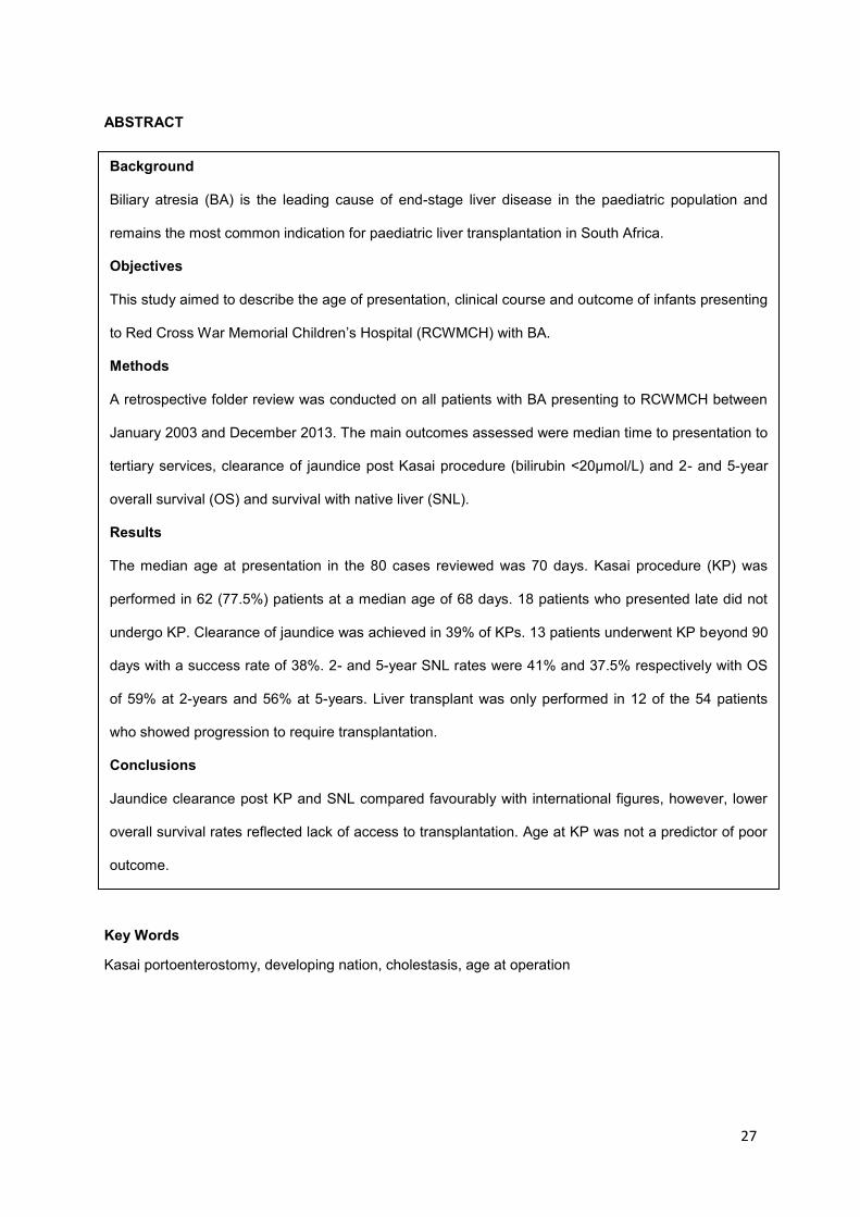
ABSTRACT
Key Words
Kasai portoenterostomy, developing nation, cholestasis, age at operation
Background
Biliary atresia (BA) is the leading cause of end-stage liver disease in the paediatric population and
remains the most common indication for paediatric liver transplantation in South Africa.
Objectives
This study aimed to describe the age of presentation, clinical course and outcome of infants presenting
to Red Cross War Memorial Children’s Hospital (RCWMCH) with BA.
Methods
A retrospective folder review was conducted on all patients with BA presenting to RCWMCH between
January 2003 and December 2013. The main outcomes assessed were median time to presentation to
tertiary services, clearance of jaundice post Kasai procedure (bilirubin <20µmol/L) and 2- and 5-year
overall survival (OS) and survival with native liver (SNL).
Results
The median age at presentation in the 80 cases reviewed was 70 days. Kasai procedure (KP) was
performed in 62 (77.5%) patients at a median age of 68 days. 18 patients who presented late did not
undergo KP. Clearance of jaundice was achieved in 39% of KPs. 13 patients underwent KP beyond 90
days with a success rate of 38%. 2- and 5-year SNL rates were 41% and 37.5% respectively with OS
of 59% at 2-years and 56% at 5-years. Liver transplant was only performed in 12 of the 54 patients
who showed progression to require transplantation.
Conclusions
Jaundice clearance post KP and SNL compared favourably with international figures, however, lower
overall survival rates reflected lack of access to transplantation. Age at KP was not a predictor of poor
outcome.
28
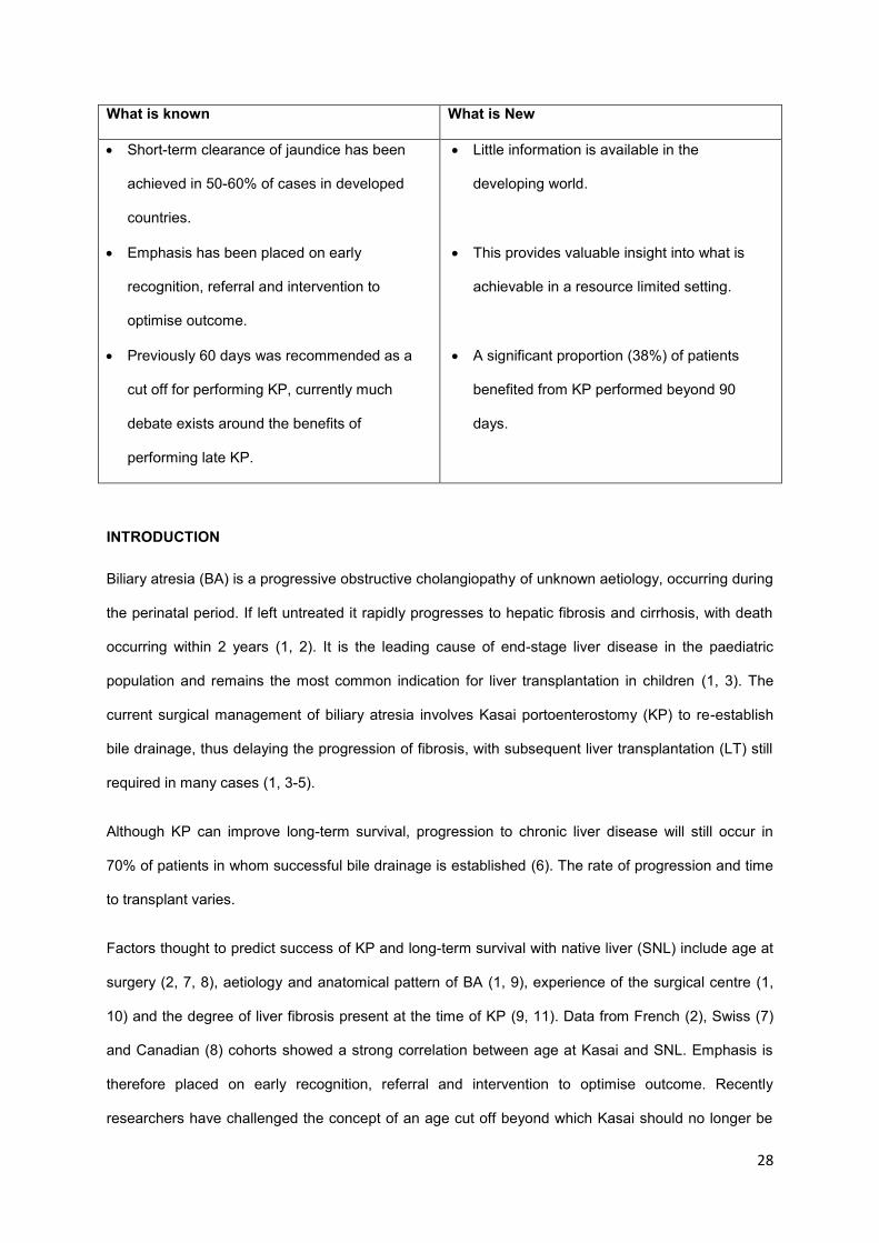
What is known What is New
Short-term clearance of jaundice has been
achieved in 50-60% of cases in developed
countries.
Emphasis has been placed on early
recognition, referral and intervention to
optimise outcome.
Previously 60 days was recommended as a
cut off for performing KP, currently much
debate exists around the benefits of
performing late KP.
Little information is available in the
developing world.
This provides valuable insight into what is
achievable in a resource limited setting.
A significant proportion (38%) of patients
benefited from KP performed beyond 90
days.
INTRODUCTION
Biliary atresia (BA) is a progressive obstructive cholangiopathy of unknown aetiology, occurring during
the perinatal period. If left untreated it rapidly progresses to hepatic fibrosis and cirrhosis, with death
occurring within 2 years (1, 2). It is the leading cause of end-stage liver disease in the paediatric
population and remains the most common indication for liver transplantation in children (1, 3). The
current surgical management of biliary atresia involves Kasai portoenterostomy (KP) to re-establish
bile drainage, thus delaying the progression of fibrosis, with subsequent liver transplantation (LT) still
required in many cases (1, 3-5).
Although KP can improve long-term survival, progression to chronic liver disease will still occur in
70% of patients in whom successful bile drainage is established (6). The rate of progression and time
to transplant varies.
Factors thought to predict success of KP and long-term survival with native liver (SNL) include age at
surgery (2, 7, 8), aetiology and anatomical pattern of BA (1, 9), experience of the surgical centre (1,
10) and the degree of liver fibrosis present at the time of KP (9, 11). Data from French (2), Swiss (7)
and Canadian (8) cohorts showed a strong correlation between age at Kasai and SNL. Emphasis is
therefore placed on early recognition, referral and intervention to optimise outcome. Recently
researchers have challenged the concept of an age cut off beyond which Kasai should no longer be
29
offered. Davenport et al reported a 5-year SNL of 40% in patients undergoing Kasai beyond 100 days
(12).
Outcomes vary world-wide, with short-term clearance of jaundice achievable in 50-60% of cases (10,
13-16), and a 10-year SNL of 30-40% (10, 13, 17, 18) reported in literature from Western countries.
Despite a wealth of information from developed countries, very little information is available in Africa
and other developing nations. The purpose of this research is to review the current experience of
biliary atresia at Red Cross War Memorial Children’s Hospital (RCWMCH) in South Africa with
emphasis on age at presentation, age at KP and outcomes in patients following KP at our institution.
METHODS
This is a retrospective descriptive study conducted within the Department of Paediatrics, RCWMCH,
Cape Town, South Africa. Patients managed within the department from January 2003 to December
2013 were included. The study was approved by the University of Cape Town Faculty of Health
Sciences Ethics Committee (HREC REF: 137/2014).
Inclusion Criteria:
Patients in whom a diagnosis of BA was made on clinical, biochemical and radiological grounds with
surgical findings and liver histology consistent with BA, including those who did not undergo KP were
analysed. Only patients managed primarily at RCWMCH during the study period were included.
Patients referred for transplant assessment after initial management at other tertiary institutions did
not form part of this study.
Analysis of Records:
Cases were identified through the RCWMCH clinical data base using the International Statistical
Classification of Diseases Code (ICD-10) allocated to BA. Records of attendance for the liver and
transplant clinics, as well as current and old transplant waiting lists were reviewed to ensure no cases
were missed. A review of all files was conducted from May to July 2014, by the primary author.
Data collected from patient charts included unique patient code, date of birth, sex, referral site, age of
jaundice onset (in weeks), date of presentation, presence of congenital anomalies, presence of

30
jaundice and stool pigment, imaging results (ultrasound, hepatobiliary scintigraphy, intraoperative
cholangiogram), whether KP was performed, date of KP, liver histology, success of KP, complications
(ascites, cholangitis, varices, fractures), tuberculosis, progression to require transplantation, suitability
for transplant listing, whether liver transplant (LT) was performed, date of LT, date of last visit and
final outcome including cause of death. Laboratory indices (full liver function tests, albumin,
haemoglobin and international normalised ratio) were recorded at time of presentation, time of KP, 3
months post KP and annually thereafter.
Liver biopsies were scored using Lee’s 15 point histological scoring system for neonatal cholestasis
(19), by a single paediatric histopathologist. Cirrhosis was classified as present if there was porto-
portal bridging involving more than 50% of the portal tracts.
Outcome measures:
The latest outcome status was documented as alive, dead, lost to follow up or transferred to another
institution. Successful KP was defined as clearance of jaundice with serum bilirubin < 20µmol/L within
3 months of surgery. Survival was expressed as 2- and 5-year survival rates, including overall survival
(OS) and survival with native liver. OS was defined as beginning at birth and ending at death or last
follow-up; SNL starting at birth and ending at death (based on how many patients could have reach 2
or 5 years of age by 31 July 2014).
Statistical Analysis:
All analyses were done using Stata Statistical Software Release 13 (StataCorp LP, College Stat).
Ages are quoted as median and interquartile range (IQR). Fisher’s exact tests were used to compare
categorical data. The distribution of numerical data was assessed and the Student t-test or Mann-
Whitney U test applied as appropriate. A P-value of 0.05 was regarded as significant. Survival rates
were estimated using the Kaplan-Meier method.
Full data was not available in all medical records, for these variables number (n) was declared. For
patients lost to follow up (LTFU) the date of their last visit was utilised when calculating survival.
Cases LTFU immediately post-KP were not included when calculating success of KP.
Limitations: Anatomical classification of BA was not recorded. Data was limited by that available in
medical records and short follow-up periods of the cohort.
31
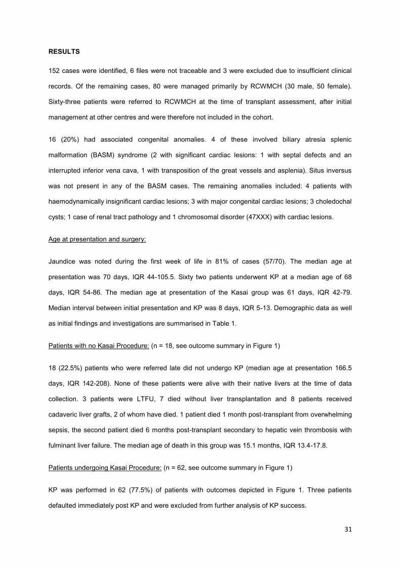
RESULTS
152 cases were identified, 6 files were not traceable and 3 were excluded due to insufficient clinical
records. Of the remaining cases, 80 were managed primarily by RCWMCH (30 male, 50 female).
Sixty-three patients were referred to RCWMCH at the time of transplant assessment, after initial
management at other centres and were therefore not included in the cohort.
16 (20%) had associated congenital anomalies. 4 of these involved biliary atresia splenic
malformation (BASM) syndrome (2 with significant cardiac lesions: 1 with septal defects and an
interrupted inferior vena cava, 1 with transposition of the great vessels and asplenia). Situs inversus
was not present in any of the BASM cases. The remaining anomalies included: 4 patients with
haemodynamically insignificant cardiac lesions; 3 with major congenital cardiac lesions; 3 choledochal
cysts; 1 case of renal tract pathology and 1 chromosomal disorder (47XXX) with cardiac lesions.
Age at presentation and surgery:
Jaundice was noted during the first week of life in 81% of cases (57/70). The median age at
presentation was 70 days, IQR 44-105.5. Sixty two patients underwent KP at a median age of 68
days, IQR 54-86. The median age at presentation of the Kasai group was 61 days, IQR 42-79.
Median interval between initial presentation and KP was 8 days, IQR 5-13. Demographic data as well
as initial findings and investigations are summarised in Table 1.
Patients with no Kasai Procedure: (n = 18, see outcome summary in Figure 1)
18 (22.5%) patients who were referred late did not undergo KP (median age at presentation 166.5
days, IQR 142-208). None of these patients were alive with their native livers at the time of data
collection. 3 patients were LTFU, 7 died without liver transplantation and 8 patients received
cadaveric liver grafts, 2 of whom have died. 1 patient died 1 month post-transplant from overwhelming
sepsis, the second patient died 6 months post-transplant secondary to hepatic vein thrombosis with
fulminant liver failure. The median age of death in this group was 15.1 months, IQR 13.4-17.8.
Patients undergoing Kasai Procedure: (n = 62, see outcome summary in Figure 1)
KP was performed in 62 (77.5%) of patients with outcomes depicted in Figure 1. Three patients
defaulted immediately post KP and were excluded from further analysis of KP success.
32
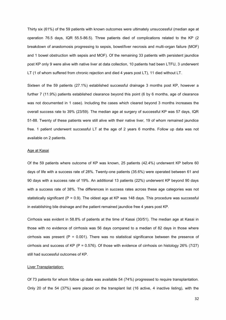
Thirty six (61%) of the 59 patients with known outcomes were ultimately unsuccessful (median age at
operation 76.5 days, IQR 55.5-86.5). Three patients died of complications related to the KP (2
breakdown of anastomosis progressing to sepsis, bowel/liver necrosis and multi-organ failure (MOF)
and 1 bowel obstruction with sepsis and MOF). Of the remaining 33 patients with persistent jaundice
post KP only 9 were alive with native liver at data collection, 10 patients had been LTFU, 3 underwent
LT (1 of whom suffered from chronic rejection and died 4 years post LT), 11 died without LT.
Sixteen of the 59 patients (27.1%) established successful drainage 3 months post KP, however a
further 7 (11.9%) patients established clearance beyond this point (6 by 6 months, age of clearance
was not documented in 1 case). Including the cases which cleared beyond 3 months increases the
overall success rate to 39% (23/59). The median age at surgery of successful KP was 57 days, IQR
51-88. Twenty of these patients were still alive with their native liver, 19 of whom remained jaundice
free. 1 patient underwent successful LT at the age of 2 years 6 months. Follow up data was not
available on 2 patients.
Age at Kasai
Of the 59 patients where outcome of KP was known, 25 patients (42.4%) underwent KP before 60
days of life with a success rate of 28%. Twenty-one patients (35.6%) were operated between 61 and
90 days with a success rate of 19%. An additional 13 patients (22%) underwent KP beyond 90 days
with a success rate of 38%. The differences in success rates across these age categories was not
statistically significant (P = 0.9). The oldest age at KP was 148 days. This procedure was successful
in establishing bile drainage and the patient remained jaundice free 4 years post KP.
Cirrhosis was evident in 58.8% of patients at the time of Kasai (30/51). The median age at Kasai in
those with no evidence of cirrhosis was 56 days compared to a median of 82 days in those where
cirrhosis was present (P = 0.001). There was no statistical significance between the presence of
cirrhosis and success of KP (P = 0.576). Of those with evidence of cirrhosis on histology 26% (7/27)
still had successful outcomes of KP.
Liver Transplantation:
Of 73 patients for whom follow up data was available 54 (74%) progressed to require transplantation.
Only 20 of the 54 (37%) were placed on the transplant list (16 active, 4 inactive listing), with the

33
remaining 34 (63%) being excluded from potential transplantation. Unfavourable social circumstances
such as poor compliance with medical treatment and follow up, inadequate sanitation, parental
substance abuse and distance from the transplant centre formed the main grounds for exclusion
(n=16, 47% of those excluded from LT; 30% of the 54 patients requiring LT). Two patients were
medically unfit to undergo LT (1 due to active tuberculosis). One patient was not from South Africa, 8
defaulted prior to completion of the transplant assessment and 4 died before they could be listed
A total of 12 patients underwent LT (15% of the total cohort, 22.2% of those requiring LT) at a median
age of 31.5 months, IQR 18.9-34.2. The outcomes of these patients were described above. (Figure 1)
Clinical course and complications:
Of the 29 patients who were alive with their native liver at the end of data collection, only 3 (10.3%)
were alive without complications. Cholangitis was the commonest complication occurring in 21 of
these patients (72.4%), of these 13 patients suffered recurrent episodes of cholangitis. Ascites
occurred in 6 of the 29 patients (20.7%). Routine endoscopy was not performed and thus varices
were only diagnosed after an episode of melaena or hematemesis. Five of the 29 patients (17.2%)
experienced variceal bleeds. One patient suffered 2 consecutive femur fractures. Pulmonary
tuberculosis occurred in 6 patients (20.7%).
The frequency of complications for the full cohort is expressed in Table 2.
Sixty two patients underwent KP and information regarding cholangitis was not available for 2 of these
patients. Of the remaining 60 patients, 44 experienced episodes of cholangitis (73.3%). Fifteen of these
patients were lost to follow up and hence outcome data was only available for 45 patients. Of the patients
that died during the study period, 84.6% had experienced at least 1 episode of cholangitis (11/13).
Cholangitis was also seen in 71.9% of patients who ultimately survived (23/32). There was no statistical
significance in the occurrence of cholangitis and the outcome of survival (P=0.467).
Reliable growth parameters were not available in many records and growth failure was thus unable to
be assessed. Bacterial peritonitis was not reported in any patients.
34
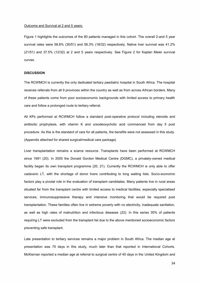
Outcome and Survival at 2 and 5 years:
Figure 1 highlights the outcomes of the 80 patients managed in this cohort. The overall 2-and 5 year
survival rates were 58.8% (30/51) and 56.3% (18/32) respectively. Native liver survival was 41.2%
(21/51) and 37.5% (12/32) at 2 and 5 years respectively. See Figure 2 for Kaplan Meier survival
curves.
DISCUSSION
The RCWMCH is currently the only dedicated tertiary paediatric hospital in South Africa. The hospital
receives referrals from all 9 provinces within the country as well as from across African borders. Many
of these patients come from poor socioeconomic backgrounds with limited access to primary health
care and follow a prolonged route to tertiary referral.
All KPs performed at RCWMCH follow a standard post-operative protocol including steroids and
antibiotic prophylaxis, with vitamin K and ursodeoxycholic acid commenced from day 5 post
procedure. As this is the standard of care for all patients, the benefits were not assessed in this study.
(Appendix attached for shared surgical/medical care package)
Liver transplantation remains a scarce resource. Transplants have been performed at RCWMCH
since 1991 (20). In 2005 the Donald Gordon Medical Centre (DGMC), a privately-owned medical
facility began its own transplant programme (20, 21). Currently the RCWMCH is only able to offer
cadaveric LT, with the shortage of donor livers contributing to long waiting lists. Socio-economic
factors play a pivotal role in the evaluation of transplant candidates. Many patients live in rural areas
situated far from the transplant centre with limited access to medical facilities, especially specialised
services; immunosuppressive therapy and intensive monitoring that would be required post
transplantation. These families often live in extreme poverty with no electricity, inadequate sanitation,
as well as high rates of malnutrition and infectious diseases (22). In this series 30% of patients
requiring LT were excluded from the transplant list due to the above mentioned socioeconomic factors
preventing safe transplant.
Late presentation to tertiary services remains a major problem in South Africa. The median age at
presentation was 70 days in this study, much later than that reported in International Cohorts.
McKiernan reported a median age at referral to surgical centre of 40 days in the United Kingdom and
35
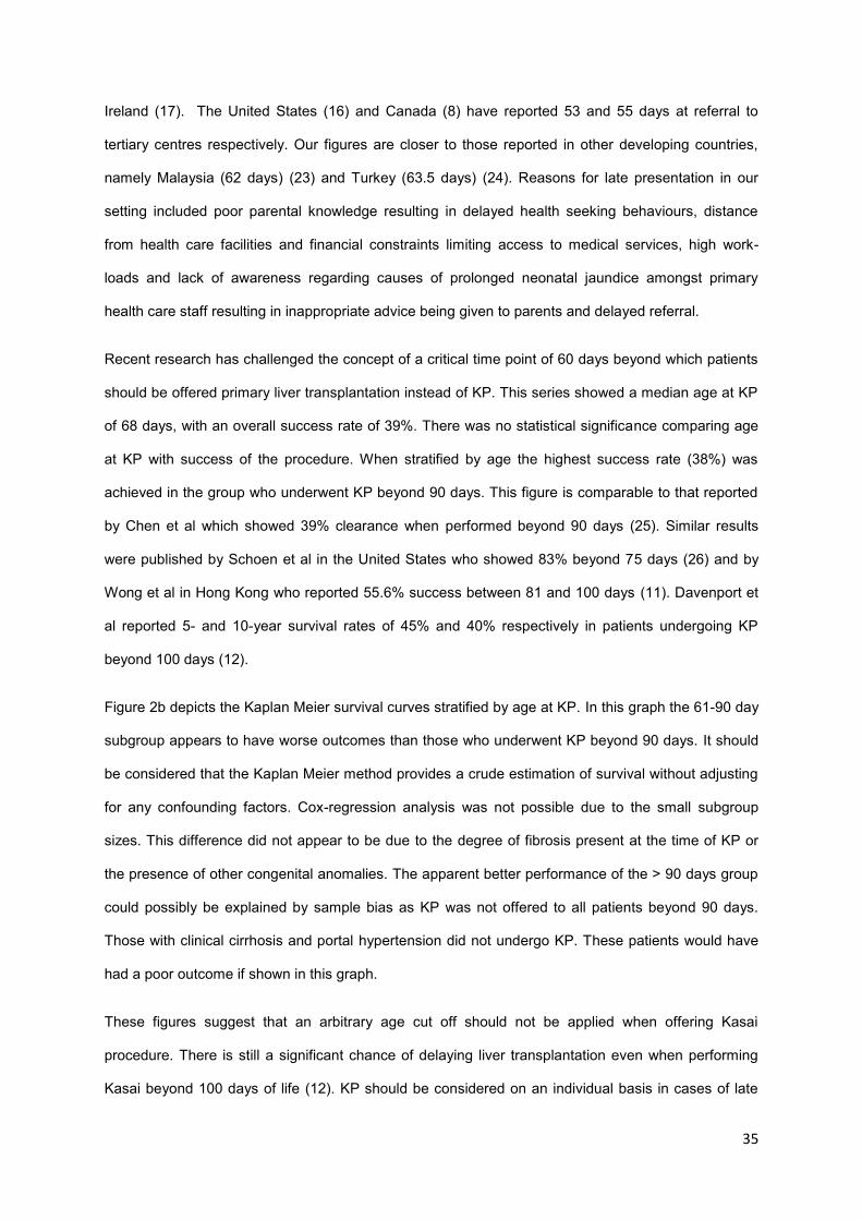
Ireland (17). The United States (16) and Canada (8) have reported 53 and 55 days at referral to
tertiary centres respectively. Our figures are closer to those reported in other developing countries,
namely Malaysia (62 days) (23) and Turkey (63.5 days) (24). Reasons for late presentation in our
setting included poor parental knowledge resulting in delayed health seeking behaviours, distance
from health care facilities and financial constraints limiting access to medical services, high work-
loads and lack of awareness regarding causes of prolonged neonatal jaundice amongst primary
health care staff resulting in inappropriate advice being given to parents and delayed referral.
Recent research has challenged the concept of a critical time point of 60 days beyond which patients
should be offered primary liver transplantation instead of KP. This series showed a median age at KP
of 68 days, with an overall success rate of 39%. There was no statistical significance comparing age
at KP with success of the procedure. When stratified by age the highest success rate (38%) was
achieved in the group who underwent KP beyond 90 days. This figure is comparable to that reported
by Chen et al which showed 39% clearance when performed beyond 90 days (25). Similar results
were published by Schoen et al in the United States who showed 83% beyond 75 days (26) and by
Wong et al in Hong Kong who reported 55.6% success between 81 and 100 days (11). Davenport et
al reported 5- and 10-year survival rates of 45% and 40% respectively in patients undergoing KP
beyond 100 days (12).
Figure 2b depicts the Kaplan Meier survival curves stratified by age at KP. In this graph the 61-90 day
subgroup appears to have worse outcomes than those who underwent KP beyond 90 days. It should
be considered that the Kaplan Meier method provides a crude estimation of survival without adjusting
for any confounding factors. Cox-regression analysis was not possible due to the small subgroup
sizes. This difference did not appear to be due to the degree of fibrosis present at the time of KP or
the presence of other congenital anomalies. The apparent better performance of the > 90 days group
could possibly be explained by sample bias as KP was not offered to all patients beyond 90 days.
Those with clinical cirrhosis and portal hypertension did not undergo KP. These patients would have
had a poor outcome if shown in this graph.
These figures suggest that an arbitrary age cut off should not be applied when offering Kasai
procedure. There is still a significant chance of delaying liver transplantation even when performing
Kasai beyond 100 days of life (12). KP should be considered on an individual basis in cases of late
36
presentation taking into account the clinical severity of disease, presence of established cirrhosis and
portal hypertension. In the South African context where access to LT is limited, life expectancy and
quality of life can be improved by offering KP even in those presenting beyond 90 days. In patients
who fulfil the social criteria for transplantation, delaying LT benefits patients by allowing time for
growth and immunisations prior to transplantation.
Definitions of successful KP vary amongst authors. Bilirubin values of <20µmol/L are the most
commonly quoted (2, 9, 11, 18, 25, 27), but <2mg/dL (34µmol/L) has also been used (26, 28). Grizelj
et al used a cut off of 3 months post KP to define successful clearance of jaundice (27), although
other authors have been more liberal in their cut off with Davenport (9), Lee (23) and Hung (29) using
6 months and McKiernan (18) and Tiao (28) accepting the target bilirubin levels at any time post KP.
When applying a cut off of 3 months in this study successful clearance of jaundice was only achieved
in 27% of cases. However, if this is expanded to include the 7 cases which cleared beyond 3 months,
the success rate increased to 39% which is comparable with jaundice clearance rates achieved in
France (40%) (15), Netherlands (38%) (30) and US (40%) (16). This is still below those achieved in
Japan (62%) (13), Taiwan (59%) (14) and the UK (55%) (10).
Cirrhosis was evident in almost 60% of patients undergoing KP. Although the presence of cirrhosis
did not have a statistical impact on the success of procedure, cirrhosis was associated with increasing
age at KP. Davenport et al reported that histological degree of fibrosis had no value in predicting
success and furthermore that the presence of cirrhosis at the time of KP had no significant survival
disadvantage, in a series reviewing the outcomes of KP beyond 100 days (12).
This cohort showed a high rate of cholangitis and complications of portal hypertension including
ascites and variceal bleeding. Cholangitis was common, occurring in 73% of those who underwent KP
(Table 2). This is comparable to figures seen in other literature (28, 29). Zhen et al (31) reported early
cholangitis to be the single most powerful predictor for prognosis, although other authors have found
no association between cholangitis and survival (16, 28). In this study cholangitis was not associated
with a worse survival outcome, although the limited numbers need to be taken into account when
interpreting the statistical significance of this data. It is essential that cholangitis is treated promptly

37
and aggressively to preserve liver function. 20% of patients in this cohort were lost from follow up.
Parents should be counselled about the potential for complications and the development of chronic
liver disease post KP, as life-long follow up is essential in order to delay progression of liver disease
and begin appropriate transplant assessment when required.
Twenty six per cent of patients were treated for pulmonary tuberculosis (PTB) in this cohort. PTB is
highly prevalent in the Western Cape and the presence of active disease is an exclusion to
transplantation until fully treated. The majority of these cases were not culture confirmed, but were
considered as probable PTB and therefore treated empirically. The higher number of TB cases in the
non-Kasai group may be due to the effects of chronic illness and poor nutritional status on immune
function.
The 37.5% 5-year SNL in this cohort is comparable with figures from Canada (33%) (8), France (40%)
(2), Switzerland (37% 4-year SNL) (7), Netherlands (42% 4-year SNL) (30) and United Kingdom
(46%) (10). However, the overall 5-year survival of 56% falls far below those seen internationally,
which range from 77% in Canada (8) to 90% in the UK (10) and 92% in Switzerland (7). The low
overall survival rates can be explained by poor access to LT with only 12% of our initial cohort
undergoing transplantation, as opposed to 40% seen in the BARC (Biliary Atresia Research
Consortium) cohort reported by Shneider et al (16) and 44% reported by Davenport et al in the UK
(10).
A previous study conducted in the surgical department at RCWMCH reviewed 39 children undergoing
Kasai procedure between January 1975 and January 1985 (32). The mean age of operation was 12.8
weeks. In the first 4 years of the study no patients established successful bile drainage. Thereafter
50% of KPs were successful in establishing drainage, however only 20% of these patients remained
alive at the time of analysis. The current cohort (2003-2013) shows a dramatic improvement in age at
KP which has decreased from 12.8 weeks to 68 days as well as in overall survival.
Delayed presentation and late referral remain a major challenge in the management of BA. Efforts
aimed at promoting earlier referral through the creation of biliary atresia awareness at the primary
health care level play an important role in improving outcomes and survival post Kasai surgery. The

38
most successful strategies have been the stool colour cards established in Taiwan in 2004 and the
“yellow alert” educational programme in the UK aimed at expediting the referral of jaundiced infants
(8, 14, 33).
Since the introduction of stool colour cards in Taiwan, BA has been diagnosed earlier with the
percentage of patients undergoing Kasai before 60 days improving from 49.4% to 65.7%. Jaundice
clearance post Kasai has improved from 34.8% to 60.8%, with 5-year jaundice free SNL improving
from 27.3% to 64.3% (33). As Taiwan has the highest incidence of BA, screening in this population is
bound to yield positive results. A US study on the cost-effectiveness of a screening programme
concluded that the use of stool colour cards resulted in lower costs and better outcomes for patients
with BA (34).
Differentiating physiological jaundice in the neonate from pathological jaundice is essential to facilitate
early referral. Pale stools, dark urine and jaundice persisting beyond 14 days of life warrants urgent
referral for further investigation (including conjugated bilirubin). In the RCWMCH cohort initial stool
colour was not documented in 20% of patients, a figure which is likely to be higher at primary health
care facilities. This highlights the need to improve awareness amongst both parents and all health
care professionals. Although it is unlikely to be financially feasible to include stool colour cards in the
current Road to Health Card Booklets, posters should be made available at antenatal services and
baby clinics as part of an educational campaign. Nursing staff administering 6 week immunisations
should be encouraged to enquire about stool colour. Although referral at 6 weeks would already be
considered late, it would still allow for KP to be performed within 60 days and would be an
improvement on our current age at presentation. Employing an algorithm for the appropriate
management of conjugated hyperbilirubinaemia at peripheral services would aid in facilitating earlier
referral.
This study has confirmed that patients can still benefit from undergoing KP beyond 90 days. In a
country with limited access to transplantation, KP should still be offered to patients presenting late.
This will allow a significant proportion (38%) of cases the opportunity for successful KP with improved

39
quality of life, as well as delaying the need for transplantation and immunosuppression and alleviating
strain on the organ donor system.
In this study age at KP, degree of fibrosis and complications were not associated with poor outcome
of KP.
In conclusion, although overall survival figures still lag behind international data, we have made
enormous progress over the last 30 years within a limited resource setting. Improved awareness and
earlier referral of infantile cholestasis will improve these figures further.

40
REFERENCES
1 Davenport M. Biliary atresia: clinical aspects. Semin Pediatr Surg 2012;21(3):175-84.
2 Chardot C, Buet C, Serinet MO, et al. Improving outcomes of biliary atresia: French national
series 1986-2009. J Hepatol 2013;58(6):1209-17.
3 Hartley JL, Davenport M, Kelly DA. Biliary atresia. Lancet 2009;374(9702):1704-13.
4 Bijl EJ, Bharwani KD, Houwen RH, et al. The long-term outcome of the Kasai operation in
patients with biliary atresia: a systematic review. Neth J Med 2013;71(4):170-3.
5 Hadzic N, Davenport M, Tizzard S, et al. Long-term survival following Kasai
portoenterostomy: is chronic liver disease inevitable? J Pediatr Gastroenterol Nutr
2003;37(4):430-3.
6 Davenport M, De Ville de Goyet J, Stringer MD, et al. Seamless management of biliary atresia
in England and Wales (1999-2002). Lancet 2004;363(9418):1354-7.
7 Wildhaber BE, Majno P, Mayr J, et al. Biliary atresia: Swiss national study, 1994-2004. J
Pediatr Gastroenterol Nutr 2008;46(3):299-307.
8 Schreiber RA, Barker CC, Roberts EA, et al. Biliary atresia: the Canadian experience. J
Pediatr 2007;151(6):659-65, 65.e1.
9 Davenport M, Caponcelli E, Livesey E, et al. Surgical outcome in biliary atresia: etiology
affects the influence of age at surgery. Ann Surg 2008;247(4):694-8.
10 Davenport M, Ong E, Sharif K, et al. Biliary atresia in England and Wales: results of
centralization and new benchmark. J Pediatr Surg 2011;46(9):1689-94.
11 Wong KK, Chung PH, Chan IH, et al. Performing Kasai portoenterostomy beyond 60 days of
life is not necessarily associated with a worse outcome. J Pediatr Gastroenterol Nutr
2010;51(5):631-4.
12 Davenport M, Puricelli V, Farrant P, et al. The outcome of the older (> or =100 days) infant
with biliary atresia. J Pediatr Surg 2004;39(4):575-81.
13 Nio M, Ohi R, Miyano T, et al. Five- and 10-year survival rates after surgery for biliary atresia:
a report from the Japanese Biliary Atresia Registry. J Pediatr Surg 2003;38(7):997-1000.
14 Hsiao CH, Chang MH, Chen HL, et al. Universal screening for biliary atresia using an infant
stool color card in Taiwan. Hepatology 2008;47(4):1233-40.

41
15 Serinet MO, Broue P, Jacquemin E, et al. Management of patients with biliary atresia in
France: results of a decentralized policy 1986-2002. Hepatology 2006;44(1):75-84.
16 Shneider BL, Brown MB, Haber B, et al. A multicenter study of the outcome of biliary atresia
in the United States, 1997 to 2000. J Pediatr 2006;148(4):467-74.e1.
17 McKiernan PJ, Baker AJ, Kelly DA. The frequency and outcome of biliary atresia in the UK
and Ireland. Lancet 2000;355(9197):25-9.
18 McKiernan PJ, Baker AJ, Lloyd C, et al. British paediatric surveillance unit study of biliary
atresia: outcome at 13 years. J Pediatr Gastroenterol Nutr 2009;48(1):78-81.
19 Lee WS, Looi LM. Usefulness of a scoring system in the interpretation of histology in neonatal
cholestasis. World J Gastroenterol 2009;15(42):5326-33.
20 Spearman CW, McCulloch M, Millar AJ, et al. Liver transplantation at Red Cross War
Memorial Children's Hospital. S Afr Med J 2006;96:960-3.
21 Loveland JA, Govender T, Botha J, et al. Paediatric liver transplantation in Johannesburg:
initial 29 cases and prospects for the future. S Afr Med J 2012;102(4):233-6.
22 Spearman CW, McCulloch MI. Challenges for paediatric transplantation in Africa. Pediatr
Transplant 2014;18(7):668-74.
23 Lee WS, Chai PF, Lim KS, et al. Outcome of biliary atresia in Malaysia: a single-centre study.
J Paediatr Child Health 2009;45(5):279-85.
24 Aydogdu S, Ozgenc F, AtIk T, et al. Biliary atresia in Turkish children. Pediatr Int
2004;46(2):158-61.
25 Chen G, Zheng S, Sun S, et al. Early surgical outcomes and pathological scoring values of
older infants (>/= 90 d old) with biliary atresia. J Pediatr Surg 2012;47(12):2184-8.
26 Schoen BT, Lee H, Sullivan K, et al. The Kasai portoenterostomy: when is it too late? J
Pediatr Surg 2001;36(1):97-99.
27 Grizelj R, Vukovic J, Novak M, et al. Biliary atresia: the Croatian experience 1992-2006. Eur J
Pediatr 2010;169(12):1529-34.
28 Tiao MM, Chuang JH, Huang LT, et al. Management of biliary atresia: experience in a single
institute. Chang Gung Med J 2007;30(2):122-7.
29 Hung PY, Chen CC, Chen WJ, et al. Long-term prognosis of patients with biliary atresia: a 25
year summary. J Pediatr Gastroenterol Nutr 2006;42(2):190-5.
42
30 de Vries W, de Langen ZJ, Groen H, et al. Biliary atresia in the Netherlands: outcome of
patients diagnosed between 1987 and 2008. J Pediatr 2012;160(4):638-44.e2.
31 Zhen C, Guoliang Q, Lishuang M, et al. Design and validation of an early scoring system for
predicting early outcomes of type III biliary atresia after Kasai's operation. Pediatr Surg Int
2015;31(6):535-42.
32 Millar AJ, Davies MR, Rode H, et al. Biliary atresia--surgical management; A 10-year review.
S Afr Med J 1986;69(5):288-93.
33 Lien TH, Chang MH, Wu JF, et al. Effects of the infant stool color card screening program on
5-year outcome of biliary atresia in Taiwan. Hepatology 2011;53(1):202-8.
34 Mogul D, Zhou M, Intihar P, et al. Cost-effective analysis of screening for biliary atresia with
the stool color card. J Pediatr Gastroenterol Nutr 2015;60(1):91-8.
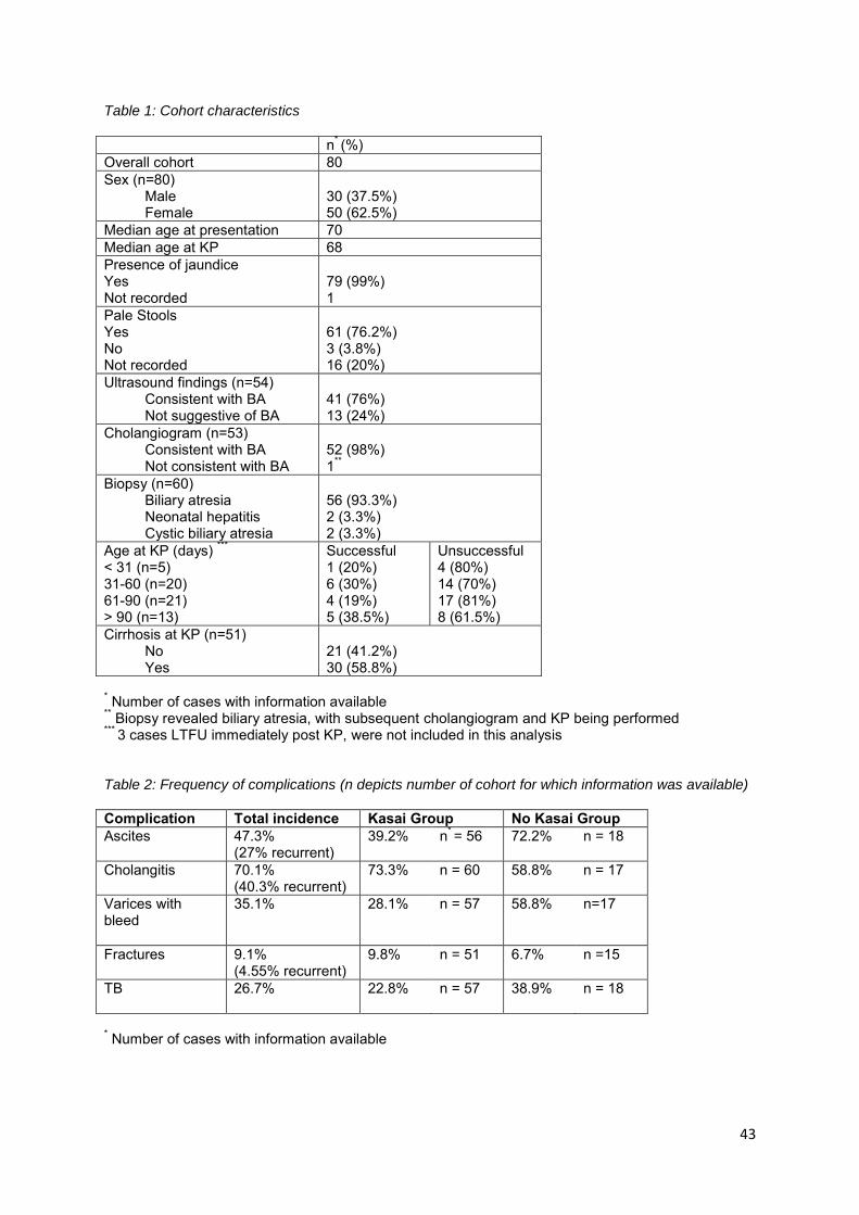
43
Table 1: Cohort characteristics
n* (%) Overall cohort 80 Sex (n=80)
Male Female
30 (37.5%) 50 (62.5%)
Median age at presentation 70 Median age at KP 68 Presence of jaundice Yes Not recorded
79 (99%) 1
Pale Stools Yes No Not recorded
61 (76.2%) 3 (3.8%) 16 (20%)
Ultrasound findings (n=54) Consistent with BA Not suggestive of BA
41 (76%) 13 (24%)
Cholangiogram (n=53) Consistent with BA Not consistent with BA
52 (98%) 1**
Biopsy (n=60) Biliary atresia Neonatal hepatitis Cystic biliary atresia
56 (93.3%) 2 (3.3%) 2 (3.3%)
Age at KP (days) *** < 31 (n=5) 31-60 (n=20) 61-90 (n=21) > 90 (n=13)
Successful 1 (20%) 6 (30%) 4 (19%) 5 (38.5%)
Unsuccessful 4 (80%) 14 (70%) 17 (81%) 8 (61.5%)
Cirrhosis at KP (n=51) No Yes
21 (41.2%) 30 (58.8%)
* Number of cases with information available ** Biopsy revealed biliary atresia, with subsequent cholangiogram and KP being performed *** 3 cases LTFU immediately post KP, were not included in this analysis Table 2: Frequency of complications (n depicts number of cohort for which information was available) Complication Total incidence Kasai Group No Kasai Group Ascites
47.3% (27% recurrent)
39.2% n* = 56 72.2% n = 18
Cholangitis 70.1% (40.3% recurrent)
73.3% n = 60 58.8% n = 17
Varices with bleed
35.1% 28.1% n = 57 58.8% n=17
Fractures
9.1% (4.55% recurrent)
9.8% n = 51 6.7% n =15
TB
26.7% 22.8% n = 57 38.9% n = 18
* Number of cases with information available

44
Figure 1: Outcomes of 80 patients with biliary atresia managed at RCWMCH 2003-2013
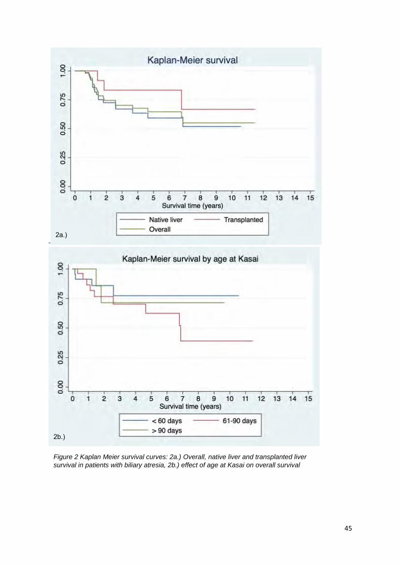
45
-
Figure 2 Kaplan Meier survival curves: 2a.) Overall, native liver and transplanted liver
survival in patients with biliary atresia, 2b.) effect of age at Kasai on overall survival
2a.)
2b.)
46
5. APPENDICES
ETHICAL APPROVAL

47
DATA CAPTURE SHEETS
Index Case Number Folder Number Age at presentation (days) Race 1 African; 2 Mixed; 3 White; 4 Other Sex 1 Male; 2 Female Source of referral 1 GP; 2 clinic/day hosp; 3 secondary; 4 tertiary; 5 other Number of visits prior to referral Date of birth Date of presentation Date of Kasai Date entered transplant list Date transplanted Date of death Presenting features Jaundice (0 no; 1 yes) Pale stools (0 no, 1 yes) Other Associated Congenital Abnormalities 0 = nil; 1 = yes (specify)
LABORATORY VALUES Bili (TB/CB) P = Presentation; K = Kasai; K3 = 3/12 post Kasai; A1/2/3/4… = annually
AST P = Presentation; K = Kasai; K3 = 3/12 post Kasai; A1/2/3/4… = annually
ALT P = Presentation; K = Kasai; K3 = 3/12 post Kasai; A1/2/3/4… = annually
ALP P = Presentation; K = Kasai; K3 = 3/12 post Kasai; A1/2/3/4… = annually
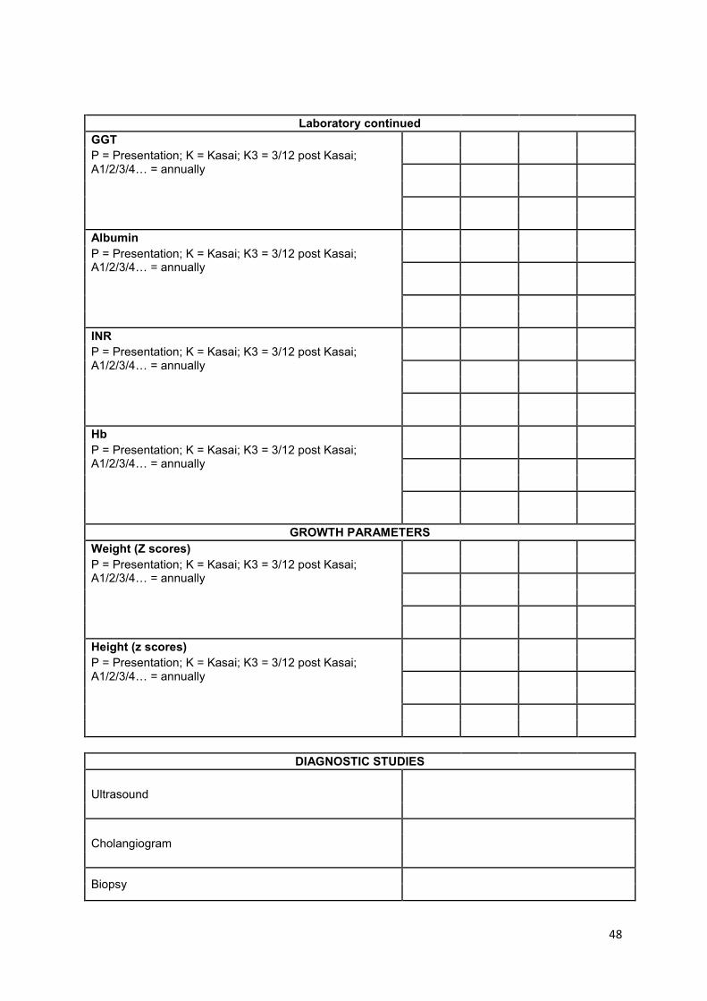
48
Laboratory continued GGT P = Presentation; K = Kasai; K3 = 3/12 post Kasai; A1/2/3/4… = annually
Albumin P = Presentation; K = Kasai; K3 = 3/12 post Kasai; A1/2/3/4… = annually
INR P = Presentation; K = Kasai; K3 = 3/12 post Kasai; A1/2/3/4… = annually
Hb P = Presentation; K = Kasai; K3 = 3/12 post Kasai; A1/2/3/4… = annually
GROWTH PARAMETERS Weight (Z scores) P = Presentation; K = Kasai; K3 = 3/12 post Kasai; A1/2/3/4… = annually
Height (z scores) P = Presentation; K = Kasai; K3 = 3/12 post Kasai; A1/2/3/4… = annually
DIAGNOSTIC STUDIES
Ultrasound
Cholangiogram
Biopsy

49
KASAI OPERATION Age in days Surgeon performing Kasai 0 = consultant; 1 = registrar Pigmented stools 3/12 post Kasai 0 = no; 1 = yes Clearance of jaundice 3/12 Post Kasai 0 = no; 1 = yes
COMPLICATIONS POST KASAI Ascites 0 = no; 1 = yes (number of episodes)
Cholangitis 0 = no; 1 = yes (number of episodes)
Varices 0 = no; 1 = yes (number of episodes)
Bacterial peritonitis 0 = no; 1 = yes (number of episodes)
Growth failure 0 = no; 1 = yes
Other (e.g. TB, rickets, specify)
OUTCOMES Successful Kasai 0 = no, 1 = yes Progression to require transplant 0 = no; 1 = yes Transplanted 0 = no; 1 = yes Alive at data collection 0= no (capture cause of death); 1 = yes; 2 = LTFU; 3 = transferred to another centre
Addendum:
Race was not captured as it is not recorded in hospital records.
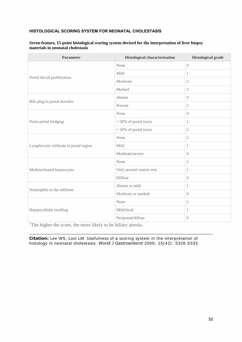
50
HISTOLOGICAL SCORING SYSTEM FOR NEONATAL CHOLESTASIS
Seven-feature, 15-point histological scoring system devised for the interpretation of liver biopsy
materials in neonatal cholestasis
Parameter Histological characterization Histological grade
Portal ductal proliferation
None 0
Mild 1
Moderate 2
Marked 3
Bile plug in portal ductules Absent 0
Present 2
Porto-portal bridging
None 0
< 50% of portal tracts 1
> 50% of portal tracts 2
Lymphocytic infiltrate in portal region
None 2
Mild 1
Moderate/severe 0
Multinucleated hepatocytes
None 2
Only around central vein 1
Diffuse 0
Neutrophils in the infiltrate Absent or mild 1
Moderate or marked 0
Hepatocellular swelling
None 2
Mild/focal 1
Periportal/diffuse 0
1The higher the score, the more likely to be biliary atresia.
Citation: Lee WS, Looi LM. Usefulness of a scoring system in the interpretation of histology in neonatal cholestasis. World J Gastroenterol 2009; 15(42): 5326-5333

51
BILIARY ATRESIA PATHOLOGICAL IMAGES
52
SHARED CARE GUIDANCE
Follow up of children post surgery for Biliary Atresia
CONTENTS
Introduction Page 2
Contact Numbers Page 2
Follow up Page 2
Medication Page 2 - 4
Vaccinations Page 4
Complications/Problems Page 4 - 5
Appendix Page 7
SHARED CARE GUIDANCE: FOLLOW UP OF CHILDREN POST SURGERY FOR BILIARY ATRESIA

53
INTRODUCTION
We look forward to sharing care of this infant who has Biliary Atresia, and underwent a Kasai Portoenterostomy. Biliary drainage is achieved in up to 80% of patients. Subsequently some of these children will develop complications of cirrhosis (portal hypertension, liver dysfunction etc.) and will be considered for liver transplant electively. Children who do not achieve bile drainage (i.e. remain jaundiced) have an unsuccessful Kasai and should be referred for liver transplant.
CONTACT NUMBERS
Ward D2, GIT Unit on Ward E2
Please feel free to discuss any problems at any time.
FOLLOW UP
2nd, 3rd & 4th week SOPD
5th week GIT follow-up in Liver OPD Monday am.
2 - 3 month GIT Liver clinic
6 month GIT Liver clinic
1 year and then yearly GIT Liver clinic
MEDICATION
The following regimen is followed:
Steroids (two week course)
Initially IV in hospital Methylprednisolone 20mg commencing on POD 1, decreasing 2.5mg daily until 5mg/day. This is then completed at a dose of 5mg orally until POD 14.
Further need for steroids to be assessed in clinic but may be reintroduced as a short course if an episode of cholangitis results in sudden cholestasis after good bile drainage.
Antibiotic prophylaxis for 5 days
From induction IVI Ampicillin 25mg/kg/dose 6hrly, Gentamycin 6mg/kg/dose daily (check levels) and Metronidazole 7.5mg/kg/dose 8hrly

54
Long term oral antibiotic prophylaxis
From Day 6 Ciprofloxacin 12.5mg/kg/dose bd for one month then daily for 3 months
Then a cycle of: Amoxycillin 25mg/kg once a day for 12 weeks (max of 1 gram)
Cephalexin 12.5mg/kg once a day for 12 weeks (max of 500 mg)
Then cycle indefinitely
Vitamin supplements
The infant will be receiving the following vitamins
Vitamin A (retinol) (started in out patients at first surgical review)
Trade Name = Arovit®
Presentation = 150,000 iu/ml (equivalent to 5,000iu/drop)
<1 year 5000 units/day oral
Vitamin D (alfacalcidol) (started in BCH out patients at first surgical review)
Trade Name = One Alpha®
Presentation = 2mcg/ml (equivalent to 100ng/drop)
Neonate + premature 50-100 nanograms/kg/daily oral
Vitamin E (alpha tocopheryl) (started in BCH out patients at first surgical review)
Trade Name = Ephynal
Presentation = 500mg/5ml suspension
<1 year 50mg/day oral
Vitamin K (phytomenadione) (From Day 5 post surgery) give as either
Trade Name = Menadiol liquid
Presentation = 5mg in 5mls
Neonate + premature 300 micrograms/kg/day oral
or
Trade Name = Konakion® MM Paediatric
Presentation = 2mg in 0.2ml
Neonate + premature 300 micrograms/kg/day oral

55
These should stop at 1 year unless failing to thrive or having significant cholangitis
See Appendix for details of suppliers and dosages for older children
(iv) Other medication (from day 5 post surgery)
Ursodeoxycholic acid
5 – 7mg/kg 2-3 times a day Max 45mg/kg/day
Suspension 250mg in 5ml
Phenobarbitone (alcohol, sugar and colour free)
3-5mg/kg daily - all ages
oral liquid 50mg in 5mls
Ranitidine
2mg/kg tds oral
Oral liquid 75mg in 5mls
Omeprazole
1mg/kg/day oral
Fish oil (empirical dose of 100mg/day for Essential Fatty Acid support)
These will be reviewed periodically and duration of use will depend on the success of the operation, but are usually given for at least 3 months after the operation.
VACCINATIONS
Vaccination may start after finishing Prednisolone. No live vaccines should be given until steroid free for 3 months. These infants should receive Prevenar® (Pneumoccal Conjugate Vaccine) This is a 3 dose course that can be given alongside the normal immunisations of DtaP/IPV/HIB+Men C. Practically to avoid 3 injections given at once time, the Prevenar can be given 2 weeks after each dose of DtaP/IPV/HIB+Men C to reduce the number of injections given at one time. There is a 2 dose schedule between 7-11 months with at least 1 month between doses. A third dose is recommended in the second year of life for whatever scheme is used. They should have Pneumovax II when they reach 2 years of age. We recommend a course of Hepatitis B once the primary vaccination course is finished. Infants who start their course of Prevenar after 1 year of age should have 2 injections separated by 2 months followed by Pneumovax II when they get to 2 years of age. We also recommended Influenza vaccination annually when 6 months of age.
56
COMPLICATIONS/PROBLEMS
Cholangitis
Suspicion if
Fever > 37.3 for 24 hours or if child is unwell and no other obvious cause (check urine, ENT and chest)
Jaundice/pale stools/abnormal transaminases, rise in ALP/GGT/increased neutrophils
Management
Septic Screen (including two blood cultures at 1 hour interval, chest x-ray, urine m + c & s) no lumber puncture unless indicated. Please discuss with the on call liver unit registrar at this point.
Start IV antibiotics
Ceftazidime 30mg/kg/dose tds for 10 days
Ampicillin 50mg/kg/dose tds for 10 days
(change antibiotic if needed according to culture)
If temperature does not settle after 2 days or returns on discontinuation of antibiotics, start CIPROFLOXACIN (10Mg/kg/8hrs) at full dose. Please discuss with on call liver unit registrar at Red Cross Hospital. The child may need an ascitic tap and liver biopsy for bacteriological culture and further therapy based on the c & s.
The second line treatment is usually
Meropenem 10-20mg/kg tds
Vancomycin 10mg/kg tds
15/kg loading dose (levels 4th dose)
Lab tests
Monitor full blood count and liver function test at least twice a week
Pruritus
This can be present in unsuccessful Kasai. If the baseline treatment of Ursodeoxycholic acid dose and Phenobarbitone dose is not adequate at their maximum doses the following can be tried up to maximum dose sequentially.
57
In general a single drug should be used with a stepwise dose increase till effective or maximum dose reached. Combination treatment should be avoided
Rifampicin
3-10mg/kg once a day liquid 100mg in 5ml Rifadin® available
Cholestryramine
Under 6 years 2g (½ sachet) day Over 6 4g (1sachet) day
Questran Light® 4g/sachet Increase dose according to response
Give as single dose or in up to 4 divided doses Other drugs especially vitamins should be given 1 hour before or 6 hours after
Odansetron
2-4mg bd up to 12 years of age then 4-8mg 12-18 year. Solution Zofran® 4mg in 5ml available
Professor Alastair J.W. Millar
APPENDIX
VITAMIN REGIMEN
VITAMIN A (RETINOL)
Trade name = Arovit® (unlicensed)
Formulation = Solution
Presentation = 150,000 iu/ml (equivalent to 5,000iu/drop)
Supplier = Idis Ltd (Tel: 0208 4100710)
<1 year 5000 units/day
>1 year up to 10,000 units/day
VITAMIN D (ALFACALCIDOL)
Trade name = Dependent on presentation
Formulation = Solution and capsules
Presentation = 1mcg capsules – One Alpha®

58
0.25mcg capsules – Alfa D
2mcg/ml drops – One Alpha®(equivalent to 100ng/drop)
Supplier = Wholesalers (e.g. AAH)
Neonate + premature 50-100 nanograms/kg/daily
Up to 20kg 25 – 50 nanograms/kg daily
Over 20kg 1 microgram/day
Up to 2 micrograms in severe cases
VITAMIN E (ALPHA TOCOPHERYL)
Trade Name = Ephynal
Formulation = Suspension and tablets
Presentation = 500mg/5ml suspension
50mg and 200mg tablets
Supplier = Wholesalers (e.g. AHH)
<1 year 50mg/day
>1 year 100 – 200mg/day
12- 18 months 200mg/day
VITAMIN K (PHYTOMENADIONE)
Trade Name = Konakion®MM Paediatric
Formulation = Injection (for IV and oral use) (Mixed micelles)
(Menadiol is the preferred formulation for oral use)
Presentation = 2mg in 0.2ml
Supplier = Wholesalers (e.g. AHH)
Trade Name = Menadiol
Formulation = Tablets Liquid (unlicensed)
Presentation = 10mg and 5mg/5ml
Supplier = Wholesalers Boots contract manufacturing
(e.g. AAH) (ex-specials)
1 – 5 mg/day

59
Maximum 10mg/day
Based on 300 micrograms/kg/day
Vitamins A and E levels are monitored in Red Cross Hospital.

60
JOURNAL OF PEDIATRIC GASTROENTEROLOGY AND NUTRITION: INSTRUCTIONS FOR THE AUTHOR
Journal of Pediatric
Gastroenterology and Nutrition Online Submission and Review System
Scope Ethical and Legal Considerations
Manuscript Submission
Article Types Manuscript Preparation
Abstract Chart Open Access Pre-Submission Checklist After Acceptance
Editorial Office Contacts
SCOPE
The Journal of Pediatric Gastroenterology and Nutrition publishes original articles, special reports,
review articles, rapid communications, case reports, letters to the editor, short communications,
and commentaries on all aspects of pediatric gastroenterology, hepatology, pancreatology, and
nutrition.
The journal follows the International Committee of Medical Journal Editors' Uniform Requirements
for Manuscripts Submitted to Biomedical Journals (URM). Manuscripts must be prepared in
accordance with the URM (N Engl J Med 1997;336:309-15 and updated at http://www.icmje.org/).
Manuscripts not prepared according to the Instructions to Authors will be returned to the author(s)
without review.
ETHICAL AND LEGAL CONSIDERATIONS
A submitted manuscript must be an original contribution not previously published (except as an
abstract), must not be under consideration for publication elsewhere, and, if accepted, it must not
be published elsewhere in similar form, in any language, without the consent of Wolters Kluwer.
Each person listed as an author is expected to have participated in the study to a significant
extent. Although the editors and reviewers make every effort to ensure the validity of published
manuscripts, the final responsibility rests with the authors, not with the Journal, its editors, or the
publisher.
Documented review and approval from a formally constituted review board (Institutional Review
Board or Ethics committee) is required for all studies involving people, medical records, and
human tissues, and for all animal studies. For authors/investigators that do not have access to
formal ethics review committees, the principles outlined in the Declaration of Helsinki should be
61
followed. If the study is judged exempt from review, a statement from the committee should be
provided. Informed consent by participants should always be sought. If not possible, an
institutional review board must decide if this is ethically acceptable.
Plagiarism detection
JPGN is a member of CrossCheck by CrossRef and iThenticate. iThenticate is a plagiarism
screening service that verifies the originality of content submitted before publication. iThenticate
checks submissions against millions of published research papers, and billions of web content.
Authors, researchers and freelancers can also use iThenticate to screen their work before
submission by visiting www.ithenticate.com.
Declaration of Funding Source
ACKNOWLEDGMENT OF GRANTS OR FINANCIAL SUPPORT MUST BE DECLARED FOR ALL
MANUSCRIPTS. In addition, authors of all articles in which the effect of a drug, appliance, or
treatment is evaluated must also acknowledge all support from the manufacturer of such drug,
appliance, or treatment or its competitor. Authors of review articles, editorials, letters to the
editor, and other commentaries must disclose any financial interests that might have an impact on
the views expressed in the article, editorial, letter, or commentary. The Declaration of Funding
Source statement will be included in the published article or commentary.
The conflict of interest disclosure and funding declaration must be included on the title page of the
manuscript and in Editorial Manager. Authors with nothing to declare should provide a statement
to that effect. Manuscripts submitted without the required disclosures will be returned to the
authors.
Compliance with NIH and Other Research Funding Agency Accessibility Requirements
A number of research funding agencies now require or request authors to submit the post-print
(the article after peer review and acceptance but not the final published article) to a repository
that is accessible online by all without charge. As a service to our authors, Wolters Kluwer will
identify to the National Library of Medicine (NLM) articles that require deposit and will transmit the
post-print of an article based on research funded in whole or in part by the National Institutes of
Health, Wellcome Trust, Howard Hughes Medical Institute, or other funding agencies to PubMed
Central. The Copyright Transfer Agreement provides the mechanism.
Patient anonymity and informed consent
It is the author's responsibility to ensure that a patient's anonymity be carefully protected and to
verify that any experimental investigation with human subjects reported in the manuscript was
performed with informed consent and following all the guidelines for experimental investigation
with human subjects required by the institution(s) with which all the authors are affiliated.
Authors should remove patients' names and other identifying information from figures. If any
identifying details appear in text, tables, and/or figures, the author must provide proof of informed
consent obtained from the patient (i.e., a signed permissions form). Photographs with bars placed
over eyes of patients should NOT be used in publication. If they are used, permission from the
patient is required.
The corresponding author of a Case Report must provide the editorial office at the time of
submission of the manuscript a written guarantee indicating that the subject(s) of the case report
or their parents (or guardians) are aware of the intent to publish and agree to it.
If the parents or guardian were unable to be located for their consent, a signed statement from
62
the Chair of the Department may be accepted. The statement must read: all attempts have been
exhausted in trying to contact the parents or guardian for the purpose of attaining their consent to
publish the Case Report.
Conflicts of Interest
Authors must state all possible conflicts of interest in the manuscript, including financial,
consultant, institutional and other relationships that might lead to bias or a conflict of interest. If
there is no conflict of interest, this should also be explicitly stated as none declared. All sources of
funding should be acknowledged in the manuscript. All relevant conflicts of interest and sources of
funding should be included on the title page of the manuscript with the heading "Conflicts of
Interest and Source of Funding:" For example:
Conflicts of Interest and Source of Funding - A has received honoraria from Company Z. B is currently receiving a grant (#12345) from Organization Y, and is on the speaker's bureau for
Organization X – the CME organizers for Company A. For the remaining authors, none are declared.
Copyright
In addition, each author must complete and submit the journal's copyright transfer agreement,
which includes a section on the disclosure of potential conflicts of interest based on the
recommendations of the International Committee of Medical Journal Editors, "Uniform
Requirements for Manuscripts Submitted to Biomedical Journals" (www.icmje.org/update.html).
A copy of the form is made available to the submitting author within the Editorial
Manager submission process. Co-authors will automatically receive an e-mail with
instructions on completing the form upon submission.
Please note that only activities presenting a possible conflict of interest with the work submitted
must be disclosed.
Permissions
Authors must submit written permission from the copyright owner (usually the publisher) to use
direct quotations, tables, or illustrations that have appeared in copyrighted form elsewhere, along
with complete details about the source. Any permissions fees that might be required by the
copyright owner are the responsibility of the authors requesting use of the borrowed material, not
the responsibility of Wolters Kluwer.
Drugs, Devices, and Other Products
Use nonproprietary names of drugs, devices, and other products, unless the specific trade name is
essential to the discussion. The trade name may appear once in the Abstract and once in the
Introduction or Methods section, followed by the nonproprietary name, manufacturer, and
manufacturer location in parentheses; all other mention of the product must use the generic
name. Trade names of drugs and other products must not appear in the article title.
MANUSCRIPT SUBMISSION
Clicking on the submission service link on this page will open our manuscript submission service
website in a new browser window.
Submit a manuscript
First-time Users
Please click the Register button in the main menu and enter the requested information. Upon

63
successful registration, you will be sent an email indicating your user name and password. Save a
copy of this information for future reference. Note: If you have received an email from us with an
assigned user ID and password, or if you are a repeat user, do not register again. Once you have
an assigned ID and password, you do not have to re-register, even if your role changes (that is,
author, reviewer, or editor). Authors please click the login button from the menu at the top of the
page and log in to the system as an Author. Submit your manuscript according to the author
instructions. You will be able to track the progress of your manuscript through the system. If you
experience any problems, please contact the appropriate Editorial Office (see below for complete
contact information) or click on the 'Contact Us' link in the header menu.
ARTICLE TYPES
Rapid Communication: This article type allows for rapid review (within 10 days) and publication
of original studies. Manuscripts considered for rapid review will be limited to reports judged to be
of general scientific or public health importance. Authors submitting Rapid Communications must
provide a detailed cover letter outlining the rationale for fast tracking their work. Authors must
state whether the findings could alter current standards of patient care (e.g., finding efficacy or
lack of efficacy of treatment), and/or if the findings suggest a novel mechanism or understanding
of disease process (e.g., new susceptibility gene identification in H pylori organism).
Rapid Communications should contain no more than 3000 words, structured abstract with no more
than 250 words and no more than four figures and tables combined (for example, a submission
may include 4 figures, 1 figure and 3 tables, 4 tables, etc., but not 2 figures and 3 tables) and no
more than 50 references. Submissions exceeding these parameters without justification or without
a detailed cover letter explaining the rationale for a Rapid Communication will be returned to the
author for correction prior to review.
Original Articles: Original articles are full-length reports of original research. Original articles are
accepted based on their scientific relevance, the originality of the work, and the priority of the
work for JPGN and its readership. Authors should aim for accuracy, clarity, and brevity. Long
introductions, repetition of data among tables, figures, and the text, and unfocused discussions
should be avoided.
Original research articles should be approximately 18 double-spaced, numbered pages, including
the title page, references, figures, and tables. Failure to comply with length restrictions may result
in a delay in the processing of your paper. The following length targets are recommended for
Original Articles:
Structured Abstract: maximum of 250 words
Introduction: 1 page
Methods: 2-3 pages
Results: 2-3 pages
Discussion: 3-5 pages
References: limited to those critical and relevant to the manuscript (not more than 50)
Tables and Figures: 4 total
Additional/supplemental content may be submitted as "Supplemental Digital Content
(SDC)"
Clinical Trials: Clinical trials are Original Articles of studies that prospectively assign human
subjects to specific intervention or comparison groups and determine the relationship between an
intervention and outcome. To ensure consistency with the guidelines of the Clinical Trial

64
Registration Statement from the International Committee of Medical Journal Editors
(http://content.nejm.org/cgi/content/full/NEJMe078110), all trials submitted to the Journal with
patient enrollment commencing after January 1, 2009 must be registered in a public trials registry
prior to enrollment of the first subject. The registry must incorporate free public access, and must
be searchable, open to prospective registrants, and have not-for-profit management. The following
information must be included in the registry: (1) unique identifying number, (2) statement of
intervention(s), (3) hypothesis, definition of primary and secondary outcome measurements,
eligibility criteria, target number of subjects, funding source, contact information for principal
investigator, and dates of registration, start and completion. Authors should provide the URL
(website address) and trial identification number on the title page of the manuscript. This
information will be published with the article. Structured Abstract (no more than 250 words). No
more than 50 references permitted. Clinical trial reports should comply with the Consolidated
Standards of Reporting Trials (CONSORT) (www.consort-statement.org).
Review Articles: Review articles are usually solicited by the Editorial Board. However, unsolicited
reviews of exceptional interest will also be considered. Authors should contact the Editors before
submitting a review to determine whether the topic and contents are appropriate for JPGN. All
proposed reviews will be approved based on a submitted list of author(s) and a brief outline for the
proposed review. Reviews should be balanced and unbiased. Review articles undergo peer review.
Please include an unstructured abstract (no more than 250 words). For Systematic reviews/Meta-
analyses structured reviews (no more than 250 words).
Short Communications: This category comprises brief reports on topics relevant to the JPGN
reader and preliminary reports of original studies of relevant scientific importance. Short
Communications must not exceed 8 manuscript pages including tables and references (calculated
at 250 words per page, and including figures, where two figures equal one typed page). Include an
unstructured abstract of 150 words or less. No more than 50 references permitted (included in the
page count).
Invited Commentary: These submissions are typically no more than 500-1000 words in length
and are usually considered by invitation only. Requests to submit a commentary regarding a 'hot
topic' should be made to one of the editors. Generally no more than 5-10 references, and 1 table
and/or 1 figure are permitted. No abstract needed.
Topic of the Month: These submissions are typically no more than 1000-1500 words in length
and are usually considered by invitation only. Unstructured abstract no more than 150 words.
References should be limited to those critical and relevant to the manuscript. No more than 30
references and a total of 2 figures and/or tables.
Societal Papers: These submissions are coordinated by ESPGHAN and/or NASPGHAN. For more
information about submitting a societal paper, please contact the ESPGHAN or NASPGHAN
Consulting Editor for Societal Papers. Abstract optional, no more than 250 words, can be
structured or unstructured.
Online Only Article Types
Please Note: Case Reports, Image of the Month, Video of the Month, and Letters to the Editor will
be reviewed and accepted as online only content. Papers in these sections will still be assigned to
an issue and listed in the issue's Table of Contents, and will appear in full on the journal website
and in all electronic versions of the journal.
65
Case Reports (online only): As of October 1, 2014, JPGN has temporarily suspended the
submission of new Case Reports until further notice. Rare exception will be made for unique
cases that are deemed important to the health of our patients or the advancement of the
knowledge base in our field. If you wish to submit a Case Report under this condition, please e-
mail a brief paragraph that explains why your case meets the above criteria to the applicable
Editorial Office. Alternately, you may submit your report as a Letter to Editor. Please review
submission parameters for Letters may include up to one figure or table.
Case Reports will be considered for publication only if they concern a hitherto unrecognized
condition or offer new insight into the pathophysiology, diagnosis, or treatment of a disease.
Patients should always be referred to as "the patient"; initials or other identification should not be
used. Case Reports must not exceed 1,000 words, and may include up to three tables and figures,
and no more than 8 references. An abstract is not required, and if present, it will be included in
the word count.
Image of the Month (online only): Submissions for the "Image of the Month" should include
one (at most two) high quality TIF endoscopic, histologic, radiologic, or photographic images of
unusual or informative findings. A brief description of no more than 200 words should accompany
the images. No more than 8 references permitted. No abstract.
Video of the Month (online only): Submissions for the "Video of the Month" should include high
quality endoscopic video of unusual or informative findings. One or two additional associated
photos, such as radiologic, pathologic, or photographic images, can also be submitted for online
publication. A brief description of no more than 200 words should accompany the video. No more
than 8 references permitted. No abstract. Videos should be uploaded as Supplemental Digital
Content with one of the following file extensions: .wmv, .mov, .qt, .mpg, .mpeg, .mp4; and
formatted with a 320 x 240 pixel minimum screen size. For more information, please review
Wolters Kluwer's requirements for submitting supplemental digital
content: http://links.lww.com/A142.
Letters to the Editor (online only): A Letter to the Editor may be in response to an article
published in JPGN or may comment on a controversial issue. Letters should be brief (less than 250
words), and will be published at the discretion of the editor. No abstract.
Filler material (in print only): Material such as historical vignettes, photographs, or brief
poems/stories/comments may be submitted as 'filler' material that are inserted by the publisher
into extra space as available. "Filler" space is available when a paper does not fill at least 50% of
the last page in the proofs of the paper. "Fillers" are accepted at the discretion of the Editors in
Chief.
MANUSCRIPT PREPARATIONManuscripts that do not adhere to the preceding guidelines and following instructions will be
returned to the corresponding author for technical revision before undergoing peer review.
Concise, clearly written articles are more likely to be accepted for publication in the Journal of
Pediatric Gastroenterology and Nutrition. Authors whose first language is not English are
STRONGLY encouraged to ask a native English-speaking colleague or a professional author's
editor, preferably with knowledge in the subject matter contained in the manuscript, to edit their
manuscript before submission. A list of editing services is available
athttp://journals.lww.com/jpgn/_layouts/1033/oaks.journals/editservices.aspx.
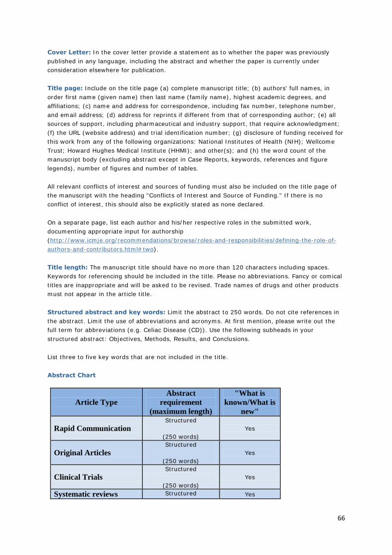
66
Cover Letter: In the cover letter provide a statement as to whether the paper was previously
published in any language, including the abstract and whether the paper is currently under
consideration elsewhere for publication.
Title page: Include on the title page (a) complete manuscript title; (b) authors' full names, in
order first name (given name) then last name (family name), highest academic degrees, and
affiliations; (c) name and address for correspondence, including fax number, telephone number,
and email address; (d) address for reprints if different from that of corresponding author; (e) all
sources of support, including pharmaceutical and industry support, that require acknowledgment;
(f) the URL (website address) and trial identification number; (g) disclosure of funding received for
this work from any of the following organizations: National Institutes of Health (NIH); Wellcome
Trust; Howard Hughes Medical Institute (HHMI); and other(s); and (h) the word count of the
manuscript body (excluding abstract except in Case Reports, keywords, references and figure
legends), number of figures and number of tables.
All relevant conflicts of interest and sources of funding must also be included on the title page of
the manuscript with the heading "Conflicts of Interest and Source of Funding." If there is no
conflict of interest, this should also be explicitly stated as none declared.
On a separate page, list each author and his/her respective roles in the submitted work,
documenting appropriate input for authorship
(http://www.icmje.org/recommendations/browse/roles-and-responsibilities/defining-the-role-of-
authors-and-contributors.html#two).
Title length: The manuscript title should have no more than 120 characters including spaces.
Keywords for referencing should be included in the title. Please no abbreviations. Fancy or comical
titles are inappropriate and will be asked to be revised. Trade names of drugs and other products
must not appear in the article title.
Structured abstract and key words: Limit the abstract to 250 words. Do not cite references in
the abstract. Limit the use of abbreviations and acronyms. At first mention, please write out the
full term for abbreviations (e.g. Celiac Disease (CD)). Use the following subheads in your
structured abstract: Objectives, Methods, Results, and Conclusions.
List three to five key words that are not included in the title.
Abstract Chart
Article Type
Abstract
requirement
(maximum length)
"What is
known/What is
new"
Rapid Communication
Structured
(250 words)
Yes
Original Articles Structured
(250 words)
Yes
Clinical Trials Structured
(250 words)
Yes
Systematic reviews Structured Yes
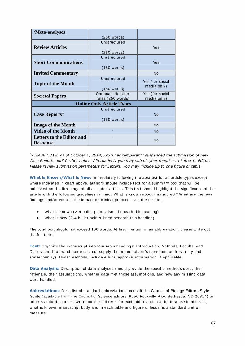
67
/Meta-analyses
(250 words)
Review Articles
Unstructured
(250 words)
Yes
Short Communications
Unstructured
(150 words)
Yes
Invited Commentary - No
Topic of the Month
Unstructured
(150 words)
Yes (for social
media only)
Societal Papers Optional -No strict
rules (250 words)
Yes (for social
media only)
Online Only Article Types
Case Reports*
Unstructured
(150 words)
No
Image of the Month - No
Video of the Month - No
Letters to the Editor and
Response
- No
*PLEASE NOTE: As of October 1, 2014, JPGN has temporarily suspended the submission of new
Case Reports until further notice. Alternatively you may submit your report as a Letter to Editor.
Please review submission parameters for Letters. You may include up to one figure or table.
What is Known/What is New: Immediately following the abstract for all article types except
where indicated in chart above, authors should include text for a summary box that will be
published on the first page of all accepted articles. This text should highlight the significance of the
article with the following guidelines in mind: What is known about this subject? What are the new
findings and/or what is the impact on clinical practice? Use the format:
What is known (2-4 bullet points listed beneath this heading)
What is new (2-4 bullet points listed beneath this heading)
The total text should not exceed 100 words. At first mention of an abbreviation, please write out
the full term.
Text: Organize the manuscript into four main headings: Introduction, Methods, Results, and
Discussion. If a brand name is cited, supply the manufacturer's name and address (city and
state/country). Under Methods, include ethical approval information, if applicable.
Data Analysis: Description of data analyses should provide the specific methods used, their
rationale, their assumptions, whether data met those assumptions, and how any missing data
were handled.
Abbreviations: For a list of standard abbreviations, consult the Council of Biology Editors Style
Guide (available from the Council of Science Editors, 9650 Rockville Pike, Bethesda, MD 20814) or
other standard sources. Write out the full term for each abbreviation at its first use in abstract,
what is known, manuscript body and in each table and figure unless it is a standard unit of
measure.

68
References: Please adhere to the reference limits noted for each article type above. The authors
are responsible for the accuracy of the references. Key the references (double-spaced) at the end
of the manuscript. Cite the references in text in the order of appearance. Cite unpublished data—
such as papers submitted but not yet accepted for publication and personal communications,
including email communications—in parentheses in the text. If there are more than three authors,
name only the first three authors and then use et al. Refer to the List of Journals Indexed in Index
Medicus for abbreviations of journal names, or access the list
athttp://www.nlm.nih.gov/tsd/serials/lji.html.
Sample references:
Journal article
I. Rautava S, Lu L, Nanthakumar NN, et al. TGF-β2 induces maturation of immature human
intestinal epithelial cells and inhibits inflammatory cytokine responses induced via the NF-κB
pathway. J Pediatr Gastroenterol Nutr 2012;54:630-8.
Book chapter
2. Todd VR. Visual information analysis: frame of reference for visual perception. In: Kramer P,
Hinojosa J, eds. Frames of Reference for Pediatric Occupational Therapy. Philadelphia: Lippincott
Williams & Wilkins; 1999:205–56.
Entire Book
3. Ming S-C, Goldman H. Pathology of the Gastrointestinal Tract. Philadelphia: Lippincott Williams
& Wilkins; 1998.
Software
4. Epi Info [computer program]. Version 6. Atlanta: Centers for Disease Control and Prevention;
1994.
Online journals
5. Friedman SA. Preeclampsia: a review of the role of prostaglandins. Obstet Gynecol [serial
online] January 1988;71: 22-37. Available from: BRS Information Technologies, McLean, VA.
Accessed December 15, 1990.
Database
6. CANCERNET-PDQ [database online]. Bethesda, MD: National Cancer Institute; 1996. Updated
March 29, 1996.
World Wide Web
7. Sullivan D. Major search engines and directories. SearchEngineWatch Web
site. http://www.searchenginewatch.com/links/article.php/2156221. Published May 8, 2011.
Accessed July 13, 2012.
Figure legends: Each figure must have a legend. Legends should be brief and should be typed on
a separate manuscript page, directly following the reference list. Use scale markers in the image
for electron micrographs, and indicate the type of stain used. Please let the editors and reviewers
know if any of the figures (e.g., figures of study design) are appropriate for the on-line
supplemental digital content (SDC) rather than needing to be in-print version.
Figures:
A) Creating Digital Artwork
69
Learn about the publication requirements for Digital Artwork: http://links.lww.com/ES/A42
Create, Scan and Save your artwork and compare your final figure to the Digital Artwork
Guideline Checklist (below).
Upload each figure to Editorial Manager in conjunction with your manuscript text and
tables.
B) Digital Artwork Guideline Checklist
Artwork should be saved in TIFF, Word Doc, PPT or EPS format (PDF is not recommended).
Artwork is created as the actual size (or slightly larger) it will appear in the journal. (To get
an idea of the size images should be when they print, study a copy of the journal to which
you wish to submit. Measure the artwork typically shown and scale your image to match.)
Crop out any white or black space surrounding the image.
Diagrams, drawings, graphs, and other line art must be vector or saved at a resolution of
at least 1200 dpi.
Photographs, radiographs and other halftone images must be saved at a resolution of at
least 300 dpi.
Photographs and radiographs with text must be saved as postscript or at a resolution of at
least 600 dpi.
Each figure must be saved and submitted as a separate file. Figures should not be
embedded in the manuscript text file.
C) Remember:
Cite figures consecutively in your manuscript.
Number figures in the figure legend in the order in which they are discussed.
Upload figures consecutively to the Editorial Manager web site and number figures
consecutively in the Description box during upload.
Tables: Cite tables consecutively in the text and number them in that order. Each table should be
submitted as a separate Word document in text format. Each table must have a title. Use
footnotes to define abbreviations and for other explanatory detail in a legend below the Tables.
Tables should be self-explanatory and must supplement, rather than duplicate, the material in the
text. Please let the editors and reviewers know if any of the tables (e.g., large data tables) are
appropriate for the on-line supplemental digital content (SDC) rather than needing to be in-print
version.
Supplemental Digital Content (SDC): Authors may submit supplemental digital content to
enhance their article's text and to be considered for online-only posting. Supplemental digital
content may include the following types of content: text documents, graphs, tables, figures,
graphics, illustrations, audio, and video. Cite all supplemental digital content consecutively in the
text. Citations should include the type of material submitted, should be clearly labeled as
"Supplemental Digital Content," should include a sequential number, and should provide a brief
description of the supplemental content. Provide a legend of supplemental digital content at the
end of the text, listing captions in the order in which the material is cited in the text. The legends
must be numbered to match the citations from the text. Include a title and a brief summary of the
content. For audio and video files, also include the author name, videographer, participants, length
(minutes), and size (MB). No patient-identifying information should be used in supplemental digital
content unless written consent from the patient, the patient's parents or the patient's guardian has
70
been obtained. Documentation regarding this consent must be submitted with the manuscript.
Copyright and Permission forms for article content including supplemental digital content must be
provided at the time of submission.
Supplemental Digital Content Size & File Type Requirements: To ensure a quality experience
for those viewing supplemental digital content, it is suggested that authors submit supplemental
digital files no larger than 10 MB each. Documents, graphs, and tables may be presented in any
format. Figures, graphics, and illustrations should be submitted with the following file extensions:
.tif, .eps, .ppt, .jpg, .pdf, .gif. Audio files should be submitted with the following file extensions:
.mp3, .wma. Video files should be submitted with the following file extensions: .wmv, .mov, .qt,
.mpg, .mpeg, .mp4. Video files should also be formatted with a 320 X 240 pixel minimum screen
size. For more information, please review Wolters Kluwer's requirements for submitting
supplemental digital content: http://links.lww.com/A142.
Reviewers: The JPGN editors encourage authors to submit names, departments, institutions, and
e-mail addresses of 3-7 potential reviewers with appropriate expertise to evaluate the manuscript.
These potential reviewers should be outside all authors' institution(s) and have no known potential
conflicts of interest. Names of persons who should not be asked to review the manuscript due to
potential conflicts of interest should also be submitted. Final choice of reviewers, however, remains
with the editors.
OPEN ACCESS
Wolters Kluwer's hybrid open access option is offered to authors whose articles have been
accepted for publication. With this choice, articles are made freely available online immediately
upon publication. Authors may take advantage of the open access option at the point of
acceptance to ensure that this choice has no influence on the peer review and acceptance process.
These articles are subject to the journal's standard peer-review process and will be accepted or
rejected based on their own merit.
Authors of accepted peer-reviewed articles have the choice to pay a fee to allow perpetual
unrestricted online access to their published article to readers globally, immediately upon
publication. The article processing charge for Journal of Pediatric Gastroenterology and Nutrition is
$3,000. The article processing charge for authors funded by the Research Councils UK (RCUK) is
$3,800. The publication fee is charged on acceptance of the article and should be paid within 30
days by credit card by the author, funding agency or institution. Payment must be received in full
for the article to be published open access.
Please note: There may be a short delay in PAP for open access articles.
Authors retain copyright
Authors retain their copyright for all articles they opt to publish open access. Authors grant
Wolters Kluwer a license to publish the article and identify itself as the original publisher.
Creative Commons license
Articles opting for open access will be freely available to read, download and share from the time
of publication. Articles are published under the terms of the Creative Commons License
Attribution-NonCommerical No Derivative 3.0 which allows readers to disseminate and reuse the
article, as well as share and reuse of the scientific material. It does not permit commercial
exploitation or the creation of derivative works without specific permission. To view a copy of this
license visit:http://creativecommons.org/licenses/by-nc-nd/3.0.
71
Compliance with NIH, RCUK, Wellcome Trust and other research funding agency
accessibility requirements
A number of research funding agencies now require or request authors to submit the post-print
(the article after peer review and acceptance but not the final published article) to a repository
that is accessible online by all without charge. As a service to our authors, Wolters Kluwer
identifies to the National Library of Medicine (NLM) articles that require deposit and transmits the
post-print of an article based on research funded in whole or in part by the National Institutes of
Health, Howard Hughes Medical Institute, or other funding agencies to PubMed Central. The
revised Copyright Transfer Agreement provides the mechanism. Wolters Kluwer ensures that
authors can fully comply with the public access requirements of major funding bodies worldwide.
Additionally, all authors who choose the open access option will have their final published article
deposited into PubMed Central.
RCUK and Wellcome funded authors can choose to publish their paper as open access with the
payment of an article process charge (gold route), or opt for their accepted manuscript to be
deposited (green route) into PMC with an embargo.
With both the gold and green open access options, the author will continue to sign the Copyright
Transfer Agreement (CTA) as it provides the mechanism for Wolters Kluwer to ensure that the
author is fully compliant with the requirements. After signature of the CTA, the author will then
sign a License to Publish where they will then own the copyright. Those authors who wish to
publish their article via the gold route will be able to publish under the terms of the Attribution 3.0
(CCBY) License. To view of a copy of this license
visit: http://creativecommons.org/licenses/by/2.0/. Those authors who wish to publish their article
via the green route will be able to publish under the rights of the Attribution Non-commercial 3.0
(CCBY NC) license (http://creativecommons.org/licenses/by-nc/2.0/).
It is the responsibility of the author to inform the Editorial Office and/or Wolters Kluwer that they
have RCUK funding. Wolters Kluwer will not be held responsible for retroactive deposits to PMC if
the author has not completed the proper forms.
FAQ for open access
http://links.lww.com/LWW-ES/A48
JPGN Editors:
Manuscripts submitted by members of the editorial board are subject to peer review. The decisions
and editorial process are handled by the alternate editorial office (with the exception of societal
papers). Manuscripts submitted with involvement of editorial board members from both the EU
and NA offices will be handled by the Consulting Editor.
JPGN Editor Submission Policy Disclaimer: Manuscript submissions authored by editors of
either editorial office will be processed by the other editorial office, with the exception of societal
manuscripts, Image/Video of the Month, and certain invited papers.
PRE-SUBMISSION CHECKLIST
Please use the following checklist to decrease the likelihood that your manuscript will be returned:
1. Title Page: Check for appropriate length and wording of the title. Title page should be on separate page from the abstract and manuscript.
2. Author and co-author information: Provide details for all of the authors/co-authors and
corresponding author on the title page. The authors should be listed as: first name first,
72
middle name second if applicable and family name last (bold surname/family name). Verify that co-author names, titles, affiliations and degrees are accurate and current.
3. On a separate page, list each author and his/her respective roles in the submitted work, documenting appropriate input for authorship
(http://www.icmje.org/recommendations/browse/roles-and-responsibilities/defining-the-role-of-authors-and-contributors.html#two).
4. Manuscript length and formatting: Check that the length of the manuscript and abstract do not exceed the maximum word count and are formatted as per instructions to authors for each. Abstract should be on a separate page from the title page and manuscript. See Abstract Chart
5. Figures: Do not embed figures in manuscript. Cite the figures in the text of the
manuscript. Provide figure legends on a separate page. Check that the figures and
supplementary figures are formatted as indicated in instructions to authors. Each figure should be in a separate file (uploaded individually).
6. Tables: Do not embed tables in manuscript. Cite the tables in the text of the manuscript. Provide table legends. Check that the tables and supplementary tables are formatted as indicated in instructions to authors. Each table should be in a separate file (uploaded
individually).
7. References: Make sure each reference is cited in the text, references are formatted per instructions to authors and the quantity does not exceed maximum for the article type. Double check the accuracy of each reference (spelling of names, page numbers, proper journal abbreviation, etc.) as any errors are the authors' responsibility.
8. Supplementary files and appendices: Make sure files are formatted per instructions to authors and cited in main text. Add legends to figure(s) and/or table legend(s) in the
manuscript.
9. JPGN recommends that authors of Original Articles including Clinical Trials comply with one of the appropriate reporting guidelines endorsed by the EQUATOR Network (e.g. CONSORT, PRISMA, etc.). More information may be found at http://www.equator-network.org. While authors are not required to submit a formal checklist at time of submission, the
editors may request it during the review process.
10. Ethics: Make sure to include the necessary ethical approval/clearance, clinical trial and/or parental or guardian approval information where applicable. If the submission is a clinical trial, include documentation that the project was registered in a public trials registry prior to enrollment of the first subject.
11. Permissions: If using previously published materials, include permission to re-use the
material obtained from the copyright holder as Supplemental Data and acknowledge the source in your legend.
12. Reviewers: Provide names, email addresses and other pertinent information for suggested reviewers and/or non-preferred reviewers, if applicable.
13. Revised manuscripts: include a point-by-point response to the comments of each reviewer in one separate file from the marked revision. The marked revised manuscript should show the revisions with hi-lighting in yellow or red text.
14. Copyright Transfer Agreement: All co-authors must respond to the e-mail regarding authorship and answer the questionnaire. If they are unable to respond to the e-mail, they must include a completed and signed copyright transfer agreement Submissions cannot be published without them.
73
AFTER ACCEPTANCE
Publication Ahead-of-Print (PAP): PAP provides rapid access to important new data, and
enables authors to cite their work promptly. All accepted submissions with completed copyright
and disclosure forms will be published in manuscript form online within approximately 5 business
days with a watermark denoting "accepted draft". To avoid errors and delays, submitted
manuscripts must be carefully proofed by authors prior to final submission. Typos and other
textual errors may delay final acceptance, or may be published online with errors that
were not detected by the reviewers and will not be corrected until the final print
version.
Page proofs and corrections: Corresponding authors will receive electronic page proofs to check
the copyedited and typeset article before publication. Portable document format (PDF) files of the
typeset pages and support documents (e.g., reprint order form) will be sent to the corresponding
author by email. Complete instructions will be provided with the email for downloading and
printing the files and for faxing the corrected page proofs to the publisher. Those authors without
an email address will receive traditional page proofs. It is the author's responsibility to ensure that
there are no errors in the proofs. Changes that have been made to conform to journal style will
stand if they do not alter the authors' meaning. Only the most critical changes to the accuracy of
the content will be made. Changes that are stylistic or are a reworking of previously accepted
material will be disallowed. The publisher reserves the right to deny any changes that do not affect
the accuracy of the content. Authors may be charged for alterations to the proofs beyond those
required to correct errors or to answer queries. Proofs must be checked carefully and corrections
faxed within 24 to 48 hours of receipt, as requested in the cover letter accompanying the page
proofs.
Reprints: Authors will receive a reprint order form and a price list with the page proofs. Reprint
requests should be faxed to the publisher with the corrected proofs, if possible. Reprints are
normally shipped 6 to 8 weeks after publication of the issue in which the item appears. Contact the
Reprint Department, Wolters Kluwer, 351 W. Camden Street, Baltimore, MD 21201; Fax:
+1.410.528.4434; Email: [email protected] with any questions.
CME: Accepted manuscripts may be selected to offer continue medical education (CME). If so, the
Editorial Office/CME Editor will send an invitation to the communicating author requesting
materials such as a list of 3-4 learning objectives and 5 multiple choice test questions. If invited, it
is important to meet the deadlines for CME submission and revisions. Failure to do so could result
in publication delays.
Fees: JPGN does not charge submission fees, page fees, or color figure fees.
EDITORIAL OFFICE CONTACTS:
Manuscript Submission:
http://jpgn.edmgr.com
NASPGHAN Editor:
Melvin B. Heyman, MD, MPH
Pediatric Gastroenterology and Nutrition and Anita Ow Wing Endowed Chairs
Professor of Pediatrics
74
Department of Pediatrics, Division of Pediatric Gastroenterology, Hepatology and Nutrition
University of California, San Francisco
San Francisco, CA
NASPGHAN Managing Editor:
Naima Stone
Email: [email protected]
Phone: 215-521-8816
Fax: 215-827-5586
ESPGHAN Editor:
Prof. Hania Szajewska, M.D.
The Medical University of Warsaw
Department of Paediatrics
Zwirki i Wigury 63A, 02-091 Warsaw, Poland
ESPGHAN Managing Editor:
Phyllis Barr
Phone: 610-205-9318, 972-52-578-3708 Fax: 972-73-726-4439
Email: [email protected]
C o p y r i g h t / D i s c l a i m e r N o t i c e • P r i v a c y P o l i c y
75
ACKNOWLEDGEMENTS
I would like to acknowledge the staff of RCWMCH records department for their assistance in locating
patient files.
I would also like to thank Dr. Rudzani Muloiwa for his invaluable assistance performing the statistical
analysis.


![Ultrasonographic findings of type IIIa biliary atresiabiliary atresia. Further, there have been only a few reports on the US findings of biliary atresia based on its types [33,34]](https://img.pdfslide.us/doc/110x75/60a90a6926e7a533947d7637/ultrasonographic-findings-of-type-iiia-biliary-atresia-biliary-atresia-further.jpg)