Embed Size (px)
Citation preview

147
INTRODUCTION
Rhabdomyosarcoma is the most common primary malignant neoplasm of the orbit in children (Shields et al., 1984). When it occurs in the orbit, it is almost
always a primary lesion (Jones et al., 1965; Kassel et al., 1965). Rhabdomyosarcoma is composed of cells with histologic features of muscle in various stages of skeletal muscle differentiation. It arises from primitive mesenchymal tissue and not from skeletal muscle. Metastatic orbital rhabdomyosarcoma uncommonly involves the extraocular muscles. The present report describes a case of metastasis to all the extraocular muscles bilaterally in a 14-year-old child with a primary non-orbital rhabdomyosarcoma.
Orbit, 29(3), 147–149, 2010Copyright © 2010 Informa Healthcare USA, Inc.ISSN: 0167-6830 print/ 1744-5108 onlineDOI: 10.3109/01676830903294917
case report
Bilateral Symmetrical Metastasis to All Extraocular Muscles from Distant
Rhabdomyosarcoma
Pankaj Gupta, MS1, Usha Singh, MS1, Shrawan Kumar Singh, MS2, Dr Rakesh Kapoor, MD3, Dr Vivek Gupta, MD4, and Dr Ashim Das, MS5
1Department of Ophthalmology, Post Graduate Institute of Medical Education and Research, Chandigarh 160012 India2Department of Urology, Post Graduate Institute of Medical Education and Research, Chandigarh 160012 India
3Department of Radiotherapy, Post Graduate Institute of Medical Education and Research, Chandigarh 160012 India4Department of Radiodiagnosis, Post Graduate Institute of Medical Education and Research, Chandigarh 160012 India5Department of Histopathology, Post Graduate Institute of Medical Education and Research, Chandigarh 160012 India
ABSTRACT
Introduction: Rhabdomyosarcoma arising in the inguinal region has high potential of metastasis. The common sites for spread from inguinal region include regional lymph nodes, lungs, bone marrow and bone cortex. Orbit is an uncommon site for such metastasis. This case report describes a patient with inguinal rhabdomyosarcoma, which metastasized to both orbits to all the extraocular muscles.Case Report: A 14-year-old male patient presented with inguinal mass involving the scrotum. The patient underwent high inguinal orchiectomy with hemiscrotectomy for the mass and histopathology revealed rhabdomyosarcoma. After 2 weeks of initial surgery the patient developed bilateral axial proptosis and radiological imaging revealed bilateral extraocular muscle thickening involving all the extraocular muscles. A biopsy of right superior rectus muscle confirmed rhabdomyosarcoma.Comment: Although rhabdomyosarcoma is the commonest primary orbital malignant mass developing in young patients, it is an uncommon metastasis. Metastasis from inguinal rhabdomyosarcoma to extraocular muscles bilaterally involving all the muscles has not been reported in the literature. The present report describes one such patient with favorable initial response to chemotherapy and muscle thickness reverting to normal. Metastasis from a distant site should be considered in differential diagnosis when evaluating a patient with bilateral enlargement of all extraocular muscles.
KEYWORDS: rhabdomyosarcoma; Metastasis; extraocular muscles; Bilateral
Received 09 August 2009; revised 22 August 2009; accepted 23 August 2009
Correspondence: Dr. Pankaj Gupta, Department of Ophthalmology, PGIMER, Chandigarh 160012. E-mail [email protected]
09 August 2009
22 August 2009
23 August 2009
© 2010 Informa Healthcare USA, Inc.
2010
Orbit
0167-68301744-5108
10.3109/01676830903294917
29
147149
3
Orb
it D
ownl
oade
d fr
om in
form
ahea
lthca
re.c
om b
y U
nive
rsity
of
Bri
tish
Col
umbi
a on
10/
29/1
4Fo
r pe
rson
al u
se o
nly.
148 P. Gupta et al.
orbit
Case Report
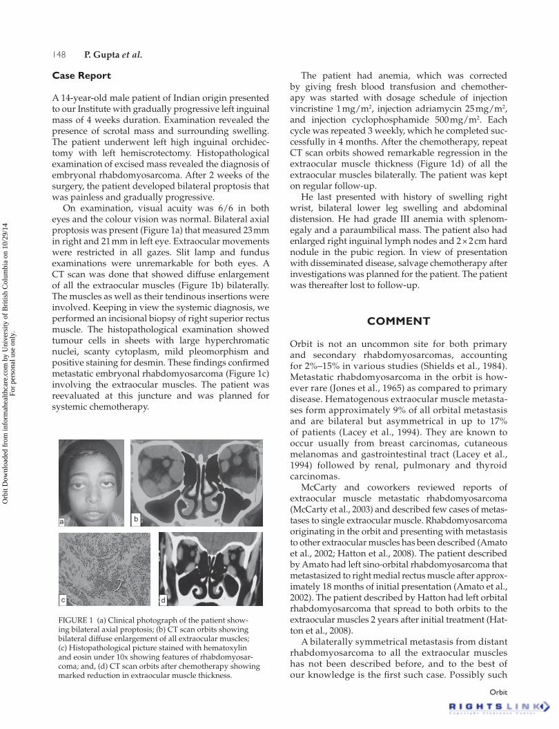
A 14-year-old male patient of Indian origin presented to our Institute with gradually progressive left inguinal mass of 4 weeks duration. Examination revealed the presence of scrotal mass and surrounding swelling. The patient underwent left high inguinal orchidec-tomy with left hemiscrotectomy. Histopathological examination of excised mass revealed the diagnosis of embryonal rhabdomyosarcoma. After 2 weeks of the surgery, the patient developed bilateral proptosis that was painless and gradually progressive.
On examination, visual acuity was 6/6 in both eyes and the colour vision was normal. Bilateral axial proptosis was present (Figure 1a) that measured 23 mm in right and 21 mm in left eye. Extraocular movements were restricted in all gazes. Slit lamp and fundus examinations were unremarkable for both eyes. A CT scan was done that showed diffuse enlargement of all the extraocular muscles (Figure 1b) bilaterally. The muscles as well as their tendinous insertions were involved. Keeping in view the systemic diagnosis, we performed an incisional biopsy of right superior rectus muscle. The histopathological examination showed tumour cells in sheets with large hyperchromatic nuclei, scanty cytoplasm, mild pleomorphism and positive staining for desmin. These findings confirmed metastatic embryonal rhabdomyosarcoma (Figure 1c) involving the extraocular muscles. The patient was reevaluated at this juncture and was planned for systemic chemotherapy.
The patient had anemia, which was corrected by giving fresh blood transfusion and chemother-apy was started with dosage schedule of injection vincristine 1 mg/m2, injection adriamycin 25 mg/m2, and injection cyclophosphamide 500 mg/m2. Each cycle was repeated 3 weekly, which he completed suc-cessfully in 4 months. After the chemotherapy, repeat CT scan orbits showed remarkable regression in the extraocular muscle thickness (Figure 1d) of all the extraocular muscles bilaterally. The patient was kept on regular follow-up.
He last presented with history of swelling right wrist, bilateral lower leg swelling and abdominal distension. He had grade III anemia with splenom-egaly and a paraumbilical mass. The patient also had enlarged right inguinal lymph nodes and 2 × 2 cm hard nodule in the pubic region. In view of presentation with disseminated disease, salvage chemotherapy after investigations was planned for the patient. The patient was thereafter lost to follow-up.
COMMENT
Orbit is not an uncommon site for both primary and secondary rhabdomyosarcomas, accounting for 2%–15% in various studies (Shields et al., 1984). Metastatic rhabdomyosarcoma in the orbit is how-ever rare (Jones et al., 1965) as compared to primary disease. Hematogenous extraocular muscle metasta-ses form approximately 9% of all orbital metastasis and are bilateral but asymmetrical in up to 17% of patients (Lacey et al., 1994). They are known to occur usually from breast carcinomas, cutaneous melanomas and gastrointestinal tract (Lacey et al., 1994) followed by renal, pulmonary and thyroid carcinomas.
McCarty and coworkers reviewed reports of extraocular muscle metastatic rhabdomyosarcoma (McCarty et al., 2003) and described few cases of metas-tases to single extraocular muscle. Rhabdomyosarcoma originating in the orbit and presenting with metastasis to other extraocular muscles has been described (Amato et al., 2002; Hatton et al., 2008). The patient described by Amato had left sino-orbital rhabdomyosarcoma that metastasized to right medial rectus muscle after approx-imately 18 months of initial presentation (Amato et al., 2002). The patient described by Hatton had left orbital rhabdomyosarcoma that spread to both orbits to the extraocular muscles 2 years after initial treatment (Hat-ton et al., 2008).
A bilaterally symmetrical metastasis from distant rhabdomyosarcoma to all the extraocular muscles has not been described before, and to the best of our knowledge is the first such case. Possibly such
a b
c d
FIGURE 1 (a) Clinical photograph of the patient show-ing bilateral axial proptosis; (b) CT scan orbits showing bilateral diffuse enlargement of all extraocular muscles; (c) Histopathological picture stained with hematoxylin and eosin under 10x showing features of rhabdomyosar-coma; and, (d) CT scan orbits after chemotherapy showing marked reduction in extraocular muscle thickness.
Orb
it D
ownl
oade
d fr
om in
form
ahea
lthca
re.c
om b
y U
nive
rsity
of
Bri
tish
Col
umbi
a on
10/
29/1
4Fo
r pe
rson
al u
se o
nly.
Bilateral Metastatis Rhabdomyosarcoma 149
© 2010 Informa Healthcare USA, Inc.
metastasis occurs due to metastatic homing of rhabdomyosarcoma to muscles though etiology of such behavior of malignant cells can only be hypoth-esised presently. An unusual feature in our patient was metastasis occurring within weeks of initial pre-sentation and involving all the extraocular muscles. The patient responded well initially to chemotherapy and extraocular muscle thickness reduced consider-ably. Symmetrical involvement of all extraocular muscles bilaterally in a patient with known history of malignancy should make the surgeons consider metastasis from a distant primary site. Histopathol-ogy played an important role in the diagnosis of our patient and such cases if diagnosed should be promptly treated.
Declaration of interest: The authors report no conflicts of interest. The authors alone are responsible for the content and writing of the paper.
REFERENCES
[1] Amato MM, Esmaeli B, Shore JW. Orbital rhabdomyosarcoma metastatic to contralateral orbit: a case report. Ophthalmology. 2002;109:753–756.
[2] Hatton MP, Green L, Boulos PR, Rubin PAD. Rhabdomyo-sarcoma metastatic to all extraocular muscles. Opthal Plast Reconstr Surg. 2008;24(4):336–338.
[3] Jones IS, Reese AB, Krout J. Orbital rhabdomyosarcoma: an analysis of sixty two cases. Trans Am Ophthalmol Soc. 1965;63:223–255.
[4] Kassel SH, Copenhaver R, Arean VM. Orbital rhabdomyo-sarcoma. Am J Ophthalmol. 1965;60:811–818.
[5] Lacey B, Chang W, Rootman J. Nonthyroid causes of extraoc-ular muscle disease. Surv Ophthalmol. 1994;44(3):187–213.
[6] McCarty ML, Wilson MW, Ibrahim F, Fuller CE, Kun LE, Haik BG. Primary perineal alveolar rhabdomyosarcoma metastatic to an extraocular muscle. Ophthal Plast Reconstr Surg. 2003;19:333–335.
[7] Shields JA, Bakewell B, Augsburger JJ, Flanagan JC. Classification and incidence of space occupying lesions of the orbit: a survey of 645 biopsies. Arch Ophthalmol. 1984;102:1606–1611.
Orb
it D
ownl
oade
d fr
om in
form
ahea
lthca
re.c
om b
y U
nive
rsity
of
Bri
tish
Col
umbi
a on
10/
29/1
4Fo
r pe
rson
al u
se o
nly.