Embed Size (px)
Citation preview

Letter to the Editor
Vol. 27, No. 1, 2015 115
Received November 22, 2012, Revised March 10, 2013, Accepted for publication April 21, 2014
Corresponding author: Jeong Deuk Lee, Department of Dermatology, Incheon St. Mary's Hospital, College of Medicine, The Catholic University of Korea, 56 Dongsu-ro, Bupyeong-gu, Incheon 403-720, Korea. Tel: 82-32-280-5842, Fax: 82-32-506-9514, E-mail: [email protected]
This is an Open Access article distributed under the terms of the Creative Commons Attribution Non-Commercial License (http:// creativecommons.org/licenses/by-nc/3.0) which permits unrestricted non-commercial use, distribution, and reproduction in any medium, provided the original work is properly cited.
well-established. It is known that SpSCCs that are asso-ciated with radiation, burn scars, and immunosuppression may correlate with aggressive clinical courses5. Further studies of this rare entity are needed to establish its bio-logical behavior, and the accumulation of more case re-ports will aid in determining a precise prognosis.
REFERENCES
1. Lewis JE, Olsen KD, Sebo TJ. Spindle cell carcinoma of the
larynx: review of 26 cases including DNA content and
immunohistochemistry. Hum Pathol 1997;28:664-673.
2. Bolognia JL, Jorizzo JL, Schaffer JV, Cerroni L, Heymann
WR, Callen JP, et al. Dermatology Vol 2. London: Mosby,
2013:1681-1685.
3. Cassarino DS, Derienzo DP, Barr RJ. Cutaneous squamous cell
carcinoma: a comprehensive clinicopathologic classification.
Part one. J Cutan Pathol 2006;33:191-206.
4. Morgan MB, Purohit C, Anglin TR. Immunohistochemical
distinction of cutaneous spindle cell carcinoma. Am J
Dermatopathol 2008;30:228-232.
5. Kurzen H, Zeier M, Zelger B, Hartschuh W. Unusual spindle
cell squamous carcinoma in a renal transplant patient. Acta
Derm Venereol 2004;84:61-64.
http://dx.doi.org/10.5021/ad.2015.27.1.115
Bilateral Segmental Neurofibromatosis on the Face
Woo Seok Jeon, Hei Sung Kim, Sang Hyun Cho, Jeong Deuk Lee
Department of Dermatology, Incheon St. Mary’s Hospital, College of Medicine, The Catholic University of Korea, Incheon, Korea
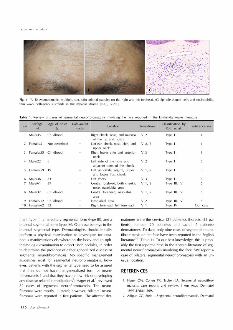
Dear Editor:Neurofibromatosis is a heterogeneous disorder clinically characterized by the presence of neurofibromas, multiple café-au-lait spots, intertriginous freckles, and Lisch nod-ules1.A 62-year-old woman presented with diffusely scattered papules on the forehead that had been present for 10 years. She had no family history of neurofibromatosis. On physical examination, asymptomatic, soft, flesh-colored papules of 3∼5 mm diameter were observed to be dis-tributed over the ophthalmic branch of the right and left trigeminal nerves (Fig. 1A, B). There were no other abnor-malities in any other body region. Her general physical examination revealed a normal status, including in-telligence, speech, auditory function, and visual acuity.
Histopathological examination showed a well-circum-scribed tumor in the dermis with a normal overlying epidermis. The tumor consisted of loosely spaced spin-dle-shaped cells and wavy collagenous strands in the myx-oid stroma. Nuclear pleomorphism and mitoses were not observed (Fig. 1C). Overall, the features were consistent with a neurofibroma. The papules on the forehead were excised for cosmetic reason.Segmental neurofibromatosis is a rare form of neuro-fibromatosis that is characterized by cafe-au-lait macules and neurofibromas, or only neurofibromas, distributed in only one dermatome, and less commonly in two or more dermatomes1. Segmental neurofibromatosis was catego-rized into four subtypes by Roth et al. in 1987: a true seg-mental form (type I), a localized form with deep involve-
Letter to the Editor
116 Ann Dermatol
Table 1. Review of cases of segmental neurofibromatosis involving the face reported in the English-language literature
CaseSex/age
(y)Age of onset
(y)Café-au-lait
spotsLocation Dermatome
Classification by Roth et al.
Reference no.
1 Male/45 Childhood − Right cheek, nose, and mucosa of the lip and nostril
V 2 Type I 1
2 Female/51 Not described − Left ear, cheek, nose, chin, and upper neck
V 2, 3 Type I 1
3 Female/35 Childhood − Right lower chin and anterior neck
V 3 Type I 1
4 Male/22 6 − Left side of the nose and adjacent parts of the cheek
V 2 Type I 5
5 Female/59 19 + Left periorbital region, upper and lower lids, cheek
V 1, 2 Type I 3
6 Male/38 33 − Left cheek V 3 Type I 4 7 Male/61 39 − Central forehead, both cheeks,
nose, nasolabial areaV 1, 2 Type III, IV 5
8 Male/37 Childhood − Central forehead, nasolabial area
V 1, 2 Type III, IV 5
9 Female/12 Childhood − Nasolabial area V 2 Type III, IV 510 Female/62 52 − Right forehead, left forehead V 1 Type IV Our case
Fig. 1. (A, B) Asymptomatic, multiple, soft, skin-colored papules on the right and left forehead. (C) Spindle-shaped cells and eosinophilic, thin wavy collagenous strands in the myxoid stroma (H&E, ×200).
ment (type II), a hereditary segmental form (type III), and a bilateral segmental form (type IV). Our case belongs to the bilateral segmental type. Dermatologists should initially perform a physical examination to investigate for cuta-neous manifestations elsewhere on the body and an oph-thalmologic examination to detect Lisch nodules, in order to determine the presence of either generalized disease or segmental neurofibromatosis. No specific management guidelines exist for segmental neurofibromatosis; how-ever, patients with the segmental type need to be assured that they do not have the generalized form of neuro-fibromatosis-1 and that they have a low risk of developing any disease-related complications2. Hager et al.1 reviewed 82 cases of segmental neurofibromatosis. The neuro-fibromas were mostly ulilateral; however, bilateral neuro-fibromas were reported in five patients. The affected der-
matomes were the cervical (31 patients), thoracic (33 pa-tients), lumbar (20 patients), and sacral (5 patients) dermatomes. To date, only nine cases of segmental neuro-fibromatosis on the face have been reported in the English literature3-5 (Table 1). To our best knowledge, this is prob-ably the first reported case in the Korean literature of seg-mental neurofibromatosis involving the face. We report a case of bilateral segmental neurofibromatosis with an un-usual location.
REFERENCES
1. Hager CM, Cohen PR, Tschen JA. Segmental neurofibro-
matosis: case reports and review. J Am Acad Dermatol
1997;37:864-869.2. Adigun CG, Stein J. Segmental neurofibromatosis. Dermatol

Letter to the Editor
Vol. 27, No. 1, 2015 117
Received June 24, 2013, Revised January 23, 2014, Accepted for publication April 27, 2014
Corresponding author: Jose Luis Torregrosa Calatayud, Department of Dermatology, General Hospital of Valencia, Avenida Tres Cruces Nº2, Valencia 46014, Spain. Tel: 34-665242548, Fax: 34-963-131-297, E-mail: [email protected]
This is an Open Access article distributed under the terms of the Creative Commons Attribution Non-Commercial License (http:// creativecommons.org/licenses/by-nc/3.0) which permits unrestricted non-commercial use, distribution, and reproduction in any medium, pro-vided the original work is properly cited.
Online J 2011;17:25.3. López-Cepeda LD, Domínguez-Gómez MA, Novales-Santa
CJ, Guarneros-Campos A. Segmental neurofibromatosis of
facial localization. Int J Dermatol 2005;44:583-586.4. Agarwal A, Thappa DM, Jayanthi S, Shivaswamy KN.
Segmental neurofibromatosis of face. Dermatol Online J 2005;11:33.
5. Jankovic I, Kovacevic P, Visnjic M, Jankovic D, Velickovic
M. A unique case of hereditary bilateral segmental neuro-fibromatosis on the face. An Bras Dermatol 2012;87:895-898.
http://dx.doi.org/10.5021/ad.2015.27.1.117
Retiform Purpura Caused by the Use of Cocaine, That Was Probably Adulterated with Levamisole
Jose Luis Torregrosa Calatayud, Juan Garcías Ladaria, Blanca De Unamuno Bustos, Violeta Zaragoza Ninet, Victor Alegre De Miquel
Department of Dermatology, General Hospital of Valencia, Valencia, Spain
Dear Editor:Retiform purpura is a dermatological condition charac-terized by reticulated, stellate, or serpentine shaped pur-ple lesions on the skin and mucous membranes. New, multiple cases of retiform purpura after the use of levami-sole adulterated cocaine have been reported. Levamisole is an anthelmintic drug with immunomodulatory and im-munostimulating properties. It has been used in humans to treat rheumatoid arthritis, cancer of the colon and neph-ritic syndrome in children. It was withdrawn from use in the United States in 2000 because of the risk of agranulo-cytosis1.We report the case of a 52-year-old woman receiving treatment with levothyroxine for hypothyroidism. Two days after consuming cocaine, she developed painful skin lesions with arthralgia on both wrists. Physical examina-tion revealed plaques and papules infiltrated to touch, pur-puric on the edges and necrotic in the center, with retic-ular and stellate lesions on both cheeks, the tip of the
nose, outer left ear, and lower limbs (Fig. 1). Biopsy re-vealed thrombotic vasculopathy of the small and medium blood vessels in the dermis and subcutaneous cell tissue (Fig. 2). Blood tests revealed leukopenia, neutropenia, and lymphopenia. Antinuclear antibodies (ANA, titer 1 : 1.280) in anti-neutrophil cytoplasm antibodies (ANCAs) against myeloperoxidase with a p-ANCA pattern (titer, >100 [0∼5]), and ANCAs against proteinase 3 with a c-ANCA pat-tern (titer, 6.8 [0∼5]) were also found. Hypocomplemen-temia of C3 was detected. The tests for thrombosis and co-agulation, serology, cryoglobulins and antiphospholipid antibodies were normal or negative. Cocaine was detected in the urine sample. The results of chest radiography and urine sediment test were normal. A diagnosis of retiform purpura resulting from the use of cocaine, that was prob-ably adulterated with levamisole was made. She was pre-scribed with low dose oral prednisone. The hematological symptoms cleared 5 days later, after one month, the skin lesions had healed without sequelae.