Embed Size (px)
Citation preview
BioMed CentralBMC Ophthalmology
ss
Open AcceCase reportBilateral neuro-retinitis following chick embryo cell anti-rabies vaccination – a case reportRohit Saxena, Harinder Singh Sethi*, Harminder Kumar Rai and Vimla MenonAddress: Dr. R. P. Centre for Ophthalmic Sciences, All India Institute of Medical Sciences, New Delhi 110029, India
Email: Rohit Saxena - [email protected]; Harinder Singh Sethi* - [email protected]; Harminder Kumar Rai - [email protected]; Vimla Menon - [email protected]
* Corresponding author
AbstractBackground: The Optic nerve is rarely involved after sheep brain anti-rabies vaccination in theform of retrobulbar neuritis or papillitis. Bilateral neuroretinitis after chick embryo cell antirabiesvaccination has not been reported.
Case presentation: We report the case of a 56 year old male who developed bilateral neuro-retinitis following three injections of antirabies vaccine prepared from the chick embryo.
Conclusion: The chick embryo cell antirabies vaccine can cause bilateral neuroretinits which hasnot been reported previously.
BackgroundThe Optic nerve is rarely involved after sheep brain anti-rabies vaccination. Neurological complications are usu-ally seen with sheep brain vaccines but can be rarely seenafter chick embryo cell vaccines [1]. The main cause insuch cases is presumed to be the antigenic cerebral tissueused in the preparation of sheep brain vaccine [2-5]. Wereport the case of a 56 year old male who developed bilat-eral neuro-retinitis following three injections of antirabiesvaccine prepared from the chick embryo. Retrobular neu-ritis and papillitis following sheep brain antirabies vac-cine have been reported [6-11]. The present reportdescribes a case with bilateral neuroretinitis after chickembryo antirabies vaccine, which to best of our knowl-edge has not been reported earlier.
Case presentationA 56 year old male presented with complaint of acuteonset painless diminution of vision in the right eye of 3days duration followed by similar complaint in left eyeafter 1 day which deteriorated over the next 2 days. Thiswas associated with headache and pain on ocular move-ments. There was a history of being bitten by a stray dog 8days before the visual symptoms, and the patient hadreceived three injections of chick embryo cell anti-rabiesvaccine (Rabipur, Hoeshst Marion Roussel) on day 0, 3and 7 after the dog bite. He developed these symptomsafter the third injection. There was no history of any otherocular or systemic problems.
The general physical examination was within normal lim-its. The best-corrected visual acuity was 6/60 in the rightand 6/24 in the left eye. IOP was 16 mm of Hg by appla-nation tonometry. Anterior segment examination using
Published: 17 August 2005
BMC Ophthalmology 2005, 5:20 doi:10.1186/1471-2415-5-20
Received: 18 May 2005Accepted: 17 August 2005
This article is available from: http://www.biomedcentral.com/1471-2415/5/20
© 2005 Saxena et al; licensee BioMed Central Ltd. This is an Open Access article distributed under the terms of the Creative Commons Attribution License (http://creativecommons.org/licenses/by/2.0), which permits unrestricted use, distribution, and reproduction in any medium, provided the original work is properly cited.
Page 1 of 3(page number not for citation purposes)
BMC Ophthalmology 2005, 5:20 http://www.biomedcentral.com/1471-2415/5/20
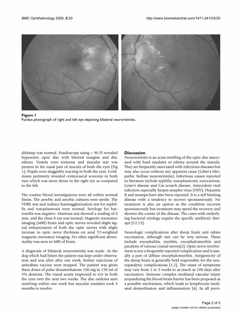
slitlamp was normal. Fundoscopy using + 90 D revealedhyperemic optic disc with blurred margins and discedema. Vessels were tortuous and macular star waspresent in the nasal part of macula of both the eyes (Fig1). Pupils were sluggishly reacting in both the eyes. Gold-mann perimetry revealed centrocaecal scotoma in botheyes which was more dense in the right eye as comparedto the left.
The routine blood investigations were all within normallimits. The aerobic and aerobic cultures were sterile. TheVDRL test and indirect haemagglutination test for syphil-lis and toxoplasmosis were normal. Serology for bar-tonella was negative. Mantoux test showed a reading of 8mm. and the chest X-ray was normal. Magnetic resonanceimaging (MRI) brain and optic nerves revealed slight sig-nal enhancement of both the optic nerves with slightincrease in optic nerve thickness on axial T2-weightedmagnetic resonance imaging. No other significant abnor-mality was seen in MRI of brain.
A diagnosis of bilateral neuroretinitis was made. As thedog which had bitten the patient was kept under observa-tion and was alive after one week, further injections ofantirabies vaccine were stopped. The patient was giventhree doses of pulse dexamethasone 100 mg in 150 ml of5% dextrose. The visual acuity improved to 6/6 in boththe eyes over the next two weeks. The disc oedema startresolving within one week but macular exudates took 4months to resolve.
DiscussionNeuroretinitis is an acute swelling of the optic disc associ-ated with hard exudates or edema around the macula.They are frequently associated with infectious diseases butmay also occur without any apparent cause (Leber's Idio-pathic Stellate neuroretinitis). Infectious causes reportedin literature include syphilis, toxoplasmosis, toxocariosis,Lyme's disease and Cat scratch disease. Antecedent viralinfection especially herpes simplex virus (HSV), HepatitisB and mumps have also been reported. It is a self limitingdisease with a tendency to recover spontaneously. Notreatment is also an option as the condition recoversspontaneously but treatment may speed the recovery andshorten the course of the disease. The cases with underly-ing bacterial etiology require the specific antibiotic ther-apy [12,13].
Neurologic complications after sheep brain anti rabiesvaccination, although rare can be very serious. Theseinclude encephalitis, myelitis, encephalomyelitis andparalysis of various cranial nerves[2]. Optic nerve involve-ment is not a frequently reported complication and is usu-ally a part of diffuse encephalomyelitis. Antigenecity ofthe sheep brain is generally held responsible for the neu-roparalytic complications [1,2]. The onset of symptomsmay vary from 1 to 3 weeks to as much as 100 days aftervaccination. Immune complex mediated vascular injuryjeopardizing the blood-brain barrier has been proposed asa possible mechanism, which leads to lymphocyte medi-ated demyelinaton and inflammation [4]. In all previ-
Fundus photograph of right and left eye depicting bilateral neuroretinitisFigure 1Fundus photograph of right and left eye depicting bilateral neuroretinitis.
Page 2 of 3(page number not for citation purposes)

BMC Ophthalmology 2005, 5:20 http://www.biomedcentral.com/1471-2415/5/20
Publish with BioMed Central and every scientist can read your work free of charge
"BioMed Central will be the most significant development for disseminating the results of biomedical research in our lifetime."
Sir Paul Nurse, Cancer Research UK
Your research papers will be:
available free of charge to the entire biomedical community
peer reviewed and published immediately upon acceptance
cited in PubMed and archived on PubMed Central
yours — you keep the copyright
Submit your manuscript here:http://www.biomedcentral.com/info/publishing_adv.asp
BioMedcentral
ously reported cases of optic neuritis following antirabiesvaccination, sheep brain vaccine had been used. Howeverour present case had received chick embryo vaccine.Chakravarty has reported a single case of neurologic ill-ness simultating Guillain Barre syndrome after post-expo-sure prophylaxis with purified chick embryo cell anti-rabies vaccine [1]. Apart from this, no published reportsare available regarding the neurological complication fol-lowing purified chick embryo cell vaccination (althoughmanufacturer's information on their websites does men-tion the risk of neurological complications with somechick embryo vaccines). This case was also unusual in thesense that patient has neuroretinits as compared to previ-ously reported cases which have retrobulbar neuritis orpapillitis [6-11]. Although good recovery is reportedspontaneously, pulse steroids speed up the recovery. Ourpatient had good visual recovery after three doses of pulsesteroids. As brain tissue and perhaps chick embryo tissue(as in our case) is thought to be the cause of the neuropar-alytic complications, it is suggested that recombinant vac-cines may offer a solution to this problem.
ConclusionThe chick embryo antirabies vaccine can cause bilateralneuroretinits which has not been reported previously.
Competing interestsThe author(s) declare that they have no competinginterests.
Authors' contributionsRS : Helped in making the clinical diagnosis and manage-ment of the case.
HSS : Maintained the follow-up record, and prepared themanuscript.
HKR : Performed the literature search and helped in doc-umentation of case
VM : Helped in making the clinical diagnosis and manage-ment of the case and supervised the case report.
All authors read and approved the final manuscript.
AcknowledgementsNone
References1. Chakravarty A: Neurologic illness following post-exposure
prophylaxis with purifiled chick embryo cell antirabiesvaccine. J Assoc Physicians India 2001, 49:927-8.
2. Raghvan Veera N: Rabies and its prevention. In Vepery, Madras,India The Diocesan Press; 1955:17.
3. Brain WR: Diseases of the Nervous System. 6th edition. NewYork: Oxford University Press; 1962.
4. Stevenson VL, Acheson JF, Ball J, Plant GT: Optic neuritis followingmeasles/rubella vaccination in two 13-year-old children. Br JOphthalmol 1996, 80:1110-1111.
5. Fenichel GM: Neurological complication after immunization.Ann Neurol 1982, 12:119-28.
6. Cormack HS, Anderson LAP: Bilateral papillitis following antir-abies innoculation recovery. Br J Ophthalmol 1934, 18:167-8.
7. Consul BN, Purohit GK, Chabra HN: Antirabies vaccine opticneuritis. Indian Med Sci 1968, 22:630-632.
8. Srisupan V, Konyama K: Bilateral retrobulbar optic neuritis fol-lowing antirabies vaccination. Sriraj Hosp Gaz 1970, 4:403-8.
9. Chayakul V, Ishikawa S, Chotibut S, Konyama K: Convergenceinsufficiency and optic neuritis due to antirabies innoculation– a case study. Jap J ophthalmol 1975, 19:307-14.
10. Gupta V, Bandyopadhyay S, Bapuraj JR, Gupta A: Bilateral opticneuritis complicating rabies vaccination. Retina 2004,24:179-181.
11. Dadeya s, Guliani BP, Gupta VS, Mallick KPS, Jain DC: Retrobulbarneuritis following rabies vaccination. Tropical doctor 2004,34:174-175.
12. Neuroretinitis: Walsh & Hoyt's clinical Neuro – ophthalmology Volume I.5th edition. Edited by: Miller NR, Newman J. williams & Wilkins Balti-more Mariland, USA; 1998:634-647.
13. Neuroretinitis: Neuro – Ophthalmology. Diagnosis and management Vol-ume . 1st edition. Edited by: Liu GT, Volpe NJ, Galette SL. WB Saun-ders company, Philadelphia, pennsylvania; 2001:140-141.
Pre-publication historyThe pre-publication history for this paper can be accessedhere:
http://www.biomedcentral.com/1471-2415/5/20/prepub
Page 3 of 3(page number not for citation purposes)