Embed Size (px)
Citation preview
Biglycan, a Danger Signal That Activates the NLRP3Inflammasome via Toll-like and P2X Receptors*□S
Received for publication, April 29, 2009, and in revised form, June 16, 2009 Published, JBC Papers in Press, July 15, 2009, DOI 10.1074/jbc.M109.014266
Andrea Babelova‡, Kristin Moreth‡§¶, Wasiliki Tsalastra-Greul‡, Jinyang Zeng-Brouwers‡§¶, Oliver Eickelberg�,Marian F. Young**, Peter Bruckner¶‡‡, Josef Pfeilschifter‡, Roland M. Schaefer§¶, Hermann-Josef Grone§§,and Liliana Schaefer‡1
From the ‡Pharmazentrum Frankfurt/ZAFES, Institut fur Allgemeine Pharmakologie und Toxikologie, Klinikum derGoethe-Universitat Frankfurt am Main, Theodor-Stern-Kai 7, 60590 Frankfurt am Main, Germany, the §Department of Medicine D,‡‡Institute of Physiological Chemistry and Pathobiochemistry, and ¶Interdisciplinary Centre for Clinical Research, University ofMuenster, 48149 Muenster, Germany, the �Comprehensive Pneumology Center, Ludwig Maximilians University Munich,85764 Neuherberg, Germany, the **Craniofacial and Skeletal Diseases Branch, NIDCR, National Institutes of Health,Bethesda, Maryland 20892, and the §§Department of Cellular and Molecular Pathology, German Cancer Research Center,69120 Heidelberg, Germany
The role of endogenous inducers of inflammation is poorlyunderstood. To produce the proinflammatory master cytokineinterleukin (IL)-1�, macrophages need double stimulation withligands to both Toll-like receptors (TLRs) for IL-1� gene tran-scription and nucleotide-binding oligomerization domain-likereceptors for activation of the inflammasome. It is particularlyintriguing to define how this complex regulation is mediated inthe absence of an infectious trigger. Biglycan, a ubiquitousleucine-rich repeat proteoglycan of the extracellular matrix,interacts with TLR2/4 on macrophages. The objective of thisstudy was to define the role of biglycan in the synthesis andactivation of IL-1�. Here we show that in macrophages, solublebiglycan induces the NLRP3/ASC inflammasome, activatingcaspase-1 and releasingmature IL-1�without the need for addi-tional costimulatory factors. This is brought about by the inter-action of biglycan with TLR2/4 and purinergic P2X4/P2X7receptors, which induces receptor cooperativity. Furthermore,reactive oxygen species formation is involved in biglycan-medi-ated activation of the inflammasome. By signaling throughTLR2/4, biglycan stimulates the expression of NLRP3 and pro-IL-1� mRNA. Both in a model of non-infectious inflammatoryrenal injury (unilateral ureteral obstruction) and in lipopolysac-charide-induced sepsis, biglycan-deficientmice displayed lowerlevels of active caspase-1 and mature IL-1� in the kidney, lung,and circulation. Our results provide evidence for direct activa-tion of the NLRP3 inflammasome by biglycan and describe afundamental paradigm of how tissue stress or injury is moni-tored by innate immune receptors detecting the release of theextracellular matrix components and turning such a signal intoa robust inflammatory response.
IL-1�2 is a proinflammatory master cytokine produced bymacrophages in response to inflammatory stimuli, such as LPS.The activity of IL-1� is regulated sequentially by synthesis ofthe 31-kDa precursor pro-IL-1�, intracellular proteolytic con-version into active IL-1� (17 kDa) by the cysteine proteasecaspase-1, also known as IL-1-converting enzyme (1, 2), and bysecretion of IL-1� (3). The synthesis of pro-IL-1� is initiated byToll-like receptor (TLR) agonists, whereas ATP stimulates cleav-ageandmaturationof IL-1� (4, 5).Activationofcaspase-1requiresthe assembly and activity of a cytosolic multiprotein complexknownas the inflammasome, consistingofnucleotide-bindingoli-gomerization-like receptor family members (NLRs; NLRPs (NLRfamily, pyrin domain-containing 3), NAIP (NLR family, apoptosisinhibitory protein), and NLRC4 (NLR family caspase recruitmentdomain-containing 4)) (6), generating functional caspase-1 p20and p10 subunits (1, 7, 8). TLRs and NLRs contain leucine-richrepeats (LRRs), which are used as ligand-sensing motifs (9, 10).NLRP3, the best characterized member of NLRs, recruitscaspase-1 to the inflammasome via the adapter molecule ASC(apoptosis-associated specklike protein containing caspase activa-tion and recruitment domain), thereby activating the inflamma-some in response to toxins and ATP (11, 12). ASC is essential foractivation of caspase-1 and secretion of mature IL-1� inresponse to various pathogen-associated molecular patterns(PAMPs) (12–15). Asbestos-, silica-, and ATP-mediatedNLRP3 inflammasome activation is triggered by ROS (16, 17).Despite the great importance of endogenous regulators of theinflammasome, very little is known about how the complex reg-
* This work was supported by Deutsche Forschungsgemeinschaft SFB 815, Pro-ject A5 (to L. S.) and SCHA 1082/2-1 (to L. S. and H.-J. G.), the Excellence ClusterCardiopulmonary System (to L. S. and O. E.), SFB 405, Project B10 (to H.-J. G.),Interdisciplinary Center of Clinical Research, Muenster, Grant Schae2/026/06(to L. S., R. M. S., and P. B.), KliFo 118 (to O. E.), and Else Kroner-Fresenius-Stif-tung (to L. S.). This work was also supported in part by the National Institutes ofHealth, NIDCR, Intramural Research Program (to M. F. Y.).
□S The on-line version of this article (available at http://www.jbc.org) containssupplemental Figs. S1–S7.
1 To whom correspondence should be addressed. Tel.: 49-69-6301-7899; Fax:49-69-6301-83027; E-mail: [email protected].
2 The abbreviations used are: IL, interleukin; BEL, bromoenol lactone;BM-M�, bone marrow-derived macrophage; ECM, extracellular matrix;GAG, glycosaminoglycan; HSP90, heat-shock protein 90; IFN�, interfer-on-�; LRR, leucine-rich repeat; NLR, nucleotide binding oligomerizationdomain-like receptor; oATP, ATP periodate oxidized sodium salt;PAMPs, pathogen-associated molecular patterns; PM�, peritonealmacrophage; ROS, reactive oxygen species; TNP-ATP, 2�3�-O-(2,4,6-trini-trophenyl) adenosine 5�-triphosphate monolithium trisodium salt; TLR,Toll-like receptor; UUO, unilateral ureteral obstruction; CMK, chlorometh-ylketone; CHAPS, 3-((3-cholamidopropyl) dimethylammonio)-1-propane-sulfonate; LPS, lipopolysaccharide; WT, wild type; MAPK, mitogen-acti-vated protein kinase; TNF, tumor necrosis factor; shRNA, short hairpin RNA;siRNA, small interfering RNA; ELISA, enzyme-linked immunosorbent assay;GAG, glycosaminoglycan.
THE JOURNAL OF BIOLOGICAL CHEMISTRY VOL. 284, NO. 36, pp. 24035–24048, September 4, 2009Printed in the U.S.A.
SEPTEMBER 4, 2009 • VOLUME 284 • NUMBER 36 JOURNAL OF BIOLOGICAL CHEMISTRY 24035
by guest on August 22, 2018
http://ww
w.jbc.org/
Dow
nloaded from
ulation of IL-1� processing and release is mediated in theabsence of an infectious trigger.Macromolecules of the extracellular matrix (ECM) are com-
monly thought to function as purely structural components.However, there is growing evidence that the ECM exerts muchmore complex functions than being a mere scaffold for cells toattach to, including direct regulation of the inflammatoryresponse reaction (18–25). Biglycan is a stationary componentof the ECM and can be found in most tissues. It is a member ofthe family of small proteoglycans and has LRRmotifs, similar toTLRs and NLRs (20, 26). However, when biglycan is releasedfrom the ECM during tissue injury or after secretion from acti-vated macrophages, biglycan becomes available in its solubleform. Similar to PAMPs, soluble biglycan is an endogenousligand for TLR4 and TLR2 in macrophages (27). The objectiveof this study was to define the role of biglycan in the regulationof IL-1� secretion by macrophages and its in vivo relevance, inorder to better understand how tissue stress or injury is recog-nized and acted upon by the innate immune system.Here we show that soluble biglycan organizes a multirecep-
tor complex consisting of Toll-like and purinergic P2X recep-tors on the cell surface of macrophages. Thereby, biglycan reg-ulates (i) the expression of NLRP3- and pro-IL-1� mRNA in aTLR2- and TLR4-dependent manner, (ii) the activation of theNLRP3/ASC inflammasome by interacting with purinergicreceptors, and (iii) caspase-1 activation and the release ofmature IL-1�. Importantly, both in a model of non-infectiousinflammatory renal injury (unilateral ureteral obstruction) andin a prototypical innate immune process, such as LPS-inducedsepsis, biglycan-deficient mice displayed lower levels of activecaspase-1 andmature IL-1�, resulting in reduced infiltration ofmononuclear cells and less damage to target organs. Thus, wepropose that soluble biglycan acts as a critical danger and stresssignal, which can activate the NLRP3 inflammasome, causingcaspase-1-dependent processing and release of IL-1�.
The present study shows that by interacting with Toll-likeand purinergic P2X receptors on the cell surface of macro-phages, soluble biglycan activates the NLRP3 inflammasomeand triggers a robust inflammatory response.
EXPERIMENTAL PROCEDURES
Mice—Male Bgn�/0 and Bgn�/0 mice (C57BL/6) (since thebiglycan gene is located on the X chromosome, male animalsare hemizygous, having only one allele) have been describedpreviously (28). TLR2�/�, TLR4�/�, TLR2�/�/TLR4-M(TLR4-M mice with a TLR4 gene point mutation, C3H/HeJ)(29), ASC�/�, and P2X7R�/� mice were kindly provided by Dr.M. Freudenberg (Max Planck Institute for Immunology,Freiburg, Germany), Dr.M. Kirschfink (Technical University ofMunich), Dr. V. M. Dixit (Genentech, San Francisco, CA), andDr. B. Sperlagh (Hungarian Academy of Sciences, Budapest,Hungary), respectively. C57BL/6 and C3H/HeN (WT strainsfor TLR4-M) were purchased from Charles River Laboratories,and Casp-1tm1Sesh/LtJ was purchased from Jackson Laborato-ries. Mice were housed in a pathogen-free facility. Obstructionof the left ureter (unilateral ureteral obstruction (UUO)) (30)and sepsis (27) were performed in 2-month-old Bgn�/0 andBgn�/0malemice. The contralateral kidney served as control in
UUO. Kidneys (n � 6 per group) were analyzed at day 3 (acute)and 21 (chronic) after induction of renal obstruction. Sepsiswasinduced by intraperitoneal injection of LPS in a dose of 200�g/g body weight (Salmonella minnesota, trichloroacetic acidextraction (Sigma) or ultrapure S. minnesota (InvivoGen)). Allanimal work was done in accordance with the German AnimalProtection Law and was approved by the Ethics Review Com-mittee for laboratory animals of the District Government ofMuenster and Darmstadt, Germany.Purification of Human Biglycan—Expression of human big-
lycan in 293 HEK cells has been described previously (31). Forpurification of the native proteoglycan, containing two chon-droitin/dermatan sulfate chains (31), the conditioned mediumwas supplemented with proteinase inhibitors (0.1 M �-amino-n-caproic acid, 10mMEDTA, 5mMbenzamidine, 10mMN-eth-ylmaleimide, 1 mM phenylmethylsulfonyl fluoride) and passedover a DEAE-Trisacryl-M (Pall) column, followed by elutionwith 20 mM Tris/HCl, pH 7.4, containing 1 M NaCl. After con-centrating the relevant fractions with Aquacide I, as instructedby themanufacturer (Calbiochem), the proteoglycanswere dia-lyzed for 2 h against 20 mM Tris-HCl, pH 7.4, containing 150mM NaCl and separated by high performance liquid chroma-tography (Prominence LC; Shimadzu) on a TSK-GEL-DEAE-5PW, 7.5-mm inner diameter� 7.5 cm, 10-�mcolumn (TosohBioscience) by a discontinuous binary NaCl gradient. Thepurity of the proteoglycans was verified by silver staining afterSDS gel electrophoresis.Cell Culture and Stimulation—Macrophages were harvested
by peritoneal lavage 5 days after injection of thioglycollate andcultured in RPMI 1640 (Invitrogen) under serum-free condi-tions as described (27).When required,macrophages were pre-incubated for 1 h at 37 °C with Ac-Tyr-Val-Ala-Asp-chlorom-ethylketone (Ac-YVAD-CMK; 10 �M; Bachem), ATP oxidizedsodium salt (oATP; 1 mM; Sigma), KN-62 (30 �M; Sigma),geldanamycin (200 nM; Axxora), IKK inhibitor III (10 �M;Merck), TNP-ATP (10 �M; Sigma), 6E-(bromomethylene)tet-rahydro-3-(1-naphtalenyl-2H-pyran-2-one (bromoenol lac-tone (BEL); 20 �M; Cayman Chemical Co.), MEK1/2 inhibitor(U0126; 10 �M; New England Biolabs), p38 MAPK inhibitor(SB203580; 10�M; Sigma),N-acetyl-L-cysteine (10mM solutionin RPMI 1640 adjusted to a pH of 7.4; Alexis), or diphenylenei-odonium chloride (0.5 �M; Sigma) and stimulated for 4 h withintact biglycan, biglycan protein core, or biglycan-derived gly-cosaminoglycan chains (all 4�g/ml) (27). In some experiments,macrophages were subsequently pulsed for 30min with ATP (5mM; Sigma). For neutralization of biglycan activity, 4 �g/mlbiglycanwas incubated for 1 h at 37 °Cwith a biglycan-blockingantibody (IgG; 0.1–10 �g/ml) or as control with non-immunerabbit serum and subsequently added to thioglycollate-elicitedmacrophages for 4 h (27). To obtain biglycan protein core, theproteoglycan was digested with chondroitinase ABC (Seika-gaku Corp.) and purified as described (32). Biglycan-derivedglycosaminoglycan chains were obtained by �-elimination fol-lowed by dialysis against RPMI 1640 medium. For quantifica-tion, protein and hexuronic acid content in glycosaminoglycanchains and intact biglycan were measured (27). Alternatively tobiglycan, macrophages were stimulated with ultrapure LPSfrom S. minnesota (2 ng/ml; InvivoGen), peptidoglycan from
Biglycan Activates the NLRP3 Inflammasome
24036 JOURNAL OF BIOLOGICAL CHEMISTRY VOLUME 284 • NUMBER 36 • SEPTEMBER 4, 2009
by guest on August 22, 2018
http://ww
w.jbc.org/
Dow
nloaded from
Staphylococcus aureus (5�g/�l; Sigma), R837 (5�g/ml; Invivo-Gen), chondroitin/dermatan sulfate (4 �g/�l; Medac), andhuman umbilical cord hyaluronan (25 �g/ml; Sigma) andpulsed for 30minwithATP. Recombinant humanTNF�, inter-feron-� (IFN�) and IL-1� (all 10 ng/ml) were from R&D Sys-tems. Synthesis of hyaluronan was inhibited with 4-methylum-belliferone (2 mM; Sigma) for 24 h. Hyaluronan content inculture media from PM�s was verified using the HyaluronanDuoSet assay development kit (R&D Systems). NLRP3�/� andNLRP3�/� bone marrow-derived macrophages (a kind giftfromDr. J. Tschopp, University of Lausanne, Switzerland) werecultured inDulbecco’smodified Eagle’smedium supplementedwith 10% fetal calf serum and 30% L929 supernatant for 5 days.Northern Blot Analysis and Reverse Transcription-PCR—To-
tal RNA was extracted using TRIzol reagent (Invitrogen).Membranes were hybridized with 32P-labeled cDNA probesgenerated by reverse transcription-PCR (33): for IL-1� encom-passing bp 257–790 (GenBankTM accession number M15131)with the primer pair 5�-CAGGCAGGCAGTATCACTCA-3�and 5�-TACCAGTTGGGGAACTCTGC-3�; for NLRP1encompassing bp 524–980 (GenBankTM accession numberNM001004142) with the primer pair 5�-GAGAGCTGGCCC-AGTATGAG-3� and 5�-ACCCAGGGAACTTCACACAG-3�;for NLRP3 encompassing bp 1905–2365 (GenBankTM acces-sion number AY355340) with the primer pair 5�-GCAGGAG-GAAGACTTTGTGC-3� and 5�-AGGAGATGTCGAAGCAG-CAT-3�; for NLRC4 encompassing bp 1613–2098 (GenBankTMaccession number NM001033367) with the primer pair 5�-GAG-GTGAGCAAAGGGAACAG-3� and 5�-TGCCTTGTCCTGT-GACTCTG-3�; for ASC encompassing bp 22–573 (Gen-BankTM accession number AB059327) with the primer pair5�-ATCCTGGACGCTCTTGAAAA-3� and 5�-CTCCAGGT-CCATCACCAAGT-3�; for caspase-1 encompassing bp 391–1163 (GenBankTM accession number BC008152) with theprimer pair 5�-ACCCTCAAGTTTTGCCCTTT-3� and5�-TCA-GCAGTGGGCATCTGTAG-3�; a mouse HPRT1 (hypoxan-thine guanine phosphoribosyl transferase 1) probe encompass-ing bp 13–503 (GenBankTM accession number NM013556)with the primer pair 5�-AGTCCCAGCGTCGTGATTAG-3�and 5�-AGAGGTCCTTTTCACCAGCA-3�. 18 S rRNA (Gen-BankTM accession number X00686; Applied Biosystems) wasused as a housekeeping gene. Northern blots and reverse tran-scription-PCR were performed and quantified as described(33). Quantitative TaqMan PCRs were performed according tothe manufacturer’s instructions (Applied Biosystems). Ampli-fication was detected by an increased fluorescent signal of5-[(N-(30-diphenylphosphinyl-40-methoxycarbonyl) phe-nyl-carbonyl) aminoacetamido] fluorescein, using the Abi-Prism 7500 sequence detection system (Applied Biosys-tems). Assay probes for NLRC4 (Mm01233151-m1), andglyceraldehyde-3-phosphate dehydrogenase (Mm03302249-g1) as well as the TaqMan Master Mix were purchased fromApplied Biosystems.Silencing of P2X4R and NLRC4 Gene Expression—P2X7R�/�
(for P2X4R) and C57BL/6 (for NLRC4) PM�s (1 � 106) wereseeded in 6-well culture plates in antibiotic-free RPMI 1640medium (Invitrogen) supplemented with 10% fetal calf serum.After 2 h, cells were washed, and only adherent cells were used
for the experiments. P2X4R short hairpin RNA (shRNA) plas-mid (sc42570SH), negative controls (control shRNA plasmid B,which encodes a scrambled shRNA sequence (sc108065), andcontrol shRNA plasmid C, which is an alternate negativescrambled shRNA sequence control (sc108066)), transfectionmedium, and reagents were obtained from Santa Cruz Biotech-nology, Inc. (Santa Cruz, CA), and the protocol for transfectionrecommended by the manufacturer was followed. ProteinP2X4R expression was verified by Western blots using an anti-P2X4R antibody (Alomone Laboratories). 24 h after transfec-tion, cells were washed, serum-free RPMI 1640 medium wasadded, and cells were stimulated with biglycan (40 �g/ml) for16 h. For silencingNLRC4 gene expression by small interferingRNA (siRNA), the siGENOME SMARTpool (M-055000-01,Dharmacon/Thermo Fisher Scientific) was used. siGENOMEnon-targeting siRNA 2 (D-001210-02-05; Dharmacon/ThermoFisher Scientific) served as a negative control. Cells were trans-fected using the X-tremeGENE siRNA transfection reagent(Roche Applied Science), following instructions of the manu-facturer. NLRC4 gene expression was verified by QuantitativeTaqMan PCRs.Immunoprecipitation, Western Blotting, and ELISA—Immu-
noprecipitations and Western and dot blots were performedand quantified as described (27, 32). The following antibodieswere used: anti-mouse IL-1� (clone B122; BD Biosciences),anti-mouse caspase-1 (Millipore for immunoprecipitation andsc-514; Santa Cruz Biotechnology for Western blotting), anti-phosphorylated and -total Erk (Thr202/204), p38 (Thr180/Tyr182)MAPK (all from Cell Signaling Technology), anti-mouse TLR4(BioCarta for immunoprecipitation) or rabbit polyclonal TLR4antibody (H-80; Santa Cruz Biotechnology for Western blot-ting), anti-mouse TLR2 (Sanbio for immunoprecipitation; Bio-Carta for Western blotting), anti-P2X4R and anti-P2X7R (bothfromAlomone Laboratories), and anti-NLRP3 (D-12) and anti-�-tubulin (both from Santa Cruz Biotechnology). For coimmu-noprecipitation of P2X7R and P2X4R with biglycan and TLR4or TLR2, thioglycollate-elicited Bgn�/0 (3 � 106) macrophageswere incubated with 8 �g of intact human biglycan for 2 h at4 °C, followed by incubation with 1mM 3,3�-dithiobis(sulfosuc-cinmidylpropionate) (Pierce) for cross-linking biglycan tobinding proteins. Cells were harvested in radioimmune precip-itation buffer, and a complex of normal rabbit serum bound toprotein A-Sepharose (Sigma) was added for 6 h to remove non-specifically precipitated proteins. Then 10 �g of rabbit anti-human biglycan (27) were added, and after 16 h, the immunecomplexes were precipitated with protein A-Sepharose. Ascontrol, a similar amount of protein A-Sepharose was incu-bated with the antibody but without cell lysate. Additionalcontrols included homogenates of P2X7R�/�, TLR2�/�, andTLR4�/� macrophages, samples incubated without cross-linker and/or without biglycan and with non-immune rabbitserum (Sigma). After washing (3� lysis buffer, 2� phosphate-buffered saline), the material containing intact biglycan wasdivided into two aliquots. One aliquot was treated with chon-droitinase ABC. Bound proteins were eluted by boiling in therespective sample buffer (27). IL-1�, TNF�, and IL-6 weremeasured in plasma and in culture media by mouse-specificELISAs (R&D Systems). Results were normalized by protein
Biglycan Activates the NLRP3 Inflammasome
SEPTEMBER 4, 2009 • VOLUME 284 • NUMBER 36 JOURNAL OF BIOLOGICAL CHEMISTRY 24037
by guest on August 22, 2018
http://ww
w.jbc.org/
Dow
nloaded from

content and cell number. All assays were performed in dupli-cate, and each experiment was carried out at least three times.ATP Assay—The ATP content in supernatants from Bgn�/0
thioglycollate-elicited PM�s incubatedwith biglycan (4�g/ml)for 10 s to 240 min was quantified with the adenosine-5�-triphosphate bioluminescent assay kit (Sigma) in the TEKANInfinite M200 luminometer (Crailsheim, Germany), using ala-methicin (50�g/ml; Sigma) as a positive control (34). The assaysensitivity was 10�9 to 10�1 mM of ATP.Caspase-1 Activity Assay and ELISA forMature IL-1� in Kid-
ney Homogenates—Kidneys from Bgn�/0 and Bgn�/0 mice 3days after UUOwere homogenized and sonicated in lysis buffercontaining 25 mM Na-HEPES, 2 mM dithiothreitol, 1 mM
EDTA, 0.1% CHAPS, 10% sucrose, 1 mM phenylmethylsulfonylfluoride, 1 �M pepstatin A, pH 7.2, for the measurement ofcaspase-1 activity. For themeasurement ofmature IL-1� buffercontaining 137 mM NaCl, 20 mM Tris-Cl, pH 8.0, 5 mM EDTA,10% glycerol, 1% Triton X-100 was used. Lysates were thencentrifuged at 4 °C at 13,000 rpm for 15 min, and supernatantswere snap-frozen and stored at �80 °C. Equal amounts of pro-tein were used for each sample. The activity of caspase-1 wasmeasured using the caspase-1 activity assay kit (R&D Systems)as instructed by the manufacturer and was expressed as thepercentage of increase of activity in ligated in relation to con-tralateral control kidneys. IL-1� levels were measured bymouse-specific IL-1� ELISA (R&D Systems) and normalized toprotein content. All assays were performed in duplicate for fouranimals in each group.Morphology and Immunohistochemistry—Serial sections
(3–5 �m) of formaldehyde-fixed and paraffin-embedded sam-ples were stained with periodic acid-Schiff reaction and werefurther processed for immunohistochemical studies. Lung tis-sue sections were incubated with goat anti-mouse IL-1� (1:30;R&D Systems), followed by the alkaline phosphatase/anti-alka-line phosphatase technique (33) andMayer’s hematoxylin solu-tion (Sigma) for counterstaining. Nonspecific staining wasdetermined by the use of the secondary antibodies alone. Toevaluate the number of infiltrating mononuclear cells in theinterstitium of individual kidneys, 10 randomly selected non-overlapping fields of renal sections were examined under highpower field magnification (�400) (Soft Imaging System; Olym-pus). Mean values of at least five kidneys per group were aver-aged. Tubulointerstitial damage was evaluated (tubular dilata-tion, epithelial cellular atrophy, and luminal cast formationwith tubulointerstitial expansions) and scored on a scale of 0–4(0, normal; 0.5, small focal area of the tubular injury; 1, involve-ment of �10% of the cortex; 2, involvement of up to 25% of thecortex; 3, involvement of up to 50–75% of the cortex; and 4,extensive damage involving �75% of the cortex), as describedpreviously (35).Other Methods—To ensure that all described effects of big-
lycan on macrophages were not attributable to contaminationof the biglycan preparation, we carefully ruled out the presenceof LPS, peptidoglycan, other proinflammatory factors, andTLR4orTLR2 ligands, as shownpreviously (27). The endotoxincontent was measured using the ENDOSAFE-PTS system,based on the Limulus amebocyte lysate test (Charles River Lab-oratories). The CytoTox non-radioactive assay kit (Promega)
was used to detect L-lactate dehydrogenase. ROS formationwasdetected in peritoneal macrophages (PM�s) incubated withthe redox-sensitive probe dihydrodichlorofluorescein diacetateby using a microplate fluorimeter (Wallac, Victor; excitation488 nm, emission 515 nm) (36). Protein concentrations weredetermined using the BCA protein assay reagent (Pierce).Statistical Analysis—Data are given as means � S.D. ana-
lyzed by the one-way analysis of variance, with Dunnett’s sig-nificance correction test (SPSS software). Differences wereconsidered significant at a p value of 0.05.
RESULTS
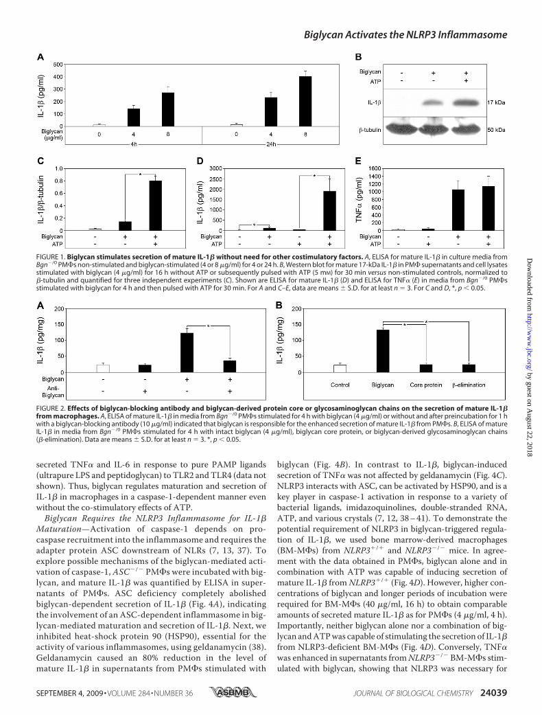
Biglycan Stimulates Maturation and Secretion of IL-1� inMacrophages without the Need for Additional CostimulatoryFactors—Stimulation of Bgn�/0 thioglycollate-elicited PM�swith intact biglycan in a time-dependent (up to 24 h) and dose-dependent (up to 80 �g/ml) manner resulted in enhancedsecretion of mature IL-1� detected by ELISA (Fig. 1A and sup-plemental Fig. S1). Western blot analysis confirmed the pres-ence of the mature 17-kDa form of IL-1� in culture superna-tants from Bgn�/0 macrophages stimulated with biglycan (Fig.1B). Similar results were obtained in resident peritoneal mac-rophages without stimulation with thioglycollate, furtherunderlining the specificity of the response to biglycan (data notshown). Due to a limited number of resident peritoneal macro-phages, in further experiments, only thioglycollate-elicitedperitoneal macrophages were used.Surprisingly, biglycan-dependent secretion of mature
IL-1� occurred without the costimulatory effect of ATP.However, when biglycan-stimulated Bgn�/0 macrophageswere additionally pulsed with 5 mM ATP for 30 min,enhanced secretion of mature IL-1� was detected by West-ern blot (Fig. 1, B and C) and ELISA (Fig. 1D) in a dose-de-pendentmanner (supplemental Fig. S1). Stimulation of IL-1�was specific for biglycan and was inhibited by biglycan-neutral-izing antibodies (Fig. 2A). Neither the biglycan protein core norbiglycan-derived glycosaminoglycan (GAG) side chains(obtained by �-elimination) alone affected IL-1�-levels (Fig.2B), suggesting that both core protein andGAG chains are nec-essary for the stimulation of macrophages.Since the cleavage of IL-1� into its mature 17-kDa form is
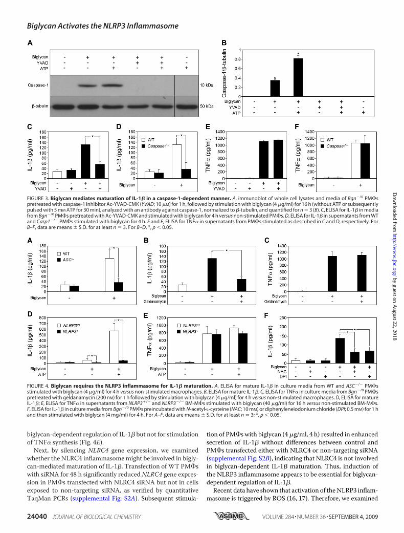
processed by activated caspase-1 (1, 2), we also examinedwhether stimulation of macrophages with biglycan wouldlead to activation of caspase-1. In fact, the p10 active form ofcaspase-1 was detected by Western blot in macrophagesincubated with biglycan alone for 16 h or additionally pulsedwith 5 mMATP for 30 min (Fig. 3,A and B). Furthermore, thecaspase-1 inhibitor Ac-YVAD-CMK profoundly blockedbiglycan-stimulated secretion of mature IL-1� (Fig. 3, A–C).Comparable inhibitory effects were observed in caspase-1�/�
(Casp-1tm1Sesh)macrophages incubatedwith biglycan (Fig. 3D).The effects of biglycan were highly specific for IL-1�, sinceneither ATP (Fig. 1E) nor a functional lack of caspase-1 (inhi-bition (Fig. 3E) or deficiency (Fig. 3F)) had any influence onbiglycan-mediated secretion of TNF� (27) from macrophages.As expected, PM�s did not secrete mature IL-1� and showedsigns of neither activation of caspase-1 nor release of endoge-nous ATP but overexpressed mRNA for pro-IL-1� and
Biglycan Activates the NLRP3 Inflammasome
24038 JOURNAL OF BIOLOGICAL CHEMISTRY VOLUME 284 • NUMBER 36 • SEPTEMBER 4, 2009
by guest on August 22, 2018
http://ww
w.jbc.org/
Dow
nloaded from
secreted TNF� and IL-6 in response to pure PAMP ligands(ultrapure LPS and peptidoglycan) to TLR2 andTLR4 (data notshown). Thus, biglycan regulates maturation and secretion ofIL-1� in macrophages in a caspase-1-dependent manner evenwithout the co-stimulatory effects of ATP.Biglycan Requires the NLRP3 Inflammasome for IL-1�
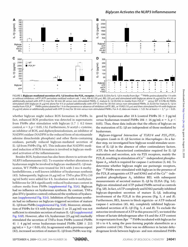
Maturation—Activation of caspase-1 depends on pro-caspase recruitment into the inflammasome and requires theadapter protein ASC downstream of NLRs (7, 13, 37). Toexplore possible mechanisms of the biglycan-mediated acti-vation of caspase-1, ASC�/� PM�s were incubated with big-lycan, and mature IL-1� was quantified by ELISA in super-natants of PM�s. ASC deficiency completely abolishedbiglycan-dependent secretion of IL-1� (Fig. 4A), indicatingthe involvement of an ASC-dependent inflammasome in big-lycan-mediated maturation and secretion of IL-1�. Next, weinhibited heat-shock protein 90 (HSP90), essential for theactivity of various inflammasomes, using geldanamycin (38).Geldanamycin caused an 80% reduction in the level ofmature IL-1� in supernatants from PM�s stimulated with
biglycan (Fig. 4B). In contrast to IL-1�, biglycan-inducedsecretion of TNF� was not affected by geldanamycin (Fig. 4C).NLRP3 interacts with ASC, can be activated by HSP90, and is akey player in caspase-1 activation in response to a variety ofbacterial ligands, imidazoquinolines, double-stranded RNA,ATP, and various crystals (7, 12, 38–41). To demonstrate thepotential requirement of NLRP3 in biglycan-triggered regula-tion of IL-1�, we used bone marrow-derived macrophages(BM-M�s) from NLRP3�/� and NLRP3�/� mice. In agree-ment with the data obtained in PM�s, biglycan alone and incombination with ATP was capable of inducing secretion ofmature IL-1� fromNLRP3�/� (Fig. 4D). However, higher con-centrations of biglycan and longer periods of incubation wererequired for BM-M�s (40 �g/ml, 16 h) to obtain comparableamounts of secreted mature IL-1� as for PM�s (4 �g/ml, 4 h).Importantly, neither biglycan alone nor a combination of big-lycan andATPwas capable of stimulating the secretion of IL-1�from NLRP3-deficient BM-M�s (Fig. 4D). Conversely, TNF�was enhanced in supernatants fromNLRP3�/� BM-M�s stim-ulated with biglycan, showing that NLRP3 was necessary for
FIGURE 1. Biglycan stimulates secretion of mature IL-1� without need for other costimulatory factors. A, ELISA for mature IL-1� in culture media fromBgn�/0 PM�s non-stimulated and biglycan-stimulated (4 or 8 �g/ml) for 4 or 24 h. B, Western blot for mature 17-kDa IL-1� in PM� supernatants and cell lysatesstimulated with biglycan (4 �g/ml) for 16 h without ATP or subsequently pulsed with ATP (5 mM) for 30 min versus non-stimulated controls, normalized to�-tubulin and quantified for three independent experiments (C). Shown are ELISA for mature IL-1� (D) and ELISA for TNF� (E) in media from Bgn�/0 PM�sstimulated with biglycan for 4 h and then pulsed with ATP for 30 min. For A and C–E, data are means � S.D. for at least n � 3. For C and D, *, p 0.05.
FIGURE 2. Effects of biglycan-blocking antibody and biglycan-derived protein core or glycosaminoglycan chains on the secretion of mature IL-1�from macrophages. A, ELISA of mature IL-1� in media from Bgn�/0 PM�s stimulated for 4 h with biglycan (4 �g/ml) or without and after preincubation for 1 hwith a biglycan-blocking antibody (10 �g/ml) indicated that biglycan is responsible for the enhanced secretion of mature IL-1� from PM�s. B, ELISA of matureIL-1� in media from Bgn�/0 PM�s stimulated for 4 h with intact biglycan (4 �g/ml), biglycan core protein, or biglycan-derived glycosaminoglycan chains(�-elimination). Data are means � S.D. for at least n � 3. *, p 0.05.
Biglycan Activates the NLRP3 Inflammasome
SEPTEMBER 4, 2009 • VOLUME 284 • NUMBER 36 JOURNAL OF BIOLOGICAL CHEMISTRY 24039
by guest on August 22, 2018
http://ww
w.jbc.org/
Dow
nloaded from
biglycan-dependent regulation of IL-1� but not for stimulationof TNF� synthesis (Fig. 4E).Next, by silencing NLRC4 gene expression, we examined
whether the NLRC4 inflammasomemight be involved in bigly-can-mediated maturation of IL-1�. Transfection ofWT PM�swith siRNA for 48 h significantly reduced NLRC4 gene expres-sion in PM�s transfected with NLRC4 siRNA but not in cellsexposed to non-targeting siRNA, as verified by quantitativeTaqMan PCRs (supplemental Fig. S2A). Subsequent stimula-
tion of PM�s with biglycan (4 �g/ml, 4 h) resulted in enhancedsecretion of IL-1� without differences between control andPM�s transfected either with NLRC4 or non-targeting siRNA(supplemental Fig. S2B), indicating that NLRC4 is not involvedin biglycan-dependent IL-1� maturation. Thus, induction ofthe NLRP3 inflammasome appears to be essential for biglycan-dependent regulation of IL-1�.
Recent data have shown that activation of theNLRP3 inflam-masome is triggered by ROS (16, 17). Therefore, we examined
FIGURE 3. Biglycan mediates maturation of IL-1� in a caspase-1-dependent manner. A, immunoblot of whole cell lysates and media of Bgn�/0 PM�spretreated with caspase-1 inhibitor Ac-YVAD-CMK (YVAD; 10 �M) for 1 h, followed by stimulation with biglycan (4 �g/ml) for 16 h (without ATP or subsequentlypulsed with 5 mM ATP for 30 min), analyzed with an antibody against caspase-1, normalized to �-tubulin, and quantified for n � 3 (B). C, ELISA for IL-1� in mediafrom Bgn�/0 PM�s pretreated with Ac-YVAD-CMK and stimulated with biglycan for 4 h versus non-stimulated PM�s. D, ELISA for IL-1� in supernatants from WTand Casp1�/� PM�s stimulated with biglycan for 4 h. E and F, ELISA for TNF� in supernatants from PM�s stimulated as described in C and D, respectively. ForB–F, data are means � S.D. for at least n � 3. For B–D, *, p 0.05.
FIGURE 4. Biglycan requires the NLRP3 inflammasome for IL-1� maturation. A, ELISA for mature IL-1� in culture media from WT and ASC�/� PM�sstimulated with biglycan (4 �g/ml) for 4 h versus non-stimulated macrophages. B, ELISA for mature IL-1�; C, ELISA for TNF� in culture media from Bgn�/0 PM�spretreated with geldanamycin (200 nM) for 1 h followed by stimulation with biglycan (4 �g/ml) for 4 h versus non-stimulated macrophages. D, ELISA for matureIL-1�; E, ELISA for TNF� in supernatants from NLRP3�/� and NLRP3�/� BM-M�s stimulated with biglycan (40 �g/ml) for 16 h versus non-stimulated BM-M�s.F, ELISA for IL-1� in culture media from Bgn�/0 PM�s preincubated with N-acetyl-L-cysteine (NAC; 10 mM) or diphenyleneiodonium chloride (DPI; 0.5 mM) for 1 hand then stimulated with biglycan (4 mg/ml) for 4 h. For A–F, data are means � S.D. for at least n � 3; *, p 0.05.
Biglycan Activates the NLRP3 Inflammasome
24040 JOURNAL OF BIOLOGICAL CHEMISTRY VOLUME 284 • NUMBER 36 • SEPTEMBER 4, 2009
by guest on August 22, 2018
http://ww
w.jbc.org/
Dow
nloaded from
whether biglycan might induce ROS formation in PM�s. Infact, enhanced ROS production was detected in supernatantsfrom PM�s after stimulation with biglycan (1.7 � 0.2 timescontrol, n� 3, p 0.05, 1 h). Furthermore,N-acetyl-L-cysteine,an inhibitor of ROS, and diphenyleneiodonium, an inhibitor ofNADPH oxidase (NADPH is the reduced form of nicotinamideadenine dinucleotide phosphate) and other flavin-containingoxidases, partially reduced biglycan-mediated secretion ofIL-1� from PM�s (Fig. 4F). This indicates that NADPH-medi-ated induction of ROS formation is involved in biglycan-medi-ated activation of the inflammasome.Besides ROS, hyaluronan has also been shown to activate the
NLRP3 inflammasome (42). To examine whether alterations inhyaluronanmight be involved in biglycan-mediated IL-1�mat-uration, WT PM�s were treated for 24 h with 2 mM 4-methy-lumbelliferone, a well known inhibitor of hyaluronan synthesis(43). Subsequently, biglycan (4 �g/ml) or TNF� plus IFN� (10ng/ml both) were added for 4 h. Incubation with 4-methylum-belliferone for 28 h significantly reduced hyaluronan content inculture media from PM�s (supplemental Fig. S3A). Biglycanhad no influence on hyaluronan synthesis. By contrast, TNF�plus IFN� (positive control) enhanced the synthesis of hyaluro-nan (supplemental Fig. S3A). Inhibition of hyaluronan synthe-sis had no influence on biglycan-triggered secretion of matureIL-1� from PM�s (supplemental Fig. S3B). Moreover, stimula-tion of PM�s for 4 h with hyaluronan (1 or 25 �g/ml) was notsufficient to stimulate secretion ofmature IL-1� (supplementalFig. S3B). However, after 4 h, hyaluronan (25 �g/ml) markedlystimulated the secretion of TNF� from PM�s (control PM�s41 � 4 pg/ml versus hyaluronan-treated PM�s 3280 � 215pg/ml; n� 3, p 0.05, 4 h). In agreementwith a previous report(42), increased secretion of mature IL-1� from PM�s was trig-
gered by hyaluronan after 18 h (control PM�s 35 � 3 pg/mlversus hyaluronan-treated PM�s 184 � 16 pg/ml; n � 3, p 0.05). Thus, these data indicate that the effects of biglycan onthe maturation of IL-1� are independent of those mediated byhyaluronan.Biglycan-triggered Interaction of TLR2/4 and P2X4/P2X7
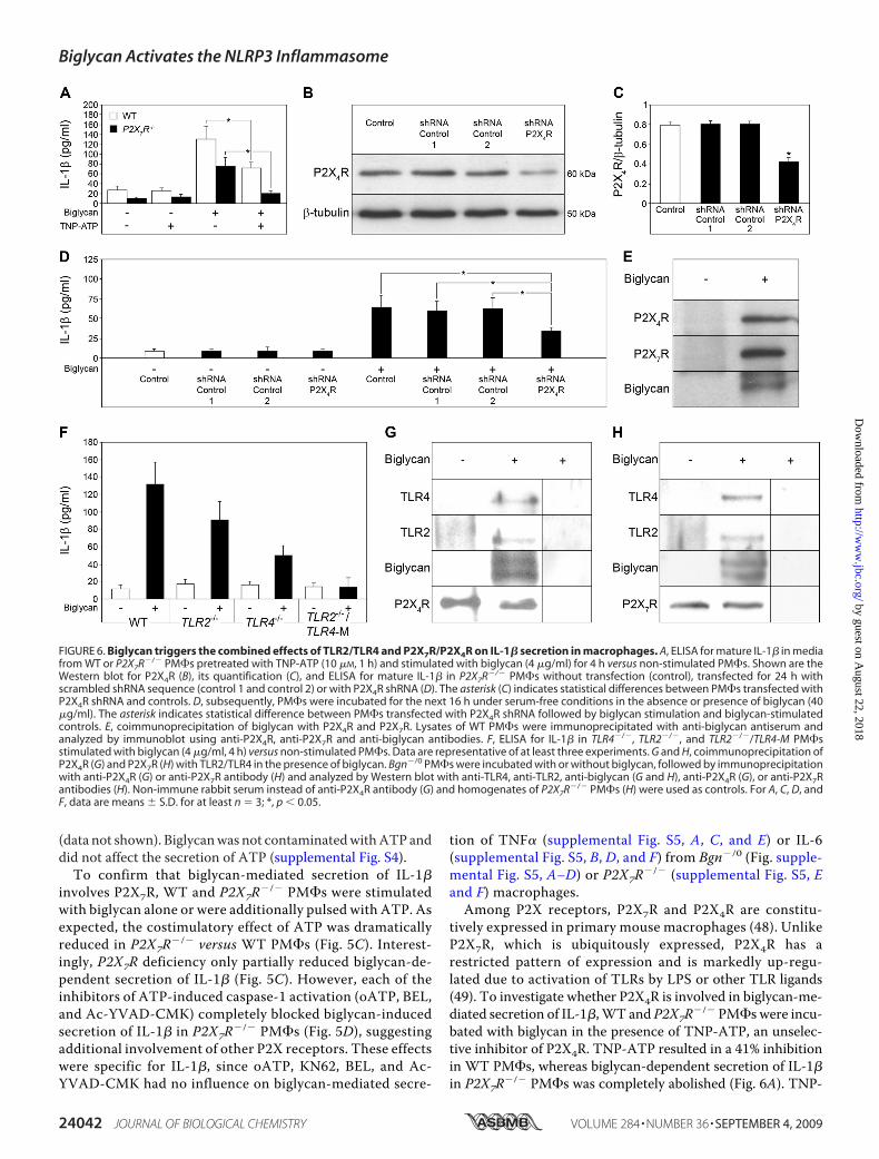
Receptors Leads to IL-1� Secretion in Macrophages—In a fur-ther step, we investigated how biglycan would stimulate secre-tion of IL-1� in the absence of other costimulatory factors.ATP, the best characterized costimulator required for IL-1�maturation and secretion, acts via P2X receptors, mainly theP2X7R, resulting in stimulation of Ca2�-independent phospho-lipase A2, which is required for caspase-1 activation (3, 44). Todetermine whether biglycan might use purinergic receptors,Bgn�/0 PM�s were stimulated with biglycan in the presence ofthe P2X7R antagonists oATP and KN62 and of the Ca2�-inde-pendent phospholipase A2 inhibitor BEL with subsequentmeasurements of secreted mature IL-1� by ELISA (Fig. 5A).Biglycan-stimulated and ATP-pulsed PM�s served as controls(Fig. 5B). In fact, oATP completely andKN62 partially inhibitedbiglycan-dependent secretion of mature IL-1�, indicatinginvolvement of the P2X7R in this process (Fig. 5, A and B).Furthermore, BEL, known to block nigericin- or ATP-inducedcaspase-1 activation (45, 46), completely inhibited biglycan-mediated secretion of IL-1� (Fig. 5, A and B). To exclude theautocrine involvement ofATP in this process (47), we tested therelease of lactate dehydrogenase after 4 h and the ATP contentin supernatants fromBgn�/0 PM�s incubatedwith biglycan for10 s to 4 h, using the pore-forming peptide alamethicin as apositive control (34). There was no difference in lactate dehy-drogenase levels between biglycan- and non-stimulated PM�s
FIGURE 5. Biglycan-mediated secretion of IL-1� involves the P2X7 receptor. A and B, ELISAs for IL-1� in media from Bgn�/0 PM�s preincubated for 1 h withor without inhibitors: oATP (ATP periodate oxidized sodium salt, 1 mM), KN-62 (30 �M), BEL (20 �M) and stimulated with biglycan alone (4 �g/ml) for 4 h (A) oradditionally pulsed with ATP (5 mM) for 30 min (B) versus non-stimulated PM�s. C, mature IL-1� (ELISA) in media from P2X7R�/� versus WT (C57BL/6) PM�sstimulated with biglycan (4 �g/ml) alone for 4 h or pulsed additionally with ATP (5 mM) for 30 min versus non-stimulated PM�s. D, ELISA for mature IL-1� inmedia from P2X7R�/� PM�s preincubated for 1 h in the presence or absence of inhibitors oATP, BEL, or YVAD (10 �M) and then stimulated for 4 h with biglycan(4 �g/ml) alone or additionally pulsed with ATP (5 mM) for 30 min versus non-stimulated PM�s. For A–D, data are means � S.D. for at least n � 3; *, p 0.05.
Biglycan Activates the NLRP3 Inflammasome
SEPTEMBER 4, 2009 • VOLUME 284 • NUMBER 36 JOURNAL OF BIOLOGICAL CHEMISTRY 24041
by guest on August 22, 2018
http://ww
w.jbc.org/
Dow
nloaded from
(data not shown). Biglycanwas not contaminatedwithATP anddid not affect the secretion of ATP (supplemental Fig. S4).To confirm that biglycan-mediated secretion of IL-1�
involves P2X7R, WT and P2X7R�/� PM�s were stimulatedwith biglycan alone or were additionally pulsed with ATP. Asexpected, the costimulatory effect of ATP was dramaticallyreduced in P2X7R�/� versus WT PM�s (Fig. 5C). Interest-ingly, P2X7R deficiency only partially reduced biglycan-de-pendent secretion of IL-1� (Fig. 5C). However, each of theinhibitors of ATP-induced caspase-1 activation (oATP, BEL,and Ac-YVAD-CMK) completely blocked biglycan-inducedsecretion of IL-1� in P2X7R�/� PM�s (Fig. 5D), suggestingadditional involvement of other P2X receptors. These effectswere specific for IL-1�, since oATP, KN62, BEL, and Ac-YVAD-CMK had no influence on biglycan-mediated secre-
tion of TNF� (supplemental Fig. S5, A, C, and E) or IL-6(supplemental Fig. S5, B, D, and F) from Bgn�/0 (Fig. supple-mental Fig. S5, A–D) or P2X7R�/� (supplemental Fig. S5, Eand F) macrophages.Among P2X receptors, P2X7R and P2X4R are constitu-
tively expressed in primary mouse macrophages (48). UnlikeP2X7R, which is ubiquitously expressed, P2X4R has arestricted pattern of expression and is markedly up-regu-lated due to activation of TLRs by LPS or other TLR ligands(49). To investigate whether P2X4R is involved in biglycan-me-diated secretion of IL-1�,WT and P2X7R�/� PM�s were incu-bated with biglycan in the presence of TNP-ATP, an unselec-tive inhibitor of P2X4R. TNP-ATP resulted in a 41% inhibitionin WT PM�s, whereas biglycan-dependent secretion of IL-1�in P2X7R�/� PM�s was completely abolished (Fig. 6A). TNP-
FIGURE 6. Biglycan triggers the combined effects of TLR2/TLR4 and P2X7R/P2X4R on IL-1� secretion in macrophages. A, ELISA for mature IL-1� in mediafrom WT or P2X7R�/� PM�s pretreated with TNP-ATP (10 �M, 1 h) and stimulated with biglycan (4 �g/ml) for 4 h versus non-stimulated PM�s. Shown are theWestern blot for P2X4R (B), its quantification (C), and ELISA for mature IL-1� in P2X7R�/� PM�s without transfection (control), transfected for 24 h withscrambled shRNA sequence (control 1 and control 2) or with P2X4R shRNA (D). The asterisk (C) indicates statistical differences between PM�s transfected withP2X4R shRNA and controls. D, subsequently, PM�s were incubated for the next 16 h under serum-free conditions in the absence or presence of biglycan (40�g/ml). The asterisk indicates statistical difference between PM�s transfected with P2X4R shRNA followed by biglycan stimulation and biglycan-stimulatedcontrols. E, coimmunoprecipitation of biglycan with P2X4R and P2X7R. Lysates of WT PM�s were immunoprecipitated with anti-biglycan antiserum andanalyzed by immunoblot using anti-P2X4R, anti-P2X7R and anti-biglycan antibodies. F, ELISA for IL-1� in TLR4�/�, TLR2�/�, and TLR2�/�/TLR4-M PM�sstimulated with biglycan (4 �g/ml, 4 h) versus non-stimulated PM�s. Data are representative of at least three experiments. G and H, coimmunoprecipitation ofP2X4R (G) and P2X7R (H) with TLR2/TLR4 in the presence of biglycan. Bgn�/0 PM�s were incubated with or without biglycan, followed by immunoprecipitationwith anti-P2X4R (G) or anti-P2X7R antibody (H) and analyzed by Western blot with anti-TLR4, anti-TLR2, anti-biglycan (G and H), anti-P2X4R (G), or anti-P2X7Rantibodies (H). Non-immune rabbit serum instead of anti-P2X4R antibody (G) and homogenates of P2X7R�/� PM�s (H) were used as controls. For A, C, D, andF, data are means � S.D. for at least n � 3; *, p 0.05.
Biglycan Activates the NLRP3 Inflammasome
24042 JOURNAL OF BIOLOGICAL CHEMISTRY VOLUME 284 • NUMBER 36 • SEPTEMBER 4, 2009
by guest on August 22, 2018
http://ww
w.jbc.org/
Dow
nloaded from
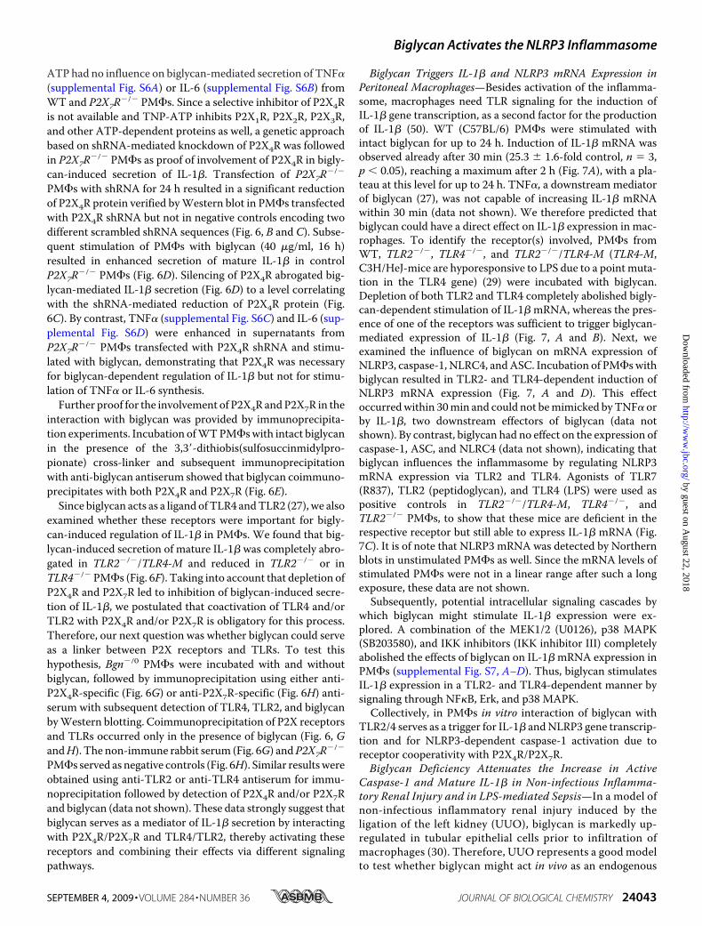
ATP had no influence on biglycan-mediated secretion of TNF�(supplemental Fig. S6A) or IL-6 (supplemental Fig. S6B) fromWT and P2X7R�/� PM�s. Since a selective inhibitor of P2X4Ris not available and TNP-ATP inhibits P2X1R, P2X2R, P2X3R,and other ATP-dependent proteins as well, a genetic approachbased on shRNA-mediated knockdown of P2X4R was followedin P2X7R�/� PM�s as proof of involvement of P2X4R in bigly-can-induced secretion of IL-1�. Transfection of P2X7R�/�
PM�s with shRNA for 24 h resulted in a significant reductionof P2X4R protein verified byWestern blot in PM�s transfectedwith P2X4R shRNA but not in negative controls encoding twodifferent scrambled shRNA sequences (Fig. 6, B and C). Subse-quent stimulation of PM�s with biglycan (40 �g/ml, 16 h)resulted in enhanced secretion of mature IL-1� in controlP2X7R�/� PM�s (Fig. 6D). Silencing of P2X4R abrogated big-lycan-mediated IL-1� secretion (Fig. 6D) to a level correlatingwith the shRNA-mediated reduction of P2X4R protein (Fig.6C). By contrast, TNF� (supplemental Fig. S6C) and IL-6 (sup-plemental Fig. S6D) were enhanced in supernatants fromP2X7R�/� PM�s transfected with P2X4R shRNA and stimu-lated with biglycan, demonstrating that P2X4R was necessaryfor biglycan-dependent regulation of IL-1� but not for stimu-lation of TNF� or IL-6 synthesis.Further proof for the involvement of P2X4R andP2X7R in the
interaction with biglycan was provided by immunoprecipita-tion experiments. Incubation ofWTPM�s with intact biglycanin the presence of the 3,3�-dithiobis(sulfosuccinmidylpro-pionate) cross-linker and subsequent immunoprecipitationwith anti-biglycan antiserum showed that biglycan coimmuno-precipitates with both P2X4R and P2X7R (Fig. 6E).Since biglycan acts as a ligand ofTLR4 andTLR2 (27), we also
examined whether these receptors were important for bigly-can-induced regulation of IL-1� in PM�s. We found that big-lycan-induced secretion of mature IL-1� was completely abro-gated in TLR2�/�/TLR4-M and reduced in TLR2�/� or inTLR4�/� PM�s (Fig. 6F). Taking into account that depletion ofP2X4R and P2X7R led to inhibition of biglycan-induced secre-tion of IL-1�, we postulated that coactivation of TLR4 and/orTLR2 with P2X4R and/or P2X7R is obligatory for this process.Therefore, our next question was whether biglycan could serveas a linker between P2X receptors and TLRs. To test thishypothesis, Bgn�/0 PM�s were incubated with and withoutbiglycan, followed by immunoprecipitation using either anti-P2X4R-specific (Fig. 6G) or anti-P2X7R-specific (Fig. 6H) anti-serum with subsequent detection of TLR4, TLR2, and biglycanbyWestern blotting. Coimmunoprecipitation of P2X receptorsand TLRs occurred only in the presence of biglycan (Fig. 6, GandH). The non-immune rabbit serum (Fig. 6G) andP2X7R�/�
PM�s served as negative controls (Fig. 6H). Similar resultswereobtained using anti-TLR2 or anti-TLR4 antiserum for immu-noprecipitation followed by detection of P2X4R and/or P2X7Rand biglycan (data not shown). These data strongly suggest thatbiglycan serves as a mediator of IL-1� secretion by interactingwith P2X4R/P2X7R and TLR4/TLR2, thereby activating thesereceptors and combining their effects via different signalingpathways.
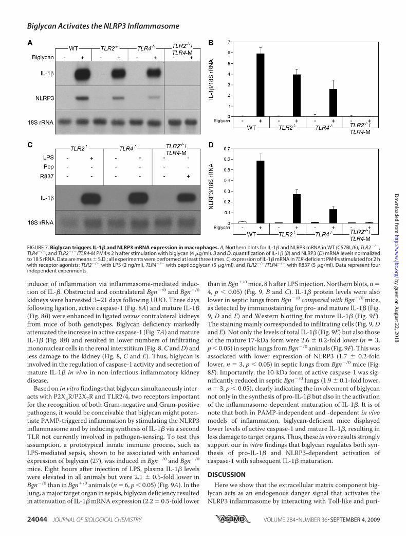
Biglycan Triggers IL-1� and NLRP3 mRNA Expression inPeritoneal Macrophages—Besides activation of the inflamma-some, macrophages need TLR signaling for the induction ofIL-1� gene transcription, as a second factor for the productionof IL-1� (50). WT (C57BL/6) PM�s were stimulated withintact biglycan for up to 24 h. Induction of IL-1� mRNA wasobserved already after 30 min (25.3 � 1.6-fold control, n � 3,p 0.05), reaching a maximum after 2 h (Fig. 7A), with a pla-teau at this level for up to 24 h. TNF�, a downstream mediatorof biglycan (27), was not capable of increasing IL-1� mRNAwithin 30 min (data not shown). We therefore predicted thatbiglycan could have a direct effect on IL-1� expression in mac-rophages. To identify the receptor(s) involved, PM�s fromWT, TLR2�/�, TLR4�/�, and TLR2�/�/TLR4-M (TLR4-M,C3H/HeJ-mice are hyporesponsive to LPS due to a point muta-tion in the TLR4 gene) (29) were incubated with biglycan.Depletion of both TLR2 and TLR4 completely abolished bigly-can-dependent stimulation of IL-1� mRNA, whereas the pres-ence of one of the receptors was sufficient to trigger biglycan-mediated expression of IL-1� (Fig. 7, A and B). Next, weexamined the influence of biglycan on mRNA expression ofNLRP3, caspase-1, NLRC4, andASC. Incubation of PM�s withbiglycan resulted in TLR2- and TLR4-dependent induction ofNLRP3 mRNA expression (Fig. 7, A and D). This effectoccurredwithin 30min and could not bemimicked byTNF� orby IL-1�, two downstream effectors of biglycan (data notshown). By contrast, biglycan had no effect on the expression ofcaspase-1, ASC, and NLRC4 (data not shown), indicating thatbiglycan influences the inflammasome by regulating NLRP3mRNA expression via TLR2 and TLR4. Agonists of TLR7(R837), TLR2 (peptidoglycan), and TLR4 (LPS) were used aspositive controls in TLR2�/�/TLR4-M, TLR4�/�, andTLR2�/� PM�s, to show that these mice are deficient in therespective receptor but still able to express IL-1� mRNA (Fig.7C). It is of note that NLRP3 mRNA was detected by Northernblots in unstimulated PM�s as well. Since the mRNA levels ofstimulated PM�s were not in a linear range after such a longexposure, these data are not shown.Subsequently, potential intracellular signaling cascades by
which biglycan might stimulate IL-1� expression were ex-plored. A combination of the MEK1/2 (U0126), p38 MAPK(SB203580), and IKK inhibitors (IKK inhibitor III) completelyabolished the effects of biglycan on IL-1� mRNA expression inPM�s (supplemental Fig. S7, A–D). Thus, biglycan stimulatesIL-1� expression in a TLR2- and TLR4-dependent manner bysignaling through NF�B, Erk, and p38 MAPK.Collectively, in PM�s in vitro interaction of biglycan with
TLR2/4 serves as a trigger for IL-1� andNLRP3 gene transcrip-tion and for NLRP3-dependent caspase-1 activation due toreceptor cooperativity with P2X4R/P2X7R.Biglycan Deficiency Attenuates the Increase in Active
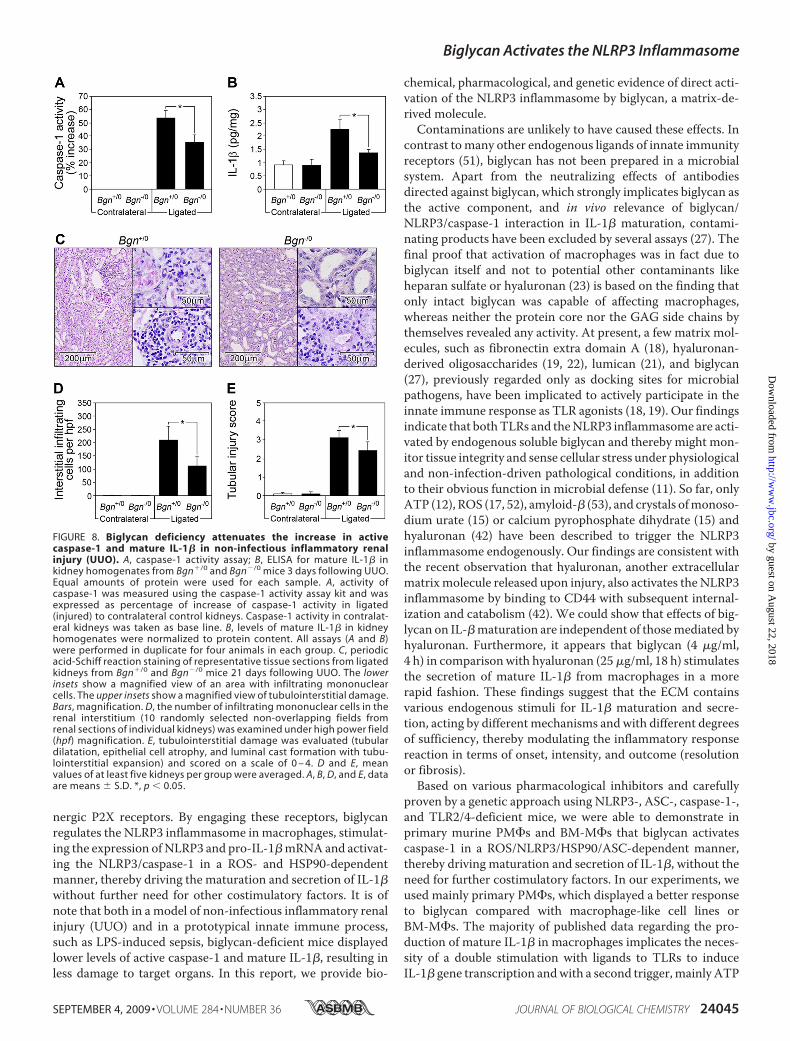
Caspase-1 and Mature IL-1� in Non-infectious Inflamma-tory Renal Injury and in LPS-mediated Sepsis—In a model ofnon-infectious inflammatory renal injury induced by theligation of the left kidney (UUO), biglycan is markedly up-regulated in tubular epithelial cells prior to infiltration ofmacrophages (30). Therefore, UUO represents a good modelto test whether biglycan might act in vivo as an endogenous
Biglycan Activates the NLRP3 Inflammasome
SEPTEMBER 4, 2009 • VOLUME 284 • NUMBER 36 JOURNAL OF BIOLOGICAL CHEMISTRY 24043
by guest on August 22, 2018
http://ww
w.jbc.org/
Dow
nloaded from
inducer of inflammation via inflammasome-mediated induc-tion of IL-�. Obstructed and contralateral Bgn�/0 and Bgn�/0
kidneys were harvested 3–21 days following UUO. Three daysfollowing ligation, active caspase-1 (Fig. 8A) and mature IL-1�(Fig. 8B) were enhanced in ligated versus contralateral kidneysfrom mice of both genotypes. Biglycan deficiency markedlyattenuated the increase in active caspase-1 (Fig. 7A) andmatureIL-1� (Fig. 8B) and resulted in lower numbers of infiltratingmononuclear cells in the renal interstitium (Fig. 8,C andD) andless damage to the kidney (Fig. 8, C and E). Thus, biglycan isinvolved in the regulation of caspase-1 activity and secretion ofmature IL-1� in vivo in non-infectious inflammatory kidneydisease.Based on in vitro findings that biglycan simultaneously inter-
acts with P2X4R/P2X7R and TLR2/4, two receptors importantfor the recognition of both Gram-negative and Gram-positivepathogens, it would be conceivable that biglycan might poten-tiate PAMP-triggered inflammation by stimulating the NLRP3inflammasome and by inducing synthesis of IL-1� via a secondTLR not currently involved in pathogen-sensing. To test thisassumption, a prototypical innate immune process, such asLPS-mediated sepsis, shown to be associated with enhancedexpression of biglycan (27), was induced in Bgn�/0 and Bgn�/0
mice. Eight hours after injection of LPS, plasma IL-1� levelswere elevated in all animals but were 2.1 � 0.5-fold lower inBgn�/0 than inBgn�/0 animals (n� 6, p 0.05) (Fig. 9A). In thelung, amajor target organ in sepsis, biglycan deficiency resultedin attenuation of IL-1�mRNA expression (2.2� 0.5-fold lower
than inBgn�/0mice, 8 h after LPS injection,Northern blots,n�4, p 0.05) (Fig. 9, B and C). IL-1� protein levels were alsolower in septic lungs from Bgn�/0 compared with Bgn�/0 mice,as detected by immunostaining for pro- andmature IL-1� (Fig.9, D and E) and Western blotting for mature IL-1� (Fig. 9F).The staining mainly corresponded to infiltrating cells (Fig. 9,Dand E). Not only the levels of total IL-1� (Fig. 9E) but also thoseof the mature 17-kDa form were 2.6 � 0.2-fold lower (n � 3,p 0.05) in septic lungs fromBgn�/0 animals (Fig. 9F). Thiswasassociated with lower expression of NLRP3 (1.7 � 0.2-foldlower, n � 3, p 0.05) in septic lungs from Bgn�/0 mice (Fig.8F). Importantly, the 10-kDa form of active caspase-1 was sig-nificantly reduced in septic Bgn�/0 lungs (1.9 � 0.1-fold lower,n � 3, p 0.05), clearly indicating the involvement of biglycannot only in the synthesis of pro-IL-1� but also in the activationof the inflammasome-dependent maturation of IL-1�. It is ofnote that both in PAMP-independent and -dependent in vivomodels of inflammation, biglycan-deficient mice displayedlower levels of active caspase-1 and mature IL-1�, resulting inless damage to target organs. Thus, these in vivo results stronglysupport our in vitro findings that biglycan regulates both syn-thesis of pro-IL-1� and NLRP3-dependent activation ofcaspase-1 with subsequent IL-1� maturation.
DISCUSSION
Here we show that the extracellular matrix component big-lycan acts as an endogenous danger signal that activates theNLRP3 inflammasome by interacting with Toll-like and puri-
FIGURE 7. Biglycan triggers IL-1� and NLRP3 mRNA expression in macrophages. A, Northern blots for IL-1� and NLRP3 mRNA in WT (C57BL/6), TLR2�/�,TLR4�/�, and TLR2�/�/TLR4-M PM�s 2 h after stimulation with biglycan (4 �g/ml). B and D, quantification of IL-1� (B) and NLRP3 (D) mRNA levels normalizedto 18 S rRNA. Data are means � S.D.; all experiments were performed at least three times. C, expression of IL-1� mRNA in TLR-deficient PM�s stimulated for 2 hwith receptor agonists: TLR2�/� with LPS (2 ng/ml), TLR4�/� with peptidoglycan (5 �g/ml), and TLR2�/�/TLR4�/� with R837 (5 �g/ml). Data represent fourindependent experiments.
Biglycan Activates the NLRP3 Inflammasome
24044 JOURNAL OF BIOLOGICAL CHEMISTRY VOLUME 284 • NUMBER 36 • SEPTEMBER 4, 2009
by guest on August 22, 2018
http://ww
w.jbc.org/
Dow
nloaded from
nergic P2X receptors. By engaging these receptors, biglycanregulates the NLRP3 inflammasome in macrophages, stimulat-ing the expression ofNLRP3 and pro-IL-1�mRNAand activat-ing the NLRP3/caspase-1 in a ROS- and HSP90-dependentmanner, thereby driving the maturation and secretion of IL-1�without further need for other costimulatory factors. It is ofnote that both in a model of non-infectious inflammatory renalinjury (UUO) and in a prototypical innate immune process,such as LPS-induced sepsis, biglycan-deficient mice displayedlower levels of active caspase-1 and mature IL-1�, resulting inless damage to target organs. In this report, we provide bio-
chemical, pharmacological, and genetic evidence of direct acti-vation of the NLRP3 inflammasome by biglycan, a matrix-de-rived molecule.Contaminations are unlikely to have caused these effects. In
contrast tomany other endogenous ligands of innate immunityreceptors (51), biglycan has not been prepared in a microbialsystem. Apart from the neutralizing effects of antibodiesdirected against biglycan, which strongly implicates biglycan asthe active component, and in vivo relevance of biglycan/NLRP3/caspase-1 interaction in IL-1� maturation, contami-nating products have been excluded by several assays (27). Thefinal proof that activation of macrophages was in fact due tobiglycan itself and not to potential other contaminants likeheparan sulfate or hyaluronan (23) is based on the finding thatonly intact biglycan was capable of affecting macrophages,whereas neither the protein core nor the GAG side chains bythemselves revealed any activity. At present, a few matrix mol-ecules, such as fibronectin extra domain A (18), hyaluronan-derived oligosaccharides (19, 22), lumican (21), and biglycan(27), previously regarded only as docking sites for microbialpathogens, have been implicated to actively participate in theinnate immune response as TLR agonists (18, 19). Our findingsindicate that bothTLRs and theNLRP3 inflammasome are acti-vated by endogenous soluble biglycan and thereby might mon-itor tissue integrity and sense cellular stress under physiologicaland non-infection-driven pathological conditions, in additionto their obvious function in microbial defense (11). So far, onlyATP (12), ROS (17, 52), amyloid-� (53), and crystals ofmonoso-dium urate (15) or calcium pyrophosphate dihydrate (15) andhyaluronan (42) have been described to trigger the NLRP3inflammasome endogenously. Our findings are consistent withthe recent observation that hyaluronan, another extracellularmatrixmolecule released upon injury, also activates theNLRP3inflammasome by binding to CD44 with subsequent internal-ization and catabolism (42). We could show that effects of big-lycan on IL-�maturation are independent of thosemediated byhyaluronan. Furthermore, it appears that biglycan (4 �g/ml,4 h) in comparisonwith hyaluronan (25�g/ml, 18 h) stimulatesthe secretion of mature IL-1� from macrophages in a morerapid fashion. These findings suggest that the ECM containsvarious endogenous stimuli for IL-1� maturation and secre-tion, acting by differentmechanisms andwith different degreesof sufficiency, thereby modulating the inflammatory responsereaction in terms of onset, intensity, and outcome (resolutionor fibrosis).Based on various pharmacological inhibitors and carefully
proven by a genetic approach using NLRP3-, ASC-, caspase-1-,and TLR2/4-deficient mice, we were able to demonstrate inprimary murine PM�s and BM-M�s that biglycan activatescaspase-1 in a ROS/NLRP3/HSP90/ASC-dependent manner,thereby driving maturation and secretion of IL-1�, without theneed for further costimulatory factors. In our experiments, weused mainly primary PM�s, which displayed a better responseto biglycan compared with macrophage-like cell lines orBM-M�s. The majority of published data regarding the pro-duction of mature IL-1� in macrophages implicates the neces-sity of a double stimulation with ligands to TLRs to induceIL-1� gene transcription andwith a second trigger,mainlyATP
FIGURE 8. Biglycan deficiency attenuates the increase in activecaspase-1 and mature IL-1� in non-infectious inflammatory renalinjury (UUO). A, caspase-1 activity assay; B, ELISA for mature IL-1� inkidney homogenates from Bgn�/0 and Bgn�/0 mice 3 days following UUO.Equal amounts of protein were used for each sample. A, activity ofcaspase-1 was measured using the caspase-1 activity assay kit and wasexpressed as percentage of increase of caspase-1 activity in ligated(injured) to contralateral control kidneys. Caspase-1 activity in contralat-eral kidneys was taken as base line. B, levels of mature IL-1� in kidneyhomogenates were normalized to protein content. All assays (A and B)were performed in duplicate for four animals in each group. C, periodicacid-Schiff reaction staining of representative tissue sections from ligatedkidneys from Bgn�/0 and Bgn�/0 mice 21 days following UUO. The lowerinsets show a magnified view of an area with infiltrating mononuclearcells. The upper insets show a magnified view of tubulointerstitial damage.Bars, magnification. D, the number of infiltrating mononuclear cells in therenal interstitium (10 randomly selected non-overlapping fields fromrenal sections of individual kidneys) was examined under high power field(hpf) magnification. E, tubulointerstitial damage was evaluated (tubulardilatation, epithelial cell atrophy, and luminal cast formation with tubu-lointerstitial expansion) and scored on a scale of 0 – 4. D and E, meanvalues of at least five kidneys per group were averaged. A, B, D, and E, dataare means � S.D. *, p 0.05.
Biglycan Activates the NLRP3 Inflammasome
SEPTEMBER 4, 2009 • VOLUME 284 • NUMBER 36 JOURNAL OF BIOLOGICAL CHEMISTRY 24045
by guest on August 22, 2018
http://ww
w.jbc.org/
Dow
nloaded from

or muramyldipeptide, for activation of the inflammasome (50).Recent studies compared the response of human monocytes,macrophages, and dendritic cells to PAMP ligands of TLR2 andTLR4 (54) and showed that only monocytes are capable ofreleasing mature IL-1� without using additional exogenouscostimulatory factors due to constitutively active caspase-1 andsecretion of endogenous ATP (47, 54). In agreement with thisreport, in our hands, murine macrophages did not secretemature IL-1� and showed no signs of activation of caspase-1 orrelease of endogenous ATP but overexpressed mRNA for pro-IL-1� in response to pure LPS (data not shown). Using highlysensitivemethods, we did not detect biglycan-dependent secre-tion of ATP, which could have acted as a costimulatory factor.Therefore, in contrast to LPS, biglycan appears to be an auton-omous trigger of IL-1�. Findings that the amounts of secretedIL-1� triggered by biglycan alone are much lower than thoseobtained by an additional pulse of 5 mMATP have to be seen inlight of the fact that physiological levels of extracellularATP areonly 10 nM and may maximally reach the micromolar rangearound living cells, creating an “ATP halo” (55). Therefore, theeffects of ATP in the millimolar range used by us and othersrather represent intracellular (3–10 mM) and not extracellularlevels of ATP (55). Furthermore, the amount of mature IL-1�secreted from PM�s stimulated by 4 �g/ml biglycan over 4 hwas comparable with the response of macrophages to 25 �g/mlof hyaluronan over 18 h and much higher than the stimulationinduced by large amounts of crude LPS (25 �g/ml, 18 h) (42),underlining the biological relevance of endogenous regulatorsin IL-1� maturation and secretion.
By binding to TLR2/4 and signaling through NF�B, Erk, andp38 MAPK, biglycan (i) stimulates the expression of NLRP3
mRNA and protein and (ii) increases the amount of pro-IL-1�,the major substrate of the inflammasome. Since various exog-enous TLR ligands induce pro-IL-1� production via NF�B (56)and NLRP3mRNA (39, 57), it is conceivable that biglycan as anendogenous ligand of TLR2/TLR4 (27) may exert similareffects. However, the somewhat surprising finding that besidesbinding to TLRs, biglycan independently interacts with puri-nergic P2X receptors, thereby inducing cumulative signalingcapacities, suggests a novel mechanism of downstream activa-tion of the NLRP3 inflammasome. Several mechanisms ofNLRP3 inflammasome activation have been postulated up tonow. Internalization with subsequent lysosomal damage ap-pears to play a role in hyaluronan-, crystal-, and amyloid-�-de-pendent inflammasome activation (42, 53, 58). By contrast,ATP activates the inflammasome via the P2X7R with a subse-quent efflux of K� (3, 7, 16). The requirement of ROS orHSP90in biglycan-dependent activation of theNLRP3 inflammasome,similar to monosodium urate, R837 (52), asbestos, silica (17),and ATP (16) as ROS mediators or to NLRP3-mediated gout-like inflammation involving HSP90 (38), indicates that HSP90and ROS play a crucial role in the activation of the NLRP3inflammasome independent of the initial trigger. The findingsthat biglycan triggers ROS formation fit well with previousobservations that TLRs are involved in ROS formation via theNADPH oxidase enzyme complex (59).Our genetic approach using TLR2/4- and P2X7R-deficient
macrophages together with shRNA for P2X4R and further sub-stantiated by biochemical methods indicates that the interac-tion of TLR2/4 with P2X7R/P2X4R occurs in the presence ofbiglycan. It is tempting to speculate that biglycanmediates clus-tering of TLR2/4 and purinergic P2X4/P2X7 receptors, induc-
FIGURE 9. Biglycan deficiency attenuates the increase in pro-IL-1� expression, active caspase-1, and mature IL-1� in LPS-mediated sepsis. A, plasmalevels of IL-1� in control mice without LPS (Bgn�/0 and Bgn�/0, ELISA, each group n � 6) and in septic Bgn�/0 and Bgn�/0 mice (each group n � 4). B, Northernblot for IL-1� mRNA in septic lungs of Bgn�/0 and Bgn�/0 mice normalized to 18 S rRNA and quantified for three experiments (C). D and E, immunostaining for(pro- and mature) IL-1� (marked by arrows) in septic lungs from Bgn�/0 (D) versus Bgn�/0 (E) mice. The lower right insets show a magnified view of cellsexpressing IL-1�. Bars, magnification. F, Western blots for mature IL-1�, active caspase-1 and NLRP3 normalized to �-tubulin in control and septic lungs fromBgn�/0 versus Bgn�/0. Quantification of F is included under “Results.” A–F, 8 h after LPS. Data are means � S.D.; *, p 0.05.
Biglycan Activates the NLRP3 Inflammasome
24046 JOURNAL OF BIOLOGICAL CHEMISTRY VOLUME 284 • NUMBER 36 • SEPTEMBER 4, 2009
by guest on August 22, 2018
http://ww
w.jbc.org/
Dow
nloaded from
ing receptor cooperativity within these newly formed multire-ceptor complexes. Although several controls have beenincluded, interpretation of the cross-linking experiments can-not totally rule out the possibility that biglycan might inde-pendently engage both receptors, being sufficient for activationof the inflammasome. The final proof for biglycan-triggeredfunctional interaction of both signaling pathways at the proxi-mal level by priming TLR2/4-deficient macrophages with a dif-ferent TLR agonist is limited by the fact that, for example, R837by itself is involved in activation of the inflammasome (41).The core protein of biglycan is rife with leucine-rich repeats,
which are considered to be involved in protein-protein interac-tions (26), and carries up to two GAG side chains. These com-plex structural features make biglycan particularly wellendowed to serve as a cross-linker between different cell sur-face receptors (20). The requirement of intact biglycan forTLR/P2X receptor clustering underlines the intricacy of this interac-tion, in which besides CD14 and MD2 other moleculesassociated with TLRs and/or P2X receptors might also beinvolved. In fact, other proteins (e.g. �2 integrin) that mightinteract with biglycan have been identified to form signalingcomplexes with P2X7R (60). Thus, soluble biglycan, as an indi-cator of tissue injury, could be a pivotal danger signal, which iscarefully monitored by the innate immune system.Most importantly, the biological relevance of biglycan as a
trigger of theNLRP3 inflammasomehas been confirmed in vivoin PAMP-independent and -dependent mouse models ofinflammation, sharing features of enhanced biglycan expres-sion and extensive macrophage infiltration (27, 30). In bothmodels, biglycan deficiency led to lower levels of activecaspase-1 and mature IL-1�, resulting in less damage to targetorgans. It is tempting to speculate that biglycan, upon releasefrom the ECM, acts as an autonomous trigger of the non-infec-tious inflammatory response due to interaction of TLR2/4 andP2X4/P2X7 receptors, followed by their alloyed signaling. Onthe other hand, in pathogen-mediated inflammation, biglycanmight potentiate PAMP-triggered inflammation by stimulatingthe NLRP3 inflammasome and by inducing synthesis of IL-1�via a second TLR not involved in pathogen sensing. In this con-text, it would be conceivable that in LPS-induced sepsis, bigly-can deficiency resulted in lower expression of IL-1� mRNAbesides reduced capase-1 activity and mature IL-1� levels.It is currently not understood how a stationary macromole-
cule of the ECMcan turn into a soluble ligand triggering cellularresponses. Biglycan undergoes strong interactions with colla-gen VI (61); however, in comparison with other LRR proteogly-cans, biglycan has a relatively low affinity with soluble, mono-molecular collagen I, the most abundant matrix component inmammalian organisms by far. No information exists on theaffinity of biglycan with collagen I, which is incorporated intobanded collagen fibrils. In addition, biglycan can form stablesoluble dimers at least in vitro (62). It is conceivable, however,that monomeric biglycan interacts with ECM components,leading to sequestration of a potentially proinflammatory sig-nal. As has been shown previously (27), stimulated macro-phages step up their synthesis of biglycan. The newly synthe-sized proteoglycanmay then displace biglycan weakly bound tothe ECMby formingmore stable soluble dimers, which, in turn,
may become organizers of multireceptor complexes involvingTLRs and P2XRs. Likewise, othermatrix proteinsmaymobilizesequestered biglycan by heterodimer formation, although suchheterodimers have not yet been reported. In the same context,it is intriguing that both TLRs andNLRs contain LRRmotifs (9,10) with the potential to interact in an analogous fashion withLRR proteins, including biglycan.Taken together, our data suggest that soluble biglycan acts
as a fundamental danger signal that signifies tissue injuryand elicits a robust proinflammatory response by the innateimmune system. By interacting with Toll-like and purinergicP2X receptors on the cell surface of macrophages, biglycansignals through these multireceptor complexes and activatesthe NLRP3 inflammasome in a ROS- and HSP90-dependentmanner, thereby driving the caspase-1-mediated maturationand secretion of IL-1�. The observations described here impli-cate biglycan as a promising target for novel anti-inflammatorystrategies. Mechanisms are conceivable whereby unseques-tered (63) biglycan is trapped, most likely by other matrix com-ponents containing LRR motifs. In this way, receptor signalingof soluble biglycan might be prevented, thereby achieving tightcontrol of its proinflammatory effects.
REFERENCES1. Martinon, F., and Tschopp, J. (2007) Cell Death Differ. 14, 10–222. Joshi, V. D., Kalvakolanu, D. V., Hebel, J. R., Hasday, J. D., and Cross, A. S.
(2002) Infect. Immun. 70, 6896–69033. Di Virgilio, F. (2007) Trends Pharmacol. Sci. 28, 465–4724. Hogquist, K. A., Nett,M. A., Unanue, E. R., andChaplin, D. D. (1991) Proc.
Natl. Acad. Sci. U.S.A. 88, 8485–84895. Dinarello, C. A. (2005) J. Exp. Med. 201, 1355–13596. Ting, J. P., Lovering, R. C., Alnemri, E. S., Bertin, J., Boss, J. M., Davis, B. K.,
Flavell, R. A., Girardin, S. E., Godzik, A., Harton, J. A., Hoffman, H. M.,Hugot, J. P., Inohara,N.,Mackenzie, A.,Maltais, L. J., Nunez,G.,Ogura, Y.,Otten, L. A., Philpott, D., Reed, J. C., Reith, W., Schreiber, S., Steimle, V.,and Ward, P. A. (2008) Immunity 28, 285–287
7. Martinon, F., Gaide, O., Petrilli, V., Mayor, A., and Tschopp, J. (2007)Semin. Immunopathol. 29, 213–229
8. Fritz, J. H., Ferrero, R. L., Philpott, D. J., and Girardin, S. E. (2006) Nat.Immunol. 7, 1250–1257
9. Gay, N. J., and Gangloff, M. (2008) Handb. Exp. Pharmacol. 181–20010. Inohara Chamaillard McDonald, C., and Nunez, G. (2005) Annu. Rev.
Biochem. 74, 355–38311. Petrilli, V., Dostert, C., Muruve, D. A., and Tschopp, J. (2007) Curr. Opin.
Immunol. 19, 615–62212. Mariathasan, S., Weiss, D. S., Newton, K., McBride, J., O’Rourke, K.,
Roose-Girma, M., Lee, W. P., Weinrauch, Y., Monack, D. M., and Dixit,V. M. (2006) Nature 440, 228–232
13. Mariathasan, S., Newton, K.,Monack, D.M., Vucic, D., French, D.M., Lee,W. P., Roose-Girma, M., Erickson, S., and Dixit, V. M. (2004)Nature 430,213–218
14. Kanneganti, T. D., Body-Malapel, M., Amer, A., Park, J. H., Whitfield, J.,Franchi, L., Taraporewala, Z. F., Miller, D., Patton, J. T., Inohara, N., andNunez, G. (2006) J. Biol. Chem. 281, 36560–36568
15. Martinon, F., Petrilli, V., Mayor, A., Tardivel, A., and Tschopp, J. (2006)Nature 440, 237–241
16. Cruz, C.M., Rinna, A., Forman, H. J., Ventura, A. L., Persechini, P.M., andOjcius, D. M. (2007) J. Biol. Chem. 282, 2871–2879
17. Dostert, C., Petrilli, V., Van Bruggen, R., Steele, C., Mossman, B. T., andTschopp, J. (2008) Science 320, 674–677
18. Okamura, Y., Watari, M., Jerud, E. S., Young, D. W., Ishizaka, S. T., Rose,J., Chow, J. C., and Strauss, J. F., 3rd (2001) J. Biol. Chem. 276,10229–10233
19. Termeer, C., Benedix, F., Sleeman, J., Fieber, C., Voith, U., Ahrens, T.,
Biglycan Activates the NLRP3 Inflammasome
SEPTEMBER 4, 2009 • VOLUME 284 • NUMBER 36 JOURNAL OF BIOLOGICAL CHEMISTRY 24047
by guest on August 22, 2018
http://ww
w.jbc.org/
Dow
nloaded from
Miyake, K., Freudenberg, M., Galanos, C., and Simon, J. C. (2002) J. Exp.Med. 195, 99–111
20. Schaefer, L., and Iozzo, R. V. (2008) J. Biol. Chem. 283, 21305–2130921. Wu, F., Vij, N., Roberts, L., Lopez-Briones, S., Joyce, S., and Chakravarti, S.
(2007) J. Biol. Chem. 282, 26409–2641722. Jiang, D., Liang, J., Fan, J., Yu, S., Chen, S., Luo, Y., Prestwich, G. D.,
Mascarenhas, M. M., Garg, H. G., Quinn, D. A., Homer, R. J., Goldstein,D. R., Bucala, R., Lee, P. J., Medzhitov, R., and Noble, P. W. (2005) Nat.Med. 11, 1173–1179
23. Jiang, D., Liang, J., and Noble, P. W. (2007) Annu. Rev. Cell Dev. Biol. 23,435–461
24. Nathan, C. (2002) Nature 420, 846–85225. Park, P.W., Pier, G. B., Hinkes,M.T., andBernfield,M. (2001)Nature411,
98–10226. Iozzo, R. V. (1999) J. Biol. Chem. 274, 18843–1884627. Schaefer, L., Babelova, A., Kiss, E., Hausser, H. J., Baliova, M., Kr-
zyzankova, M., Marsche, G., Young, M. F., Mihalik, D., Gotte, M., Malle,E., Schaefer, R. M., and Grone, H. J. (2005) J. Clin. Invest. 115, 2223–2233
28. Xu, T., Bianco, P., Fisher, L. W., Longenecker, G., Smith, E., Goldstein, S.,Bonadio, J., Boskey, A., Heegaard, A. M., Sommer, B., Satomura, K.,Dominguez, P., Zhao, C., Kulkarni, A. B., Robey, P. G., and Young, M. F.(1998) Nat. Genet. 20, 78–82
29. Poltorak, A., He, X., Smirnova, I., Liu, M. Y., Van Huffel, C., Du, X., Bird-well, D., Alejos, E., Silva, M., Galanos, C., Freudenberg, M., Ricciardi-Castagnoli, P., Layton, B., and Beutler, B. (1998) Science 282, 2085–2088
30. Schaefer, L., Macakova, K., Raslik, I., Micegova, M., Grone, H. J., Schon-herr, E., Robenek, H., Echtermeyer, F. G., Grassel, S., Bruckner, P.,Schaefer, R. M., Iozzo, R. V., and Kresse, H. (2002) Am. J. Pathol. 160,1181–1191
31. Kresse, H., Seidler, D. G., Muller, M., Breuer, E., Hausser, H., Roughley,P. J., and Schonherr, E. (2001) J. Biol. Chem. 276, 13411–13416
32. Schaefer, L., Beck, K. F., Raslik, I., Walpen, S., Mihalik, D., Micegova, M.,Macakova, K., Schonherr, E., Seidler, D. G., Varga, G., Schaefer, R. M.,Kresse, H., and Pfeilschifter, J. (2003) J. Biol. Chem. 278, 26227–26237
33. Schaefer, L., Raslik, I., Grone, H. J., Schonherr, E., Macakova, K., Ugorca-kova, J., Budny, S., Schaefer, R. M., and Kresse, H. (2001) FASEB J. 15,559–561
34. Mathew, M. K., and Balaram, P. (1983)Mol. Cell. Biochem. 50, 47–6435. Adams, J., Kiss, E., Arroyo, A. B., Bonrouhi, M., Sun, Q., Li, Z., Gretz, N.,
Schnitger, A., Zouboulis, C. C., Wiesel, M., Wagner, J., Nelson, P. J., andGrone, H. J. (2005) Am. J. Pathol. 167, 285–298
36. Pleskova, M., Beck, K. F., Behrens, M. H., Huwiler, A., Fichtlscherer, B.,Wingerter, O., Brandes, R. P., Mulsch, A., and Pfeilschifter, J. (2006)FASEB J. 20, 139–141
37. Martinon, F. (2007) Eur. J. Immunol. 37, 3003–300638. Mayor, A., Martinon, F., De Smedt, T., Petrilli, V., and Tschopp, J. (2007)
Nat. Immunol. 8, 497–50339. Sutterwala, F. S., Ogura, Y., Szczepanik, M., Lara-Tejero, M., Lichten-
berger, G. S., Grant, E. P., Bertin, J., Coyle, A. J., Galan, J. E., Askenase,P. W., and Flavell, R. A. (2006) Immunity 24, 317–327
40. Lamkanfi, M., Kanneganti, T. D., Franchi, L., and Nunez, G. (2007) J. Leu-kocyte Biol. 82, 220–225
41. Kanneganti, T. D., Ozoren, N., Body-Malapel, M., Amer, A., Park, J. H.,Franchi, L., Whitfield, J., Barchet, W., Colonna, M., Vandenabeele, P.,Bertin, J., Coyle, A., Grant, E. P., Akira, S., and Nunez, G. (2006) Nature440, 233–236
42. Yamasaki, K., Muto, J., Taylor, K. R., Cogen, A. L., Audish, D., Bertin, J.,Grant, E. P., Coyle, A. J., Misaghi, A., Hoffman, H. M., and Gallo, R. L.(2009) J. Biol. Chem. 284, 12762–12771
43. Vigetti, D., Rizzi, M., Viola, M., Karousou, E., Genasetti, A., Clerici, M.,Bartolini, B., Hascall, V. C., De Luca, G., and Passi, A. (2009) Glycobiology19, 537–546
44. Andrei, C., Margiocco, P., Poggi, A., Lotti, L. V., Torrisi, M. R., and Rubar-telli, A. (2004) Proc. Natl. Acad. Sci. U.S.A. 101, 9745–9750
45. Walev, I., Klein, J., Husmann, M., Valeva, A., Strauch, S., Wirtz, H.,Weichel, O., and Bhakdi, S. (2000) J. Immunol. 164, 5120–5124
46. Kahlenberg, J.M., andDubyak, G. R. (2004) J. Leukocyte Biol. 76, 676–68447. Piccini, A., Carta, S., Tassi, S., Lasiglie, D., Fossati, G., and Rubartelli, A.
(2008) Proc. Natl. Acad. Sci. U.S.A. 105, 8067–807248. Guo, C., Masin, M., Qureshi, O. S., and Murrell-Lagnado, R. D. (2007)
Mol. Pharmacol. 72, 1447–145649. Raouf, R., Chabot-Dore, A. J., Ase, A. R., Blais, D., and Seguela, P. (2007)
Neuropharmacology 53, 496–50450. Netea, M. G., van de Veerdonk, F. L., Kullberg, B. J., Van der Meer, J. W.,
and Joosten, L. A. (2008) Expert Opin. Biol. Ther. 8, 1867–187251. Tsan, M. F., and Gao, B. (2004) J. Leukocyte Biol. 76, 514–51952. Petrilli, V., Papin, S., Dostert, C., Mayor, A., Martinon, F., and Tschopp, J.
(2007) Cell Death Differ. 14, 1583–158953. Halle, A., Hornung, V., Petzold, G. C., Stewart, C. R., Monks, B. G., Rein-
heckel, T., Fitzgerald, K. A., Latz, E., Moore, K. J., and Golenbock, D. T.(2008) Nat. Immunol. 9, 857–865
54. Netea, M. G., Nold-Petry, C. A., Nold, M. F., Joosten, L. A., Opitz, B., vanderMeer, J. H., van de Veerdonk, F. L., Ferwerda, G., Heinhuis, B., Devesa,I., Funk, C. J., Mason, R. J., Kullberg, B. J., Rubartelli, A., Van der Meer,J. W., and Dinarello, C. A. (2009) Blood 113, 2324–2335
55. Trautmann, A. (2009) Sci. Signal. 2, pe656. Creagh, E. M., and O’Neill, L. A. (2006) Trends Immunol. 27, 352–35757. O’Connor, W., Jr., Harton, J. A., Zhu, X., Linhoff, M. W., and Ting, J. P.
(2003) J. Immunol. 171, 6329–633358. Hornung, V., Bauernfeind, F., Halle, A., Samstad, E. O., Kono, H., Rock,
K. L., Fitzgerald, K. A., and Latz, E. (2008) Nat. Immunol. 9, 847–85659. Ogier-Denis, E., Mkaddem, S. B., and Vandewalle, A. (2008) Semin. Im-
munopathol. 30, 291–30060. Kim,M., Jiang, L. H.,Wilson,H. L., North, R. A., and Surprenant, A. (2001)
EMBO J. 20, 6347–635861. Wiberg, C., Heinegård, D., Wenglen, C., Timpl, R., and Morgelin, M.
(2002) J. Biol. Chem. 277, 49120–4912662. Scott, P. G., Dodd, C. M., Bergmann, E. M., Sheehan, J. K., and Bishop,
P. N. (2006) J. Biol. Chem. 281, 13324–1333263. Medzhitov, R. (2008) Nature 454, 428–435
Biglycan Activates the NLRP3 Inflammasome
24048 JOURNAL OF BIOLOGICAL CHEMISTRY VOLUME 284 • NUMBER 36 • SEPTEMBER 4, 2009
by guest on August 22, 2018
http://ww
w.jbc.org/
Dow
nloaded from
Schaefer, Hermann-Josef Gröne and Liliana SchaeferOliver Eickelberg, Marian F. Young, Peter Bruckner, Josef Pfeilschifter, Roland M.
Andrea Babelova, Kristin Moreth, Wasiliki Tsalastra-Greul, Jinyang Zeng-Brouwers,and P2X Receptors
Biglycan, a Danger Signal That Activates the NLRP3 Inflammasome via Toll-like
doi: 10.1074/jbc.M109.014266 originally published online July 15, 20092009, 284:24035-24048.J. Biol. Chem.
10.1074/jbc.M109.014266Access the most updated version of this article at doi:
Alerts:
When a correction for this article is posted•
When this article is cited•
to choose from all of JBC's e-mail alertsClick here
Supplemental material:
http://www.jbc.org/content/suppl/2009/07/15/M109.014266.DC1
http://www.jbc.org/content/284/36/24035.full.html#ref-list-1
This article cites 62 references, 25 of which can be accessed free at
by guest on August 22, 2018
http://ww
w.jbc.org/
Dow
nloaded from