Embed Size (px)
Citation preview
1
Bicuspid Aortic ValveDilated Aortic Root
Bicuspid Aortic ValveDilated Aortic Root
Amr E Abbas, MD, FACC, FSCAI, FASE, FSVMDirector, Interventional Cardiology Research
Co-Director, EchocardiographyBeaumont Health
Associate Professor of Medicine, OU/WB School of Medicine
Amr E Abbas, MD, FACC, FSCAI, FASE, FSVMDirector, Interventional Cardiology Research
Co-Director, EchocardiographyBeaumont Health
Associate Professor of Medicine, OU/WB School of Medicine
2
Relevant Financial Relationship(s)
NoneOff Label Usage
None
Relevant Financial Relationship(s)
NoneOff Label Usage
None
Pre Questions (1)Pre Questions (1)
• The Difference between Doppler MIG and catheterization PPG
A. Is due to pressure recovery
B. Is due to different measurement timing of the LV and aortic pressures
C. Occurs only in patients with small aortas
D. Is used to calculate aortic valve area
• The Difference between Doppler MIG and catheterization PPG
A. Is due to pressure recovery
B. Is due to different measurement timing of the LV and aortic pressures
C. Occurs only in patients with small aortas
D. Is used to calculate aortic valve area
3
Pre Questions (2)Pre Questions (2)• The Difference between Doppler MIG and
catheterization PPGA. Is due to pressure recoveryB. Is due to difference in the timing of
the aortic pressure measurement between cath and echo
C. Is due to difference in the timing of the LV pressure measurement between cath and echo
D. Is related to the severity of aortic stenosis
• The Difference between Doppler MIG and catheterization PPG
A. Is due to pressure recoveryB. Is due to difference in the timing of
the aortic pressure measurement between cath and echo
C. Is due to difference in the timing of the LV pressure measurement between cath and echo
D. Is related to the severity of aortic stenosis
Pre Questions (3)Pre Questions (3)
• Catheter-Doppler Discordance maybe due to
A. Pressure recoveryB. Eccentric jetC. Very severe aortic stenosisD. HOCM
• Catheter-Doppler Discordance maybe due to
A. Pressure recoveryB. Eccentric jetC. Very severe aortic stenosisD. HOCM
4
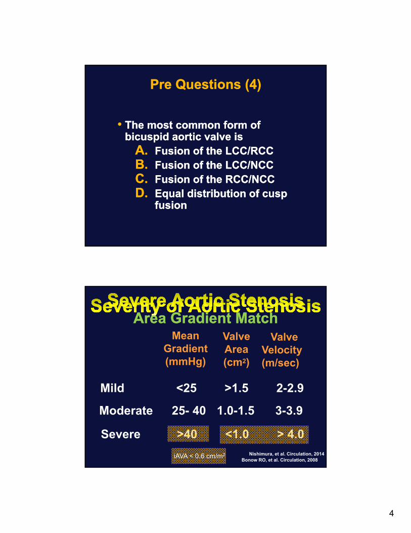
Pre Questions (4)Pre Questions (4)
• The most common form of bicuspid aortic valve is
A. Fusion of the LCC/RCCB. Fusion of the LCC/NCCC. Fusion of the RCC/NCCD. Equal distribution of cusp
fusion
• The most common form of bicuspid aortic valve is
A. Fusion of the LCC/RCCB. Fusion of the LCC/NCCC. Fusion of the RCC/NCCD. Equal distribution of cusp
fusion
Mean Gradient (mmHg)
Valve Area (cm2)
Valve Velocity (m/sec)
Mild <25 >1.5 2-2.9
Moderate 25- 40 1.0-1.5 3-3.9
Severe >40 <1.0 > 4.0
Bonow RO, et al. Circulation, 2008
Area Gradient MatchArea Gradient Match
iAVA < 0.6 cm/m2 Nishimura, et al. Circulation, 2014
5
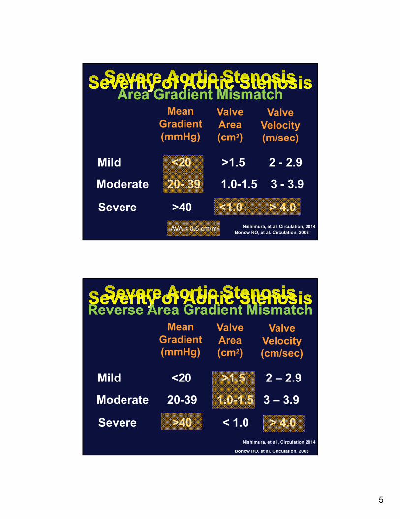
Mean Gradient (mmHg)
Valve Area (cm2)
Valve Velocity (m/sec)
Mild <20 >1.5 2 - 2.9
Moderate 20- 39 1.0-1.5 3 - 3.9
Severe >40 <1.0 > 4.0
Nishimura, et al. Circulation, 2014
Area Gradient MismatchArea Gradient Mismatch
iAVA < 0.6 cm/m2Bonow RO, et al. Circulation, 2008
Mean Gradient (mmHg)
Valve Area (cm2)
Valve Velocity (cm/sec)
Mild <20 >1.5 2 – 2.9
Moderate 20-39 1.0-1.5 3 – 3.9
Severe >40 < 1.0 > 4.0
Nishimura, et al., Circulation 2014
Reverse Area Gradient MismatchReverse Area Gradient Mismatch
Bonow RO, et al. Circulation, 2008
6
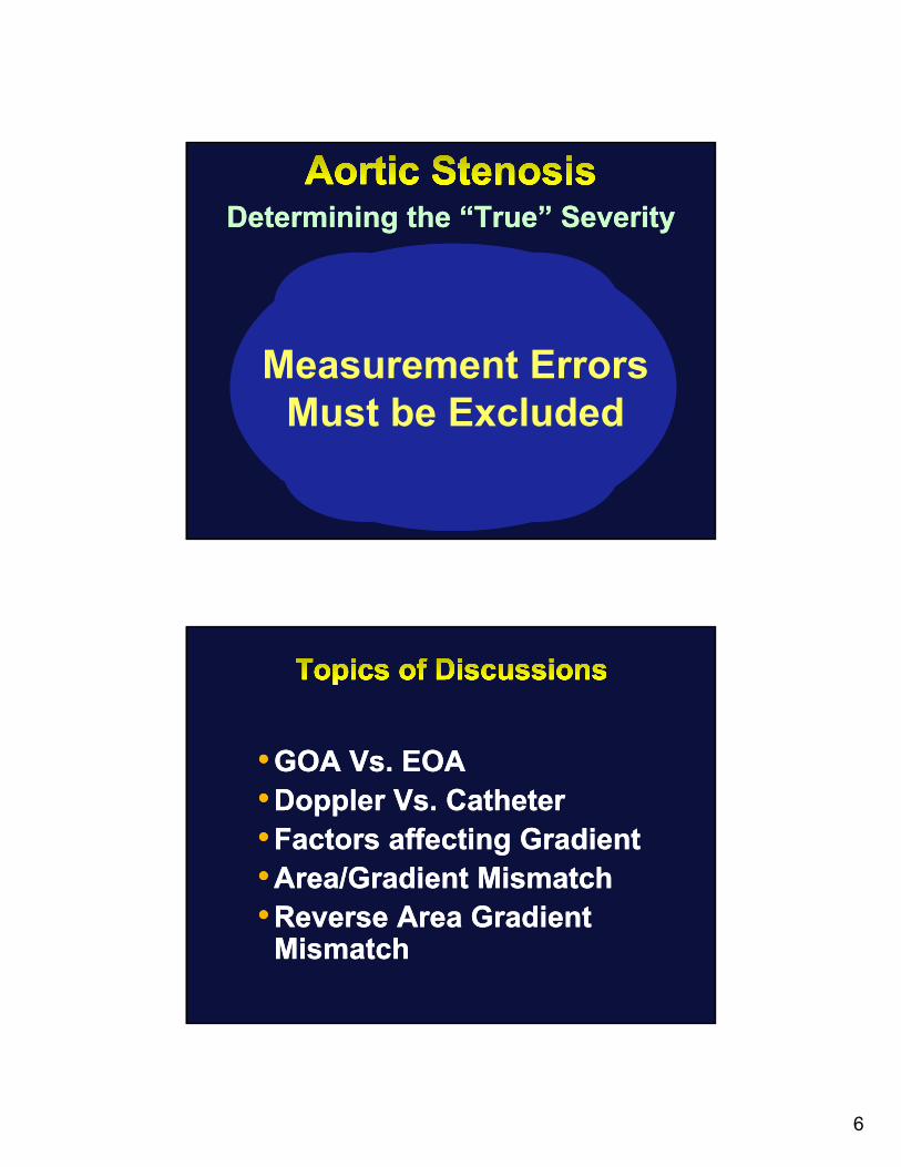
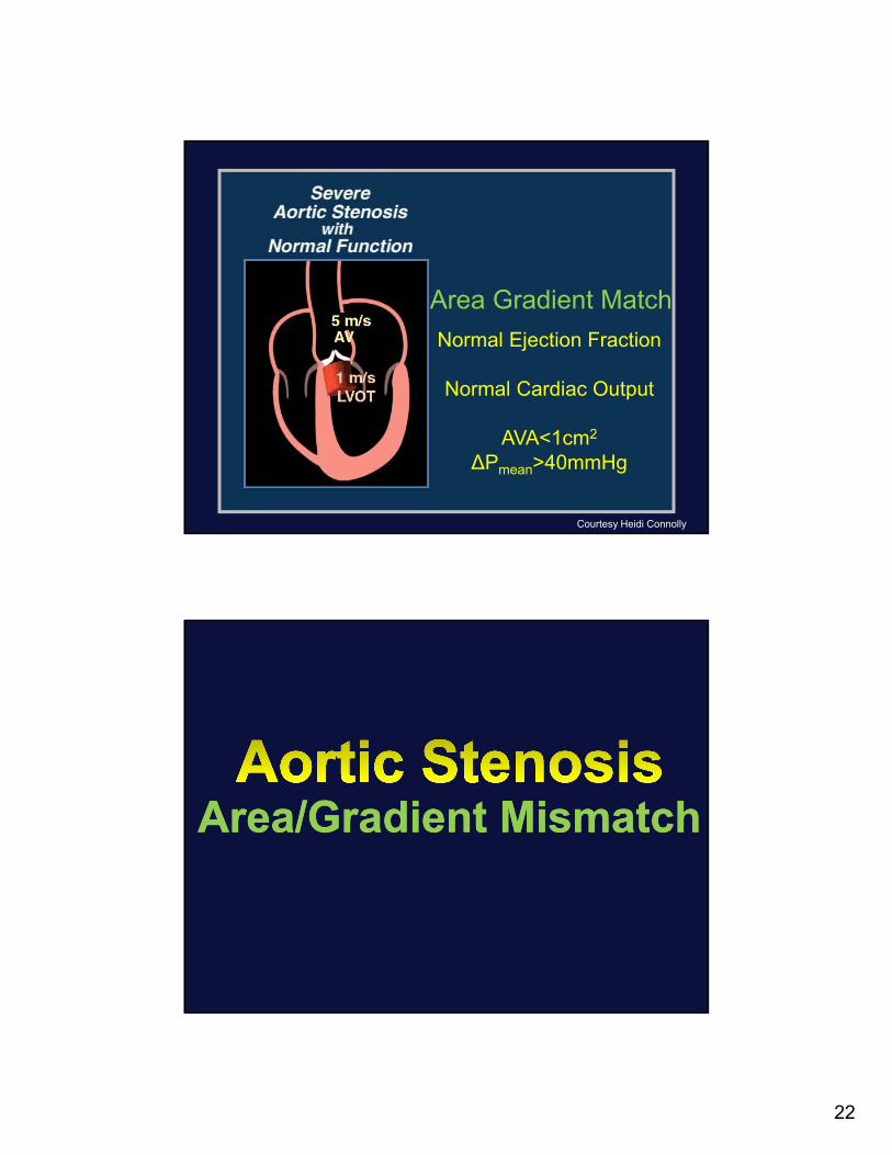
Determining the “True” SeverityDetermining the “True” Severity
Area (cm2)Gradient(mmHg)
Flow Amount/Direction
(l/min)
Pressure Recovery
Doppler Catheterization
Global LV After LoadClinical Presentation
Aortic ValveAortic Regurgitation
Measurement ErrorsMust be Excluded
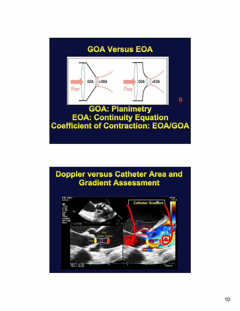
•GOA Vs. EOA•Doppler Vs. Catheter•Factors affecting Gradient•Area/Gradient Mismatch•Reverse Area Gradient
Mismatch
•GOA Vs. EOA•Doppler Vs. Catheter•Factors affecting Gradient•Area/Gradient Mismatch•Reverse Area Gradient
Mismatch
7
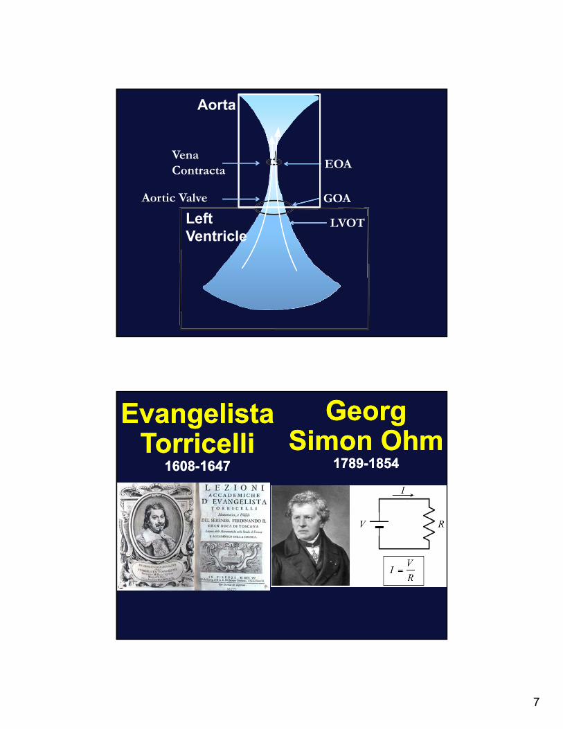
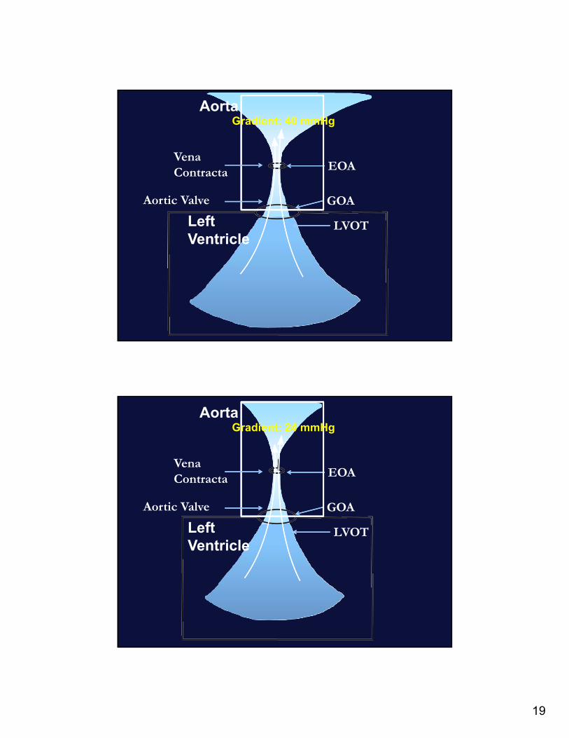
Left Ventricle
GOA
EOA
Aorta
Vena Contracta
Aortic Valve
LVOT
Evangelista Torricelli
1608-1647
Evangelista Torricelli
1608-1647
Georg Simon Ohm
1789-1854
Georg Simon Ohm
1789-1854
8
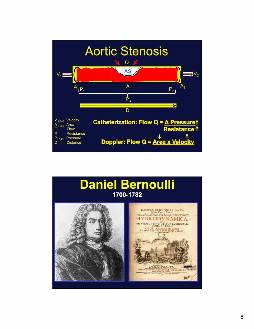
Q
RV2V1
A1A2A3
V3
V 1,2&3 VelocityA 1,2&3 AreaQ FlowR ResistanceP1,2&3 PressureD Distance
P2P1
D
P3
Aortic Stenosis
AS
Daniel Bernoulli1700-1782
Daniel Bernoulli1700-1782
9
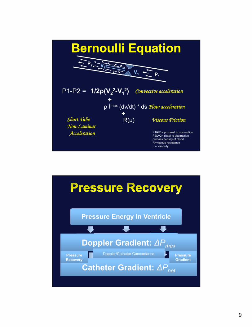
P1-P2 = 1/2ρ(V22-V1
2)
P1&V1= proximal to obstructionP2&V2= distal to obstructionρ=mass density of bloodR=viscous resistanceμ = viscosity
Convective acceleration
+ρ ∫max (dv/dt) * ds Flow acceleration
+R(μ) Viscous Friction
V1 P1
V2P2
Bernoulli EquationBernoulli Equation
Short TubeNon-LaminarAcceleration
Pressure Energy In VentriclePressure Energy In Ventricle
Kinetic Energy
Aortic Valve
Kinetic Energy
Aortic Valve
Pressure Energy
Aorta
Pressure Energy
Aorta
Heat & Friction
Turbulence& Vortices
Lost
Doppler Gradient: ΔPmax
Catheter Gradient: ΔPnet
Pressure Recovery
Pressure Gradient
Doppler/Catheter Concordance
10
B
GOA
Doppler Catheter
Catheter Gradient
Doppler Gradient
EOA
11
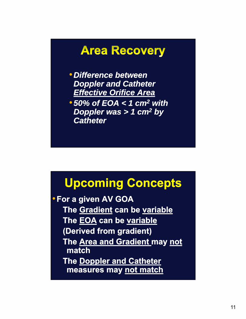
•Difference between Doppler and Catheter Effective Orifice Area
•50% of EOA < 1 cm2 with Doppler was > 1 cm2 by Catheter
•Difference between Doppler and Catheter Effective Orifice Area
•50% of EOA < 1 cm2 with Doppler was > 1 cm2 by Catheter
Upcoming ConceptsUpcoming Concepts•For a given AV GOA
The Gradient can be variableThe EOA can be variable(Derived from gradient)The Area and Gradient may not match
The Doppler and Catheter measures may not match
•For a given AV GOAThe Gradient can be variableThe EOA can be variable(Derived from gradient)The Area and Gradient may not match
The Doppler and Catheter measures may not match
12
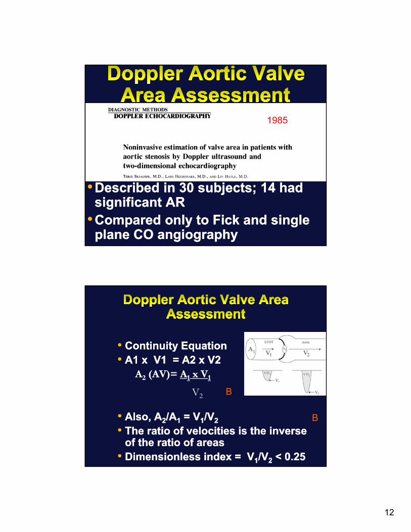
•Described in 30 subjects; 14 had significant AR
•Compared only to Fick and single plane CO angiography
•Described in 30 subjects; 14 had significant AR
•Compared only to Fick and single plane CO angiography
1985
• Continuity Equation• A1 x V1 = A2 x V2
A2 (AV)= A1 x V1
• Also, A2/A1 = V1/V2
• The ratio of velocities is the inverse of the ratio of areas
• Dimensionless index = V1/V2 < 0.25
• Continuity Equation• A1 x V1 = A2 x V2
A2 (AV)= A1 x V1
• Also, A2/A1 = V1/V2
• The ratio of velocities is the inverse of the ratio of areas
• Dimensionless index = V1/V2 < 0.25
V2 B
B
13
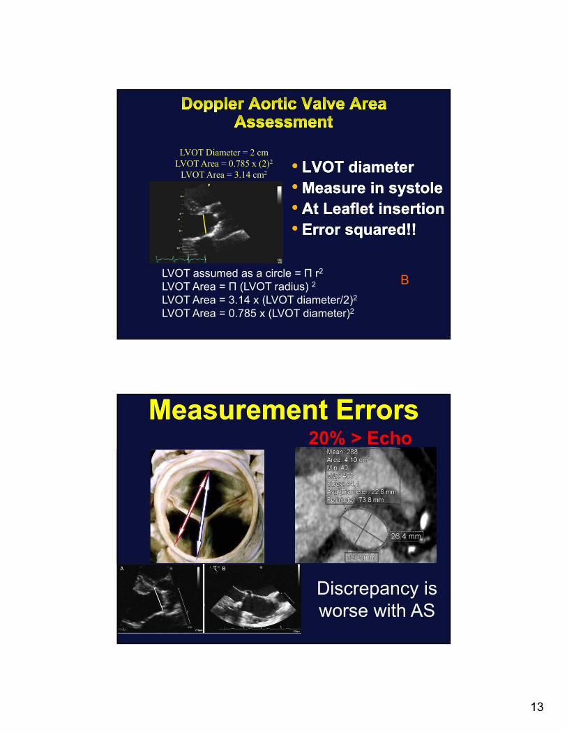
• LVOT diameter• Measure in systole• At Leaflet insertion • Error squared!!
• LVOT diameter• Measure in systole• At Leaflet insertion • Error squared!!
LVOT assumed as a circle = Π r2
LVOT Area = Π (LVOT radius) 2
LVOT Area = 3.14 x (LVOT diameter/2)2
LVOT Area = 0.785 x (LVOT diameter)2
LVOT Diameter = 2 cmLVOT Area = 0.785 x (2)2
LVOT Area = 3.14 cm2
B
Measurement ErrorsMeasurement Errors
Discrepancy is worse with AS
20% > Echo
14
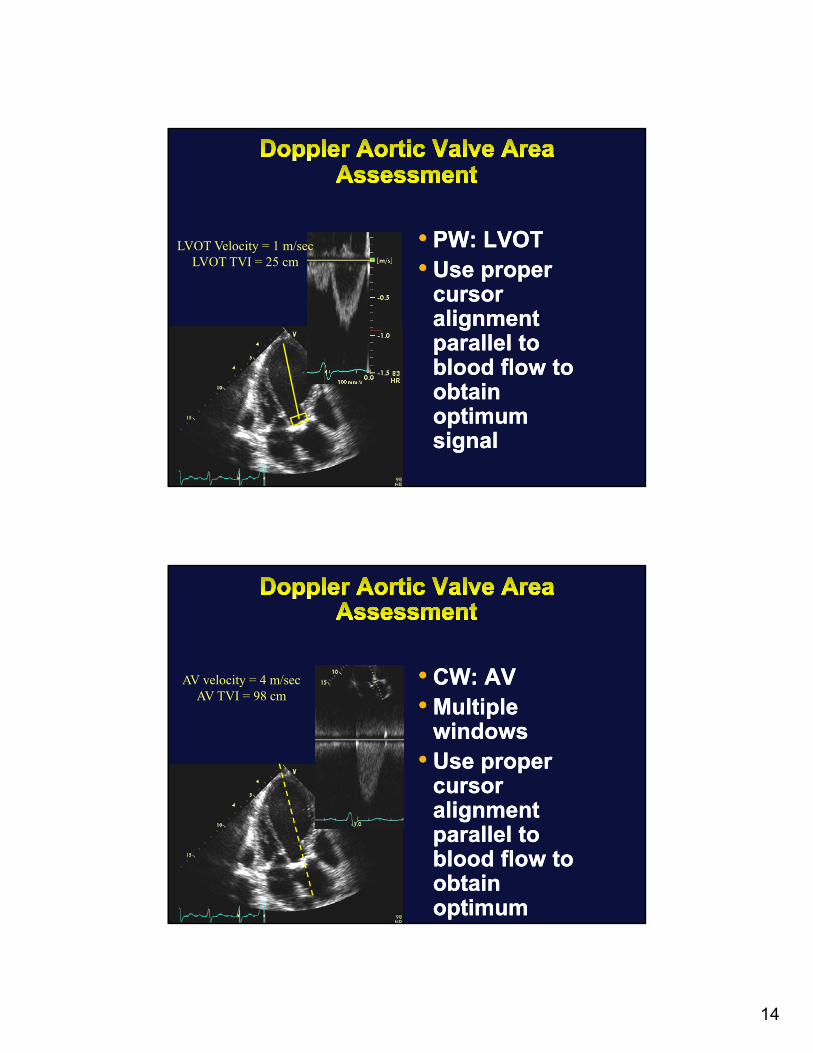
• PW: LVOT• Use proper
cursor alignment parallel to blood flow to obtain optimum signal
• PW: LVOT• Use proper
cursor alignment parallel to blood flow to obtain optimum signal
LVOT Velocity = 1 m/secLVOT TVI = 25 cm
• CW: AV• Multiple
windows• Use proper
cursor alignment parallel to blood flow to obtain optimum i l
• CW: AV• Multiple
windows• Use proper
cursor alignment parallel to blood flow to obtain optimum i l
AV velocity = 4 m/secAV TVI = 98 cm
15
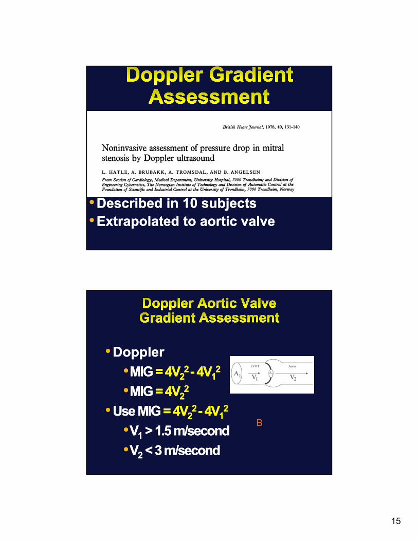
•Described in 10 subjects•Extrapolated to aortic valve•Described in 10 subjects•Extrapolated to aortic valve
•Doppler
•MIG = 4V22 -4V1
2
•MIG= 4V22
•Use MIG = 4V22 -4V1
2
•V1 > 1.5 m/second
•V2 < 3 m/second
•Doppler
•MIG = 4V22 -4V1
2
•MIG= 4V22
•Use MIG = 4V22 -4V1
2
•V1 > 1.5 m/second
•V2 < 3 m/second
B
16
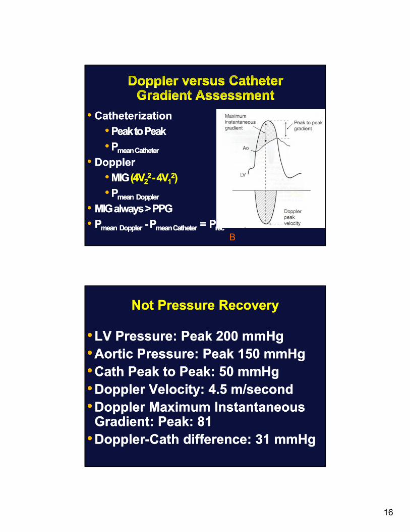
• Catheterization
• Peak to Peak
• Pmean Catheter
• Doppler
• MIG (4V22 -4V1
2)
• Pmean Doppler
• MIG always > PPG
• Pmean Doppler -Pmean Catheter = Prec
• Catheterization
• Peak to Peak
• Pmean Catheter
• Doppler
• MIG (4V22 -4V1
2)
• Pmean Doppler
• MIG always > PPG
• Pmean Doppler -Pmean Catheter = PrecB
Not Pressure RecoveryNot Pressure Recovery
•LV Pressure: Peak 200 mmHg•Aortic Pressure: Peak 150 mmHg•Cath Peak to Peak: 50 mmHg•Doppler Velocity: 4.5 m/second•Doppler Maximum Instantaneous
Gradient: Peak: 81•Doppler-Cath difference: 31 mmHg
•LV Pressure: Peak 200 mmHg•Aortic Pressure: Peak 150 mmHg•Cath Peak to Peak: 50 mmHg•Doppler Velocity: 4.5 m/second•Doppler Maximum Instantaneous
Gradient: Peak: 81•Doppler-Cath difference: 31 mmHg
17
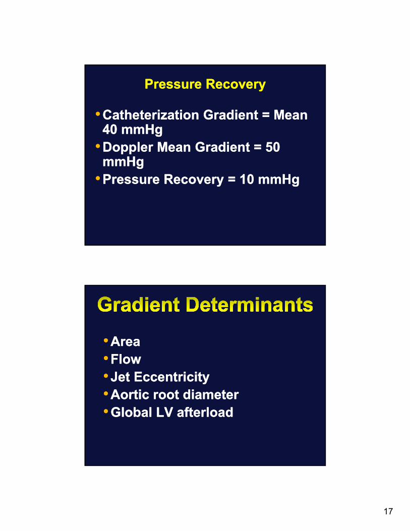
Pressure RecoveryPressure Recovery
•Catheterization Gradient = Mean 40 mmHg
•Doppler Mean Gradient = 50 mmHg
•Pressure Recovery = 10 mmHg
•Catheterization Gradient = Mean 40 mmHg
•Doppler Mean Gradient = 50 mmHg
•Pressure Recovery = 10 mmHg
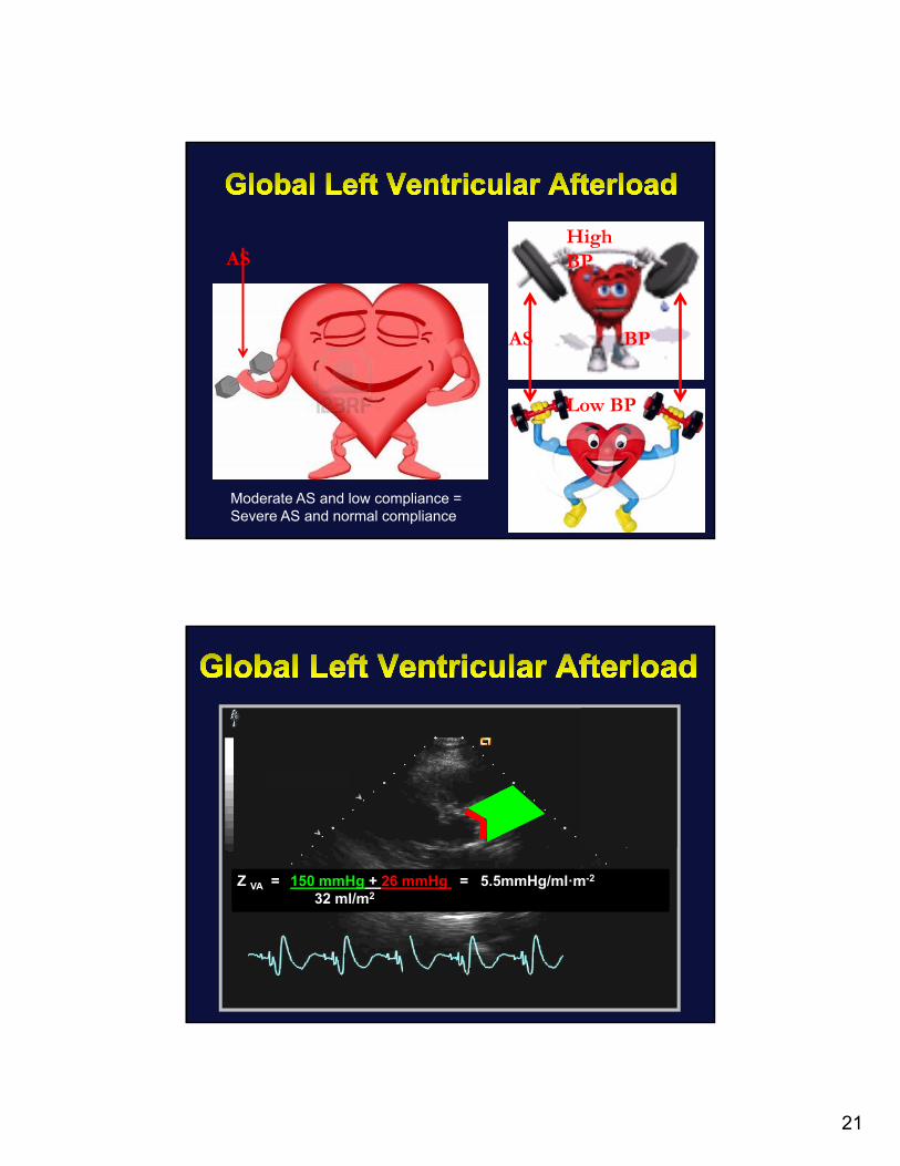
•Area•Flow•Jet Eccentricity•Aortic root diameter•Global LV afterload
•Area•Flow•Jet Eccentricity•Aortic root diameter•Global LV afterload
18
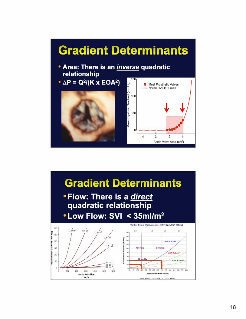
• Area: There is an inverse quadratic relationship
• ∆P = Q2/(K x EOA2)
• Area: There is an inverse quadratic relationship
• ∆P = Q2/(K x EOA2)
160
140
120
100
80
60
40
20
250 ml/sec
•Flow: There is a directquadratic relationship
•Low Flow: SVI < 35ml/m2
•Flow: There is a directquadratic relationship
•Low Flow: SVI < 35ml/m2
AVA 0.7 cm2
AVA 1.0 cm2
AVA 1.5 cm2
250 ml/s125 ml/s
40 mmHg
19
Left Ventricle
GOA
EOA
Aorta
Vena Contracta
Aortic Valve
LVOT
Gradient: 40 mmHg
Left Ventricle
GOA
EOA
Aorta
Vena Contracta
Aortic Valve
LVOT
Gradient: 24 mmHg
20
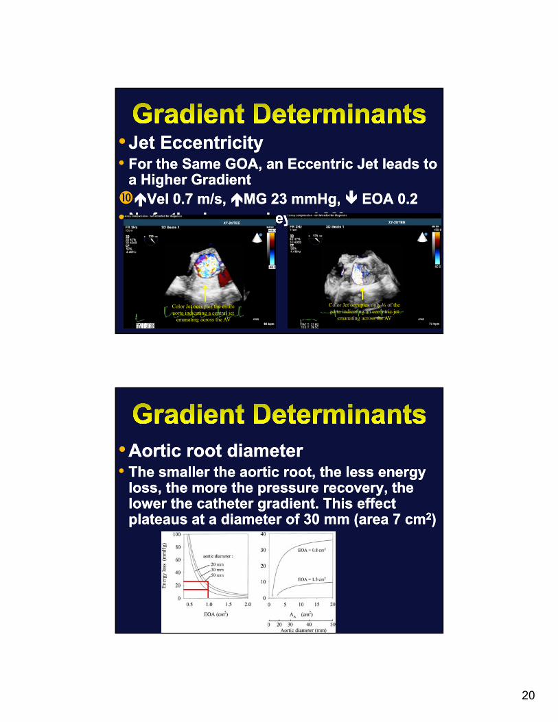
•Jet Eccentricity• For the Same GOA, an Eccentric Jet leads to
a Higher GradientVel 0.7 m/s, MG 23 mmHg, EOA 0.2• No further increase beyond 30°
•Jet Eccentricity• For the Same GOA, an Eccentric Jet leads to
a Higher GradientVel 0.7 m/s, MG 23 mmHg, EOA 0.2• No further increase beyond 30°
Color Jet occupies the entire aorta indicating a central jet
emanating across the AV
Color Jet occupies only ½ of the aorta indicating an eccentric jet
emanating across the AV
•Aortic root diameter• The smaller the aortic root, the less energy
loss, the more the pressure recovery, the lower the catheter gradient. This effect plateaus at a diameter of 30 mm (area 7 cm2)
•Aortic root diameter• The smaller the aortic root, the less energy
loss, the more the pressure recovery, the lower the catheter gradient. This effect plateaus at a diameter of 30 mm (area 7 cm2)
21
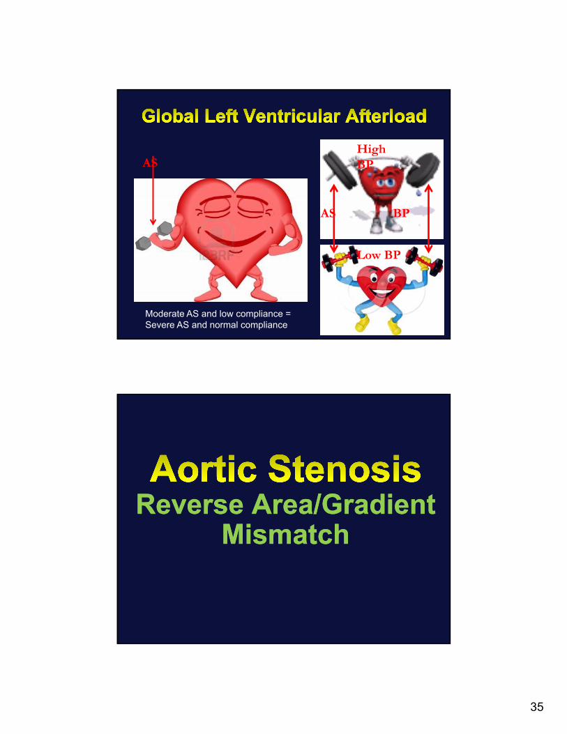
AS
BPAS
High BP
Low BP
Moderate AS and low compliance = Severe AS and normal compliance
Z VA = SBP + Mean Gradient = (xx) mmHg/ml·m-2
Stroke Volume IndexZ VA = 150 mmHg + 26 mmHg = 5.5mmHg/ml·m-2
32 ml/m2
22
Courtesy Heidi Connolly
Normal Ejection Fraction
Normal Cardiac Output
AVA<1cm2
ΔPmean>40mmHg
Area Gradient Match
Area/Gradient Mismatch Area/Gradient Mismatch
23
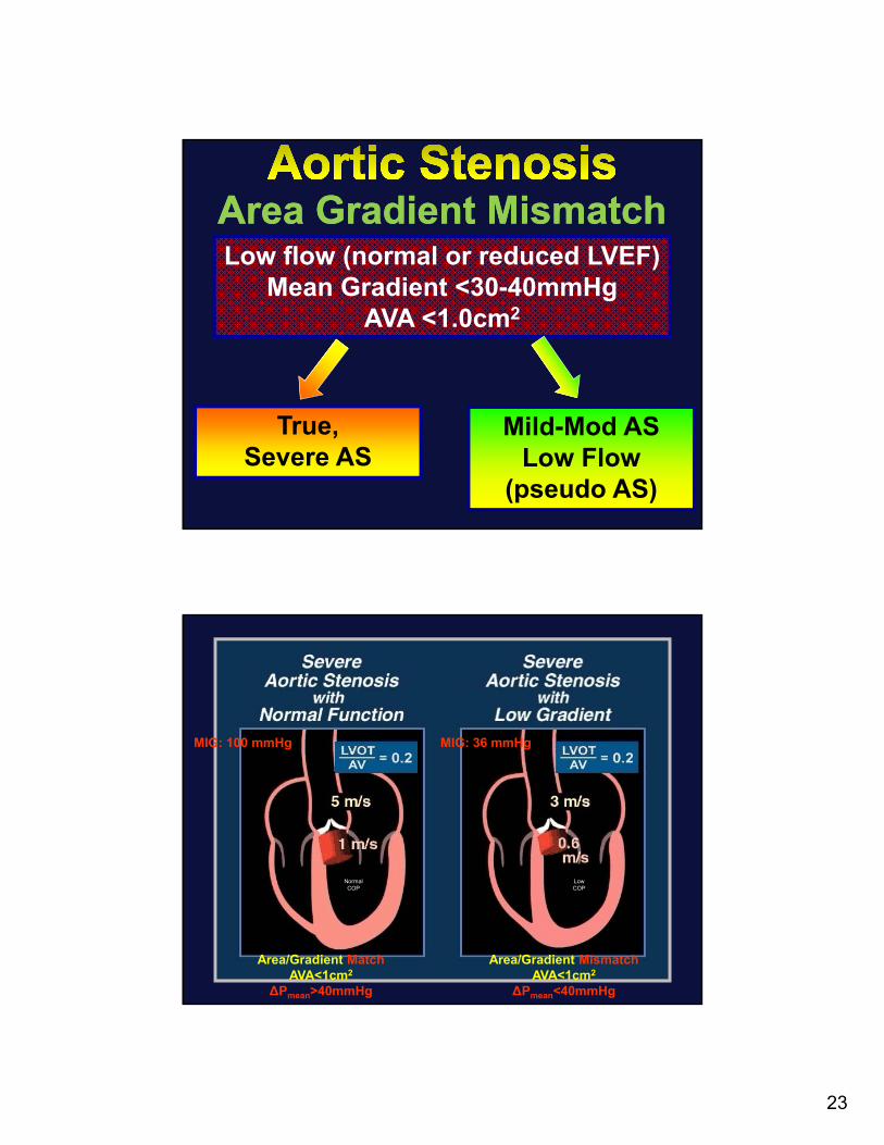
Area Gradient MismatchArea Gradient MismatchLow flow (normal or reduced LVEF)
Mean Gradient <30-40mmHgAVA <1.0cm2
True, Severe AS
Mild-Mod ASLow Flow
(pseudo AS)
Area/Gradient MatchAVA<1cm2
ΔPmean>40mmHg
Area/Gradient MismatchAVA<1cm2
ΔPmean<40mmHg
MIG: 100 mmHg MIG: 36 mmHg
NormalCOP
LowCOP
24
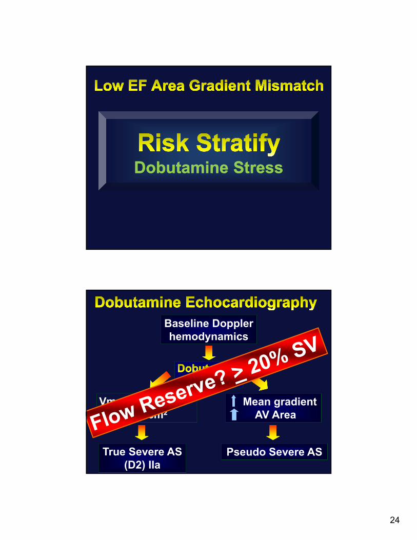
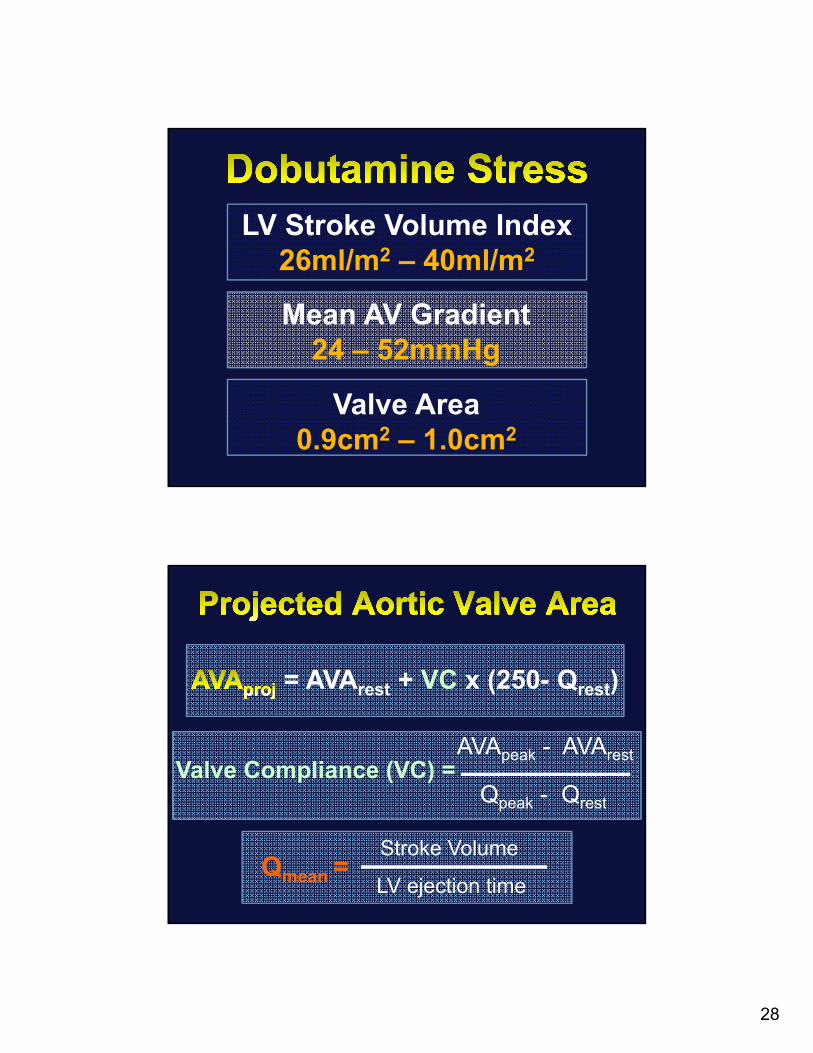
Dobutamine StressDobutamine Stress
Baseline Dopplerhemodynamics
Mean gradient AV Area
Mean gradient AV Area
True Severe AS (D2) IIa
Pseudo Severe AS
Vmax > 4.0m/sec AVA < 1.0cm2
25
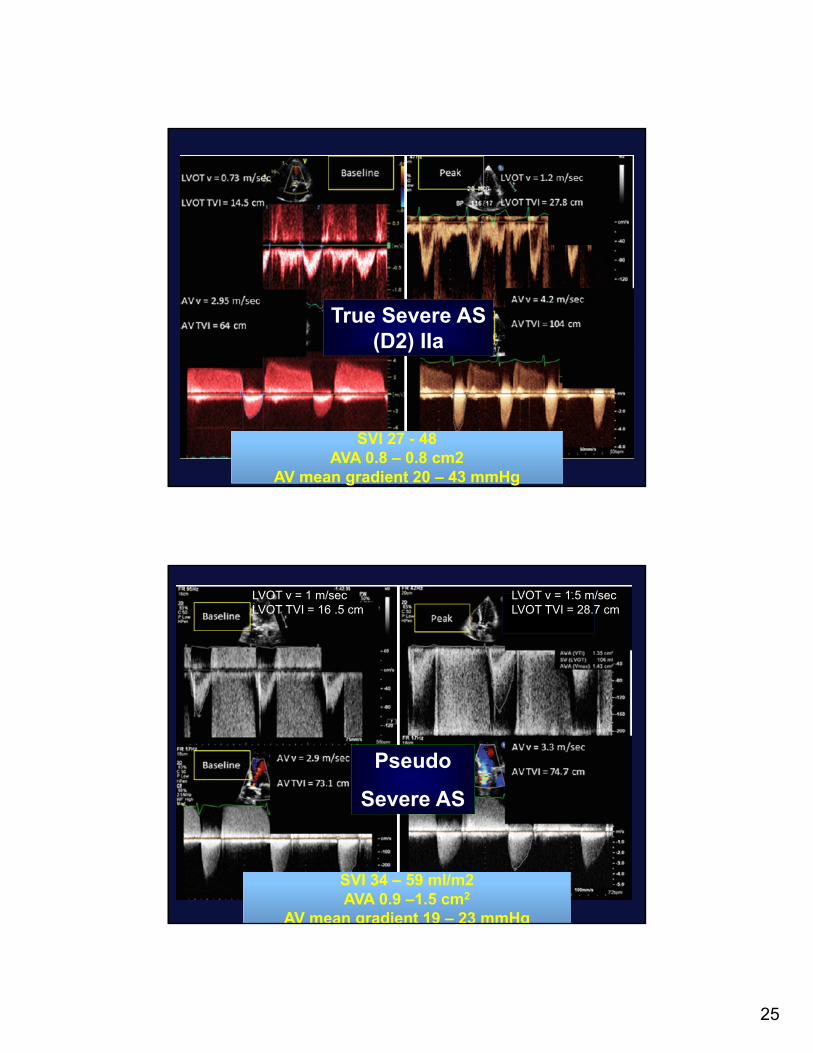
g g
SVI 27 - 48AVA 0.8 – 0.8 cm2
AV mean gradient 20 – 43 mmHg
True Severe AS (D2) IIa
LVOT v = 1 m/secLVOT TVI = 16 .5 cm
LVOT v = 1.5 m/secLVOT TVI = 28.7 cm
SVI 34 – 59 ml/m2AVA 0.9 –1.5 cm2
AV mean gradient 19 – 23 mmHg
Pseudo
Severe AS
26
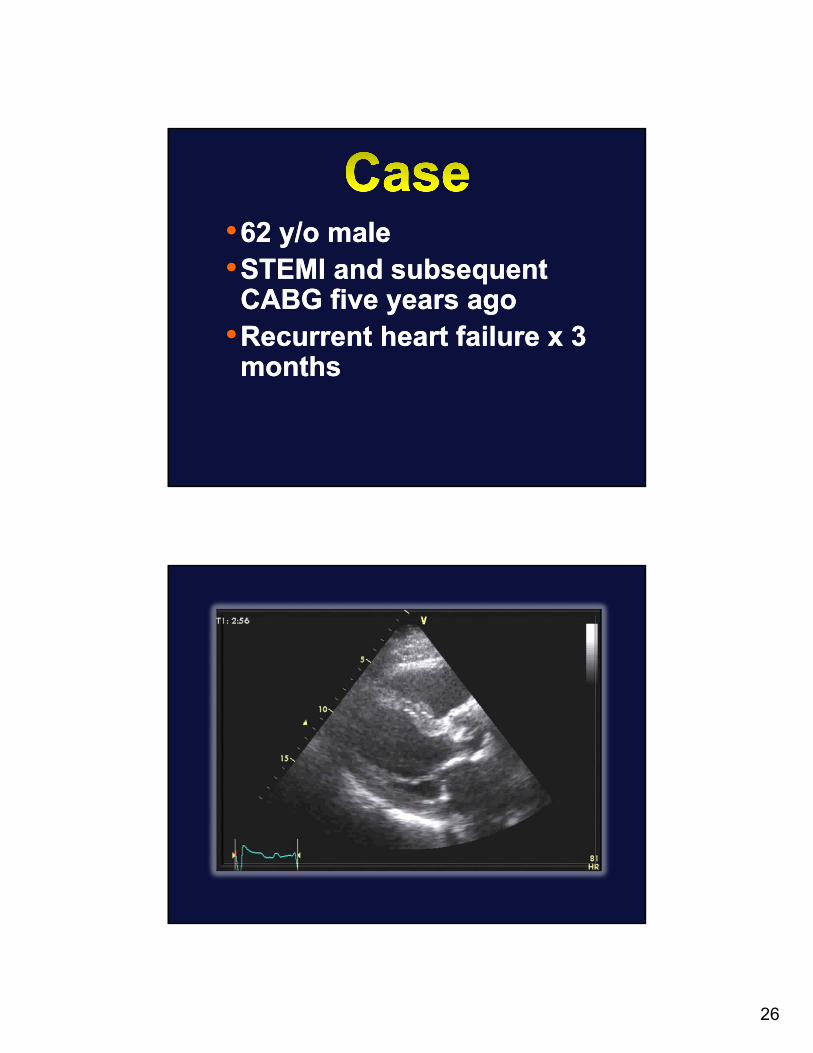
•62 y/o male•STEMI and subsequent CABG five years ago
•Recurrent heart failure x 3 months
•62 y/o male•STEMI and subsequent CABG five years ago
•Recurrent heart failure x 3 months
27
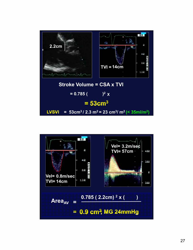
2.2cm
TVI = 14cm
Stroke Volume = CSA x TVI
= 0.785 ( )2 X
= 53cm3 / 2.3 m2 = 23 cm3/ m2 (< 35ml/m2)
Vel= TVI=
0.8m/sec
Vel= TVI=
3.2m/sec
AreaAV0.785 ( 2.2cm) 2 x ( )
=
14cm
57cm
28
Mean AV Gradient24 – 52mmHg
Valve Area0.9cm2 – 1.0cm2
LV Stroke Volume Index26ml/m2 – 40ml/m2
Qmean =Stroke Volume
LV ejection time
= AVArest + VC x (250- Qrest)
Valve Compliance (VC) =AVApeak - AVArest
Qpeak - Qrest
29
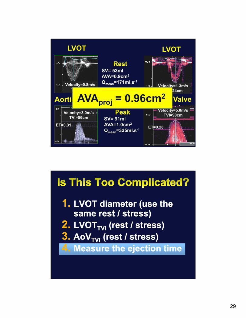
LVOT
Aortic Valve
Velocity=0.8m/sTVI=14cm
Velocity=3.0m/sTVI=56cm
LVOT
Aortic Valve
Velocity=1.3m/sTVI=24cm
Velocity=5.0m/sTVI=90cm
ET=0.31 ET=0.28
SV= 53mlAVA=0.9cm2
Qmean=171ml.s-1
SV= 91mlAVA=1.0cm2
Qmean=325ml.s-1
AVAproj = 0.96cm2
1. LVOT diameter (use the same rest / stress)
2. LVOTTVI (rest / stress)3. AoVTVI (rest / stress)4. Measure the ejection time
1. LVOT diameter (use the same rest / stress)
2. LVOTTVI (rest / stress)3. AoVTVI (rest / stress)4. Measure the ejection time
30
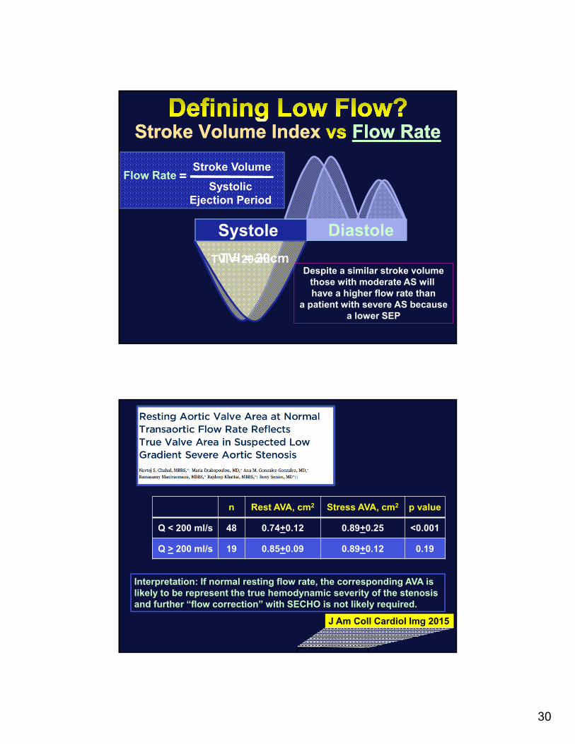
Systole Diastole
Stroke Volume Index Flow RateStroke Volume Index Flow Rate
Systole Diastole
TVI = 20cmTVI = 20cm
Flow Rate Stroke Volume
SystolicEjection Period
=
Despite a similar stroke volumethose with moderate AS will have a higher flow rate than
a patient with severe AS because a lower SEP
n Rest AVA, cm2 Stress AVA, cm2 p value
Q < 200 ml/s 48 0.74+0.12 0.89+0.25 <0.001
Q > 200 ml/s 19 0.85+0.09 0.89+0.12 0.19
Interpretation: If normal resting flow rate, the corresponding AVA is likely to be represent the true hemodynamic severity of the stenosis and further “flow correction” with SECHO is not likely required.
J Am Coll Cardiol Img 2015
31
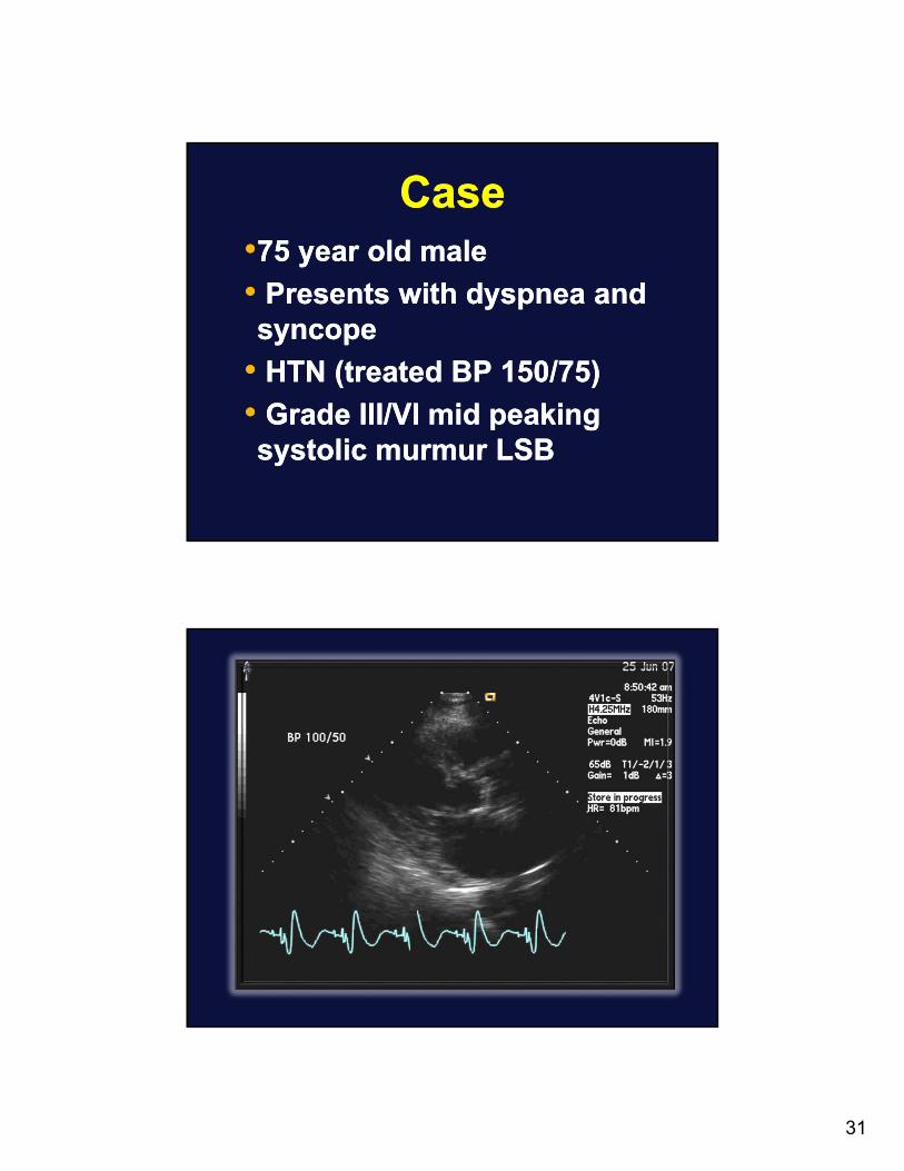
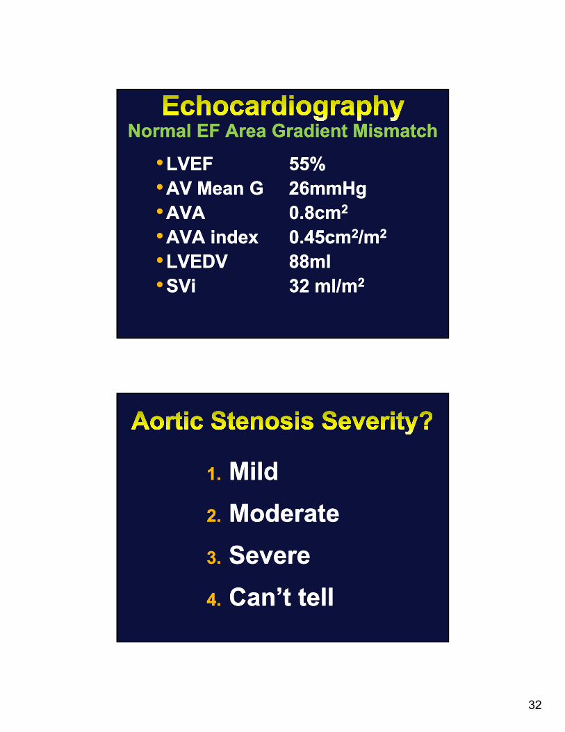
CaseCase•75 year old male
• Presents with dyspnea and syncope
• HTN (treated BP 150/75)
• Grade III/VI mid peaking systolic murmur LSB
•75 year old male
• Presents with dyspnea and syncope
• HTN (treated BP 150/75)
• Grade III/VI mid peaking systolic murmur LSB
32
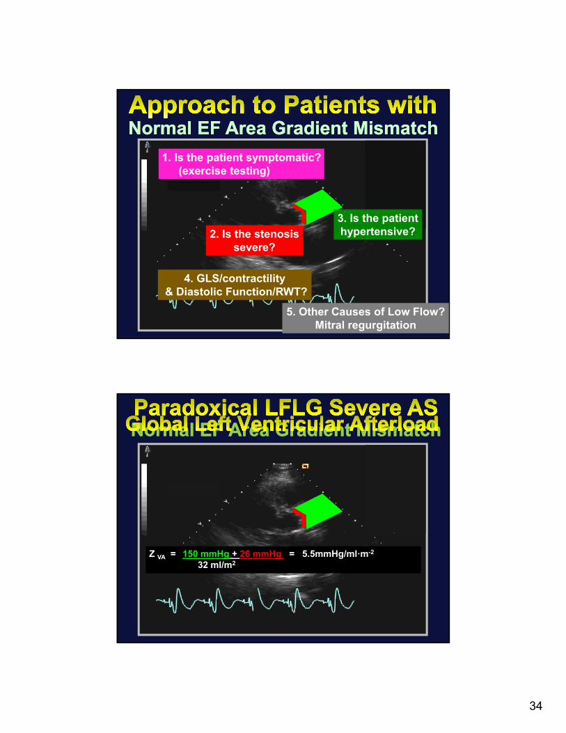
Normal EF Area Gradient MismatchNormal EF Area Gradient Mismatch
•LVEF 55%•AV Mean G 26mmHg•AVA 0.8cm2
•AVA index 0.45cm2/m2
•LVEDV 88ml•SVi 32 ml/m2
•LVEF 55%•AV Mean G 26mmHg•AVA 0.8cm2
•AVA index 0.45cm2/m2
•LVEDV 88ml•SVi 32 ml/m2
1. Mild
2. Moderate
3. Severe
4. Can’t tell
1. Mild
2. Moderate
3. Severe
4. Can’t tell
33
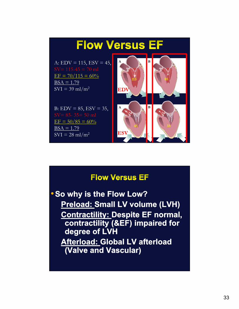
A: EDV = 115, ESV = 45, SV= 115-45 = 70 mlEF = 70/115 = 60%BSA = 1.79SVI = 39 ml/m2
B: EDV = 85, ESV = 35, SV= 85- 35= 50 mlEF = 50/85 = 60%BSA = 1.79SVI = 28 ml/m2
A
A
B
B
115 85
3545
EDV
ESV
•So why is the Flow Low?Preload: Small LV volume (LVH)Contractility: Despite EF normal, contractility (&EF) impaired for degree of LVH
Afterload: Global LV afterload (Valve and Vascular)
•So why is the Flow Low?Preload: Small LV volume (LVH)Contractility: Despite EF normal, contractility (&EF) impaired for degree of LVH
Afterload: Global LV afterload (Valve and Vascular)
34
Normal EF Area Gradient MismatchNormal EF Area Gradient Mismatch
1. Is the patient symptomatic?(exercise testing)
3. Is the patienthypertensive?2. Is the stenosis
severe?
4. GLS/contractility& Diastolic Function/RWT?
5. Other Causes of Low Flow?Mitral regurgitation
Z VA = SBP + Mean Gradient = (xx) mmHg/ml·m-2
Stroke Volume IndexZ VA = 150 mmHg + 26 mmHg = 5.5mmHg/ml·m-2
32 ml/m2
Normal EF Area Gradient MismatchNormal EF Area Gradient Mismatch
35
AS
BPAS
High BP
Low BP
Moderate AS and low compliance = Severe AS and normal compliance
Reverse Area/Gradient Mismatch
Reverse Area/Gradient Mismatch
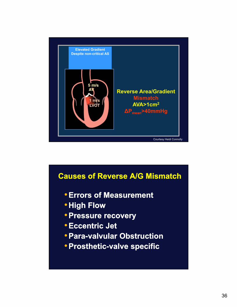
36
Courtesy Heidi Connolly
Elevated Gradient Despite non-critical AS
Reverse Area/Gradient MismatchAVA>1cm2
ΔPmean>40mmHg
Causes of Reverse A/G MismatchCauses of Reverse A/G Mismatch
•Errors of Measurement•High Flow•Pressure recovery•Eccentric Jet•Para-valvular Obstruction•Prosthetic-valve specific
•Errors of Measurement•High Flow•Pressure recovery•Eccentric Jet•Para-valvular Obstruction•Prosthetic-valve specific
37
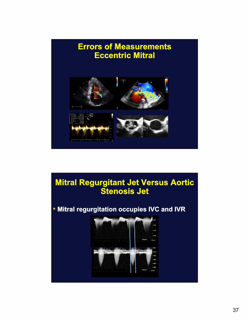
Errors of MeasurementsEccentric Mitral
Errors of MeasurementsEccentric Mitral
Mitral Regurgitant Jet Versus Aortic Stenosis Jet
Mitral Regurgitant Jet Versus Aortic Stenosis Jet
• Mitral regurgitation occupies IVC and IVR• Mitral regurgitation occupies IVC and IVR
38
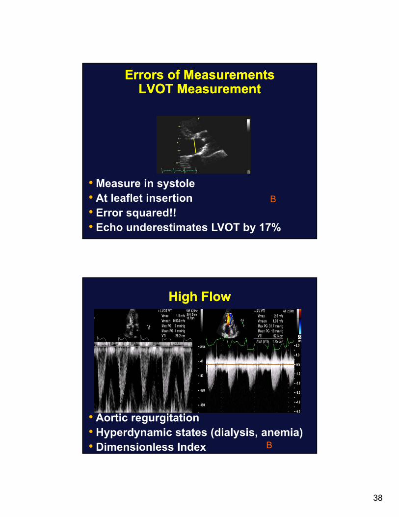
Errors of MeasurementsLVOT Measurement
Errors of MeasurementsLVOT Measurement
• Measure in systole• At leaflet insertion • Error squared!!• Echo underestimates LVOT by 17%
B
High FlowHigh Flow
• Aortic regurgitation• Hyperdynamic states (dialysis, anemia)• Dimensionless Index B
39
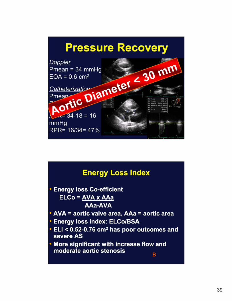
Pressure RecoveryPressure Recovery
GOA 1.1 cm2
DopplerPmean = 34 mmHgEOA = 0.6 cm2
CatheterizationPmean = 18 mmHgEOA = 1 cm2
APR= 34-18 = 16 mmHgRPR= 16/34= 47%
Energy Loss IndexEnergy Loss Index
• Energy loss Co-efficientELCo = AVA x AAa
AAa-AVA• AVA = aortic valve area, AAa = aortic area• Energy loss index: ELCo/BSA• ELI < 0.52-0.76 cm2 has poor outcomes and
severe AS• More significant with increase flow and
moderate aortic stenosis
• Energy loss Co-efficientELCo = AVA x AAa
AAa-AVA• AVA = aortic valve area, AAa = aortic area• Energy loss index: ELCo/BSA• ELI < 0.52-0.76 cm2 has poor outcomes and
severe AS• More significant with increase flow and
moderate aortic stenosisB
40
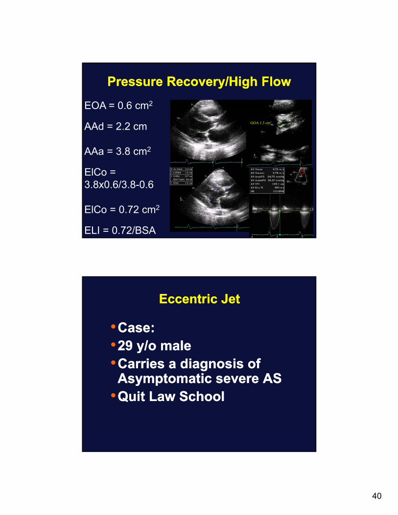
Pressure Recovery/High FlowPressure Recovery/High Flow
GOA 1.3 cm2
EOA = 0.6 cm2
AAd = 2.2 cm
AAa = 3.8 cm2
ElCo = 3.8x0.6/3.8-0.6
ElCo = 0.72 cm2
ELI = 0.72/BSA
Eccentric JetEccentric Jet
•Case:•29 y/o male•Carries a diagnosis of Asymptomatic severe AS
•Quit Law School
•Case:•29 y/o male•Carries a diagnosis of Asymptomatic severe AS
•Quit Law School
41
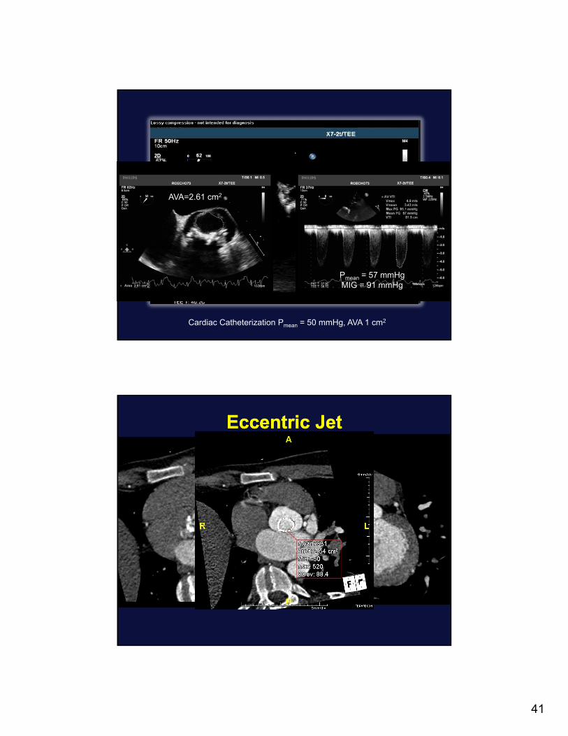
Cardiac Catheterization Pmean = 50 mmHg, AVA 1 cm2
AVA=2.61 cm2
Pmean = 57 mmHgMIG = 91 mmHg
Eccentric JetEccentric Jet
42
Aortic Diameter = 4.0 cm Eccentric Jet
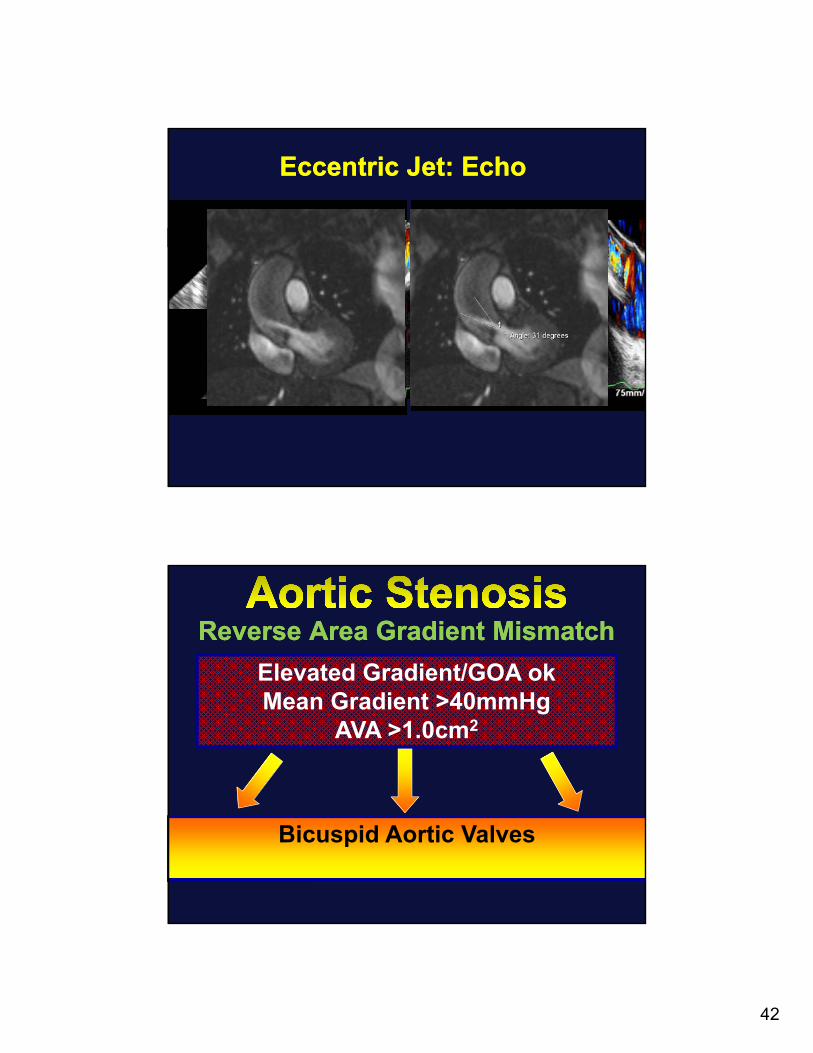
Eccentric Jet: EchoEccentric Jet: Echo
Reverse Area Gradient MismatchReverse Area Gradient Mismatch
Elevated Gradient/GOA okMean Gradient >40mmHg
AVA >1.0cm2
Increased Flow
Eccentric Jet
Dilated Aorta
Bicuspid Aortic Valves
43
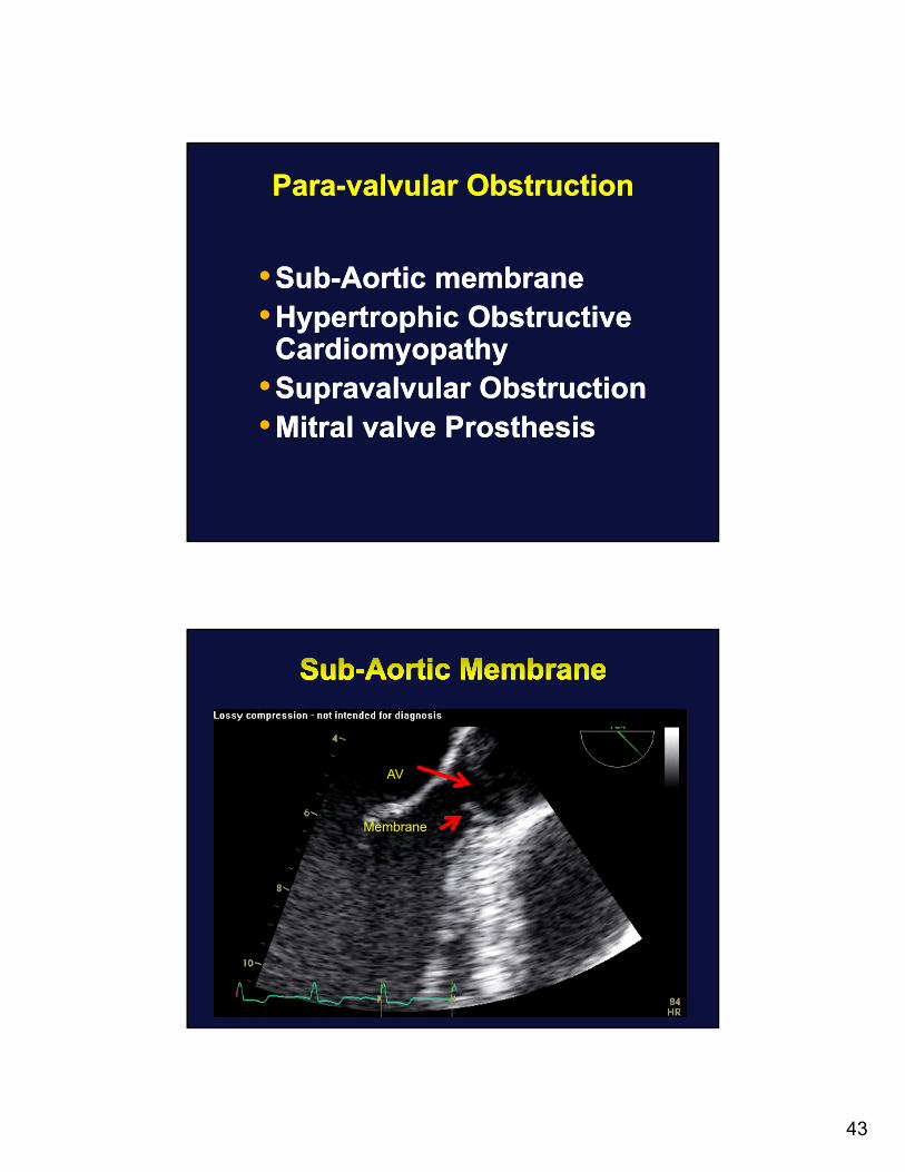
Para-valvular ObstructionPara-valvular Obstruction
•Sub-Aortic membrane•Hypertrophic Obstructive
Cardiomyopathy•Supravalvular Obstruction•Mitral valve Prosthesis
•Sub-Aortic membrane•Hypertrophic Obstructive
Cardiomyopathy•Supravalvular Obstruction•Mitral valve Prosthesis
Membrane
AV
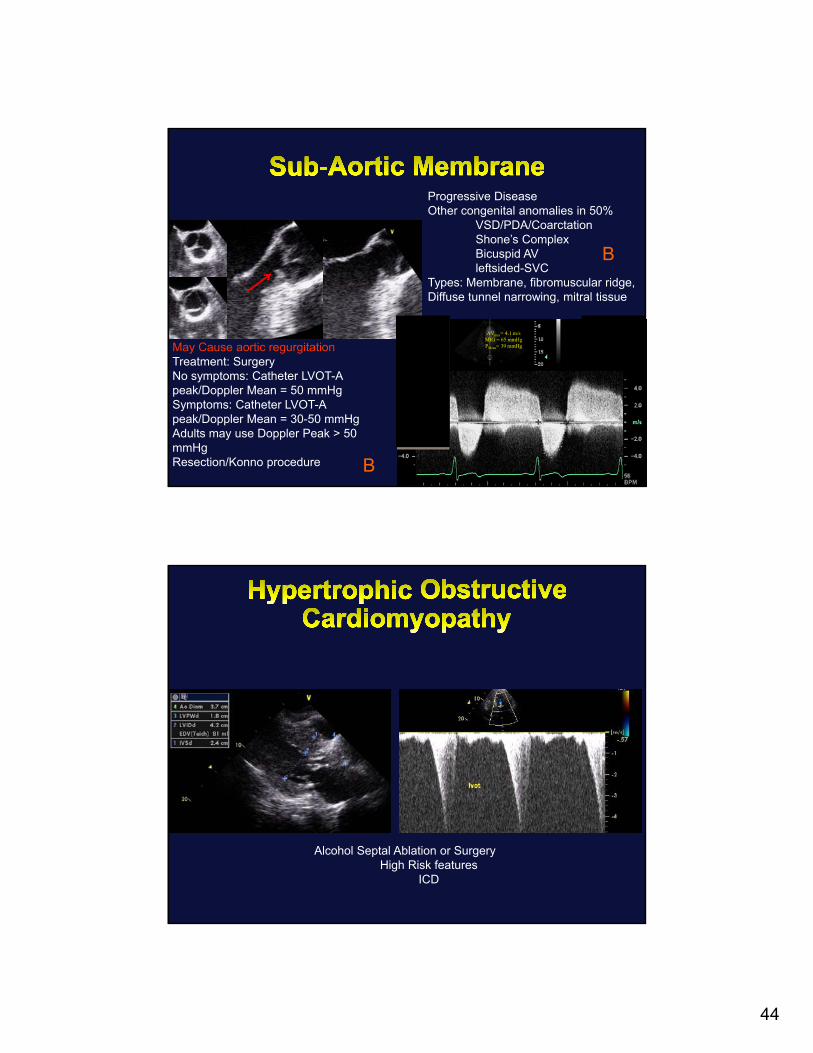
44
AVmax= 4.1 m/sMIG = 65 mmHgPMean= 39 mmHg
Progressive DiseaseOther congenital anomalies in 50%
VSD/PDA/CoarctationShone’s ComplexBicuspid AVleftsided-SVC
Types: Membrane, fibromuscular ridge, Diffuse tunnel narrowing, mitral tissue
May Cause aortic regurgitationTreatment: SurgeryNo symptoms: Catheter LVOT-A peak/Doppler Mean = 50 mmHgSymptoms: Catheter LVOT-A peak/Doppler Mean = 30-50 mmHgAdults may use Doppler Peak > 50 mmHgResection/Konno procedure B
B
Alcohol Septal Ablation or SurgeryHigh Risk features
ICD
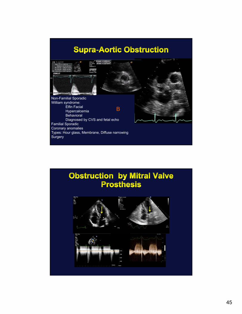
45
Non-Familial SporadicWilliam syndrome:
Elfin FacialHypercalcemiaBehavioralDiagnosed by CVS and fetal echo
Familial SporadicCoronary anomaliesTypes: Hour glass, Membrane, Diffuse narrowingSurgery
B
46
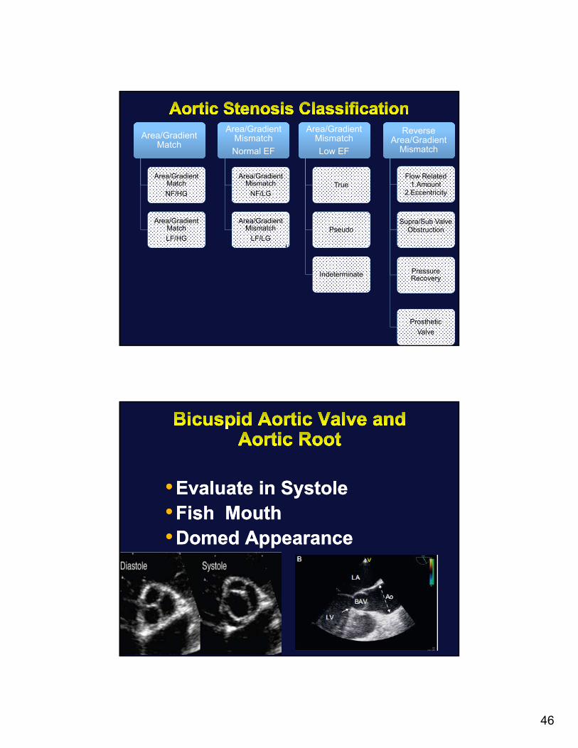
Area/Gradient Mismatch
Normal EF
Area/Gradient Mismatch
Normal EF
Area/Gradient Mismatch
NF/LG
Area/Gradient Mismatch
LF/LG
Reverse Area/Gradient
Mismatch
Reverse Area/Gradient
Mismatch
Flow Related1.Amount
2.Eccentricity
Supra/Sub Valve Obstruction
Pressure Recovery
Prosthetic
Valve
Area/Gradient Mismatch
Low EF
Area/Gradient Mismatch
Low EF
True
Pseudo
Indeterminate
Area/Gradient Match
Area/Gradient Match
Area/Gradient Match
NF/HG
Area/Gradient Match
LF/HG
•Evaluate in Systole•Fish Mouth•Domed Appearance
•Evaluate in Systole•Fish Mouth•Domed Appearance
47
48
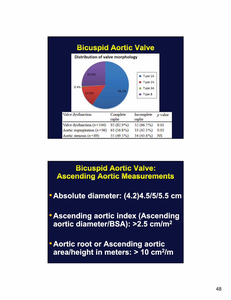
•Absolute diameter: (4.2)4.5/5/5.5 cm
•Ascending aortic index (Ascending aortic diameter/BSA): >2.5 cm/m2
•Aortic root or Ascending aortic area/height in meters: > 10 cm2/m
•Absolute diameter: (4.2)4.5/5/5.5 cm
•Ascending aortic index (Ascending aortic diameter/BSA): >2.5 cm/m2
•Aortic root or Ascending aortic area/height in meters: > 10 cm2/m
49
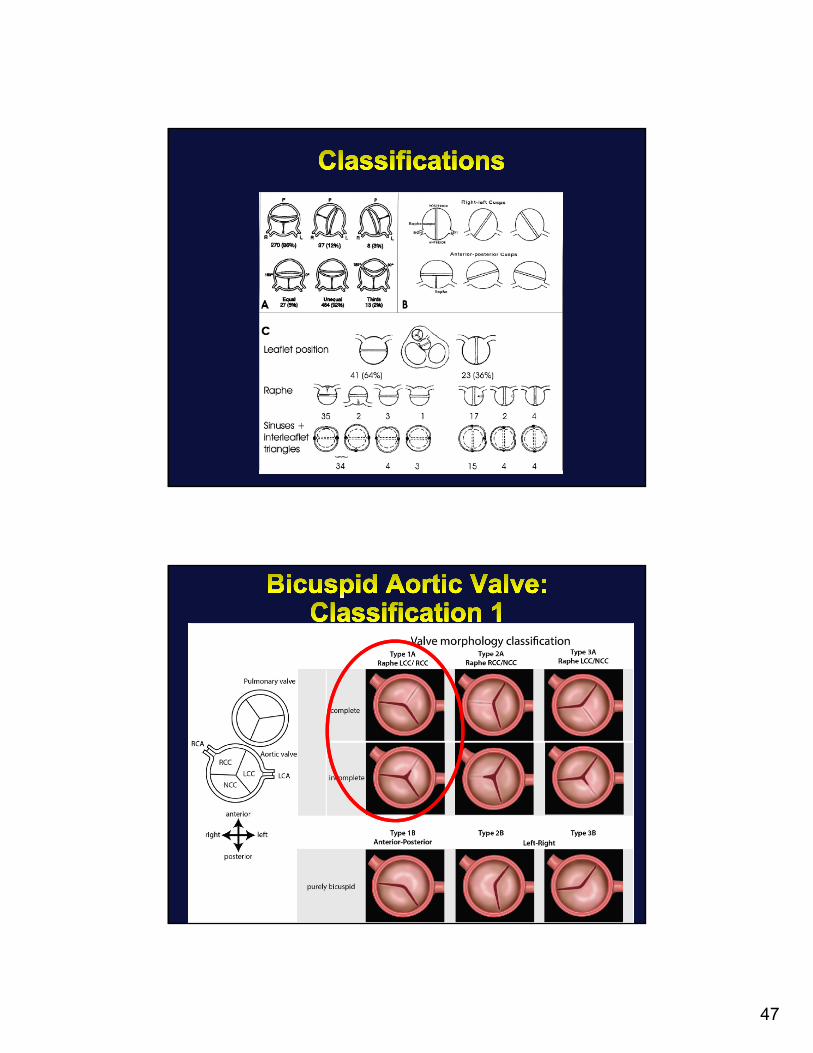
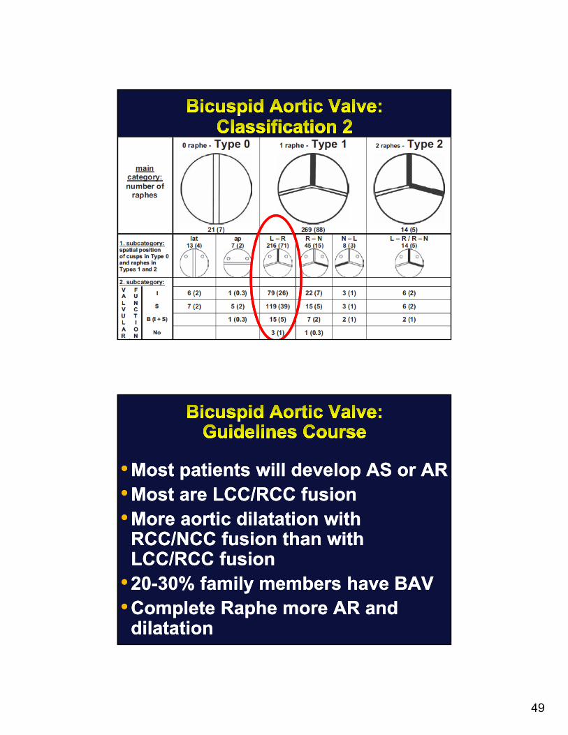
•Most patients will develop AS or AR•Most are LCC/RCC fusion•More aortic dilatation with
RCC/NCC fusion than with LCC/RCC fusion
•20-30% family members have BAV•Complete Raphe more AR and
dilatation
•Most patients will develop AS or AR•Most are LCC/RCC fusion•More aortic dilatation with
RCC/NCC fusion than with LCC/RCC fusion
•20-30% family members have BAV•Complete Raphe more AR and
dilatation
50
Aortic Root Mean (cm) SD Method
Female 3.5-3.72 0.38 CT
Male 3.63-3.91 0.38 CT
Syndrome Gene Features
Marfan FBN1 Skeletal FeaturesEctopia lentis
Loeys-Dietz TGFBR1TGFBR2
Skeletal FeaturesCleft palate/uvula
ACTA2 Livedo reticularisPDA/BAV
MYH11 PDA
Vascular Ehlers-Danlos
COL3A1 Thin skinGI/uterine rupture
Turner 45,X Skeletal FeatureBAV/Coarctation
• Increased wall stress: HTN
CocainePheoWeight liftingTrauma and deceleration
Coarctation
• Increased wall stress: HTN
CocainePheoWeight liftingTrauma and deceleration
Coarctation
51
•Media abnormalities: GeneticInflammatoryTakayasu arteritisGiant cell arteritisBehcet arteritis
Other:Pregnancy/PCKD/steroids
•Media abnormalities: GeneticInflammatoryTakayasu arteritisGiant cell arteritisBehcet arteritis
Other:Pregnancy/PCKD/steroids
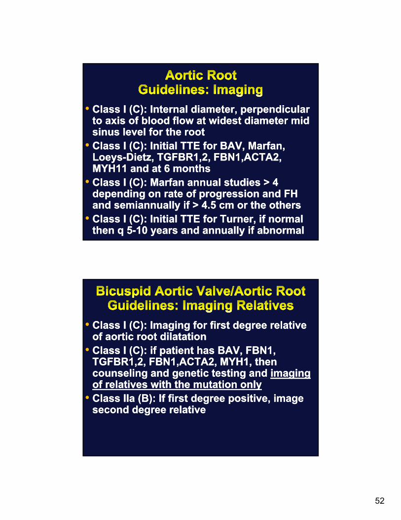
• Class I (C): Initial TTE for morphology, AS/AR, sinuses, ascending aorta and timing for intervention
• Class I (C): Serial studies > 4 depending on rate of progression and FH and annually if > 4.5 cm
• Class I (C): Internal diameter, perpendicular to axis of blood flow at widest diameter mid sinus level for the root
52
• Class I (C): Internal diameter, perpendicular to axis of blood flow at widest diameter mid sinus level for the root
• Class I (C): Initial TTE for BAV, Marfan, Loeys-Dietz, TGFBR1,2, FBN1,ACTA2, MYH11 and at 6 months
• Class I (C): Marfan annual studies > 4 depending on rate of progression and FH and semiannually if > 4.5 cm or the others
• Class I (C): Initial TTE for Turner, if normal then q 5-10 years and annually if abnormal
• Class I (C): Imaging for first degree relative of aortic root dilatation
• Class I (C): if patient has BAV, FBN1, TGFBR1,2, FBN1,ACTA2, MYH1, then counseling and genetic testing and imaging of relatives with the mutation only
• Class IIa (B): If first degree positive, image second degree relative
• Class I (C): Imaging for first degree relative of aortic root dilatation
• Class I (C): if patient has BAV, FBN1, TGFBR1,2, FBN1,ACTA2, MYH1, then counseling and genetic testing and imaging of relatives with the mutation only
• Class IIa (B): If first degree positive, image second degree relative
53
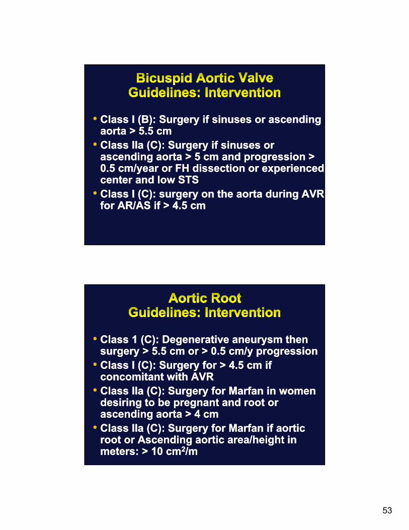
• Class I (B): Surgery if sinuses or ascending aorta > 5.5 cm
• Class IIa (C): Surgery if sinuses or ascending aorta > 5 cm and progression > 0.5 cm/year or FH dissection or experienced center and low STS
• Class I (C): surgery on the aorta during AVR for AR/AS if > 4.5 cm
• Class I (B): Surgery if sinuses or ascending aorta > 5.5 cm
• Class IIa (C): Surgery if sinuses or ascending aorta > 5 cm and progression > 0.5 cm/year or FH dissection or experienced center and low STS
• Class I (C): surgery on the aorta during AVR for AR/AS if > 4.5 cm
• Class 1 (C): Degenerative aneurysm then surgery > 5.5 cm or > 0.5 cm/y progression
• Class I (C): Surgery for > 4.5 cm if concomitant with AVR
• Class IIa (C): Surgery for Marfan in women desiring to be pregnant and root or ascending aorta > 4 cm
• Class IIa (C): Surgery for Marfan if aortic root or Ascending aortic area/height in meters: > 10 cm2/m
54
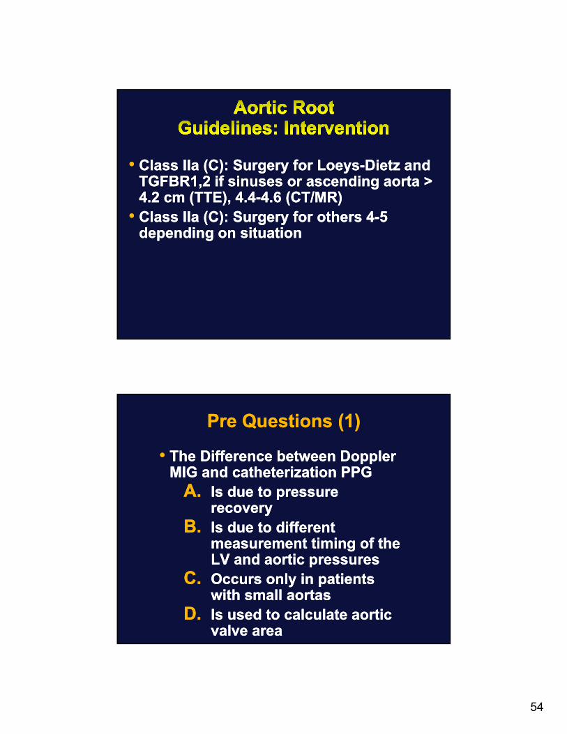
• Class IIa (C): Surgery for Loeys-Dietz and TGFBR1,2 if sinuses or ascending aorta > 4.2 cm (TTE), 4.4-4.6 (CT/MR)
• Class IIa (C): Surgery for others 4-5 depending on situation
• Class IIa (C): Surgery for Loeys-Dietz and TGFBR1,2 if sinuses or ascending aorta > 4.2 cm (TTE), 4.4-4.6 (CT/MR)
• Class IIa (C): Surgery for others 4-5 depending on situation
Pre Questions (1)Pre Questions (1)
• The Difference between Doppler MIG and catheterization PPG
A. Is due to pressure recovery
B. Is due to different measurement timing of the LV and aortic pressures
C. Occurs only in patients with small aortas
D. Is used to calculate aortic valve area
• The Difference between Doppler MIG and catheterization PPG
A. Is due to pressure recovery
B. Is due to different measurement timing of the LV and aortic pressures
C. Occurs only in patients with small aortas
D. Is used to calculate aortic valve area
55
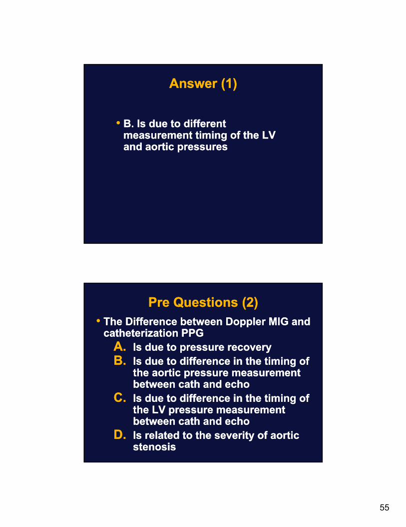
Answer (1)Answer (1)
• B. Is due to different measurement timing of the LV and aortic pressures
• B. Is due to different measurement timing of the LV and aortic pressures
Pre Questions (2)Pre Questions (2)• The Difference between Doppler MIG and
catheterization PPGA. Is due to pressure recoveryB. Is due to difference in the timing of
the aortic pressure measurement between cath and echo
C. Is due to difference in the timing of the LV pressure measurement between cath and echo
D. Is related to the severity of aortic stenosis
• The Difference between Doppler MIG and catheterization PPG
A. Is due to pressure recoveryB. Is due to difference in the timing of
the aortic pressure measurement between cath and echo
C. Is due to difference in the timing of the LV pressure measurement between cath and echo
D. Is related to the severity of aortic stenosis
56
Pre Questions (2)Pre Questions (2)
B. Is due to difference in the timing of the aortic pressure measurement between cathand echo
B. Is due to difference in the timing of the aortic pressure measurement between cathand echo
Pre Questions (3)Pre Questions (3)
• Catheter-Doppler Discordance maybe due to
A. Pressure recoveryB. Eccentric jetC. Very severe aortic stenosisD. HOCM
• Catheter-Doppler Discordance maybe due to
A. Pressure recoveryB. Eccentric jetC. Very severe aortic stenosisD. HOCM
57
Pre Questions (3)Pre Questions (3)
• A. Pressure recovery• A. Pressure recovery
Pre Questions (4)Pre Questions (4)
• The most common form of bicuspid aortic valve is
A. Fusion of the LCC/RCCB. Fusion of the LCC/NCCC. Fusion of the RCC/NCCD. Equal distribution of cusp
fusion
• The most common form of bicuspid aortic valve is
A. Fusion of the LCC/RCCB. Fusion of the LCC/NCCC. Fusion of the RCC/NCCD. Equal distribution of cusp
fusion
58
Pre Questions (4)Pre Questions (4)
A. Fusion of the LCC/RCCA. Fusion of the LCC/RCC


![Native Aortic Valve Endocarditis—A Case Report · aortic cusps, resulting in a bicuspid aortic valve and a weakened aortic root 3], [which may complicate infective endocarditis](https://img.pdfslide.us/doc/110x75/6015ccdee1b3dd30591e4f45/native-aortic-valve-endocarditisaa-case-report-aortic-cusps-resulting-in-a-bicuspid.jpg)


![Effect of Bicuspid Aortic Valve Cusp Fusion on Aorta Wall ...The congenital bicuspid aortic valve (BAV) is a valvular defect present in 1% - 2% of the general population[1]. While](https://img.pdfslide.us/doc/110x75/5f34ae6844f7a3568d255217/effect-of-bicuspid-aortic-valve-cusp-fusion-on-aorta-wall-the-congenital-bicuspid.jpg)