Embed Size (px)
Citation preview
REVIEWpublished: 13 March 2019
doi: 10.3389/fendo.2019.00148
Frontiers in Endocrinology | www.frontiersin.org 1 March 2019 | Volume 10 | Article 148
Edited by:
Daniele Santi,
University of Modena and Reggio
Emilia, Italy
Reviewed by:
Karen J. Gregory,
Monash University, Australia
Brice Campo,
Medicines for Malaria Venture,
Switzerland
James A. Dias,
University at Albany, United States
*Correspondence:
Eric Reiter
Specialty section:
This article was submitted to
Reproduction,
a section of the journal
Frontiers in Endocrinology
Received: 10 January 2019
Accepted: 19 February 2019
Published: 13 March 2019
Citation:
Landomiel F, De Pascali F, Raynaud P,
Jean-Alphonse F, Yvinec R,
Pellissier LP, Bozon V, Bruneau G,
Crépieux P, Poupon A and Reiter E
(2019) Biased Signaling and Allosteric
Modulation at the FSHR.
Front. Endocrinol. 10:148.
doi: 10.3389/fendo.2019.00148
Biased Signaling and AllostericModulation at the FSHRFlavie Landomiel, Francesco De Pascali, Pauline Raynaud, Frédéric Jean-Alphonse,
Romain Yvinec, Lucie P. Pellissier, Véronique Bozon, Gilles Bruneau, Pascale Crépieux,
Anne Poupon and Eric Reiter*
PRC, INRA, CNRS, IFCE, Université de Tours, Nouzilly, France
Knowledge on G protein-coupled receptor (GPCRs) structure and mechanism of
activation has profoundly evolved over the past years. The way drugs targeting this
family of receptors are discovered and used has also changed. Ligands appear to
bind a growing number of GPCRs in a competitive or allosteric manner to elicit
balanced signaling or biased signaling (i.e., differential efficacy in activating or inhibiting
selective signaling pathway(s) compared to the reference ligand). These novel concepts
and developments transform our understanding of the follicle-stimulating hormone
(FSH) receptor (FSHR) biology and the way it could be pharmacologically modulated
in the future. The FSHR is expressed in somatic cells of the gonads and plays a
major role in reproduction. When compared to classical GPCRs, the FSHR exhibits
intrinsic peculiarities, such as a very large NH2-terminal extracellular domain that
binds a naturally heterogeneous, large heterodimeric glycoprotein, namely FSH. Once
activated, the FSHR couples to Gαs and, in some instances, to other Gα subunits. G
protein-coupled receptor kinases and β-arrestins are also recruited to this receptor and
account for its desensitization, trafficking, and intracellular signaling. Different classes of
pharmacological tools capable of biasing FSHR signaling have been reported and open
promising prospects both in basic research and for therapeutic applications. Here we
provide an updated review of the most salient peculiarities of FSHR signaling and its
selective modulation.
Keywords: GPCR, reproduction, follicle-stimulating hormone, β-arrestin, G protein, signaling, bias, trafficking
INTRODUCTION
Follicle stimulating hormone (FSH) plays a crucial role in the control of male and femalereproduction. FSH is a heterodimeric glycoprotein consisting of an α-subunit non-covalentlyassociated with a β-subunit. The α-subunit is shared with luteinizing hormone (LH), chorionicgonadotropin (CG) and thyroid-stimulating hormone (TSH), whereas the β chain is specific ofeach glycoprotein hormone (1). FSH is synthesized and secreted by the pituitary and binds to aplasma membrane receptor (FSHR) that belongs to the class A of the G protein-coupled receptor(GPCR) superfamily. The FSHR displays a high degree of tissue specificity as it is expressed inSertoli and granulosa cells located in the male and female gonads, respectively (2). FSH is requiredfor normal growth and maturation of ovarian follicles in women and for normal spermatogenesisin men (3). Female mice with FSHβ or FSHR gene knockout present an incomplete follicledevelopment leading to infertility, whereas males display oligozoospermia and subfertility (4, 5).
Landomiel et al. Biased Signaling and Allosteric Modulation at the FSHR
Consistently, women expressing non-functional variants of theFSHR are infertile while men are oligozoospermic, yet fertile(6). To date, only native forms of FSH, either purified fromurine or by using recombinant technology, are being used inreproductive medicine with no other pharmacological agentsbeing currently available in clinic (7–9). Novel classes of FSHRagonists with varying pharmacological profiles could potentiallyhelp improving the overall efficiency of assisted reproductivetechnology. On the other hand, FSHR antagonists couldrepresent an avenue for non-steroidal approach to contraception(10). This paper offers an updated overview of the way FSHRsignals and of how selective modulation of its signaling canbe achieved.
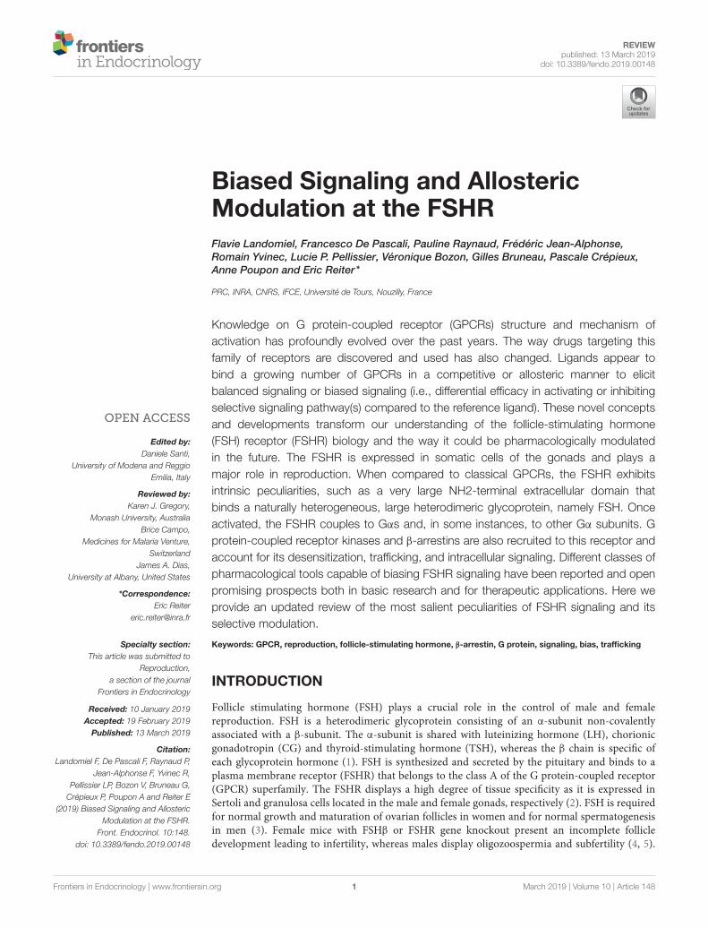
STRUCTURE OF THE FSHR
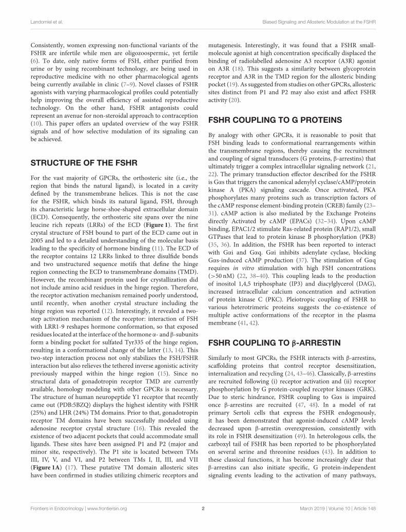
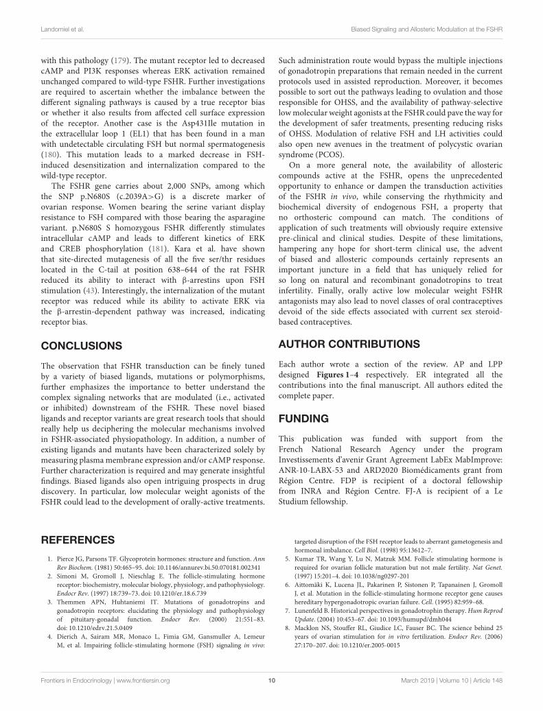
For the vast majority of GPCRs, the orthosteric site (i.e., theregion that binds the natural ligand), is located in a cavitydefined by the transmembrane helices. This is not the casefor the FSHR, which binds its natural ligand, FSH, throughits characteristic large horse-shoe-shaped extracellular domain(ECD). Consequently, the orthosteric site spans over the nineleucine rich repeats (LRRs) of the ECD (Figure 1). The firstcrystal structure of FSH bound to part of the ECD came out in2005 and led to a detailed understanding of the molecular basisleading to the specificity of hormone binding (11). The ECD ofthe receptor contains 12 LRRs linked to three disulfide bondsand two unstructured sequence motifs that define the hingeregion connecting the ECD to transmembrane domains (TMD).However, the recombinant protein used for crystallization didnot include amino acid residues in the hinge region. Therefore,the receptor activation mechanism remained poorly understood,until recently, when another crystal structure including thehinge region was reported (12). Interestingly, it revealed a two-step activation mechanism of the receptor: interaction of FSHwith LRR1-9 reshapes hormone conformation, so that exposedresidues located at the interface of the hormone α- and β-subunitsform a binding pocket for sulfated Tyr335 of the hinge region,resulting in a conformational change of the latter (13, 14). Thistwo-step interaction process not only stabilizes the FSH/FSHRinteraction but also relieves the tethered inverse agonistic activitypreviously mapped within the hinge region (15). Since nostructural data of gonadotropin receptor TMD are currentlyavailable, homology modeling with other GPCRs is necessary.The structure of human neuropeptide Y1 receptor that recentlycame out (PDB:5BZQ) displays the highest identity with FSHR(25%) and LHR (24%) TM domains. Prior to that, gonadotropinreceptor TM domains have been successfully modeled usingadenosine receptor crystal structure (16). This revealed theexistence of two adjacent pockets that could accommodate smallligands. These sites have been assigned P1 and P2 (major andminor site, respectively). The P1 site is located between TMsIII, IV, V, and VI, and P2 between TMs I, II, III, and VII(Figure 1A) (17). These putative TM domain allosteric siteshave been confirmed in studies utilizing chimeric receptors and
mutagenesis. Interestingly, it was found that a FSHR small-molecule agonist at high concentration specifically displaced thebinding of radiolabelled adenosine A3 receptor (A3R) agoniston A3R (18). This suggests a similarity between glycoproteinreceptor and A3R in the TMD region for the allosteric bindingpocket (19). As suggested from studies on other GPCRs, allostericsites distinct from P1 and P2 may also exist and affect FSHRactivity (20).
FSHR COUPLING TO G PROTEINS
By analogy with other GPCRs, it is reasonable to posit thatFSH binding leads to conformational rearrangements withinthe transmembrane regions, thereby causing the recruitmentand coupling of signal transducers (G proteins, β-arrestins) thatultimately trigger a complex intracellular signaling network (21,22). The primary transduction effector described for the FSHRis Gαs that triggers the canonical adenylyl cyclase/cAMP/proteinkinase A (PKA) signaling cascade. Once activated, PKAphosphorylates many proteins such as transcription factors ofthe cAMP response element-binding protein (CREB) family (23–31). cAMP action is also mediated by the Exchange Proteinsdirectly Activated by cAMP (EPACs) (32–34). Upon cAMPbinding, EPAC1/2 stimulate Ras-related protein (RAP1/2), smallGTPases that lead to protein kinase B phosphorylation (PKB)(35, 36). In addition, the FSHR has been reported to interactwith Gαi and Gαq. Gαi inhibits adenylate cyclase, blockingGαs-induced cAMP production (37). The stimulation of Gαqrequires in vitro stimulation with high FSH concentrations(>50 nM) (22, 38–40). This coupling leads to the productionof inositol 1,4,5 triphosphate (IP3) and diacylglycerol (DAG),increased intracellular calcium concentration and activationof protein kinase C (PKC). Pleiotropic coupling of FSHR tovarious heterotrimeric proteins suggests the co-existence ofmultiple active conformations of the receptor in the plasmamembrane (41, 42).
FSHR COUPLING TO β-ARRESTIN
Similarly to most GPCRs, the FSHR interacts with β-arrestins,scaffolding proteins that control receptor desensitization,internalization and recycling (24, 43–46). Classically, β-arrestinsare recruited following (i) receptor activation and (ii) receptorphosphorylation by G protein-coupled receptor kinases (GRK).Due to steric hindrance, FSHR coupling to Gαs is impairedonce β-arrestins are recruited (47, 48). In a model of ratprimary Sertoli cells that express the FSHR endogenously,it has been demonstrated that agonist-induced cAMP levelsdecreased upon β-arrestin overexpression, consistently withits role in FSHR desensitization (49). In heterologous cells, thecarboxyl tail of FSHR has been reported to be phosphorylatedon several serine and threonine residues (43). In addition tothese classical functions, it has become increasingly clear thatβ-arrestins can also initiate specific, G protein-independentsignaling events leading to the activation of many pathways,
Frontiers in Endocrinology | www.frontiersin.org 2 March 2019 | Volume 10 | Article 148
Landomiel et al. Biased Signaling and Allosteric Modulation at the FSHR
FIGURE 1 | Orthosteric and allosteric sites in the FSHR. (A) Cartoon and surface view of the transmembrane regions of the FSHR showing P1 and P2 allosteric sites.
(B) Complex between the ectodomain of the FSHR (gray) and FSH (violet: alpha chain, pink: beta chain). The colored spheres represent sulphated Tyr355. (C)
Residues involved in FSH binding are shown in red. (D) Close-up on the interaction between sulphated Tyr335 (colored spheres) and FSH.
amongst which the ERK (Extracellular signal-Regulated Kinase)MAP (Mitogen-Activated Protein) kinase pathway has beenthe most studied (50). Of note, ERK activation kinetics at theFSHR has been reported to vary in heterologous cells as afunction of the upstream transduction mechanism involved:β-arrestin-mediated ERK activation is delayed but moresustained compared to Gαs-dependent ERK activation, whichoccurs early but is transient (43). Consistent with the concept of“phosphorylation barcode” which links particular GRK-mediatedphosphorylation signatures at the receptor level to the activationof distinct β-arrestin-dependent functions (51, 52), a relationshiphas been found between the subtype of GRK involved inFSHR phosphorylation and the nature of β-arrestin-mediatedactions. In particular, β-arrestins recruited to GRK2 or GRK3-phosphorylated FSHR favor receptor desensitization whereasGRK5 or GRK6-mediated phosphorylation of FSHR wereinvolved in β-arrestin-dependent ERK activation (43, 53, 54).Recently, phosphorylation of Tyrosine383 in β-arrestin 2 hasproved to be crucial for β-arrestin-mediated ERK activation bythe FSHR and other GPCRs. More precisely, ligand-inducedreceptor activation provokes MEK (Mitogen-activated proteinkinase kinase)-mediated phosphorylation of Tyr383, necessary
for β-arrestin 2-mediated ERK recruitment and activation(55). β-arrestins also play a role in FSHR-induced translation,mediated by a β-arrestin/p70S6K/ribosomal S6 complex thatassembles in heterologous and in primary Sertoli cells. UponFSH stimulation, activation of G protein-dependent signalingenhances p70S6K activity within the β-arrestin/p70S6K/rpS6preassembled complex, leading to the fast and robust translationof 5′ oligopyrimidine track (5′TOP) mRNA (56). In addition,the balance between FSHR-mediated proliferation vs apoptosisseems to be regulated by β-arrestins. In hGL5 human granulosacells, silencing of β-arrestins leads to an increase in cAMP/PKAand a decrease in β-arrestin-mediated proliferative pathway,resulting in cell death (57). Evidence reported for other GPCRsdemonstrated that the internalized receptor can form molecularcomplexes involving simultaneous interactions with Gαs to thecore domain and β-arrestin to the C-tail of the receptor (58).These complexes, named “megaplexes,” are able to signal fromthe endosome by inducing a second wave of cAMP (58, 59).Based on structural evidence, a two-step mechanism for β-arrestin recruitment has been proposed (60). First, β-arrestinsare recruited to the phosphorylated C-tail, resulting in a so-called“partially engaged” complex which the authors reported to be
Frontiers in Endocrinology | www.frontiersin.org 3 March 2019 | Volume 10 | Article 148
Landomiel et al. Biased Signaling and Allosteric Modulation at the FSHR
sufficient for ERK signaling and internalization. Interestingly,this conformation allows the receptor to simultaneously coupleto G protein α subunit. Second, a conformational rearrangementof β-arrestins allows them to interact with the receptor coredomain, forming a “fully engaged complex” incompatible withfurther G protein coupling (58, 60–62). More recently, a separatestudy uncovered another mechanism of β-arrestin activation thatthe authors called “catalytic activation.” Upon ligand-inducedrecruitment of inactivated β-arrestin to the receptor core domain,a conformational change in β-arrestin occurs that exposes aPIP2-bindingmotif and allows β-arrestin to bindmembrane lipidrafts independently of the receptor. Interestingly, the authorsnoticed an accumulation of active β-arrestin in clathrin-coatedendocytic structures in the absence of the receptor, revealingthe existence of a receptor C-tail-independent β-arrestinactivation mechanism (63). No evidence currently exist that theaforementioned mechanisms also apply to the FSHR. Furtherstudies will be necessary to clarify the molecular mechanismsinvolved in β-arrestin recruitment and activation at the FSHRand to determine their possible peculiarities.
FSHR INTERACTION WITHOTHER PARTNERS
Beside G proteins, GRKs and β-arrestins, the signal is alsotransduced at the FSHR by other direct binding partners (44).For example, adaptor protein, phosphotyrosine interacting withthe Adapter protein with PH domain, PTB domain, and leucinezipper 1 (APPL1) binds intracellular loop 1 of the FSHR (64).This protein has lately retained the greatest attention in thegonadotropin community for two main reasons. The first oneis that this adapter protein links the FSHR directly to inositidephosphate metabolism and Ca2+ release in granulosa cells (65),hence it induces cAMP-independent signaling; the second isthat, like β-arrestins, APPL1 recruitment plays a role in thesubcellular routing of FSHR. This discovery had been heralded bythe previously identified interaction between GAIP-interactingprotein C-tail (GIPC) adaptor and the FSHR (or the LHR),presumably requiring the carboxyterminal end of the receptor.Interestingly, GIPC reroutes the internalized FSHR from EarlyEndosomes (EE) to recycling Very Early Endosomes (VEE), andby these means, enables sustained ERK phosphorylation (66).Likewise, in HEK293 cells, APPL1 has been shown to conveyinternalized FSHR, as well as LHR, to VEE for recycling, andPKA-dependent phosphorylation of APPL1 leads to endosomalcAMP signaling (67). These two sets of observations on ERKMAP kinases and cAMP suggest that spatially restricted FSHsignaling may be generalized to several of its components. Inaddition, 14-3-3τ has been shown to interact directly withthe second intracellular loop of the receptor FSHR (68, 69).The 14-3-3τ interaction site on the FSHR encompasses theERW motif involved in G protein association (70), that isconsistent with the observation that 14-3-3τ overexpression inHEK293 cells reduces FSH-induced cAMP response (68). The co-occurrence of these direct binding partners as well as G protein,GRK and β-arrestins, raises fundamental questions about their
sequence/dynamics of interaction on a single FSHR or aboutthe possibility that FSHR oligomers might cluster transductionassembly of variable composition at the plasma membrane andin intracellular compartments.
TRAFFICKING ANDENDOSOMAL SIGNALING
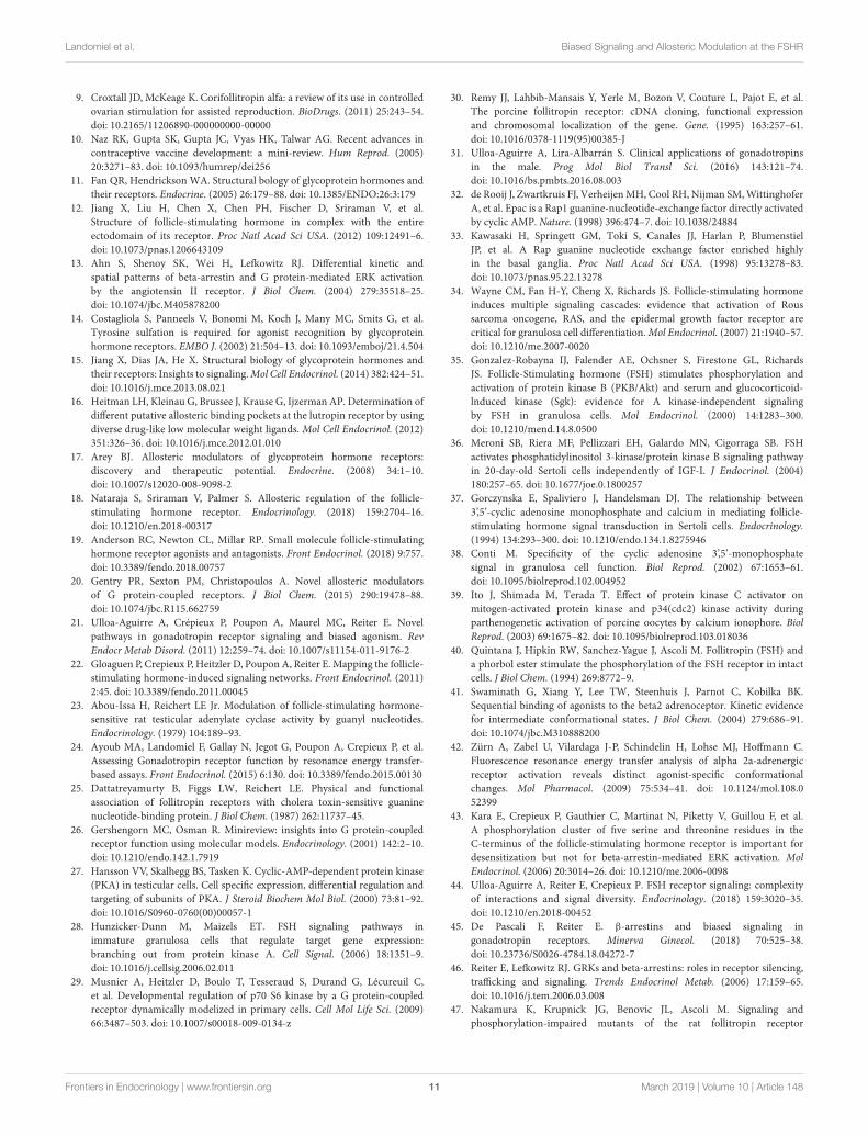
Compartmentalization of signaling is now viewed as animportant feature for many signaling proteins and playskey roles in cellular responses. This is particularly the casefor membrane receptors such as GPCRs since, in the pastyears, several examples have revealed connections betweenmembrane compartmentalization, endocytic trafficking andsignaling patterns. Originally thought to function solely atthe plasma membrane, the multifunctional protein β-arrestinassemble signaling molecules (e.g., MAPK, Src, etc) that directGPCRs to the endocytic pathway and regulate their post-endocytic fate, as mentioned above. For some GPCRs forminga stable interaction with β-arrestin, β-arrestin/receptor/signalingmolecule complexes are found in endosomes, allowing prolongedsignaling from these intracellular structures (71–73). The natureof the β-arrestin binding motifs, in particular serine/threonineclusters in the C-tail of the receptor, regulates the stability ofthis interaction. GPCRs that display high affinity binding toβ-arrestin, are classified as class B (74, 75). In the FSHR, acluster of 5 serines/threonines is involved in both internalizationand binding of β-arrestin to the receptor and is consistentwith the class B definition. Such interaction was confirmed bybioluminescence energy transfer experiments (BRET) and co-immunoprecipitation experiments, however no imaging datahave confirmed the existence of a functional complex inendosomes (24, 43). In addition, it is unclear whether β-arrestin-mediated ERK signaling by FSHR requires β-arrestin localizationin endosomes. Recently, both the FSHR and LHR were reportedto predominantly localize to an atypical endosome denotedas VEE (Figure 2). Alteration of this endosomal trafficking byblocking internalization inhibits activation of ERK through theLHR, suggesting that VEE are a location for signaling (66). Theseparticular endosomes are upstream of the classical endosomesand are devoid of typical early endosomes markers such as theRab5 GTPase or the phosphatidylinositol-3-phosphate (PI(3)P)or the PI(3)P-bound EEA1 proteins. Morphologically, they aresmaller (<400 nm) than conventional sorting EE but theirexact nature remains to be defined. Interestingly, gonadotropinreceptor localization in VEE requires an intact receptor C-tail and the GIPC PDZ-domain protein (66). PDZ motifs arefound in several GPCRs to regulate their spatial localization ortrafficking (76). The PDZ motif of the LHR directly binds GIPCthat sequesters the receptor into VEE following agonist-inducedinternalization. In fact, in cells depleted in GIPC or expressingLHR lacking the distal PDZ motif in its C-tail, the receptorsare rerouted and accumulated into the classical EE where theyfail to recycle back to the plasma membrane. In addition, theywere not able to signal to ERKMAP kinases (66), suggesting thatendosomal ERK activity occurs from this specific compartment.
Frontiers in Endocrinology | www.frontiersin.org 4 March 2019 | Volume 10 | Article 148
Landomiel et al. Biased Signaling and Allosteric Modulation at the FSHR
It is worth noting that the FSHR does not display a knownPDZ ligand in its C-tail and the exact mechanism on how GIPCcontrols FSHR fate remains to be determined. However, APPL1,a known FSHR binding partner, localizes to a subset of VEE anddisplays a PDZ motif previously shown to interact with GIPC(64, 65, 77). A possible scenario would be that FSHR, via itsinteraction with APPL1, connects with GIPC and is targeted toVEE where it activates ERK (78). Earlier work supports the ideathat endosomal APPL1 defines a signaling platform upstreamof the Rab5/PI(3)P endosomes. Disruption of EE leads to theaccumulation of the EGFR Tyrosine kinase receptor in APPL1vesicles, leading to a sustained activation of ERK from thiscompartment (79, 80). As shown for the LHR, the endosomalcAMP/PKA dependent phosphorylation of APPL1 on Ser410 isnecessary for the recycling of the receptor (67).
This concept of endosomal signaling compartmentalizationwas further supported by the findings that GPCR can inducea second phase of G protein activation following theirinternalization (81–83). This allowed the advent of a newparadigm where some GPCRs do not only transduce andactivate G proteins from the plasma membrane but also fromendocytic compartments (the so-called “megaplex” mentionedabove). Interestingly, the two other members of the glycoproteinhormone receptor family, TSHR and LHR, were both shownto transduce via Gαs and promote sustained cAMP productionfrom endocytic compartments (59, 67, 84, 85). While the TSHRacts from EE and trans-Golgi compartments, the LHR signalingis restricted to the VEE. It has yet to be shown whether the FSHRalso activates G proteins from the VEE but the fact that it sharesseveral features with GPCRs known to signal from endosomes,including LHR, V2R or PTHR, supports this possibility. FSHRtrafficking mimics the LHR as discussed above and it displaysa phosphorylation code similar to those found in the C-tail ofV2R and PTHR. More precisely, the formation of a “megaplex”has been shown to induce cAMP from the EE in responseto PTHR activation (86, 87). That the FSHR could signal inendocytic compartments through G proteins in a similar way asthe PTHR, but from VEE, is conceivable, but further studies areneeded to demonstrate this possibility. Despite the identificationof structural determinants in the FSHR C-tail that regulateits trafficking (88), very little is known about the mechanismsinvolved in the post-endocytic trafficking of this receptor.
BIASED SIGNALING ANDITS QUANTIFICATION
The action of a given ligand on its cognate receptor has classicallybeen characterized by its effect on downstream effectors (secondmessengers). Compared to the reference ligand (generally, thephysiological ligand), a pharmacological agent can be eitheran agonist (it produces a biological response similar to thatof the reference ligand), an antagonist (it blocks the biologicalresponse elicited by the reference ligand) or an inverse agonist (itproduces an opposite biological response that leads to a decreasein the receptor basal activity). Importantly, it has long beenthought that these characteristics hold irrespectively of effector
measured (89). However, some ligands did not match with oneof these categories, because they displayed both agonist andantagonist (or inverse agonist) activities at the same receptor,depending on the downstream pathway measured. For instance,carvedilol, a clinically used β-blocker, has a clear inverse agonistprofile on Gαs-dependent activity at the β2 adrenergic receptorwhile being a weak partial agonist for β-arrestin-dependent ERKactivation (90). To deal with these discrepancies, the conceptof biased signaling or functional selectivity, recently came tothe fore (89, 91, 92). According to this concept, a ligand isbiased when it triggers imbalanced responses, compared to areference ligand acting on the same receptor (classically theendogenous ligand). Importantly, a biased ligand can selectivelyactivate only a subset of the biological responses or activate allof them but with different efficacies compared to the referenceligand. As these ideas hold profound implications and potentialfor the design of new therapeutics, biased signaling is a veryactive area of research in pharmacology (93, 94). Over the lastdecade, biased signaling has been evidenced in many differentreceptors, including gonadotropin receptors, as will be discussedin a forthcoming section. By analogy with a ligand bias, thenotion of receptor bias has been proposed (95). Two receptors,diverging only by a mutation or a polymorphism, once activatedby the same ligand may induce two signaling pathways withdifferent relative efficacies. Importantly, biased signaling hasbeen extended to allosteric ligands, which can modulate notonly the efficacy of a given ligand-induced receptor signal butalso select and bias the activation of the receptor toward asubset of the biological responses (93). Ligand bias and receptorbias must be set apart from system bias or observational bias(95). System bias refers to bias that are due to the particularbiological system used (some transducer molecules, such as Gproteins, may be expressed differentially in different cell typesfor instance). Observational bias refers to the modification oramplification of the signals inherent to the specific assays usedfor the measurements. Besides being supported by numerousexperimental evidences, biased signaling is consistent with thereceptor conformation theory, which views a receptor populationas an ensemble of conformations that evolve dynamically,according to some energy landscape and subjected to externalperturbations (96). In such theory, ligand-induced receptoractivation is concomitant with a stabilization of some receptorconformations and a modification of the receptor conformationenergy landscape, resulting from the interaction of the ligandwith its receptor. Several studies have thus shown that receptorconformational equilibriummodels, such as the extended ternarycomplex model, can satisfactorily explain ligand bias (97, 98).Several groups have attempted to address the problem of biasquantification. Considering a given receptor and two signalingpathways (A and B), the objective is to be able to classifyligand bias, as having for instance a low, a moderate or a highbias toward pathway A vs pathway B, when compared to areference ligand. The most popular method to quantify ligandbias uses dose-response data and the so-called operational model(99, 100). The latter is widely used to perform regression ondose-response data, which have in many cases a sigmoid shape.The parameters of the operational model are derived from a
Frontiers in Endocrinology | www.frontiersin.org 5 March 2019 | Volume 10 | Article 148
Landomiel et al. Biased Signaling and Allosteric Modulation at the FSHR
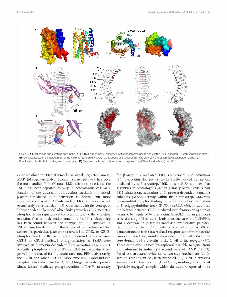
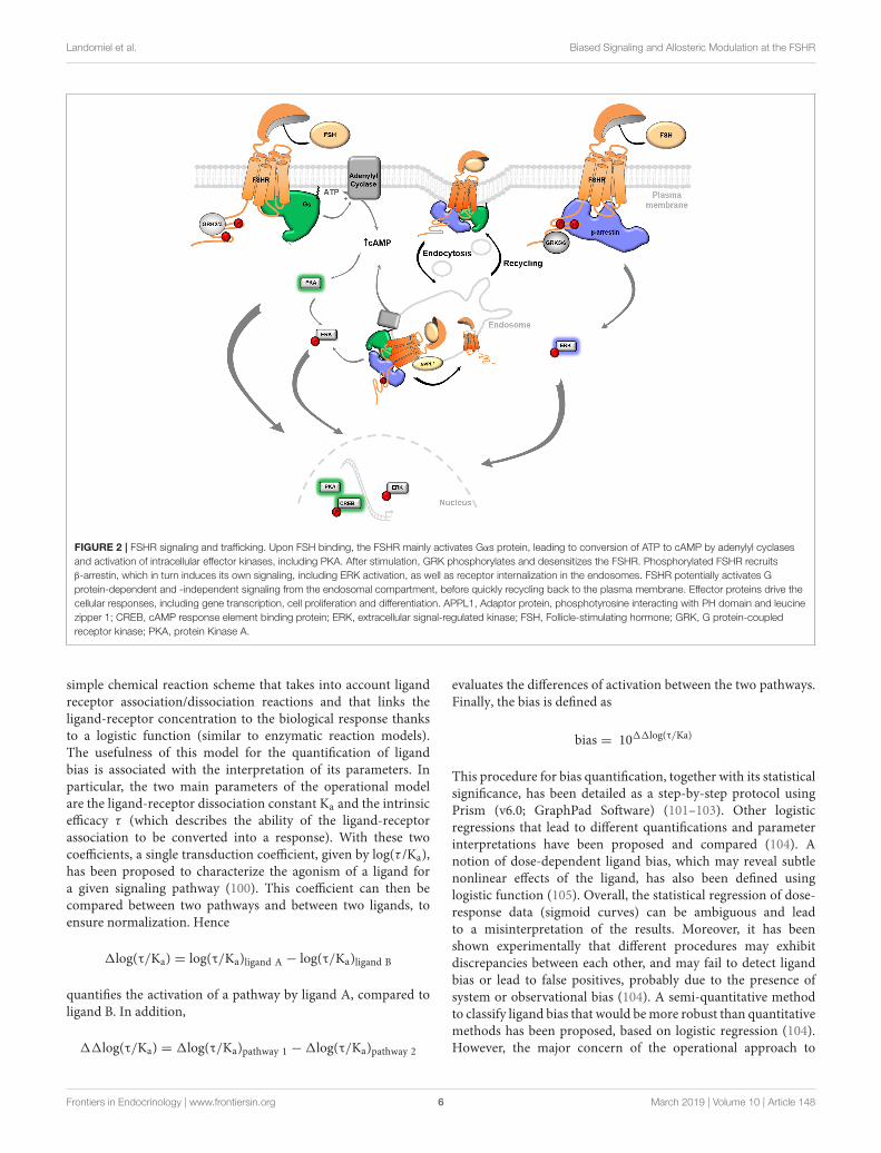
FIGURE 2 | FSHR signaling and trafficking. Upon FSH binding, the FSHR mainly activates Gαs protein, leading to conversion of ATP to cAMP by adenylyl cyclases
and activation of intracellular effector kinases, including PKA. After stimulation, GRK phosphorylates and desensitizes the FSHR. Phosphorylated FSHR recruits
β-arrestin, which in turn induces its own signaling, including ERK activation, as well as receptor internalization in the endosomes. FSHR potentially activates G
protein-dependent and -independent signaling from the endosomal compartment, before quickly recycling back to the plasma membrane. Effector proteins drive the
cellular responses, including gene transcription, cell proliferation and differentiation. APPL1, Adaptor protein, phosphotyrosine interacting with PH domain and leucine
zipper 1; CREB, cAMP response element binding protein; ERK, extracellular signal-regulated kinase; FSH, Follicle-stimulating hormone; GRK, G protein-coupled
receptor kinase; PKA, protein Kinase A.
simple chemical reaction scheme that takes into account ligandreceptor association/dissociation reactions and that links theligand-receptor concentration to the biological response thanksto a logistic function (similar to enzymatic reaction models).The usefulness of this model for the quantification of ligandbias is associated with the interpretation of its parameters. Inparticular, the two main parameters of the operational modelare the ligand-receptor dissociation constant Ka and the intrinsicefficacy τ (which describes the ability of the ligand-receptorassociation to be converted into a response). With these twocoefficients, a single transduction coefficient, given by log(τ /Ka),has been proposed to characterize the agonism of a ligand fora given signaling pathway (100). This coefficient can then becompared between two pathways and between two ligands, toensure normalization. Hence
1log(τ/Ka) = log(τ/Ka)ligand A − log(τ/Ka)ligand B
quantifies the activation of a pathway by ligand A, compared toligand B. In addition,
11log(τ/Ka) = 1log(τ/Ka)pathway 1 − 1log(τ/Ka)pathway 2
evaluates the differences of activation between the two pathways.Finally, the bias is defined as
bias = 1011log(τ/Ka)
This procedure for bias quantification, together with its statisticalsignificance, has been detailed as a step-by-step protocol usingPrism (v6.0; GraphPad Software) (101–103). Other logisticregressions that lead to different quantifications and parameterinterpretations have been proposed and compared (104). Anotion of dose-dependent ligand bias, which may reveal subtlenonlinear effects of the ligand, has also been defined usinglogistic function (105). Overall, the statistical regression of dose-response data (sigmoid curves) can be ambiguous and leadto a misinterpretation of the results. Moreover, it has beenshown experimentally that different procedures may exhibitdiscrepancies between each other, and may fail to detect ligandbias or lead to false positives, probably due to the presence ofsystem or observational bias (104). A semi-quantitative methodto classify ligand bias that would bemore robust than quantitativemethods has been proposed, based on logistic regression (104).However, the major concern of the operational approach to
Frontiers in Endocrinology | www.frontiersin.org 6 March 2019 | Volume 10 | Article 148
Landomiel et al. Biased Signaling and Allosteric Modulation at the FSHR
quantify ligand bias is that it disregards an important aspectof signaling pathways, namely the temporal activation of thedifferent signaling processes (106). This has been revealed by therelatively simple observation that the bias value, as calculatedwith the operational model, could change as a function ofthe kinetics of response (107). Actually, the apparent bias caneven be in an opposite direction for two different time pointswhen the biological responses are measured. While part of theexplanation of this phenomena resides in the different timescales at stake within a signal transduction pathway (bindingkinetics, second messenger and effector kinetics), it also revealsthe whole complexity of a receptor trafficking system (108), thatcan certainly not be condensed into a single number. Thus,methodological developments such as dynamical versions of theoperational model and/or the extended ternary complex model(109, 110) must be developed to address this complexity andallow better characterization of the effect of a ligand on itscognate receptor.
BIASED SIGNALING AT THE FSHR
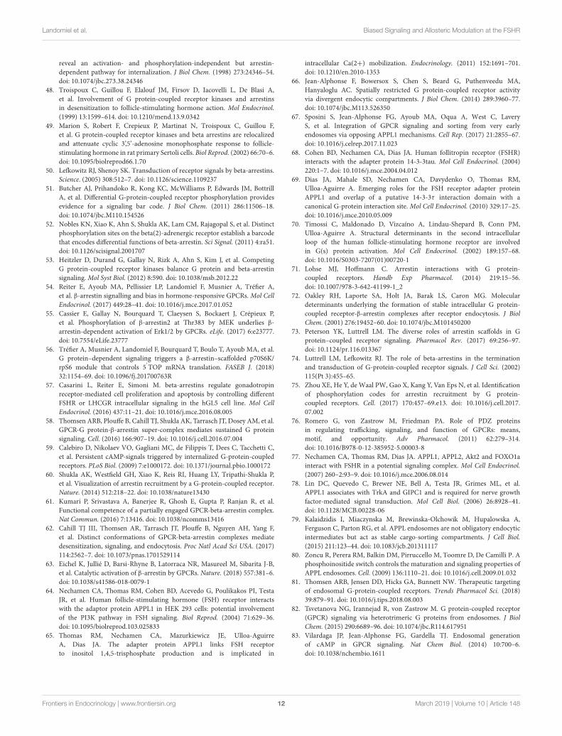
To date, different classes of biases have been reported to elicitselective modulation at the FSHR (Figure 3). Ligand bias canbe provoked by small molecule ligands, glycosylation variants ofFSH or by antibodies acting at FSH or FSHR. Receptor bias dueto mutations or single nucleotide polymorphisms (SNP) at theFSHR have also been reported.
Small Molecule LigandsSeveral classes of chemical compounds exhibiting the abilityto modulate FSHR-mediated signaling upon binding have beenidentified to date. Readers interested in the chemical diversityof currently known FSHR small molecules classes can referto Figure 2 of Anderson et al. in the same special issue ofFrontiers in Endocrinology (19). According to their mode ofaction and effect on the receptor, they can be divided infour classes: allosteric agonists, positive allosteric modulators(PAMs), negative allosteric modulators (NAMs) and neutralallosteric ligands (NALs) (111). While PAMs or NAMs needthe presence of FSH to detect the enhancement or the decreaseof FSHR activation, respectively, agonists have the capacityto activate it on their own. Even though NALs do notinfluence signaling, they can potentially prevent other allostericmodulators from binding (112). Thiazolidinones, identified byscreening combinatorial chemical scaffolds, were the first classof FSHR allosteric agonists to be reported (113). The allostericnature of thiazolidinone derivatives was confirmed thanks toexperiments involving FSHR/TSHR chimeras, which showedthat their binding site was localized in the TMD (114). Ananomolar potent thiazolidinone FSHR agonist was reportedto trigger signaling pathways similar to FSH, both in vitroand in vivo (115). Interestingly, some thiazolidinone analogsdemonstrated biased agonism by mobilizing the Gαi proteininstead of Gαs or both as observed for other thiazolidinoneanalogs or FSH preparations (116). Besides, high throughputscreening on substituted benzamides allowed the identificationof a series of FSHR PAMs that showed improved selectivity
against LHR and TSHR. Interesting pharmacokinetic propertieswere also described for two selected compounds (117). Adihydropyridine compound, Org 24444-0, is another PAM,which displayed a good selectivity toward FSHR and inducedcAMP production in presence of FSH (118). The compoundwas also able to reproduce the effects of FSH on the folliclephase maturation in mature female rats. Among the currentlyknown NAMs, tetrahydroquinolines constitute a good exampleof biased signaling. It was indeed established that the compoundsinhibited FSHR-induced cAMP production, without inhibitingFSH binding (119). Unfortunately, the tetrahydroquinolinesdid not display any in vivo activity. Three other NAMs havebeen characterized by Dias et al. (120, 121). The first one,ADX61623, was reported to inhibit cAMP and progesterone butnot estradiol production in rat granulosa primary cells. Using125I-hFSH, it was established that ADX61623 did not competewith FSH, but rather increased FSH binding, suggesting that itdoes not bind the extracellular domain of FSHR. When testedin vivo, the compound was not able to decrease FSH-inducedpreovulatory follicle development (120). Two similar compoundswere described later: ADX68692 and ADX68693. Both werereported to inhibit cAMP and progesterone production in ratgranulosa primary cells, but while ADX68692 also affectedestradiol and decreased the number of oocytes recovered inmature female rat, ADX68693 had no effect on estradiol, nor onthe number of retrieved oocytes (121). Interestingly, ADX68692and ADX68693 were also reported to exert similar actions on theLHR (122). The first FSHR competitive antagonist described inscientific literature, suramin, was reported to inhibit testosteroneproduction and FSHR signaling, by competing with FSH binding(123). Another non-competitive antagonist of human and ratFSHR showing the same behavior was later identified (124).
Glycosylation VariantsGonadotropins present natural heterogeneity in their glycanmoieties that contribute up to nearly 30% of the hormone’s mass(125–128). The presence of glycans has important outcomeson the in vivo half-life of the hormone because, by doublingits diameter, it limits its glomerular filtration. FSH containstwo potential N-linked oligosaccharides on each subunit thatare sources of heterogeneities. Importantly, these glycan chainsare involved in FSH folding, assembly, stability, quality control,secretion, transport as well as the biological activity andpotency (15, 129–138). The α chain is glycosylated at asparagine52 (Asn52) and Asn78, while the FSH β subunit can beglycosylated at Asn7 and Asn24. Partially glycosylated variantsthat are missing either one or both of these oligosaccharideson FSHβ have been reported in equine FSHβ, human FSHβ
(hFSH β), rhesus FSHβ and Japanese macaque FSHβ (139–142).Glycosylation profile of each subunit plays a critical role inthe activity and clearance of FSH (131, 143, 144). Interestingly,while FSHα subunit amino-acid sequences are identical to LH,TSH and CG α-subunits, the N-glycan populations at Asn52and Asn78 differ from those of the other hormones (145–147). FSHβ subunit shares 34–40% of sequence homologywith the other human glycoprotein hormone β-subunits, yetthe main structural hallmarks (i.e., six disulfide bonds, cystine
Frontiers in Endocrinology | www.frontiersin.org 7 March 2019 | Volume 10 | Article 148
Landomiel et al. Biased Signaling and Allosteric Modulation at the FSHR
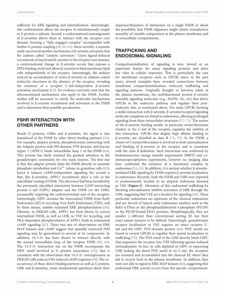
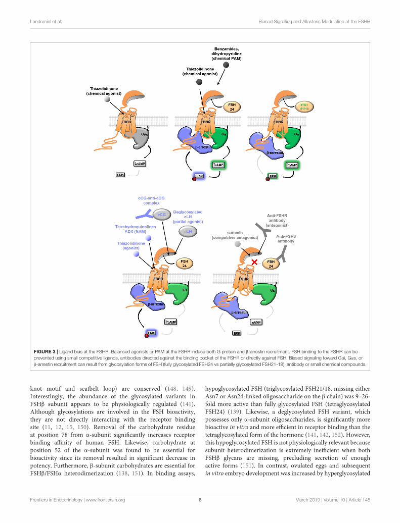
FIGURE 3 | Ligand bias at the FSHR. Balanced agonists or PAM at the FSHR induce both G protein and β-arrestin recruitment. FSH binding to the FSHR can be
prevented using small competitive ligands, antibodies directed against the binding pocket of the FSHR or directly against FSH. Biased signaling toward Gαi, Gαs, or
β-arrestin recruitment can result from glycosylation forms of FSH (fully glycosylated FSH24 vs partially glycosylated FSH21-18), antibody or small chemical compounds.
knot motif and seatbelt loop) are conserved (148, 149).Interestingly, the abundance of the glycosylated variants inFSHβ subunit appears to be physiologically regulated (141).Although glycosylations are involved in the FSH bioactivity,they are not directly interacting with the receptor bindingsite (11, 12, 15, 150). Removal of the carbohydrate residueat position 78 from α-subunit significantly increases receptorbinding affinity of human FSH. Likewise, carbohydrate atposition 52 of the α-subunit was found to be essential forbioactivity since its removal resulted in significant decrease inpotency. Furthermore, β-subunit carbohydrates are essential forFSHβ/FSHα heterodimerization (138, 151). In binding assays,
hypoglycosylated FSH (triglycosylated FSH21/18, missing eitherAsn7 or Asn24-linked oligosaccharide on the β chain) was 9–26-fold more active than fully glycosylated FSH (tetraglycosylatedFSH24) (139). Likewise, a deglycosylated FSH variant, whichpossesses only α-subunit oligosaccharides, is significantly morebioactive in vitro and more efficient in receptor binding than thetetraglycosylated form of the hormone (141, 142, 152). However,this hypoglycosylated FSH is not physiologically relevant becausesubunit heterodimerization is extremely inefficient when bothFSHβ glycans are missing, precluding secretion of enoughactive forms (151). In contrast, ovulated eggs and subsequentin vitro embryo development was increased by hyperglycosylated
Frontiers in Endocrinology | www.frontiersin.org 8 March 2019 | Volume 10 | Article 148
Landomiel et al. Biased Signaling and Allosteric Modulation at the FSHR
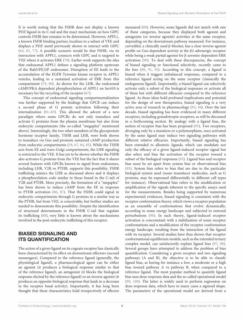
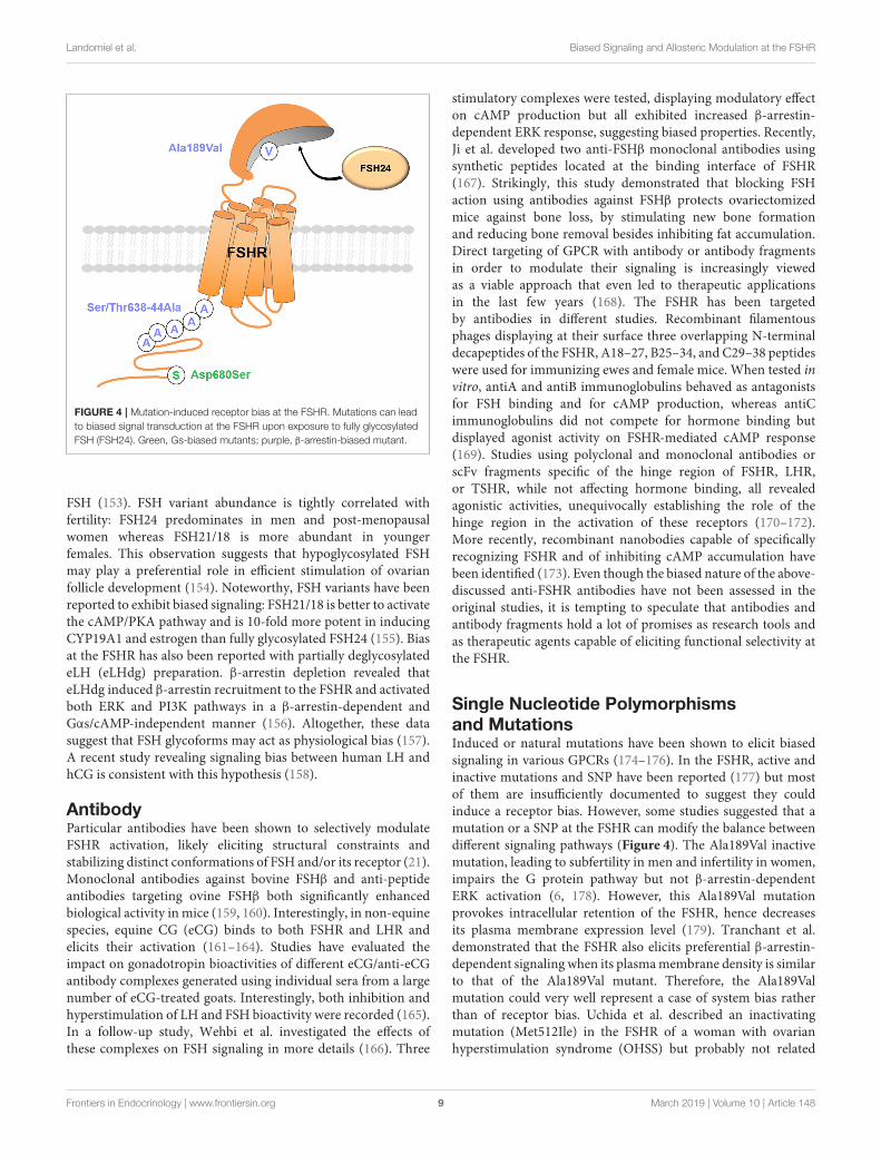
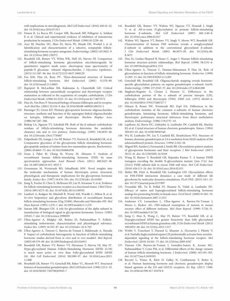
FIGURE 4 | Mutation-induced receptor bias at the FSHR. Mutations can lead
to biased signal transduction at the FSHR upon exposure to fully glycosylated
FSH (FSH24). Green, Gs-biased mutants; purple, β-arrestin-biased mutant.
FSH (153). FSH variant abundance is tightly correlated withfertility: FSH24 predominates in men and post-menopausalwomen whereas FSH21/18 is more abundant in youngerfemales. This observation suggests that hypoglycosylated FSHmay play a preferential role in efficient stimulation of ovarianfollicle development (154). Noteworthy, FSH variants have beenreported to exhibit biased signaling: FSH21/18 is better to activatethe cAMP/PKA pathway and is 10-fold more potent in inducingCYP19A1 and estrogen than fully glycosylated FSH24 (155). Biasat the FSHR has also been reported with partially deglycosylatedeLH (eLHdg) preparation. β-arrestin depletion revealed thateLHdg induced β-arrestin recruitment to the FSHR and activatedboth ERK and PI3K pathways in a β-arrestin-dependent andGαs/cAMP-independent manner (156). Altogether, these datasuggest that FSH glycoforms may act as physiological bias (157).A recent study revealing signaling bias between human LH andhCG is consistent with this hypothesis (158).
AntibodyParticular antibodies have been shown to selectively modulateFSHR activation, likely eliciting structural constraints andstabilizing distinct conformations of FSH and/or its receptor (21).Monoclonal antibodies against bovine FSHβ and anti-peptideantibodies targeting ovine FSHβ both significantly enhancedbiological activity in mice (159, 160). Interestingly, in non-equinespecies, equine CG (eCG) binds to both FSHR and LHR andelicits their activation (161–164). Studies have evaluated theimpact on gonadotropin bioactivities of different eCG/anti-eCGantibody complexes generated using individual sera from a largenumber of eCG-treated goats. Interestingly, both inhibition andhyperstimulation of LH and FSH bioactivity were recorded (165).In a follow-up study, Wehbi et al. investigated the effects ofthese complexes on FSH signaling in more details (166). Three
stimulatory complexes were tested, displaying modulatory effecton cAMP production but all exhibited increased β-arrestin-dependent ERK response, suggesting biased properties. Recently,Ji et al. developed two anti-FSHβ monoclonal antibodies usingsynthetic peptides located at the binding interface of FSHR(167). Strikingly, this study demonstrated that blocking FSHaction using antibodies against FSHβ protects ovariectomizedmice against bone loss, by stimulating new bone formationand reducing bone removal besides inhibiting fat accumulation.Direct targeting of GPCR with antibody or antibody fragmentsin order to modulate their signaling is increasingly viewedas a viable approach that even led to therapeutic applicationsin the last few years (168). The FSHR has been targetedby antibodies in different studies. Recombinant filamentousphages displaying at their surface three overlapping N-terminaldecapeptides of the FSHR, A18–27, B25–34, and C29–38 peptideswere used for immunizing ewes and female mice. When tested invitro, antiA and antiB immunoglobulins behaved as antagonistsfor FSH binding and for cAMP production, whereas antiCimmunoglobulins did not compete for hormone binding butdisplayed agonist activity on FSHR-mediated cAMP response(169). Studies using polyclonal and monoclonal antibodies orscFv fragments specific of the hinge region of FSHR, LHR,or TSHR, while not affecting hormone binding, all revealedagonistic activities, unequivocally establishing the role of thehinge region in the activation of these receptors (170–172).More recently, recombinant nanobodies capable of specificallyrecognizing FSHR and of inhibiting cAMP accumulation havebeen identified (173). Even though the biased nature of the above-discussed anti-FSHR antibodies have not been assessed in theoriginal studies, it is tempting to speculate that antibodies andantibody fragments hold a lot of promises as research tools andas therapeutic agents capable of eliciting functional selectivity atthe FSHR.
Single Nucleotide Polymorphismsand MutationsInduced or natural mutations have been shown to elicit biasedsignaling in various GPCRs (174–176). In the FSHR, active andinactive mutations and SNP have been reported (177) but mostof them are insufficiently documented to suggest they couldinduce a receptor bias. However, some studies suggested that amutation or a SNP at the FSHR can modify the balance betweendifferent signaling pathways (Figure 4). The Ala189Val inactivemutation, leading to subfertility in men and infertility in women,impairs the G protein pathway but not β-arrestin-dependentERK activation (6, 178). However, this Ala189Val mutationprovokes intracellular retention of the FSHR, hence decreasesits plasma membrane expression level (179). Tranchant et al.demonstrated that the FSHR also elicits preferential β-arrestin-dependent signaling when its plasmamembrane density is similarto that of the Ala189Val mutant. Therefore, the Ala189Valmutation could very well represent a case of system bias ratherthan of receptor bias. Uchida et al. described an inactivatingmutation (Met512Ile) in the FSHR of a woman with ovarianhyperstimulation syndrome (OHSS) but probably not related
Frontiers in Endocrinology | www.frontiersin.org 9 March 2019 | Volume 10 | Article 148
Landomiel et al. Biased Signaling and Allosteric Modulation at the FSHR
with this pathology (179). The mutant receptor led to decreasedcAMP and PI3K responses whereas ERK activation remainedunchanged compared to wild-type FSHR. Further investigationsare required to ascertain whether the imbalance between thedifferent signaling pathways is caused by a true receptor biasor whether it also results from affected cell surface expressionof the receptor. Another case is the Asp431Ile mutation inthe extracellular loop 1 (EL1) that has been found in a manwith undetectable circulating FSH but normal spermatogenesis(180). This mutation leads to a marked decrease in FSH-induced desensitization and internalization compared to thewild-type receptor.
The FSHR gene carries about 2,000 SNPs, among whichthe SNP p.N680S (c.2039A>G) is a discrete marker ofovarian response. Women bearing the serine variant displayresistance to FSH compared with those bearing the asparaginevariant. p.N680S S homozygous FSHR differently stimulatesintracellular cAMP and leads to different kinetics of ERKand CREB phosphorylation (181). Kara et al. have shownthat site-directed mutagenesis of all the five ser/thr residueslocated in the C-tail at position 638–644 of the rat FSHRreduced its ability to interact with β-arrestins upon FSHstimulation (43). Interestingly, the internalization of the mutantreceptor was reduced while its ability to activate ERK viathe β-arrestin-dependent pathway was increased, indicatingreceptor bias.
CONCLUSIONS
The observation that FSHR transduction can be finely tunedby a variety of biased ligands, mutations or polymorphisms,further emphasizes the importance to better understand thecomplex signaling networks that are modulated (i.e., activatedor inhibited) downstream of the FSHR. These novel biasedligands and receptor variants are great research tools that shouldreally help us deciphering the molecular mechanisms involvedin FSHR-associated physiopathology. In addition, a number ofexisting ligands and mutants have been characterized solely bymeasuring plasmamembrane expression and/or cAMP response.Further characterization is required and may generate insightfulfindings. Biased ligands also open intriguing prospects in drugdiscovery. In particular, low molecular weight agonists of theFSHR could lead to the development of orally-active treatments.
Such administration route would bypass the multiple injectionsof gonadotropin preparations that remain needed in the currentprotocols used in assisted reproduction. Moreover, it becomespossible to sort out the pathways leading to ovulation and thoseresponsible for OHSS, and the availability of pathway-selectivelowmolecular weight agonists at the FSHR could pave the way forthe development of safer treatments, presenting reducing risksof OHSS. Modulation of relative FSH and LH activities couldalso open new avenues in the treatment of polycystic ovariansyndrome (PCOS).
On a more general note, the availability of allostericcompounds active at the FSHR, opens the unprecedentedopportunity to enhance or dampen the transduction activitiesof the FSHR in vivo, while conserving the rhythmicity andbiochemical diversity of endogenous FSH, a property thatno orthosteric compound can match. The conditions ofapplication of such treatments will obviously require extensivepre-clinical and clinical studies. Despite of these limitations,hampering any hope for short-term clinical use, the adventof biased and allosteric compounds certainly represents animportant juncture in a field that has uniquely relied forso long on natural and recombinant gonadotropins to treatinfertility. Finally, orally active low molecular weight FSHRantagonists may also lead to novel classes of oral contraceptivesdevoid of the side effects associated with current sex steroid-based contraceptives.
AUTHOR CONTRIBUTIONS
Each author wrote a section of the review. AP and LPPdesigned Figures 1–4 respectively. ER integrated all thecontributions into the final manuscript. All authors edited thecomplete paper.
FUNDING
This publication was funded with support from theFrench National Research Agency under the programInvestissements d’avenir Grant Agreement LabEx MabImprove:ANR-10-LABX-53 and ARD2020 Biomédicaments grant fromRégion Centre. FDP is recipient of a doctoral fellowshipfrom INRA and Région Centre. FJ-A is recipient of a LeStudium fellowship.
REFERENCES
1. Pierce JG, Parsons TF. Glycoprotein hormones: structure and function. AnnRev Biochem. (1981) 50:465–95. doi: 10.1146/annurev.bi.50.070181.002341
2. Simoni M, Gromoll J, Nieschlag E. The follicle-stimulating hormonereceptor: biochemistry, molecular biology, physiology, and pathophysiology.Endocr Rev. (1997) 18:739–73. doi: 10.1210/er.18.6.739
3. Themmen APN, Huhtaniemi IT. Mutations of gonadotropins andgonadotropin receptors: elucidating the physiology and pathophysiologyof pituitary-gonadal function. Endocr Rev. (2000) 21:551–83.doi: 10.1210/edrv.21.5.0409
4. Dierich A, Sairam MR, Monaco L, Fimia GM, Gansmuller A, LemeurM, et al. Impairing follicle-stimulating hormone (FSH) signaling in vivo:
targeted disruption of the FSH receptor leads to aberrant gametogenesis andhormonal imbalance. Cell Biol. (1998) 95:13612–7.
5. Kumar TR, Wang Y, Lu N, Matzuk MM. Follicle stimulating hormone isrequired for ovarian follicle maturation but not male fertility. Nat Genet.(1997) 15:201–4. doi: 10.1038/ng0297-201
6. Aittomäki K, Lucena JL, Pakarinen P, Sistonen P, Tapanainen J, GromollJ, et al. Mutation in the follicle-stimulating hormone receptor gene causeshereditary hypergonadotropic ovarian failure. Cell. (1995) 82:959–68.
7. Lunenfeld B. Historical perspectives in gonadotrophin therapy.Hum Reprod
Update. (2004) 10:453–67. doi: 10.1093/humupd/dmh0448. Macklon NS, Stouffer RL, Giudice LC, Fauser BC. The science behind 25
years of ovarian stimulation for in vitro fertilization. Endocr Rev. (2006)27:170–207. doi: 10.1210/er.2005-0015
Frontiers in Endocrinology | www.frontiersin.org 10 March 2019 | Volume 10 | Article 148
Landomiel et al. Biased Signaling and Allosteric Modulation at the FSHR
9. Croxtall JD, McKeage K. Corifollitropin alfa: a review of its use in controlledovarian stimulation for assisted reproduction. BioDrugs. (2011) 25:243–54.doi: 10.2165/11206890-000000000-00000
10. Naz RK, Gupta SK, Gupta JC, Vyas HK, Talwar AG. Recent advances incontraceptive vaccine development: a mini-review. Hum Reprod. (2005)20:3271–83. doi: 10.1093/humrep/dei256
11. Fan QR, Hendrickson WA. Structural bology of glycoprotein hormones andtheir receptors. Endocrine. (2005) 26:179–88. doi: 10.1385/ENDO:26:3:179
12. Jiang X, Liu H, Chen X, Chen PH, Fischer D, Sriraman V, et al.Structure of follicle-stimulating hormone in complex with the entireectodomain of its receptor. Proc Natl Acad Sci USA. (2012) 109:12491–6.doi: 10.1073/pnas.1206643109
13. Ahn S, Shenoy SK, Wei H, Lefkowitz RJ. Differential kinetic andspatial patterns of beta-arrestin and G protein-mediated ERK activationby the angiotensin II receptor. J Biol Chem. (2004) 279:35518–25.doi: 10.1074/jbc.M405878200
14. Costagliola S, Panneels V, Bonomi M, Koch J, Many MC, Smits G, et al.Tyrosine sulfation is required for agonist recognition by glycoproteinhormone receptors. EMBO J. (2002) 21:504–13. doi: 10.1093/emboj/21.4.504
15. Jiang X, Dias JA, He X. Structural biology of glycoprotein hormones andtheir receptors: Insights to signaling.Mol Cell Endocrinol. (2014) 382:424–51.doi: 10.1016/j.mce.2013.08.021
16. Heitman LH, Kleinau G, Brussee J, Krause G, Ijzerman AP. Determination ofdifferent putative allosteric binding pockets at the lutropin receptor by usingdiverse drug-like low molecular weight ligands. Mol Cell Endocrinol. (2012)351:326–36. doi: 10.1016/j.mce.2012.01.010
17. Arey BJ. Allosteric modulators of glycoprotein hormone receptors:discovery and therapeutic potential. Endocrine. (2008) 34:1–10.doi: 10.1007/s12020-008-9098-2
18. Nataraja S, Sriraman V, Palmer S. Allosteric regulation of the follicle-stimulating hormone receptor. Endocrinology. (2018) 159:2704–16.doi: 10.1210/en.2018-00317
19. Anderson RC, Newton CL, Millar RP. Small molecule follicle-stimulatinghormone receptor agonists and antagonists. Front Endocrinol. (2018) 9:757.doi: 10.3389/fendo.2018.00757
20. Gentry PR, Sexton PM, Christopoulos A. Novel allosteric modulatorsof G protein-coupled receptors. J Biol Chem. (2015) 290:19478–88.doi: 10.1074/jbc.R115.662759
21. Ulloa-Aguirre A, Crépieux P, Poupon A, Maurel MC, Reiter E. Novelpathways in gonadotropin receptor signaling and biased agonism. Rev
Endocr Metab Disord. (2011) 12:259–74. doi: 10.1007/s11154-011-9176-222. Gloaguen P, Crepieux P, Heitzler D, Poupon A, Reiter E.Mapping the follicle-
stimulating hormone-induced signaling networks. Front Endocrinol. (2011)2:45. doi: 10.3389/fendo.2011.00045
23. Abou-Issa H, Reichert LE Jr. Modulation of follicle-stimulating hormone-sensitive rat testicular adenylate cyclase activity by guanyl nucleotides.Endocrinology. (1979) 104:189–93.
24. Ayoub MA, Landomiel F, Gallay N, Jegot G, Poupon A, Crepieux P, et al.Assessing Gonadotropin receptor function by resonance energy transfer-based assays. Front Endocrinol. (2015) 6:130. doi: 10.3389/fendo.2015.00130
25. Dattatreyamurty B, Figgs LW, Reichert LE. Physical and functionalassociation of follitropin receptors with cholera toxin-sensitive guaninenucleotide-binding protein. J Biol Chem. (1987) 262:11737–45.
26. Gershengorn MC, Osman R. Minireview: insights into G protein-coupledreceptor function using molecular models. Endocrinology. (2001) 142:2–10.doi: 10.1210/endo.142.1.7919
27. Hansson VV, Skalhegg BS, Tasken K. Cyclic-AMP-dependent protein kinase(PKA) in testicular cells. Cell specific expression, differential regulation andtargeting of subunits of PKA. J Steroid Biochem Mol Biol. (2000) 73:81–92.doi: 10.1016/S0960-0760(00)00057-1
28. Hunzicker-Dunn M, Maizels ET. FSH signaling pathways inimmature granulosa cells that regulate target gene expression:branching out from protein kinase A. Cell Signal. (2006) 18:1351–9.doi: 10.1016/j.cellsig.2006.02.011
29. Musnier A, Heitzler D, Boulo T, Tesseraud S, Durand G, Lécureuil C,et al. Developmental regulation of p70 S6 kinase by a G protein-coupledreceptor dynamically modelized in primary cells. Cell Mol Life Sci. (2009)66:3487–503. doi: 10.1007/s00018-009-0134-z
30. Remy JJ, Lahbib-Mansais Y, Yerle M, Bozon V, Couture L, Pajot E, et al.The porcine follitropin receptor: cDNA cloning, functional expressionand chromosomal localization of the gene. Gene. (1995) 163:257–61.doi: 10.1016/0378-1119(95)00385-J
31. Ulloa-Aguirre A, Lira-Albarrán S. Clinical applications of gonadotropinsin the male. Prog Mol Biol Transl Sci. (2016) 143:121–74.doi: 10.1016/bs.pmbts.2016.08.003
32. de Rooij J, Zwartkruis FJ, VerheijenMH, Cool RH, Nijman SM,WittinghoferA, et al. Epac is a Rap1 guanine-nucleotide-exchange factor directly activatedby cyclic AMP. Nature. (1998) 396:474–7. doi: 10.1038/24884
33. Kawasaki H, Springett GM, Toki S, Canales JJ, Harlan P, BlumenstielJP, et al. A Rap guanine nucleotide exchange factor enriched highlyin the basal ganglia. Proc Natl Acad Sci USA. (1998) 95:13278–83.doi: 10.1073/pnas.95.22.13278
34. Wayne CM, Fan H-Y, Cheng X, Richards JS. Follicle-stimulating hormoneinduces multiple signaling cascades: evidence that activation of Roussarcoma oncogene, RAS, and the epidermal growth factor receptor arecritical for granulosa cell differentiation.Mol Endocrinol. (2007) 21:1940–57.doi: 10.1210/me.2007-0020
35. Gonzalez-Robayna IJ, Falender AE, Ochsner S, Firestone GL, RichardsJS. Follicle-Stimulating hormone (FSH) stimulates phosphorylation andactivation of protein kinase B (PKB/Akt) and serum and glucocorticoid-lnduced kinase (Sgk): evidence for A kinase-independent signalingby FSH in granulosa cells. Mol Endocrinol. (2000) 14:1283–300.doi: 10.1210/mend.14.8.0500
36. Meroni SB, Riera MF, Pellizzari EH, Galardo MN, Cigorraga SB. FSHactivates phosphatidylinositol 3-kinase/protein kinase B signaling pathwayin 20-day-old Sertoli cells independently of IGF-I. J Endocrinol. (2004)180:257–65. doi: 10.1677/joe.0.1800257
37. Gorczynska E, Spaliviero J, Handelsman DJ. The relationship between3’,5’-cyclic adenosine monophosphate and calcium in mediating follicle-stimulating hormone signal transduction in Sertoli cells. Endocrinology.(1994) 134:293–300. doi: 10.1210/endo.134.1.8275946
38. Conti M. Specificity of the cyclic adenosine 3’,5’-monophosphatesignal in granulosa cell function. Biol Reprod. (2002) 67:1653–61.doi: 10.1095/biolreprod.102.004952
39. Ito J, Shimada M, Terada T. Effect of protein kinase C activator onmitogen-activated protein kinase and p34(cdc2) kinase activity duringparthenogenetic activation of porcine oocytes by calcium ionophore. BiolReprod. (2003) 69:1675–82. doi: 10.1095/biolreprod.103.018036
40. Quintana J, Hipkin RW, Sanchez-Yague J, Ascoli M. Follitropin (FSH) anda phorbol ester stimulate the phosphorylation of the FSH receptor in intactcells. J Biol Chem. (1994) 269:8772–9.
41. Swaminath G, Xiang Y, Lee TW, Steenhuis J, Parnot C, Kobilka BK.Sequential binding of agonists to the beta2 adrenoceptor. Kinetic evidencefor intermediate conformational states. J Biol Chem. (2004) 279:686–91.doi: 10.1074/jbc.M310888200
42. Zürn A, Zabel U, Vilardaga J-P, Schindelin H, Lohse MJ, Hoffmann C.Fluorescence resonance energy transfer analysis of alpha 2a-adrenergicreceptor activation reveals distinct agonist-specific conformationalchanges. Mol Pharmacol. (2009) 75:534–41. doi: 10.1124/mol.108.052399
43. Kara E, Crepieux P, Gauthier C, Martinat N, Piketty V, Guillou F, et al.A phosphorylation cluster of five serine and threonine residues in theC-terminus of the follicle-stimulating hormone receptor is important fordesensitization but not for beta-arrestin-mediated ERK activation. Mol
Endocrinol. (2006) 20:3014–26. doi: 10.1210/me.2006-009844. Ulloa-Aguirre A, Reiter E, Crepieux P. FSH receptor signaling: complexity
of interactions and signal diversity. Endocrinology. (2018) 159:3020–35.doi: 10.1210/en.2018-00452
45. De Pascali F, Reiter E. β-arrestins and biased signaling ingonadotropin receptors. Minerva Ginecol. (2018) 70:525–38.doi: 10.23736/S0026-4784.18.04272-7
46. Reiter E, Lefkowitz RJ. GRKs and beta-arrestins: roles in receptor silencing,trafficking and signaling. Trends Endocrinol Metab. (2006) 17:159–65.doi: 10.1016/j.tem.2006.03.008
47. Nakamura K, Krupnick JG, Benovic JL, Ascoli M. Signaling andphosphorylation-impaired mutants of the rat follitropin receptor
Frontiers in Endocrinology | www.frontiersin.org 11 March 2019 | Volume 10 | Article 148
Landomiel et al. Biased Signaling and Allosteric Modulation at the FSHR
reveal an activation- and phosphorylation-independent but arrestin-dependent pathway for internalization. J Biol Chem. (1998) 273:24346–54.doi: 10.1074/jbc.273.38.24346
48. Troispoux C, Guillou F, Elalouf JM, Firsov D, Iacovelli L, De Blasi A,et al. Involvement of G protein-coupled receptor kinases and arrestinsin desensitization to follicle-stimulating hormone action. Mol Endocrinol.
(1999) 13:1599–614. doi: 10.1210/mend.13.9.034249. Marion S, Robert F, Crepieux P, Martinat N, Troispoux C, Guillou F,
et al. G protein-coupled receptor kinases and beta arrestins are relocalizedand attenuate cyclic 3’,5’-adenosine monophosphate response to follicle-stimulating hormone in rat primary Sertoli cells. Biol Reprod. (2002) 66:70–6.doi: 10.1095/biolreprod66.1.70
50. Lefkowitz RJ, Shenoy SK. Transduction of receptor signals by beta-arrestins.Science. (2005) 308:512–7. doi: 10.1126/science.1109237
51. Butcher AJ, Prihandoko R, Kong KC, McWilliams P, Edwards JM, BottrillA, et al. Differential G-protein-coupled receptor phosphorylation providesevidence for a signaling bar code. J Biol Chem. (2011) 286:11506–18.doi: 10.1074/jbc.M110.154526
52. Nobles KN, Xiao K, Ahn S, Shukla AK, Lam CM, Rajagopal S, et al. Distinctphosphorylation sites on the beta(2)-adrenergic receptor establish a barcodethat encodes differential functions of beta-arrestin. Sci Signal. (2011) 4:ra51.doi: 10.1126/scisignal.2001707
53. Heitzler D, Durand G, Gallay N, Rizk A, Ahn S, Kim J, et al. CompetingG protein-coupled receptor kinases balance G protein and beta-arrestinsignaling.Mol Syst Biol. (2012) 8:590. doi: 10.1038/msb.2012.22
54. Reiter E, Ayoub MA, Pellissier LP, Landomiel F, Musnier A, Tréfier A,et al. β-arrestin signalling and bias in hormone-responsive GPCRs.Mol Cell
Endocrinol. (2017) 449:28–41. doi: 10.1016/j.mce.2017.01.05255. Cassier E, Gallay N, Bourquard T, Claeysen S, Bockaert J, Crépieux P,
et al. Phosphorylation of β-arrestin2 at Thr383 by MEK underlies β-arrestin-dependent activation of Erk1/2 by GPCRs. eLife. (2017) 6:e23777.doi: 10.7554/eLife.23777
56. Tréfier A, Musnier A, Landomiel F, Bourquard T, Boulo T, Ayoub MA, et al.G protein–dependent signaling triggers a β-arrestin–scaffolded p70S6K/rpS6 module that controls 5
′
TOP mRNA translation. FASEB J. (2018)32:1154–69. doi: 10.1096/fj.201700763R
57. Casarini L, Reiter E, Simoni M. beta-arrestins regulate gonadotropinreceptor-mediated cell proliferation and apoptosis by controlling differentFSHR or LHCGR intracellular signaling in the hGL5 cell line. Mol Cell
Endocrinol. (2016) 437:11–21. doi: 10.1016/j.mce.2016.08.00558. Thomsen ARB, Plouffe B, Cahill TJ, Shukla AK, Tarrasch JT, Dosey AM, et al.
GPCR-G protein-β-arrestin super-complex mediates sustained G proteinsignaling. Cell. (2016) 166:907–19. doi: 10.1016/j.cell.2016.07.004
59. Calebiro D, Nikolaev VO, Gagliani MC, de Filippis T, Dees C, Tacchetti C,et al. Persistent cAMP-signals triggered by internalized G-protein-coupledreceptors. PLoS Biol. (2009) 7:e1000172. doi: 10.1371/journal.pbio.1000172
60. Shukla AK, Westfield GH, Xiao K, Reis RI, Huang LY, Tripathi-Shukla P,et al. Visualization of arrestin recruitment by a G-protein-coupled receptor.Nature. (2014) 512:218–22. doi: 10.1038/nature13430
61. Kumari P, Srivastava A, Banerjee R, Ghosh E, Gupta P, Ranjan R, et al.Functional competence of a partially engaged GPCR-beta-arrestin complex.Nat Commun. (2016) 7:13416. doi: 10.1038/ncomms13416
62. Cahill TJ III, Thomsen AR, Tarrasch JT, Plouffe B, Nguyen AH, Yang F,et al. Distinct conformations of GPCR-beta-arrestin complexes mediatedesensitization, signaling, and endocytosis. Proc Natl Acad Sci USA. (2017)114:2562–7. doi: 10.1073/pnas.1701529114
63. Eichel K, Jullié D, Barsi-Rhyne B, Latorraca NR, Masureel M, Sibarita J-B,et al. Catalytic activation of β-arrestin by GPCRs. Nature. (2018) 557:381–6.doi: 10.1038/s41586-018-0079-1
64. Nechamen CA, Thomas RM, Cohen BD, Acevedo G, Poulikakos PI, TestaJR, et al. Human follicle-stimulating hormone (FSH) receptor interactswith the adaptor protein APPL1 in HEK 293 cells: potential involvementof the PI3K pathway in FSH signaling. Biol Reprod. (2004) 71:629–36.doi: 10.1095/biolreprod.103.025833
65. Thomas RM, Nechamen CA, Mazurkiewicz JE, Ulloa-AguirreA, Dias JA. The adapter protein APPL1 links FSH receptorto inositol 1,4,5-trisphosphate production and is implicated in
intracellular Ca(2+) mobilization. Endocrinology. (2011) 152:1691–701.doi: 10.1210/en.2010-1353
66. Jean-Alphonse F, Bowersox S, Chen S, Beard G, Puthenveedu MA,Hanyaloglu AC. Spatially restricted G protein-coupled receptor activityvia divergent endocytic compartments. J Biol Chem. (2014) 289:3960–77.doi: 10.1074/jbc.M113.526350
67. Sposini S, Jean-Alphonse FG, Ayoub MA, Oqua A, West C, LaveryS, et al. Integration of GPCR signaling and sorting from very earlyendosomes via opposing APPL1 mechanisms. Cell Rep. (2017) 21:2855–67.doi: 10.1016/j.celrep.2017.11.023
68. Cohen BD, Nechamen CA, Dias JA. Human follitropin receptor (FSHR)interacts with the adapter protein 14-3-3tau. Mol Cell Endocrinol. (2004)220:1–7. doi: 10.1016/j.mce.2004.04.012
69. Dias JA, Mahale SD, Nechamen CA, Davydenko O, Thomas RM,Ulloa-Aguirre A. Emerging roles for the FSH receptor adapter proteinAPPL1 and overlap of a putative 14-3-3τ interaction domain with acanonical G-protein interaction site.Mol Cell Endocrinol. (2010) 329:17–25.doi: 10.1016/j.mce.2010.05.009
70. Timossi C, Maldonado D, Vizcaíno A, Lindau-Shepard B, Conn PM,Ulloa-Aguirre A. Structural determinants in the second intracellularloop of the human follicle-stimulating hormone receptor are involvedin G(s) protein activation. Mol Cell Endocrinol. (2002) 189:157–68.doi: 10.1016/S0303-7207(01)00720-1
71. Lohse MJ, Hoffmann C. Arrestin interactions with G protein-coupled receptors. Handb Exp Pharmacol. (2014) 219:15–56.doi: 10.1007/978-3-642-41199-1_2
72. Oakley RH, Laporte SA, Holt JA, Barak LS, Caron MG. Moleculardeterminants underlying the formation of stable intracellular G protein-coupled receptor-β-arrestin complexes after receptor endocytosis. J Biol
Chem. (2001) 276:19452–60. doi: 10.1074/jbc.M10145020073. Peterson YK, Luttrell LM. The diverse roles of arrestin scaffolds in G
protein–coupled receptor signaling. Pharmacol Rev. (2017) 69:256–97.doi: 10.1124/pr.116.013367
74. Luttrell LM, Lefkowitz RJ. The role of beta-arrestins in the terminationand transduction of G-protein-coupled receptor signals. J Cell Sci. (2002)115(Pt 3):455–65.
75. Zhou XE, He Y, de Waal PW, Gao X, Kang Y, Van Eps N, et al. Identificationof phosphorylation codes for arrestin recruitment by G protein-coupled receptors. Cell. (2017) 170:457–69.e13. doi: 10.1016/j.cell.2017.07.002
76. Romero G, von Zastrow M, Friedman PA. Role of PDZ proteinsin regulating trafficking, signaling, and function of GPCRs: means,motif, and opportunity. Adv Pharmacol. (2011) 62:279–314.doi: 10.1016/B978-0-12-385952-5.00003-8
77. Nechamen CA, Thomas RM, Dias JA. APPL1, APPL2, Akt2 and FOXO1ainteract with FSHR in a potential signaling complex. Mol Cell Endocrinol.
(2007) 260–2:93–9. doi: 10.1016/j.mce.2006.08.01478. Lin DC, Quevedo C, Brewer NE, Bell A, Testa JR, Grimes ML, et al.
APPL1 associates with TrkA and GIPC1 and is required for nerve growthfactor-mediated signal transduction. Mol Cell Biol. (2006) 26:8928–41.doi: 10.1128/MCB.00228-06
79. Kalaidzidis I, Miaczynska M, Brewinska-Olchowik M, Hupalowska A,Ferguson C, Parton RG, et al. APPL endosomes are not obligatory endocyticintermediates but act as stable cargo-sorting compartments. J Cell Biol.
(2015) 211:123–44. doi: 10.1083/jcb.20131111780. Zoncu R, Perera RM, Balkin DM, Pirruccello M, Toomre D, De Camilli P. A
phosphoinositide switch controls the maturation and signaling properties ofAPPL endosomes. Cell. (2009) 136:1110–21. doi: 10.1016/j.cell.2009.01.032
81. Thomsen ARB, Jensen DD, Hicks GA, Bunnett NW. Therapeutic targetingof endosomal G-protein-coupled receptors. Trends Pharmacol Sci. (2018)39:879–91. doi: 10.1016/j.tips.2018.08.003
82. Tsvetanova NG, Irannejad R, von Zastrow M. G protein-coupled receptor(GPCR) signaling via heterotrimeric G proteins from endosomes. J Biol
Chem. (2015) 290:6689–96. doi: 10.1074/jbc.R114.61795183. Vilardaga JP, Jean-Alphonse FG, Gardella TJ. Endosomal generation
of cAMP in GPCR signaling. Nat Chem Biol. (2014) 10:700–6.doi: 10.1038/nchembio.1611
Frontiers in Endocrinology | www.frontiersin.org 12 March 2019 | Volume 10 | Article 148

Landomiel et al. Biased Signaling and Allosteric Modulation at the FSHR
84. Godbole A, Lyga S, Lohse MJ, Calebiro D. Internalized TSH receptors enroute to the TGN induce local Gs-protein signaling and gene transcription.Nat Commun. (2017) 8:443. doi: 10.1038/s41467-017-00357-2
85. Lyga S, Volpe S, Werthmann RC, Gotz K, Sungkaworn T, LohseMJ, et al. Persistent cAMP signaling by internalized LH receptors inovarian follicles. Endocrinology. (2016) 157:1613–21. doi: 10.1210/en.2015-1945
86. Wehbi VL, Stevenson HP, Feinstein TN, Calero G, Romero G, VilardagaJP. Noncanonical GPCR signaling arising from a PTH receptor-arrestin-Gbetagamma complex. Proc Natl Acad Sci USA. (2013) 110:1530–5.doi: 10.1073/pnas.1205756110
87. Jean-Alphonse FG, Wehbi VL, Chen J, Noda M, Taboas JM, Xiao K, et al.beta2-adrenergic receptor control of endosomal PTH receptor signaling viaGbetagamma.Nat ChemBiol. (2017) 13:259–61. doi: 10.1038/nchembio.2267
88. Krishnamurthy H, Kishi H, Shi M, Galet C, Bhaskaran RS, HirakawaT, et al. Postendocytotic trafficking of the follicle-stimulating hormone(FSH)-FSH receptor complex. Mol Endocrinol. (2003) 17:2162–76.doi: 10.1210/me.2003-0118
89. Urban JD, Clarke WP, von Zastrow M, Nichols DE, Kobilka B,Weinstein H, et al. Functional selectivity and classical concepts ofquantitative pharmacology. J Pharmacol Exp Ther. (2006) 320:1–13.doi: 10.1124/jpet.106.104463
90. Wisler JW, DeWire SM, Whalen EJ, Violin JD, Drake MT, Ahn S,et al. A unique mechanism of beta-blocker action: carvedilol stimulatesbeta-arrestin signaling. Proc Natl Acad Sci USA. (2007) 104:16657–62.doi: 10.1073/pnas.0707936104
91. Evans BA, Sato M, Sarwar M, Hutchinson DS, Summers RJ. Ligand-directed signalling at β-adrenoceptors. Br J Pharmacol. (2010) 159:1022–38.doi: 10.1111/j.1476-5381.2009.00602.x
92. Kenakin T. Functional selectivity and biased receptor signaling. J Pharmacol
Exp Ther. (2011) 336:296–302. doi: 10.1124/jpet.110.17394893. Kenakin T, Christopoulos A. Signalling bias in new drug discovery: detection,
quantification and therapeutic impact. Nat Rev Drug Discov. (2013) 12:205–16. doi: 10.1038/nrd3954
94. Michel MC, Charlton SJ. Biased agonism in drug discovery - isit too soon to choose a path? Mol Pharmacol. (2018) 93:259–65.doi: 10.1124/mol.117.110890
95. Smith JS, Lefkowitz RJ, Rajagopal S. Biased signalling: from simple switchesto allosteric microprocessors. Nat Rev Drug Discov. (2018) 17:243–60.doi: 10.1038/nrd.2017.229
96. Kenakin T. Theoretical aspects of GPCR–ligand complex pharmacology.Chem Rev. (2017) 117:4–20. doi: 10.1021/acs.chemrev.5b00561
97. Edelstein SJ, Changeux J-P. Biased allostery. Biophys J. (2016) 111:902–8.doi: 10.1016/j.bpj.2016.07.044
98. Roth S, Bruggeman FJ. A conformation-equilibrium model captures ligand–ligand interactions and ligand-biased signalling by G-protein coupledreceptors. FEBS J. (2014) 281:4659–71. doi: 10.1111/febs.12970
99. Black JW, Leff P. Operational models of pharmacological agonism. Proc RSoc Lond Series B Biol Sci. (1983) 220:141–62. doi: 10.1098/rspb.1983.0093
100. Kenakin T, Watson C, Muniz-Medina V, Christopoulos A, Novick S. Asimple method for quantifying functional selectivity and agonist bias. ACSChem Neurosci. (2012) 3:193–203. doi: 10.1021/cn200111m
101. van der Westhuizen ET, Breton B, Christopoulos A, Bouvier M.Quantification of ligand bias for clinically relevant 2-adrenergic receptorligands: implications for drug taxonomy.Mol Pharmacol. (2014) 85:492–509.doi: 10.1124/mol.113.088880
102. Namkung Y, Le Gouill C, Lukashova V, Kobayashi H, Hogue M, Khoury E,et al. Monitoring G protein-coupled receptor and beta-arrestin traffickingin live cells using enhanced bystander BRET. Nat Commun. (2016) 7:12178.doi: 10.1038/ncomms12178
103. Rajagopal S, Ahn S, Rominger DH, Gowen-MacDonald W, Lam CM,DeWire SM, et al. Quantifying ligand bias at seven-transmembranereceptors.Mol Pharmacol. (2011) 80:367–77. doi: 10.1124/mol.111.072801
104. Onaran HO, Ambrosio C, Ugur Ö, Koncz EM, Grò MC, Vezzi V, et al.Systematic errors in detecting biased agonism: analysis of current methodsand development of a new model-free approach. Sci Rep. (2017) 7:44247.doi: 10.1038/srep44247
105. Barak LS, Peterson S. Modeling of bias for the analysis of receptorsignaling in biochemical systems. Biochemistry. (2012) 51:1114–25.doi: 10.1021/bi201308s
106. Grundmann M, Kostenis E. Temporal bias: time-encoded dynamicGPCR signaling. Trends Pharmacol Sci. (2017) 38:1110–24.doi: 10.1016/j.tips.2017.09.004
107. Klein Herenbrink C, Sykes DA, Donthamsetti P, Canals M, Coudrat T,Shonberg J, et al. The role of kinetic context in apparent biased agonism atGPCRs. Nat Commun. (2016) 7:10842. doi: 10.1038/ncomms10842
108. Lane JR, May LT, Parton RG, Sexton PM, Christopoulos A. A kinetic viewof GPCR allostery and biased agonism. Nat Chem Biol. (2017) 13:929–37.doi: 10.1038/nchembio.2431
109. Bridge LJ, Mead J, Frattini E, Winfield I, Ladds G. Modelling and simulationof biased agonism dynamics at a G protein-coupled receptor. J Theor Biol.(2018) 442:44–65. doi: 10.1016/j.jtbi.2018.01.010
110. Hoare SRJ, Pierre N, Moya AG, Larson B. Kinetic operational models ofagonism for G-protein-coupled receptors. J Theor Biol. (2018) 446:168–204.doi: 10.1016/j.jtbi.2018.02.014
111. Christopoulos A, Changeux JP, Catterall WA, Fabbro D, Burris TP,Cidlowski JA, et al. International Union of Basic and Clinical Pharmacology.XC. multisite pharmacology: recommendations for the nomenclature ofreceptor allosterism and allosteric ligands. Pharmacol Rev. (2014) 66:918–47.doi: 10.1124/pr.114.008862
112. Burford NT, Watson J, Bertekap R, Alt A. Strategies for the identification ofallosteric modulators of G-protein-coupled receptors. Biochem Pharmacol.
(2011) 81:691–702. doi: 10.1016/j.bcp.2010.12.012113. Maclean D, Holden F, Davis AM, Scheuerman RA, Yanofsky S, Holmes
CP, et al. Agonists of the follicle stimulating hormone receptor froman encoded thiazolidinone library. J Combin Chem. (2004) 6:196–206.doi: 10.1021/cc0300154
114. Yanofsky SD, Shen ES, Holden F, Whitehorn E, Aguilar B, Tate E, et al.Allosteric activation of the Follicle-stimulating Hormone (FSH) receptorby selective, nonpeptide agonists. J Biol Chem. (2006) 281:13226–33.doi: 10.1074/jbc.M600601200
115. Sriraman V, Denis D, de Matos D, Yu H, Palmer S, Nataraja S.Investigation of a thiazolidinone derivative as an allosteric modulator offollicle stimulating hormone receptor: evidence for its ability to supportfollicular development and ovulation. Biochem Pharmacol. (2014) 89:266–75.doi: 10.1016/j.bcp.2014.02.023
116. Arey BJ, Yanofsky SD, Claudia Pérez M, Holmes CP, Wrobel J, GopalsamyA, et al. Differing pharmacological activities of thiazolidinone analogsat the FSH receptor. Biochem Biophys Res Commun. (2008) 368:723–8.doi: 10.1016/j.bbrc.2008.01.119
117. Yu HN, Richardson TE, Nataraja S, Fischer DJ, Sriraman V, Jiang X,et al. Discovery of substituted benzamides as follicle stimulating hormonereceptor allosteric modulators. Bioorgan Med Chem Lett. (2014) 24:2168–72.doi: 10.1016/j.bmcl.2014.03.018
118. van Koppen CJ, Verbost PM, van de Lagemaat R, Karstens W-JF, LoozenHJJ, van Achterberg TAE, et al. Signaling of an allosteric, nanomolar potent,low molecular weight agonist for the follicle-stimulating hormone receptor.Biochem Pharmacol. (2013) 85:1162–70. doi: 10.1016/j.bcp.2013.02.001
119. van Straten NCR, van Berkel THJ, Demont DR, Karstens W-JF,Merkx R, Oosterom J, et al. Identification of substituted 6-amino-4-phenyltetrahydroquinoline derivatives: potent antagonists for thefollicle-stimulating hormone receptor. J Med Chem. (2005) 48:1697–700.doi: 10.1021/jm049676l
120. Dias JA, Bonnet B, Weaver BA, Watts J, Kluetzman K, Thomas RM, et al. Anegative allosteric modulator demonstrates biased antagonism of the folliclestimulating hormone receptor. Mol Cell Endocrinol. (2011) 333:143–50.doi: 10.1016/j.mce.2010.12.023
121. Dias JA, Campo B, Weaver BA, Watts J, Kluetzman K, Thomas RM,et al. Inhibition of follicle-stimulating hormone-induced preovulatoryfollicles in rats treated with a nonsteroidal negative allosteric modulatorof follicle-stimulating hormone receptor1. Biol Reprod. (2014) 90:19.doi: 10.1095/biolreprod.113.109397
122. Ayoub MA, Yvinec R, Jegot G, Dias JA, Poli SM, Poupon A, et al. Profiling ofFSHR negative allosteric modulators on LH/CGR reveals biased antagonism
Frontiers in Endocrinology | www.frontiersin.org 13 March 2019 | Volume 10 | Article 148
Landomiel et al. Biased Signaling and Allosteric Modulation at the FSHR
with implications in steroidogenesis.Mol Cell Endocrinol. (2016) 436:10–22.doi: 10.1016/j.mce.2016.07.013
123. Danesi R, La Rocca RV, Cooper MR, Ricciardi MP, Pellegrini A, SoldaniP, et al. Clinical and experimental evidence of inhibition of testosteroneproduction by suramin. J Clin Endocrinol Metab. (1996) 81:2238–46.
124. Arey BJ, Deecher DC, Shen ES, Stevis PE, Meade EH, Wrobel J, et al.Identification and characterization of a selective, nonpeptide follicle-stimulating hormone receptor antagonist. Endocrinology. (2002) 143:3822–9.doi: 10.1210/en.2002-220372
125. Bousfield GR, Butnev VY, White WK, Hall AS, Harvey DJ. Comparisonof follicle-stimulating hormone glycosylation microheterogenity byquantitative negative mode nano- electrospray mass spectrometry ofpeptide-N glycanase- released oligosaccharides. J Glycomics Lipidomics.(2015) 311:587–96. doi: 10.4172/2153-0637.1000129
126. Fox KM, Dias JA, Roey PV. Three-dimensional structure of humanfollicle-stimulating hormone. Mol Endocrinol. (2001) 15:378–89.doi: 10.1210/mend.15.3.0603
127. Rapoport B, McLachlan SM, Kakinuma A, Chazenbalk GD. Criticalrelationship between autoantibody recognition and thyrotropin receptormaturation as reflected in the acquisition of complex carbohydrate. J ClinEndocrinol Metab. (1996) 81:2525–33. doi: 10.1210/jcem.81.7.8675572
128. Dias JA, Van Roey P. Structural biology of human follitropin and its receptor.Arch Med Res. (2001) 32:510–9. doi: 10.1016/S0188-4409(01)00333-2
129. Baenziger JU, Green ED. Pituitary glycoprotein hormone oligosaccharides:structure, synthesis and function of the asparagine-linked oligosaccharideson lutropin, follitropin and thyrotropin. Biochim Biophys Acta.
(1988) 947:287–306.130. Bishop LA, Nguyen TV, Schofield PR. Both of the β-subunit carbohydrate
residues of follicle-stimulating hormone determine the metabolicclearance rate and in vivo potency. Endocrinology. (1995) 136:2635–40.doi: 10.1210/endo.136.6.7750487
131. Dalpathado DS, Irungu J, Go EP, Butnev VY, Norton K, Bousfield GR, et al.Comparative glycomics of the glycoprotein follicle stimulating hormone:glycopeptide analysis of isolates from two mammalian species. Biochemistry.
(2006) 45:8665–73. doi: 10.1021/bi060435k132. Grass J, Pabst M, Chang M, Wozny M, Altmann F. Analysis of
recombinant human follicle-stimulating hormone (FSH) by massspectrometric approaches. Anal Bioanal Chem. (2011) 400:2427–38.doi: 10.1007/s00216-011-4923-5
133. Grossmann M, Weintraub BD, Szkudlinski MW. Novel insights intothe molecular mechanisms of human thyrotropin action: structural,physiological, and therapeutic implications for the glycoprotein hormonefamily. Endocr Rev. (1997) 18:476–501. doi: 10.1210/edrv.18.4.0305
134. Jiang X, Fischer D, Chen X, McKenna SD, Liu H, Sriraman V, et al. Evidencefor follicle-stimulating hormone receptor as a functional trimer. J Biol Chem.
(2014) 289:14273–82. doi: 10.1074/jbc.M114.549592135. Lambert A, Rodgers M, Mitchell R, Wood AM, Wardle C, Hilton B, et al.
In-vitro biopotency and glycoform distribution of recombinant humanfollicle stimulating hormone (Org 32489), Metrodin and Metrodin-HP.Mol
Hum Reprod. (1995) 1:270–7. doi: 10.1093/molehr/1.5.270136. Sairam MR, Bhargavi GN. A role for glycosylation of the alpha subunit in
transduction of biological signal in glycoprotein hormones. Science. (1985)229:65–7. doi: 10.1126/science.2990039
137. Ulloa-Aguirre A, Midgley AR, Beitins IZ, Padmanabhan V. Follicle-stimulating isohormones: characterization and physiological relevance.Endocr Rev. (1995) 16:765–87. doi: 10.1210/edrv-16-6-765
138. Ulloa-Aguirre A, Timossi C, Barrios-de-Tomasi J, Maldonado A, NayuduP. Impact of carbohydrate heterogeneity in function of follicle-stimulatinghormone: studies derived from in vitro and in vivo models1. Biol Reprod.(2003) 69:379–89. doi: 10.1095/biolreprod.103.016915
139. Bousfield GR, Butnev VY, Butnev VY, Hiromasa Y, Harvey DJ, May JV.Hypo-glycosylated Human Follicle-Stimulating Hormone (hFSH 21/18)is much more active in vitro than Fully-glycosylated hFSH (hFSH24). Mol Cell Endocrinol. (2014) 382:989–97. doi: 10.1016/j.mce.2013.11.008
140. Bousfield GR, Butnev VY, Gotschall RR, Baker VL, Mooreb WT. Structuralfeatures ofmammalian gonadotropins.Mol Cell Endocrinol. (1996) 125:3–19.doi: 10.1016/S0303-7207(96)03945-7
141. Bousfield GR, Butnev VY, Walton WJ, Nguyen VT, Huneidi J, SinghV, et al. All-or-none N-glycosylation in primate follicle-stimulatinghormone β-subunits. Mol Cell Endocrinol. (2007) 260–2:40–8.doi: 10.1016/j.mce.2006.02.017
142. Walton WJ, Nguyen VT, Butnev VY, Singh V, Moore WT, Bousfield GR.Characterization of human FSH isoforms reveals a nonglycosylatedβ-subunit in addition to the conventional glycosylated β-subunit.J Clin Endocrinol Metab. (2001) 86:3675–85. doi: 10.1210/jc.86.8.3675
143. Dias JA, Lindau-Shepard B, Hauer C, Auger I. Human follicle-stimulatinghormone structure-activity relationships. Biol Reprod. (1998) 58:1331–6.doi: 10.1095/biolreprod58.6.1331
144. Ulloa-Aguirre A, Timossi C, Damian-Matsumura P, Dias JA. Role ofglycosylation in function of follicle-stimulating hormone. Endocrine. (1999)11:205–15. doi: 10.1385/ENDO:11:3:205
145. Gotschall RR, Bousfield GR. Oligosaccharide mapping reveals hormone-specific glycosylation patterns on equine gonadotropin α-subunit Asn56.Endocrinology. (1996) 137:2543–57. doi: 10.1210/endo.137.6.8641208
146. Maghuin-Rogister G, Closset J, Hennen G. Differences in thecarbohydrate portion of the α subunit of porcine lutropin (LH),follitropin (FSH) and thyrotropin (TSH). FEBS Lett. (1975) 60:263–6.doi: 10.1016/0014-5793(75)80727-7
147. Nilsson B, Rosen SW, Weintraub BD, Zopf DA. Differences in thecarbohydrate moieties of the common α-subunits of human chorionicgonadotropin, luteinizing hormone, follicle-stimulating hormone, andthyrotropin: preliminary structural inferences from direct methylationanalysis. Endocrinology. (1986) 119:2737–43.
148. Lapthorn AJ, Harris DC, Littlejohn A, Lustbader JW, Canfield RE, MachinKJ, et al. Crystal structure of human chorionic gonadotropin. Nature. (1994)369:455–61. doi: 10.1038/369455a0
149. Wu H, Lustbader JW, Liu Y, Canfield RE, Hendrickson WA. Structure ofhuman chorionic gonadotropin at 2.6 å resolution fromMAD analysis of theselenomethionyl protein. Structure. (1994) 2:545–58.
150. Miguel RN, Sanders J, Furmaniak J, Smith BR. Glycosylation pattern analysisof glycoprotein hormones and their receptors. J Mol Endocrinol. (2017)58:25–41. doi: 10.1530/JME-16-0169
151. Wang H, Butnev V, Bousfield GR, Rajendra Kumar T. A human FSHBtransgene encoding the double N-glycosylation mutant [Asn (71) Asn(241)] FSHβ subunit fails to rescue Fshb null mice. Mol Cell Endocrinol.
(2016) 426:113–24. doi: 10.1016/j.mce.2016.02.015152. Meher BR, Dixit A, Bousfield GR, Lushington GH. Glycosylation effects
on FSH-FSHR interaction dynamics: a case study of different fshglycoforms by molecular dynamics simulations. PLoS ONE. (2015) 10:1–23.doi: 10.1371/journal.pone.0137897
153. Trousdale RK, Yu B, Pollak SV, Husami N, Vidali A, Lustbader JW.Efficacy of native and hyperglycosylated follicle-stimulating hormoneanalogs for promoting fertility in female mice. Fertil Steril. (2009) 91:265–70.doi: 10.1016/j.fertnstert.2007.11.013
154. Andersen CY, Leonardsen L, Ulloa-Aguirre A, Barrios-De-Tomasi J,Moore L, Byskov AG. FSH-induced resumption of meiosis in mouseoocytes: effect of different isoforms. Mol Hum Reprod. (1999) 5:726–31.doi: 10.1093/molehr/5.8.726
155. Jiang C, Hou X, Wang C, May JV, Butnev VY, Bousfield GR, et al.Hypoglycosylated hFSH has greater bioactivity than fully glycosylatedrecombinant hFSH in human granulosa cells. J Clin EndocrinolMetab. (2015)100:E852–60. doi: 10.1210/jc.2015-1317
156. Wehbi V, Tranchant T, Durand G, Musnier A, Decourtye J, Piketty V,et al. Partially deglycosylated equine LH preferentially activates beta-arrestin-dependent signaling at the follicle-stimulating hormone receptor. Mol
Endocrinol. (2010) 24:561–73. doi: 10.1210/me.2009-0347157. Timossi CM, Barrios-de-Tomasi J, González-Suárez R, Arranz MC,
Padmanabhan V, Conn PM, et al. Differential effects of the charge variantsof human follicle-stimulating hormone. J Endocrinol. (2000) 165:193–205.doi: 10.1677/joe.0.1650193
158. Riccetti L, Yvinec R, Klett D, Gallay N, Combarnous Y, Reiter E,et al. Human luteinizing hormone and chorionic gonadotropin displaybiased agonism at the LH and LH/CG receptors. Sci Rep. (2017) 7:940.doi: 10.1038/s41598-017-01078-8
Frontiers in Endocrinology | www.frontiersin.org 14 March 2019 | Volume 10 | Article 148
Landomiel et al. Biased Signaling and Allosteric Modulation at the FSHR
159. Ferasin L, Gabai G, Beattie J, Bono G, Holder AT. Enhancement of FSHbioactivity in vivo using site-specific antisera. J Endocrinol. (1997) 152:355–63. doi: 10.1677/joe.0.1520355
160. Glencross RG, Lovell RD, Holder AT. Monoclonal antibody enhancementof fsh-induced uterine growth in snell dwarf mice. J Endocrinol. (1993)136:R5–7. doi: 10.1677/joe.0.136R005
161. Combarnous Y, Guillou F, Martinat N. Comparison of in vitro Follicle-Stimulating Hormone (FSH) activity of equine gonadotropins (LuteinizingHormone, FSH, and Chorionic Gonadotropin) in male and female rats.Endocrinology. (1984) 115:1821–7. doi: 10.1210/endo-115-5-1821
162. Guillou F, Combarnous Y. Purification of equine gonadotropins andcomparative study of their acid-dissociation and receptor-binding specificity.Biochim Biophys Acta. (1983) 755:229–36.
163. Combarnous Y, Hennen G, Ketelslegers M. Pregnant mareserum gonadotropin exhibits higher affinity for lutropin than forfollitropin receptors of porcine testis. FEBS Lett. (1978) 90:65–8.doi: 10.1016/0014-5793(78)80299-3
164. Licht P, Gallo AB, Aggarwal BB, Farmer SW, Castelino JB, Papkoff H.Biological and binding activities of equine pituitary gonadotrophins andpregnant mare serum gonadotrophin. J Endocrinol. (1979) 83:311–22.doi: 10.1677/joe.0.0830311
165. Hervé V, Roy F, Bertin J, Guillou F, Maurel M-C. Antiequine chorionicgonadotropin (eCG) antibodies generated in goats treated with eCG forthe induction of ovulation modulate the luteinizing hormone and follicle-stimulating hormone bioactivities of eCG differently. Endocrinology. (2004)145:294–303. doi: 10.1210/en.2003-0595
166. Wehbi V, Decourtye J, Piketty V, Durand G, Reiter E, Maurel M-C. Selectivemodulation of follicle-stimulating hormone signaling pathways withenhancing equine chorionic gonadotropin/antibody immune complexes.Endocrinology. (2010) 151:2788–99. doi: 10.1210/en.2009-0892
167. Ji Y, Liu P, Yuen T, Haider S, He J, Romero R, et al. Epitope-specificmonoclonal antibodies to FSHβ increase bone mass. Proc Natl Acad Sci USA.(2018) 115:2192–7. doi: 10.1073/pnas.1718144115
168. Hutchings CJ, Koglin M, Olson WC, Marshall FH. Opportunities fortherapeutic antibodies directed at G-protein-coupled receptors. Nat Rev
Drug Discov. (2017) 16:787–810. doi: 10.1038/nrd.2017.91169. Abdennebi L, Couture L, Grebert D, Pajot E, Salesse R, Remy JJ.
Generating FSH antagonists and agonists through immunization againstFSH receptor N-terminal decapeptides. J Mol Endocrinol. (1999) 22:151–9.doi: 10.1677/jme.0.0220151
170. Agrawal G, Dighe RR. Critical Involvement of the Hinge region of thefollicle-stimulating hormone receptor in the activation of the receptor. J BiolChem. (2009) 284:2636–47. doi: 10.1074/jbc.M808199200
171. Dhar N, Mohan A, Thakur C, Chandra NR, Dighe RR. Dissecting thestructural and functional features of the Luteinizing hormone receptor usingreceptor specific single chain fragment variables.Mol Cell Endocrinol. (2016)427:1–12. doi: 10.1016/j.mce.2016.02.022
172. Majumdar R, Railkar R, Dighe RR. Insights into differential modulation ofreceptor function by hinge region using novel agonistic lutropin receptorand inverse agonistic thyrotropin receptor antibodies. FEBS Lett. (2012)586:810–7. doi: 10.1016/j.febslet.2012.01.052
173. Crepin R, Veggiani G, Djender S, Beugnet A, Planeix F, Pichon C,et al. Whole-cell biopanning with a synthetic phage display libraryof nanobodies enabled the recovery of follicle-stimulating hormonereceptor inhibitors. Biochem Biophys Res Commun. (2017) 493:1567–72.doi: 10.1016/j.bbrc.2017.10.036
174. Chen X, Bai B, Tian Y, DuH, Chen J. Identification of serine 348 on the apelinreceptor as a novel regulatory phosphorylation site in apelin-13-induced Gprotein-independent biased signaling. J Biol Chem. (2014) 289:31173–87.doi: 10.1074/jbc.M114.574020
175. Gorvin CM, Babinsky VN, Malinauskas T, Nissen PH, Schou AJ,Hanyaloglu AC, et al. A calcium-sensing receptor mutation causinghypocalcemia disrupts a transmembrane salt bridge to activate beta-arrestin-biased signaling. Sci Signal. (2018) 11:518. doi: 10.1126/scisignal.aan3714
176. Woo AY, Jozwiak K, Toll L, Tanga MJ, Kozocas JA, Jimenez L,et al. Tyrosine 308 is necessary for ligand-directed Gs protein-biasedsignaling of beta2-adrenoceptor. J Biol Chem. (2014) 289:19351–63.doi: 10.1074/jbc.M114.558882
177. Desai SS, Roy BS, Mahale SD. Mutations and polymorphisms in FSHreceptor: functional implications in human reproduction. Reproduction.(2013) 146:R235–48. doi: 10.1530/REP-13-0351
178. Tranchant T, Durand G, Gauthier C, Crepieux P, Ulloa-Aguirre A, RoyereD, et al. Preferential beta-arrestin signalling at low receptor density revealedby functional characterization of the human FSH receptor A189Vmutation.Mol Cell Endocrinol. (2011) 331:109–18. doi: 10.1016/j.mce.2010.08.016
179. Uchida S, Uchida H, Maruyama T, Kajitani T, Oda H, Miyazaki K,et al. Molecular analysis of a mutated FSH receptor detected in a patientwith spontaneous ovarian hyperstimulation syndrome. PLoS ONE. (2013)8:e75478. doi: 10.1371/journal.pone.0075478
180. Casas-Gonzalez P, Scaglia HE, Perez-Solis MA, Durand G, Scaglia J, ZarinanT, et al. Normal testicular function without detectable follicle-stimulatinghormone. A novel mutation in the follicle-stimulating hormone receptorgene leading to apparent constitutive activity and impaired agonist-induceddesensitization and internalization. Mol Cell Endocrinol. (2012) 364:71–82.doi: 10.1016/j.mce.2012.08.011
181. Casarini L, Moriondo V,MarinoM, Adversi F, Capodanno F, Grisolia C, et al.FSHR polymorphism p.N680S mediates different responses to FSH in vitro.Mol Cell Endocrinol. (2014) 393:83–91. doi: 10.1016/j.mce.2014.06.013
Conflict of Interest Statement: The authors declare that the research wasconducted in the absence of any commercial or financial relationships that couldbe construed as a potential conflict of interest.
Copyright © 2019 Landomiel, De Pascali, Raynaud, Jean-Alphonse, Yvinec, Pellissier,
Bozon, Bruneau, Crépieux, Poupon and Reiter. This is an open-access article
distributed under the terms of the Creative Commons Attribution License (CC BY).
The use, distribution or reproduction in other forums is permitted, provided the
original author(s) and the copyright owner(s) are credited and that the original
publication in this journal is cited, in accordance with accepted academic practice.
No use, distribution or reproduction is permitted which does not comply with these
terms.
Frontiers in Endocrinology | www.frontiersin.org 15 March 2019 | Volume 10 | Article 148