Embed Size (px)
Citation preview

Beyond Neurons: Evidence That Immune and Glial CellsContribute to Pathological Pain States
LINDA R. WATKINS AND STEVEN F. MAIER
Department of Psychology and the Center for Neuroscience,
University of Colorado at Boulder, Boulder, Colorado
I. Introduction 981II. Peripheral Nerve Trunks as Targets of Immune Activation 982
A. Overview of peripheral nerve anatomy and immunology 983B. Painful neuropathies involving nerve trauma and inflammation 986C. Painful neuropathies involving antibody attack of peripheral nerves 996D. Painful neuropathies involving immune attack of peripheral nerve blood vessels 999
III. Dorsal Root Ganglia and Dorsal Roots as Targets of Immune Activation 1000A. Overview of DRG and dorsal root anatomy and immunology 1000B. Painful conditions involving immune effects on DRG and dorsal roots 1001
IV. Conclusions and Implications 1002
Watkins, Linda R., and Steven F. Maier. Beyond Neurons: Evidence That Immune and Glial Cells Contribute toPathological Pain States. Physiol Rev 82: 981–1011, 2002; 10.1152/physrev.00011.2002.—Chronic pain can occur afterperipheral nerve injury, infection, or inflammation. Under such neuropathic pain conditions, sensory processing inthe affected body region becomes grossly abnormal. Despite decades of research, currently available drugs largelyfail to control such pain. This review explores the possibility that the reason for this failure lies in the fact that suchdrugs were designed to target neurons rather than immune or glial cells. It describes how immune cells are a naturaland inextricable part of skin, peripheral nerves, dorsal root ganglia, and spinal cord. It then examines how immuneand glial activation may participate in the etiology and symptomatology of diverse pathological pain states in bothhumans and laboratory animals. Of the variety of substances released by activated immune and glial cells,proinflammatory cytokines (tumor necrosis factor, interleukin-1, interleukin-6) appear to be of special importancein the creation of peripheral nerve and neuronal hyperexcitability. Although this review focuses on immunemodulation of pain, the implications are pervasive. Indeed, all nerves and neurons regardless of modality or functionare likely affected by immune and glial activation in the ways described for pain.
I. INTRODUCTION
Pain is so simple, until it goes wrong. In healthyindividuals, pain serves highly adaptive, survival-orientedpurposes. The first purpose of pain is to warn of actual orimpending threat of bodily harm, such as contacting sharpor dangerously hot objects. Peripheral nerves transmitthis information from the body tissue to the spinal cord.Here, neurons in the spinal cord dorsal horn both relaythe information to the brain and, simultaneously, triggerwithdrawal reflexes to remove the endangered body partfrom the painful stimulus. Going hand-in-hand with thisspinally mediated protective reflex is the supraspinallymediated perception that the danger arises from some-thing in the environment that should be defended against.In contrast, the second purpose of pain is to encourage
recuperative behaviors in response to pain arising fromwithin the body itself. Here, bodily damage has alreadyoccurred and the damaged area is now inflamed or in-fected. This information, in contrast to signals about en-vironmental threats, fails to trigger spinally mediatedwithdrawal reflexes, as there is no external source fromwhich to withdraw. Instead, the information is relayed tohigher brain centers that organize the appropriate recu-perative behaviors to protect and facilitate healing of thedamaged body site. Such behaviors include disuse andprotection of an injured limb and licking/cleansing thewound. In either case, pain arises when appropriate; inhealthy, normal organisms pain rarely occurs in the ab-sence of a threatening or inflammatory signal.
The chronic pain experienced following injury, in-fection, or inflammation of peripheral nerves, called
Physiol Rev
82: 981–1011, 2002; 10.1152/physrev.00011.2002.
www.prv.org 9810031-9333/02 $15.00 Copyright © 2002 the American Physiological Society

neuropathic pain, sharply contrasts with normal pain.Here, sensory processing for the affected body region isgrossly abnormal. Environmental stimuli (e.g., thermal,touch/pressure) that would normally never create thesensation of pain now do so (allodynia), and environ-mental stimuli that are normally perceived as painfulelicit exaggerated perceptions of pain (hyperalgesia).In addition, environmental stimuli can elicit abnormalsensations similar to electric tingling or shocks (par-aesthesias) and/or sensations having unusually un-pleasant qualities (dysesthesias). Lastly, pain of varyingqualities and from varying perceived bodily locations isfrequently spontaneous; that is, there is no known stim-ulus to account for the pain.
Neuropathic pain patients were not born that way.Their pain perceptions were once normal. So, how doesthis occur? Animal models of nerve trauma have providedinsights into the neural changes that occur in response toperipheral nerve damage. They have revealed a remark-able degree of plasticity in both the sensory neurons andspinal cord (346). For example, pain-responsive periph-eral nerve fibers develop spontaneous activity. This spon-taneous activity can arise not only near their peripheralnerve terminals but also midaxonally from the site ofnerve damage or even from the neuronal cell bodies farfrom the nerve injury site (133, 171). These “pain” neuronsalso exhibit altered peripheral terminal receptor functionthat increases their responsiveness to pain-inducing sub-stances (78, 79). In addition, neurons that do not normallysignal pain exhibit altered gene expression such that theynow, for the first time, begin producing “pain neurotrans-mitters” for signaling the spinal cord (212). Furthermore,the spinal cord pain-responsive neurons show plasticityalong similar lines (166, 354). On the basis of such insightsof neuronal changes in response to traumatic nerve in-jury, a variety of drugs have been tested in hopes ofcontrolling chronic neuropathic pain. None ends the pain.Some work partially in some patients (187, 188, 281).Even when combinations of drugs are given that targetdifferent putative causes of the pain, they fail (281).
So, why do the therapies fail? Are conclusions drawnfrom the animal models wrong? Alternatively, could therebe another critical factor influencing the creation andmaintenance of chronic pain?
One potentially critical factor that has been lacking,until very recently, has been an appreciation for the roleof the immune system in pathological pain. With neurop-athy as the example, it has been estimated that approxi-mately one-half of the clinical cases are associated withinfection/inflammation of peripheral nerves rather thannerve trauma (259). Yet, until just the past few years, noanimal model has taken that fact into account. Further-more, even trauma activates immune processes, yet thepotential implication of this fact for pain was never ex-plored.
Within the past few years, an explosion of researchhas delineated the dynamic and powerful effects of im-mune activation on pain. This work has explored actionsof immune-derived substances at peripheral nerve termi-nals, along midaxonal sites, on sensory neuron cell bod-ies, and within the spinal cord. From this work it is nowclear that each of these sites is powerfully modulated byactivation of peripheral immune cells and/or immunelikeglial cells and that immune activation may indeed be acritical factor in the creation and maintenance of patho-logical pain. The general argument will be that althoughimmune processes are highly adaptive when directedagainst pathogens or cancer cells, they can also come tobe directed against peripheral nerves, dorsal root ganglia,and dorsal roots, with pathological pain as the result.
The purpose of this review is to explore these issues.We focus first on sensory nerve fibers and then on sensorynerve somas as the targets of immune actions. The im-mune-neuronal interactions that are described have farbroader implications than only for pain. The effects de-scribed occur wherever immune-derived substancescome in close contact with axons or nerve cell bodies.The implications of immune-produced alterations in neu-ral structure and function in both the peripheral nervoussystem and central nervous system are predicted to occurin all other sensory and sensory-related systems as well.
II. PERIPHERAL NERVE TRUNKS AS TARGETS
OF IMMUNE ACTIVATION
Peripheral nerves are the origin of almost all forms ofneuropathic pain. This section is divided into two majorsubsections. Section IIA provides an overview of periph-eral nerve anatomy and immunology, by addressing issuesof 1) anatomy and immune surveillance, as well as nervedamage caused by 2) antibodies, 3) complement, 4) Tlymphocytes, and 5) trauma. The purpose is to providethe background for the clinically relevant discussion thatfollows. Section IIB focuses on painful neuropathies thatinvolve nerve trauma and/or inflammation. Within this,three clinical neuropathic pain syndromes are examined:1) complex regional pain syndromes associated with pe-ripheral nerve trauma and/or inflammation, 2) autoim-mune neuropathies, and 3) vasculitic neuropathies. Foreach, an examination of the clinical findings is first dis-cussed followed by a summary of data from relevantanimal models. The argument to be developed is thatimmune attack of peripheral nerves, or even simply im-mune activation near peripheral nerves, is sufficient tocreate increases in peripheral nerve hyperexcitabilityand/or damage so as to be considered a significant con-tributor to the neuropathic pain observed.
982 LINDA R. WATKINS AND STEVEN F. MAIER
Physiol Rev • VOL 82 • OCTOBER 2002 • www.prv.org

A. Overview of Peripheral Nerve Anatomy
and Immunology
1. Anatomy and immune surveillance
The anatomy of peripheral nerves creates a microen-vironment unique from most bodily tissues. Each periph-eral nerve trunk is composed of numerous nerve fasci-cles. Each fascicle within the nerve is surrounded by aperineurium. The connective tissue in which these peri-neurium-enwrapped fascicles lie is the endoneurium. Fi-nally, the entire bundle of endoneurium-embedded fasci-cles is surrounded by the epineurium (226). A complexnetwork of blood vessels penetrates the nerve, providingboth nutrients as well as potential access of circulatingimmune factors to nerve tissue (10). This access is limitedunder normal conditions as the microenvironment of thenerve is protected by the blood-nerve barrier, althoughnot as strictly limited as by the blood-brain barrier (227).Whereas the vascular supply to the epineurium includessome fenestrated capillaries and so does not excludeantibodies or other large proteins, this is not the case forthe nerve interior. The endoneurium, as a general rule, isnearly impermeable to circulating immune cells, antibod-ies, and other plasma proteins (227, 270). However, itsimpenetrability does vary considerably between species,individuals, and even among fascicles (226, 227, 235).Furthermore, there is an exception to exclusion of im-mune cells in that peripheral nerves are under constantimmune surveillance by circulating activated T lympho-cytes (112).
Immune surveillance also occurs within the nerveitself (Table 1). The major nonneuronal cells in the endo-neurium are fibroblasts (226). One function of these cellsis removal of myelin and other cellular debris after tissuedamage (268). Upon activation, these cells produce avariety of substances involved in host defense. Theseinclude chemoattractant molecules [e.g., macrophage in-flammatory protein-2 and monocyte chemoattractant pro-tein-1 (312)] that recruit immune cells (primarily neutro-
phils and macrophages) from the circulation into thenerve, proinflammatory cytokines that orchestrate theearly immune response by communicating between im-mune cells, and nitric oxide (NO) and reactive oxygenspecies (ROS) which kill pathogens (e.g., viruses andbacteria) by damaging mitochondria, DNA, and other cel-lular machinery (189, 208, 268, 305, 332). However, theeffects of proinflammatory cytokines, NO, and ROS ex-tend beyond pathogen killing, because these fibroblast-derived substances can also directly increase nerve excit-ability (160, 288, 291), damage myelin (202, 249, 259, 283),and/or alter the blood-nerve barrier (83, 315). This lattereffect leads to edema and infiltration of immune cells,antibodies, and other immune products (198).
Beyond fibroblasts, the endoneurium contains nu-merous resident macrophages, dendritic cells, mast cells,and endothelial cells (112, 226) (Table 1). Each of thesecell types, upon activation, also releases proinflammatorycytokines, NO, ROS, and immune cell chemoattractants(155, 305). Activated macrophages and mast cells in ad-dition release a variety of proteases and other degradativeenzymes that evolved to destroy pathogens. In keepingwith this role, release of these enzymes is stimulated by avariety of immunologic stimuli, including bacteria andparasites. However, pathogens are not the only trigger forrelease. To the detriment of peripheral nerves, these deg-radative enzymes are also released by immune cells upondetection of peripheral nerve proteins (P0 and P2). De-tection of P0 and P2 occurs only after nerve damage asthese peripheral nerve proteins are normally buriedwithin the myelin sheath and thus “hidden” from immunesurveillance. Their novel exposure during nerve damageleads them to be responded to by immune cells as “non-self” (130). Once released, macrophage- and mast cell-derived enzymes attack myelin and disrupt the blood-nerve barrier, allowing egress of blood-borne immunecells into the site (226).
Lastly, Schwann cells, which enwrap peripheralnerves, are macrophage-like in many respects; that is,
TABLE 1. Profile of major actions exerted by immune cells resident in peripheral nerves
Cell Type
Release Remove
PIC NO ROS Chemoattract. Enzy. AcidsDamaged
myelinCellulardebris
Dendritic cells X X X XEndothelium X X X XFibroblasts X X X X X XMacrophages X X X X X X X XMast cells X X X X XSchwann cells X X X X X X X
Immune cell actions are divided into categories of substances released by these cell types and of phagocytic actions to remove damaged tissuesfrom the site. PIC, proinflammatory cytokines (tumor necrosis factor, interleukin-1, interleukin-6); NO, nitric oxide; ROS, reactive oxygen species;Chemoattract., chemoattractant molecules that recruit immune cells to the site; Enzy., digestive and destructive enzymes.
IMMUNE AND GLIAL CELL INVOLVEMENT IN PATHOLOGICAL PAIN 983
Physiol Rev • VOL 82 • OCTOBER 2002 • www.prv.org

they detect the presence of nonself substances andpresent them to T lymphocytes to further activate theseimmune cells (338). In addition, Schwann cells participatein the removal of damaged myelin and cellular debris.Upon activation, these cells release chemoattractants,proinflammatory cytokines, ROS, and NO (13, 80) (Table1). Monocyte chemoattractant protein-1 is notable amongthe chemoattractants released by Schwann cells. Thisprotein is rapidly produced by Schwann cells upon nervedamage and serves to selectively recruit monocytes (cir-culating macrophages) from the systemic circulation tothe site of nerve degeneration (112).
Taken together, it is clear that there are numerousintraneurial and circulating immune cell types that canpotentially effect peripheral nerves. The following sec-tions briefly review immune responses relevant to under-standing immune-related neuropathies.
2. Antibody-mediated nerve damage
Most humans do not have antibodies in their blood-stream that attack peripheral nerves. Even when thisoccurs it is often without clinical consequence, likely duein large part to the integrity of the blood-nerve barrier(243). Antibodies that attack peripheral nerves often ariseby “molecular mimicry” (Table 2). Molecular mimicry re-fers to similarities between the three-dimensional struc-tures (epitopes) expressed on the external surface ofnormal nerve versus those expressed by either pathogenssuch as viruses and bacteria (243) or cancer cells (e.g.,small cell lung carcinoma, melanoma, neuroblastoma;Ref. 325). Epitopes of pathogens and cancer cells that arerecognized as nonself stimulate the formation of antibod-ies that specifically bind to them. If these nonself epitopesare sufficiently similar to epitopes expressed by periph-eral nerve, the antibodies may cross-react and attackperipheral nerves as well. As the antibodies are nowattacking “self,” they are referred to as “autoantibodies”which create autoimmune neuropathies. Under such cir-cumstances, killing the pathogen does not relieve the
neuropathic symptoms since the autoantibodies have al-ready formed and the immune cells that generate themare long-lived.
Antiperipheral nerve antibodies can also arise as aresult of nerve trauma which exposes P0 and P2 to im-mune surveillance (see sect. IIA5). As noted in sectionIIA1, these peripheral nerve proteins are not normallyencountered by the immune system and so are respondedto as nonself when they are exposed by nerve damage,hence generating an immune response (150). Indeed, P0and P2 are the peripheral nerve proteins that are injectedinto laboratory animals to create the autoimmune neurop-athy model called “experimental allergic neuritis” (EAN)(88). EAN is discussed further in section IIC2A.
Finally, antibodies may be directed against patho-gens that have invaded the nerve (Table 2). In this case,the immune response triggered by antibodies is not di-rected at the nerve, but rather by the recognition ofnonself within the nerve bundle. In this case, peripheralnerves can suffer “innocent-bystander” damage; that is,substances released during the ensuing immune responseto the pathogen (e.g., proinflammatory cytokines, NO,ROS, degradative enzymes, etc. ) can alter the structureand function of nearby nerve fibers as well.
As noted in section IIA1, antibodies do not readilyaccess the microenvironment of peripheral nerves undernormal conditions; rather, they primarily gain entry to thenerve interior upon breakdown of the blood-nerve barrier.Upon entry, antibodies that recognize epitopes within thenerve bundle bind to them. Being bound to an epitopeallows these antibodies to be recognized by specific re-ceptors expressed on macrophages and Schwann cells(Table 2). Binding of these immune cells to bound anti-body triggers the extracellular release of a variety ofhighly toxic substances. These macrophage and Schwanncell-derived substances include acids, ROS (superoxide,hydrogen peroxide, singlet oxygen, hydroxyl radicals, hy-pohalite), NO, and so forth (69, 165). In addition, macro-phages and Schwann cells are phagocytic cells; that is,they engulf nonself to destroy it. Binding of these immunecells to bound antibody triggers phagocytosis of the anti-body-bound site (123, 322). If the antibody-bound entity istoo large to be engulfed (a myelinated axon, for example),then the bound phagocyte releases digestive enzymes intothe extracellular space toward the antibody-bound site.While the purpose of these digestive enzymes and toxicsubstances is to kill pathogens, these immune cell prod-ucts will also cause innocent-bystander damage to allnearby cells, including nerves (95).
Of the destructive weaponry wielded by immunecells following their detection of bound antibody, NO andROS are especially relevant to neuropathies as they dam-age subcellular organelles, membranes, and enzymesthrough actions on proteins, lipids, and DNA (283) (Table3). Myelin is a preferential target due to its high lipid-to-
TABLE 2. Profile of antibody generation and actions
as they relate to neuropathies
Antibodies
Generated in response toMolecular mimicryPO exposurePathogens (bacteria, viruses)
Bound antibodies stimulateEngulfment by macrophages/Schwann cellsRelease of degradative substances by macrophages/Schwann cellsComplement activation
PO, a major peripheral nerve protein that is normally “hidden”from immune detection and so is responded to as “nonself” whenexposed by nerve damage.
984 LINDA R. WATKINS AND STEVEN F. MAIER
Physiol Rev • VOL 82 • OCTOBER 2002 • www.prv.org
protein ratio (21). Moreover, antioxidant levels and activ-ities are lower in nerve than in other tissues, makingnerves an “at risk” site for NO- and ROS-mediated effects(253, 254). NO- and ROS-induced damage causes decom-paction of myelin lamellae (21); that is, while such axonsare normally enwrapped by multiple tightly packed layersof myelin, NO- and ROS-induced damage causes thesemyelin layers to split apart and physically separate. Thisrenders the myelin susceptible to degradation by extra-cellular proteases liberated by macrophages. Lipid peroxi-dation and protein nitration are the most destructiveresult of ROS and NO, as these damage a variety ofperipheral nerve structures, including ion channels,ATPases, ion transporters, ion exchangers, glucose trans-porters, membrane-bound enzymes, and mitochondria(149, 154, 184). Damaged ion channels and transport sys-tems increase neuronal excitability (154, 184, 283) andalter a variety of second messenger systems via increasesin intracellular calcium (154). Increases in inducible im-mune-derived NO synthase (iNOS) and ROS-generatingenzyme activity are correlated with the expression ofneuropathic pain behaviors in animals (145, 165). Indeed,disrupting NO and ROS production reduces peripheralnerve damage and pain associated with animal models ofneuropathy (95, 145, 154, 283, 304, 307, 326).
3. Complement-mediated nerve damage
Complement is a family of proteins each of which iscalled a complement component. These proteins are nor-mally present in serum and extracellular fluid, and underbasal conditions are largely synthesized by the liver toprovide a background readiness in case of immune chal-lenge. In addition, complement components are releasedby a variety of activated immune cells (67) and Schwanncells (151) so that levels at sites of infection can beimmediately elevated. In fact, the major source of com-plement at inflammatory foci is local production (95).
There is more than one way to activate complement(Table 4). Several types of antibody associated with neu-ropathies (IgM, IgG1, and IgG3) are termed “complementfixing” as, once bound, they activate the complementcascade. The complement cascade can also be activated,independently of antibody, by the presence of variousviruses, yeasts, and bacteria (200) and by contact withperipheral nerve protein P0 (153).
Activation of the complement cascade creates wide-ranging effects on nerves (Table 4). Complement compo-nents recruit macrophages and neutrophils from the gen-eral circulation into nerve (353), “coat” bound antibody toenhance its destruction by phagocytes (67), and disruptSchwann cell function (50). Although complement, by andlarge, does not kill Schwann cells, it inhibits the expres-sion of Schwann cell genes important in myelin formationand compaction. For example, complement disrupts tran-scription of P0 as well as enhancing degradation of P0mRNA (50).
In addition, complement activation causes the forma-tion of membrane attack complexes (MACs) (150). MACsinsert into lipid membranes, forming cation pores. Theirinsertion helps kill pathogens. However, MACs are indis-criminate, as they insert into the host’s own tissues aswell. Many of the hosts’ cells are fairly well protectedfrom MAC-induced cell death by specific cytoplasmic fac-tors that rapidly expel the MACs (150, 169). However,myelin sheaths have no such protection against MACattack (28, 150). MAC insertion into the myelin sheath isassociated with morphological changes of the myelin,wherein the sheath’s lamellae are split, showing signs ofdecompaction (Table 4). In the course of these changes inmyelin morphology, the major peripheral nerve proteins(e.g., P0, P2) are exposed. These activate the complementcascade (95, 150), leading to further myelin damage, fur-ther exposure of P0 and P2, and so on (95, 150). Thisprovides a local means of perseveration of peripheralnerve damage and excitability beyond the presence of the
TABLE 4. Profile of complement activation and effects
as regards neuropathies
Complement Activation
Occurs in response toBound antibodyPathogensPO exposure
EffectsRecruits immune cellsEnhances phagocytosisInhibits Schwann cell myelin production/compactionMAC-induced myelin destructionMAC-induced calcium entry and calpain activation
PO, a major peripheral nerve protein that is normally “hidden”from immune detection and so is responded to as “nonself” whenexposed by nerve damage; MAC, membrane attack complexes which arean end product of activation of the complement cascade.
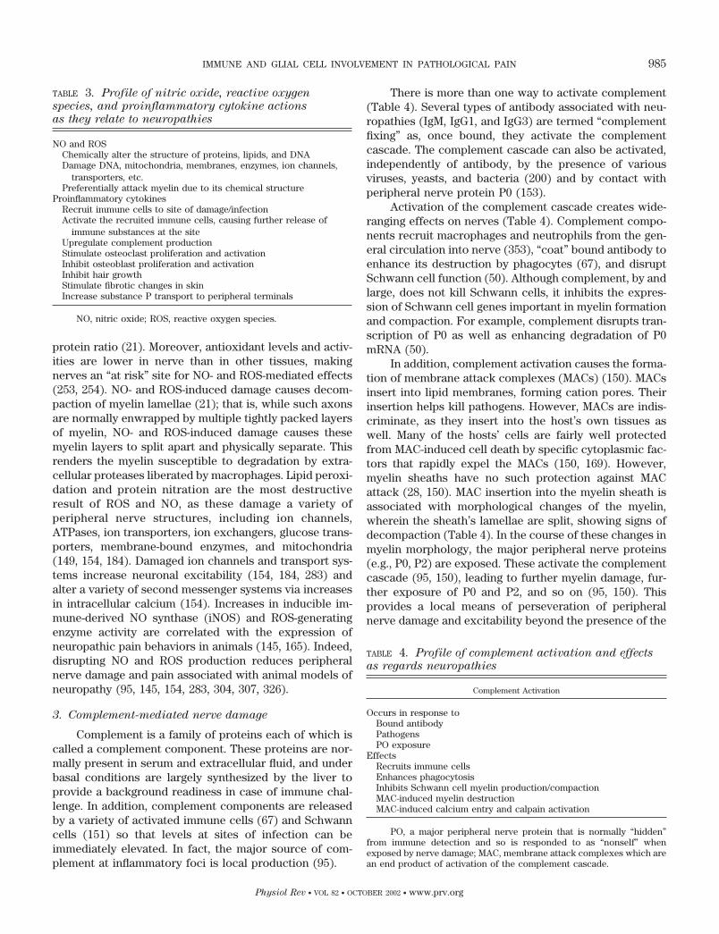
TABLE 3. Profile of nitric oxide, reactive oxygen
species, and proinflammatory cytokine actions
as they relate to neuropathies
NO and ROSChemically alter the structure of proteins, lipids, and DNADamage DNA, mitochondria, membranes, enzymes, ion channels,
transporters, etc.Preferentially attack myelin due to its chemical structure
Proinflammatory cytokinesRecruit immune cells to site of damage/infectionActivate the recruited immune cells, causing further release of
immune substances at the siteUpregulate complement productionStimulate osteoclast proliferation and activationInhibit osteoblast proliferation and activationInhibit hair growthStimulate fibrotic changes in skinIncrease substance P transport to peripheral terminals
NO, nitric oxide; ROS, reactive oxygen species.
IMMUNE AND GLIAL CELL INVOLVEMENT IN PATHOLOGICAL PAIN 985
Physiol Rev • VOL 82 • OCTOBER 2002 • www.prv.org
original immune activator (95). Indeed, complement acti-vation in close association with peripheral nerves hasbeen linked to the development of neuropathic pain (267).
Finally, MAC attack on myelin, axons, and Schwanncells leads to calcium entry and activation of calcium-sensi-tive enzymes such as phospholipase A2 and calpain (Table4). Calpain, a calcium-activated neutral protease, is found inmyelin of peripheral nerves (168), in Schwann cells (183),and in both myelinated and unmyelinated peripheral nerveaxons (6). As noted above, myelin is highly susceptible toMAC-associated damage. Indeed, intraneurial injection ofcalcium ionophore is sufficient to cause demyelination invivo as calpain activation causes the myelin to self-destruct(90). Calpain has been implicated in Wallerian degeneration(112) and has been found to be necessary and sufficient foraxonal degeneration (77).
4. T lymphocyte-mediated nerve damage
T lymphocytes have been implicated in animal mod-els of neuropathy such as EAN (see above). Each T lym-phocyte can bind to and become activated by a singleepitope. As activated T lymphocytes move into andthrough peripheral nerves in the normal course of im-mune surveillance, they exit if they do not identify theirepitope. On the other hand, if the epitope is detected, theT lymphocytes remain at the site and begin to both pro-liferate and release a variety of substances. Some of thesesubstances disrupt the blood-nerve barrier, allowing entryof antibodies and immune cells (290). Others, such asinterleukin (IL)-2, tumor necrosis factor (TNF), and inter-feron-�, stimulate Schwann cells and macrophages to en-hance their antigen-presenting capabilities and to beginreleasing substances such as proinflammatory cytokinesand ROS (19, 209). In addition, these T-cell productsstimulate a subset of T lymphocytes, called “cytotoxic Tcells.” Upon binding to the epitope expressed on a cell,the activated cytotoxic T cells release specialized lyticgranules directly at the cell. These lytic granules containthe cytotoxic protein perforin and a family of destructiveproteases called granzymes. Perforin creates transmem-brane pores in the target cell membrane, disrupting ionbalances and allowing granzymes to enter. Once inside,granzymes trigger programmed cell death (apoptosis) ofthe target cell (89). The killed cells are then engulfed anddestroyed by phagocytic immune cells, including macro-phages and Schwann cells. As with other immune pro-cesses, this cytotoxic T-cell response is adaptive whendirected against threats, such as bacteria, viruses, or can-cer cells. However, nearby nerves can also be destroyed.
5. Trauma-induced nerve damage
Traumatic nerve injury leads in many cases to post-traumatic neuropathic pain. In traumatic injury, there isnot only trauma-induced tissue destruction, but likely
bacterial contamination of the injury site as well. Manybacteria activate the complement cascade, as notedabove. In addition, the presence of bacteria causes releaseof chemoattractants that recruit and activate phagocyticcells (neutrophils and macrophages). These phagocytesexpress surface receptors that recognize and bind to evo-lutionarily conserved epitopes on bacterial surfaces.Binding triggers phagocytosis as well as release of NO,ROS, and proinflammatory cytokines (IL-1, IL-6, andTNF). NO and ROS are key antibacterial products as theydamage DNA, mitochondria, and other cellular machineryleading to bacterial demise (Table 3).
On the other hand, IL-1, IL-6, and TNF orchestrate theearly immune response to infection and damage by serv-ing as chemoattractants to recruit immune cells to the sitefrom the general circulation (229) (Table 3). These im-mune-derived proteins also activate the recruited immunecells to release a variety of substances that enhance hostdefense. Thus, in response to these proinflammatory cy-tokines, there is further release of NO and ROS (169, 249)and upregulation of the production of complement com-ponents by recruited immune cells and Schwann cells(169). In turn, engulfment of damaged myelin by recruitedand resident phagocytes further increases their produc-tion of proinflammatory cytokines long after the originalimmune stimulus (169). As reviewed in section IIB2,proinflammatory cytokines have been repeatedly impli-cated in demyelination and degeneration of peripheralnerves, increases in sensory afferent excitability, and cre-ation of neuropathic pain.
Beyond introducing bacteria, traumatic injury stimu-lates immune responses in two ways. First, nerve damageexposes the peripheral nerve proteins P0 and P2. As de-scribed in sect. IIA1, these are responded to as nonself by theimmune system, so this initiates an immune response similarto that triggered by pathogens. Second, physical injury andischemic injury that occur with trauma lead to cell disinte-gration (necrosis). In peripheral nerve, this is associatedwith Wallerian degeneration characterized by demyelinationand denervation followed by remyelination and renervation(296, 326). This process has been extensively studied andfound to involve edema-associated disruption of the blood-nerve barrier and the activation of recruited and residentmacrophages, fibroblasts, and Schwann cells (296). All ofthese cell types are active in phagocytosing necrotic periph-eral nerve tissue (268). Locally produced proinflammatorycytokines are intimately involved in the Wallerian degener-ation process as well (279, 280).
B. Painful Neuropathies Involving Nerve Trauma
and Inflammation
As documented in section IIA, multiple immunologicmechanisms exist in peripheral nerves that, upon activa-
986 LINDA R. WATKINS AND STEVEN F. MAIER
Physiol Rev • VOL 82 • OCTOBER 2002 • www.prv.org

tion, can extensively damage or destroy its function. Be-low we discuss how these may relate to clinical peripheralnerve neuropathies and their associated animal models.What will be presented are examples of immunologicallyrelated neuropathies. The discussion is not intended to beinclusive. However, the examples were chosen to illus-trate the types of immunological changes that occur un-der a number of very different precipitating events.
1. Clinical correlations: complex regional pain
syndromes (causalgia and reflex
sympathetic dystrophy)
A) ETIOLOGY AND GENERAL SYMPTOMOLOGY. Reflex sympa-thetic dystrophy (RSD) and causalgia have recently beencontroversially reclassified as complex regional pain syn-drome (CRPS) I and II, respectively (8). While the CRPS Iand II terminology will be followed here, the readershould be clear that RSD and causalgia are the syndromesdiscussed. CRPS I and II are painful conditions that ap-pear to regionally and typically affect limbs rather thanthe body trunk. There is no consensus on the pathophys-iological mechanisms underlying these pain syndromes(8, 293). Not surprisingly, given the mysteries surroundingCRPS I and II, current drug therapies targeting neurons asthe basis of these syndromes fail to control the neuro-pathic pains (293, 329). The symptomology to be de-scribed below raises the possibility of an immunologicalbasis of these syndromes.
CRPS I and II have many features in common. Theprincipal feature that distinguishes them is that CRPS II(causalgia) develops after partial injury of a peripheralnerve trunk. In contrast, CRPS I (RSD) occurs in theapparent absence of known injury to nerve trunks. Minorinjuries to the limb, injuries to remote body regions, low-grade infection, frostbite, burns, myocardial infarction,stroke, neurologic and rheumatologic diseases, fractures,surgery, or even a minor sprain or contusion can precedethe onset of CRPS I symptoms (8, 228). Indeed, no iden-tifiable precipitating event can be identified in 35% ofCRPS I cases (320). It is not known whether minor inju-ries or unidentified events also contribute to the CRPS IIpain assumed to arise from nerve trauma. This possibilityexists as CRPS II pain can arise from body regions outsideof known nerve trauma. For both CRPS I and II, themagnitude and duration of pain greatly exceeds that pre-dicted by the inciting injury, and there is variable progres-sion of pain over time (8). Also, in both syndromes, theaffected region is characterized by abnormalities in bloodflow and sweating, swelling, trophic skin changes (e.g.,thinning, shiny), fibrosis, either decreased or increasedhair growth, and patchy bone demineralization (osteopo-rosis) (8). Many but not all patients exhibit altered sym-pathetic function as well (see sect. IIB1C) (228).
The pain associated with CRPS includes both a spon-taneous burning sensation as well as allodynia to bothtouch/pressure and cold stimuli; heat hyperalgesia is alsoobserved in some patients (8, 239, 240). A peripheraltrigger for the pain of at least CRPS II is supported by thereport that local anesthetic block of the site of priortrauma blocks mechanical allodynia, cold allodynia, andspontaneous pain perceived from sites beyond the area ofanesthesia (82). Pain returns upon loss of anesthesia atthe trauma site. Based on these findings, it has beenproposed that ongoing sensory information arising fromsuch pain-triggering foci create and maintain pathologicalpain of CRPS by actions on the spinal cord (82).
One striking feature of the pain of CRPS I and II isthat it changes with time. Typically, pain begins at arelatively focal site. A hallmark of CRPS is that the painfularea does not follow either neural, vascular, or muscularpatterns (23). The pain expands along the limb and/ormigrates to other body parts in nearly 70% of patients, andbilateral pain occurs in �50% of cases (137). Indeed, thepain may expand to encompass a body quadrant or eventhe entire body (158, 323). Such “anatomically impossi-ble” patterns of pain led to the hypothesis that CRPS wasof psychological rather than physical origin, but this ex-planation has been dismissed (363). An alternative hy-pothesis is that the anatomically impossible pain distribu-tions and expansions of the painful region with time arecreated by spinal cord sensitization, likely involving im-munelike glial cells (335, 336a). Here, spinal sensitizationrefers to dynamic changes that occur in the spinal corddorsal horn in response to intense and/or prolonged painsignals received from peripheral nerves. These intense/prolonged signals arriving at dorsal horn pain responsiveneurons cause these spinal neurons to become hyperex-citable; that is, these neurons now respond to stimuli thatare not normally perceived as painful as if they werepainful, and overreact to stimuli that are normally per-ceived as painful. The possibility that glial cells drive thisspinal hyperexcitable state will be addressed in sectionIIB2D.
B) EVIDENCE AND IMPLICATIONS OF A PERSEVERATIVE INFLAMMATORY
STATE. The central sensitization of CRPS may, at least inpart, be sustained by the presence of a chronic inflamma-tory state in the affected body region. There are manyfeatures of CRPS that suggest that the pathological regionis exhibiting an excessive inflammatory response for atleast the first several months of the disease process (Ta-ble 5). As typical for an inflammatory event, the affectedregion exhibits increased blood flow, increased vascularpermeability, edema of soft tissues and bone, hypervas-cularity in synovium and skeletal muscle, impaired localoxygen utilization leading to ischemic oxidative stress ofthe involved tissues, tissue accumulation of antibodiesand immune cells (neutrophils), and degenerative tissuechanges due to localized ROS-induced lipid peroxidation
IMMUNE AND GLIAL CELL INVOLVEMENT IN PATHOLOGICAL PAIN 987
Physiol Rev • VOL 82 • OCTOBER 2002 • www.prv.org

(158, 161, 228, 251, 318, 361). Patients may exhibit in-creased circulating levels of bradykinin, which has beenassociated with inflammatory pain (18). Furthermore,shifts in acute phase protein concentrations in blood andblood cell counts are consistent with a subacute inflam-matory process (161). Supportive of inflammatory medi-ation of CRPS, scavengers of ROS decrease the symptoms(81). The clinical finding that treatment with immunosup-pressive doses of corticosteroid decreases CRPS com-plaints is also supportive of an inflammatory basis ofCRPS (318).
The patchy osteoporosis, proliferation of epidermalimmune cells, and alterations in skin and hair growthobserved in CRPS patients are also consistent with aregional inflammatory process (362). Both IL-1 and IL-6cause proliferation and activation of osteoclasts (the cellsthat mobilize calcium via bone destruction) and suppressthe activity of osteoblasts (the cells that create new bone)(179, 297). Furthermore, skin biopsies of CRPS patientsshow striking increases in the numbers of epidermalLangerhans cells (31) which, like keratinocytes and sev-eral other cell types in epidermis, can release immune cellchemoattractants and proinflammatory cytokines (44,109). Indeed, denervation of the skin causes rapid activa-tion and proliferation of Langerhans cells and keratino-cytes that continues until reinnervation occurs (117, 292).This suggests that such cell proliferation in CRPS mayreflect partial denervation of the affected region. Lastly,skin and hair changes may be proinflammatory cytokinerelated. Recall that CRPS is characterized by the seem-ingly strange symptom of both decreased and increasedhair growth. But, in actuality, both decreased and in-creased hair growth can be created by proinflammatorycytokines by direct and indirect pathways, respectively.TNF and IL-1 directly inhibit hair growth in that they arehighly potent inhibitors of both the growth of human hairfollicles and elongation of the hair shaft (284, 351). Kera-
tinocyte-derived TNF and IL-6 cause retarded hair growth,signs of fibrosis, and immune infiltration of the dermis(34, 314), as observed in CRPS patients. Proinflammatorycytokines can also exert the counterintuitive effect ofindirectly stimulating hair growth. In humans, proinflam-matory cytokines can stimulate the release of hepatocytegrowth factor/scatter factor from hair follicle cells, result-ing in enhanced hair growth (276).
Given the inflammatory profile of CRPS, it was nat-ural to consider whether an infective or autoimmuneprocess underlies the disease. Thus attempts have beenmade to link CRPS to specific preceding infections. Al-though a few cases of CRPS have been noted to followBorrelia infections (23) and spirochetal infections (211),no links to other pathogens have been reported, nor haveantiperipheral nerve antibodies been identified in thesepatients.
Because autoimmunity and infection do not accountfor CRPS, exaggerated neurogenic inflammation has beenproposed (14). Neurogenic inflammation refers to the factthat painful stimulation of the receptive fields of certainpain responsive fibers (termed C fibers) causes two re-sults. The first is sending a pain message to the spinalcord, leading to sensation/perception. The second is re-lease of substances by these same nerve terminals intotheir own receptive fields. These neurally released sub-stances (e.g., substance P) trigger all of the cardinal signsof inflammation: reddening of the area, swelling, and pain.Because this inflammatory response is created by a localnerve “reflex,” this has been called neurogenic inflamma-tion. Exaggerated neurogenic inflammation would exag-gerate the release of substance P from peripheral nerveterminals. In turn, substance P would cause local swell-ing, redness, and pain, consistent with symptoms of CRPS(14). Such an exaggerated release of substance P is in-triguing from an immunological viewpoint, as this wouldpotentially provide an additional mechanism for creatingpain. Substance P induces proinflammatory cytokine re-lease from a variety of immune cells (175, 223, 360) andhas been shown to induce at least TNF and IL-1 releasefrom human skin (26, 222). In turn, proinflammatory cy-tokines induce pain by activating pain-responsive sensorynerve terminals (46, 68, 345). Indeed, a substance P-proin-flammatory cytokine positive-feedback loop would bepredicted in CRPS, given that even a single intraplantarinjection of IL-1 produces a long-term increase in axonaltransport of substance P to cutaneous nerve terminals(128). Such a hypothesized positive feedback loop wouldbe predicted to provide a perseverative “drive” to createand maintain spinal cord sensitization.
If true, such a substance P-proinflammatory cytokinepositive feedback loop would also have implications forthe elevated bradykinin levels observed in CRPS patients(18). This CRPS-related elevated systemic bradykinin maypotentially interact with the proposed substance P-proin-
TABLE 5. Summary of the signs of perseverative,
exaggerated inflammation of body regions affected
by complex regional pain syndromes I and II
Signs of Inflammation in CRPS
Increased blood flowIncreased vascular permeabilityEdema of soft tissues and boneHypervascularity of synovium and muscleOxidative stressAccumulation of immune cellsROS production and consequent lipid peroxidationPatchy osteoporosisProliferation of epidermal immune cellsAltered skin, nail, and hair growth
ROS, reactive oxygen species; CRPS, complex regional pain syn-dromes I and II (reflex sympathetic dystrophy and causalgia, respec-tively).
988 LINDA R. WATKINS AND STEVEN F. MAIER
Physiol Rev • VOL 82 • OCTOBER 2002 • www.prv.org

flammatory cytokine positive-feedback loop becauseproinflammatory cytokines upregulate the expression ofbradykinin receptors in a variety of tissues (9, 86), andbradykinin receptors contribute to pain hypersensitivity(52). Bradykinin may also further stimulate the proposedsubstance P-proinflammatory cytokine loop, as bradyki-nin increases IL-1, TNF, and IL-6 (199, 310).
Taken together, numerous lines of evidence suggestthat prolonged localized release of proinflammatory cyto-kines may occur in body regions affected by CRPS. Al-though clearly speculative, if this does occur, it suggeststhat such perseverative proinflammatory cytokine releasecould, by stimulation of sensory nerves, be a contributingfactor to the maintenance of central sensitization ob-served in CRPS patients.
C) IMPLICATIONS OF SYMPATHETIC NERVOUS SYSTEM INVOLVEMENT
FOR IMMUNE RESPONSES. Although controversial (215, 294),CRPS is often reported to be a sympathetically main-tained pain syndrome (8, 239). Sympathetic involvementin CRPS is supported by the facts that 1) there is overlapof body regions exhibiting pain and autonomic dysfunc-tion (sweating, temperature, and blood flow abnormali-ties) and 2) blocking sympathetic function relieves pain(293, 329). While an increase in sympathetic activity wasoriginally thought to occur, more recent evidence sug-gests instead that there is reduced sympathetic activity inthe affected region that, with time, develops supersensi-tivity to catecholamines (60). While catecholamines andsympathetic activation do not cause pain in normal hu-mans, they do create pain in CRPS patients (2). This painresponse is thought to be due, at least in part, to novelexpression of �1-adrenergic receptors on pain-responsivesensory fibers (8). Blocking sympathetic function,whether by surgical sympathectomy, systemic phentol-amine, or systemic guanethidine, relieves partial nerveinjury-induced neuropathic pain in laboratory animalmodels as well as humans (8, 35, 146, 239, 278). Indeed,sympathectomy does not just relieve pathological pain inthe body region ipsilateral to the CRPS-initiating event;rather, it also relieves pain arising from anatomicallyimpossible mirror-image sites, that is, the identical bodyregion contralateral to the initiating event (278). Thussympathetectomy must somehow quiet the contralateralspread of spinal cord hyperexcitability underlying mirror-image pain.
Alterations in sympathetic fibers rapidly follow pe-ripheral nerve injury. This occurs as sprouting of sympa-thetic fibers, creating aberrant communication pathwaysfrom the new sympathetic terminals to sensory neurons(35). Sympathetic sprouting has been documented in theregion of peripheral terminal fields of sensory neurons(262), at the site of nerve trauma (57), and within thedorsal root ganglia (DRG) containing cell bodies of sen-sory neurons (248, 343). Each of these sites develops
spontaneous activity and sensitivity for catecholaminesand sympathetic activation (8, 53).
The clearest evidence that immune activation partic-ipates in sympathetic sprouting comes from studies of theDRG. DRG cells receive signals that peripheral nerveinjury has occurred via retrograde axonal transport fromthe trauma site. These retrogradely transported signalstrigger sympathetic nerve sprouting into DRG (205, 308).As a result of nerve damage-induced retrogradely trans-ported signals, glial cells within the DRG (called satellitecells) proliferate (248) and become activated (343); mac-rophages are recruited to the DRG as well (63, 176). Inturn, the activated satellite glial cells (and, presumably,the macrophages) release proinflammatory cytokines anda variety of growth factors into the extracellular fluid ofthe DRG (206, 246–248, 258, 277, 308, 358). These sub-stances stimulate and direct the growth of sympatheticfibers, which form basket-like terminals around the satel-lite cells that, in turn, surround neuronal cell bodies (247,248, 343). For discussion of satellite cell functions, seesection IIIA.
Until recently, the sympathetic sprouting, rather thanthe glial (satellite cell) activation, has attracted the atten-tion of pain researchers. The satellite cells were ignoredas they were thought to be irrelevant to the creation ofexaggerated pain states. However, it may be speculatedthat the satellite cells, rather than the sympatheticsprouts, have the most impact on pain. Although electricalstimulation of these DRG sympathetic sprouts does exciteDRG neurons (8), other observations cast doubt on therelevance of these sprouts for pathological pain. First,these sympathetic sprouts predominantly form terminalfields around large-diameter neurons that, as a class, donot transmit pain information (247, 358). Second, thedensity of sympathetic sprouts in the DRG does not cor-relate with neuropathic pain intensity (8). Given that 1)DRG neurons express receptors for satellite cell-derivedproinflammatory cytokines and growth factors (204, 216)and 2) these proinflammatory cytokines and growth fac-tors act in a paracrine fashion to influence large numbersof cells, perhaps it may be that these satellite cell-derivedsubstances are really the basis for altered pain, ratherthan the sympathetic sprouts. From this perspective, sym-pathetic sprouting into DRGs may simply be a “side ef-fect” of glial activation.
If this is true, then satellite cell-derived substancesshould have demonstrable effects consistent with en-hanced pain. Satellite cell-derived substances include, forexample, nerve growth factor (NGF) (159, 358), gliallyderived neurotrophic factor (GDNF) (91), brain-derivedneurotrophic factor (BDNF) (339), neurotrophin-3 (NT-3)(244, 358), and proinflammatory cytokines (42). Each ofthese does indeed exert effects consistent with enhancedpain. 1) GDNF upregulates the expression of pain-rele-vant sodium receptor subtypes in DRG neurons (45); 2)
IMMUNE AND GLIAL CELL INVOLVEMENT IN PATHOLOGICAL PAIN 989
Physiol Rev • VOL 82 • OCTOBER 2002 • www.prv.org

intra-DRG injection of NGF and BDNF each induces me-chanical allodynia in the absence of nerve damage (359);3) antibodies to NGF, NT-3, and BDNF each reducesmechanical allodynia induced by nerve damage (359); 4)IL-1 induces the release of substance P from DRG neuronsin cell culture (125); 5) neuropathic pain is reduced in IL-6knock-out mice (247); 6) almost all DRG neurons expressIL-6 receptors (204); and 7) TNF induces abnormal spon-taneous activity in DRG neurons (170, 357). Thus theactions of satellite cell-derived substances strongly sug-gest that immune activation in the DRG can facilitate pain.
Beyond the DRG, peripheral nerve injury inducessympathetic nerve sprouting into the upper dermis as well(256). This aberrant pattern of sympathetic innervation ofthe skin has been proposed to have important implica-tions for sympathetic interactions with pain-responsivesensory terminals (256). In support of this perspective,intradermal injection of norepinephrine, while having noeffect on normals, produces pain in CRPS patients (2),and subcutaneous norepinephrine excites pain-respon-sive C-fiber terminals in the skin of rodents after, but notbefore, peripheral nerve damage (262). As noted above,�1-adrenoceptors have been implicated in sympatheticallymaintained pain (2).
This remarkable change in catecholaminergic sensi-tivity of the skin in CRPS and nerve damage is potentiallyintriguing from an immunologic point of view. Undernormal conditions, catecholamines act via �2-adrenergicreceptors on immune cells to inhibit the production andrelease of proinflammatory cytokines (106). These cellsdo not express �1-adrenergic receptors under basal con-ditions (138). However, the situation can dramaticallychange in chronic inflammation. Now, immune cellsdownregulate their expression of �2-adrenergic receptorsand upregulate their expression of �1-adrenergic recep-tors over time (106). Such a shift to a predominant �1-expression may potentially have implications for inflam-mation and pain associated with CRPS and nerve damage.This is because �1-receptors stimulate the production andrelease of proinflammatory cytokines (106, 138). Clearly,if �1-adrenoceptors were to become expressed by theresident and/or recruited immune or immunocompetentcells of the affected CRPS sites (synoviocytes, endothelialcells, Langerhans cells, keratinocytes, fibroblasts, classi-cal immune cells, etc.), then sympathetic activation wouldbe predicted to cause pain, at least in part, via proinflam-matory cytokine release (237). Indeed, while the densityof �1-adrenoceptors is known to increase in hyperalgesicskin of CRPS patients, these studies have either usedtissue homogenates or low-resolution autoradiographythat preclude the authors from identifying the cell type(s)expressing the receptors (60, 252). Although the autora-diographic study shows marked increases in �1-adrener-gic expression in skin regions not accounted for by pe-ripheral nerves (60), this finding has never been explored
to define whether �1-expressing immune cells may indeedaccount for this upregulated receptor expression.
Thus, from a variety of angles, immune activationwithin skin, peripheral nerves, DRG, and spinal cord mayrepresent under-appreciated sources of pain in CRPS.Although highly speculative, the evidence suggests thatinvestigations into the potential involvement of immuneactivation in CRPS are warranted. As is reviewed in thefollowing sections, evidence from animal models providesfurther support for the plausibility of this proposal.
2. Animal models
A) IMMUNE INVOLVEMENT IN PAIN FROM NERVE TRAUMA: FROM
APLYSIA TO RATS. Evidence for altered pain processing due toimmune activation near peripheral nerve trunks hasarisen primarily from two animals: rats and the simplerAplysia. Regarding Aplysia, it has been known since themid 1980s that sensory nerve damage creates prolongedenhanced pain responses (for review, see Ref. 331) (Table6). These studies led to the discovery that large numbersof immunocytes (immune-like cells of Aplysia) are at-tracted to the site of nerve damage (36). This is intriguingsince immunocytes are strikingly similar in function tomammalian macrophages in that they are phagocytes thatrelease proinflammatory cytokine-like molecules (IL-1-like and TNF-like) upon activation by foreign (nonself)substances (37). The link to IL-1 and TNF is interestingsince these proinflammatory cytokines alter ion channelsin Aplysia neurons, causing hyperexcitability (264, 301)(Table 7). These findings led to the discovery that 1) hyper-excitability of injured nerves is significantly greater in the
TABLE 6. Summary of evidence that sensory nerve
damage in Aplysia and rat is associated with
exaggerated pain responses, which are linked
with recruitment of immune cells to the site
Sensory nerve damage in Aplysia
Exaggerates pain responsesExaggerates electrical responses of pain-responsive neuronsAssociated with macrophage-like immune cell (immunocyte)
invasion of the injury siteAssociated with release of interleukin-1-like and tumor necrosis
factor-like substances from immunocytesNerve hyperexcitability is enhanced by the presence of activated
immunocytesSensory nerve damage in rat
Exaggerates pain responsesExaggerates electrical responses of pain-responsive neuronsAssociated with macrophage invasion of the injury siteAssociated with activation of resident immune cellsAssociated with release of interleukin-1, tumor necrosis factor, and
interleukin-6 from immune cellsNerve hyperexcitability is enhanced by the presence of activated
immune cells
These immune cells appear to contribute to the exaggerated painresponses that ensue.
990 LINDA R. WATKINS AND STEVEN F. MAIER
Physiol Rev • VOL 82 • OCTOBER 2002 • www.prv.org
presence of activated immunocytes (39) and 2) IL-1 ap-plication enhances nerve injury hyperexcitability (38).
During this same time period, parallel findings beganto appear with rodents. A number of partial nerve injurymodels were developed in rodents (12, 147, 271), becausecausalgia (CRPS II) and other posttraumatic neuropathiesare associated with partial nerve injuries. Similaritieshave been noted between such models and CRPS symp-toms. For example, partial nerve injury in rodents causesvasodilation in the limb that is associated with increasedvascular permeability and edema (47, 48, 158), accumula-tion of immune cells (neutrophils) in the nerve-injuredhindpaw and muscle (47, 48), spinal sensitization (180),enhanced release of substance P from peripheral endingsof activated C fibers (158), sympathetic dysfunction withdenervation-induced supersensitivity to catecholamines(157), and increased local production of cytokines (76,328). In parallel with human studies implicating ROS inthe generation of CRPS (81), ROS are implicated in CRPS-like pain in rodents since 1) the generation of ROS inhealthy rat hindlimbs elicits pain and other symptoms ofCRPS (317) and 2) blockade of ROS attenuates neuro-pathic pain from partial nerve injury (173).
By the mid-1980s it was known that partial nerveinjury causes exaggerated pain states and axonal hyper-excitability as well as Wallerian degeneration (231) (Table6). Both trauma-induced Wallerian degeneration (15) andenhanced pain (174) have been linked to the activity ofmacrophages recruited to the site of injury. Indeed, sim-ply delaying the recruitment of macrophages to the areaof damage delays the development of both neuropathicpain and Wallerian degeneration (210, 245). In contrast,neuropathic pain behaviors are enhanced by attractingactivated immune cells to the injury (40, 186). Of thevarious neuroactive substances released by activatedmacrophages, most evidence implicates the proinflamma-tory cytokines IL-1, TNF, and IL-6 (Table 7). These in-crease at sites of nerve trauma, via production bySchwann cells, endothelial cells, and resident and re-cruited macrophages (19, 164, 255, 279). Blockade of IL-1(286) or TNF (122, 285, 287) activity after sciatic injuryreduces thermal hyperalgesia and mechanical allodynia.
Blockade of IL-1 and TNF at the level of the sciatic nervealso prevents and reverses pain changes induced by sci-atic inflammatory neuropathy (267). Furthermore, neuro-pathic pain is prevented in IL-6 knock-out mice (247), andthe magnitude of mechanical allodynia that develops afterperipheral nerve injury directly correlates with both thenumber of activated macrophages and the number ofIL-6-producing cells at the injury site (43). In addition toproinflammatory cytokines, ROS (peroxynitrite) have alsobeen implicated in creating nerve trauma-induced exag-gerated pain states through their actions at the site ofnerve injury (173).
B) IMMUNE INVOLVEMENT IN PAIN FROM NERVE INFLAMMATION IN THE
ABSENCE OF TRAUMA. Pain facilitation can occur even in theabsence of apparent physical trauma to peripheral nerves.The simple placement of immunologically activated im-munocytes near healthy Aplysia sensory nerves increasestheir excitability (36). Furthermore, exposing healthy ratsciatic nerve to gut suture (186), killed bacteria (64), algaeprotein (carrageenan) (64), yeast cell walls (zymosan)(33, 75), or the HIV-1 envelope glycoprotein gp120 (108)increases behavioral responsivity to touch/pressureand/or heat stimuli; that is, these manipulations inducemechanical allodynia and thermal hyperalgesia. Suchchanges are mimicked by proinflammatory cytokines.TNF injected into the sciatic produces thermal hyperal-gesia and mechanical allodynia (327) as well as endoneur-ial inflammation, demyelination, and axonal degeneration(249). Furthermore, TNF applied to the sciatic inducesectopic activity in single primary afferent nociceptive fi-bers (288). TNF is not alone in this regard as ATP alsoectopically activates peripheral nerves, including fibersassociated with pain transmission (126). ROS also appearsufficient to drive exaggerated pain states in the absenceof physical trauma to nerves as intra-arterial infusion offree radical donors into one hindlimb of rats causes in-creased sensitivity to mechanical and thermal stimuli aswell as spontaneous pain (317).
Although it is clear that proinflammatory cytokinesinduce pain, how they do this from midaxonal sites iscontroversial. While proinflammatory cytokine receptorsare known to be expressed on DRG cell bodies (236),whether these receptors are expressed along the courseof their peripheral nerve fibers has never been investi-gated. Of the proinflammatory cytokines, TNF has re-ceived the most study to date and is known to rapidlyalter neural activity, suggesting that it may directly affectaxonal excitability (160, 288). Indeed, studies of the struc-ture of TNF indicate that it can insert into lipid mem-branes to form a central porelike region due to its three-dimensional conformation (131). Insertion is facilitatedby a physiologically relevant lowering of pH, which oc-curs at sites of inflammation (131). The inserted TNFmolecules form voltage-dependent sodium channels(131). Other evidence suggests that TNF interacts with
TABLE 7. Summary of evidence that Aplysia and rat
proinflammatory cytokines enhance responses
to pain stimuli
Proinflammatory cytokine-like substances in Aplysia
Exaggerate pain responsesCreate neuronal hyperexcitability via alterations in ion channelsEnhance injury-induced hyperexcitability, just as activated
immunocytes doProinflammatory cytokines in rat
Exaggerate pain responsesCreate neuronal hyperexcitability, likely via alterations in ion
channelsImplicated in neuropathy-induced pain
IMMUNE AND GLIAL CELL INVOLVEMENT IN PATHOLOGICAL PAIN 991
Physiol Rev • VOL 82 • OCTOBER 2002 • www.prv.org
endogenous sodium and calcium channels to increasemembrane conductance (316, 341). Like TNF, IL-1 rapidlyincreases neuronal excitation (219) and produces long-lasting increases in conductance of voltage-sensitive so-dium and calcium channels (266, 341). IL-6 enhances con-ductance of these ion channels as well (242). Whether IL-1or IL-6 can insert into lipid membranes has not beenreported.
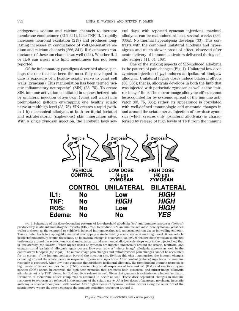
Of the inflammatory paradigms described above, per-haps the one that has been the most fully developed todate is exposure of a healthy sciatic nerve to yeast cellwalls (zymosan). This manipulation has been termed “sci-atic inflammatory neuropathy” (SIN) (33, 75). To createSIN, immune activation is initiated in unanesthetized ratsby unilateral injection of zymosan (yeast cell walls) intopreimplanted gelfoam enwrapping one healthy sciaticnerve at midthigh level (33, 75). SIN creates a rapid (with-in 1 h) mechanical allodynia at both territorial (sciatic)and extraterritorial (saphenous) skin innervation sites.With a single zymosan injection, the allodynia lasts sev-
eral days; with repeated zymosan injections, maximalallodynia can be maintained at least several weeks (336,336a). No thermal hyperalgesia develops (33). This con-trasts with the combined unilateral allodynia and hyper-algesia and much slower onset of effect, observed afteracute delivery of immune activators delivered during sci-atic surgery (11, 64, 108).
One of the striking aspects of SIN-induced allodyniais the pattern of pain changes (Fig. 1). Unilateral low-dosezymosan injection (4 �g) induces an ipsilateral hindpawallodynia. Unilateral higher doses induce bilateral effects(33, 336); that is, allodynia develops in both the limb thatwas injected with perisciatic zymosan as well as the “mir-ror-image” limb. The mirror-image allodynic effect cannotbe accounted for by systemic spread of the immune acti-vator (33, 75, 336); rather, its appearance is correlatedwith well-defined immunologic and anatomic changes inand around the sciatic nerve. Injection of low-dose zymo-san (which creates only ipsilateral allodynia) is charac-terized by release of high levels of TNF from the immune
FIG. 1. Schematic of the dose-dependent patterns of low-threshold allodynia (top) and immune responses (bottom)produced by sciatic inflammatory neuropathy (SIN). Top: to produce SIN, an immune activator [here zymosan (yeast cellwalls) is shown as the example] or vehicle is injected into unanesthetized, unrestrained rats via an indwelling catheter.This catheter leads to a spongelike material enwrapping a single healthy sciatic nerve at mid-thigh level. When vehicleis injected unilaterally around the sciatic, no behavioral change is observed (top left). When low-dose zymosan is injectedunilaterally around the sciatic, territorial and extraterritorial mechanical allodynia develops only in the injected leg; thatis, ipsilaterally (top middle). When higher doses of zymosan are injected unilaterally around the sciatic, territorial andextraterritorial ipsilateral allodynia again occurs. However, now a “mirror image” allodynia appears as well in thecontralateral hindpaw (top right). The mirror-image pain changes and extraterritorial pain changes cannot be accountedfor by spread of the immune activator beyond the injection site. Bottom: this chart summarizes the immune changesoccurring around the sciatic nerve in response to perisciatic injections. After control (vehicle) injections, no immuneresponse is produced. After low-dose zymosan that produces ipsilateral allodynia, the predominant immune response ishigh levels of tumor necrosis factor (TNF) release. Only small responses of interleukin-1 (IL-1) and reactive oxygenspecies (ROS) occur. In contrast, the high-dose zymosan that produces both ipsilateral and mirror-image allodyniastimulates not only TNF release, but IL-1 and ROS release as well. Given that zymosan is a classic complement activator,formation of membrane attack complexes is assumed to occur as well. These dose-dependent changes in immuneresponses to zymosan are reflected in the anatomy of the sciatic nerve. After low doses of zymosan, no change in sciaticanatomy is observed compared with control. After higher doses of zymosan, edema occurs along the outer rim of thesciatic nerve where the nerve contacts the immune activation occurring around it.
992 LINDA R. WATKINS AND STEVEN F. MAIER
Physiol Rev • VOL 82 • OCTOBER 2002 • www.prv.org

cells around the sciatic nerve (macrophages and neutro-phils), and no morphological change in the sciatic nerve isdetected (75) (Fig. 1). In contrast, response to higherzymosan doses (which create ipsilateral plus mirror-im-age contralateral allodynia) is characterized not only byhigh levels of TNF but also high levels of IL-1 and ROSaround the nerve (75). In addition, complement-derivedMACs assumed to be produced as zymosan are well es-tablished to potently trigger MAC production (49). Atleast perisciatic complement and proinflammatory cyto-kines are involved in producing SIN-induced allodynia,since perisciatic delivery of antagonists against these im-mune-derived substances blocks the pain changes (267).The immunological profile for the higher zymosan dose isassociated with distinctive pathology of the sciatic nerve.Edema is observed 24 h after higher dose zymosan peri-sciatic injection. Strikingly, this edema occurs along theouter rim of the nerve where the nerve is in contact withsubstances released by zymosan-activated immune cells(75) (Fig. 2). Thus the appearance of mirror-image painco-occurs with distinctive immunological and anatomicchanges at the level of the sciatic nerve.
C) IMPACT OF TRAUMA AND INFLAMMATION ON NEIGHBORING INTACT
NERVE FIBERS. One aspect of partial nerve injuries that hasonly recently received attention is the fascinating ques-tion of whether neuropathic pain is being generated solelyby the damaged nerves; that is, could pathological pain becreated because the function of the remaining intact neu-rons is being altered by immune-derived substances re-leased during demyelination and degeneration of the in-termingled damaged peripheral nerves and/or associatedchanges in the damaged cell bodies in the DRG? While anumber of widely used partial nerve injury models existfor rats, many cause nerve injury in such a way that there
is intermingling of damaged and intact peripheral nervesas well as intermingling of damaged and intact DRG so-mas (12, 271). Thus the relative contributions of signalsfrom damaged peripheral nerves versus DRG somas ofdamaged nerves cannot be dissociated.
In response, new models have recently developedthat either have 1) damaged peripheral nerves intermin-gled with intact peripheral nerves, yet maintain completesegregation of damaged versus intact sensory nerve so-mas (73, 347), or 2) damaged DRG somas intermingledwith intact DRG somas, yet maintain almost completesegregation of damaged versus intact peripheral nerves(54). Thus the influence of damaged nerves and the influ-ence of damaged somas can be studied independently.
The effect that intermingled damaged axons haveon intact neuronal function has been examined by se-lectively damaging the fifth lumbar (L5) spinal nerve(167, 347). This creates intermingling of damaged L5
and undamaged L4 fibers within the sciatic nerve. At thesame time, separate DRG locations of the injured (L5)and intact (L4) DRG somas are maintained (147, 347).Thus peripheral nerves arising from L4 somas are ex-posed to immune-derived substances released in theirvicinity as a result of the demyelination and degenera-tion of sciatic L5-derived nerves and from the intermin-gling of their receptive fields in the skin (347). Intrigu-ing changes in the intact L4 nerves result. Within 1 dayafter L5 spinal nerve damage (the earliest time tested),L4 spinal nerve fibers develop spontaneous activity (3).Mechanical allodynia develops correlated with thischange and is abolished by transection of L4 spinalnerves, indicating that intact L4 has become the drivingforce for creating allodynia (167). Spontaneous activityalso develops in monkey uninjured nerve fibers follow-
FIG. 2. Example of the edema pat-tern produced 24 h after perisciatic ad-ministration of high-dose zymosan. Re-call that this is the intensity of immuneactivation that creates mirror-image painchanges. Under normal conditions, theedge of the nerve (left side of image)would look identical to deeper portionsof the nerve (right side of image). Edemacauses the fascicles in the nerve rim toappear to be spaced further apart thanare deeper fascicles. [From Gazda et al.(75). Reprinted with permission fromJournal of the Peripheral Nervous Sys-
tem.]
IMMUNE AND GLIAL CELL INVOLVEMENT IN PATHOLOGICAL PAIN 993
Physiol Rev • VOL 82 • OCTOBER 2002 • www.prv.org
ing a similar procedure (3). This nerve fiber activitydevelops in the cutaneous receptive field region andalong the peripheral nerve, rather than within the DRG (3).
As discussed in section IIA5, nerve damage results inimmune activation and the release of a host of neuroac-tive substances along the length of the degenerating fi-bers. These could have direct influences on the electricalactivity of intermingled uninjured axons. Alternatively,such substances may serve as retrogradely transportedsignals to influence gene activation in intact DRG somas.Indeed, a number of changes have been detected in the L4
DRG somas of spared axons in partial nerve injury para-digms. First, immune-derived substances such as leuke-mia inhibitory factor (LIF) (306), IL-6 (127), and NGF(163, 172, 261) are released at the injury site and areretrogradely transported by both intact as well as injuredaxons (72, 204, 248, 308). These retrogradely transportedsignals in intact L4 nerves result in increased DRG neuro-nal expression of BDNF mRNA and protein, vanilloidreceptors type 1 (VR1) mRNA and protein, calcitoningene-related peptide (CGRP) mRNA, preprotachykinin(PPT) mRNA and its protein product substance P, andgalanin (72, 73, 118, 177, 308, 309), as well as increasedexpression of PN3, a tetrodotoxin-resistant sodium chan-nel subunit (238). This pattern contrasts what is observedafter transection of all nerves in a bundle (axotomy),namely, downregulation of DRG expression of VR1 (191),substance P (272), CGRP (272), and PN3 (22). The in-creases in expression of these factors after partial (ratherthan complete) nerve injury may be relevant to patholog-ical pain as increased VR1 expression suggests increasedresponsivity of the peripheral nerves to heat stimuli, PN3could increase neuronal excitability, and substance P andCGRP are pain transmitters of sensory neurons. Further-more, BDNF administered to intact DRG causes mechan-ical allodynia (359), and increased DRG neuronal expres-sion of BDNF has been linked to neuropathic pain asintrathecal delivery of anti-BDNF antibodies attenuatesthermal hyperalgesia in a spinal nerve injury model (72).Indeed, BDNF release in spinal cord phosphorylates spi-nal N-methyl-D-aspartate (NMDA) receptors, which is oneof the mechanisms known to create and maintain centralsensitization (72).
In the second type of spared nerve injury model, thetibial and common peroneal terminal branches of thesciatic are lesioned, leaving the sural nerve intact (54). Indoing so, there is minimal comingling of injured andnoninjured peripheral nerves, yet considerable DRG co-mingling of injured tibial and common peroneal somaswith noninjured sural nerve somas (54). Thus this allowsexamination of possible paracrine signals arising frominjured somas acting on nearby noninjured ones to altertheir function and/or excitability. With this procedure,mechanical allodynia without thermal hyperalgesia devel-ops from the uninjured sural sensory neurons within 24 h,
an effect that lasts for over 6 mo (54). While satellite cellproducts are potentially involved, such studies have notyet been done.
D) SPINAL IMMUNELIKE GLIAL CELL INVOLVEMENT IN TERRITORIAL,
EXTRATERRITORIAL, AND MIRROR-IMAGE NEUROPATHIC PAIN. There isgrowing recognition that immune involvement in patho-logical pain states occurs within the spinal cord as well asin the periphery (335). In spinal cord, immunelike glialcells (astrocytes and microglia) are activated in responseto diverse conditions that create exaggerated pain states:subcutaneous inflammation, peripheral nerve trauma, pe-ripheral nerve inflammation, spinal nerve trauma, andspinal nerve inflammation (41, 71, 74, 99, 190, 192, 298,334, 342). Activation of glia after nerve trauma can occuras a consequence of degeneration of central terminals ofthe dying sensory neurons. In addition, glia express re-ceptors for a host of substances released by incomingpain-responsive sensory afferents: substance P, excita-tory amino acids, CGRP, and ATP (111, 148, 181, 182, 230,274, 348) (Fig. 3). In addition, they may be activated inresponse to substances released by pain-responsive neu-rons in the dorsal horn, such as prostaglandins, NO, andfractalkine (114, 194, 233).
These glia are not only activated but involved in thecreation and maintenance of pathological pain states.Drugs that disrupt glial activation or block the actions ofglially released proinflammatory cytokines (TNF, IL-1,IL-6) prevent and/or reverse exaggerated pain states pro-duced by subcutaneous inflammation, peripheral nervetrauma, peripheral nerve inflammation, spinal nervetrauma, and spinal nerve inflammation (5, 100, 192, 299,300, 334, 336a). Indeed, proinflammatory cytokines ad-ministered perispinally (intrathecally) can create exagger-ated pain responses (56, 250, 302) as well as create hy-perexcitability of pain-responsive neurons in spinal corddorsal horns (250). Activated glia also release (or increasethe extracellular concentration of) a number of othersubstances implicated in the creation and maintenance ofpathological pain states, including NO, ROS, prostaglan-dins, and excitatory amino acids (134, 195, 214). Lastly,glial products can enhance the release of pain transmit-ters from primary afferent terminals (125). Thus gliallyderived, as well as neuronally derived, sources of thesesubstances likely contribute to the pathological painstates that ensue.
While it is easy to understand how peripheral nerveinjury and inflammation lead to hyperexcitability in thedorsal horn termination area of the involved sensorynerves, the phenomena of extraterritorial pain and mirror-image pain have proven enigmatic. These are importantphenomena to understand as both extraterritorial andmirror-image pain changes are reported by neuropathicpain patients, including those with CRPS I and II (178,323, 324). The recent discovery of the importance ofspinal cord glia to neuropathic pain has led to new in-
994 LINDA R. WATKINS AND STEVEN F. MAIER
Physiol Rev • VOL 82 • OCTOBER 2002 • www.prv.org

sights into extraterritorial and mirror-image pain as well.As noted in section IIB2B, the SIN model creates bothextraterritorial (saphenous nerve innervation sites) andmirror-image (contralateral) pain (33). Glia appear to beinvolved, as drugs that disrupt glial activation abolish notonly the sciatic territorial pain changes, but extraterrito-rial and mirror-image pain changes as well (336, 336a).Furthermore, blocking the activity of glially releasedproinflammatory cytokines blocks extraterritorial andmirror-image pain changes, as well as territorial pain (336,336a). Notably, even well-established chronic SIN painsare reversed by intrathecal proinflammatory cytokine an-tagonists, supporting the idea that these glially derivedsubstances are important for maintenance as well as cre-ation of neuropathic pain (335, 336, 336a).
Glia are especially well suited for creating extrater-ritorial and mirror-image pain for two reasons. First,proinflammatory cytokines classically act in a paracrine,rather than synaptic, fashion; that is, these immune-de-rived substances are released into the extracellular fluidso as to affect surrounding populations of cells well be-yond their site of release (16, 333). Thus they couldreadily influence neurons in neighboring spinal termina-tion regions, creating extraterritorial pain changes. Sec-ond, glia are organized into widespread networks via gapjunctions and propagated calcium waves. Thus excitationof glia at one site can lead to the activation of distant glia,consistent with the creation of both extraterritorial andmirror-image effects. Activation in such a manner canlead, in turn, to release of pain-enhancing glial products(101, 124, 232).
3. Summary
Neuropathic pain can occur as a consequence offrank nerve trauma, and almost all animal models devel-oped to date have focused on such peripheral nerve inju-ries. Certainly, the physical damage to nerves, in its ownright, alters pain perception and the functioning of paintransmission pathways. However, neuropathic pain alsodevelops in the absence of detectable physical injury tonerves. Here pathological pain follows peripheral immuneactivation and inflammation. New animal models of suchinflammatory neuropathic conditions are providing in-sights into how immune activation may create such painchanges. Recognition that immune activation can modu-late peripheral nerve function has implications for neuro-pathic pain arising from nerve trauma as well. This isbecause immune activation will by necessity occur when-ever there is damage to peripheral nerves or associatedtissues. Hence, immune activation is a natural componentof all forms of traumatic and inflammatory neuropathicpain conditions.
From animal models of both traumatic and inflam-matory neuropathies, a consistent picture is beginning toemerge for immune involvement in pain. In both trau-matic and inflammatory models, the key immune cellsinvolved at the level of the peripheral nerve are likelyneutrophils and macrophages recruited into the affectedarea from the general circulation, plus a host of residentimmune cells. These include fibroblasts, endothelial cells,Schwann cells, mast cells, resident macrophages, andresident dendritic cells. The importance of proinflamma-tory cytokines (TNF, IL-1, IL-6) in the creation and main-
FIG. 3. Schematic of spinal cord glial regulation ofpain. Glia (microglia and astrocytes) can be activated byeither 1) pathogens (viruses; bacteria) in their role as im-munelike cells; 2) substances released by incoming pri-mary afferents that relay “pain” information [ATP, excita-tory amino acids (EAAs), substance P], or 3) substancesreleased by spinal cord dorsal horn neurons that relay paininformation [pain transmission neurons, fractalkine, nitricoxide (NO), prostaglandins (PG)]. Once activated, glia re-lease a host of substances, including proinflammatory cy-tokines, reactive oxygen species (ROS), NO, PG, EAAs, andATP. Of these, the proinflammatory cytokines act in aparacrine fashion to affect cells far beyond their site ofrelease. Glia form positive-feedback circuits whereby sub-stances they release further activate these cells, creatingperseverative responses. In turn, glially derived substancesincrease pain by 1) increasing the release of “pain” trans-mitters from incoming primary afferents and 2) increasingthe excitability of pain transmission neurons. It should benoted that glia are interconnected into large networks viagap junctions and propagated calcium waves as well (notshown). Taken together, glia are perfectly positioned tocreate perseverative and widespread pain changes in spinalcord.
IMMUNE AND GLIAL CELL INVOLVEMENT IN PATHOLOGICAL PAIN 995
Physiol Rev • VOL 82 • OCTOBER 2002 • www.prv.org

tenance of pathological pain is the most consistent findingacross models. NO, ROS, and complement have also beenimplicated, at least in the few models in which they havebeen tested to date. The potential involvement of othersubstances released under traumatic and inflammatoryconditions (acids, proteases, digestive enzymes, chemoat-tractant molecules) has not yet been assessed, other thannoting that decreasing pH (increasing acidity) near pe-ripheral nerves enhances pain (185).
It is also clear from studies of traumatic and inflam-matory neuropathies that immune activation is not re-stricted to the periphery; rather, spinal cord immune in-volvement also occurs in the form of glial activation.Here, immunelike astrocytes and microglia are key play-ers in the creation and maintenance of diverse neuro-pathic pain states. Once again, proinflammatory cytokineshave been identified as key mediators, this time releasedby glia. In addition, these glia enhance neurotransmitterrelease from primary afferent terminals and release a hostof substances classically implicated in pain, includingATP, numerous excitatory amino acids, NO, prostaglan-dins, and ROS. Indeed, glia may well provide an answer tothe mystery of extraterritorial and mirror-image painsreported by patients with traumatic and inflammatoryneuropathies. Glia are perfectly suited to create suchanatomically impossible pain phenomena as their activa-tion leads both to the release of proinflammatory cyto-kines that act in a paracrine fashion and to widespreadglial activation via gap junctions and propagated calciumwaves connecting widespread glial networks (335).
C. Painful Neuropathies Involving Antibody Attack
of Peripheral Nerves
1. Clinical correlation: Guillain-Barre syndrome
A) GENERAL SYMPTOMOLOGY. Guillain-Barre syndrome(GBS) is an acute inflammatory neuropathy that can si-multaneously attack peripheral nerves throughout thebody. GBS is made life-threatening by the fact that motorand autonomic, as well as sensory, nerves are affected. Inaddition, neuropathic pain is a common symptom. Pain,including abnormal sensations such as electric shocklikesensations (paraesthesias), sensations having unusuallyunpleasant qualities (dysesthesias), muscle pain, jointpain, and visceral pain, occurs in 70–90% of adult cases(201, 234). In children, pain is often the major symptom,with 80% of children reporting pain on admission and100% of children reporting pain during the course of thedisease (213).
B) EVIDENCE FOR INVOLVEMENT OF AUTOIMMUNE ANTIBODIES.
Which immune factor(s) are critical in GBS remains amatter of some debate. T lymphocytes (see sect. IIA4)have long been posited as playing a major role, based bothon GBS nerve autopsy and biopsy studies indicating the
presence of large numbers of T lymphocytes and, morespecifically, on the presence of T lymphocytes sensitizedto myelin epitopes (98). T-lymphocyte products such asIL-2 are elevated in the serum of GBS patients, supportingactivation of this immune cell type (98). However, otherautopsy/biopsy cases fail to reveal T-lymphocytic infiltra-tion, especially at the initial stages of nerve lesions (241).The absence of T lymphocytes at the stage of nerve lesionformation led to the idea that T-lymphocyte invasion oc-curs after the initial peripheral nerve damage has alreadyoccurred. In support, some GBS patients have activated Tcells that respond to the major peripheral nerve proteinsP0 and P2 (119, 243). This is noteworthy since, as dis-cussed in section IIA1, P0 and P2 are enwrapped withinthe myelin sheath of intact nerves and so are not normallydetected by T lymphocytes. Hence, nerve damage musthave occurred before T lymphocytes could become sen-sitized to these peripheral nerve proteins. The role of Tlymphocytes may be to further the damage that has al-ready begun, by releasing substances that destroy theirtargets, attract immune cells to the site, and facilitatefurther entry of other immune cells and products by dis-rupting the blood-nerve barrier.
Given the efficacy of exchanging patients’ plasmawith artificial plasma (i.e., plasmapheresis) for relievingGBS symptoms, some soluble factor in the general circu-lation does appear important to the etiology of at leastsome forms of GBS (112). Circulating antibodies are themost likely candidates (see sect. IIA2). Indeed, numerousexamples of autoimmune attack of peripheral nerves ex-ist in the clinical literature. Clinically, high titers of serumantibodies directed against peripheral nerve and/or sen-sory nerve somas occur in a variety of neuropathies,including monoclonal gammopathy, inflammatory poly-neuropathies (including, but not limited to, GBS), andparaneoplastic neuropathies (e.g., anti-Hu neuropathy as-sociated with small cell lung carcinoma). The presence ofsuch autoantibodies is a useful diagnostic. These antibod-ies typically bind to glycolipids, glycoproteins, and gly-cosaminoglycans, but some (e.g., anti-Hu) are directedagainst intracellular proteins as well (243). Antibody in-teractions with nerves can cause a variety of dysfunc-tions. For example, antibodies against GM1 ganglioside (acomponent of peripheral nerve myelin sheaths) are asso-ciated with motor neuropathies, anti-MAG antibodies areassociated with sensory or sensorimotor neuropathies,and antibodies directed against GD3, GD2, GD1a, andGQ1b gangliosides, chondroitin sulfate, and sulfatides areall associated with sensory neuropathies (243, 295). Ininflammatory polyneuropathies such as GBS, it is notuncommon to find multiple types of autoimmune antibod-ies in the general circulation of individual patients, eachdirected against different nerve targets (243, 295).
“Molecular mimicry” is posited as the underlyingcause of antibody-directed attack of peripheral nerves in
996 LINDA R. WATKINS AND STEVEN F. MAIER
Physiol Rev • VOL 82 • OCTOBER 2002 • www.prv.org

several forms of GBS (355). Molecular mimicry has beenidentified between peripheral motor nerve gangliosideGM1 and the lipopolysaccharide component of Campy-
lobacter jejuni, between the sensorimotor nerve ganglio-side GD2 and cytomegalovirus, and between the periph-eral nerve major protein P0 and several viruses linked toGBS including cytomegalovirus, Epstein-Barr virus, vari-cella zoster, and HIV-1 (119, 319).
In support of a role of autoimmune antibodies in theetiology of this disorder, GBS sera with high antiperiph-eral nerve myelin antibody activity have been found tocause 50–80% demyelination in rat DRG cultures, and thedegree of demyelination correlates with antiperipheralnerve myelin activity and damage (265). Indeed, evenwhen sera are not screened for high antiperipheral nervemyelin antibody activity, one-third of GBS patient serasamples cause myelin breakdown in rat DRG neuronalcultures and rat Schwann cell cultures (196).
While autoimmune antibodies are most commonlythought of in terms of their direct peripheral nerve dam-aging effects, they can also disrupt nerve function in otherways. For example, antibodies in the sera of GBS patientsalter presynaptic neurotransmitter release and disruptactivation of postsynaptic ion channels in mouse hemidi-aphragm preparations (29). Some forms of GBS antibod-ies can also bind to nodes of Ranvier, altering sodiumchannel function, and resulting in conduction block inde-pendent of anatomic damage to the axons (112). The factthat plasma exchange can sometimes produce spectacu-lar results in GBS patients, with a bed-ridden patientreturning to near-normal walking within a few hours,clearly demonstrates that at least some GBS-associatedantibodies (or other serum components) can cause pe-ripheral nerve dysfunction rather than demyelination/damage that could not be so rapidly reversed (112).
C) EVIDENCE FOR INVOLVEMENT OF OTHER IMMUNE PRODUCTS. Sol-uble immune substances other than antibodies likely alsocontribute to GBS. Substances released from mast cellshave only recently been proposed to exert a major role inGBS, predominantly in nerve demyelination (58). Also, inthe acute stages of GBS, there is upregulation of immunecell adhesion molecules involved in migration of immunecells out of the blood and into sites of inflammation (96,217, 313). Circulating levels of TNF and IL-6 are likewiseelevated in GBS patients (94, 275). However, their poten-tial involvement in pain or other symptomology of thedisease is not yet clear. What has been reported is thatplasma TNF levels correlate with severity of clinical andnerve conduction deficits (66, 273). Interest in the poten-tial role of proinflammatory cytokines in GBS originallyarose due to their known neurotoxic and neuroexcitatoryeffects (249, 259). However, given that these same proin-flammatory cytokines are upregulated locally in Schwanncells, fibroblasts and macrophages of GBS nerve biopsies(208), it may be that proinflammatory cytokines in nerves
rather than in the general circulation are linked to GBSsymptomology.
In contrast, involvement of the complement cascadein GBS is indicated by multiple findings. 1) The antipe-ripheral nerve myelin antibodies of GBS patients are com-plement-fixing; that is, they activate the complement cas-cade and bind products formed by complement activation(265). As noted in section IIA3, this facilitates macrophageattack of the antibody-bound myelin (241), creates che-moattractants that recruit and activate immune cells, anddisrupts the blood-nerve barrier as is known to occur inGBS patients (86). 2) MACs, the destructive end productof complement activation, are found in 100% of GBS serawhile being nonexistent in controls (152). This sign ofcomplement activation is linked to the expression of an-tiperipheral nerve antibody in these patients, and MACsare bound to peripheral nerves of GBS patient biopsies(152). 3) Blockade of MAC formation prevents GBS serafrom inducing myelin disruption in rat DRG cultures(265). 4) Autopsy of GBS patients who died 3–9 days afterdisease onset reveals products of complement activation(complement activation marker C3d and MACs) bound tothe outer rim of many peripheral nerve fibers andSchwann cell membranes (87). In these patients, disrup-tion of myelin occurs after complement activation butbefore the invasion of the region by macrophages (87).This indicates that the initial nerve damage is complementmediated and associated with MAC, rather than macro-phage, attack (87). This damage is then amplified by mac-rophages which, in addition to engulfing myelin, mayfurther the damage by releasing injurious molecules suchas ROS, NO, proteases, acids, eicosanoids, and proinflam-matory cytokines (97).
Based on the evidence supporting a role for bothautoimmune antibodies and complement in GBS, it hasbeen proposed that GBS-associated nerve damage is ini-tiated by autoimmune antibody binding to the outersurface of Schwann cells (112). The antibody, in turn,activates complement. Formation of MACs creates trans-membrane pores. This leads to calcium entry, activatingcalcium-sensitive enzymes, such as phospholipase A2, cal-pain, and proteases that degrade myelin proteins. Macro-phages are recruited by the complement-bound antibodyand complement-driven myelin damage, leading to furthermyelin damage and phagocytosis (112). What is clearlymissing from this schema is an answer to what causes theinitial breakdown of the blood-nerve barrier to allow theentry of autoimmune antibody. Given that nerve fasciclesvary in the “tightness” of their blood-nerve barriers (seesect. IIA1), it may simply be that the pathological processbegins in fascicles where the blood-nerve barrier is weak,and the ensuing immune cascade then leads to disruptionof the blood-nerve barrier more broadly.
IMMUNE AND GLIAL CELL INVOLVEMENT IN PATHOLOGICAL PAIN 997
Physiol Rev • VOL 82 • OCTOBER 2002 • www.prv.org

2. Animal models
A) EAN: A MODEL OF GBS PATHOLOGY. Interest in developinganimal models of antibody-driven attack of peripheralneurons arose upon recognition of numerous clinical syn-dromes associated with circulating levels of neurotoxicantibodies. EAN has provided an animal model for themotoric and axonal damage associated with GBS. How-ever, it is quite striking from a review of this literaturethat altered pain processing has not been a focus of study.One reason may be that EAN can be so severe as to createcomplete nerve conduction blocks (107, 113, 290), makingexamination of sensory changes moot. Despite the lack ofinformation about potential pain changes in this model,EAN is still instructive from an immunological point ofview, and so is included here.
EAN develops as a consequence of immunizing ro-dents with either P0 or P2, the two major peripheral nerveproteins that, under normal circumstances, are seques-tered within the nerve and hence functionally invisible toimmune surveillance (119). Development of anti-P0 andanti-P2 antibodies leads to destructive attack of periph-eral nerve myelin, conduction block, and paralysis similarto GBS. Indeed, T-cell responses to myelin proteins aloneare sufficient to produce EAN, since T-cell infiltration andnerve demyelination begin rapidly after intravenous trans-fer of activated anti-P0 T lymphocytes to immunologicallynaive animals (119). However, the combination of auto-antibodies plus adoptive transfer of activated peripheralnerve-specific T cells elicits a more severe disease withmore rampant demyelination than that produced by adop-tive transfer of the T cells alone (119). Thus, in keepingwith the classic but disputed view that GBS is a T cell-mediated neuropathy, activated anti-P0/P2 T cells havebeen proposed to cross the blood-nerve barrier in thenormal course of immunologic surveillance of the nerve.These cells then become further activated upon recogni-tion of their epitope, releasing substances that open theblood-nerve barrier and allow egress of antibodies. Anti-bodies that recognize cell surface molecules then bind,and complement activation ensues. Also similar to GBS,vascular permeability increases in EAN as the diseaseprogresses, allowing additional damage by circulating fac-tors that normally have no to limited access to peripheralnerve (119).
One point that has been clarified by the rodent EANmodel is the importance of the blood-nerve barrier indisease progression. The endoneurium is critical for theprotection of peripheral nerves from substances in thesystemic circulation. The strength of this blood-nerve bar-rier is species dependent. EAN damage is greater inguinea pigs than rabbits, for example, due to speciesdifferences in blood-nerve barrier (88). The integrity ofthe blood-nerve barrier has led to the direct intraneurial
injection of purified antibody and GBS patient sera to testtheir effects on laboratory animal nerves. This is requiredin immunologically naive animals since they do not pos-sess the nerve-specific activated T cells present in EANimmunized subjects. Activated T cells, but not nonacti-vated T cells, are allowed to cross the blood-nerve barrierduring normal immune surveillance and release blood-nerve disrupting factors upon detection of their epitopes(e.g., P0, P2) (112).
B) ANTI-GD2-INDUCED SENSORY NEUROPATHY: FROM CLINICAL SIDE
EFFECT TO ANIMAL MODEL OF NEUROPATHIC PAIN. An animal modelhas recently been developed in which antiperipheralnerve antibody causes pain characteristic of neuropathy.This rat model was created based on two clinical obser-vations. First, gangliosides had been administered to hu-mans in an attempt to aid their recuperation from variousneurological disorders. However, some of these patientsinstead developed antibodies against gangliosides thatattacked their peripheral nerves, causing GBS symptom-ology, including pathological pain (355). Later, intrave-nous monoclonal antibodies directed against the GD2ganglioside were tested in clinical trials as a therapeuticapproach for melanoma, small cell lung carcinoma, andneuroblastoma (325). In response, patients developed ademyelinating neuropathy affecting sensory and motornerves throughout the body (i.e., sensorimotor demyeli-nating polyneuropathy). This was associated with aching/burning pain (207), severe shooting pain (207), intensemechanical allodynia (330), moderate-to-severe abdomi-nal pain (325), joint pain (92), moderate-to-severe extrem-ity pain, headache, sensations having unusually unpleas-ant qualities (dysesthesia), and muscle pain (myalgia)(207). Studies that followed revealed that anti-GD2 re-acted with peripheral nerve myelin sheaths and DRGneuronal cell bodies (325, 356).
Administering anti-GD2 intravenously to rats rapidlycreates mechanical allodynia, and whole body touchevoked agitation (282, 350). Single sensory nerve fiberrecordings reveal that anti-GD2 induces high-frequencyspontaneous activity and hyperexcitability of pain-re-sponsive peripheral nerves (350). Although similar effectson DRG somas would be expected, this has not yet beentested. Because this antibody activates the complementcascade (7), complement has a potential role as well.
3. Summary
Neuropathic pain can occur as a consequence ofantibody attack on peripheral nerves. Antibodies exerttwo distinct types of effects on nerves. First is alterationof nerve function via antibody binding-induced changes inion channel function. This effect of antibodies has not yetbeen assessed for its potential involvement in pain mod-ulation. Second is immune-driven nerve damage that oc-curs secondary to complement activation and macro-
998 LINDA R. WATKINS AND STEVEN F. MAIER
Physiol Rev • VOL 82 • OCTOBER 2002 • www.prv.org

phage attack of the antibody-bound sites. Complementactivation disrupts the blood-nerve barrier, creates MACsthat punch holes in nerves, and facilitates macrophagedestruction of antibody-bound sites. Macrophages furtheramplify the damage begun by complement activation byreleasing injurious molecules such as ROS, NO, proteases,acids, eicosanoids, and proinflammatory cytokines.Which aspect(s) of this antibody-induced cascade ofevents is directly involved in the creation of neuropathicpain is unknown, but several are excellent candidates.
To date, EAN has been the most widely studiedmodel of pathology associated with GBS. Unfortunately,the model is so severe as to create complete conductionblocks in peripheral nerves, negating its usefulness forstudying neuropathic pain associated with autoimmuneattack. While variable degrees of conduction blocks withassociated paralysis and autonomic dysfunction are hall-marks of GBS, one should not overlook the fact that80–100% of GBS patients suffer from neuropathic pain.Models are needed to understand this aspect of GBS aswell. Toward that end, a new animal model appears ap-propriate for this goal. Here, antibody against the periph-eral nerve ganglioside GD2 is administered intravenously,producing allodynia and electrophysiological signs of pe-ripheral nerve hyperexcitability. Hopefully, future studiesof this model will clarify which immunologic changes areresponsible for creating the neuropathic pains observed.
D. Painful Neuropathies Involving Immune Attack
of Peripheral Nerve Blood Vessels
1. Clinical correlation: vasculitic neuropathy
A) GENERAL SYMPTOMOLOGY. Vasculitic neuropathies (VN)result from vasculitis, an immune attack of peripheralnerve blood vessels. Vasculitis refers to a broad group ofsyndromes characterized by inflammation and necrosis ofblood vessel walls, causing narrowing and occlusion ofthe vessel lumen (120). Although some forms of vasculitisselectively target the blood vessels of nerves (51, 62), themore typical syndrome includes widespread attack ofvessels throughout the body (120). Pain is a frequentsymptom in these neuropathies (103).
B) EVIDENCE FOR IMMUNE INVOLVEMENT. Immune involvementin vasculitis is complex. Some forms of vasculitis arethought to result from viral infections, including HIV-1and hepatitis C (85). While cytotoxic T lymphocytes havebeen proposed as the mediator of some forms of vascu-litis (102), several studies fail to find evidence of T-lym-phocyte involvement (24, 110). Other forms of vasculitismay derive from the expression of autoantibodies. An-tineutrophil cytoplasmic antibodies (ANCA) are antibod-ies directed against any of several neutrophil constitu-ents, monocytes, and endothelial cells (84). Molecularmimicry of these antibodies to viruses or bacteria has
never been shown; hence, how these autoantibodies formis unknown (84). However, their presence is diagnostic ofseveral forms of vasculitis including polyarteritis nodosa,microscopic polyarteritis, necrotizing glomerulonephritis,Churg-Straus syndrome, systemic lupus erythematosus,and cryoglobulinemic vasculitis (120, 129, 269). Theseautoantibodies are suspected to be important in thepathogenesis of vasculitis (84). ANCA, for example, havebeen proposed to induce vasculitis by causing proinflam-matory cytokine release from neutrophils. This in turncauses the upregulation of adhesion factors [intercellularadhesion molecules (ICAMs)]. ICAMs cause attachmentof immune cells to vessel lumen walls. They then damagethese vessel walls via release of neutrophil-derived ROSand degranulation products, including degradative en-zymes (269). Furthermore, attachment of IgG or IgMANCA to vessel walls causes the activation of the com-plement cascade. This recruits more immune cells to thearea and directly damages the endothelium (20).
C) EVIDENCE FOR ISCHEMIA-INDUCED NERVE AND IMMUNE CHANGES.
Whatever the underlying etiology, induction of vasculitisin the peripheral nerve blood vessels results in an envi-ronment characterized by widespread occlusion of bloodvessels due to clot formation within the lumen of thesevessels (i.e., disseminated intravascular coagulation), in-creased attachment of immune cells to vessel walls, anddisruption of the blood-nerve barrier leading to edemaand endoneurial immune cell migration (263, 311). VNtypically occurs at multiple sites throughout the body,scattered randomly across nerves (i.e., an asymmetricalpolyneuropathy) (102, 337). It is caused primarily as aresult of the ischemia that follows blood vessel injury,coagulation, and necrosis (110). VN is characterized byacute pain as well as edema in the affected portion of thelimb. There is a partial loss of both myelinated and un-myelinated nerve fibers (102). Macrophage infiltrates arefound in nerve biopsies of VN patients (65). These cellsare likely there as a result of ischemic injury and death ofnerves and resident cells of the nerve. As demyelinationand cell death ensue, immune complexes and comple-ment products may be deposited in VN nerves (51, 105),leading to more efficient clearing of the debris by acti-vated macrophages. Whether there is an increase in Tlymphocytes relative to normal controls is controversial(24, 321). If present, T cells may have been attracted tothe region primarily as the result of the underlying vas-culitis and appear to be infiltrating into the nerve due toischemic-induced blood-nerve barrier breakdown. If anti-gen-specific T cells were being actively recruited into thenerve by an immune stimulus and/or proliferating there inresponse to a local immune stimulus (i.e., clonal expan-sion), one would expect that many of the T cells presentin the biopsy would express receptors for the sameepitopes. Since they do not (25), the more likely explana-
IMMUNE AND GLIAL CELL INVOLVEMENT IN PATHOLOGICAL PAIN 999
Physiol Rev • VOL 82 • OCTOBER 2002 • www.prv.org

tion of increased T lymphocytes in the region rests simplyon blood-nerve barrier breakdown.
2. Animal models
An animal model relevant to necrotizing VN pain hasrecently been developed. This is a photochemical isch-emia model in which the sciatic nerve is unilaterally irra-diated with an argon laser after intravenous administra-tion of a photosensitizing dye, erythrosin B (93). Thus,although there is not an immune attack on the nerveblood vessels per se, vessel coagulation with resultantischemia-induced nerve damage does result. Earlier ana-tomic work had shown that this type of ischemic lesionproduced axonal neurofilament disintegration within 4 h,followed by Wallerian degeneration-induced macrophageinfiltration and phagocytosis of the necrotic debris (340).Although no studies have yet been done to examine po-tential immune mediators at either peripheral or centralsites, what is known is that damage occurs to both my-elinated and unmyelinated fibers and this ischemic nervelesion indeed produces both mechanical allodynia andthermal hyperalgesia (93, 156). In this model, these painchanges can be observed in the mirror-image (contralat-eral) limb as well (93). Furthermore, enhanced pain iscorrelated with the partial demyelination and axon degen-eration (93, 156). Thus, although not yet studied, involve-ment of immune-derived substances in the neuropathicpain changes would be predicted at both peripheral andspinal sites.
3. Summary
Neuropathic pain can arise as a result of immuneattack of peripheral blood vessels. While blood vessels ofnerves can occasionally be specifically targeted, it is farmore common that vessels throughout the body are at-tacked. Diverse immune cells have been implicated in VN,and diverse immune cell products are released as a result.Which of these underlies the associated neuropathic painhas not yet been determined. Clearly, though, there aremultiple candidate immune mediators with known pain-enhancing effects.
In terms of animal models, no model yet exists forimmune attack on blood vessels. However, if the assump-tion is true that localized ischemic damage of the nerveand consequent immune activation may be proximatecauses of pain, one recently developed animal model maybe relevant. This model causes ischemic changes in pe-ripheral nerves with associated immune activation in theaffected region. It is hoped that future studies of thismodel will reveal whether immune-derived substancesare involved in the resultant pain state.
III. DORSAL ROOT GANGLIA AND
DORSAL ROOTS AS TARGETS
OF IMMUNE ACTIVATION
DRG and dorsal roots can be the origin of pain forherniated disks and related back pathologies. This sectionis divided into two major subsections. Section IIIA pro-vides an overview of DRG and dorsal root anatomy andimmunology. This will provide the background for theclinically relevant discussion that follows. Section IIIB
focuses on pain arising from herniated discs. Within this,an examination of the clinical findings will first be dis-cussed followed by a summary of data from relevantanimal models. The argument to be developed is thatimmune-derived substances created and released as aresult of disc herniation are sufficient to create increasesin peripheral nerve hyperexcitability and/or damage so asto be considered a significant contributor to the patholog-ical pain observed.
A. Overview of DRG and Dorsal Root Anatomy
and Immunology
DRG consist of cell bodies of sensory neurons, gliallyderived satellite cells, dendritic cells, macrophages, andendothelial cells (226). The dorsal roots contain the prox-imal axons of DRG neurons, and the anatomy of theseaxon bundles is consistent with that described above forperipheral nerves. Each neuronal cell body in the DRG isencapsulated by a layer of satellite cells, with a basementlamina separating neighboring glially encapsulated neuro-nal soma (226). Satellite cells are glial cells and as suchshare many of their regulatory and immunelike functions.For example, satellite cells regulate uptake of extracellu-lar glutamate and aspartate as well as the availability ofthe glutamate precursor glutamine to DRG neurons (61,132), are a reservoir of the nitric oxide precursor L-argi-nine and thought to regulate L-arginine availability to DRGneurons (4), upregulate their expression of the astrocyte-associated activation marker glial fibrillary acidic protein(GFAP) in response to peripheral nerve damage (344),and communicate, in part, via gap junctions (260). Inaddition, as noted previously, peripheral nerve injurystimulates activated satellite cells (and presumably mac-rophages) to release proinflammatory cytokines and avariety of growth factors in the DRG (206, 246–248, 258,277, 308, 358). Thus these satellite cells are well posi-tioned to regulate neuronal activity in the DRG.
In normal DRG, the great majority of resident mac-rophages contact the neuron/satellite cell complexes; theremaining few are mostly perivascular (226). An imper-meable capsule surrounds the entire DRG. This capsule iscomposed of connective tissue and a perineurium similarto that of peripheral nerves (226). This capsule keeps the
1000 LINDA R. WATKINS AND STEVEN F. MAIER
Physiol Rev • VOL 82 • OCTOBER 2002 • www.prv.org

DRG microenvironment separate from surrounding extra-cellular fluid under normal circumstances (226).
DRG blood vessels do not share the properties of thevessels in the central nervous system; that is, they areoutside of the blood-brain barrier and so allow large-molecular-weight molecules to pass easily (226). In bothhuman and rat DRG, almost every neuron soma is bor-dered by a rich network of capillaries (226). Indeed, theDRG has a far denser blood supply than does eitherperipheral nerve or dorsal root (226).
As the dorsal roots begin to course toward the spinalcord via the intervertebral space, they pierce the menin-ges and enter the cerebrospinal fluid. Thus the dorsalroots, but not DRG somas, are in direct contact withcerebrospinal fluid constituents. The dorsal roots alsobecome governed by the blood-brain barrier, which se-verely limits what blood-borne substances can contactdorsal root fibers (226).
B. Painful Conditions Involving Immune Effects
on DRG and Dorsal Roots
1. Clinical correlation: herniated discs
A) GENERAL SYMPTOMOLOGY. Back pain remains an elusiveclinical problem in that �80% of all back pain episodesare of unknown etiology (55, 203). Dislocation of inter-vertebral disc tissue (nucleus pulposus: the distensiblematerial that serves as “shock absorbers” of the spine) byprotrusion or extrusion (i.e., herniated disc) is a commonsource of severe pain, occurring in 1–3% of the generalpopulation (32). This condition results in a surgery rate of450–900 per 100,000 in the United States (32). Herniationof the nucleus pulposus causes it to contact and compressthe DRG and spinal root that enters at that vertebral level(32). Acute mechanical compression is sufficient to pro-duce spontaneous activity in the sensory afferents oflaboratory animals (116), supporting the classic assump-tion that mechanical compression is the cause of pain andother neurological symptoms. This mechanical compres-sion has been posited to account for the ischemia, edema,and demyelination that subsequently occur in DRG anddorsal root (257). In addition, pain may arise from nerveendings in the outer annulus fibrosus, longitudinal liga-ments, facet capsules, and paraspinal muscles (135).
B) EVIDENCE FOR IMMUNE INVOLVEMENT. Extending the classi-cal views of disc pain, there is mounting evidence thatimmune factors may be involved as well. Herniated discsare reacted to as “foreign” and so produce an autoimmuneinflammatory response (59). Herniated discs spontane-ously produce a variety of pain-producing substances in-cluding NO, proinflammatory cytokines (TNF, IL-1, IL-6,IL-8), as well as cyclooxygenase-2 (COX-2), phospho-lipase A2, thromboxanes, leukotrienes, and PGE2 (1, 70,136, 140, 197, 303). These are produced not only by infil-
trating macrophages (59), but also by histiocytes, fibro-blasts, endothelial cells, and chondrocytes of the discitself (135, 303). In addition, herniated discs produce ele-vated levels of matrix metalloproteinases (MMPs), whichare important in the release of bioactive TNF into theextracellular fluid (136). These MMPs, released by bothTNF and IL-1, are also implicated in the breakdown ofcartilage and inhibiting the biosynthesis of proteoglycans(135). Furthermore, herniated discs become hyperrespon-sive to inflammatory stimuli. For example, they showexaggerated release of NO, IL-6, and PGE2 upon stimula-tion with IL-1 (136). Such findings have led to the sugges-tion that such disc-associated proinflammatory sub-stances may be a major factor in the creation of back pain(1, 30).
If proinflammatory cytokines indeed contribute topain and neuropathological changes in the sensory neu-rons, this is important as this may provide a much-neededalternative approach to treatment. Surgery for herniateddiscs is not without cost. The surgical failure rate rangesfrom 5 to 50% (289). In addition to failure to relieve thepain, postoperative complications include death (0.3%),surgical damage to nerve roots (0.5–3%), motor weakness(�5%), disc inflammation experienced as violent spas-modic back pain induced by body movement, (1–2%), andinflammation of spinal meninges, which is experienced asconstant burning or cramping back pain radiating to oneor both legs (289, 349). As a result, except in emergencies(e.g., rupture of the disc into the spinal canal), surgicaltreatment of disc herniation is advised only if nonsurgicaltreatments fail after being attempted for several months(289). Thus patients are often in severe pain for prolongedperiods.
2. Animal models
There is also a growing laboratory animal literaturethat DRG and nerve roots are vulnerable to immune-derived substances released from herniated discs. Thedisc nucleus pulposus is implicated as a mediator ofneuropathological changes as its application to L5 nerveroot causes edema and altered conduction velocity (225,352) as well as spontaneous electrical activity in sensoryafferents (32). Exposure of nerve roots to autologousnucleus pulposus (that is, collected from genetically iden-tical organisms) leads to altered gene expression in DRG,with increases in mRNA for IL-1, IL-6, iNOS, and phos-pholipase A2 (139). Furthermore, autologous nucleus pul-posus applied to nerve roots produces mechanical allo-dynia (142). Combining nucleus pulposus exposure witheither compression of the nerve root (142) or disc annulusfibrosis application (144) results in both mechanical allo-dynia and thermal hyperalgesia. Indeed, mechanical allo-dynia correlates with DRG mRNA changes noted above(139).
IMMUNE AND GLIAL CELL INVOLVEMENT IN PATHOLOGICAL PAIN 1001
Physiol Rev • VOL 82 • OCTOBER 2002 • www.prv.org

Given that TNF is a major inflammatory mediatorexpressed by human herniated nucleus pulposus (224), itwas natural to examine the effect of this proinflammatorycytokine in animal models. Disrupting TNF function pre-vents edema and altered conduction velocity produced byautologous nucleus pulpous application (225, 352). Topi-cal application of TNF to DRG and proximal nerve rootselicits abnormal spontaneous activity (170, 357), presum-ably via binding to TNF receptors known to be expressedby DRG neurons (236). Indeed, applying TNF in vivo to ratnerve roots produces the same neuropathologicalchanges as does autologous nucleus pulposus (121); thatis, TNF produces Schwann cell activation, edema, andmacrophage recruitment as well as triggering further pro-duction of endoneurial TNF (121). TNF applied to spinalnerve produces mechanical allodynia; combining TNFwith compression of the nerve root results in both me-chanical allodynia and thermal hyperalgesia (115, 121).Blocking endogenous TNF activity delays the develop-ment of this mechanical allodynia (115).
Little is known regarding the impact of immune fac-tors other than TNF on pain from herniated discs. To date,all that is known is that inhibitors of thromboxane, leu-kotrine, and phospholipase A2 reduce mechanical allo-dynia produced by applying nucleus pulposus to spinalroots (140, 141, 143). Although not yet tested for its po-tential pain-reducing effects, an iNOS inhibitor has re-cently been reported to decrease autologous nucleus pul-posus-induced spinal nerve edema and conductionvelocity changes (27), so effects on pain would not beunexpected. The only study to date on IL-1 relevant toherniated discs relates to the finding (above) that expo-sure of spinal roots to autologous nucleus pulposus in-creases IL-1 mRNA in DRG, likely in satellite glial cells.This is intriguing since in vitro exposure of DRG to IL-1causes release of the pain transmitter substance P (125),suggesting that IL-1 induction in DRG may contribute topain. No studies have yet examined the potential effectsof IL-6 or other immune-derived substances. However, itis notable that these proinflammatory cytokines havebeen implicated in exaggerated pain states created byinnocent bystander effects of immune activation nearhealthy peripheral nerves (see sect. IIIB2B). Hence, itwould not be surprising if these factors exert the samepain-inducing effects on nerve roots that they do in pe-ripheral nerve.
3. Summary
Pathological pain can arise as a result of protrusionof the nucleus pulposus so as to contact the DRG anddorsal root. While pressure per se has classically beenconsidered as a major basis of pain, there is growingevidence that immune-derived substances may be in-volved as well. Diverse immune cells and equally diverse
immune cell products are potential mediators. Of these,proinflammatory cytokines have received by far the mostattention. Data to date suggest a strong case in support ofproinflammatory cytokine involvement in the pain of her-niated discs. How the proinflammatory cytokines do thismay well be related to two factors. First is expression ofproinflammatory cytokine receptors within DRGs. WithinDRGs, although given the anatomy of the DRG capsule, itis not clear that proinflammatory cytokines induced ex-trinsic to the DRG could penetrate. Second, is axonalinteractions of proinflammatory cytokines with the sen-sory nerve fibers as innocent bystanders. Here, proinflam-matory cytokines could form novel cation channelsand/or increase the conductivity of endogenously ex-pressed cation channels as described previously.
IV. CONCLUSIONS AND IMPLICATIONS
Although the present review has focused on immunemodulation of pain, it should be clear that there is nothinga priori unique about the nerves or neurons involved. Allperipheral nerves, regardless of modality or function,would be expected to be affected by immune activation inthe ways described for pain. Glial modulation of centralnervous system function likewise is not the purview ofpain. Indeed, there is growing recognition of the powerfulinfluences glia wield over neurons at all levels of theneuraxis (17, 104, 162).
This review has approached immune interactionswith pain systems at multiple levels. It described howimmune cells are a natural and inextricable part of 1)skin, where nerves form their terminal receptive fieldsand express receptors for immune products; 2) peripheralnerves, where multiple types of immune cells are in con-stant intimate contact with the nerve fibers and can mid-axonally alter nerve anatomy and physiology; 3) dorsalroot ganglia, where they ensheath and modulate everyneuronal cell body; and 4) spinal cord, where they formdynamic networks well suited for maintaining and spread-ing excitation. This review has also attempted to definethe varied ways that immune activation can both damageperipheral nerves and enhance their excitability. Finally,it illustrated how such immune-derived changes mightparticipate in the etiology and symptomatology of variouspathological pain states.
There are several major points covered during thecourse of this review that bear reemphasis. First andforemost is that immune activation is important for neu-ropathic pain. Classical immune cells (e.g., macrophages,T lymphocytes, mast cells, dendritic cells, etc.), immuno-competent cells that can release factors classicallythought of as immune-cell products (e.g., endothelialcells, fibroblasts, keratinocytes, Schwann cells, etc.), andimmunelike spinal cord glial cells (astrocytes and micro-
1002 LINDA R. WATKINS AND STEVEN F. MAIER
Physiol Rev • VOL 82 • OCTOBER 2002 • www.prv.org

glia) must all be considered when one examines the po-tential role of immune activation in neuropathic pain. Therecent recognition of peripheral and spinal immune in-volvement in neuropathic pain potentially has profoundimplications for the understanding of how such painstates occur. Equally profound are the implications thatimmune involvement pose for the future development ofdrug therapies aimed at controlling neuropathic pain.Given that all available pain therapies exclusively targetneurons, the recognition of peripheral and central im-mune cell involvement in neuropathic pain of diverseetiologies offers hope for whole new approaches for paincontrol.
The second major point is the pervasiveness of proin-flammatory cytokine involvement in diverse neuropathicpain conditions. There are multiple sites and situationswhere these immune-derived proteins (TNF, IL-1, IL-6)are correlated with and likely causal to neuropathic painconditions. Some “sites and situations” currently enjoysubstantial experimental support, such as the pain-en-hancing effects of proinflammatory cytokines at sites ofperipheral nerve trauma and herniated discs. Some areunexpected and as yet unstudied with regard to neuro-pathic pain. An example is the immune cell phenotypicshift from �2- to �1-adrenergic receptors that can occur insome chronic inflammatory conditions. If such a changewere to occur in association with neuropathic pain con-ditions, it may provide a novel source for proinflamma-tory cytokine production via �1-adrenergic stimulation. Ifsuch a shift occurs in “sympathetically maintained” painstates such as CRPS, this predicts that proinflammatorycytokines may contribute to such pain. Finally, some sitesand situations are only recently recognized but striking intheir implications. An example is pain enhancement byimmunelike glial cells in the spinal cord. Indeed, spinalcord may well not be alone in the importance of glia inpain modulation. There certainly is no a priori reason toassume a preeminence for spinal cord glia over glia inhigher centers. While beyond the scope of the presentreview, there is growing evidence that supraspinal gliaand glially derived proinflammatory cytokines can havemajor influences on pain modulation as well (218, 220,221). Taken together, the pervasive and potentially keyinvolvement of proinflammatory cytokines from periph-ery to higher centers suggests that targeting these im-mune products either locally within an affected bodyregion or within the spinal cord may be a desirable strat-egy for drug development (for discussion of drug therapyapproaches, see Ref. 336).
The last major point is that investigation of immuneinvolvement in neuropathic pain is in its infancy. Manymore immune cells and immune-derived substances areimplicated in the etiology of pathological pain syndromesthan have been studied for their potential involvement inthe ensuing long-lasting neuropathic pains. Furthermore,
precious little is understood about the immunelike glialcells within the pain modulatory laminae of the spinalcord dorsal horn. This is because it has only very recentlybeen recognized that glia residing in various sites in thecentral nervous system are not all the same; rather, theirreceptor expression and function reflect their microenvi-ronment and so must be understood in this context. Giventhat studies of glial function have almost exclusively usedglia isolated from supraspinal sites, much remains to belearned about the dynamics of dorsal spinal cord glialfunction as regards pain modulation and neural functionmore generally.
Address for reprint requests and other correspondence:L. R. Watkins, Dept. of Psychology, Campus Box 345, Univ. ofColorado at Boulder, Boulder, CO 80309-0345 (E-mail:[email protected]).
REFERENCES
1. AHN SH, CHO YW, AHN MW, JANG SH, SOHN YK, AND KIM HS. mRNAexpression of cytokines and chemokines in herniated lumbar in-tervertebral discs. Spine 27: 911–917, 2002.
2. ALI Z, RAJA SN, WESSELMANN U, FUCHS PN, MEYER RA, AND CAMPBELL
JN. Intradermal injection of norepinephrine evokes pain in patientswith sympathetically maintained pain. Pain 88: 161–168, 2000.
3. ALI Z, RINGKAMP M, HARTKE TV, CHIEN HF, FLAVAHAN NA, CAMPBELL
JN, AND MEYER RA. Uninjured C-fiber nociceptors develop sponta-neous activity and alpha adrenergic sensitivity following L6 spinalnerve ligation in the monkey. J Neurophysiol 81: 455–466, 1999.
4. AOKI E, SEMBA R, AND KASHIWAMATA S. Evidence for the presence ofL-arginine in the glial components of the peripheral nervous sys-tem. Brain Res 559: 159–162, 1991.
5. ARRUDA JL, RUTKOWSKI MD, SWEITZER SM, AND DELEO JA. Anti-IL6antibody and IgG attenuates mechanical allodynia in a mononeu-ropathy model in the rat: potential role of immune modulation inneuropathic pain. Brain Res 879: 216–225, 2000.
6. BADALAMENTE MA, HURST LC, AND STRACHER A. Localization andinhibition of calcium-activated neutral protease (CANP) in primateskeletal muscle and peripheral nerve. Exp Neurol 98: 357–369,1987.
7. BARKER E, MUELLER BM, HANDGRETINGER R, HERTER M, YU AL, AND
REISFELD RA. Effect of a chimeric anti-ganglioside GD2 antibody oncell-mediated lysis of human neuroblastoma cells. Cancer Res 51:144–149, 1991.
8. BARON R, LEVINE JD, AND FIELDS HL. Causalgia and reflex sympa-thetic dystrophy: does the sympathetic nervous system contributeto the generation of pain? Muscle Nerve 22: 678–695, 1999.
9. BATHON JM, MANNING DC, GOLDMAN DW, TOWNS MC, AND PROUD D.Characterization of kinin receptors on human synovial cells andupregulation of receptor number by interleukin-1. J Pharmacol
Exp Ther 260: 384–392, 1992.10. BELL MA AND WEDDELL AG. A morphometric study of intrafascicular
vessels of mammalian sciatic nerve. Muscle Nerve 7: 524–534, 1984.11. BENNETT GJ. A neuroimmune interaction in painful peripheral neu-
ropathy. Clin J Pain 16: S139–S143, 2000.12. BENNETT GJ AND XIE YK. A peripheral mononeuropathy in rat that
produces disorders of pain sensation like those seen in man. Pain
33: 87–107, 1988.13. BERGSTEINSDOTTIR K, KINGSTON A, MIRSKY R, AND JESSEN KR. Rat
Schwann cells produce interleukin-1. J Neuroimmunol 34: 15–23,1991.
14. BERTHELOT JM, GLEMAREC J, GUILLOT P, MAUGARS Y, AND PROST A.Algodystrophy (reflex sympathetic dystrophy syndrome) and cau-salgia: novel concepts regarding the nosology, pathophysiology,and pathogenesis of complex regional pain syndromes. Is the sym-pathetic hyperactivity hypothesis wrong? Rev Rhum Engl Ed 64:481–491, 1997.
IMMUNE AND GLIAL CELL INVOLVEMENT IN PATHOLOGICAL PAIN 1003
Physiol Rev • VOL 82 • OCTOBER 2002 • www.prv.org

15. BEUCHE W AND FRIEDE RL. The role of non-resident cells in Walle-rian degeneration. J Neurocytol 13: 767–796, 1984.
16. BEZZI P, DOMERCQ M, BRAMBILLA L, GALLI R, SCHOLS D, DECLERCQ E,VESCOVI A, BAGETTA G, KOLLIAS G, MELDOLESI J, AND VOLTERRA A.Cxcr4-activated astrocyte glutamate release via TNFalpha: ampli-fication by microglia triggers neurotoxicity. Nature Neurosci 4:702–710, 2001.
17. BEZZI P AND VOLTERRA A. A neuron-glia signalling network in theactive brain. Curr Opin Neurobiol 11: 387–394, 2001.
18. BLAIR SJ, CHINTHAGADA M, HOPPENSTEHDT D, KIJOWSKI R, AND FAREED
J. Role of neuropeptides in pathogenesis of reflex sympatheticdystrophy. Acta Orthop Belg 64: 448–451, 1998.
19. BOLIN LM, VERITY AN, SILVER JE, SHOOTER EM, AND ABRAMS JS.Interleukin-6 production by Schwann cells and induction in sciaticnerve injury. J Neurochem 64: 850–858, 1995.
20. BONETTI B, INVERNIZZI F, RIZZUTO N, BONAZZI ML, ZANUSSO GL, CHI-NAGLIA G, AND MONACO S. T-cell-mediated epineurial vasculitis andhumoral-mediated microangiopathy in cryoglobulinemic neuropa-thy. J Neuroimmunol 73: 145–154, 1997.
21. BONGARZONE ER, PASQUINI JM, AND SOTO EF. Oxidative damage toproteins and lipids of CNS myelin produced by in vitro generatedreactive oxygen species. J Neurosci Res 41: 213–221, 1995.
22. BONGENHIELM U, NOSRAT CA, NOSRAT I, ERIKSSON J, FJELL J, AND FRIED
K. Expression of sodium channel SNS/PN3 and ankyrin(G) mRNAsin the trigeminal ganglion after inferior alveolar nerve injury in therat. Exp Neurol 164: 384–395, 2000.
23. BORG AA. Reflex sympathetic dystrophy syndrome: diagnosis andtreatment. Disabil Rehabil 18: 174–180, 1996.
24. BOSBOOM WM, VAN DEN BERG LH, DE BOER L, VAN SON MJ, VELDMAN
H, FRANSSEN H, LOGTENBERG T, AND WOKKE JH. The diagnostic valueof sural nerve T cells in chronic inflammatory demyelinating poly-neuropathy. Neurology 53: 837–845, 1999.
25. BOSBOOM WM, VAN DEN BERG LH, MOLLEE I, SASKER LD, JANSEN J,WOKKE JH, AND LOGTENBERG T. Sural nerve T-cell receptor V� geneutilization in chronic inflammatory demyelinating polyneuropathyand vasculitic neuropathy. Neurology 56: 74–81, 2000.
26. BRANCHET-GUMILA MC, BOISNIC S, LE CHARPENTIER Y, NONOTTE I,MONTASTIER C, AND BRETON L. Neurogenic modifications induced bysubstance P in an organ culture of human skin. Skin Pharmacol
Appl Skin Physiol 12: 211–220, 1999.27. BRISBY H, BYROD G, OLMARKER K, MILLER VM, AOKI Y, AND RYDEVIK B.
Nitric oxide as a mediator of nucleus pulposus-induced effects onspinal nerve roots. J Orthop Res 18: 815–820, 2000.
28. BRUCK W, BRUCK Y, DIEDERICH U, AND PIDDLESDEN SJ. The membraneattack complex of complement mediates peripheral nervous sys-tem demyelination in vitro. Acta Neuropathol 90: 601–607, 1995.
29. BUCHWALD B, TOYKA KV, ZIELASEK J, WEISHAUPT A, SCHWEIGER S, AND
DUDEL J. Neuromuscular blockage by IgG antibodies from patientswith Guillain-Barr syndrome: a macro-patch-clamp study. Ann
Neurol 44: 913–922, 1998.30. BURKE JG, WATSON RW, MCCORMACK D, DOWLING FE, WALSH MG, AND
FIZPATRICK JM. Intervertebral discs which cause low back painsecrete high levels of proinflammatory mediators. J Bone Joint
Surg 84: 196–201, 2002.31. CALDER JS, HOLTEN I, AND MCALLISTER RM. Evidence for immune
system involvement in reflex sympathetic dystrophy. J Hand Surg
23: 147–150, 1998.32. CAVANAUGH JM. Neural mechanisms of lumbar pain. Spine 20: 1804–
1809, 1995.33. CHACUR M, MILLIGAN ED, GAZDA L, ARMSTRONG CB, WANG H, TRACEY
KJ, MAIER SF, AND WATKINS LR. A new model of sciatic inflammatoryneuritis (SIN): induction of unilateral and bilateral mechanicalallodynia following acute unilateral peri-sciatic immune activationin rats. Pain 94: 231–244, 2001.
34. CHENG J, TURKSEN K, YU QC, SCHREIBER H, TENG M, AND FUCHS E.Cachexia and graft-vs.-host-disease-type skin changes in keratinpromoter-driven TNF alpha transgenic mice. Genes Dev 6: 1444–1456, 1992.
35. CHUNG K, LEE BH, YOON YW, AND CHUNG JM. Sympathetic sproutingin the dorsal root ganglia of the injured peripheral nerve in a ratneuropathic pain model. J Comp Neurol 376: 241–252, 1996.
36. CLATWORTHY AL, CASTRO GA, BUDELMANN BU, AND WALTERS ET. In-duction of a cellular defense reaction is accompanied by an in-
crease in sensory neuron excitability in Aplysia. J Neurosci 14:3263–3270, 1994.
37. CLATWORTHY AL AND GROSE E. Immune-mediated alterations in no-ciceptive sensory function in Aplysia californica. J Exp Biol 202:623–630, 1999.
38. CLATWORTHY AL, HUGHES TK, BUDELMANN BU, CASTRO GA, AND
WALTERS ET. Cytokines may act as signals for the injury-inducedhyperexcitability in nociceptive sensory neurons in Aplysia. Soc
Neurosci Abstr 23: 166, 1994.39. CLATWORTHY AL, HUGHES TK, BUDELMANN BU, CASTRO GA, AND
WALTERS ET. Induction of a cellular defense reaction is accompa-nied by an increase in sensory neuron excitability in Aplysia.J Neurosci 14: 3263–3270, 1994.
40. CLATWORTHY AL, ILLICH PA, CASTRO GA, AND WALTERS ET. Role ofperiaxonal inflammation in the development of thermal hyperalge-sia and guarding behavior in a rat model of neuropathic pain.Neurosci Lett 184: 5–8, 1995.
41. COLBURN RW, RICKMAN AJ, AND DELEO JA. The effect of site and typeof nerve injury on spinal glial activation and neuropathic painbehavior. Exp Neurol 157: 289–304, 1999.
42. COPRAY JC, MANTINGH I, BROUWER N, BIBER K, KUST BM, LIEM RS,HUITINGA I, TILDERS FJ, VAN DAM AM, AND BODDEKE HW. Expressionof interleukin-1 beta in rat dorsal root ganglia. J Neuroimmunol
118: 203–211, 2001.43. CUI JG, HOLMIN S, MATHIESEN T, MEYERSON BA, AND LINDEROTH B.
Possible role of inflammatory mediators in tactile hypersensitivityin rat models of mononeuropathy. Pain 88: 239–248, 2000.
44. CUMBERBATCH M, DEARMAN RJ, AND KIMBER I. Constitutive and induc-ible expression of interleukin-6 by Langerhans cells and lymphnode dendritic cells. Immunology 87: 513–518, 1996.
45. CUMMINS TR, BLACK JA, DIB-HAJJ SD, AND WAXMAN SG. Glial-derivedneurotrophic factor upregulates expression of functional SNS andNaN sodium channels and their currents in axotomized dorsal rootganglion neurons. J Neurosci 20: 8754–8761, 2000.
46. CUNHA FQ, POOLE S, LORENZETTI BB, AND FERREIRA SH. The pivotalrole of tumour necrosis factor alpha in the development of inflam-matory hyperalgesia. Br J Pharmacol 107: 660–664, 1992.
47. DAEMEN M, KURVERS H, BULLENS P, BARENDSE G, VAN KLEEF M, AND
VAN DEN WILDENBERG F. Neurogenic inflammation and reflex sym-pathetic dystrophy (in vivo and in vitro assessment in an experi-mental model). Acta Orthop Belg 64: 441–447, 1998.
48. DAEMEN MARC, KURVERS HAJM, AND KITSLAAR PJEHM. Neurogenicinflammation in an animal model of neuropathic pain. Neurol Res
20: 41–45, 1998.49. DAHA MR, STUFFERS-HEIMAN M, KIJLSTRA A, AND VAN ES LA. Isolation
and characterization of the third component of rat complement.Immunology 36: 63–70, 1979.
50. DASHIELL SM AND KOSKI CL. Sublytic terminal complement com-plexes decrease P0 gene expression in Schwann cells. J Neuro-
chem 73: 2321–2330, 1999.51. DAVIES L, SPIES JM, POLLARD JD, AND MCLEOD JG. Vasculitis confined
to peripheral nerves. Brain 119: 1441–1448, 1996.52. DAVIS CL, NAEEM S, PHAGOO SB, CAMPBELL EA, URBAN L, AND BURGESS
GM. B1 bradykinin receptors and sensory neurones. Br J Pharma-
col 118: 1469–1476, 1996.53. DAVIS KD, TREEDE RD, RAJA SN, MEYER RA, AND CAMPBELL JN.
Topical application of clonidine relieves hyperalgesia in patientswith sympathetically maintained pain. Pain 47: 309–317, 1991.
54. DECOSTERD I AND WOOLF CJ. Spared nerve injury: an animal model ofpersistent peripheral neuropathic pain. Pain 87: 149–158, 2000.
55. DELEO JA AND COLBURN RW. The role of cytokines in nociceptionand chronic pain. In: Low Back Pain: A Scientific and Clinical
Overview, edited by Weinstein JN and Gordon SL. Rosemont, IL:Am Acad Orthopaedic Surgeons, 1996, p. 163–186.
56. DELEO JA, COLBURN RW, NICHOLS M, AND MALHOTRA A. Interleukin(IL)-6 mediated hyperalgesia/alloydnia and increased spinal IL-6 ina rat mononeuropathy model. J Interferon Cytokine Res 16: 695–700, 1996.
57. DEVOR M. Nerve pathophysiology and mechanisms of pain in cau-salgia. J Auton Nerv Syst 7: 371–384, 1983.
58. DINES KC AND POWELL HC. Mast cell interactions with the nervoussystem: relationship to mechanisms of disease. J Neuropathol Exp
Neurol 56: 627–640, 1997.
1004 LINDA R. WATKINS AND STEVEN F. MAIER
Physiol Rev • VOL 82 • OCTOBER 2002 • www.prv.org

59. DOITA M, KANATANI T, HARADA T, AND MIZUNO K. Immunohistologicstudy of the ruptured intervertebral disc of the lumbar spine. Spine
21: 235–241, 1996.60. DRUMMOND PD, SKIPWORTH S, AND FINCH PM. Alpha 1-adrenoceptors
in normal and hyperalgesic human skin. Clin Sci 91: 73–77, 1996.61. DUCE IR AND KEEN P. Selective uptake of [3H]glutamine and [3H]glu-
tamate in neurons and satelite cells of dorsal root ganglia in vitro.Neuroscience 8: 861–866, 1983.
62. DYCK PJ, BENSTEAD TJ, CONN DL, STEVENS JC, WINDEBANK AJ, AND
LOW PA. Nonsystemic vasculitic neuropathy. Brain 110: 843–853,1987.
63. ECKART A, SEGOND VON BANCHET G, SOPPER S, AND PETERSEN M.Spatio-temporal pattern of induction of bradykinin receptors andinflammation in rat dorsal root ganglia after unilateral nerve liga-tion. Pain 83: 487–497, 1999.
64. ELIAV E, HERZBERG U, RUDA MA, AND BENNETT GJ. Neuropathic painfrom an experimental neuritis of the rat sciatic nerve. Pain 83:169–182, 1999.
65. ENGELHARDT A, LORLER H, AND NEUNDORFER B. Immunohistochemi-cal findings in vasculitic neuropathies. Acta Neurol Scand 87: 318–321, 1993.
66. EXLEY AR, SMITH N, AND WINER JB. Tumour necrosis factor-alphaand other cytokines in Guillain-Barre syndrome. J Neurol Neuro-
surg Psychiatry 57: 1118–1120, 1994.67. EZEKOWITZ RA, SIM RB, MACPHERSON GG, AND GORDON S. Interaction
of human monocytes, macrophages, and polymorphonuclear leu-kocytes with zymosan in vitro. Role of type 3 complement recep-tors and macrophage-derived complement. J Clin Invest 76: 2368–2376, 1985.
68. FERREIRA SH, LORFENZETTI BB, BRISTOW AF, AND POOLE S. Interleu-kin-1� as a potent hyperalgesic agent antagonized by a tripeptideanalogue. Nature 334: 698–700, 1988.
69. FORMAN HJ AND TORRES M. Redox signaling in macrophages. Mol
Aspects Med 22: 189–216, 2001.70. FRANSON RC, SAAL JS, AND SAAL JA. Human disc phospholipase A2 is
inflammatory. Spine 17: S129–S132, 1992.71. FU Y, LIGHT AR, MATSUSHIMA GK, AND MAIXNER W. Microglial reac-
tions after subcutaneous formalin injection into the rat hind paw.Brain Res 825: 59–67, 1999.
72. FUKUOKA T, KONDI E, DAI Y, HASHIMOTO N, AND NOGUCHI K. Brain-derived neurotrophic factor increases in the uninjured dorsal rootganglion neurons in selective spinal nerve ligation model. J Neu-
rosci 21: 4891–4900, 2001.73. FUKUOKA T, TOKUNAGA A, KONDO E, AND NOGUCHI K. The role of
neighboring intact dorsal root ganglion neurons in a rat neuro-pathic pain model. Prog Pain Res Mgmt 16: 137–146, 2000.
74. GARRISON CJ, DOUGHERTY PM, KAJANDER KC, AND CARLTON SM. Stain-ing of glial fibrillary acidic protein (GFAP) in lumbar spinal cordincreases following a sciatic nerve constriction injury. Brain Res
565: 1–7, 1991.75. GAZDA LS, MILLIGAN ED, HANSEN MK, TWINING C, PAULOS N, CHACUR
M, O’CONNOR KA, ARMSTRONG C, MAIER SF, WATKINS LR, AND MYERS
RR. Sciatic inflammatory neuritis (SIN): behavioral allodynia isparalleled by peri-sciatic proinflammatory cytokine and superoxideproduction. J Peripheral Nerv Syst 6: 1–19, 2001.
76. GEORGE A, SCHMIDT C, WEISHAUPT A, TOYKA KV, AND SOMMER C. Serialdetermination of tumor necrosis factor-alpha content in rat sciaticnerve after chronic constriction injury. Exp Neurol 160: 124–132,1999.
77. GEORGE EB, GLASS JD, AND GRIFFIN JW. Axotomy-induced axonaldegeneration is mediated by calcium influx through ion-specificchannels. J Neurosci 15: 6445–6452, 1995.
78. GOLD MS, LEVINE JD, AND CORREA AM. Modulation of TTX-R INa byPKC and PKA and their role in PGE2-induced sensitization of ratsensory neurons in vitro. J Neurosci 18: 10345–10355, 1998.
79. GOLD MS, REICHLING DB, SHUSTER MJ, AND LEVINE JD. Hyperalgesicagents increase a tetrodotoxin-resistant Na� current in nocicep-tors. Proc Natl Acad Sci USA 98: 1108–1112, 1996.
80. GOLD R, ZIELASEK J, KIEFER R, TOYKA KV, AND HARTUNG HP. Secretionof nitrite by Schwann cells and its effect on T-cell activation invitro. Cell Immunol 168: 69–77, 1996.
81. GORIS RJ, DONGEN LMV, AND WINTERS HAH. Are toxic oxygen radi-
cals involved in the pathogenesis of reflex sympathetic dystrophy?Free Radical Res Commun 3: 13–18, 1987.
82. GRACELY RH, LYNCH SA, AND BENNETT GJ. Painful neuropathy: al-tered central processing maintained dynamically by peripheral in-put. Pain 51: 175–194, 1992.
83. GREENACRE S, RIDGER V, WILSONCROFT P, AND BRAIN SD. Peroxyni-trite: a mediator of increased microvascular permeability? Clin
Exp Pharmacol Physiol 24: 880–882, 1997.84. GROSS WL, CSERNOK E, AND HELMCHEN U. Antineutrophil cytoplasmic
autoantibodies, autoantigens, and systemic vasculitis. APMIS 103:81–97, 1995.
85. GUILLEVIN L, LHOTE F, AND GHERARDI R. The spectrum and treatmentof virus-associated vasculitides. Curr Opin Rheumatol 9: 31–36,1997.
86. HADDAD EB, FOX AJ, ROUSELL J, BURGESS G, MCINTYRE P, BARNES PJ,AND CHUNG KF. Post-transcriptional regulation of bradykinin B1 andB2 receptor gene expression in human lung fibroblasts by tumornecrosis factor-alpha: modulation by dexamethasone. Mol Phar-
macol 57: 1123–1131, 2000.87. HAFER-MACKO CE, SHEIKH KA, LI CY, HO TW, CORNBLATH DR, MC-
KHANN GM, ASBURY AK, AND GRIFFIN JW. Immune attack on theSchwann cell surface in acute inflammatory demyelinating poly-neuropathy. Ann Neurol 39: 625–635, 1996.
88. HAHN AF. Experimental allergic neuritis (EAN) as a model for theimmune-mediated demyelinating neuropathies. Rev Neurol 152:328–332, 1996.
89. HAHN S, GEHRI R, AND ERB P. Mechanism and biological significanceof CD4-mediated cytotoxicity. Immunol Rev 146: 57–79, 1995.
90. HALL SM AND GREGSON NA. The in vivo and ultrastructural effects ofinjection of lysophosphatidylcholine into myelinated peripheralnerve fibres of the adult mouse. J Cell Sci 9: 769–789, 1971.
91. HAMMARBERG H, PIEHL F, CULLHEIM S, FJELL J, HOKFELT T, AND FRIED
K. GDNF mRNA in Schwann cells and DRG satellite cells afterchronic sciatic nerve injury. Neuroreport 7: 857–860, 1996.
92. HANDGRETINGER R, ANDERSON K, LANG P, DOPFER R, KLINGEBIEL T,SCHRAPPE M, REULAND P, GILLIES SD, REISFELD RA, AND NEITHAMMER
D. A phase I study of human/mouse chimeric antiganglioside GD2antibody ch14.18 in patients with neuroblastoma. Eur J Cancer
31A: 261–267, 1995.93. HAO JX, BLAKEMAN KH, YU W, HULTENBY K, XU XJ, AND WIESENFELD-
HALLIN Z. Development of a mouse model of neuropathic painfollowing photochemically induced ischemia in the sciatic nerve.Exp Neurol 163: 231–238, 2000.
94. HARTUNG H-P. Immune-mediated demyelination. Ann Neurol 33:563–567, 1993.
95. HARTUNG H-P, JUNG S, STOLL GJZ, SCHMIDT B, ARCHELOS JJ, AND TOKYA
KV. Inflammatory mediators in demyelinating disorders of the CNSand PNS. J Neuroimmunol 40: 197–210, 1992.
96. HARTUNG H-P, REINERS K, MICHELS M, HUGHES RA, HEIDENREICH F,ZIELASEK J, ENDERS U, AND TOYKA KV. Serum levels of soluble E-selectin (ELAM-1) in immune-mediated neuropathies. Neurology
44: 1153–1158, 1994.97. HARTUNG H-P, ZIELASEK J, JUNG S, AND TOYKA KV. Effector mecha-
nisms in demyelinating neuropathies. Rev Neurol 152: 320–327,1996.
98. HARVEY GK, GOLD R, HARTUNG HP, AND TOYKA KV. Non-neural-specific T lymphocytes can orchestrate inflammatory peripheralneuropathy. Brain 118: 1263–1272, 1995.
99. HASHIZUME H, DELEO JA, COLBURN RW, AND WEINSTEIN JN. Spinal glialactivation and cytokine expression after lumbar root injury in therat. Spine 25: 1206–1217, 2000.
100. HASHIZUME H, RUTKOWSKI MD, WEINSTEIN JN, AND DELEO JA. Centraladministration of methotrexate reduces mechanical allodynia in ananimal model of low pack pain associated with radiculopathy.Pain. In press.
101. HASSINGER TD, ATKINSON PB, STRECKER GJ, WHALEN LR, DUDEK FE,KOSSEL AH, AND KATER SB. Evidence for glutamate-mediated acti-vation of hippocampal neurons by glial calcium waves. J Neurobiol
28: 159–170, 1995.102. HATTORI N, ICHIMURA M, NAGAMATSU M, LI M, YAMAMOTO K, KUMAZAWA
K, MITSUMA T, AND SOBUE G. Clinicopathological features of Churg-Strauss syndrome-associated neuropathy. Brain 122: 427–439,1999.
IMMUNE AND GLIAL CELL INVOLVEMENT IN PATHOLOGICAL PAIN 1005
Physiol Rev • VOL 82 • OCTOBER 2002 • www.prv.org

103. HAWKE SH, DAVIES L, PAMPHLETT R, GUO YP, POLLARD JD, AND MCLEOD
JG. Vasculitic neuropathy. A clinical and pathological study. Brain
114: 2175–2190, 1991.104. HAYDON PG. Glia: listening and talking to the synapse. Nature Rev
Neurosci 2: 185–193, 2001.105. HAYS AP, LEE SS, AND LATOV N. Immune reactive C3d on the surface
of myelin sheaths in neuropathy. J Neuroimmunol 18: 231–244,1988.
106. HEIJNEN CJ, ROUPPE VAN DER VOORT C, WULFFRAAT N, VAN DER NET
J, KUIS W, AND KAVELAARS A. Functional alpha 1-adrenergic recep-tors on leukocytes of patients with polyarticular juvenile rheuma-toid arthritis. J Neuroimmunol 71: 223–226, 1996.
107. HEININGER K, STOLL G, LININGTON C, TOYKA KV, AND WEKERLE H.Conduction failure and nerve conduction slowing in experimentalallergic neuritis induced by P2-specific T-cell lines. Ann Neurol 19:44–49, 1986.
108. HERZBERG U AND SAGEN J. Peripheral nerve exposure to HIV viralenvelope protein gp120 induces neuropathic pain and spinal gliosis.J Neuroimmunol 116: 29–39, 2001.
109. HEUFLER C, TOPAR G, KOCH F, TROCKENBACHER B, KAMPGEN E, ROMANI
N, AND SCHULER G. Cytokine gene expression in murine epidermalcell suspensions: interleukin 1 beta and macrophage inflammatoryprotein 1 alpha are selectively expressed in Langerhans cells butare differentially regulated in culture. J Exp Med 176: 1221–1226,1992.
110. HEUSS D, PROBST-COUSIN S, KAYSER C, AND NEUNDORFER B. Cell deathin vasculitic neuropathy. Muscle Nerve 23: 999–1004, 2000.
111. HIDE I, TANAKA M, INOUE A, NAKAJIMA K, KOHSAKA S, INOUE K, AND
NAKATA Y. Extracellular ATP triggers tumor necrosis factor-alpharelease from rat microglia. J Neurochem 2000: 965–972, 2000.
112. HO TW, MCKANN GM, AND GRIFFIN JW. Human autoimmune neurop-athies. Annu Rev Neurosci 21: 187–226, 1998.
113. HODGKINSON S, WESTLAND KW, AND POLLARD JD. Transfer of experi-mental allergic neuritis by intraneural injection of sensitized lym-phocytes. J Neurol Sci 123: 162–172, 1994.
114. HOLGUIN A, O’CONNOR KA, HANSEN MK, MILLIGAN ED, CHEEPSUNTHORN
P, BRAVMANN C, COCHRAN K, BIEDENKAPP J, MARTIN D, MAIER SF, AND
WATKINS LR. Nitric oxide mediates HIV-1 gp120-induced spinalproinflammatory cytokine production and low threshold mechani-cal allodynia. J Pain 2 Suppl 2: 2002.
115. HOMMA Y, BRULL SJ, AND ZHANG JM. A comparison of chronic painbehavior following local application of tumor necrosis factor � tothe normal and mechanically compressed lumbar ganglia in the rat.Pain. In press.
116. HOWE JF, LOESER JD, AND CALVIN WH. Mechanosensitivity of dorsalroot ganglia and chronically injured axons: a physiological basis forthe radicular pain of nerve root compression. Pain 3: 25–41, 1977.
117. HSIEH ST, CHOI S, LIN WM, CHANG YC, MCARTHUR JC, AND GRIFFIN JW.Epidermal denervation and its effects on keratinocytes and Lang-erhans cells. J Neurocytol 25: 513–524, 1996.
118. HUDSON LJ, BEVAN S, WOTHERSPOON G, GENTRY C, FOX A, AND WINTER
J. Vr1 protein expression increases in undamaged DRG neuronsafter partial nerve injury. Eur J Neurosci 13: 2105–2114, 2001.
119. HUGHES RA, HADDEN RDM, GREGSON NA, AND SMITH KJ. Pathogenesisof Guillain-Barre syndrome. J Neuroimmunol 100: 74–97, 1999.
120. HUNDER G. Vasculitis: diagnosis and therapy. Am J Med 100: 37S–45S, 1996.
121. IGARASHI T, KIKUCHI S, SHUBAYEV V, AND MYERS RR. Exogenous tumornecrosis factor-alpha mimics nucleus pulposus-induced neuropa-thology. Molecular histologic, and behavioral comparisons in rats.Spine 25: 2975–2980, 2000.
122. ILLICH PA, MARTIN D, CASTRO GA, AND CLATWORTHY AL. TNF bindingprotein attenuates thermal hyperalgesia but not guarding behaviorfollowing loose ligation of rat sciatic nerve. Soc Neurosci Abstr 23:166, 1997.
123. INDIK ZK, PARK JG, HUNTER S, AND SCHREIBER AD. Structure/functionrelationships of Fc gamma receptors in phagocytosis. Semin Im-
munol 7: 45–54, 1995.124. INNOCENTI B, PARPURA V, AND HAYDON PG. Imaging extracellular
waves of glutamate during calcium signaling in cultured astrocytes.J Neurosci 2000: 1800–1808, 2000.
125. INOUE A, IKOMA K, MORIOKA N, KUMAGAI K, HASHIMOTO T, HIDE I, AND
NAKATA Y. Interleukin-1� induces substance P release from primary
afferent neurons through the cyclooxygenase-2 system. J Neuro-
chem 73: 2206–2213, 1999.126. INRNICH D, TRACEY DJ, POLTEN J, BURGSTAHLER R, AND GRAFE P. ATP
stimulates peripheral axons in human, rat and mouse—differentialinvolvement of A2B adenosine and P2X purinergic receptors. Neu-
roscience 110: 123–129, 2002.127. JANEWAY CA, TRAVERS P, WALPORT M, AND CAPRA JD,. Immunobiol-
ogy: The Immune System in Health and Disease (4th ed.). NewYork: Garland, 1999.
128. JEANJEAN AP, MOUSSAOUI SM, MALOTEAUX JM, AND LADURON PM.Interleukin-1 beta induces long-term increase of axonally trans-ported opiate receptors and substance P. Neuroscience 68: 151–157, 1995.
129. JENNETTE JC, THOMAS DB, AND FALK RJ. Microscopic polyangiitis.Semin Diagn Pathol 18: 3–13, 2001.
130. JOHNSON D, WEINER HL, AND SEELDRAYERS PA. Role of mast cells inperipheral nervous system demyelination. Ann NY Acad Sci 540:727–728, 1988.
131. KAGAN BL, BALDWIN RL, MUNOZ D, AND WISNIESKI BJ. Formation ofion-permeable channels by tumor necrosis factor-alpha. Science
255: 1427–1430, 1992.132. KAI-KAI MA AND HOWE R. Glutamate-immunoreactivity in the trigem-
inal and dorsal root ganglia, and intraspinal neurons and fibres inthe dorsal horn of the rat. Histochem J 23: 171–179, 1991.
133. KAJANDER KC AND BENNETT GJ. Onset of a painful peripheral neu-ropathy in rat: a partial and differential deafferentation and spon-taneous discharge in A� and A� primary afferent neurons. J Neu-
rophysiol 68: 734–744, 1992.134. KANG J, PARK EJ, JOU I, KIM JH, AND JOE EH. Reactive oxygen species
mediate A beta(25–35)-induced activation of BV-2 microglia. Neu-
roreport 12: 1449–1452, 2001.135. KANG JD AND EVANS CH. Cytokines, nitric oxide, prostaglandin E2,
and idiopathic low back pain. In: Low Back Pain: A Scientific and
Clinical Overview, edited by Weinstein JN and Gordon SL. Rose-mont, IL: Am Acad Orthopaedic Surgeons, 1996, p. 529–538.
136. KANG JD, STEFANOVIC-RACIC M, MCINTYRE LA, GEORGESCU HI, AND
EVANS CH. Toward a biochemical understanding of human inter-vertebral disc degeneration and herniation. Contributions of nitricoxide, interleukins, prostaglandin E2, and matrix metalloprotein-ases. Spine 22: 1065–1073, 1997.
137. KASDAN ML AND JOHNSON AL. Reflex sympathetic dystrophy. Occup
Med 13: 521–531, 1998.138. KAVELAARS A, DE JONG-DE VOS VAN STEENWIJK T, KUIS W, AND HEIJNEN
CJ. The reactivity of the cardiovascular system and immunomodu-lation by catecholamines in juvenile chronic arthritis. Ann NY Acad
Sci 840: 698–704, 1998.139. KAWAKAMI M, MATSUMOTO T, KURIBAYASHI K, AND TAMAKI T. mRNA
expression of interleukins, phospholipase A2, and nitric oxide syn-thase in the nerve root and dorsal root ganglion induced by autol-ogous nucleus pulposus in the rat. J Orthop Res 17: 941–946, 1999.
140. KAWAKAMI M, MATSUMOTO T, AND TAMAKI T. Roles of thromboxane A2
and leukotriene B4 in radicular pain induced by herniated nucleuspulposus. J Orthop Res 19: 472–477, 2001.
141. KAWAKAMI M, TAMAKI T, HASHIZUME H, WEINSTEIN JN, AND MELLER ST.The role of phospholipase A2 and nitric oxide in pain-related be-havior produced by an allograft of intervertebral disc material tothe sciatic nerve of the rat. Spine 22: 1074–1079, 1997.
142. KAWAKAMI M, TAMAKI T, HAYASHI N, HASHIZUME H, MATSUMOTO T,MINAMIDE A, AND KIHIRA T. Mechanical compression of the lumbarnerve root alters pain-related behaviors induced by the nucleuspulposus in the rat. J Orthop Res 18: 257–264, 2000.
143. KAWAKAMI M, TAMAKI T, HAYASHI N, HASHIZUME H, AND NISHI H.Possible mechanism of painful radiculopathy in lumbar disc herni-ation. Clin Orthop 352: 241–251, 1998.
144. KAWAKAMI M, TAMAKI T, WEINSTEIN JN, HASHIZUME H, NISHI H, AND
MELLER ST. Pathomechanism of pain-related behavior produced byallografts of invertebral disc in the rat. Spine 21: 2101–2107, 1996.
145. KHALIL Z, LIU T, AND HELME RD. Free radicals contribute to thereduction in peripheral vascular responses and the maintenance ofthermal hyperalgesia in rats with chronic constriction injury. Pain
79: 31–37, 1999.146. KIM SH AND CHUNG JM. Sympathectomy alleviates mechanical allo-
1006 LINDA R. WATKINS AND STEVEN F. MAIER
Physiol Rev • VOL 82 • OCTOBER 2002 • www.prv.org

dynia in an experimental animal model for neuropathy in the rat.Neurosci Lett 134: 131–134, 1991.
147. KIM SH AND CHUNG JM. An experimental model for peripheralneuropathy produced by segmental spinal nerve ligation in the rat.Pain 50: 355–363, 1992.
148. KIRIYAMA Y, MURAYAMA T, TOKUMISU Y, AND NOMURA Y. Protein kinaseA-dependent IL-6 production induced by calcitonin in human glio-blastoma A172 cells. J Neuroimmunol 76: 139–144, 1997.
149. KONAT G, GANTT G, GORMAN A, AND WIGGINS RC. Peroxidative aggre-gation of myelin membrane proteins. Metab Brain Dis 1: 177–185,1986.
150. KOSKI CL. Humoral mechanisms in immune neuropathies. Neurol
Clin 10: 629–649, 1992.151. KOSKI CL. Mechanisms of Schwann cell damage in inflammatory
neuropathy. J Infect Dis 176 Suppl: S169–S172, 1997.152. KOSKI CL, SANDERS ME, SWOVELAND PT, LAWLEY TJ, SHIN ML, FRANK
MM, AND JOINER KA. Activation of terminal components of comple-ment in patients with Guillain-Barre syndrome and other demyeli-nating neuropathies. J Clin Invest 80: 1492–1497, 1987.
153. KOSKI CL, VANGURI P, AND SHIN ML. Activation of the alternativepathway of complement by human peripheral nerve myelin. J Im-
munol 134: 1810–1814, 1985.154. KOURIE JI. Interaction of reactive oxygen species with ion transport
mechanisms. Am J Physiol Cell Physiol 275: C1–C24, 1998.155. KUBY J. Immunology. New York: Freeman, 1992.156. KUPERS R, YU W, PERSSON JK, XU XJ, AND WIESENFELD-HALLIN Z.
Photochemically-induced ischemia of the rat sciatic nerve pro-duces a dose-dependent and highly reproducible mechanical, heatand cold allodynia, and signs of spontaneous pain. Pain 76: 45–59,1998.
157. KURVERS H, DAEMEN M, SLAAF DW, STASSEN F, VAN DEN WILDENBERG
F, AND KITSLAAR P. Partial peripheral neuropathy and denervationinduced adrenoceptor supersensitivity. Functional studies in anexperimental model. Acta Orthop Belg 64: 64–70, 1998.
158. KURVERS HA. Reflex sympathetic dystrophy: facts and hypotheses.Vasc Med 3: 207–214, 1998.
159. LEE SE, SHEN H, TAGLIATATELA G, CHUNG JM, AND CHUNG K. Expres-sion of nerve growth factor in the dorsal root ganglion after pe-ripheral nerve injury. Brain Res 796: 99–106, 1998.
160. LEEM J-G AND BOVE GM. Mid-axonal tumor necrosis factor-alphainduces ectopic activity in a subset of slowly conducting cutaneousand deep afferent neurons. J Pain 3: 45–49, 2002.
161. LEITHA T, KORPAN M, STAUDENHERZ A, WUNDERBALDINGER P, AND FI-ALKA V. Five phase bone scintigraphy supports the pathophysiolog-ical concept of a subclinical inflammatory process in reflex sym-pathetic dystrophy. Q J Nucl Med 40: 188–193, 1996.
162. LEMKE G. Glial control of neuronal development. Annu Rev Neu-
rosci 24: 87–105, 2001.163. LEON A, BURIANI A, DALTOSO R, FABRIS M, ROMANANELLO S, ALOE L,
AND LEVI-MONTALCINI R. Mast cells synthesize, store, and releasenerve growth factor. Proc Natl Acad Sci USA 91: 3739–3743, 1994.
164. LESKOVAR A, MORIARTY LJ, TUREK JJ, SCHOENLEIN IA, AND BORGENS RB.The macrophage in acute neural injury: changes in cell numbersover time and levels of cytokine production in mammalian centraland peripheral nervous systems. J Exp Biol 203: 1783–1795, 2000.
165. LEVY D, HOKE A, AND ZOCHODNE DW. Local expression of induciblenitric oxide synthase in an animal model of neuropathic pain.Neurosci Lett 260: 207–209, 1999.
166. LI P, KERCHNER GA, SALA C, WEI F, HUETTNER JE, SHENG M, AND ZHUO
M. AMPA receptor-PDZ interactions in facilitation of spinal sensorysynapses. Nat Neurosci 2: 972–977, 1999.
167. LI Y, DORSI MJ, MEYER RA, AND BELZBERG AJ. Mechanical hyperal-gesia after an L5 spinal nerve lesion in the rat is not dependent oninput from injured nerve fibers. Pain 85: 493–502, 2000.
168. LI Z AND BANIK NL. The localization of mcalpain in myelin: immu-nocytochemical evidence in different areas of rat brain and nerves.Brain Res 697: 112–121, 1995.
169. LISAK RP, SKUNDRIC D, BEALMEAR B, AND RAGHEB S. The role ofcytokines in Schwann cell damage, protection, and repair. J Infect
Dis 176 Suppl: S173–S179, 1997.170. LIU B-G, HASTINGS SL, LI H, JIA Y-P, BRULL SJ, AND ZHANG JM. Tumor
necrosis factor � enhances the excitability of dorsal root ganglioncells with unmyelinated axons. Soc Neurosci Abstr 26: 1695, 2000.
171. LIU CN, WALL PD, BEN-DOR E, MICHAELIS M, AMIR R, AND DEVOR M.Tactile allodynia in the absence of C-fiber activation: altered firingproperties of DRG neurons following spinal nerve injury. Pain 85:503–521, 2000.
172. LIU HM, LEI HY, AND KAO KP. Correlation between NGF levels inwound chamber fluid and cytological localization of NGF and NGFreceptor in axotomized rat sciatic nerve. Exp Neurol 132: 24–32,1995.
173. LIU T, KNIGHT KR, AND TRACEY DJ. Hyperalgesia due to nerve injury:role of peroxynitrite. Neuroscience 97: 125–131, 2000.
174. LIU T, VANROOIJEN N, AND TRACEY DJ. Depletion of macrophagesreduces axonal degeneration and hyperalgesia following nerve in-jury. Pain 86: 25–32, 2000.
175. LOTZ M, VAUGHAN JH, AND CARSON DA. Effect of neuropeptides onproduction of inflammatory cytokines by human monocytes. Sci-
ence 241: 1218–1221, 1988.176. LU X AND RICHARDSON PM. Responses of macrophages in rat dorsal
root ganglia following peripheral nerve injury. J Neurocytol 22:334–341, 1993.
177. MA W AND BISBY MA. Increase of preprotachykinin mRNA andsubstance P immunoreactivity in spared dorsal root ganglion neu-rons following partial sciatic nerve injury. Eur J Neurosci 10:2388–2399, 1998.
178. MALEKI J, LEBEL AA, BENNETT GJ, AND SCHWARTZMAN RJ. Patterns ofspread in complex regional pain syndrome, type I (reflex sympa-thetic dystrophy). Pain 88: 259–266, 2000.
179. MANOLAGAS SC AND JILKA RL. Bone marrow, cytokines, and boneremodeling. Emerging insights into the pathophysiology of osteo-porosis. N Engl J Med 332: 305–311, 1995.
180. MAO J, PRICE DD, COGHILL RC, MAYER DJ, AND HAYES RL. Spatialpatterns of spinal cord [14C]-2-deoxyglucose metabolic activity in arat model of painful peripheral mononeuropathy. Pain 50: 89–100,1992.
181. MARRIOTT D, WILKIN GP, COOTE PR, AND WOOD JN. Eicosanoid syn-thesis by spinal cord astrocytes is evoked by substance P: possibleimplications for nociception and pain. Adv Prostaglandin Throm-
boxane Leukotriene Res 21: 739–741, 1991.182. MARTIN FC, CHARLES AC, SANDERSON MJ, AND MERRILL JE. Substance
P stimulates IL-1 production by astrocytes via intracellular calcium.Brain Res 599: 13–18, 1992.
183. MATA M, KUPINA N, AND FINK DJ. Calpain II in rat peripheral nerve.Brain Res 564: 328–331, 1991.
184. MATTSON MP. Modification of ion homeostasis by lipid peroxida-tion: roles in neuronal degeneration and adaptive plasticity. Trends
Neurosci 21: 53–57, 1998.185. MAVES TJ, GEBHART GF, AND MELLER ST. Continuous infusion of
acidified saline around the rat sciatic nerve produces thermal hy-peralgesia. Neurosci Lett 194: 45–48, 1995.
186. MAVES TJ, PECHMAN PS, GEBHART GF, AND MELLER ST. Possiblechemical contribution from chromic gut sutures produces disor-ders of pain sensation like those seen in man. Pain 54: 57–69, 1993.
187. MCQUAY H, CARROLL D, JADAD AR, WIFFEN P, AND MOORE A. Anticon-vulsant drugs for management of pain: a systematic review. Br
Med J 311: 1047–1052, 1995.188. MCQUAY HJ, TRAMER M, NYE BA, CARROLL D, WIFFEN PJ, AND MOORE
RA. A systematic review of antidepressants in neuropathic pain.Pain 68: 217–227, 1996.
189. MEIER B, RADEKE HH, SELLE S, YOUNES M, SIES H, RESCH K, AND
HABERMEHL GG. Human fibroblasts release reactive oxygen speciesin response to interleukin-1 or tumour necrosis factor-alpha. Bio-
chem J 263: 539–545, 1989.190. MELLER ST, DYSKSTRA C, GRZYBYCKI D, MURPHY S, AND GEBHART GF.
The possible role of glia in nociceptive processing and hyperalgesiain the spinal cord of the rat. Neuropharmacology 33: 1471–1478,1994.
191. MICHAEL GJ AND PRIESTLEY JC. Differential expression of the mRNAfor the vanilloid receptor subtype 1 in cells of the adult rat dorsalroot and nodose ganglia and its downregulation by axotomy. J Neu-
rosci 19: 1844–1855, 1999.192. MILLIGAN ED, O’CONNOR KA, NGUYEN KT, ARMSTRONG CB, TWINING C,
GAYKEMA RPA, HOLGUIN A, MARTIN D, MAIER SF, AND WATKINS LR.Intrathecal HIV-1 envelope glycoprotein gp120 induces enhance
IMMUNE AND GLIAL CELL INVOLVEMENT IN PATHOLOGICAL PAIN 1007
Physiol Rev • VOL 82 • OCTOBER 2002 • www.prv.org
pain states mediated by spinal proinflammatory cytokines. J Neu-
rosci 21: 2808–2819, 2001.194. MILLIGAN ED, VERGE G, TWINING C, CHAPMAN G, MAIER SF, NAEVE G,
AND WATKINS LR. The potential role of fractalkine, a neural chemo-kine, in creating spinally mediated exaggerated pain states. J Pain
Suppl 2: 30, 2002.195. MINGHETTI L, NICOLINI A, POLAZZI E, GRECO A, PERRETTI M, PARENTE L,
AND LEVI G. Down-regulation of microglial cyclo-oxygenase-2 andinducible nitric oxide synthase expresion by lipocortin 1. Br J
Pharmacol 126: 1307–1314, 1999.196. MITHEN FA, ILYAS AA, BIRCHEM R, AND COOK SD. Effects of Guillain-
Barre sera containing antibodies against glycolipids in cultures ofrat Schwann cells and sensory neurons. J Neurol Sci 112: 223–232,1992.
197. MIYAMOTO H, SAURA R, HARADA T, DOITA M, AND MIZUNO K. The roleof cyclooxygenase-2 and inflammatory cytokines in pain inductionof herniated lumbar intervertebral disc. Kobe J Med Sci 46: 13–28,2000.
198. MIZISIN AP, KALICHMAN MW, MYERS RR, AND POWELL HC. Role of theblood-nerve barrier in experimental nerve edema. Toxicol Pathol
18: 170–185, 1990.199. MODEER T, ANDUREN I, AND YUCEL-LINDBERG T. Bradykinin synergis-
tically stimulates interleukin 6 production in human gingival fibro-blasts challenged with interleukin 1 or tumour necrosis factoralpha. Cytokine 10: 26–31, 1998.
200. MORGAN BP. Physiology and pathophysiology of complement:progress and trends. Crit Rev Clin Lab Sci 32: 265–298, 1995.
201. MOULIN DE, HAGEN N, FEASBY TE, AMIREH R, AND HAHN A. Pain inGuillain-Barre syndrome. Neurology 48: 328–331, 1997.
202. MUIJSERS RBR, FOLKERTS G, HENRICKS PAJ, SADEGHI-HASHJIN G, AND
NIJKAMP FP. Peroxynitrite: a two-faced metabolite of nitric oxide.Life Sci 60: 1833–1845, 1997.
203. MULLER G. Problems of diagnostic assessment in low back patients.Schmerz 15: 435–441, 2001.
204. MURPHY PG, BORTHWICK LA, ALTARES M, GAULDIE J, KAPLAN D, AND
RICHARDSON PM. Reciprocal actions of interleukin-6 and brain-de-rived neurotrophic factor on rat and mouse primary sensory neu-rons. Eur J Neurosci 12: 1891–1899, 2000.
205. MURPHY PG, BORTHWICK LS, JOHNSTON RS, KUCHEL G, AND RICHARDSON
PM. Nature of the retrograde signal from injured nerves that in-duces interleukin-6 mRNA in neurons. J Neurosci 19: 3791–3800,1999.
206. MURPHY PG, GRONDIN J, ALTARES M, AND RICHARDSON PM. Inductionof interleukin-6 in axotomized sensory neurons. J Neurosci 15:5130–5138, 1995.
207. MURRAY JL, KLEINERMAN ES, JIA SF, ROSENBLUM MG, ETON O, BUZAID
A, LEGHA S, ROSS MI, THOMPSON L, MUJOO K, RIEGER PT, SALEH M,KHAZAELI MB, AND VADHAN-RAJ S. Phase Ia/Ib trial of anti-GD2 chi-meric monoclonal antibody 14 18 (ch14 18) and recombinant hu-man granulocyte-macrophage colony-stimulating factor (rhGM-CSF) in metastatic melanoma. J Immunother Emphasis Tumor
Immunol 19: 206–217, 1996.208. MURWANI R AND ARMATI PJ. Peripheral nerve fibroblasts as a source
of IL-6, TNF� and IL-1 and their modulation by IFN�. J Neurol Sci
161: 99–109, 1998.209. MURWANI R, HODGKINSON S, AND ARMATI P. Tumor necrosis factor
alpha and interleukin-6 mRNA expression in neonatal Lewis ratSchwann cells and a neonatal rat Schwann cell line followinginterferon gamma stimulation. J Neuroimmunol 71: 65–71, 1996.
210. MYERS RR, HECKMAN HM, AND RODRIGUEZ M. Reduced hyperalgesiain nerve-injured WLD mice: relationship to nerve fiber phagocyto-sis, axonal degeneration and regeneration in normal mice. Exp
Neurol 141: 94–101, 1996.211. NEUMANN RA, ABERER E, AND STANEK G. Evidence for spirochaetal
origin of Sudeck’s atrophy (algodystrophy, reflex sympathetic dys-trophy). Arch Orthop Trauma Surg 108: 314–316, 1989.
212. NEUMANN S, DOUBELL TP, LESLIE T, AND WOOLF CJ. Inflammatory painhypersensitivity mediated by phenotypic switch in myelinated pri-mary sensory neurons. Nature 384: 360–364, 1996.
213. NGUYEN DK, AGENARIOTI-BELANGER S, AND VANASSE M. Pain and theGuillain-Barre syndrome in children under 6 years old. J Pediatr
134: 773–776, 1999.214. NOACK H, POSSEL H, RETHFELDT C, KEILHOFF G, AND WOLF G. Per-
oxynitrite mediated damage and lowered superoxide tolerance inprimary cortical glial cultures after induction of the inducible iso-form of NOS. Glia 28: 13–24, 1999.
215. OCHOA J. Reflex sympathetic dystrophy: a tragic error in medicalscience. Hippocrates’ Lantern 3: 1–6, 1995.
216. OGUN-MUYIWA P, HELLIWELL R, MCINTYRE P, AND WINTER J. Gial cellline derived neurotrophic factor (GDNF) regulates VR1 and sub-stance P in cultured sensory neurons. Neuroreport 10: 2107–2111,1999.
217. OKA N, AKIGUCHI I, KAWASAKI T, OHNISHI K, AND KIMURA J. Elevatedserum levels of endothelial leukocyte adhesion molecules in Guil-lain-Barre syndrome and chronic inflammatory demyelinating poly-neuropathy. Ann Neurol 35: 621–624, 1994.
218. OKA T, AOU S, AND HORI T. Intracerebroventricular injection ofinterleukin-1� induces hyperalgesia in rats. Brain Res 624: 61–68,1993.
219. OKA T, AOU S, AND HORI T. Intracerebroventricular injection ofinterleukin-1� enhances nociceptive neuronal responses of thetrigeminal nucleus caudalis in rats. Brain Res 656: 236–244, 1994.
220. OKA T, OKA K, HOSOI M, AOU S, AND HORI T. Intraventricular injectionof interleukin-6 induces thermal hyperalgesia in rats. Brain Res
692: 123–128, 1995.221. OKA T, WAKUGAWA Y, HOSOI M, OKA K, AND HORI T. Intracerebroven-
tricular injections of tumor necrosis factor-alpha induces thermalhyperalgesia in rats. Neuroimmunomodulation 3: 135–140, 1996.
222. OKABE T, HIDE M, KORO O, AND YAMAMOTO S. Substance P inducestumor necrosis factor-alpha release from human skin via mitogen-activated protein kinase. Eur J Pharmacol 398: 309–315, 2000.
223. OKAYAMA Y, ONO Y, NAKAZAWA T, CHURCH MK, AND MORI M. Humanskin mast cells produce TNF-alpha by substance P. Int Arch Al-
lergy Immunol 117: 48–51, 1998.224. OLMARKER K AND LARSSON K. Tumor necrosis factor alpha and nu-
cleus-pulposus-induced nerve root injury. Spine 23: 2538–2544,1998.
225. OLMARKER K AND RYDEVIK B. Selective inhibition of tumor necrosisfactor-alpha prevents nucleus pulposus-induced thrombus forma-tion, intraneural edema, and reduction of nerve conduction veloc-ity: possible implications for future pharmacologic treatment strat-egies of sciatica. Spine 26: 863–869, 2001.
226. OLSSON Y. Microenvironment of the peripheral nervous systemunder normal and pathological conditions. Crit Rev Neurobiol 5:265–311, 1990.
227. OLSSON Y AND KRISTENSSON K. Permeability of blood vessels andconnective tissue sheaths in the peripheral nervous system toexogenous proteins. Acta Neuropathol 5 Suppl 5: 61–69, 1971.
228. OYEN WJ, ARNTZ IE, CLAESSENS RM, VAN ER MEER JW, CORSTENS FH,AND GORIS RJ. Reflex sympathetic dystrophy of the hand: an exces-sive inflammatory response? Pain 55: 151–157, 1993.
229. OZAKI Y, OHASHI T, AND KUME S. Potentiation of neutrophil functionby recombinant DNA-produced interleukin 1a. J Leukoc Biol 42:621–627, 1987.
230. PALMA C, MINGHETTI L, ASTOLFI M, AMBROSINI E, SILBERSTEIN FC,MANZINI S, LEVI G, AND ALOISI F. Functional characterization ofsubstance P receptors on cultured human spinal cord astrocytes:synergism of substance P with cytokines in inducing interleukin-6and prostaglandin E2 production. Glia 21: 183–193, 1997.
231. PAPIR-KRICHELI D AND DEVOR M. Abnormal impulse discharge inprimary afferent axons injured in the peripheral versus the centralnervous system. Somatosens Motor Res 6: 63–77, 1988.
232. PARRI HR, GOULD TM, AND CRUNELLI V. Spontaneous astrocytic Ca2�
oscillations in situ drive NMDAR-mediated neuronal excitation.Nat Neurosci 4: 803–812, 2001.
233. PATRIZIO M, COLUCCI M, AND LEVI G. Protein kinase C activationreduces microglial cyclic AMP response to prostaglandin E2 byinterfering with EP2 receptors. J Neurochem 74: 400–405, 2000.
234. PENTLAND B AND DONALD SM. Pain in the Guillain-Barre syndrome: aclinical review. Pain 59: 159–164, 1994.
235. PODUSLO JF, CURRAN GL, AND BERG CT. Macromolecular permeabil-ity across the blood-nerve and blood-brain barriers. Proc Natl Acad
Sci USA 91: 5705–5709, 1994.236. POLLOCK J, MCFARLANE SM, CONNELL MC, ZEHAVI U, VANDENABEELE P,
MACEWAN DJ, AND SCOTT RH. TNF-alpha receptors simultaneously
1008 LINDA R. WATKINS AND STEVEN F. MAIER
Physiol Rev • VOL 82 • OCTOBER 2002 • www.prv.org

activate Ca2� mobilisation and stress kinases in cultured sensoryneurones. Neuropharmacology 42: 93–106, 2002.
237. POOLE S, CUNHA FQ, AND FERREIRA SH. Hyperalgesia from subcuta-neous cytokines. In: Cytokines and Pain, edited by Watkins LR andMaier SF. Berlin: Birkhauser, 1999, p. 89–132.
238. PORRECA F, LAI J, BIAN D, WEGERT S, OSSIPOV MH, EGLEN RM,KASSOTAKIS L, NOVAKOVIC S, RABERT DK, SANGAMESWARAN L, AND
HUNTER JC. A comparison of the potential role of the tetrodotoxin-insensitive sodium channels, PN3/SNS and NaN/SNS2, in rat mod-els of chronic pain. Proc Natl Acad Sci USA 96: 7640–7644, 1999.
239. PRICE DD, BENNETT GJ, AND RAFII A. Psychophysical observations onpatients with neuropathic pain relieved by a sympathetic block.Pain 36: 273–288, 1989.
240. PRICE DD, LONG S, AND HUITT C. Sensory testing of pathophysiolog-ical mechanisms of pain in patients with reflex sympathetic dys-trophy. Pain 49: 163–173, 1992.
241. PRINEAS JW. Pathology of inflammatory demyelinating neuropa-thies. Baillieres Clin Neurol 3: 1–24, 1994.
242. QIU Z, SWEENEY DD, NETZEBAND JG, AND GRUOL DL. Chronic inter-leukin-6 alters NMDA receptor-mediated membrane responses andenhances neurotoxicity in developing CNS neurons. J Neurosci 18:10445–10456, 1998.
243. QUARLES RH AND WEISS MD. Autoantibodies associated with periph-eral neuropathy. Muscle Nerve 22: 800–822, 1999.
244. QUARTU M, GEIC M, AND DEL FIACCO M. Neurotrophin-like immuno-reactivity in the human trigeminal ganglion. Neuroreport 8: 3611–3617, 1997.
245. RAMER MS, FRENCH GD, AND BISBY MA. Wallerian degeneration isrequired for both neuropathic pain and sympathetic sprouting intothe DRG. Pain 72: 71–78, 1997.
246. RAMER MS, KAWAJA MD, HENDERSON JT, RODER JC, AND BISBY MA.Glial overexpression of NGF enhances neuropathic pain and ad-renergic sprouting into DRG following chronic sciatic constrictionin mice. Neurosci Lett 251: 53–56, 1998.
247. RAMER MS, MURPHY PG, RICHARDSON PM, AND BISBY MA. Spinal nervelesion-induced mechanoallodynia and adrenergic sprouting in sen-sory ganglia are attenuated in interleukin-6 knockout mice. Pain
78: 115–121, 1998.248. RAMER MS, THOMPSON SW, AND MCMAHON SB. Causes and conse-
quences of sympathetic basket formation in dorsal root ganglia.Pain 6 Suppl: S111–S120, 1999.
249. REDFORD EJ, HALL SM, AND SMITH KJ. Vascular changes and demy-elination induced by the intraneural injection of tumour necrosisfactor. Brain 118: 869–878, 1995.
250. REEVE AJ, PATEL S, FOX A, WALKER K, AND URBAN L. Intrathecallyadministered endotoxin or cytokines produce allodynia, hyperalge-sia and changes in spinal cord neuronal responses to nociceptivestimuli in the rat. Eur J Pain 4: 247–257, 2000.
251. RENIER JC, BASLE M, ARLET J, SERET P, ACQUAVIVA P, SCHIANO A, ROUX
C, SERRATRICE G, AMOR B, MAY V, RYCKEWAERT A, DELCAMBRE B,D’ESHOUGUES R, VINCENT G, DUCASTELLE DESPROGES GOTTERON R, LE
GOFF P, GOUGEON J, AND HAUTIN MELOUX RAMPON S. Bone andphosphoro-calcium metabolism in reflex sympathetic dystrophy.Rev Rhum Mal Osteoartic 50: 23–31, 1983.
252. RODRIGUEZ GP, CLAUS-WALKER J, KENT MC, AND STAL S. Adrenergicreceptors in insensitive skin of spinal cord injured patients. Arch
Phys Med Rehabil 67: 177–180, 1986.253. ROMERO FJ. Antioxidants in peripheral nerve. Free Rad Biol Med 20:
925–932, 1996.254. ROMERO FJ, MONSALVE E, HERMEENEGILDO C, PUERTAS FJ, HIGUERAS V,
NIES E, SEGURA-AGUILAR J, AND ROMA J. Oxygen toxicity in nervoustissue: comparison of the antioxidant defense of rat brain andsciatic nerve. Neurochem Res 16: 157–161, 1991.
255. ROTSHENKER S, AAMAR S, AND BARAK V. Interleukin-1 activity inlesioned peripheral nerve. J Neuroimmunol 39: 75–80, 1992.
256. RUOCCO I, CUELLO AC, AND RIBEIRO-DA-SILVA A. Peripheral nerveinjury leads to the establishment of a novel pattern of sympatheticfibre innervation in the rat skin. J Comp Neurol 422: 287–296, 2000.
257. RYDEVIK B, BROWN MD, AND LUNDBORG G. Pathoanatomy and patho-physiology of nerve root compression. Spine 9: 7–15, 1984.
258. RYOKE K, OCHI M, IWATA A, UCHIO Y, YAMAMOTO S, AND YAMAGUCHI H.A conditioning lesion promotes in vivo nerve regeneration in the
contralateral sciatic nerve of rats. Biochem Biophys Res Commun
267: 715–718, 2000.259. SAID G AND HONTEBEYRIE-JOSKOWICZ M. Nerve lesions induced by
macrophage activation. Res Immunol 143: 589–599, 1992.260. SAKUMA E, WANG HJ, ASAI Y, TAMAKI D, AMANO K, MABUCHI Y, HER-
BERT DC, AND SOJI T. Gap junctional communication between thesatellite cells of rat dorsal root ganglia. Kaibogaku Zasshi 76:297–302, 2001.
261. SANTAMBROGIO L, BENEDETTI M, CHAO MV, MUZAFFAR R, KULIG K,GABELLINI N, AND HOCHWALD G. Nerve growth factor production bylymphocytes. J Immunol 153: 4488–4495, 1994.
262. SATO J AND PERL ER. Adrenergic excitation of cutaneous painreceptors induced by peripheral nerve injury. Science 251: 1608–1610, 1991.
263. SAVAGE CO AND COOKE SP. The role of the endothelium in systemicvasculitis. J Autoimmune 6: 237–249, 1993.
264. SAWADA M, HARA N, AND MAENO T. Extracellular tumor necrosisfactor induces a decreased K� conductance in an identified neuronof Aplysia kurodai. Neurosci Lett 115: 219–225, 1990.
265. SAWANT-MANE S, CLARK MB, AND KOSKI CL. In vitro demyelination byserum antibody from patients with Guillain-Barre syndrome re-quires terminal complement complexes. Ann Neurol 29: 397–404,1991.
266. SCHETTINI G, MEUCCI O, FLORIO T, SCALA G, LANDOLFI E, AND GRIMALDI
M. Effect of interleukin 1 beta on transducing mechanisms in 235–1clonal pituitary cells. Part II. Modulation of calcium fluxes. Bio-
chem Biophys Res Commun 155: 1097–1104, 1988.267. SCHOENIGER D, TWINING C, MILLIGAN E, MARTIN D, PATTEY C, MAIER S,
AND WATKINS LR. Peri-sciatic proinflammatory cytokines mediateallodynia produced by sciatic inflammatory neuropathy (SIN). Proc
Soc Neurosci. In press.268. SCHUBERT T AND FRIEDE RL. The role of endoneurial fibroblasts in
myelin degradation. J Neuropathol Exp Neurol 40: 134–154, 1981.269. SCHULTZ DR AND TOZMAN EC. Antineutrophil cytoplasmic antibodies:
major autoantigens, pathophysiology, and disease associations. Se-
min Arthritis Rheum 25: 143–159, 1995.270. SEITZ RJ, HEININGER K, SCHWENDEMANN G, TOYKA KV, AND WECHSLER
W. The mouse blood-brain barrier and blood-nerve barrier for IgG:a tracer study by use of the avidin-biotin system. Acta Neuropathol
68: 15–21, 1985.271. SELTZER Z, DUBNER G, AND SHIR Y. A novel behavioral model of
neuropathic pain disorders produced in rats by partial sciatic nerveinjury. Pain 43: 205–218, 1990.
272. SHADIACK AM, SUN Y, AND ZIGMOND RE. Nerve growth factor anti-serum induces axotomy-like changes in neuropeptide expression inintact sympathetic and sensory neurons. J Neurosci 21: 363–371,2001.
273. SHARIEF MK, INGRAM DA, AND SWASH M. Circulating tumor necrosisfactor-alpha correlates with electrodiagnostic abnormalities inGuillain-Barre syndrome. Ann Neurol 42: 68–73, 1997.
274. SHIGEMOTO-MOGAMI Y, KOIZUMI S, TSUDA M, OHSAWA K, KOHSAKA S,AND INOUE K. Mechanisms underlying extracellular ATP-evokedinterleukin-6 release in mouse microglial cell line, MG-5. J Neuro-
chem 78: 1339–1349, 2001.275. SHIMADA K, KOH CS, AND YANAGISAWA N. Detection of interleukin-6 in
serum and cerebrospinal fluid of patients with neuroimmunologicaldiseases. Arerugi 42: 934–940, 1993.
276. SHIMAOKA S, TSUBOI R, JINDO T, IMAI R, TAKAMORI K, RUBIN JS, AND
OGAWA H. Hepatocyte growth factor/scatter factor expressed infollicular papilla cells stimulates human hair growth in vitro. J Cell
Physiol 165: 333–338, 1995.277. SHINDER V, GOVRIN-LIPPMANN R, COHEN S, BELENKY M, ILIN P, FRIED K,
WILKINSON HA, AND DEVOR M. Structural basis of sympathetic-sen-sory coupling in rat and human dorsal root ganglia following pe-ripheral nerve injury. J Neurocytol 28: 743–761, 1999.
278. SHIR Y AND SELTZER Z. Effects of sympathectomy in a model ofcausalgiform pain produced by partial sciatic nerve injury in rats.Pain 45: 309–320, 1991.
279. SHUBAYEV VI AND MYERS RR. Upregulation and interaction of TNF�and gelatinases A and B in painful peripheral nerve injury. Brain
Res 855: 83–89, 2000.280. SHUBAYEV VI AND MYERS RR. Endoneurial remodeling by TNF�- and
IMMUNE AND GLIAL CELL INVOLVEMENT IN PATHOLOGICAL PAIN 1009
Physiol Rev • VOL 82 • OCTOBER 2002 • www.prv.org

TNF�-releasing proteases. A spatial and temporal co-localizationstudy in painful neuropathy. J Peripheral Nerv Syst 7: 28–36, 2002.
281. SINDRUP SH AND JENSEN TS. Efficacy of pharmacological treatmentsof neuropathic pain: an update and effect releated to mechanism ofdrug action. Pain 83: 389–400, 1999.
282. SLART R, YU AL, YAKSH TL, AND SORKIN LS. An animal model of painproduced by systemic administration of an immunotherapeuticanti-ganglioside antibody. Pain 69: 119–125, 1997.
283. SMITH KJ, KAPOOR R, AND FELTS PA. Demyelination: the role ofreactive oxygen and nitrogen species. Brain Pathol 9: 69–92, 1999.
284. SOMA T, OGO M, SUZUKI J, TAKAHASHI T, AND HIBINO T. Analysis ofapoptotic cell death in human hair follicles in vivo and in vitro.J Invest Dermatol 111: 948–954, 1998.
285. SOMMER C, MARZINIAK M, AND MYERS RR. The effect of thalidomidetreatment on vascular pathology and hyperalgesia caused bychronic constriction injury of rat nerve. Pain 74: 83–91, 1998.
286. SOMMER C, PETRAUSCH S, LINDENLAUB T, AND TOYKA KV. Neutralizingantibodies to interleukin 1-receptor reduce pain associated behav-ior in mice with experimental neuropathy. Neurosci Lett 270: 25–28, 1999.
287. SOMMER C, SCHMIDT C, GEORGE A, AND TOYKA KV. A metalloprotease-inhibitor reduces pain associated behavior in mice with experimen-tal neuropathy. Neurosci Lett 237: 45–48, 1997.
288. SORKIN LS, XIAO WH, WAGNER R, AND MYERS RR. Tumor necrosisfactor-alpha induces ectopic activity in nociceptive primary affer-ent fibers. Neuroscience 81: 255–262, 1997.
289. SPANGFORT E. Disc surgery. In: Textbook of Pain, edited by Wall PDand Melzack R. New York: Churchill Livingstone, 1994, p. 1067–1074.
290. SPIES JM, WESTLAND KW, BONNER JG, AND POLLARD JD. Intraneuralactivated T cells cause focal breakdown of the blood-nerve barrier.Brain 118: 857–868, 1995.
291. STAHL GL, PAN HL, AND LONGHURST JC. Activation of ischemia- andreperfusion-sensitive abdominal visceral C fiber afferents. Role ofhydrogen peroxide and hydroxyl radicals. Circ Res 72: 1266–1275,1993.
292. STANKOVIC N, JOHANSSON O, AND HILDEBRAND C. Increased occurrenceof PGP 9.5-immunoreactive epidermal Langerhans cells in rat plan-tar skin after sciatic nerve injury. Cell Tissue Res 298: 255–260,1999.
293. STANTON-HICKS M. Complex regional pain syndrome (type I, RSD;type II, causalgia): controversies. Clin J Pain 16 Suppl: S33–S40,2000.
294. STANTON-HICKS M, JANIG W, HASSENBUSCH S, HADDOX JD, BOAS R, AND
WILSON P. Reflex sympathetic dystrophy: changing concepts andtaxonomy. Pain 63: 127–133, 1995.
295. STECK AJ AND KUNTZER T. Anti-glycoconjugate antibodies and dys-globulinemic or dysimmune peripheral neuropathies. Rev Neurol
152: 400–404, 1996.296. STOLL G, JANDER S, AND MYERS RR. Degeneration and regeneration of
the peripheral nervous system: from Augustus Waller’s observa-tions to neuroinflammation. J Peripheral Nerv Syst 7: 13–27, 2002.
297. SUZUKI Y, TANIHARA M, ICHIKAWA Y, OSANAI A, NAKAGAWA M, IDE M,AND MIZUSHIMA Y. Periarticular osteopenia in adjuvant induced ar-thritis: role of interleukin-1 in decreased osteogenic and increasedresorptive potential of bone marrow cells. Ann Rheum Dis 54:484–490, 1995.
298. SWEITZER SM, COLBURN RW, RUTKOWSKI M, AND DELEO JA. Acuteperipheral inflammation induces moderate glial activation and spi-nal IL-1 beta expression that correlates with pain behavior in therat. Brain Res 829: 209–221, 1999.
299. SWEITZER SM, MARTIN D, AND DELEO JA. Intrathecal interleukin-1receptor antagonist in combination with soluble tumor necrosisfactor receptor exhibits an anti-allodynic action in a rat model ofneuropathic pain. Neuroscience 103: 529–539, 2001.
300. SWEITZER SM, SCHUBERT P, AND DELEO JA. Propentofylline, a glialmodulating agent, exhibits anti-allodynic properties in a rat modelof neuropathic pain. J Pharmacol Exp Ther. In press.
301. SZUCS A, STEFANO GB, HUGHES TK, AND ROZSA KS. Modulation ofvoltage activated ion currents on identified neurons of Helix po-
matia by interleukin 1. Cell Mol Biol 12: 429–438, 1992.302. TADANO T, NAMIOKA M, NAKAGAWASAI O, TAN-NO K, MATSUSHIMA K,
ENDO Y, AND KISARA K. Induction of nociceptive responses by intra-thecal injection of interleukin-1 in mice. Life Sci 65: 255–261, 1999.
303. TAKAHASHI H, SUGURO T, OKAZIMA Y, MOTEGI M, OKADA Y, AND KAKI-UCHI T. Inflammatory cytokines in the herniated disc of the lumbarspine. Spine 21: 218–224, 1996.
304. TAL M. A novel antioxidant alleviates heat hyperalgesia in rats withan experimental painful peripheral neuropathy. Neuroreport 7:1382–1384, 1996.
305. TASKINEN HS AND ROYTTA M. Increased expression of chemokines(MCP-1, MIP-1alpha, RANTES) after peripheral nerve transection. J
Peripheral Nerv Syst 5: 75–81, 2000.306. TAUPIN JL, PITARD V, DECHANET J, MIOSSEC V, GUALDE N, AND MOREAU
JF. Leukemia inhibitory factor: part of a large ingathering family.Int Rev Immunol 16: 397–426, 1998.
307. THOMAS DA, REN K, BESSE D, RUDA MA, AND DUBNER R. Applicationof nitric oxide synthase inhibitor, N�-nitro-L-arginine methyl ester,on injured nerve attenuates neuropathy-induced thermal hyperal-gesia in rats. Neurosci Lett 210: 124–126, 1996.
308. THOMPSON SW AND MAJITHIA AA. Leukemia inhibitory factor inducessympathetic sprouting in intact dorsal root ganglia in the adult ratin vivo. J Physiol 506: 809–816, 1998.
309. THOMPSON SW, PRIESTLY JV, AND SOUTHALL A. gp130 cytokines, leu-kemia inhibitory factor and interleukin-6, induce neuropeptide ex-pression in intact adult rat sensory neurons in vivo: time-course,specificity and comparison with sciatic nerve axotomy. Neuro-
science 84: 1247–1255, 1998.310. TIFFANY CW AND BURCH RM. Bradykinin stimulates tumor necrosis
factor and interleukin-1 release from macrophages. FEBS Lett 247:189–192, 1989.
311. TOSCHI V, FIORINI GF, MOTTA A, RENOLDI P, PARACCHINI ML, AND
GIBELLI A. Clinical significance of endothelial damage markers inessential mixed cryoglobulinemia. Acta Haematol 86: 90–94, 1991.
312. TRIFILIEFF A, FUJITANI Y, MENTZ F, DUGAS B, FUENTES M, AND BER-TRAND C. Inducible nitric oxide synthase inhibitors suppress airwayinflammation in mice through down-regulation of chemokine ex-pression. J Immunol 165: 1526–1533, 2000.
313. TROJANO M, AVOLIO C, RUGGIERI M, DE ROBERTIS F, GIULIANI F, PA-OLICELLI D, AND LIVREA P. Soluble intercellular adhesion molecule-I(sICAM-I) in serum and cerebrospinal fluid of demyelinating dis-eases of the central and peripheral nervous system. Mult Scler 4:39–44, 1998.
314. TURKSEN K, KUPPER T, DEGENSTEIN L, WILLIAMS I, AND FUCHS E.Interleukin 6: insights to its function in skin by overexpression intransgenic mice. Proc Natl Acad Sci USA 89: 5068–5072, 1992.
315. UNCINI A, DI MUZIO A, DI GUGLIELMO G, DE ANGELIS MV, DE LUCA G,LUGARESI A, AND GAMBI D. Effect of rhTNF-alpha injection into ratsciatic nerve. J Neuroimmunol 94: 88–94, 1999.
316. VAN DER GOOT FG, PUGIN J, HRIBAR M, FRANSEN L, DUNANT Y, DE
BAETSELIER P, BLOC A, AND LUCAS R. Membrane interaction of TNF isnot sufficient to trigger increase in membrane conductance inmammalian cells. FEBS Lett 460: 107–111, 1999.
317. VAN DER LAAN L, KAPITEIN P, VERHOFSTAD A, HENDRIKS T, AND GORIS
RJ. Clinical signs and symptoms of actue reflex sympathetic dys-trophy in one hindlimb of the rat, induced by infusion of a free-radical donor. Acta Orthop Belg 64: 210–217, 1998.
318. VAN DER LAAN L, KAPITEIN PJC, AND OYEN WJ. A novel animal modelto evaluate oxygen derived free radical damage in soft tissue. Free
Radical Res 26: 363–372, 1997.319. VAN DER MECHE FG, VISSER LH, JACOBS BC, ENDTZ HP, MEULSTEE J,
AND VAN DOORN PA. Guillain-Barre syndrome: multifactorial mech-anisms versus defined subgroups. J Infect Dis 176 Suppl: S99–S102, 1997.
320. VAN LAERE M AND CLAESSENS M. The treatment of reflex sympatheticdystrophy syndrome: current concepts. Acta Orthop Belg 58: 259–261, 1992.
321. VAN RHIJN I, VAN DEN BERG LH, BOSBOOM WM, OTTEN HG, AND
LOGTENBERG T. Expression of accessory molecules for T-cell acti-vation in peripheral nerve of patients with CIDP and vasculiticneuropathy. Brain 123: 2020–2029, 2000.
322. VEDELER CA AND FITZPATRICK-KLOVE L. Receptors for immunoglobu-lin G demonstrated on human peripheral nerve fibres by electronmicroscopy. Neurosci Lett 115: 167–170, 1990.
323. VELDMAN PH AND GORIS RJ. Multiple reflex sympathetic dystrophy.
1010 LINDA R. WATKINS AND STEVEN F. MAIER
Physiol Rev • VOL 82 • OCTOBER 2002 • www.prv.org

Which patients are at risk for developing a recurrence of reflexsympathetic dystrophy in the same or another limb. Pain 64:463–466, 1996.
324. VELDMAN PHJM, REYNEN HM, ARNTZ IE, AND GORIS RJ. Signs andsymptoms of reflex sympathetic dystrophy: prospective study of829 patients. Lancet 342: 1012–1016, 1993.
325. VRIESENDORP FJ, QUADRI SM, FLYNN RE, MALONE MR, CROMEENS DM,STEPHENS LC, AND VRIESENDORP HM. Preclinical analysis of radiola-beled anti-GD2 immunoglobulin G. Cancer 80: 2642–2649, 1997.
326. WAGNER R, HECKMAN HM, AND MYERS RR. Wallerian degenerationand hyperalgesia after peripheral nerve injury are glutathione-de-pendent. Pain 77: 173–179, 1998.
327. WAGNER R AND MYERS RR. Endoneurial injection of TNF-alphaproduces neuropathic pain behaviors. Neuroreport 7: 2897–2901,1996.
328. WAGNER R AND MYERS RR. Schwann cells produce tumor necrosisfactor alpha: expression in injured and non-injured nerves. Neuro-
science 73: 625–629, 1996.329. WALKER SM AND COUSINS MJ. Complex regional pain syndromes:
including “reflex sympathetic dystrophy” and “causalgia.” Anaesth
Intensive Care 25: 113–125, 1997.330. WALLACE MS, LEE JE, SORKIN LS, DUNN JS, YAKSH TL, AND YU A.
Intravenous lidocaine: effects on controlling pain after anti-GD2antibody therapy in children with neuroblastoma—a report of aseries. Anesth Analg 85: 794–796, 1997.
331. WALTERS ET. Injury-related behavior and neuronal plasticity: anevolutionary perspective on sensitization, hyperalgesia, and anal-gesia. Int Rev Neurobiol 36: 325–427, 1994.
332. WANG R, GHAHARY A, SHEN YJ, SCOTT PG, AND TREDGET EE. Humandermal fibroblasts produce nitric oxide and express both constitu-tive and inducible nitric oxide synthase isoforms. J Invest Derma-
tol 106: 419–427, 1996.333. WATKINS LR, HANSEN MK, NGUYEN KT, LEE JE, AND MAIER SF. Dy-
namic regulation of the proinflammatory cytokine, interleukin-1�:molecular biology for non-molecular biologists. Life Sci 65: 449–481, 1999.
334. WATKINS LR, MARTIN D, ULRICH P, TRACEY KJ, AND MAIER SF. Evi-dence for the involvement of spinal cord glia in subcutaneousformalin induced hyperalgesia in the rat. Pain 71: 225–235, 1997.
335. WATKINS LR, MILLIGAN ED, AND MAIER SF. Glial activation: a drivingforce for pathological pain. Trends Neurosci 24: 450–455, 2001.
336. WATKINS LR, MILLIGAN ED, AND MAIER SF. Glial proinflammatorycytokines mediate exaggerated pain states: implications for clinicalpain. In: Immune Mechanisms of Analgesia, edited by MachelskaH and Stein C. Georgetown, TX: Landes Bioscience, 2002. In press.
336a.WATKINS LR, MILLIGAN ED, TWINING C, CHACUR M, GAZDA L, ARM-STRONG CB, HANSEN MK, MARTIN D, MYERS RR, AND MAIER SF. Uni-lateral and bilateral allodynia induced by sciatic inflammatory neu-ropathy (SIN): characterization of potential peripheral and centralmediators. J Pain 2: 22, 2001.
337. WEES SJ, SUNWOO IN, AND OH SJ. Sural nerve biopsy in systemicnecrotizing vasculitis. Am J Med 71: 525–532, 1981.
338. WEKERLE H, SCHWAB M, LININGTON C, AND MEYERMANN R. Antigenpresentation in the peripheral nervous system: Schwann cellspresent endogenous myelin autoantigens to lymphocyte. Eur J Im-
munol 16: 1551–1557, 1986.339. WETMORE C AND OLSON L. Neuronal and nonneuronal expression of
neurotrophins and their receptors in sensory and sympathetic gan-glia suggest new intercellular trophic interactions. J Comp Neurol
353: 143–159, 1995.340. WIETHOLTER H, KRUGER J, MELVILLE C, AND CORNELIUS CP. Photo-
chemically induced experimental ischemic neuropathy: a clinical,electrophysiological and immunohistochemical study. J Neurol Sci
117: 68–73, 1993.341. WILKINSON MF, EARLE ML, TRIGGLE CR, AND BARNES S. Interleukin-1�,
tumor necrosis factor-�, and LPS enhance calcium channel currentin isolated vascular smooth cells of rat tail artery. FASEB J 10:785–791, 1996.
342. WINKELSTEIN BA, RUTKOWSKI MD, SWEITZER SM, PAHL JL, AND DELEO
JA. Nerve injury proximal or distal to the DRG induces similarspinal glial activation and selective cytokine expression but differ-ential behavioral responses to pharmacologic treatment. J Comp
Neurol 439: 127–139, 2001.343. WOODHAM P, ANDERSON PN, NADIM W, AND TURMAINE M. Satellite cells
surrounding axotomised rat dorsal root ganglion cells increaseexpression of a GFAP-like protein. Neurosci Lett 98: 8–12, 1989.
345. WOOLF CJ, ALLCHORNE A, GARABDIAN BS, AND POOLE S. Cytokines,nerve growth factor and inflammatory hyperalgesia: the contribu-tion of tumour necrosis factor alpha. Br J Pharmacol 121: 417–424,1997.
346. WOOLF CJ AND SALTER MW. Neuronal plasticity: increasing the gainin pain. Science 288: 1765–1769, 2000.
347. WU G, RINGKAMP M, HARTKE TV, MURINSON BB, CAMPBELL JN, GRIFFIN
JW, AND MEYER RA. Early onset of spontaneous activity in uninjuredC-fiber nociceptors after injury to neighboring nerve fibers. J Neu-
rosci 21: 1–5, 2001.348. WU L, CAO Z, AND ZHU X. Effects of glutamate and ATP on interleu-
kin 6 production by cultured neonatal rat astrocyte. Hunan I Ko Ta
Hsueh Hsueh Pao 22: 116–118, 1997.349. WYNN PARRY CB. The failed back. In: Textbook of Pain, edited by
Wall PD and Melzack R. New York: Churchill Livingstone, 1994, p.1075–1094.
350. XIAO WH, YU AL, AND SORKIN LS. Electrophysiological characteris-tics of primary afferent fibers after systemic administration ofanti-GD2 ganglioside antibody. Pain 69: 145–151, 1997.
351. XIONG Y AND HARMON CS. Interleukin-1� is differentially expressedby human dermal papilla cells in response to PKC activation and isa potent inhibitor of human hair follicle growth in organ culture.J Interferon Cytokine Res 17: 151–157, 1997.
352. YABUKI S, ONDA A, KIKUCHI S, AND MYERS RR. Prevention of com-partment syndrome in dorsal root ganglia caused by exposure tonucleus pulposus. Spine 26: 870–875, 2001.
353. YANCEY KB, HAMMER CH, HARVATH L, RENFER L, FRANK MM, AND
LAWLEY TJ. Studies of human C5a as a mediator of inflammation innormal human skin. J Clin Invest 75: 486–495, 1985.
354. YU XM, ASKALAN R, KEIL GJ, AND SALTER MW. NMDA channel regu-lation by channel-associated protein tyrosine kinase Src. Science
275: 674–678, 1997.355. YUKI N. Anti-ganglioside antibody and neuropathy: review of our
research. J Peripheral Nerv Syst 3: 3–18, 1998.356. YUKI N, YAMADA M, TAGAWA Y, TAKAHASHI H, AND HANDA S. Pathogen-
esis of the neurotoxicity caused by anti-GD1 antibody therapy.J Neurol Sci 149: 127–130, 1997.
357. ZHANG JM, LI H, LIU B-G, AND BRULL SJ. Tumor necrosis factor �mediates spontaneous activity in chronically compressed dorsalroot ganglion neurons in the rat. ASA Ann Meet 2000: 246, 2000.
358. ZHOU XF, DENG YS, CHIE ET, XUE Q, ZHONG JH, MCLACHLAN EM, RUSH
RA, AND XIAN CJ. Satellite-cell-derived nerve growth factor andneurotrophin-3 are involved in noradrenergic sprouting in the dor-sal root ganglia following peripheral nerve injury in the rat. Eur
J Neurosci 11: 1711–1722, 1999.359. ZHOU XF, DENG YS, XIAN CJ, AND ZHONG JH. Neurotrophins from
dorsal root ganglia trigger allodynia after spinal nerve injury in rats.Eur J Neurosci 12: 100–105, 2000.
360. ZHU GF, CHANCELLOR-FREELAND C, BERMAN AS, KAGE R, LEEMAN SE,BELLER DI, AND BLACK PH. Endogenous substance P mediates coldwater stress-induced increase in interleukin-6 secretion from peri-toneal macrophages. J Neurosci 16: 3745–3752, 1996.
361. ZYLUK A. Inflammation theory for etiopathogenesis of algodystro-phy. Chir Narzadow Ruchu Ortop Pol 61: 493–498, 1996.
362. ZYLUK A. Historical review of algodystrophy. Part I. Chir Narzadow
Ruchu Ortop Pol 62: 269–274, 1997.363. ZYLUK A. Are mental disorders the cause of reflex sympathetic
dystrophy: a review. Wiad Lek 52: 500–507, 1999.
IMMUNE AND GLIAL CELL INVOLVEMENT IN PATHOLOGICAL PAIN 1011
Physiol Rev • VOL 82 • OCTOBER 2002 • www.prv.org