Embed Size (px)
Citation preview

Best practice in cryosurgeryA statement for healthcare professionals
Cryosurgery supp.mjjp2C.indd 1 06/06/2011 18:54

Dermatological NursiNg Best Practice
s2 Dermatological Nursing, 2011, Vol 10, No 2 (suppl)
This supplement is produced as part of Dermatological Nursing Volume 10 Issue 2.
AuthorsH Perfect — Watford General HospitalAM Price — Crawley HospitalS Reeken — Kingston Hospital, KingstonS Ryan — St Vincents Hospital, DublinK Stephen — Ninewells Hospital and Medical School, DundeeC Woodward — Darlington Primary Care Centre
This supplement was reviewed by the following experts:
British Association of Dermatologists Clinical Services Skin Cancer Committee
Dr Tim Cunliffe, GPwSI in Dermatology & Skin Surgery, Middlesbrough Specialist Skin Service, One Life Centre, Middlesbrough
Professor Steven Ersser, Professor of Nursing Development & Skin Care Research and Director, Centre for Wellbeing & Quality of Life at the School of Health & Social Care, Bournemouth University
This supplement of Dermatological Nursing is published by the BDNG, 21 Tower Street, London WC2H 9NS. Tel: 020 7836 0022, www.bdng.org.uk
All rights reserved. No part of this publication may be reproduced, stored or transmitted in any form or by any means without the prior written permission of the BDNG. Opinions expressed in articles are those of the authors and do not necessarily reflect those of the BDNG or the editorial/advisory board.
Acknowledgements: The majority of clinical images were supplied by DermQuest.com/Galderma; thanks also to the authors who supplied images.
Printed in Great Britain by IOS (Innovative Output Solutions), Evolution House, Choats Road, Dagenham, Essex RM9 6BF
Cryosurgery supp.mjjp2C.indd 2 06/06/2011 18:54
Dermatological NursiNg Best Practice
s3Dermatological Nursing, 2011, Vol 10, No 2 (suppl)
Best Practice iN cryosurgery
contents
Introduction and background to cryosurgery s4
Equipment and techniques s4
Procedures for administration s6
Benign lesions: viral warts: seborrhoeic keratosis, molluscum contagiosum, sebaceous hyperplasia, milia s8
Pre-malignant lesions: actinic/solar keratoses, Bowen’s disease, actinic cheilitis s10
Malignant lesions: superficial basal cell carcinoma s11
Practical considerations in relation to equipment s12
Side-effects s13
Safety aspects s13
Precautions s14
Emergency action and procedures s14
Medicolegal aspects s15
Appendix 1: Assess competency according to WASP framework s16
Appendix 2: Methods for removal of keratin s18
Appendix 3: Check list for cryotherapy s19
Cryosurgery supp.mjjp2C.indd 3 06/06/2011 18:54

introduction There is a need for guidance to inform practice in all healthcare settings in delivering cryosurgery services. This document can be used to support your individual nursing practice and provide a framework for competency. It is designed to be a flexible document based on the best available evidence at the time of publication.
The BDNG has provided this best practice guidance document. However, training to undertake cryosurgery should take place with appropriate supervision by an experienced and competent practitioner (ie, Consultant Dermatologist, Associate Specialist, and appropriately trained Clinical Nurse Specialist). Examples of competency frameworks are available in Appendix 1. Background to cryosurgeryCryosurgery is the destruction of skin lesions by a cold substance, most commonly liquid nitrogen. The technique produces selective destruction of tissue, but leaves collagen and cartilage undamaged, providing the framework for repair of the wound (Kuflik, 1994). Cells vary in their sensitivity to cold damage, melanocytes are most susceptible to damage and viruses are least susceptible. It is thought that cell death is due to a combination of extracellular and intracellular ice formation with rapid freezing and slow thawing being more destructive. There is also some evidence that low temperatures can induce an effective immune recognition of remaining viral or tumour cells. Cryosurgery, therefore, might also stimulate the host immune system (Jackson et al, 2006).
indications for useCryosurgery can be used to treat benign, premalignant and malignant lesions, however it should never be used
to treat lesions of uncertain diagnosis (Jackson et al, 2006) or melanocytic lesions as it can change the appearance of the lesion and cause difficulty with subsequent diagnosis. Table 1 outlines some of the common conditions responsive to cryosurgery.
While this is not an exhaustive list, cryosurgery is also recommended for certain scars and chronic nodules/cysts often in conjunction with intralesional steroids.
contraindications Complications can be minimised by proper patient selection, and it is important to know what and who not to treat (Elton, 1983).
There are no absolute contradictions in cryosurgery. However, caution is needed when treating patients with the conditions in Table 2:
equipment cryogens8 Liquid nitrogen (LN2) with a
boiling point of -196oC is the most suitable refrigerant for consistent cell destruction (Sterling et al, 2001).
8 Dimethyl Ether/propane mixtures, eg Histofreeze with a boiling point of -57oC are sometimes used because of their convenience, but their efficacy in inducing tissue temperatures adequate for cell necrosis appears low (Sterling et al, 2001).
When referring to cryosurgery within this document it will be assumed that the practitioner is using liquid nitrogen.
The methods for administration are:8 Open spray < –40oC8 Cotton bud –20oC8 Metal Forceps –15oC The equipment that you will require for cryosurgery is listed in Table 3.
Cryosurgery — a best practice guide
Dermatological NursiNg Best Practice
s4 Dermatological Nursing, 2011, Vol 10, No 2 (suppl)
table 1.
some of the common conditions responsive to cryosurgery.
Benign lesions8 Viral Warts8 Skin tags8 Seborrhoeic keratoses8 Sebaceous hyperplasia 8 Molluscum Contagiosum8 Milia
Pre-malignant lesions8 Actinic/solar keratoses8 Bowens disease (Intra-epithelial
carcinoma)8 Actinic cheilitis
Malignant Lesions8 Superficial basal cell carcinomas
table 2.
contraindications (Jackson et al, 2006)
8 Agammaglobulinaemia8 Cold intolerance 8 Cold Urticaria8 Concurrent treatment with renal
dialysis 8 Collagen and autoimmune disease8 The immunosuppressed8 Cryoglobulinaemia8 Multiple myeloma8 Platelet deficiency disease8 Pyoderma gangrenosum8 Raynaud’s disease
Cryosurgery supp.mjjp2C.indd 4 06/06/2011 18:54

Dermatological NursiNg Best Practice
s5Dermatological Nursing, 2011, Vol 10, No 2 (suppl)
techniquesWhen treating lesions with cryosurgery, a freeze/thaw cycle is used. This is when an icefield is formed and maintained for the appropriate length of time. Then the skin is allowed to thaw (skin returns to normal colour and temperature). This thawing should be allowed to happen naturally as this is part of the cell destruction process. This cycle may be repeated depending on pathology and thickness of the lesion.
sprayCryosprays deliver liquid nitrogen to the skin through nozzles of varying aperture. There are three methods of directional spray to treat lesions of differing sizes:8 The spot-freeze 8 Paint-spray 8Spiral technique
The spot-freeze method is most commonly used. This involves the liquid nitrogen spray tip being held approximately 1cm from the skin over the centre of the area to be treated. Spraying is commenced forming a
circular ice field, which should include a rim of normal tissue (Table 4).
When the ice has developed within the desired field the spray is continued intermittently to maintain the field size for the length of time considered adequate — from 5 to 30 seconds depending on the clinical assessment, thickness and anatomical site of the lesion. Freeze times start from when the target area is white, not from when the cryosurgery began, and stop when the skin returns to its normal colour.
The spot-freeze method is only suitable for fields of up to 2cm diameter. In larger lesions, the temperature is not low enough to give adequate cell destruction. If the lesion to be treated is greater than 2cm diameter then the ‘field’ is divided into overlapping circles of 2cm diameter that are each treated separately (Jackson et al, 2006). Alternatively, the paint-spray or spiral methods can be used (Figure 4).
(Continues on page S7...)
table 3.
equipment required for cryosurgery.
8 Cryospray with a selection of nozzles8 Flasks 8 Guards8 Auroscope earpieces/open cones 8 Cotton wool balls/orange sticks8 Plastic Teaspoon8 Metal forceps8 Disposable scalpels (size 15)8 Gallipot/Styrofoam cup8 Magnifying glass/Dermatoscope8 Examination light8 Non-sterile gloves8 Aprons8 Sharps disposal bin8 Suitable dressings8 Gauze
table 4.
rim of normal skin (andrews MD, 2004).
Benign lesions 1-2mm
Premalignant lesions 2-3mm Actinic keratosis and BowensDo not use cryosurgery to treatlentigo maligna
Malignant lesions* 5mm Superficial BCC
*Reminder: melanocytic lesions are not suitable forcryosurgery
Figure 1. equipment.
Figure 2. Methods of application.
Figure 3. the spot-freeze method. Figure 4. Methods of directional spraying.
Cryosurgery supp.mjjp2C.indd 5 06/06/2011 18:54

Dermatological NursiNg Best Practice
s6 Dermatological Nursing, 2011, Vol 10, No 2 (suppl)
table 5.
Procedures for administration. The following procedures must be used in conjunction with local policies and procedures.
Action Rationale
General patient assessment and skin surveillance including lesion assessment
To assess suitability of both patient (age and mental capacity) and lesion for treatment
Provide verbal and written explanation of the procedure to the patient/guardian/carer and gain informed consent
To ensure the patient fully understands what the treatment involves and to elicit concordance and reduce anxiety.
Assess lesion(s) for treatment and document; this could involve:measurements, photographs, body maps, diagrams
Essential for monitoring efficacy of treatment and record keeping
Hyperkeratotic lesions should be pared down prior to treatmentSee Appendix 2
Keratin can act as an insulator and can make the treatment less effective
Select and prepare appropriate equipment To ensure that treatment is delivered in a safe, effective and efficient manner
Spray technique
8 Select appropriate nozzle according to the size of the lesion This is to ensure maximum effect and best possible clinical outcome from procedure
8 Hold tip 1cm from the skin over the centre of the lesion
8 Spray gently until lesion and 1mm-5mm rim of healthy tissue becomes white (frozen)
Some patients may have low tolerance to cryosurgery and may experience extreme reactions to treatment ranging from discomfort to pain. Therefore in some cases it may be advisable to administer a test dose on the lesion
8 Maintain freeze with intermittent spraying for 5-30 seconds as appropriate
8 Allow to thaw
8 Repeat with second freeze as above if necessary
8 If indicated apply topical steroid as prescribed Application of topical steroids may reduce post inflammatory reaction particularly in facial areas
Cotton bud technique This is to ensure maximum effect and best possible clinical outcome from procedure
8 Prepare applicator using orange stick and cotton wool
8 The area of the tip of the cotton wool should be slightly smaller than the area to be treated
To ensure the applicator is tailored to the individual lesion therefore minimising trauma to healthysurrounding skin
8 Decant a small amount of liquid nitrogen into a non-metallic vessel such as a gallipot or a Styrofoam cup. The flask should never be used as the reservoir due to risk of cross-contamination
A fresh cotton bud and vessel must be used for each patient, otherwise contamination will occur. Viruses such as human papilloma virus, herpes virus and hepatitis strains can remain viable at temperatures as low as -196oC
8 The cotton bud is dipped into the liquid nitrogen for a minimum of 10 seconds
8 Immediately apply firmly and vertically to lesion
8 Continue until whole lesion and appropriate margin is frozen
8 Repeat as above if required to maintain an appropriate ice field
8 The liquid nitrogen should be allowed to evaporate prior to disposal of vessel
To prevent thermal injury to practitioner
Cryosurgery supp.mjjp2C.indd 6 06/06/2011 18:54

Dermatological NursiNg Best Practice
s7Dermatological Nursing, 2011, Vol 10, No 2 (suppl)
Forceps method This is to ensure maximum effect and best possible clinical outcome from procedures
8 Wrap gauze around the handle of the metal forceps To prevent thermal injury to practitioner
8 Decant a small amount of liquid nitrogen into a non-metallic vessel such as a gallipot or a Styrofoam cup
A fresh pair of forceps and vessel must be used for each patient, otherwise contamination will occur. Viruses such as human papilloma virus, herpes virus and hepatitis strains can remain viable at temperatures as low as –196oC
8 Dip the forceps into a vessel filled with liquid nitrogen and leave until it becomes frosted (this will take approximately one minute)
8 Grasp the lesion, including the base, and pinch until ice ball is formed and keep in place for 5-10 seconds
8 Repeat as above if required to maintain an appropriate ice field
8 For resistant lesions this technique can be used in conjunction with the spray method to the base
If required, cover treated lesions with sterile, dry dressing To reduce the risk of infection if skin is brokenPatient may like the area protected following treatment
Give patient written after-care advice and recommended adjunct therapies
To ensure that patients can self-manage minor expected adverse effects and how/where to seekhelp
Document:8 Effects of previous treatments (if any)8 Lesion treated8 Technique used8 Nozzle size if appropriate8 Number of freeze-thaw cycle 8 Length of freeze time8 Patient tolerability8 Adverse events8 After-care instructions/adjunct therapiesA checklist may be useful. See example in Appendix 3
Records of treatment should be completed with the patient present if possible to ensure agreement. They should be clear and accurate to allow them to be interpreted by others.To standardise practice between practitioners
table 6.
indications for cryosurgery.
Benign lesions Pre-malignant lesions Malignant lesions (under the care of specialist dermatology services)
8Viral warts8Skin tags8Seborrhoeic keratoses8Sebaceous hyperplasia8Molluscum Contagiosum8Milia
8 Actinic/solar keratoses8 Bowen’s disease (Intra-
epithelial carcinoma)8 Actinic cheilitis
8 Superficial basal cell carcinomas
The paint-spray method involves spraying in a zig-zag pattern, maintaining the icefield, and the spiral method, as the name suggests, involves spraying in a spiral pattern. The method used is dependent on the practitioner’s clinical assessment of the lesion.
cotton budThis technique involves the use of a cotton bud applicator dipped in liquid nitrogen and applied directly to the lesion. It is only suitable for treating relatively small, superficial benign
skin lesions. It can also be useful when treating young children or for sensitive areas, eg periorbital region. Temperatures lower than –20oC and depth below 2-3mm cannot be obtained using the cotton bud technique (Jackson et al, 2006).
Caution :the use of the giant cotton bud is not advised as this can create a dripping reservoir of liquid nitrogen. This can produce a hard freeze and a deeply penetrating ice ball.
ForcepsThe forceps method is useful for the removal of pedunculated lesions such as skin tags or filliform warts. A vessel and metal forceps are needed to perform this procedure.
Cryosurgery supp.mjjp2C.indd 7 06/06/2011 18:54

Dermatological NursiNg Best Practice
s8 Dermatological Nursing, 2011, Vol 10, No 2 (suppl)
BeniGn LesionsViral warts (Figure 5)Viral warts are caused by an infection of the epidermis with human papilloma virus (HPV). Different HPV types may infect either the cornified stratified squamous epithelium of the skin or the uncornified mucous membranes.
epidemiology and aetiology Most people will experience infection with HPV at some time in their life; cutaneous warts are common in children, young adults and immunosuppressed patients. The prevalence of viral warts in children and adolescents in the United Kingdom (UK) has been recorded at between 3.9% and 4.9%. There is a marked regional difference in wart prevalence, rates being higher in the north than in the south of the UK (Sterling et al, 2001).The appearance of the lesion is influenced not only by viral type but also environmental and host factors (Sterling et al, 2001)
Warts are spread by contact either directly or via formatives (capable of growth and differentiation) left on surfaces. Infection via the environment is most likely to occur if the skin is macerated and in contact with a rough surface, such as swimming pools, resulting in plantar warts.
clinical features Diagnosis is usually based on clinical examination, excluding any differential diagnosis of conditions such as corns, lichen planus, epidermal naevi or molluscum contagiosum (Sterling et al, 2001). Beware of the solitary wart in the elderly as squamous cell carcinoma (SCC) can present as a solitary warty growth (Marks, 1999). Approximately 50% of renal transplant patients develop warts within 5 years of transplantation (Rudlinger et al, 1986). Warts are not harmful and usually go away in time (without treatment) but they are unattractive and can be painful.
There is no single treatment that is 100% effective and different types of treatments may be combined (Gibbs et al, 2002). Wart paints containing salicylic acid are cheap and readily available, but are slow to work. Cryosurgery, usually using liquid nitrogen, is often considered more effective but more expensive then wart paints.
Certain principles in the treatment of warts should be observed:8 Not all warts need to be treated8 Indications for treatment are: pain,
interference with function, cosmetic embarrassment and risk of malignancy
8 No treatment has a very high success rate, average 60-70% clearance in three months
8 An immune response is usually essential for clearance, immune compromised individuals may never show wart clearance
8 High clearance rates for various treatments are usually in younger individuals who have a shorter duration of infection (Sterling et al, 2001)
8 Care should be taken when treating the periungual area due to the risk of damage to underlying structures.
treatment Cryosurgery can be applied by either a cotton wool bud or a spray.
Facial warts: cryosurgery should be applied to the face using a cotton bud.There is some evidence suggesting two freeze-thaw cycles are more effective in achieving clearing (Schofield et al, 2006)
Suggested time: Common digital or plantar warts 10 (or sometimes longer) seconds using the direct open or spiral spray methods. Repeated treatment visits may be needed. Re-treatment every 2-3 weeks is advised.
skin tags (fibro-epithelial polyp) (Figure 6) Skin tags are common, soft, harmless lesions (Dermnet NZ, 2010) made up of loosely arranged collagen and
table 7.
common forms of viral warts.
Clinical type Appearance
Common
Firm, rough papules and nodules on any skin surface. May be single or grouped
Plane (flat) 2-4 mm in diameter, slightly elevated but most commonly flat-topped papules with minimal scaling
Intermediate Have features of common and plane warts
Myrmecia (verruca) Deep burrowing warts
Plantar May start as ‘sago grain-like’ papules, which develop a more typical keratotic surface with a collar of thickened keratin
Mosaic Occur when palmer and plantar warts coalesce into larger plaques
Figure 6. skin tags.
Figure 5. Viral warts.
Cryosurgery supp.mjjp2C.indd 8 06/06/2011 18:54

Dermatological Nursing, 2011, Vol 10, No 2 (suppl) s9
Dermatological NursiNg Best Practice
blood vessels surrounded by thickened epidermis.
epidemiology and aetiology There appears to be a correlation between people who are at more risk of developing seborrhoeic keratosis (basal cell papilloma) and the development of skin tags.
They are often seen on or around the neck, in the flexures and around the eyes of middle-aged and elderly men and women (Schofield et al, 2006) and appear to occur more frequently in predisposed individuals:8 Chafing and irritation from friction8 Insulin resistant (syndrome X) 8 Insulin-dependent diabetics8 Human papilloma virus (wart virus)8 Clinically obese
clinical features These are often small, fleshy, pedunculated lesions. Their colour variations range from skin-coloured to darker and they appear in sizes from 1mm to 5cm. Skin tags are unsightly and may catch on clothing or jewellery leading to inflammation within the lesions (Hunter el al, 1995). Explanation of origin of lesion, as well as reassurance, is sometimes all that is necessary and skin tags may be left alone. However, when lesions are multiple they can often become a nuisance and are easily traumatised because of their distribution.
treatments Cryosurgery can be a very effective way of treating these (Schofield et al, 2006) as well as providing improved quality of life for the individual patients.
Skin tags are generally easily treated with cryosurgery using the forceps method or directly through the base of the lesion, using a forceps to hold the skin tag away from the base.
This process will guarantee that blood supply to the skin tag is compromised, eventually leading to cell death.
Usually one freeze/thaw cycle is adequate, lasting 5-8 seconds.
seborrhoeic keratosis (basal cell papilloma/senile keratosis or warts/seborrhoeic warts) (Figure 7)Seborrhoeic keratosis or basal cell papillomas are very common benign lesions usually starting during adult life in the fifth decade (Dermnet NZ, 2010, Schofield et al, 2006) and are often associated with the skin aging process. Both terms are acceptable and can be used inter-changeably. However, seborrhoeic keratosis is the most commonly used term for these lesions and will be used within this document. Nonetheless, the histological finding is reported as a basal cell papilloma (British Association of Dermatologists, 2008, Schofield et al, 2006).
epidemiology and aetiology Seborrhoeic keratosis appears mainly on the upper trunk and face and favours white skin over dark-skinned people (Schofield et al 2006). Seborrhoeic keratosis usually occurs equally in female and male. Some people appear to have a predisposition for seborrhoeic keratosis and they can often run in families, which may suggest a genetic link (Hunter et al, 1995).
clinical features Clinically they vary greatly in size, typically starting as flat lesions, which slowly develop a rough warty surface, and typically look as if they are stuck on the skin (Ashton, Leppard, 2005). The surface of the lesion may be scaly, greasy and contain keratin plugs that may be visible to the naked eye. The colour may vary between skin colour to dark brown to black as well as sometimes having several variations of colour within the lesions. They often slowly darken and can eventually turn black; when this occurs
it can be alarming to the patients who often seek medical advice and, to the untrained eye, these are often mistaken for melanomas (Schofield et al, 2006). Seborrhoeic keratosis that becomes enlarged can become inflamed as the lesions catch on clothing, jewellery, etc. Many patients also describe itching as a significant symptom, which causes them distress as well as discomfort.
treatment Explanation of origin of lesion as well as reassurance is sometimes all that is necessary and seborrhoeic keratosis may be left alone. However, when lesions are multiple they often can become a nuisance and are easily traumatised because of their distribution. Cryosurgery can be a very effective way of treating these (Schofield et al, 2006) as well as providing improved quality of life for the individual patients.
Usually one freeze/thaw cycle is adequate lasting 5-10 seconds (Schofield et al, 2006). Thicker lesions may require 15-20 seconds using the direct open spray or spiral methods, however for larger lesions sometimes several treatments are necessary to ensure complete clearing.
Molluscum contagiosum (Figure 8) These small (1-5mm) lesions are caused by a pox virus infection of the skin.
epidemiology and aetiology They are more common in people with atopic eczema and can be extensive in the immunosuppressed and are most commonly seen in children, although
Figure 7. seborrhoeic keratosis.
Figure 8. Molluscum contagiosum.
Cryosurgery supp.mjjp2C.indd 9 06/06/2011 18:54

Dermatological Nursing, 2011, Vol 10, No 2 (suppl)s10
Dermatological NursiNg Best Practice
they can affect all ages. Molluscum contagiosum, as the name suggests, are contagious and are spread through direct contact or shared objects, such as towels, and can affect any body site. They are harmless lesions, which usually last for approximately 6-18 months before clearing spontaneously, therefore treatment is not necessary.
clinical features On examination they are often seen in clusters as white or pink umbilicated papules, which can become enlarged and inflamed when irritated (Ashton, Leppard 2005, BAD, 2008).
treatment Cryotherapy can be used to treat molluscum contagiosum but it must be remembered that when treating children this is a painful treatment and these lesions will resolve spontaneously in time, so should only be treated when troublesome (BAD, 2008).
A single freeze thaw cycle of 5 seconds applied only to the lesion as a single treatment can be considered (Dawber et al, 2007) using the direct open spray.
sebaceous hyperplasia (Figure 9) Sebaceous hyperplasia are enlarged sebaceous glands.
epidemiology and aetiology Although most commonly seen in the middle-aged or elderly patient, they can also be prevalent in the immunosuppressed (Dermnet NZ, 2010) or in patients with Torre-Muir Syndrome. This is a rare, inherited
condition in which there are sebaceous (oil gland) skin tumours in association with internal cancer. The most common organ involved is the gastrointestinal tract, with almost one half of patients having colorectal cancer. The second most common site is cancer of the genitourinary tract.
clinical features These small (3mm) yellow, shiny papules can be confused with BCC but on close examination have a central hair follicle and under dermoscopy have distinctive yellow lobules, as well as small blood vessels generally found on the forehead and cheeks.
treatment These are benign lesions, which do not require treatment, but some patients are very self-conscious regarding their appearance and the tissue can be susceptible to freezing (Dawber et al, 2007).
A single treatment using an open spray to the lesion alone for 5 seconds as a single freeze-thaw cycle is recommended if treatment is required (Dawber et al, 2007).
Milia (Figure 10) Milia are tiny, superficial, keratin-filled epidermal cysts (Fitzpatrick et al, 2001).
epidemiology and aetiology Milia are commonly-occurring congenital or acquired lesions seen in both infants and adults (Thomas et al, 2008). Acquired lesions can result from physical trauma to the skin or arise spontaneously (Graham-Brown, Bourke
1998). They result from pilosebaceous or eccrine sweat duct plugging (Thomas et al, 2008).
clinical features Milia are 1-2mm white to yellow papules (Fitzpatrick et al, 2001). In adults, they are commonly located on cheeks, eyelids and trauma sites (Graham-Brown, Bourke, 1998). In infants, milia are seen on the face and mucosa (Thomas et al, 2008).
treatment Congenital milia usually resolve spontaneously (Thomas et al, 2008). Acquired Milia may also resolve spontaneously.
A single freeze-thaw cycle of 5 seconds is usually sufficient (Jackson et al, 2006). Spray or cotton bud can be used to induce local inflammation.
Pre-MaLiGnant Lesions As stated before in this document, cryosurgery should never be used to treat melanocytic lesions or lesions of unknown diagnosis (Jackson et al, 2006).
actinic/solar keratoses (Figure 11) Hyperkeratotic lesions occurring on chronically light-exposed adult skin.
epidemiology and aetiology The relationship between sun exposure and the development of solar keratosis is well documented and is more common in light-skinned individuals. Individual solar keratoses are focal points of abnormal proliferation and differentiation that carry a low risk of
Figure 9. sebaceous hyperplasia. Figure 10. Milia. Figure 11. actinic/solar keratoses.
Cryosurgery supp.mjjp2C.indd 10 06/06/2011 18:54
Dermatological NursiNg Best Practice
s11Dermatological Nursing, 2011, Vol 10, No 2 (suppl)
progression to invasive squamous cell carcinoma.
clinical features Solar keratosis has the appearance of pink, scaly, warty or crusted lesions on sun-exposed areas, particularly on face, scalp, ears and back of hands. Occasionally they present as a cutaneous horn (cutaneous horns should always have diagnosis confirmed with histology). Lesions are often multiple and increase during the summer months.
treatment Cryosurgery is the treatment of choice for small numbers of superficial lesions and generally gives excellent cosmetic results.
A single freeze-thaw cycle of 5-15 seconds using open spray depending on size and extent of lesion (Schofield et al, 2006). In rare instances, use 2 cycles.
elderly. These plaques can grow and reach several centimetres in diameter if left untreated.
treatment Single or two freeze-thaw cycles with open spray lasting 5-10 seconds; this may have to be repeated on subsequent visit. Evidence, however, is poor for freeze times. Bowen’s has a high recurrence rate and a single spray of 5-10 seconds may not be sufficient in the long-term. If patients do not want to return for a second treatment, consider giving a single 20-second treatment for smaller lesions.
Prolonged single freeze-thaw cycles for larger lesions can be painful and may be better performed under local anaesthetic.
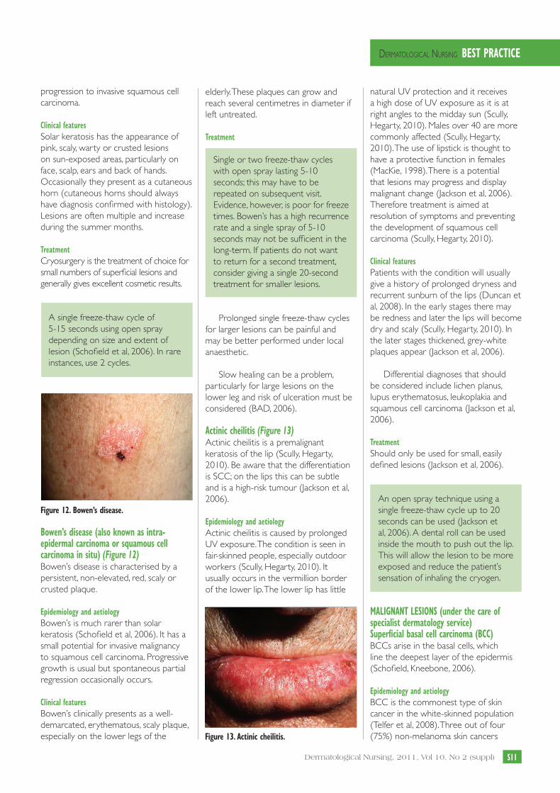
Slow healing can be a problem, particularly for large lesions on the lower leg and risk of ulceration must be considered (BAD, 2006). actinic cheilitis (Figure 13) Actinic cheilitis is a premalignant keratosis of the lip (Scully, Hegarty, 2010). Be aware that the differentiation is SCC; on the lips this can be subtle and is a high-risk tumour (Jackson et al, 2006).
epidemiology and aetiology Actinic cheilitis is caused by prolonged UV exposure. The condition is seen in fair-skinned people, especially outdoor workers (Scully, Hegarty, 2010). It usually occurs in the vermillion border of the lower lip. The lower lip has little
natural UV protection and it receives a high dose of UV exposure as it is at right angles to the midday sun (Scully, Hegarty, 2010). Males over 40 are more commonly affected (Scully, Hegarty, 2010). The use of lipstick is thought to have a protective function in females (MacKie, 1998). There is a potential that lesions may progress and display malignant change (Jackson et al, 2006). Therefore treatment is aimed at resolution of symptoms and preventing the development of squamous cell carcinoma (Scully, Hegarty, 2010). clinical features Patients with the condition will usually give a history of prolonged dryness and recurrent sunburn of the lips (Duncan et al, 2008). In the early stages there may be redness and later the lips will become dry and scaly (Scully, Hegarty, 2010). In the later stages thickened, grey-white plaques appear (Jackson et al, 2006).
Differential diagnoses that should be considered include lichen planus, lupus erythematosus, leukoplakia and squamous cell carcinoma (Jackson et al, 2006).
treatment Should only be used for small, easily defined lesions (Jackson et al, 2006).
An open spray technique using a single freeze-thaw cycle up to 20 seconds can be used (Jackson et al, 2006). A dental roll can be used inside the mouth to push out the lip. This will allow the lesion to be more exposed and reduce the patient’s sensation of inhaling the cryogen.
MaLiGnant Lesions (under the care of specialist dermatology service) superficial basal cell carcinoma (Bcc) BCCs arise in the basal cells, which line the deepest layer of the epidermis (Schofield, Kneebone, 2006).
epidemiology and aetiology BCC is the commonest type of skin cancer in the white-skinned population (Telfer et al, 2008). Three out of four (75%) non-melanoma skin cancers
Figure 12. Bowen’s disease.
Bowen’s disease (also known as intra-epidermal carcinoma or squamous cell carcinoma in situ) (Figure 12) Bowen’s disease is characterised by a persistent, non-elevated, red, scaly or crusted plaque.
epidemiology and aetiology Bowen’s is much rarer than solar keratosis (Schofield et al, 2006). It has a small potential for invasive malignancy to squamous cell carcinoma. Progressive growth is usual but spontaneous partial regression occasionally occurs.
clinical features Bowen’s clinically presents as a well-demarcated, erythematous, scaly plaque, especially on the lower legs of the Figure 13. actinic cheilitis.
Cryosurgery supp.mjjp2C.indd 11 06/06/2011 18:54

Figure 14. superficial basal cell carcinoma.
Dermatological Nursing, 2011, Vol 10, No 2 (suppl)s12
Dermatological NursiNg Best Practice
diagnosed are this type. BCCs are slow growing and rarely metastasise to vital organs, however, they can become large and sometimes cause significant local destruction and subsequent disfigurement, and some BCCs are more aggressive than others. They are commonly found in the fair-skinned older population but in recent years are seen more frequently in younger patients.
BCC sub-types are: 8 Superficial8 Nodular/cystic8 Pigmented8 Morphoeic
Main causes:8Ultraviolet radiation/sunbeds8Immunosuppression 8Fair skin and the propensity to freckle
or burn rather than tan 8Past exposure to arsenic 8Radiation damage 8Rare genetic diseases such as
xeroderma pigmentosa and Gorlin’s syndrome (Telfer et al, 2008)
clinical features BCCs can occur on any part of the body but most frequently affect areas that have been exposed to the sun, such as: the face, ears, neck, scalp, shoulders and back.
It is essential that the correct diagnosis is made prior to treatment. If in doubt, cryotherapy should not be used and a diagnostic biopsy may be necessary.
Superficial BCCs are often found on the trunk, can be single or multiple and are very different from the other types of BCC. A rolled edge may be visible at the lesion’s periphery. It is, however, easy to mistake these often erythematous lesions for eczema, psoriasis, Bowen’s disease or tinea corporis (Schofield, Kneebone, 2006).
treatment The following cryosurgery treatment schedule is only suitable for superficial BCCs.
Two freeze-thaw cycles of 25 to 30 seconds using open spray, with a thawing period of 2–4 minutes in between, depending on the size and thickness of the lesion.
caution:Long freezes can cause the patient significant discomfort and may lead to swelling, blistering and exuding wounds. Local anaesthetic may be helpful and clear aftercare instructions should be
provided, verbally and in writing. Special care must be taken if treating lesions on the face.
Practical considerations in relation to equipment All equipment used for treatment with Liquid Nitrogen must be well maintained (according to manufacturers’ manual/instructions) and inspected before use for damage of any kind. Defective and/or poorly assembled equipment can lead to an undesired thermal injury (Elton, 1983). Common equipment faults and user errors to be aware of include;
spray method 8Blocked spray attachment — can be
unblocked with a fine pin or staple. If a commonly occurring problem, it may be caused by contaminants in your liquid nitrogen storage dewar. To resolve, discuss with manufacturer of dewar and supplier of liquid nitrogen.
8Inappropriate spray attachment size.8No spray attachment — note, this
will cause serious thermal injury. It is important that you always check that the spray attachment is of the correct size and appropriately fastened.
8Escape of nitrogen from dewar — this will occur if the flask is not appropriately closed or if the flask is knocked over. In the case of the flask been knocked or inverted, the nitrogen will be seen to explosively escape. To correct, reposition the flask to its correct vertical position as soon as possible.
cotton bud method 8Bud wrong size for lesion.8Unsuitable vehicle used to store
liquid nitrogen.
Forceps method 8Forceps tip too fine for purpose.
Note, forceps tip must be able to encase lesion.
8Thermal injury to user — always ensure that forceps handle is covered by gauze.
equipment to protect unaffected skin 8Absorbent materials, such as drapes
and gauze, should not be used to protect surrounding skin as they may allow liquid nitrogen to accumulate
Cryosurgery supp.mjjp2C.indd 12 06/06/2011 18:54

Dermatological NursiNg Best Practice
s13Dermatological Nursing, 2011, Vol 10, No 2 (suppl)
at its borders or underneath causing ‘cryo’ injury, which may go unnoticed until after completion of the procedure (Elton, 1983).
8Metal as a good ‘cryo’ conductor should equally not be used.
side-effects Cryosurgery with liquid nitrogen is a safe and effective therapy in competent hands (Elton, 1983). There are, however, side-effects and potential complications that practitioners must be familiar with prior to commencing cryosurgery treatment.
Patients must always be forewarned of these side-effects and possible complications of treatment to allow informed consent to be given.
Pain A burning pain is usually felt on freezing with a more intense pain on thawing. The pain of freezing is of short duration due to the self-anaesthetising feature of the freeze and the pain generally settles within minutes after the thaw (Elton, 1983). Headache, often migraine-like, is not uncommon with freezing on the forehead, temples and scalp — this is usually transient but occasionally lasts for several hours (Jackson et al, 2006). For some patients the use of a topical anaesthetic may significantly minimise pain (Jackson et al, 2006). EMLA can be applied up to 2 hours before and Ametop (tetracaine) 30 minutes prior. For patients requiring analgesia post-treatment, simple analgesia such as paracetamol is normally recommended. In the treatment of children under the age of 12 years with multiple warts, application of EMLA cream may be considered good practice.
oedema/blister formation Oedema follows cryosurgery, and blistering may occur after longer freezes. Exaggerated oedema frequently occurs in the periorbital, forehead, temples and anterior scalp areas. In some dermatology centres, to lessen swelling, especially on the face, a single application of very potent topical steroids is applied following the freeze-thawing cycle (Hunter et al, 1995). Cool compresses may help alleviate some oedema but antihistamines do not
(Elton, 1983). Current medical practice within Dermatology and Plastic Surgery suggests blisters, if tense, should be burst with a sterile needle to let out fluid and a sterile dressing applied.
UlcerationSlow wound healing may be an issue, especially on lesions on the lower limbs in elderly patients, where circulation may be poor. The potential risk of ulceration should be considered and must be discussed with the patient (BAD, 2006).
nerve/tendon damage The most serious long-term complication of cryosurgery is nerve damage. Special care must be used in treating areas in which nerves or tendons lie superficially, including the sides of the fingers, angle of the jaw, postauricular area and the ulnar fossa of the elbow (Elton, 1983). Nerve tissue is sensitive to freezing damage.
Pigment change Pigmentary problems following cryosurgery occur much more commonly in patients with naturally deeply pigmented skin. While localised temporary depigmentation is normal, permanent pigmentary change may occur with deep freeze. Cryosurgery should be used with caution in dark-skinned patients, and the patient must be forewarned (Elton, 1983).
scarring Scarring may occur if a prolonged freeze-thaw cycle is delivered. In periungual warts the nail bed is especially at risk of scarring, causing permanent damage to the nail as it grows (Schofield et al, 2006).
infection If a blister occurs, weeping in the area is likely for the next few days. This can lead to broken skin and increased risk of infection. A smear of antiseptic cream (eg, Savlon or Germaline cream) may then be applied twice a day to reduce the very small risk of infection. An adhesive dressing such as a plaster can be used to cover the area.
Clean area daily with lukewarm water and soap. Alternatively, you may
use non-alcoholic baby wipes to gently remove any debris from the area affected during the day as this can lead to bacteria being trapped beneath the crust, leading to a higher risk of localised infection.
Urticaria Urticaria induced by cryosurgery is not documented in the literature. However, one of our authors has observed the phenomenon. In this author’s experience the urticarial response occurs minutes after treatment, usually following the spray technique. The patient may not be aware of the reaction as it usually settles quickly or will think that it is a cryosurgery-induced blister. Subsequently the patient may experience a more vigorous blistering reaction than expected. Freezing cycles in patients where this phenomenon is observed should be reduced or alternatively the cotton bud method can be used
safety aspects The storage and handling of liquid nitrogen is subject to Control of Substances Hazardous to Health (COSHH) regulations. These are generic for hazardous substances but can be applied specifically to liquid nitrogen. A risk assessment must be completed.
Properties and hazards Liquid nitrogen is pure nitrogen in a liquid state, produced industrially in large quantities by the process of air separation. It is a colourless, odourless, non-toxic, inert gas with a boiling point of -196°C. As a cryogenic fluid it causes rapid frostbite on contact with the skin, producing a destructive effect on the tissue.
Hazards include:8Asphyxiation — This is one of the
main dangers associated with liquid nitrogen, especially in relation to storage in poorly ventilated areas. Liquid nitrogen should always be stored in a well-ventilated room in an appropriate dewar (BOC, 2004). The dewar should be kept in good condition and have provision for venting the gas that boils off from the liquid (Jackson et al, 2006). Where
Cryosurgery supp.mjjp2C.indd 13 06/06/2011 18:54

Dermatological Nursing, 2011, Vol 10, No 2 (suppl)s14
Dermatological NursiNg Best Practice
large quantities of liquid nitrogen are stored, the use of an oxygen monitor and alarm should be considered. Liquid nitrogen should never be transported inside a passenger vehicle as it poses a serious risk to life (Jackson et al, 2006).
8Cryogenic burns and frost bite — liquid nitrogen can cause cryogenic burns and frost bite when the substance itself or surfaces that have been in contact with it (eg, metal surfaces) come in contact with the skin.
8Hypothermia — susceptibility to hypothermia is dependant on temperature, exposure to liquid nitrogen and the general health and age of the individual concerned. Its occurrence is greater where there is a large volume of liquid nitrogen storage.
Precautions Storage — Liquid nitrogen should always be stored in a well ventilated room. Personal protective equipment — should be available and be used when decanting liquid nitrogen. When decanting liquid nitrogen, there is a risk of cryogenic burns from a liquid nitrogen splash. To protect the user, non-absorbent insulated gloves and a full face visor should be worn. Open-toed shoes should not be worn when pouring liquid nitrogen as there is a high risk of a liquid nitrogen splash to the feet. Transportation — If liquid nitrogen is to be transported in a vehicle, the driver must be aware of potential hazards, especially asphyxiation, and know what to do in the event of an accident or emergency (BOC, 2004).
Liquid nitrogen should never be transported in the passenger compartment. It should only be transported where the load space is separated from the driver and passenger compartment.
Liquid nitrogen containers should be transported in a secure upright position in a well-ventilated area (BOC, 2004).
emergency action and procedures Emergency procedures should be in place and staff trained on the risks of cold burns, frostbite, asphyxiation and hypothermia. Hazard warning signs are displayed and comply with the Health and Safety Regulations 1996 and BS5378.
First aid measures for: Inhalation — Remove victim to uncontaminated area. Rescuers should not put themselves at risk — a contaminated area should not be entered unless considered safe. Breathing apparatus may be used by trained personnel. Keep victim warm
Figure 15. Methods of transportation.
Cryosurgery supp.mjjp2C.indd 14 06/06/2011 18:54

Dermatological NursiNg Best Practice
s15Dermatological Nursing, 2011, Vol 10, No 2 (suppl)
and rested until medical attention is obtained. If breathing stopped, commence artificial resuscitation (BOC, 2004).Skin/Eye Contact — Remove any clothing that may constrict the circulation to the frozen area (BOC, 2002). As soon as possible, the affected area should be immersed in tepid water (42-45oC) for at least 15 minutes (BOC, 2002). The aim of treatment is to warm the area slowly; do not use dry heat as this will superimpose a burn on frozen tissue. When affected area has thawed, cover with a dry, sterile dressing and seek medical attention.
In the event of eye involvement, immediately flush eyes thoroughly for at least 15 minutes (BOC, 2004).
Medicolegal aspects Accountability requires each registered practitioner to explain and justify his/her actions and clinical decisions. This means that practitioners are answerable for actions and omissions or departure from good professional practice, regardless of advice or direction from another professional (NMC, 2008).
In law, there are four areas whereby the practitioner may be called to account for his/her actions and decisions. These include:8Accountability via civil Law8Accountability via criminal law8Professional accountability to NMC8Accountability to employer
The registered practitioner must be fully cognisant with his/her legal responsibilities in relation to their role and the duty of care owed to his/her patients.
The new Code introduced in May 2008 offers nurses the opportunity to develop their role to deliver effective care that is responsive to the needs of the patient. As the role of the nurse evolves, so too must the education and clinical support required to prepare practitioners to acquire skills, competencies, knowledge and assume responsibility to manage advances in modern health care.
referencesAndrewsMD(2004)Cryosurgeryforcommonskinconditions.Am Fam Phys69(10):2365-72
AshtonR,LeppardB(2005)Differential Diagnosis in Dermatology.3rdEdition.RadcliffePublishingLtd.Oxon
BritishAssociationofDermatologists(2006)GuidelinesformanagementofBowen’sdisease:2006update.NHCox,DJEedy,CAMortononbehalfoftheBritishAssocationofDermatologistsTherapyGuidelinesandAuditSubcommittee.Br J Dermatol(2007)156:11-21
BritishAssociationofDermatologists(2008)SeborrhoeicWarts[online]BritishAssociationofDermatologists.Availableathttp://www.bad.org.uk/site/873/Default.aspx(accessed24.10.2010)
BritishAssociationofDermatologists(2008)MolluscumContagiosum[online]BritishAssociationofDermatologists.Availableathttp://www.bad.org.uk/site/845/Default.aspx(accessed10.12.2010)
BritishAssociationofDermatologists(2007)Dermatofibroma[online]BritishAssociationofDermatologists.Availableathttp://www.bad.org.uk/site/809/Default.aspx(accessed10.12.2010)
BOC(2002)Exposuretoverycoldliquefiedgases[online]BOC.Availableathttp://www.boc.ebcnet.co.uk/p/liquefiedGases-exposure.pdf(accessed28.11.2010)
BOC(2004)SafetyDataSheetLiquidNitrogen[online]BOC.Availableathttp://www.bocsds.com/uk/sds/industrial/Nitrogen_liquid.pdf(Accessed28.11.2010)
DawberR,ColverG,JacksonA,PringleF(2007)Cutaneous Cryosurgery.2ndEdition.MartinDunitz.London
DermnetNZ(2010)SkinTags[online]NewZealandDermatologicalSocietyIncorporated.Availableathttp://www.dermnetnz.org/lesions/skin-tags.html(accessed25.10.2010)
DermnetNZ(2010)SebaceousHyperplasia[online]NewZealandDermatologicalSocietyIncorporated.Availableathttp://www.dermnetnz.org/acne/sebaceous-hyperplasia.html(accessed2.10.2010)
DuncanKO,GeisseJK,Leffell,DJ(2008)Epithelialprecancerouslesions.In:WolfK,GoldsmithLA,KatzSI,GilchrestBA,PallerAS,Leffell,DJ(eds.)Fitzpatrick’s Dermatology in General Medicine,7thEdition,McGrawHill,NewYork
EltonRF(1983)Complicationsofcutaneouscryosurgery.J Am Acad Dermatol 8:513-19
FitzpatrickTB,JohnsonRA,WolffK,SuurmondD(2001)Color Atlas & Synopsis
of Clinical Dermatology.4thEdition.McGrawHill,NewYork.
GibbsS,HarveyI,SterlingJ,StarkR(2002)LocalTreatmentsforCutaneousWarts:SystemicReview.BMJ 325:461doi:10.1136/bmj.325.7362.461(Published31.8.2002)
Graham-BrownR,BourkeJF(1998)Mosby’s Color Atlas and Text of Dermatology.Mosby,London
HunterJAA,SavinJA,DahlMV(1995)Clinical Dermatology.2ndEdition.BlackwellScience,London
JacksonA,ColverG,DawberR(2006)Cutaneous Cryosurgery: Principles and Clinical Practice. 3rdEdition.Taylor&Francis,London
KuflikEG(1994)CryosurgeryUpdated. J Am Acad Dermatol31:925-44
LubaMC,BangsSA,MohlerA,StulbergD(2005)Commonbenignskintumours.Am Fam Phys67(4):729-738
MacKieR(1998)EpidermalSkinTumours.In:ChampionRH,BurtonJL,BurnsDA,BreathnachSM(eds.)Rook/Wilkinson/Ebling Textbook of Dermatology,6thEdition.BlackwellScience.London
MarksR(1999)Practical Problems in Dermatology,inPracticalProblemsinMedicineseries
NursingandMidwiferyCouncil(2008)TheCode:Standardsofconduct,performanceandethicsfornursesandmidwives.NMC,London
RudlingerR,SmithIW,etal(1986)Humanpapillomavirusinfectionsinagroupofrenaltransplantrecipients.Br J Dermatol115(6): 681-692
SchofieldJ,KneeboneR(2006)Skin Lesions. A practical guide to diagnosis, management and minor surgery.2ndEdition.SchofieldandKneebone,Rickmansworth
ScullyC,HegartyA(2010)TheOralCavityandLips.In:BurnsTBreathnachS,CoxN,GriffithsC(eds)Rook’s Textbook of Dermatology,8thEdition.Wiley-Blackwell,London
SterlingJC,Handfield-JonesS,HudsonPM(2001)Guidelinesforthemanagementofcutaneouswarts.Br J Dermatol 144(1): 4-11
TelferNR,ColverGB,MortonCA(2008)GuidelinesfortheManagementofBasalCellCarcinomas.Br J Dermatol159(1):35-48
ThomasVD,SwansonNA,LeeKK(2008)Benignepithelialtumors,hamartomasandhyperplasias.In:WolffK,GoldsmithLA,KatzSI,Gilchrest,BA,PallerAS,LeffellDJ(eds.)Fitzpatrick’s Dermatology in General Medicine,7thEdition,McGrawHill,NewYorkDN
Cryosurgery supp.mjjp2C.indd 15 06/06/2011 18:54

appendix 1
s16 Dermatological Nursing, 2011, Vol 10, No 2 (suppl)
Dermatological NursiNg Best Practice
assess competency according to WasP Framework.
W
WITNESSED Observe or witness the competency — it is considered good practice that the nurse will have had the opportunity to observe the procedure prior to being supervised.
A ASSIMILATED Understand the elements of the competency.
S SUPERVISED Practice under supervision to demonstrate understanding: score as follows:1 = NEEDS FURTHER PRACTICE2 = SHOWS APTITUDE3 = PROFICIENT
P PROFICIENT Competent in both knowledge and skill elements of the competency.
ACTION RATIONALE W A S SCORE
P
The practitioner is able to demonstrate practical and, theoretical knowledge of cryosurgery and indications for use.
KSF: C2, C4, C5
To ensure the practitioner has appropriate knowledge and skills to undertake the treatment.
The practitioner is able to prepare the clinical area according to local health and safety policies.
KSF: C3, C5
To ensure the health and safety of the patient and the person undertaking the procedure.
The practitioner is able to demonstrate knowledge of safety in the care of patients undergoing cryosurgery and is aware of the COSHH safety regulations with regard to storage, transportation and use of liquid nitrogen.
KSF: C3
Liquid nitrogen must be used in accordance with the control of substances hazardous to health policy. A risk assessment for the use, storage, disposal and emergency measures should be carried out in accordance with local policy.
The practitioner is able to assess the patient’s suitability for treatment.
KSF: C1, HWB5, HWB6, HWB7
To ensure the patient is able to cope with cryosurgery in terms of understanding due to age or mental capacity, and ability to tolerate the treatment.
The practitioner is able to locate and assess the lesion(s) to be treated as per referral letter or clinical notes. He/she recognises lesions unsuitable for cryosurgery or requiring medical review.
KSF: C6, HWB6, HWB7
Important to have knowledge about the visual and dermatoscopic (if available) features of lesions suitable for treatment with cryosurgery to ensure correct lesion/s are treated.Inappropriate cryosurgery can damage underlying structures and alter the appearance of lesions, causing diagnostic difficulty in future.
The practitioner is able to demonstrate adequate explanation to the patient of procedure and treatment expectations.
KSF: C1, C5, HWB5, HWB6, HWB7
The NMC code of conduct states ‘The people in your care must be able to trust you with their health and well-being’.
Cryosurgery supp.mjjp2C.indd 16 06/06/2011 18:54

appendix 1 (cont’d)
Dermatological NursiNg Best Practice
s17Dermatological Nursing, 2011, Vol 10, No 2 (suppl)
W
WITNESSED Observe or witness the competency — it is considered good practice that the nurse will have had the opportunity to observe the procedure prior to being supervised.
A ASSIMILATED Understand the elements of the competency.
S SUPERVISED Practice under supervision to demonstrate understanding: score as follows:1 = NEEDS FURTHER PRACTICE2 = SHOWS APTITUDE3 = PROFICIENT
P PROFICIENT Competent in both knowledge and skill elements of the competency.
ACTION RATIONALE W A S SCORE
P
The practitioner is able to demonstrate practical and, theoretical knowledge of cryosurgery and indications for use.
KSF: C2, C4, C5
To ensure the practitioner has appropriate knowledge and skills to undertake the treatment.
The practitioner is able to prepare the clinical area according to local health and safety policies.
KSF: C3, C5
To ensure the health and safety of the patient and the person undertaking the procedure.
The practitioner is able to demonstrate knowledge of safety in the care of patients undergoing cryosurgery and is aware of the COSHH safety regulations with regard to storage, transportation and use of liquid nitrogen.
KSF: C3
Liquid nitrogen must be used in accordance with the control of substances hazardous to health policy. A risk assessment for the use, storage, disposal and emergency measures should be carried out in accordance with local policy.
The practitioner is able to assess the patient’s suitability for treatment.
KSF: C1, HWB5, HWB6, HWB7
To ensure the patient is able to cope with cryosurgery in terms of understanding due to age or mental capacity, and ability to tolerate the treatment.
The practitioner is able to locate and assess the lesion(s) to be treated as per referral letter or clinical notes. He/she recognises lesions unsuitable for cryosurgery or requiring medical review.
KSF: C6, HWB6, HWB7
Important to have knowledge about the visual and dermatoscopic (if available) features of lesions suitable for treatment with cryosurgery to ensure correct lesion/s are treated.Inappropriate cryosurgery can damage underlying structures and alter the appearance of lesions, causing diagnostic difficulty in future.
The practitioner is able to demonstrate adequate explanation to the patient of procedure and treatment expectations.
KSF: C1, C5, HWB5, HWB6, HWB7
The NMC code of conduct states ‘The people in your care must be able to trust you with their health and well-being’.
The practitioner understands indications and contra-indications of cryosurgery and obtains written/verbal consent prior to treatment and checks known allergies.
KSF: C1, C3, HWB5, HWB6, HWB7
To ensure a high standard of practice and care at all times.Patients must be given enough information to enable them to make a decision regarding their treatment.Consent must be under the free will of the patient and not influenced by others.The patient has the right to refuse treatment.Consent must be obtained in accordance with local policy.
The practitioner is able to select and prepare the appropriate equipment for the procedure. KSF: C3, HWB6, HWB7
Regard for patient safety and comfort.Commitment to delivering high quality of care and service delivery.
The practitioner is able to prepare lesions and administer liquid nitrogen treatment (as per clinic protocol) and select an appropriate dressing if necessary.KSF: HWB7
Regard for patient safety and comfort.Commitment to delivering high quality of care and service delivery.
The practitioner is able to state the possible adverse effects of cryosurgery and management options.KSF: C3, HWB7
Cryosurgery can be a painful experience for patients and there is a risk of common side-effects, such as blistering and infection. Practitioners must be able to manage complications effectively.
The practitioner is able to arrange further appointments if appropriate and in accordance with local protocol.KSF: HWB7
It is recommended that warts are treated every 2–3 weeks.
Other lesions should be reviewed 6 weeks after treatment or at practitioner’s discretion. Lesions that do not respond to treatment should be reviewed by a dermatologist.
The practitioner is able to accurately document the treatment in the patient’s record in accordance with local documentation and record keeping policies.KSF:C1, C5, HWB6, HWB7
Patient records should be factual, consistent and accurate.
The practitioner is actively involved in cryosurgery data collection.KSF: C5, HWB7, IK2
To be able to contribute to the improvement, transformation and innovation of services.To monitor quality and efficacy of service.
DATE
ASSESSOR SIGNATURE
STAFF MEMBER SIGNATURE
Cryosurgery supp.mjjp2C.indd 17 06/06/2011 18:54

Dermatological Nursing, 2011, Vol 10, No 2 (suppl)s18
Dermatological NursiNg Best Practice
Methods for removal of keratin.
appendix 2
Using scalpel (suitable for warts):
1 Use a sterile, small, curved scalpel (size 15).2 Holding the scalpel parallel to the lesion, gently remove thin layers of
hard skin (keratin) by pulling the scalpel across the lesion.3 Continue paring until blood vessels become visible (these will appear
as small dark dots or may bleed) or if the patient experiences any discomfort.
4 Keep your non-dominant hand away from the scalpel blade in case it slips (not as shown).
NB. Paring is a useful method of distinguishing warts from corns. Warts have bleeding points, whereas corns have a central hard core of keratin without any blood vessels.
Using forceps and an emollient (suitable for actinic keratoses):1 Soak lesion with an ointment/oil-based emollient.2 Leave for 5-10 minutes, or longer if required.3 Gently lift keratin with forceps or gauze.
Patients should be encouraged to file warts prior to clinic appointment.
Figure 16. removal of keratin.
Cryosurgery supp.mjjp2C.indd 18 06/06/2011 18:54
Dermatological Nursing, 2011, Vol 10, No 2 (suppl) s19
Dermatological NursiNg Best Practice
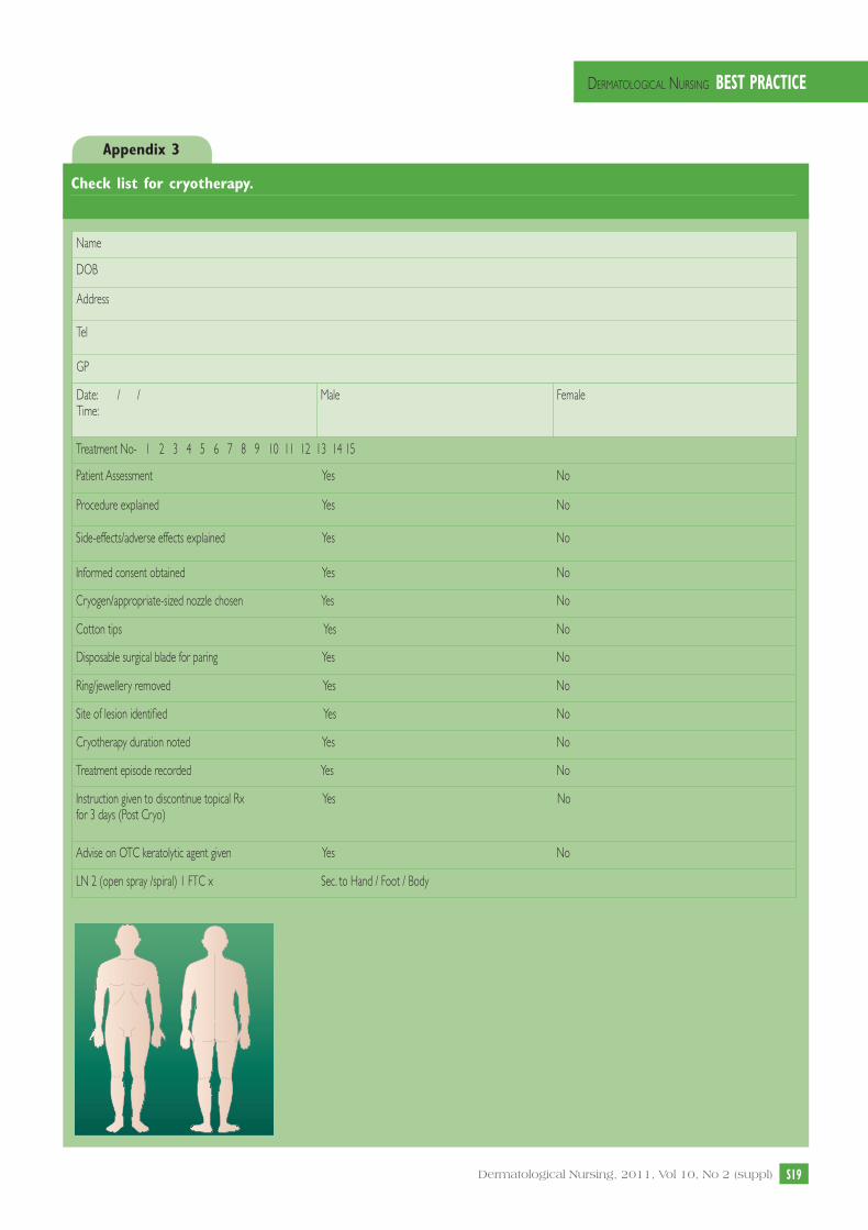
check list for cryotherapy.
appendix 3
Name
DOB
Address
Tel
GP
Date: / / Time:
Male Female
Treatment No- 1 2 3 4 5 6 7 8 9 10 11 12 13 14 15
Patient Assessment Yes No
Procedure explained Yes No
Side-effects/adverse effects explained Yes No
Informed consent obtained Yes No
Cryogen/appropriate-sized nozzle chosen Yes No
Cotton tips Yes No
Disposable surgical blade for paring Yes No
Ring/jewellery removed Yes No
Site of lesion identified Yes No
Cryotherapy duration noted Yes No
Treatment episode recorded Yes No
Instruction given to discontinue topical Rx Yes Nofor 3 days (Post Cryo)
Advise on OTC keratolytic agent given Yes No
LN 2 (open spray /spiral) 1 FTC x Sec. to Hand / Foot / Body
Cryosurgery supp.mjjp2C.indd 19 06/06/2011 18:54

BDNG
21 Tower Street, London WC2H 9NS
Tel: 020 7836 0022
www.bdng.org.uk
Cryosurgery supp.mjjp2C.indd 20 06/06/2011 18:54


![Cryosurgery in the treatment of oro-facial lesionspain. This clinical application of cryosurgery is known as.[8] Cryoneurotomy is also used for the treatment of intractable neurogenic](https://img.pdfslide.us/doc/110x75/5f02d6327e708231d406414e/cryosurgery-in-the-treatment-of-oro-facial-lesions-pain-this-clinical-application.jpg)