Embed Size (px)
Citation preview
3 BEST PRACTICE GUIDELINES FOR SKIN AND WOUND CARE IN EPIDERMOLYSIS BULLOSA
BEST PRACTICE GUIDELINES: WOUND MANAGEMENT IN DIABETIC FOOT ULCERS
I N T E R N A T I O N A L B E S T P R A C T I C E
3 BEST PRACTICE GUIDELINES FOR SKIN AND WOUND CARE IN EPIDERMOLYSIS BULLOSAC BEST PRACTICE GUIDELINES: WOUND MANAGEMENT IN DIABETIC FOOT ULCERS
Supported by an educational grant from B Braun
The views presented in this document are the work of the authors and do not necessarily reflect the opinions of B Braun.
Published byWounds InternationalA division of Schofield Healthcare Media LimitedEnterprise House1–2 Hatfields London SE1 9PG, UKwww.woundsinternational.com
To cite this document. International Best Practice Guidelines: Wound Manage-ment in Diabetic Foot Ulcers. Wounds International, 2013. Available from: www.woundsinternational.com
FOREWORD
This document focuses on wound management best practice for diabetic foot ulcers (DFUs). It aims to offer specialists and non-specialists everywhere a practical, relevant clinical guide to appropriate decision making and effec-tive wound healing in people presenting with a DFU.
In recognition of the gap in the literature in the field of wound manage-ment, this document concentrates on the importance of wound assessment, debridement and cleansing, recognition and treatment of infection and appropriate dressing selection to achieve optimal healing for patients. How-ever, it acknowledges that healing of the ulcer is only one aspect of manage-ment and the role of diabetic control, offloading strategies and an integrated wound care approach to DFU management (which are all covered exten-sively elsewhere) are also addressed. Prevention of DFUs is not discussed in this document.
The scope of the many local and international guidelines on managing DFUs is limited by the lack of high-quality research. This document aims to go further than existing guidance by drawing, in addition, from the wide-ranging experience of an extensive international panel of expert practitioners. How-ever, it is not intended to represent a consensus, but rather a best practice guide that can be tailored to the individual needs and limitations of different healthcare systems and to suit regional practice.
EXPERT WORKING GROUPDevelopment group Paul Chadwick, Principal Podiatrist, Salford Royal Foundation Trust, UK Michael Edmonds, Professor of Diabetes and Endocrinology, Diabetic Foot Clinic, King's College Hospital, London, UKJoanne McCardle, Advanced Clinical and Research Diabetes Podiatrist, NHS Lothian University Hospital, Edinburgh, UKDavid Armstrong, Professor of Surgery and Director, Southern Arizona Limb Salvage Alliance (SALSA), University of Arizona College of Medicine, Arizona, USA
Review groupJan Apelqvist, Senior Consultant, Department of Endocrinology, Skåne University Hospital, Malmo, SwedenMariam Botros, Director, Diabetic Foot Canada, Canadian Wound Care Association and Clinical Coordinator, Women's College Wound Healing Clinic, Toronto, CanadaGiacomo Clerici, Chief Diabetic Foot Clinic, IRCC Casa di Cura Multimedica, Milan, ItalyJill Cundell, Lecturer/Practitioner, University of Ulster, Belfast Health and Social Care Trust, Northern IrelandSolange Ehrler, Functional Rehabilitation Department, IUR Clémenceau (Institut Universitaire de Réadaptation Clémenceau), Strasbourg, FranceMichael Hummel, MD, Diabetes Center Rosenheim & Institute of Diabetes Research, Helmholtz Zentrum München, GermanyBenjamin A Lipsky, Emeritus Professor of Medicine, University of Washington, USA; Visiting Professor, Infectious Diseases, University of Geneva, Switzerland; Teaching Associate, University of Oxford and Deputy Director, Graduate Entry Course, University of Oxford Medical School, UKJosé Luis Lázaro Martinez, Full Time Professor, Diabetic Foot Unit, Complutense University, Madrid, SpainRosalyn Thomas, Deputy Head of Podiatry, Abertawe Bro Morgannwg University Health Board, Swansea, WalesSusan Tulley, Senior Podiatrist, Mafraq Hospital, Abu Dhabi, United Arab Emirates

BEST PRACTICE GUIDELINES: WOUND MANAGEMENT IN DIABETIC FOOT ULCERS 1
INTRODUCTION
Introduction
DFUs are complex, chronic wounds, which have a major long-term impact on the morbidity, mortality and quality of patients’ lives1,2. Individuals who develop a DFU are at greater risk of premature death, myocardial infarction and fatal stroke than those without a history of DFU3. Unlike other chronic wounds, the development and progression of a DFU is often complicated by wide-ranging diabetic changes, such as neuropathy and vascular disease. These, along with the altered neutrophil function, diminished tissue perfusion and defective protein synthesis that frequently accompany diabetes, present practitioners with specific and unique man-agement challenges1.
DFUs are relatively common — in the UK, 5–7% of people with diabetes currently have or have had a DFU4,5. Furthermore, around 25% of people with diabetes will develop a DFU during their lifetime6. Globally, around 370 million people have diabetes and this number is increasing in every country7. Dia-betes UK estimates that by 2030 some 552 million people worldwide will have diabetes8.
DFUs have a major economic impact. A US study in 1999 estimated the average out-patient cost of treating one DFU episode as $28,000 USD over a two–year period9. Aver-age inpatient costs for lower limb complica-tions in 1997 were reported as $16,580 USD for DFUs, $25,241 USD for toe or toe plus other distal amputations and $31,436 USD for major amputations10,11.
The EURODIALE study examined total direct and indirect costs for one year across several European countries. Average total costs based on 821 patients were approximately 10,000 euros, with hospitalisation represent-ing the highest direct cost. Based on preva-lence data for Europe, they estimated that costs associated with treatment of DFUs may be as high as 10 billion euros per year12. In England, foot complications account for 20% of the total National Health Service spend on diabetes care, which equates to around £650 million per year (or £1 in every £150)5. Of course, these figures do not take account of the indirect costs to patients,
such as the effect on physical, psychological and social wellbeing and the fact that many patients are unable to work long term as a result of their wounds6.
A DFU is a pivotal event in the life of a person with diabetes and a marker of serious disease and comorbidities. Without early and optimal intervention, the wound can rapidly deteriorate, leading to amputation of the affected limb5,13.
It has been estimated that every 20 seconds a lower limb is amputated due to complica-tions of diabetes14.
In Europe, the annual amputation rate for people with diabetes has been cited as 0.5-0.8%1,15, and in the US it has been reported that around 85% of lower-extremity amputations due to diabetes begin with foot ulceration16,17.
Mortality following amputation increases with level of amputation18 and ranges from 50–68% at five years, which is comparable or worse than for most malignancies13,19 (Figure 1).
The statistics need not make for such grim reading. With appropriate and careful management it is possible to delay or avoid most serious complications of DFUs1.
Prostate
cance
r
Breast
cance
r
Hodgkin's
lymphoma
Neuropath
ic DFU
Amputation
Colon cance
r
Ischae
mic DFU
Periphera
l arte
rial d
iseas
e
Lung ca
ncer
Pancre
atic c
ance
r
FIGURE 1: Relative five-year mortality (%) (adapted from19)
3 BEST PRACTICE GUIDELINES FOR SKIN AND WOUND CARE IN EPIDERMOLYSIS BULLOSA2 BEST PRACTICE GUIDELINES: WOUND MANAGEMENT IN DIABETIC FOOT ULCERS
It has been suggested that up to 85% of amputations can be avoided when an effec-tive care plan is adopted20. Unfortunately, insufficient training, suboptimal assessment and treatment methods, failure to refer patients appropriately and poor access to spe-cialist footcare teams hinder the prospects of achieving optimal outcomes21,22.
Successful diagnosis and treatment of patients with DFUs involves a holistic approach that includes:
Optimal diabetes control Effective local wound care Infection control
Pressure relieving strategiesRestoring pulsatile blood flow.
Many studies have shown that planned in-tervention aimed at healing of DFUs is most effective in the context of a multidisciplinary team with the patient at the centre of this care.
One of the key tenets underpinning this document is that infection is a major threat to DFUs — much more so than to wounds
of other aetiologies not subject to diabetic changes. A European-wide study found that 58% of patients attending a foot clinic with a new ulcer had a clinically infected wound23. Similarly a single-centre US study found that about 56% of DFUs were clinically infected24. This study also showed the risk of hospitalisa-tion and lower-extremity amputation to be 56–155 times greater for diabetes patients with a foot infection than those without24.
Recognising the importance of starting treat-ment early may allow practitioners to prevent progression to severe and limb-threatening infection and potentially halt the inevitable pathway to amputation25.
This document offers a global wound care plan for practitioners (page 20), which includes a series of steps for preventing complications through active management — namely prompt and appropriate treatment of infection, referral to a vascular specialist to manage ischaemia and optimal wound care. This should be combined with appropriate patient education and an integrated approach to care.
INTRODUCTION
BEST PRACTICE GUIDELINES: WOUND MANAGEMENT IN DIABETIC FOOT ULCERS 3
AETIOLOGY OF DFUs
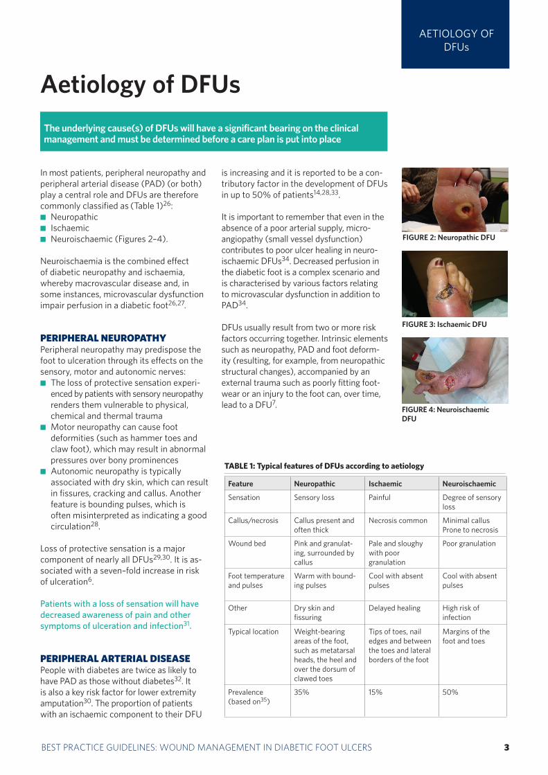
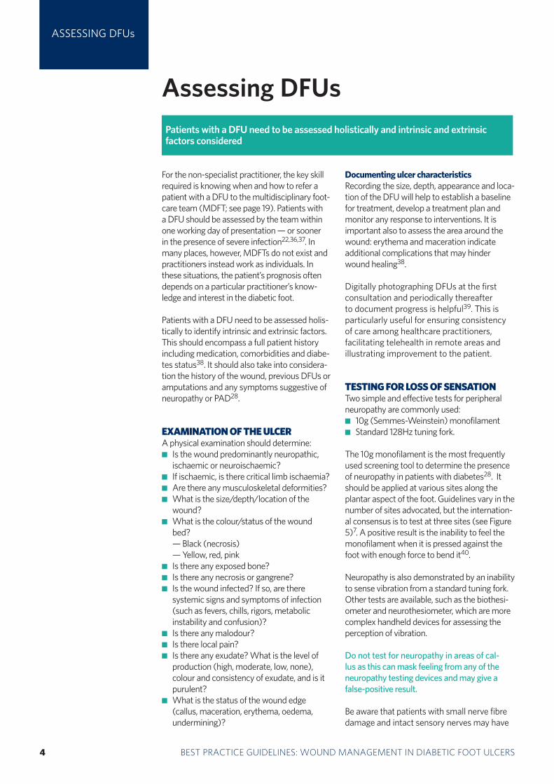
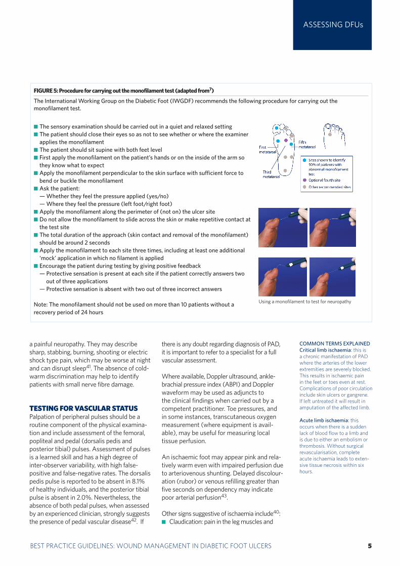
In most patients, peripheral neuropathy and peripheral arterial disease (PAD) (or both) play a central role and DFUs are therefore commonly classified as (Table 1)26:
Neuropathic Ischaemic Neuroischaemic (Figures 2–4).
Neuroischaemia is the combined effect of diabetic neuropathy and ischaemia, whereby macrovascular disease and, in some instances, microvascular dysfunction impair perfusion in a diabetic foot26,27.
PERIPHERAL NEUROPATHYPeripheral neuropathy may predispose the foot to ulceration through its effects on the sensory, motor and autonomic nerves:
The loss of protective sensation experi-enced by patients with sensory neuropathy renders them vulnerable to physical, chemical and thermal trauma
Motor neuropathy can cause foot deformities (such as hammer toes and claw foot), which may result in abnormal pressures over bony prominences
Autonomic neuropathy is typically associated with dry skin, which can result in fissures, cracking and callus. Another feature is bounding pulses, which is often misinterpreted as indicating a good circulation28.
Loss of protective sensation is a major component of nearly all DFUs29,30. It is as-sociated with a seven–fold increase in risk of ulceration6.
Patients with a loss of sensation will have decreased awareness of pain and other symptoms of ulceration and infection31.
PERIPHERAL ARTERIAL DISEASEPeople with diabetes are twice as likely to have PAD as those without diabetes32. It is also a key risk factor for lower extremity amputation30. The proportion of patients with an ischaemic component to their DFU
is increasing and it is reported to be a con-tributory factor in the development of DFUs in up to 50% of patients14,28,33.
It is important to remember that even in the absence of a poor arterial supply, micro- angiopathy (small vessel dysfunction) contributes to poor ulcer healing in neuro-ischaemic DFUs34. Decreased perfusion in the diabetic foot is a complex scenario and is characterised by various factors relating to microvascular dysfunction in addition to PAD34.
DFUs usually result from two or more risk factors occurring together. Intrinsic elements such as neuropathy, PAD and foot deform-ity (resulting, for example, from neuropathic structural changes), accompanied by an external trauma such as poorly fitting foot-wear or an injury to the foot can, over time, lead to a DFU7.
Aetiology of DFUsThe underlying cause(s) of DFUs will have a significant bearing on the clinical management and must be determined before a care plan is put into place
TABLE 1: Typical features of DFUs according to aetiology
Feature Neuropathic Ischaemic Neuroischaemic
Sensation Sensory loss Painful Degree of sensory loss
Callus/necrosis Callus present and often thick
Necrosis common Minimal callusProne to necrosis
Wound bed Pink and granulat-ing, surrounded by callus
Pale and sloughy with poor granulation
Poor granulation
Foot temperature and pulses
Warm with bound-ing pulses
Cool with absent pulses
Cool with absent pulses
Other Dry skin and fissuring
Delayed healing High risk of infection
Typical location Weight-bearing areas of the foot, such as metatarsal heads, the heel and over the dorsum of clawed toes
Tips of toes, nail edges and between the toes and lateral borders of the foot
Margins of the foot and toes
Prevalence (based on35)
35% 15% 50%
FIGURE 2: Neuropathic DFU
FIGURE 3: Ischaemic DFU
FIGURE 4: Neuroischaemic DFU
3 BEST PRACTICE GUIDELINES FOR SKIN AND WOUND CARE IN EPIDERMOLYSIS BULLOSA4 BEST PRACTICE GUIDELINES: WOUND MANAGEMENT IN DIABETIC FOOT ULCERS
ASSESSING DFUs
Assessing DFUsPatients with a DFU need to be assessed holistically and intrinsic and extrinsic factors considered
For the non-specialist practitioner, the key skill required is knowing when and how to refer a patient with a DFU to the multidisciplinary foot-care team (MDFT; see page 19). Patients with a DFU should be assessed by the team within one working day of presentation — or sooner in the presence of severe infection22,36,37. In many places, however, MDFTs do not exist and practitioners instead work as individuals. In these situations, the patient’s prognosis often depends on a particular practitioner’s know-ledge and interest in the diabetic foot.
Patients with a DFU need to be assessed holis-tically to identify intrinsic and extrinsic factors. This should encompass a full patient history including medication, comorbidities and diabe-tes status38. It should also take into considera-tion the history of the wound, previous DFUs or amputations and any symptoms suggestive of neuropathy or PAD28.
EXAMINATION OF THE ULCERA physical examination should determine:
Is the wound predominantly neuropathic, ischaemic or neuroischaemic?If ischaemic, is there critical limb ischaemia?Are there any musculoskeletal deformities?What is the size/depth/location of the wound?What is the colour/status of the wound bed?
— Black (necrosis) — Yellow, red, pink
Is there any exposed bone? Is there any necrosis or gangrene? Is the wound infected? If so, are there
systemic signs and symptoms of infection (such as fevers, chills, rigors, metabolic instability and confusion)?
Is there any malodour? Is there local pain? Is there any exudate? What is the level of
production (high, moderate, low, none), colour and consistency of exudate, and is it purulent? What is the status of the wound edge (callus, maceration, erythema, oedema, undermining)?
Documenting ulcer characteristicsRecording the size, depth, appearance and loca-tion of the DFU will help to establish a baseline for treatment, develop a treatment plan and monitor any response to interventions. It is important also to assess the area around the wound: erythema and maceration indicate additional complications that may hinder wound healing38.
Digitally photographing DFUs at the first consultation and periodically thereafter to document progress is helpful39. This is particularly useful for ensuring consistency of care among healthcare practitioners, facilitating telehealth in remote areas and illustrating improvement to the patient.
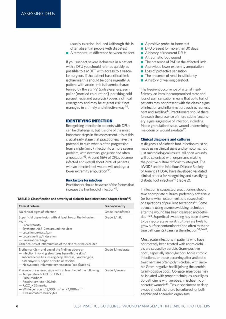
TESTING FOR LOSS OF SENSATIONTwo simple and effective tests for peripheral neuropathy are commonly used:
10g (Semmes-Weinstein) monofilament Standard 128Hz tuning fork.
The 10g monofilament is the most frequently used screening tool to determine the presence of neuropathy in patients with diabetes28. It should be applied at various sites along the plantar aspect of the foot. Guidelines vary in the number of sites advocated, but the internation-al consensus is to test at three sites (see Figure 5)7. A positive result is the inability to feel the monofilament when it is pressed against the foot with enough force to bend it40.
Neuropathy is also demonstrated by an inability to sense vibration from a standard tuning fork. Other tests are available, such as the biothesi-ometer and neurothesiometer, which are more complex handheld devices for assessing the perception of vibration.
Do not test for neuropathy in areas of cal-lus as this can mask feeling from any of the neuropathy testing devices and may give a false-positive result.
Be aware that patients with small nerve fibre damage and intact sensory nerves may have
BEST PRACTICE GUIDELINES: WOUND MANAGEMENT IN DIABETIC FOOT ULCERS 5
ASSESSING DFUs
a painful neuropathy. They may describe sharp, stabbing, burning, shooting or electric shock type pain, which may be worse at night and can disrupt sleep41. The absence of cold-warm discrimination may help to identify patients with small nerve fibre damage.
TESTING FOR VASCULAR STATUS Palpation of peripheral pulses should be a routine component of the physical examina-tion and include assessment of the femoral, popliteal and pedal (dorsalis pedis and posterior tibial) pulses. Assessment of pulses is a learned skill and has a high degree of inter-observer variability, with high false-positive and false-negative rates. The dorsalis pedis pulse is reported to be absent in 8.1% of healthy individuals, and the posterior tibial pulse is absent in 2.0%. Nevertheless, the absence of both pedal pulses, when assessed by an experienced clinician, strongly suggests the presence of pedal vascular disease42. If
there is any doubt regarding diagnosis of PAD, it is important to refer to a specialist for a full vascular assessment.
Where available, Doppler ultrasound, ankle-brachial pressure index (ABPI) and Doppler waveform may be used as adjuncts to the clinical findings when carried out by a competent practitioner. Toe pressures, and in some instances, transcutaneous oxygen measurement (where equipment is avail-able), may be useful for measuring local tissue perfusion.
An ischaemic foot may appear pink and rela-tively warm even with impaired perfusion due to arteriovenous shunting. Delayed discolour-ation (rubor) or venous refilling greater than five seconds on dependency may indicate poor arterial perfusion43.
Other signs suggestive of ischaemia include40: Claudication: pain in the leg muscles and
FIGURE 5: Procedure for carrying out the monofilament test (adapted from7)
The International Working Group on the Diabetic Foot (IWGDF) recommends the following procedure for carrying out the monofilament test.
COMMON TERMS EXPLAINEDCritical limb ischaemia: this is a chronic manifestation of PAD where the arteries of the lower extremities are severely blocked. This results in ischaemic pain in the feet or toes even at rest. Complications of poor circulation include skin ulcers or gangrene. If left untreated it will result in amputation of the affected limb.
Acute limb ischaemia: this occurs when there is a sudden lack of blood flow to a limb and is due to either an embolism or thrombosis. Without surgical revascularisation, complete acute ischaemia leads to exten-sive tissue necrosis within six hours.
The sensory examination should be carried out in a quiet and relaxed setting The patient should close their eyes so as not to see whether or where the examiner applies the monofilament
The patient should sit supine with both feet level First apply the monofilament on the patient’s hands or on the inside of the arm so they know what to expect
Apply the monofilament perpendicular to the skin surface with sufficient force to bend or buckle the monofilament
Ask the patient: — Whether they feel the pressure applied (yes/no) — Where they feel the pressure (left foot/right foot)
Apply the monofilament along the perimeter of (not on) the ulcer site Do not allow the monofilament to slide across the skin or make repetitive contact at the test site
The total duration of the approach (skin contact and removal of the monofilament) should be around 2 seconds
Apply the monofilament to each site three times, including at least one additional ‘mock’ application in which no filament is applied
Encourage the patient during testing by giving positive feedback — Protective sensation is present at each site if the patient correctly answers two
out of three applications — Protective sensation is absent with two out of three incorrect answers
Note: The monofilament should not be used on more than 10 patients without a recovery period of 24 hours
Using a monofilament to test for neuropathy
3 BEST PRACTICE GUIDELINES FOR SKIN AND WOUND CARE IN EPIDERMOLYSIS BULLOSA6 BEST PRACTICE GUIDELINES: WOUND MANAGEMENT IN DIABETIC FOOT ULCERS
usually exercise-induced (although this is often absent in people with diabetes)
A temperature difference between the feet.
If you suspect severe ischaemia in a patient with a DFU you should refer as quickly as possible to a MDFT with access to a vascu-lar surgeon. If the patient has critical limb ischaemia this should be done urgently. A patient with acute limb ischaemia charac-terised by the six ‘Ps’ (pulselessness, pain, pallor [mottled colouration], perishing cold, paraesthesia and paralysis) poses a clinical emergency and may be at great risk if not managed in a timely and effective way44.
IDENTIFYING INFECTIONRecognising infection in patients with DFUs can be challenging, but it is one of the most important steps in the assessment. It is at this crucial early stage that practitioners have the potential to curb what is often progression from simple (mild) infection to a more severe problem, with necrosis, gangrene and often amputation45. Around 56% of DFUs become infected and overall about 20% of patients with an infected foot wound will undergo a lower extremity amputation30.
Risk factors for infectionPractitioners should be aware of the factors that increase the likelihood of infection46:
A positive probe-to-bone test DFU present for more than 30 days A history of recurrent DFUs A traumatic foot wound The presence of PAD in the affected limb A previous lower extremity amputation Loss of protective sensation The presence of renal insufficiency A history of walking barefoot.
The frequent occurrence of arterial insuf-ficiency, an immunocompromised state and loss of pain sensation means that up to half of patients may not present with the classic signs of infection and inflammation, such as redness, heat and swelling47. Practitioners should there-fore seek the presence of more subtle 'second-ary' signs suggestive of infection, including friable granulation tissue, wound undermining, malodour or wound exudate47.
Clinical diagnosis and culturesA diagnosis of diabetic foot infection must be made using clinical signs and symptoms, not just microbiological results. All open wounds will be colonised with organisms, making the positive culture difficult to interpret. The IWGDF and the Infectious Disease Society of America (IDSA) have developed validated clinical criteria for recognising and classifying diabetic foot infection46 (Table 2).
If infection is suspected, practitioners should take appropriate cultures, preferably soft tissue (or bone when osteomyelitis is suspected), or aspirations of purulent secretions46. Some advocate using a deep swabbing technique after the wound has been cleansed and debri-ded17,38. Superficial swabbing has been shown to be inaccurate as swab cultures are likely to grow surface contaminants and often miss the true pathogen(s) causing the infection38,46,48.
Most acute infections in patients who have not recently been treated with antimicrobi-als are caused by aerobic Gram-positive cocci, especially staphylococci. More chronic infections, or those occurring after antibiotic treatment are often polymicrobial, with aero-bic Gram-negative bacilli joining the aerobic Gram-positive cocci. Obligate anaerobes may be isolated with proper techniques, usually as co-pathogens with aerobes, in ischaemic or necrotic wounds46. Tissue specimens or deep swabs should therefore be cultured for both aerobic and anaerobic organisms.
ASSESSING DFUs
TABLE 2: Classification and severity of diabetic foot infections (adapted from46)
Clinical criteria Grade/severity
No clinical signs of infection Grade 1/uninfected
Superficial tissue lesion with at least two of the following signs:— Local warmth— Erythema >0.5–2cm around the ulcer— Local tenderness/pain— Local swelling/induration— Purulent dischargeOther causes of inflammation of the skin must be excluded
Grade 2/mild
Erythema >2cm and one of the findings above or:— Infection involving structures beneath the skin/ subcutaneous tissues (eg deep abscess, lymphangitis, osteomyelitis, septic arthritis or fascitis)— No systemic inflammatory response (see Grade 4)
Grade 3/moderate
Presence of systemic signs with at least two of the following:— Temperature >39°C or <36°C— Pulse >90bpm— Respiratory rate >20/min— PaCO2 <32mmHg— White cell count 12,000mm3 or <4,000mm3
— 10% immature leukocytes
Grade 4/severe
BEST PRACTICE GUIDELINES: WOUND MANAGEMENT IN DIABETIC FOOT ULCERS 7
ASSESSING DFUs
Cultures should not be taken from clinically non-infected wounds as all ulcers will be con-taminated; microbiological sampling cannot discriminate colonisation from infection.
Extensive inflammation, crepitus, bullae, necro-sis or gangrene are signs suggestive of severe foot infections50. Refer patients immediately to an MDFT if you suspect a deep or limb-threatening infection. Where there is no MDFT, the referral should be to the most appropriate practitioner, notably the person(s) championing the cause of the diabetic foot, for example an experienced foot surgeon.
Refer patients urgently to a member of the specialist foot care team for urgent surgical treatment and prompt revascularisation if there is acute spreading infection (Box 1), critical limb ischaemia, wet gangrene or an unexplained hot, red, swollen foot with or without the presence of pain37,51. These clinical signs and symptoms are potentially limb- and even life-threatening.
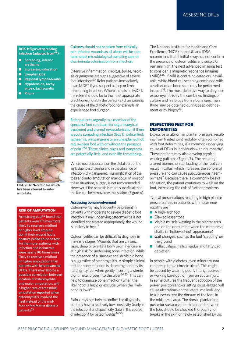
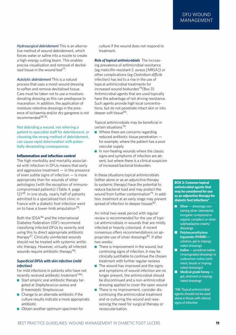
Where necrosis occurs on the distal part of the limb due to ischaemia and in the absence of infection (dry gangrene), mummification of the toes and auto-amputation may occur. In most of these situations, surgery is not recommended. However, if the necrosis is more superficial then the toe can be removed with a scalpel (Figure 6).
Assessing bone involvementOsteomyelitis may frequently be present in patients with moderate to severe diabetic foot infection. If any underlying osteomyelitis is not identified and treated appropriately, the wound is unlikely to heal17.
Osteomyelitis can be difficult to diagnose in the early stages. Wounds that are chronic, large, deep or overlie a bony prominence are at high risk for underlying bone infection, while the presence of a 'sausage toe' or visible bone is suggestive of osteomyelitis. A simple clinical test for bone infection is detecting bone by its hard, gritty feel when gently inserting a sterile blunt metal probe into the ulcer54,55. This can help to diagnose bone infection (when the likelihood is high) or exclude (when the likeli-hood is low)46.
Plain x-rays can help to confirm the diagnosis, but they have a relatively low sensitivity (early in the infection) and specificity (late in the course of infection) for osteomyelitis46,56.
The National Institute for Health and Care Excellence (NICE) in the UK and IDSA recommend that if initial x-rays do not confirm the presence of osteomyelitis and suspicion remains high, the next advanced imaging test to consider is magnetic resonance imaging (MRI)1,46. If MRI is contraindicated or unavail-able, white blood cell scanning combined with a radionuclide bone scan may be performed instead46. The most definitive way to diagnose osteomyelitis is by the combined findings of culture and histology from a bone specimen. Bone may be obtained during deep debride-ment or by biopsy46.
INSPECTING FEET FOR DEFORMITIESExcessive or abnormal plantar pressure, result-ing from limited joint mobility, often combined with foot deformities, is a common underlying cause of DFUs in individuals with neuropathy6. These patients may also develop atypical walking patterns (Figure 7). The resulting altered biomechanical loading of the foot can result in callus, which increases the abnormal pressure and can cause subcutaneous haem-orrhage7. Because there is commonly loss of sensation, the patient continues to walk on the foot, increasing the risk of further problems.
Typical presentations resulting in high plantar pressure areas in patients with motor neu-ropathy are7:
A high-arch foot Clawed lesser toes Visible muscle wasting in the plantar arch and on the dorsum between the metatarsal shafts (a ‘hollowed-out’ appearance)
Gait changes, such as the foot ‘slapping’ on the ground
Hallux valgus, hallux rigidus and fatty pad depletion.
In people with diabetes, even minor trauma can precipitate a chronic ulcer7. This might be caused by wearing poorly fitting footwear or walking barefoot, or from an acute injury. In some cultures the frequent adoption of the prayer position and/or sitting cross-legged will cause ulcerations on the lateral malleoli, and to a lesser extent the dorsum of the foot, in the mid-tarsal area. The dorsal, plantar and posterior surfaces of both feet and between the toes should be checked thoroughly for breaks in the skin or newly established DFUs.
BOX 1: Signs of spreading infection (adapted from49)
Spreading, intense erythemaIncreasing indurationLymphangitisRegional lymphadenitisHypotension, tachy-pnoea, tachycardiaRigors
RISK OF AMPUTATION
Armstrong et al52 found that patients were 11 times more likely to receive a midfoot or higher level amputa-tion if their wound had a positive probe-to-bone test. Furthermore, patients with infection and ischaemia were nearly 90 times more likely to receive a midfoot or higher amputation than patients with less advanced DFUs. There may also be a possible correlation between location of osteomyelitis and major amputation, with a higher rate of transtibial amputation reported when osteomyelitis involved the heel instead of the mid-foot or forefoot in diabetic patients53.
FIGURE 6: Necrotic toe which has been allowed to auto-amputate
3 BEST PRACTICE GUIDELINES FOR SKIN AND WOUND CARE IN EPIDERMOLYSIS BULLOSA8 BEST PRACTICE GUIDELINES: WOUND MANAGEMENT IN DIABETIC FOOT ULCERS
ASSESSING DFUs
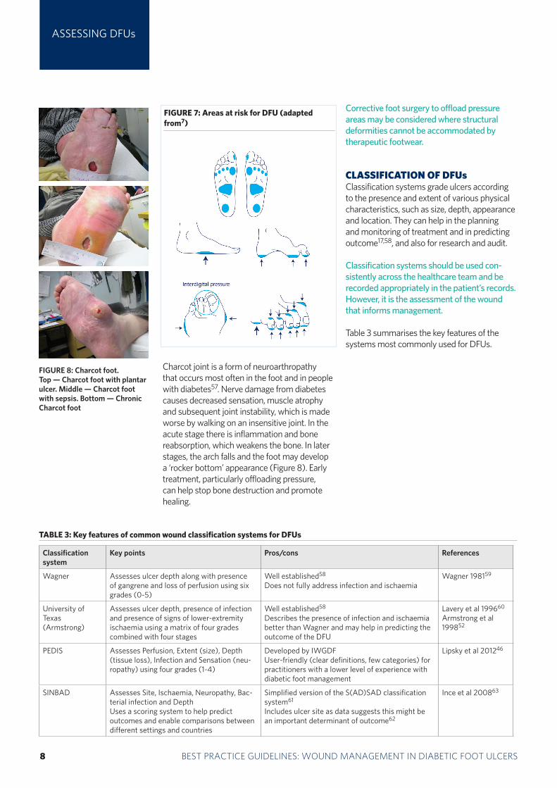
Charcot joint is a form of neuroarthropathy that occurs most often in the foot and in people with diabetes57. Nerve damage from diabetes causes decreased sensation, muscle atrophy and subsequent joint instability, which is made worse by walking on an insensitive joint. In the acute stage there is inflammation and bone reabsorption, which weakens the bone. In later stages, the arch falls and the foot may develop a ‘rocker bottom’ appearance (Figure 8). Early treatment, particularly offloading pressure, can help stop bone destruction and promote healing.
Corrective foot surgery to offload pressure areas may be considered where structural deformities cannot be accommodated by therapeutic footwear.
CLASSIFICATION OF DFUsClassification systems grade ulcers according to the presence and extent of various physical characteristics, such as size, depth, appearance and location. They can help in the planning and monitoring of treatment and in predicting outcome17,58, and also for research and audit.
Classification systems should be used con-sistently across the healthcare team and be recorded appropriately in the patient’s records. However, it is the assessment of the wound that informs management.
Table 3 summarises the key features of the systems most commonly used for DFUs.
TABLE 3: Key features of common wound classification systems for DFUs
Classification system
Key points Pros/cons References
Wagner Assesses ulcer depth along with presence of gangrene and loss of perfusion using six grades (0-5)
Well established58
Does not fully address infection and ischaemiaWagner 198159
University of Texas (Armstrong)
Assesses ulcer depth, presence of infection and presence of signs of lower-extremity ischaemia using a matrix of four grades combined with four stages
Well established58
Describes the presence of infection and ischaemia better than Wagner and may help in predicting the outcome of the DFU
Lavery et al 199660 Armstrong et al 199852
PEDIS Assesses Perfusion, Extent (size), Depth (tissue loss), Infection and Sensation (neu-ropathy) using four grades (1-4)
Developed by IWGDFUser-friendly (clear definitions, few categories) for practitioners with a lower level of experience with diabetic foot management
Lipsky et al 201246
SINBAD Assesses Site, Ischaemia, Neuropathy, Bac-terial infection and DepthUses a scoring system to help predict outcomes and enable comparisons between different settings and countries
Simplified version of the S(AD)SAD classification system61
Includes ulcer site as data suggests this might be an important determinant of outcome62
Ince et al 200863
FIGURE 7: Areas at risk for DFU (adapted from7)
FIGURE 8: Charcot foot.Top — Charcot foot with plantar ulcer. Middle — Charcot foot with sepsis. Bottom — Chronic Charcot foot
BEST PRACTICE GUIDELINES: WOUND MANAGEMENT IN DIABETIC FOOT ULCERS 9
DFU WOUND MANAGEMENT
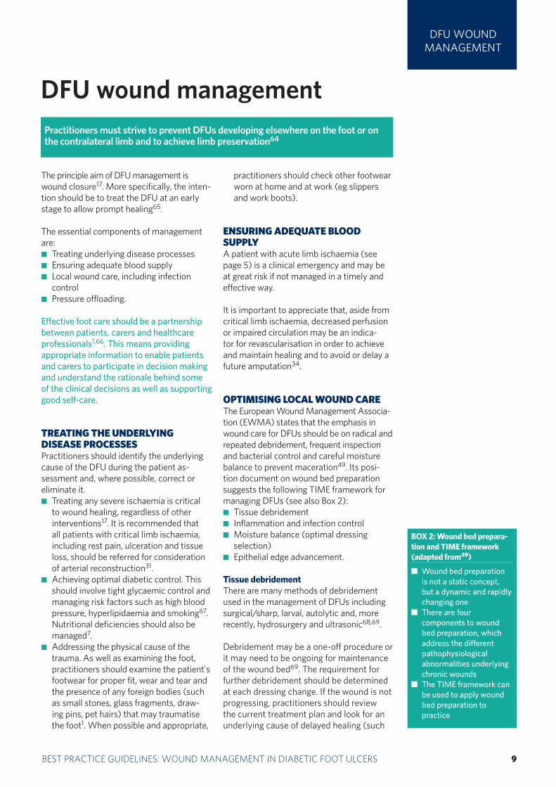
DFU wound management Practitioners must strive to prevent DFUs developing elsewhere on the foot or on the contralateral limb and to achieve limb preservation64
The principle aim of DFU management is wound closure17. More specifically, the inten-tion should be to treat the DFU at an early stage to allow prompt healing65.
The essential components of management are:
Treating underlying disease processes Ensuring adequate blood supply Local wound care, including infection control
Pressure offloading.
Effective foot care should be a partnership between patients, carers and healthcare professionals1,66. This means providing appropriate information to enable patients and carers to participate in decision making and understand the rationale behind some of the clinical decisions as well as supporting good self-care.
TREATING THE UNDERLYING DISEASE PROCESSESPractitioners should identify the underlying cause of the DFU during the patient as-sessment and, where possible, correct or eliminate it.
Treating any severe ischaemia is critical to wound healing, regardless of other interventions17. It is recommended that all patients with critical limb ischaemia, including rest pain, ulceration and tissue loss, should be referred for consideration of arterial reconstruction31.
Achieving optimal diabetic control. This should involve tight glycaemic control and managing risk factors such as high blood pressure, hyperlipidaemia and smoking67. Nutritional deficiencies should also be managed7.
Addressing the physical cause of the trauma. As well as examining the foot, practitioners should examine the patient's footwear for proper fit, wear and tear and the presence of any foreign bodies (such as small stones, glass fragments, draw-ing pins, pet hairs) that may traumatise the foot1. When possible and appropriate,
practitioners should check other footwear worn at home and at work (eg slippers and work boots).
ENSURING ADEQUATE BLOOD SUPPLYA patient with acute limb ischaemia (see page 5) is a clinical emergency and may be at great risk if not managed in a timely and effective way.
It is important to appreciate that, aside from critical limb ischaemia, decreased perfusion or impaired circulation may be an indica-tor for revascularisation in order to achieve and maintain healing and to avoid or delay a future amputation34.
OPTIMISING LOCAL WOUND CAREThe European Wound Management Associa-tion (EWMA) states that the emphasis in wound care for DFUs should be on radical and repeated debridement, frequent inspection and bacterial control and careful moisture balance to prevent maceration49. Its posi-tion document on wound bed preparation suggests the following TIME framework for managing DFUs (see also Box 2):
Tissue debridement Inflammation and infection control Moisture balance (optimal dressing
selection) Epithelial edge advancement.
Tissue debridement There are many methods of debridement used in the management of DFUs including surgical/sharp, larval, autolytic and, more recently, hydrosurgery and ultrasonic68,69.
Debridement may be a one-off procedure or it may need to be ongoing for maintenance of the wound bed69. The requirement for further debridement should be determined at each dressing change. If the wound is not progressing, practitioners should review the current treatment plan and look for an underlying cause of delayed healing (such
BOX 2: Wound bed prepara-tion and TIME framework (adapted from49)
Wound bed preparation is not a static concept, but a dynamic and rapidly changing oneThere are four components to wound bed preparation, which address the different pathophysiological abnormalities underlying chronic wounds
The TIME framework can be used to apply wound bed preparation to practice
3 BEST PRACTICE GUIDELINES FOR SKIN AND WOUND CARE IN EPIDERMOLYSIS BULLOSA10 BEST PRACTICE GUIDELINES: WOUND MANAGEMENT IN DIABETIC FOOT ULCERS
as ischaemia, infection or inflammation) and consider patient concordance with recommended treatment regimens (such as not wearing offloading devices or not taking antidiabetic medication)69.
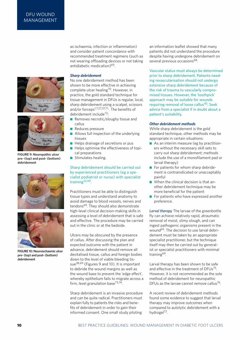
Sharp debridementNo one debridement method has been shown to be more effective in achieving complete ulcer healing70. However, in practice, the gold standard technique for tissue management in DFUs is regular, local, sharp debridement using a scalpel, scissors and/or forceps1,7,27,37,71,. The benefits of debridement include72:
Removes necrotic/sloughy tissue and callus
Reduces pressure Allows full inspection of the underlying
tissues Helps drainage of secretions or pus Helps optimise the effectiveness of topi-cal preparations
Stimulates healing.
Sharp debridement should be carried out by experienced practitioners (eg a spe-cialist podiatrist or nurse) with specialist training22,69.
Practitioners must be able to distinguish tissue types and understand anatomy to avoid damage to blood vessels, nerves and tendons69. They should also demonstrate high-level clinical decision-making skills in assessing a level of debridement that is safe and effective. The procedure may be carried out in the clinic or at the bedside.
Ulcers may be obscured by the presence of callus. After discussing the plan and expected outcome with the patient in advance, debridement should remove all devitalised tissue, callus and foreign bodies down to the level of viable bleeding tis-sue38,69 (Figures 9 and 10). It is important to debride the wound margins as well as the wound base to prevent the ‘edge effect’, whereby epithelium fails to migrate across a firm, level granulation base73,74.
Sharp debridement is an invasive procedure and can be quite radical. Practitioners must explain fully to patients the risks and bene-fits of debridement in order to gain their informed consent. One small study piloting
an information leaflet showed that many patients did not understand the procedure despite having undergone debridement on several previous occasions68.
Vascular status must always be determined prior to sharp debridement. Patients need-ing revascularisation should not undergo extensive sharp debridement because of the risk of trauma to vascularly compro-mised tissues. However, the ‘toothpick’ approach may be suitable for wounds requiring removal of loose callus45. Seek advice from a specialist if in doubt about a patient’s suitability.
Other debridement methodsWhile sharp debridement is the gold standard technique, other methods may be appropriate in certain situations:
As an interim measure (eg by practition-ers without the necessary skill sets to carry out sharp debridement; methods include the use of a monofilament pad or larval therapy)
For patients for whom sharp debride-ment is contraindicated or unacceptably painfulWhen the clinical decision is that an-other debridement technique may be more beneficial for the patient
For patients who have expressed another preference.
Larval therapy The larvae of the greenbottle fly can achieve relatively rapid, atraumatic removal of moist, slimy slough, and can ingest pathogenic organisms present in the wound69. The decision to use larval debri-dement must be taken by an appropriate specialist practitioner, but the technique itself may then be carried out by general-ist or specialist practitioners with minimal training69.
Larval therapy has been shown to be safe and effective in the treatment of DFUs75. However, it is not recommended as the sole method of debridement for neuropathic DFUs as the larvae cannot remove callus76.
A recent review of debridement methods found some evidence to suggest that larval therapy may improve outcomes when compared to autolytic debridement with a hydrogel72.
DFU WOUND MANAGEMENT
FIGURE 9: Neuropathic ulcer pre- (top) and post- (bottom) debridement
FIGURE 10: Neuroischaemic ulcer pre- (top) and post- (bottom) debridement
BEST PRACTICE GUIDELINES: WOUND MANAGEMENT IN DIABETIC FOOT ULCERS 11
DFU WOUND MANAGEMENT
Hydrosurgical debridement This is an alterna-tive method of wound debridement, which forces water or saline into a nozzle to create a high-energy cutting beam. This enables precise visualisation and removal of devital-ised tissue in the wound bed77.
Autolytic debridement This is a natural process that uses a moist wound dressing to soften and remove devitalised tissue. Care must be taken not to use a moisture-donating dressing as this can predispose to maceration. In addition, the application of moisture-retentive dressings in the pres-ence of ischaemia and/or dry gangrene is not recommended38,76.
Not debriding a wound, not referring a patient to specialist staff for debridement, or choosing the wrong method of debridement, can cause rapid deterioration with poten-tially devastating consequences.
Inflammation and infection controlThe high morbidity and mortality associat-ed with infection in DFUs means that early and aggressive treatment — in the presence of even subtle signs of infection — is more appropriate than for wounds of other aetiologies (with the exception of immuno-compromised patients) (Table 4, page 12)38. In one study, nearly half of patients admitted to a specialised foot clinic in France with a diabetic foot infection went on to have a lower-limb amputation78.
Both the IDSA46 and the International Diabetes Federation (IDF) recommend classifying infected DFUs by severity and using this to direct appropriate antibiotic therapy27. Clinically uninfected wounds should not be treated with systemic antibi-otic therapy. However, virtually all infected wounds require antibiotic therapy46.
Superficial DFUs with skin infection (mild infection)For mild infections in patients who have not recently received antibiotic treatment7,46:
Start empiric oral antibiotic therapy tar-geted at Staphylococcus aureus and ß-haemolytic Streptococcus
Change to an alternate antibiotic if the culture results indicate a more appropriate antibiotic
Obtain another optimum specimen for
culture if the wound does not respond to treatment.
Role of topical antimicrobials The increas-ing prevalence of antimicrobial resistance (eg meticillin-resistant S. aureus [MRSA]) or other complications (eg Clostridium difficile infection) has led to a rise in the use of topical antimicrobial treatments for increased wound bioburden79(Box 3). Antimicrobial agents that are used topically have the advantage of not driving resistance. Such agents provide high local concentra-tions, but do not penetrate intact skin or into deeper soft tissue80.
Topical antimicrobials may be beneficial in certain situations79:
Where there are concerns regarding reduced antibiotic tissue penetration — for example, where the patient has a poor vascular supply
In non-healing wounds where the classic signs and symptoms of infection are ab-sent, but where there is a clinical suspicion of increased bacterial bioburden.
In these situations topical antimicrobials (either alone or as an adjunctive therapy to systemic therapy) have the potential to reduce bacterial load and may protect the wound from further contamination79. In addi-tion, treatment at an early stage may prevent spread of infection to deeper tissues82.
An initial two-week period with regular review is recommended for the use of topi-cal antimicrobials in wounds that are mildly infected or heavily colonised. A recent consensus offers recommendations on ap-propriate use of silver dressings83. If after two weeks:
There is improvement in the wound, but continuing signs of infection, it may be clinically justifiable to continue the chosen treatment with further regular reviews
The wound has improved and the signs and symptoms of wound infection are no longer present, the antimicrobial should be discontinued and a non-antimicrobial dressing applied to cover the open wound
There is no improvement, consider dis-continuing the antimicrobial treatment and re-culturing the wound and reas-sessing the need for surgical therapy or revascularisation.
BOX 3: Common topical antimicrobial agents that may be considered for use as an adjunctive therapy for diabetic foot infections*
Silver — dressings con-taining silver (elemental, inorganic compound or organic complex) or silver sulphadiazine cream/dressingsPolyhexamethylene biguanide (PHMB) — solution, gel or impreg-nated dressingsIodine — povidone iodine (impregnated dressing) or cadexomer iodine (oint-ment, beads or impreg-nated dressings) Medical-grade honey — gel, ointment or impreg-nated dressings
*NB: Topical antimicrobial agents should not be used alone in those with clinical signs of infection
3 BEST PRACTICE GUIDELINES FOR SKIN AND WOUND CARE IN EPIDERMOLYSIS BULLOSA12 BEST PRACTICE GUIDELINES: WOUND MANAGEMENT IN DIABETIC FOOT ULCERS
DFU WOUND MANAGEMENT
If there are clinical signs of infection at dressing change, systemic antibiotic therapy should be started. Topical antimicrobials are not indicated as the only anti-infective treatment for moderate or severe infection of deep tissue or bone38,46.
Patients may also require debridement to remove infected material. In addition, in-fected wounds should be cleansed at each dressing change with saline or an appropri-ate antiseptic wound cleansing agent.
Deep tissue infection (moderate to severe infection)For treating deep tissue infection (cellulitis, lymphangitis, septic arthritis, fasciitis):
Start patients quickly on broad-spectrum antibiotics, commensurate with the clini-cal history and according to local proto-cols where possible37
Take deep tissue specimens or aspirates of purulent secretions for cultures at the start of treatment to identify specific organisms in the wound, but do not wait for results before initiating therapy1,37
Change to an alternate antibiotic if: — indicated by microbiology results46
— the signs of inflammation are not improving84
Administer antibiotics parenterally for all severe and some moderate infections,
and switch to the oral route when the patient is systemically well and culture results are available46
Continue antibiotic therapy until the infec-tion resolves, but not through to complete healing46. In most cases 1–3 weeks of therapy is sufficient for soft tissue infections
Consider giving empiric therapy directed against MRSA46:
— in patients with a prior history of MRSA infection — when the local prevalence of MRSA colonisation or infection is high — if the infection is clinically severe.
Note that the optimal duration of antibi-otic treatment is not clearly defined and will depend on the severity of infection and response to treatment84.
Infection in a neuroischaemic foot is often more serious than in a neuropathic foot (which has a good blood supply), and this should influence antibiotic policy49. Antibiot-ic therapy should not be given as a preventive measure in the absence of signs of infection (see Box 4). This is likely to cause infection with more resistant pathogens.
Obtain an urgent consultation with experts (eg foot surgeon) for patients who have a rapidly deteriorating wound that is not responding to antibiotic therapy. Infections accompanied by a deep abscess, extensive bone or joint involvement, crepitus, sub-stantial necrosis or gangrene, or necrotising fasciitis, need prompt surgical intervention along with appropriate antibiotic therapy, to reduce the risk of major amputation51,85.
Biofilms and chronic persistent infectionPolymicrobial infections predominate in severe diabetic foot infections and this diversity of bacterial populations in chronic wounds, such as DFUs, may be an important contributor to chronicity86,87. Biofilms are complex polymicrobial communities that develop on the surface of chronic wounds, which may lack the overt clinical signs of infec-tion34. They are not visible to the naked eye and cannot be detected by routine cultures88.
The microbes produce an extra-polymeric substance that contributes to the structure of the biofilm. This matrix acts as a thick, slimy protective barrier, making it very difficult for
TABLE 4: General principles of bacterial management (adapted from49)
At initial presentation of infection it is important to assess its severity, take appropri-ate cultures and consider need for surgical procedures
Optimal specimens for culture should be taken after initial cleansing and debride-ment of necrotic material
Patients with severe infection require empiric broad-spectrum antibiotic therapy, pending culture results. Those with mild (and many with moderate) infection can be treated with a more focused and narrow-spectrum antibioticPatients with diabetes have immunological disturbances; therefore even bacteria re-garded as skin commensals can cause severe tissue damage and should be regarded as pathogens when isolated from correctly obtained tissue specimens
Gram-negative bacteria, especially when isolated from an ulcer swab, are often colonising organisms that do not require targeted therapy unless the person is at risk for infection with those organisms
Blood cultures should be sent if fever and systemic toxicity are present Even with appropriate treatment, the wound should be inspected regularly for early signs of infection or spreading infection
Clinical microbiologists/infectious diseases specialists have a crucial role; laboratory results should be used in combination with the clinical presentation and history to guide antibiotic selection
Timely surgical intervention is crucial for deep abscesses, necrotic tissue and for some bone infections
BOX 4: Guidelines for the use of systemic antibiotic therapy
Antibiotics should be pre-scribed using local protocols and, in complex cases, the advice of a clinical microbiol-ogist or infectious diseases specialist. Avoid prescribing antibiotics for uninfected ulcerations. IDSA46 offers evidence-based suggestions, which can be adapted to local needs.http://www.idsociety.org/uploadedFiles/IDSA/Guide-lines-Patient_Care/PDF_Li-brary/2012%20Diabetic%20Foot%20Infections%20Guideline.pdf
BEST PRACTICE GUIDELINES: WOUND MANAGEMENT IN DIABETIC FOOT ULCERS 13
antimicrobial agents to penetrate it89. The impact of biofilms may depend on which spe-cies are present rather than the bioburden34.
Treatment should aim to88:Disrupt the biofilm burden through regular, repeated debridement and vigorous wound cleansing Prevent reformation and attachment of the biofilm by using antimicrobial dressings.
Appropriate wound bed preparation remains the gold standard for biofilm removal90.
Moisture balance: optimal dressing selectionMost dressings are designed to create a moist wound environment and support progres-sion towards wound healing. They are not a substitute for sharp debridement, managing systemic infection, offloading devices and diabetic control.
Moist wound healing has the potential to address multiple factors that affect wound healing. It involves maintaining a balanced wound environment that is not too moist or too dry. Dressings that can help to manage wound exudate optimally and promote a balanced environment are key to improving outcomes91. However, a dressing that may be ideal for wounds of other aetiologies may be entirely inappropriate for certain DFUs. The dressing selected may have a considerable effect on outcome and, due to the varying complexities of DFUs, there is no single dressing to suit all scenarios.
Many practitioners are confused by the great range of dressings available. Impressive claims are rarely supported by scientific studies and there is often a lack of high- quality evidence to support decision making. One inherent problem is whether the characteristics of each wound randomised to a specific dressing in a trial correspond to the characteristics that the dressing was designed to manage92. Many dressings are designed for non-foot areas of the body and may be difficult to apply between or over the toes or plantar surface. In addition, most practitioners have historically had little specific, practical guidance on selecting dressings.
In the absence of strong evidence of clinical or cost effectiveness, healthcare professionals
should use wound dressings that best match the clinical appearance and site of the wound, as well as patient preferences1. Dressing choice must begin with a thorough patient and wound assessment. Factors to consider include:
Location of the wound Extent (size/depth) of the wound Amount and type of exudate The predominant tissue type on the wound
surface Condition of the periwound skin Compatibility with other therapies (eg
contact casts)Wound bioburden and risk of infection
Avoidance of pain and trauma at dressing changes
Quality of life and patient wellbeing.
The status of the diabetic foot can change very quickly, especially if infection has not been appropriately addressed. The need for regular inspection and assessment means that dressings designed to be left in situ for more than five days are not usually appropri-ate for DFU management.
Practitioners should also consider the follow-ing questions93.
Does the dressing: Stay intact and remain in place throughout
wear time? Prevent leakage between dressing
changes? Cause maceration/allergy or sensitivity? Reduce pain? Reduce odour?
Retain fluid? Trap exudate components?
Is the dressing:Comfortable, conformable, flexible and of a bulk/weight that can be accommodated in an offloading device/footwear?Suitable for leaving in place for the required duration?
Easy to remove (does not traumatise the surrounding skin or wound bed)?
Easy to apply?Cost effective?Likely to cause iatrogenic lesions?
Tables 5 and 6 (pages 14-15) provide advice on type of dressing and how to select accord-ing to tissue type (see also Figures 11–14).
DFU WOUND MANAGEMENT
FIGURE 11: Dry necrotic wound. Select dressing to rehydrate and soften the eschar
FIGURE 12: Sloughy wound bed with areas of necrosis. Select dressing to control moisture and promote debridement of devitalised tissue
FIGURE 13: Infected wound with evidence of swelling and exudate. Start empiric antibi-otic therapy and take cultures. Consider selecting an anti-microbial dressing to reduce wound bioburden and manage exudate
FIGURE 14: A newly epitheli-alising DFU. It is important to protect new tissue growth
3 BEST PRACTICE GUIDELINES FOR SKIN AND WOUND CARE IN EPIDERMOLYSIS BULLOSA14 BEST PRACTICE GUIDELINES: WOUND MANAGEMENT IN DIABETIC FOOT ULCERS
DFU WOUND MANAGEMENT
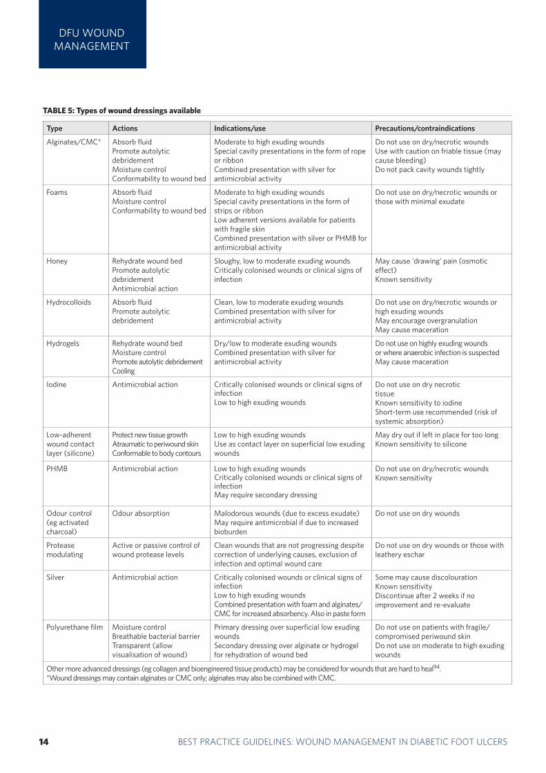
TABLE 5: Types of wound dressings available
Type Actions Indications/use Precautions/contraindications
Alginates/CMC* Absorb fluidPromote autolytic debridementMoisture controlConformability to wound bed
Moderate to high exuding woundsSpecial cavity presentations in the form of rope or ribbon Combined presentation with silver for antimicrobial activity
Do not use on dry/necrotic woundsUse with caution on friable tissue (may cause bleeding)Do not pack cavity wounds tightly
Foams Absorb fluidMoisture controlConformability to wound bed
Moderate to high exuding wounds Special cavity presentations in the form of strips or ribbonLow adherent versions available for patients with fragile skinCombined presentation with silver or PHMB for antimicrobial activity
Do not use on dry/necrotic wounds or those with minimal exudate
Honey Rehydrate wound bed Promote autolytic debridementAntimicrobial action
Sloughy, low to moderate exuding woundsCritically colonised wounds or clinical signs of infection
May cause 'drawing' pain (osmotic effect) Known sensitivity
Hydrocolloids Absorb fluidPromote autolytic debridement
Clean, low to moderate exuding woundsCombined presentation with silver for antimicrobial activity
Do not use on dry/necrotic wounds or high exuding woundsMay encourage overgranulationMay cause maceration
Hydrogels Rehydrate wound bedMoisture controlPromote autolytic debridementCooling
Dry/low to moderate exuding woundsCombined presentation with silver for antimicrobial activity
Do not use on highly exuding woundsor where anaerobic infection is suspectedMay cause maceration
Iodine Antimicrobial action Critically colonised wounds or clinical signs of infection Low to high exuding wounds
Do not use on dry necrotic tissueKnown sensitivity to iodineShort-term use recommended (risk of systemic absorption)
Low-adherent wound contact layer (silicone)
Protect new tissue growthAtraumatic to periwound skinConformable to body contours
Low to high exuding woundsUse as contact layer on superficial low exuding wounds
May dry out if left in place for too longKnown sensitivity to silicone
PHMB Antimicrobial action Low to high exuding wounds Critically colonised wounds or clinical signs of infectionMay require secondary dressing
Do not use on dry/necrotic woundsKnown sensitivity
Odour control (eg activated charcoal)
Odour absorption Malodorous wounds (due to excess exudate)May require antimicrobial if due to increased bioburden
Do not use on dry wounds
Protease modulating
Active or passive control of wound protease levels
Clean wounds that are not progressing despite correction of underlying causes, exclusion of infection and optimal wound care
Do not use on dry wounds or those with leathery eschar
Silver Antimicrobial action Critically colonised wounds or clinical signs of infection Low to high exuding woundsCombined presentation with foam and alginates/CMC for increased absorbency. Also in paste form
Some may cause discolourationKnown sensitivityDiscontinue after 2 weeks if no improvement and re-evaluate
Polyurethane film Moisture controlBreathable bacterial barrierTransparent (allow visualisation of wound)
Primary dressing over superficial low exuding woundsSecondary dressing over alginate or hydrogel for rehydration of wound bed
Do not use on patients with fragile/ compromised periwound skinDo not use on moderate to high exuding wounds
Other more advanced dressings (eg collagen and bioengineered tissue products) may be considered for wounds that are hard to heal94. *Wound dressings may contain alginates or CMC only; alginates may also be combined with CMC.
BEST PRACTICE GUIDELINES: WOUND MANAGEMENT IN DIABETIC FOOT ULCERS 15
DFU WOUND MANAGEMENT
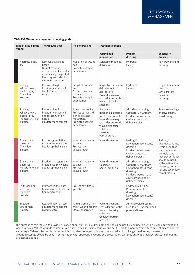
TABLE 6: Wound management dressing guide
Type of tissue in the wound
Therapeutic goal Role of dressing Treatment options
Wound bed preparation
Primary dressing
Secondary dressing
Necrotic, black, dry
Remove devitalised tissue Do not attempt debridement if vascular insufficiency suspected Keep dry and refer for vascular assessment
Hydration of wound bedPromote autolytic debridement
Surgical or mechnical debridement
HydrogelHoney
Polyurethane film dressing
Sloughy, yellow, brown, black or greyDry to low exudate
Remove sloughProvide clean wound bed for granulation tissue
Rehydrate wound bed Control moisture balancePromote autolytic debridement
Surgical or mechanicaldebridement if appropriateWound cleansing (consider antiseptic wound cleansing solution)
HydrogelHoney
Polyurethane film dressingLow adherent (silicone) dressing
Sloughy, yellow, brown, black or greyModerate to high exudate
Remove sloughProvide clean wound bed for granulation tissueExudate management
Absorb excess fluid Protect periwound skin to prevent macerationPromote autolytic debridement
Surgical or mechanical debride-ment if appropriateWound cleansing (consider antiseptic wound cleansing solution)Consider barrier products
Absorbent dressing (alginate/CMC/foam)For deep wounds, use cavity strips, rope or ribbon versions
Retention bandage or polyurethane film dressing
Granulating, clean, red Dry to low exudate
Promote granulationProvide healthy wound bed for epithelialisation
Maintain moisture balanceProtect new tissue growth
Wound cleansing HydrogelLow adherent (silicone) dressingFor deep wounds use cavity strips, rope or ribbon versions
Pad and/or retention bandage. Avoid bandages that may cause occlusion and maceration. Tapes should be used with caution due to allergy poten-tial and secondary complications
Granulating, clean, red Moderate to high exudate
Exudate managementProvide healthy wound bed for epithelialisation
Maintain moisture balanceProtect new tissue growth
Wound cleansingConsider barrier products
Absorbent dressing (alginate/CMC/foam)Low adherent (silicone) dressingFor deep wounds, use cavity strips, rope or ribbon versions
Epithelialising, red, pinkNo to low exudate
Promote epithelialisa-tion and wound matura-tion (contraction)
Protect new tissue growth
Hydrocolloid (thin) Polyurethane film dressingLow adherent (silicone) dressing
InfectedLow to high exudate
Reduce bacterial loadExudate managementOdour control
Antimicrobial action Moist wound healingOdour absorption
Wound cleansing (consider antiseptic wound cleansing solution)Consider barrier products
Antimicrobial dressing (see Table 5 for combined presentations)
The purpose of this table is to provide guidance about appropriate dressings and should be used in conjunction with clinical judgement and local protocols. Where wounds contain mixed tissue types, it is important to consider the predominant factors affecting healing and address accordingly. Where infection is suspected it is important to regularly inspect the wound and to change the dressing frequently. Wound dressings should be used in combination with appropriate wound bed preparation, systemic antibiotic therapy, pressure offloading and diabetic control
3 BEST PRACTICE GUIDELINES FOR SKIN AND WOUND CARE IN EPIDERMOLYSIS BULLOSA16 BEST PRACTICE GUIDELINES: WOUND MANAGEMENT IN DIABETIC FOOT ULCERS
DFU WOUND MANAGEMENT
Dressing application and wound monitoringRegularly reviewing a patient's wound and dressing is vital. For infected or highly exud-ing wounds, a healthcare professional should inspect the wound and change the dressing daily, and then every two or three days once the infection is stable. A different type of dressing may be needed as the status of the wound changes.
Some patients, especially those with mobility issues or work commitments may prefer to change their dressings themselves, or have a relative or carer to do it. These patients should be advised about using aseptic technique and the wound should continue to be reviewed at regular intervals by the MDFT or other healthcare team members. Patients should be encouraged to look out for signs of deteriora-tion, such as increased pain, swelling, odour, purulence or septic symptoms. In some cases (eg in the first few days of antibiotic therapy) it is a good idea to mark the extent of any celluli-tis with an indelible marker and tell the patient to contact the footcare team immediately if the redness moves substantially beyond the line.
When applying dressings: Avoid bandaging over toes as this may
cause a tourniquet effect (instead, layer gauze over the toes and secure with a band-age from the metatarsal heads to a suitable point on foot)
Use appropriate techniques (eg avoiding creases and being too bulky) and take care when dressing weight-bearing areas
Avoid strong adhesive tapes on fragile skin
Avoid tight bandaging at the fifth toe and the fifth metatarsal head (trim the bandage back)
Ensure wound dead space is eliminated (eg use a dressing that conforms to the contours of the wound bed)
Remember that footwear needs to accom-modate any dressing.
Wounds should be cleansed at each dressing change and after debridement with a wound cleansing solution or saline. Cleansing can help remove devitalised tissue, re-balance the bioburden and reduce exudate to help prepare the wound bed for healing98. It may also help to remove biofilms88.
Managing pain at dressing changesIt is now acknowledged that many patients — even those with neuropathy or neuroischaemia — can feel pain due to their wound or a proce-dure99. It is important to incorporate strategies to prevent trauma and minimise wound-related pain during dressing changes100. This may include the use of soft silicone dressings and avoiding unnecessary manipulation of the wound99. Remember also that patients who have lost the protective pain sensation are at greater risk of trauma at dressing change99.
When appropriate, use low- or non-adherent dressings99. If a dressing becomes encrusted or is difficult to remove, it is important to soak the dressing with saline or a wound irrigation solution and check the wound and surrounding skin for evidence of trauma and infection on dressing removal99.
Epithelial edge advancementIt is important to debride the edges of the ulcer to remove potential physical barriers to the growth of the epithelium across the ulcer bed74. The demarcation line between any necrotic tissue or gangrene and healthy tissue may become a site of infection48. Similar problems can be seen when a gangrenous toe touches a healthy toe50.
Conversely, ‘die-back’ is an abnormal response to over-aggressive sharp debridement. It involves necrosis at the wound edge and extends through previously healthy tissue50.
If the wound does not respond to standard wound management interventions despite treatment of the underlying cause and
BOX 5: The use of advanced therapies
Adjunctive treatments such as negative pressure wound therapy (NPWT), biologi-cal dressings, bioengineered skin equivalents, hyperbaric oxygen therapy, platelet rich plasma and growth factors may be considered, if appropriate and where avail-able for DFUs that are not progressing95. These techniques require advanced clinical decision making and should be carried out only by practitioners with appropriate skills and anatomical knowledge22. However, such therapies represent considerable greater product cost than stand-ard therapy. These costs may be justified if they result in improved ulcer healing, reduced morbidity, fewer lower-extremity amputations and improved patient functional status95. There is a good level of evidence for some biological skin equivalents95 as well as for the use of NPWT in DFU patients without significant infection96. More recently, NPWT with instillation therapy (NPWTi) using anti-septic agents (eg PHMB) has become available. Although there are limited data on its benefits, it could be considered when there is a need for wound cleansing or treatment with topical antimicrobials97.
BEST PRACTICE GUIDELINES: WOUND MANAGEMENT IN DIABETIC FOOT ULCERS 17
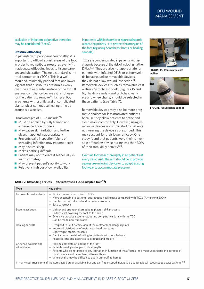
exclusion of infection, adjunctive therapies may be considered (Box 5).e underlying cause and exclusion of infection,Pressure offloading In patients with peripheral neuropathy, it is important to offload at-risk areas of the foot in order to redistribute pressures evenly101. Inadequate offloading leads to tissue dam-age and ulceration. The gold standard is the total contact cast (TCC). This is a well-moulded, minimally padded foot and lower leg cast that distributes pressures evenly over the entire plantar surface of the foot. It ensures compliance because it is not easy for the patient to remove74. Using a TCC in patients with a unilateral uncomplicated plantar ulcer can reduce healing time by around six weeks37.
Disadvantages of TCCs include74: Must be applied by fully trained and
experienced practitioners May cause skin irritation and further
ulcers if applied inappropriatelyPrevents daily inspection (signs of spreading infection may go unnoticed)May disturb sleepMakes bathing difficultPatient may not tolerate it (especially in warm climates)May prevent patient's ability to workRelatively high cost/low availability.
In patients with ischaemic or neuroischaemic ulcers, the priority is to protect the margins of the foot (eg using Scotchcast boots or healing sandals).
TCCs are contraindicated in patients with is-chaemia because of the risk of inducing further DFUs102. They are also not appropriate for patients with infected DFUs or osteomyeli-tis because, unlike removable devices, they do not allow wound inspection74. Removable devices (such as removable cast walkers, Scotchcast boots (Figures 15 and 16), healing sandals and crutches, walk-ers and wheelchairs) should be selected in these patients (see Table 7).
Removable devices may also be more prag-matic choices for less motivated patients because they allow patients to bathe and sleep more comfortably. However, using re-movable devices is complicated by patients not wearing the device as prescribed. This may account for their lower efficacy. One study found that patients wore their remov-able offloading device during less than 30% of their total daily activity103.
Examine footwear thoroughly in all patients at every clinic visit. The aim should be to provide a pressure-relieving device or to adapt existing footwear to accommodate pressure.
DFU WOUND MANAGEMENT
TABLE 7: Offloading devices — alternatives to TCCs (adapted from73)
Type Key points
Removable cast walkers — Similar pressure reduction to TCCs— More acceptable to patients, but reduced healing rate compared with TCCs (Armstrong 2001)— Can be used on infected and ischaemic wounds— Easy to remove
Scotchcast boots — Lighter and stronger alternative to plaster-of-Paris casts — Padded cast covering the foot to the ankle— Extensive practice experience, but no comparative data with the TCC— Can be made non-removable
Healing sandals — Designed to limit dorsiflexion of the metatarsophalangeal joints— Improved distribution of metatarsal head pressures— Lightweight, stable, reusable— Can increase the risk of falling for patients with poor balance— Requires time and expertise to produce and modify
Crutches, walkers and wheelchairs
— Provide complete offloading of the foot— Patients need good upper body strength— Patients who do not perceive any limitation in function of the affected limb must understand the purpose of
these devices and be motivated to use them— Wheelchairs may be difficult to use in unmodified homes
In many countries some of the items listed are unavailable, but one can find inspired individuals adapting local resources to assist patients104
FIGURE 15: Removable cast walker
FIGURE 16: Scotchcast boot
3 BEST PRACTICE GUIDELINES FOR SKIN AND WOUND CARE IN EPIDERMOLYSIS BULLOSA18 BEST PRACTICE GUIDELINES: WOUND MANAGEMENT IN DIABETIC FOOT ULCERS
DFU WOUND MANAGEMENT
Recommendations from the IWGDF26 on the use of offloading interventions in treating un-complicated neuropathic foot ulcers are:
Pressure relief should always be part of the treatment plan for an existing ulcer
TCCs and non-removable walkers are the preferred interventions
Forefoot offloading shoes or cast shoes may be used when above ankle devices are contraindicated
Conventional or standard therapeutic foot-wear should not be used101.
However, in many countries, recommended devices are not available and all that can be of-fered is cushioning constructed from items from local shops (eg, kitchen sponges, upholstery foams etc). In many regions of the world, walk-ing barefoot or with poorly protective sandals is
normal. Replacing these by advising shoe wear may be culturally unacceptable or create other foot problems105. The use of trainers or sports shoes is recommended by some clinicians, which may provide another option to custom-built footwear where this is not accessible106. Patients should also be advised to limit standing and walking and to rest with the foot elevated7.
The introduction of medical insurance schemes that do not pay for preventative care has been a significant factor in lack of care in patients with diabetes in recent years. These schemes also limit what equipment can be offered to a patient.
The hallmark of an appropriately offloaded wound is a noticeable lack of undermining at the wound’s edge at follow up74.
According to the IDF guideline, amputation should not be considered unless a detailed vascular assessment has been performed by vascular staff27.
Amputation may be indicated in the following circumstances27:
Ischaemic rest pain that cannot be managed by analgesia or revascularisation
A life-threatening foot infection that cannot be managed by other measures
A non-healing ulcer that is accompanied by a higher burden of disease than would result from amputation. In some cases, for exam-ple, complications in a diabetic foot render it functionally useless and a well performed amputation is a better alternative for the patient.
Around half of patients who undergo an amputa-tion will develop a further DFU on the contralat-eral limb within 18 months of amputation. The three–year mortality rate after a first amputation is 20–50%107. In a six-year follow-up study, almost 50% of patients developed critical limb
ischaemia in the contralateral limb, but the severity of the DFU and amputation level was significantly lower than in the unilateral limb. This may have been due to prompt intervention made possible by increased patient awareness108.
Patients at high risk for ulceration (such as patients who have undergone an amputation for a DFU) should be reviewed 1–3 monthly by a foot protection team1. At each review patients' feet should be inspected and the need for vascular assessment reviewed. Provision should be made for intensified footcare education, specialist foot-wear and insoles, and skin and nail care. Special arrangements should be made for people with disabilities or immobility1. The Scottish Intercol-legiate Guidelines Network (SIGN) recommends specialist diabetes podiatrist input for patients with a history of amputation and ulceration37.
Although amputation incidence may not reflect the quality of local healthcare delivery, there is a need for more consistent delivery of diabetes care70, with the involvement of an MDFT and patient education.
Amputation and post-amputation careLower-extremity amputation often results in disability and a loss of independence; amputation is often more costly than limb salvage25
BEST PRACTICE GUIDELINES: WOUND MANAGEMENT IN DIABETIC FOOT ULCERS 19
INTEGRATEDCARE APPROACH
MULTIDISCIPLINARY FOOTCARE TEAMEvidence consistently highlights the benefits of MDFTs in the outcomes of DFUs. Over 11 years, one study found total amputations fell by 70% following improvements in footcare services, including multidisciplinary team work109.
However, in England around one-fifth of hospitals providing inpatient care for people with diabetes have no MDFT5. Furthermore, in many areas of the country there are no clear pathways for referring patients at increased risk or high risk of developing DFUs, as recom-mended by NICE5.
All the major guidelines recommend that patients identified with new DFUs should be referred to a dedicated MDFT1,4,7,26,27,37,110. There are many different considered opinions about which disciplines should be incorporated in an MDFT.
The IDF recommends that a specialist footcare team will include doctors with a special inter-est in diabetes, people with educational skills and people with formal training in foot care (usually diabetes podiatrists and trained nurs-es). For comprehensive care, this team would be enhanced by vascular surgeons, orthopae-dic surgeons, infection specialists, orthotists, social workers and psychologists (Box 6).
Guidelines aside, it will be local resources that dictate the skill mix and scope of any footcare team. In the UK there is a move towards hav-ing a core team of specialist diabetes podia-trists, medical specialty consultants, orthotists and surgeons, which works with additional relevant disciplines (such as nurses and gen-eral practitioners) almost in a virtual manner. The key is the ability to gain immediate access to relevant healthcare professionals (such as a vascular surgeon) as needed.
In many countries it is not only specialist equipment that may be unavailable, but also the specialist practitioners themselves, such as podiatrists, vascular surgeons or plaster technicians and so on. While the MDFT will be managing the ongoing challenges of DFU
care, non-specialist practitioners can play a key role in the early detection of problems and prompt referral to the team.
PATIENT FOOTCARE EDUCATIONPatient education should be an integral part of management and prevention. Treatment outcomes will be directly influenced by patients’ knowledge of their own medical status, their ability to care for their wound and concordance with their treatment13,38. It is vital that patients should know who to contact if a DFU develops or recurs, including emergency numbers for the MDFT and out-of-hours contact details37.
The development of an ulcer is a major event and a sign of progressive disease. It is impor-tant to discuss the impact of the ulcer on life expectancy with the patient. Education should be offered on ways in which patients can help to improve outcomes by making lifestyle changes (eg smoking cessation) and working with practitioners to reduce the risk of recur-rence and life-threatening complications13.
A Cochrane systematic review found that educating people with diabetes about the need to look after their feet improves their footcare knowledge and behaviour in the short term. There was insufficient evidence that education alone, without any additional preventive measures, effectively reduces the occurrence of ulcers and amputations111.
According to the IWGDF, patient education should be provided in several sessions using a variety of methods based on standard effective communication techniques. It is essential to evaluate whether the patient has understood the messages, is motivated to act and has sufficient self-care skills7. Remember that elderly and disabled patients may need home or special care45.
Practitioners should ensure patients under-stand the aims of treatment, how to recog-nise and report the signs and symptoms of (worsening) infection and the need for prompt treatment of new wounds7,17.
Integrated care approachDFUs are a multifaceted condition and no one individual or clinical specialty should be expected (or should attempt) to address all aspects of management in isolation
BOX 6: Recommended levels of foot care in acute and com-munity settings7
1. General practitioner, diabe-tes podiatrist and diabetic nurse
2. Diabetologist, surgeon (general and/or vascular, plastic and/or orthopaedic), infectious dieases/micro-biology specialist, diabetes podiatrist and diabetic nurse
3. Specialised foot centre with multiple disciplines special-ised in foot care
3 BEST PRACTICE GUIDELINES FOR SKIN AND WOUND CARE IN EPIDERMOLYSIS BULLOSA20 BEST PRACTICE GUIDELINES: WOUND MANAGEMENT IN DIABETIC FOOT ULCERS
GLOBAL WOUND CARE PLAN
Steps to avoid amputation: implementing a global wound care plan
A Diagnosis of diabetes (+/_ peripheral sensory neuropathy) AIM: Prevent the development of a DFU 1. Implement DFU prevention care plan that includes treatment of co-morbidities, good glycaemic control and pressure offloading 2. Annually perform general foot examination: — Use 10g monofilament to assess sensory status — Inspection of the feet for deformities — Inspection of footwear for wear and tear and foreign objects that may traumatise foot — Maintain skin hydration (consider emollient therapy) for skin health — Offer patient education on checking feet for trauma 3. Ensure regular review and provide patient education
B Development of DFU AIM: Treat the ulcer and prevent infection 1. Determine cause of ulcer 2. Agree treatment aims with patient and implement wound care plan: — Debride and regularly cleanse the wound — Take appropriate tissue samples for culture if infection is suspected — Select dressings to maintain moist wound environment and manage exudate effectively 3. Initiate antibiotic treatment if infection suspected and consider topical antimicrobial therapy if increased bioburden is suspected 4. Review offloading device and ensure footwear accommodates dressing 5. Optimise glycaemic control for diabetes management 6. Refer for vascular assessment if clinically significant limb ischaemia is suspected 7. Offer patient education on how to self-manage and when to raise concerns
C Development of vascular disease AIM: Prevent complications associated with ischaemia 1. Ensure early referral to vascular specialist for arterial reconstruction to improve blood flow in patients with an ischaemic or neuroischaemic ulcer 2. Optimise diabetes control
D Ulcer becomes infected AIM: Prevent life- or limb-threatening complications 1. For superficial (mild) infections — treat with systemic antibiotics and consider topical antimicrobials in selected cases 2. For deep (moderate or severe) infections — treat with appropriately selected empiric systemic
antibiotics, modified by the results of culture and sensitivity reports 3. Offload pressure correctly and optimise glycaemic control for diabetes management 4. Consider therapy directed at biofilm in wounds that are slow to heal
ACTIVE MANAGEMENT OF THE ULCER AND CO-MORBIDITIES SHOULD AIM TO PREVENT AMPUTATIONWhere amputation is not avoidable: 1. Implement skin and wound care plan to manage surgical wound and optimise healing 2. Review regularly and implement prevention care plan to reduce risk of recurrence or further DFU
on contralateral limb
BEST PRACTICE GUIDELINES: WOUND MANAGEMENT IN DIABETIC FOOT ULCERS 21
REFERENCES
1. National Institute for Health and Clinical Excellence. Diabetic foot problems: inpatient management of diabetic foot problems. Clinical guide-line 119. London: NICE, 2011. Available at: http://publications.nice.org.uk/diabetic-foot-problems-cg119. Accessed March 2013
2. Abetz L, Sutton M, Brady L, et al. The diabetic foot ulcer scale: a quality of life instrument for use in clinical trials. Pract Diab Int 2002; 19: 167-75.
3. Brownrigg JR, Davey J, Holt et al. The association of ulceration of the foot with cardiovascular and all-cause mortality in patients with diabe-tes: a meta-analysis. Diabetologia 2012; 55(11): 2906-12.
4. Diabetes UK. Putting feet first: national minimum skills framework. Joint initiative from the Diabetes UK, Foot in Diabetes UK, NHS Diabetes, the Association of British Clinical Diabetologists, the Primary Care Diabetes Society, the Society of Chiropodists and Podiatrists. London: Diabetes UK, 2011. Available at: http://diabetes.org.uk/putting-feet-first. Accessed March 2013.
5. Kerr M. Foot care for people with diabetes: the economic case for change. NHS Diabetes, Newcastle-upon-Tyne, 2012. Available at: http://bit.ly/xjY7FS. Accessed March 2013.
6. Singh N, Armstrong DA, Lipsky BA. Preventing foot ulcers in patients with diabetes. JAMA 2005; 293: 217-28.
7. Bakker K, Apelqvist J, Schaper NC on behalf of the International Work-ing Group on the Diabetic Foot Editorial Board. Practical guidelines on the management and prevention of the diabetic foot 2011. Diabetes Metab Res Rev 2012; 28(Suppl 1): 225-31.
8. Diabetes UK. State of the nation 2012 – England. London: Diabetes UK, 2012. Available at: http://bit.ly/Kcg0TU. Accessed March 2013.
9. Ramsay SD, Newton K, Blough D, et al. Incidence, outcomes, and cost of foot ulcers in patients with diabetes. Diabetes Care 1999; 22: 382-87.
10. Assal JP, Mehnert H, Tritschler HS, et al. ‘On your feet’ workshop on the diabetic foot. J Diabet Comp 2002; 16: 183-94.
11. Rathur HM, Boulton AJM. The diabetic foot. Clin Dermatol 2007; 25: 109-20.
12. Prompers L, Huijberts M, Schaper N, et al. Resource utilisation and costs associated with the treatment of diabetic foot ulcers. Prospec-tive data from the EURODIALE Study. Diabetologia 2008; 51: 1826-34.
13. Young MJ, McCardle JE, Randall LE, et al. Improved survival of diabetic foot ulcer patients 1995-2008: possible impact of aggressive cardiovascular risk management. Diabetes Care 2008; 31: 2143-47.
14. Hinchcliffe RJ, Andros G, Apelqvist J, et al. A systematic review of the effectiveness of revascularisation of the ulcerated foot in patients with diabetes and peripheral arterial disease. Diabetes Metab Res Rev 2012; 28(Suppl 1): 179-217.
15. Muller IS, Bartelink ML, Wim JC, et al. Foot ulceration and lower limb amputation in type 2 diabetic patients in Dutch Primary Health Care. Diabetes Care 2002; 25(3): 570-74.
16. Boulton AJ, Vileikyte L, Ragnarson-Tennvall G, et al. The global burden of diabetic foot disease. Lancet 2005; 366: 1719-1724.
17. Frykberg RG. Diabetic foot ulcers: pathogenesis and management. Am Fam Physician 2002; 66(9): 1655-62.
18. Berthel M, Ehrler S. Aspects épidémiologiques de l'amputation de membre inférieur en france. Kinesitherapie Scientifique 2010; 7(512): 5-8.
19. Armstrong DG, Wrobel J, Robbins JM. Guest editorial: are diabetes-related wounds and amputations worse than cancer? Int Wound J 2007; 4: 286-87.
20. Pecoraro RE, Reiber GE, Burgess EM. Pathways to diabetic limb ampu-tation. Basis for prevention. Diabetes Care 1990; 13(5): 513-21.
21. Chadwick P, Jeffcoate W, McIntosh C. How can we improve the care of the diabetic foot? Wounds UK 2008; 4(4): 144-48.
22. TRIEPodD-UK. Podiatry competency framework for integrated diabetic foot care — a user’s guide. London: TRIEpodD-UK, 2012.
23. Prompers L, Schaper N, Apelqvist J, et al. Prediction of outcome in individuals with diabetic foot ulcers: focus on the differences between individuals with and without peripheral arterial disease. The EURODI-ALE Study. Diabetologia 2008; 51(5): 747-55.
24. Lavery LA, Armstrong DA, Wunderlich RP, et al. Risk factors for foot infections in individuals with diabetes. Diabetes Care 2006; 29(6): 1288-93.
25. Rogers LC. Preventing amputation in patients with diabetes. Podiatry Today 2008; 21(3): 44-50.
26. International Working Group on the Diabetic Foot. International consen-sus on the diabetic foot and practical guidelines on the management and the prevention of the diabetic foot. Amsterdam, the Netherlands, 2011.
27. International Diabetes Federation Clinical Guidelines Taskforce. Global guideline for type 2 diabetes. Brussels: IDF, 2012. Available at: http://www.idf.org. Accessed March 2013.
28. Boulton AJ, Armstrong DG, Albert SF, et al. Comprehensive foot exami-nation and risk assessment. Diabetes Care 2008; 31: 1679-85.
29. Reiber GE, Vileikyte L, Boyko EJ, et al. Causal pathways for incident lower-extremity ulcers in patients with diabetes from two settings. Diabetes Care 1999; 22: 157–62.
30. Wu S, Driver VR, Wrobel JS, et al. Foot ulcers in the diabetic patient, prevention and treatment. Vasc Health Risk Manag 2007; 3(1): 65–76.
31. Boulton AJM. What you can’t feel can hurt you. J Am Pod Med Assoc 2010; 100(5): 349-52.
32. Gregg EW, Sorlie P, Paulose-Ram R, et al. Prevalence of lower-extremity disease in the US adult population !40 years of age with and without diabetes: 1999-2000 national health and nutrition examination survey. Diabetes Care 2004; 27: 1591–97.
33. Huijberts MS, Schaper NC, Schalkwijk CG. Advanced glycation end products and diabetic foot disease. Diabetes Metab Res Rev 2008; 24(Suppl 1): S19-S24.
34. Apelqvist J. Diagnostics and treatment of the diabetic foot. Endocrine 2012; 41(3): 384-97.
35. Armstrong DG, Cohen K, Courric S, et al. Diabetic foot ulcers and vas-cular insufficiency: our population has changed, but our methods have not. J Diabetes Sci Technol 2011; 5(6): 1591-95.
36. AWMF [National clinical practice guideline Type 2 diabetes: prevention and treatment strategies for foot complications] Guideline in German. AWMF online 2011. Available from: www.awmf.org/leitlinien/detail/ll/nvl-001c.html Accessed April 2013.
37. Scottish Intercollegiate Guidelines Network. Management of diabetes. A national clinical guideline. Guideline no 116. Edinburgh: SIGN, 2010. Available at: http:// http://www.sign.ac.uk/guidelines/fulltext/116/index.html. Accessed March 2013.
38. Mulder G, Armstrong D, Seaman S. Standard, appropriate, and ad-vanced care and medical-legal considerations: part one — diabetic foot ulcerations. Wounds 2003; 15(4): 92-106.
39. Ousey K, Cook L. Wound assessment Made Easy. Wounds UK 2012; 8(2). Available at: http://www.wounds-uk.com/made-easy/wound-assessment-made-easy. Accessed April 2013.
40. Clayton W, Elasy TA. A review of the pathophysiology, classification, and treatment of foot ulcers in diabetic patients. Clin Diabetes 2009; 27(2): 52-58.
41. Malik R, Baker N, Bartlett K, et al. Diabetic Foot J 2010; 13(4): S1-S7.42. Armstrong DW, Tobin C, Matangi MF. The accuracy of the physical
examination for the detection of lower extremity peripheral arterial disease. Can J Cardiol 2010; 26(10): e346-50.
43. LoGerfo FW, Coffman JD. Vascular and microvascular disease of the foot in diabetes. N Engl J Med 1984; 311: 1615-19.
44. Hirsch AT, Haskal ZJ, Hertzer NR, et al. ACC/AHA guidelines for the management of patients with peripheral arterial disease (lower extremity, renal, mesenteric, and abdominal aortic): a collaborative report from the American Association for Vascular Surgery/Society for Vascular Surgery, Society for Cardiovascular Angiography and Interven-tions, Society of Interventional Radiology, Society for Vascular Medicine and Biology, and the American College of Cardiology/American Heart Association Task Force on Practice Guidelines. J Am Coll Cardiol 2006; 47(6): 1239-1312.
45. Edmonds ME, Foster AVM. Managing the diabetic foot. Oxford: Blackwell Science, 2005.
3 BEST PRACTICE GUIDELINES FOR SKIN AND WOUND CARE IN EPIDERMOLYSIS BULLOSA22 BEST PRACTICE GUIDELINES: WOUND MANAGEMENT IN DIABETIC FOOT ULCERS
REFERENCES
46. Lipsky B, Berendt A, Cornia PB. Infectious Diseases Society of America clinical practice guideline for the diagnosis and treatment of diabetic foot infections. IDSA guidelines. Clin Infect Dis 2012; 54(12): 132-73.
47. Edmonds M, Foster AVM, Vowden P. Wound bed preparation for diabetic foot ulcers. In: EWMA Position Document. Wound bed prepara-tion in practice. London: MEP Ltd, 2004. Available at: http://www.woundsinternational.com Accessed April 2013.
48. O’Meara S, Nelson EA, Golder S, et al. Diabetic Med 2006; 23(4): 341-47.49. European Wound Management Association (EWMA). Position docu-
ment: Wound bed preparation in practice. London: MEP Ltd, 2004. Avail-able at http://woundsinternational.com Accessed March 2013
50. Lipsky BA. Medical treatment of diabetic foot infections. Clin Infect Dis 2004; 39: S104-S114.
51. Faglia E, Clerici G, Caminiti M, et al. The role of early surgical debride-ment and revascularization in patients with diabetes and deep foot space abscess: retrospective review of 106 patients with diabetes. J Foot Ankle Surg 2006; 45(4): 220-26.
52. Armstrong DG, Lavery LA, Harkless LB. Validation of a diabetic wound classification system. The contribution of depth, infection, and ischemia to risk of amputation. Diabetes Care 1998; 21(5): 855-9.
53. Faglia E, Clerici G, Caminiti M. Influence of osteomyelitis location in the foot of diabetic patients with transtibial amputation. Foot Ankle Int 2013; 34(2): 222-27. Epub 2013 Jan 10.
54. Grayson ML, Gibbons GW, Balogh K, et al. Probing to bone in infected pedal ulcers: a clinical sign of underlying osteomyelitis in diabetic patients. JAMA 1995; 273: 721-23.
55. Lozano RM, Fernandes ML, Hernandez D, et al. Validating the probe to bone test and other tests for diagnosing chronic osteomyelitis in the diabetic foot. Diabetes Care 2010; 33(10): 2140-45.
56. Aragón-Sánchez J, Lipsky BA, Lázaro-Martínez J. Diagnosing diabetic foot osteomyelitis: is the combination of probe-to-bone test and plain radiography sufficient for high-risk inpatients? Diabet Med 2011; 28: 191–94.
57. Frykberg RG, Belczyk R. Epidemiology of the Charcot foot. Clin Podiatr Med Surg 2008; 25(1): 17-28.
58. Oyibo SO, Jude EB, Tarawneh I, et al. A comparison of two diabetic foot ulcer classification systems. Diabetes Care 2001; 24(1): 84-88.
59. Wagner FW. The dysvascular foot: a system of diagnosis and treat-ment. Foot Ankle 1981; 2: 64-122.
60. Lavery LA, Armstrong DG, Harkless LB. Classification of diabetic foot wounds. J Foot Ankle Surg 1996; 35: 528-31.
61. Treece KA, Macfarlane RM, Pound P, et al. Validation of a system of foot ulcer classification in diabetes mellitus. Diabet Med 2004; 21: 987–91.
62. Ince P, Kendrick D, Game F, Jeffcoate W. The association between base-line characteristics and the outcome of foot lesions in a UK population with diabetes. Diabet Med 2007; 24: 977–81.
63. Ince P, Abbas ZG, Lutale JK, et al. Use of the SINBAD classification system and score in comparing outcome of foot ulcer management on three continents. Diabetes Care 2008; 31(5): 964-67.
64. Jeffcoate WJ, Harding KG. Diabetic foot ulcers. Lancet 2003; 361: 1545-51.
65. Vuorisalo S, Venermo M, Lepantälo M. Treatment of diabetic foot ulcers. J Cardiovasc Surg 2009; 50(3): 275-91.
66. Graffy J, Eaton S, Sturt J, Chadwick P. Personalized care planning for diabetes: policy lessons from systematic reviews of consultation and self-management interventions. Primary Health Care Res Dev 2009; 10(3): 210-22.
67. United Kingdom Prospective Diabetes Study Group. Tight blood pres-sure control and risk of macrovascular and microvascular complications in type 2 diabetes. BMJ 1997; 317: 703-13.
68. Haycocks S, Chadwick P. Sharp debridement of diabetic foot ulcers and the importance of meaningful informed consent. Wounds UK 2008; 4(1): 51-56.
69. Wounds UK. Effective debridement in a changing NHS: a UK consensus. London: Wounds UK, 2013. Available from: www.wounds-uk.com. Accessed March 2013.
70. National Institute for Health and Care Excellence. NHS Evidence. Diabetic foot problems: evidence update March 2013. Available at: http://www. evidence.nhs.uk. Accessed April 2013.
71. Steed DL, Donohoe D, Webster MW, et al. Effect of extensive debride-ment and treatment on healing of diabetic foot ulcers. J Am Coll Surg 1996; 183: 61-64.
72. Edwards J, Stapley S. Debridement of diabetic foot ulcers. Cochrane Database Syst Rev 2010; 1: CD003556. doi:10.1002/14651858.
73. Armstrong DG, Athanasiou KA. The edge effect: how and why wounds grow in size and depth. Clin Podiatr Med Surg 1998; 15(1): 105-08.
74. Armstrong DG, Lavery LA, Nixon BP, et al. It’s not what you put on, but what you take off: techniques for debriding and off-loading the diabetic foot wound. Clin Infect Dis 2004; 39(Suppl 2): S92-S99.
75. Gottrup F, Jorgensen B. Maggot debridement: an alternative method for debridement. Eplasty 2011; 11: e33.
76. Game F. The advantages and disadvantages of non-surgical manage-ment of the diabetic foot. Diabetes Metab Res Rev 2008; 24(Suppl 1); S72-S75.
77. Haycock S, Chadwick P. Debridement of diabetic foot wounds. Nursing Standard 2012; 26, 24, 51-58.
78. Richards JL, Lavigne JP, Got I, et al. Management of patients hospital-ized for diabetic foot infection: results of the French OPIDIA study. Diabetes Metab 2011; 37(3): 208-15.
79. Chadwick P. International case series: using Askina® Calgitrol® Paste in the treatment of diabetic foot infection: case studies. London: Wounds International, 2013. Available at: http://www.woundsinternational.com. Accessed March 2013.
80. Lipsky BA, Holroyd KJ, Zasloff M. Topical antimicrobial therapy for treating chronic wounds. Clin Infect Dis 2009; 49(10): 1541-49.
81. Chadwick P. International case series: using Askina® Calgitrol® Paste in the treatment of diabetic foot infection: case studies. London: Wounds International, 2013. Available at: http://www.woundsinternational.com. Accessed March 2013.
82. World Union of Wound Healing Societies (WUWHS). Wound infection in clinical practice. An international consensus. London: MEP Ltd, 2008. Available at http://woundsinternational.com Accessed March 2013
83. International Consensus. Appropriate use of silver dressings in wounds. An expert working group review. Wounds International 2012. Available at: http://www.woundsinternational.com Accessed March 2013.
84. Richards JL, Sotto A, Lavigne JP. New insights in diabetic foot infec-tion. World J Diabetes 2011; 2(2): 24-32.
85. Lepantalo M, Apelqvist J, Stacci C et al. Diabetic Foot. Eur J Vasc Endo Surg 2011; 42(S2): S60-74.
86. James GA, Swogger E, Wolcott R, et al. Biofilms in chronic wounds. Wound Repair Regen 2008; 16(1): 37-44.
87. Neut D, Tijdens-Creusen EJA, Bulstra SK, et al. Biofilms in chronic diabetic foot ulcers — a study of two cases. Acta Orthop 2011; 82(3): 383-85.
88. Phillips PL, Wolcott RD, Fletcher J, et al. Biofilms Made Easy. Wounds International 2010; 1(3): Available at: http://www.woundsinternational.com. Accessed March 2013.
89. Davis SC, Martinez L, Kirsner R. The diabetic foot: the importance of biofilms and wound bed preparation. Curr Diab Rep 2006; 6(6): 439-45.
90. Kim S, Rahman M, Seol SY, et al. Pseudomonas aeruginosa bacte-riophage PA1Ø requires type-IV pili for infection and shows broad bacterial and biofilm-removal activity. Appl Environ Microbiol 2012; 78(17): 6380-85.
91. Bishop SM, Walker M, Rogers AA, Chen WYJ. Importance of moisture balance at the wound-dressing interface. J Wound Care 2993; 12(4): 125-28.
92. Timmons J, Chadwick P. Right product, right wound, right time? Dia-betic Foot J 2010; 13(2): 62-66.
93. World Union of Wound Healing Societies (WUWHS). Principles of best practice: wound exudate and the role of dressings. A consensus document. London: MEP Ltd, 2007. Available at http://woundsinternational.com. Accessed March 2013.
BEST PRACTICE GUIDELINES: WOUND MANAGEMENT IN DIABETIC FOOT ULCERS 23
94. International Consensus. Acellular matrices for the treatment of wounds. An expert working group review. Wounds International 2010. Available at http://woundsinternational.com Accessed March 2013
95. Greer N, Foman N, Dorrian J, et al. Advanced wound care therapies for non-healing diabetic, venous, and arterial ulcers: A systematic review. Washington (DC): Department of Veterans Affairs, 2012.
96. Game FL, Hinchliffe RJ, Apelqvist J et al. (2012) A systematic review of interventions to enhance the healing of chronic ulcers of the foot in diabetes. Diabetes/Metabolism Research and Reviews 28(Suppl 1): 119–41.
97. Rycerz A, Vowden K, Warner V, et al. V.A.C.Ulta® NPWT System Made Easy. Wounds International 2012; 3(3). Available at http://woundsinternational.com. Accessed March 2013.
98. Wolcott RD, Kennedy JP, Dowd SE. Regular debridement is the main tool for maintaining a healthy wound bed in most chronic wounds. J Wound Care 2009; 18(2): 54-56.
99. Baker N. Implications of dressing-related trauma and pain in patients with diabetes. Diabetic Foot J 2012; 15(Suppl): S1-S8.
100. World Union of Wound Healing Societies (WUWHS). Minimising pain at dressing-related procedures. Implementation of pain relieving strategies. WoundPedia Inc, 2007.
101. Cavanagh PR, Bus SA. Offloading the diabetic foot for ulcer prevention and healing. J Vasc Surg 2010; 52: 37S-43S.
102. National Institute for Health and Clinical Excellence. Type 2 diabetes prevention and management of foot problems. Clinical guideline 10. London: NICE, 2004. Available at: http://publications.nice.org.uk/type-2-diabetes-foot-problems-cg10. Accessed March 2013.
103. Armstrong DG, Lavery LA, Kimbriel HR,et al. Activity patterns of pa-
tients with diabetic foot ulceration: patients with active ulceration may not adhere to a standard pressure offloading regimen. Diabetes Care 2003; 26: 12595-97.
104. Shankhdhar K, Shankhdhar U, Shankhdhar S. Improving diabetic foot outcomes in India. Wounds International 2010; 1(2). Available at http://woundsinternational.com. Accessed March 2013.
105. Tulley S. Appropriate footwear: sandals or shoes? Diabetes Voice 2005; 50(Special issue): 35.
106. Cavanagh P. Footwear for people with diabetes: where are we now? Diabet Foot J 2007; 10(4): 193-94.
107. Reiber GE, Boyko EJ, Smith DG. Lower-extremity foot ulcers and amputations in diabetes. In: Diabetes in America. Second edition. Bethesda, MD: Institutes of Health, 1995: 409-28.
108. Faglia E, Clerici G, Mantero M, et al. Incidence of critical limb ischae-mia and amputation outcome in contralateral limb in diabetes patients hospitalized for unilateral critical limb ischemia during 1999-2003 and followed-up until 2005. Diabetes Res Clin Pract 2007; 77(3): 445-50.
109. Krishnan S, Nash F, Baker N, et al. Reduction in diabetic amputations over 11 years in a defined UK population: benefits of multidisciplinary team work and continuous prospective audit. Diabetes Care 2008; 31(1): 99-101.
110. Canadian Diabetes Association Clinical Practice Guidelines Expert Committee. Canadian Diabetes Association 2008 clinical practice guidelines for the prevention and management of diabetes in Canada. Can J Diabetes 2008; 32(Suppl 1): S1-S201.
111. Dorresteijn JA, Kriegsman DM, Assendelft WJ, et al. Patient education for preventing diabetic foot ulceration. Cochrane Database Syst Rev 2012; 10: CD001488. doi: 10.1002/14651858.CD001488.pub4.
REFERENCES

A Wounds International publicationwww.woundsinternational.com