Embed Size (px)
Citation preview
NeuroImage 59 (2012) 2733–2742
Contents lists available at SciVerse ScienceDirect
NeuroImage
j ourna l homepage: www.e lsev ie r .com/ locate /yn img
Sleep spindle-related reactivation of category-specific cortical regions after learningface-scene associations
Til O. Bergmann a,b,⁎, Matthias Mölle c, Jens Diedrichs a, Jan Born c,d, Hartwig R. Siebner a,e,f
a Department of Neurology, Christian-Albrechts University of Kiel, Arnold-Heller-Straße 3, Haus 41, D-24105 Kiel, Germanyb Donders Institute for Brain, Cognition and Behaviour, Radboud University Nijmegen, NL-6500 HB Nijmegen, The Netherlandsc Department of Neuroendocrinology, University of Lübeck, Ratzeburger Allee 160, Haus 50, D-23538 Lübeck, Germanyd Institute of Medical Psychology and Behavioral Neurobiology, University of Tübingen, Germanye Danish Research Center for Magnetic Resonance, Copenhagen University Hospital Hvidovre, Kettegaard Allé 30, DK-2650 Hvidovre, Denmarkf Institute of Neurology, Psychiatry and Senses, University of Copenhagen, Blegdamsvej 3B, DK-2200 Copenhagen, Denmark
⁎ Corresponding author at: Centre for Cognitive NeuroBrain, Cognition and Behaviour, Radboud University NijmNijmegen, The Netherlands. Fax: +31 24 36 10989.
E-mail address: [email protected] (T.O. Ber
1053-8119/$ – see front matter © 2011 Elsevier Inc. Alldoi:10.1016/j.neuroimage.2011.10.036
a b s t r a c t
a r t i c l e i n f oArticle history:Received 28 July 2011Revised 8 October 2011Accepted 11 October 2011Available online 20 October 2011
Keywords:EEG-fMRIFusiform face area (FFA)Parahippocampal place area (PPA)HippocampusPaired associate learning
Newly acquired declarative memory traces are believed to be reactivated during NonREM sleep to promotetheir hippocampo-neocortical transfer for long-term storage. Yet it remains a major challenge to unravelthe underlying neuronal mechanisms. Using simultaneous electroencephalography (EEG) and functionalmagnetic resonance imaging (fMRI) recordings in humans, we show that sleep spindles play a key role inthe reactivation of memory-related neocortical representations. On separate days, participants either learnedface-scene associations or performed a visuomotor control task. Spindle-coupled reactivation of brain regionsrepresenting the specific task stimuli was traced during subsequent NonREM sleep with EEG-informed fMRI.Relative to the control task, learning face-scene associations triggered a stronger combined activation of neo-cortical and hippocampal regions during subsequent sleep. Notably, reactivation did not only occur in tempo-ral synchrony with spindle events but was tuned by ongoing variations in spindle amplitude. These learning-related increases in spindle-coupled neocortical activity were topographically specific because reactivationwas restricted to the face- and scene-selective visual cortical areas previously activated during pre-sleeplearning. Spindle-coupled hippocampal activation was stronger the better the participant had performed atprior learning. These results are in agreement with the notion that sleep spindles orchestrate the reactivationof new hippocampal–neocortical memories during sleep.
© 2011 Elsevier Inc. All rights reserved.
Introduction
When encoding new declarative memories, the hippocampusrapidly binds neocortical representations to integrated memorytraces (Eichenbaum, 2000). During subsequent offline periods ini-tially labile traces are conjointly reactivated in hippocampus andneocortex to promote the formation of hippocampus-independentcortico-cortical connections for long-term storage (Buzsaki, 1996;Frankland and Bontempi, 2005; McClelland et al., 1995, Rasch andBorn, 2007). This hippocampo-neocortical reactivation preferential-ly occurs during non-rapid eye movement (NonREM) sleep which iswell known to benefit memory consolidation from numerous behav-ioral and neurophysiological studies (Diekelmann and Born, 2010;Maquet, 2001; Stickgold, 2005). Yet it remains a major challengeto unravel the neuronal processes that actually mediate sleep-dependent memory consolidation.
imaging, Donders Institute foregen, P.O. Box 9101, 6500 HB
gmann).
rights reserved.
One candidate mechanism for orchestrating neuronal plasticity inthe hippocampal–neocortical circuitry is the sleep spindle (Sejnowskiand Destexhe, 2000; Steriade and Timofeev, 2003), a transient oscilla-tory pattern of 12–16 Hz with waxing and waning amplitude that isgenerated in the thalamo-cortical system. Synchronized by the neo-cortical slow oscillation (b 1 Hz; SO) (Achermann and Borbely,1997; Steriade et al., 1993) spindles typically coincide with sharpwave-ripples (> 100 Hz) which accompany memory trace reactiva-tion in the hippocampus (Clemens et al., 2007; Mölle et al., 2002,2006; Sirota et al., 2003; Steriade, 2006; Sutherland and McNaughton,2000; Wierzynski et al., 2009). It has been proposed that the phase-locking of hippocampal ripples to single spindle cycles (Clemenset al., 2010) provides a temporal frame during which reactivatedmemory information is fed from the hippocampus into selected neo-cortical circuits (Diekelmann and Born, 2010; Mölle and Born, 2009;Siapas and Wilson, 1998; Sirota et al., 2003; Wierzynski et al., 2009).Indeed, learning-related increases in spindle number and amplitudehave been observed in the human EEG (Clemens et al., 2005;Eschenko et al., 2006; Fogel and Smith, 2006; Gais et al., 2002;Schabus et al., 2004). In line with recent animal data suggesting ele-vated replay during periods of increased spindle activity (Johnson
2734 T.O. Bergmann et al. / NeuroImage 59 (2012) 2733–2742
et al., 2010; Peyrache et al., 2009) this study was designed to provideneuroimaging evidence for a direct involvement of sleep spindles inthe reactivation of newly acquired hippocampal–neocortical memorytraces in the human brain.
To tackle this issue, we performed simultaneous EEG and fMRI re-cordings during NonREM sleep in healthy humans. This enabled us totrace spindle-related changes in blood oxygenation level dependent(BOLD) signal (indexing regional neural activity) at high temporospa-tial resolution concurrently in both hippocampal and neocortical net-works (Andrade et al., 2011; Dang-Vu et al., 2008; Schabus et al.,2007). EEG-informed fMRI analysis took advantage of the natural var-iability in spindle amplitude, modeling not only the occurrence butalso the corresponding amplitude of each spindle-event. This single-trial EEG-fMRI approach (Debener et al., 2006) enabled us to identifybrain regions coupled eminently to the spontaneous expression ofthalamo-cortical sleep spindles. We focused on the fast centroparietalspindles which have a more pronounced spectral peak and are morelikely associated with hippocampal activity as opposed to slowfrontal spindles (Andrade et al., 2011; Clemens et al., 2010; Schabuset al., 2007). To test our hypothesis that the reactivation of newlyencoded hippocampal–neocortical memory traces is embedded indiscrete spindle events, subjects learned paired face-scene associates(Fig. 1A) or performed a non-learning visuomotor control task(Fig. 1B) on separate days before sleeping in the MRI scanner. Theencoded memory traces were therefore expected to be highly specif-ic, comprising the well-localizable face- and scene-selective regionsof the ventral visual cortex (i.e., fusiform/occipital face area, FFA/OFA (Gauthier et al., 2000; Kanwisher et al., 1997) and parahippo-campal place area, PPA) (Epstein and Kanwisher, 1998), as well asthe hippocampus binding together these neocortical representations(Eichenbaum, 2000).
Material and methods
Participants
Twenty-four healthy volunteers participated after giving writteninformed consent. Participants were right-handed, free of medication,and had no history of neurological or psychiatric disease. They werenot allowed to drink alcohol or caffeine during the day precedingthe experiment and had to restrict their sleep to 4 h during the pre-ceding night to increase sleep pressure. Experimental proceduresconformed to the Declaration of Helsinki and were approved by theEthics Committee of the University of Kiel. Fifteen subjects fell asleepunder continuous EEG-fMRI recording in the adaptation night. Data
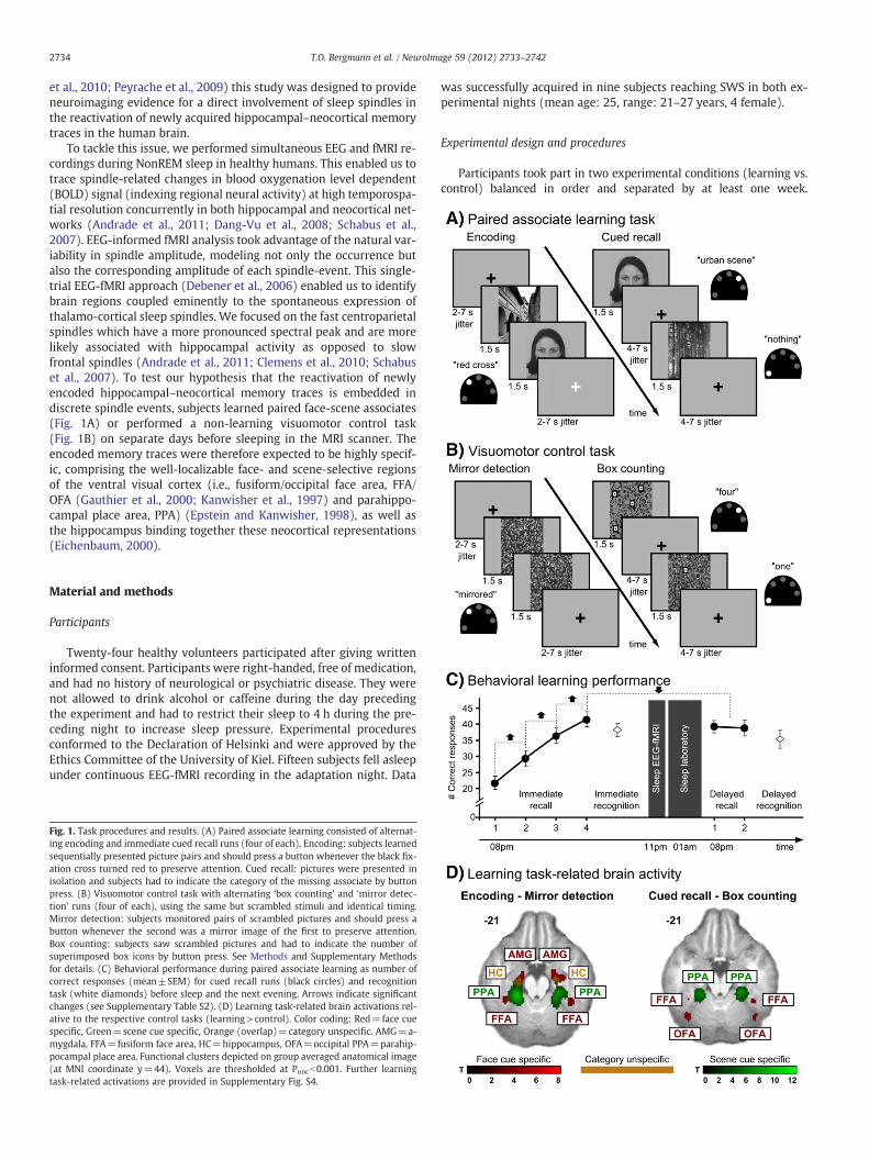
Fig. 1. Task procedures and results. (A) Paired associate learning consisted of alternat-ing encoding and immediate cued recall runs (four of each). Encoding: subjects learnedsequentially presented picture pairs and should press a button whenever the black fix-ation cross turned red to preserve attention. Cued recall: pictures were presented inisolation and subjects had to indicate the category of the missing associate by buttonpress. (B) Visuomotor control task with alternating ‘box counting’ and ‘mirror detec-tion’ runs (four of each), using the same but scrambled stimuli and identical timing.Mirror detection: subjects monitored pairs of scrambled pictures and should press abutton whenever the second was a mirror image of the first to preserve attention.Box counting: subjects saw scrambled pictures and had to indicate the number ofsuperimposed box icons by button press. See Methods and Supplementary Methodsfor details. (C) Behavioral performance during paired associate learning as number ofcorrect responses (mean±SEM) for cued recall runs (black circles) and recognitiontask (white diamonds) before sleep and the next evening. Arrows indicate significantchanges (see Supplementary Table S2). (D) Learning task-related brain activations rel-ative to the respective control tasks (learning>control). Color coding: Red=face cuespecific, Green=scene cue specific, Orange (overlap)=category unspecific. AMG=a-mygdala, FFA=fusiform face area, HC=hippocampus, OFA=occipital PPA=parahip-pocampal place area. Functional clusters depicted on group averaged anatomical image(at MNI coordinate y=44). Voxels are thresholded at Puncb0.001. Further learningtask-related activations are provided in Supplementary Fig. S4.
was successfully acquired in nine subjects reaching SWS in both ex-perimental nights (mean age: 25, range: 21–27 years, 4 female).
Experimental design and procedures
Participants took part in two experimental conditions (learning vs.control) balanced in order and separated by at least one week.
2735T.O. Bergmann et al. / NeuroImage 59 (2012) 2733–2742
Procedures were identical in both conditions except for the behavioraltask (Fig. 1). Subjects arrived around 7 pm and performed a thoroughtraining of the upcoming task to learn the assignment of responsekeys and stimulus categories until no errorsweremade anymorewithin20 consecutive trials. Around 8 pm they completed the Stanford Sleepi-ness Scale (Hoddes et al., 1973) and started the respective task-fMRI ses-sion (lasting ~70 min). In the learning condition it was followed by arecognition task outside the scanner (Supplementary Fig. S1). Aftermounting the EEG cap subjects returned to the scanner around 11 pmto sleep formax. 2.5 h during continuous EEG-fMRI recordings. Subjectswore light sleeping clothes and earplugs (~35 dB sound attenuation),were bedded comfortably on a viscoelastic mattress and covered witha light blanket. Viscoelastic foam stabilized the head inside the coiland prevented unpleasant pressure from EEG electrodes. Lights weredimmed and subjects were equipped with an alarm bell to quit the ex-periment at any time. After awakening, subjects spent the remainingnight in the adjacent sleep laboratory. They left the next morning andreturned again in the evening around 8 pm for delayed cued recall(two fMRI recall or corresponding control runs) and recognition testing(outside the scanner in the learning condition).
Behavioral tasks
The paired associate learning task consisted of four runs of mem-ory encoding, each followed by an immediate cued recall (Fig. 1A).During encoding, subjects learned 28 gray scaled picture pairs (com-binations of male faces, female faces, urban scenes, rural scenes, and‘nothing’, i.e., scrambled images) which were presented on a lightgray background and in pseudorandomized order. Paired pictureswere presented in direct succession (1.5 s per picture) with a jitteredinter-pair interval of 2–7 s (black fixation cross) and all pairs werealso presented in reversed within-pair order, making up a total of56 learning trials plus eight trials with scrambled pairs only andeight null events (fixation cross only). Subjects were asked to remem-ber the pairs as accurately as possible by visually imagining the twopictures together (neglecting within-pair order and considering pic-tures paired with a scrambled image as unpaired). It was emphasizedthat the exact stimulus combinations instead of mere categoriesshould be learned as they would be tested later (recognition task).Further, subjects had to press a button whenever the fixation crossbriefly turned red (9 times per run) to foster sustained attention. Dur-ing recall, all face and scene stimuli (48 in total) were presented inisolation (1.5 s each, inter-stimulus interval 4–7 s) and subjectswere asked to retrieve the missing associate as clearly as possibleand to indicate its category by pressing the corresponding of five but-tons. Accuracy was strongly emphasized over speed and subjectsshould also respond when they could only guess. Eight pauses of15.5 s (fixation cross) were intermingled between trials. Total runtime was 9.3 min for encoding and 8 min for recall runs, resulting ina total scan time of 69.3 min.
The visuomotor control tasks were constructed in parallel (withidentical timing) employing two alternating types of simple monitor-ing tasks, i.e., ‘mirror detection’ (cf. encoding) and ‘box counting’ (cf.cued recall). Control tasks were matched for basic visual input andmotor output (the same but scrambled stimuli were presented withidentical order and timing and responses with the same fingerswere made at the same time) and required a certain level of attentionand stimulus processing but no associative learning and no face- orscene-processing (Fig. 1B). During ‘mirror detection’, pairs of scram-bled pictures were presented and subjects had to press a buttonwhenever the second was a horizontal mirror image of the first (9times per run). During ‘box counting’, one to five high-contrast boxicons were presented on top of the scrambled images and subjectshad to indicate their number by pressing one out of five buttons. Nofeedback of response accuracy was provided in any of the tasks.
In the recognition task subjects had to identify for each stimulusthe corresponding paired associate from the complete picture set(Supplementary Fig. S1). See Supplementary Methods for a descrip-tion of the stimulus material.
EEG data acquisition
32-channel EEGwas acquired viaMR-compatible Ag–AgCl ring elec-trodes with 5 kΩ safety resistors from the following locations (10–20systems): Fp1, Fpz, Fp2, F7, F3, Fz, F4, F8, FC5, FC1, FC2, FC6, C3, Cz, C4,CP5, CP1, CP2, CP6, P7, P3, Pz, P4, P8, POz, O1, Oz, O2, T7, T8, TP9,TP10; referenced to FCz; ground ~1 cm below Oz (BrainCap MR, Easy-Cap, Munich, Germany). Skin resistance was kept below 5 kΩ (plus5 kΩ safety resistors) usingAbralyt HiCl electrode paste (Easycap) to en-sure stable EEG recordings throughout the whole night. Bipolarrecordings of vertical (below and above right eye) and horizontal elec-trooculogram (outer canthi), electromyogram (chin), and electrocar-diogram (on the backbone, ~25 cm below and above the heart) wereacquired via MR-compatible Ag–AgCl cup electrodes with 10 kΩ safetyresistors (EasyCap). Data were recorded using battery-driven BrainAmpMR plus DC and bipolar BrainAmp ExG MR amplifiers, respectively(BrainProducts, Munich, Germany), transmitted via fiber optic cablesto a laptop outside the scanner room, analog-filtered (0.016–250 Hz),and digitized with a resolution of 0.5 μV/bit at 5 kHz synchronized tothe MR scanner clock (BrainVision Recorder V.1.10; BrainProducts).EEG electrodes were coregistered to individual anatomical MR imagesusing frameless stereotaxy for consistent positioning across sessions(TMS-Navigator; Localite, Sankt Augustin, Germany).
EEG data analyses
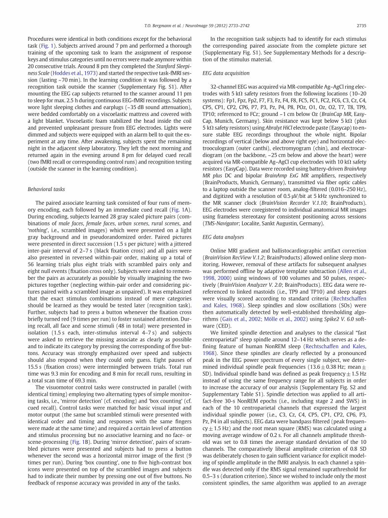
Online MRI gradient and ballistocardiographic artifact correction(BrainVision RecView V.1.2; BrainProducts) allowed online sleep mon-itoring. However, removal of these artifacts for subsequent analyseswas performed offline by adaptive template subtraction (Allen et al.,1998, 2000) using windows of 100 volumes and 50 pulses, respec-tively (BrainVision Analyzer V. 2.0; BrainProducts). EEG data were re-referenced to linked mastoids (i.e., TP9 and TP10) and sleep stageswere visually scored according to standard criteria (Rechtschaffenand Kales, 1968). Sleep spindles and slow oscillations (SOs) werethen automatically detected by well-established thresholding algo-rithms (Gais et al., 2002; Mölle et al., 2002) using Spike2 V. 6.0 soft-ware (CED).
We limited spindle detection and analyses to the classical “fastcentroparietal” sleep spindle around 12–14 Hz which serves as a de-fining feature of human NonREM sleep (Rechtschaffen and Kales,1968). Since these spindles are clearly reflected by a pronouncedpeak in the EEG power spectrum of every single subject, we deter-mined individual spindle peak frequencies (13.6±0.38 Hz; mean±SD). Individual spindle band was defined as peak frequency±1.5 Hzinstead of using the same frequency range for all subjects in orderto increase the accuracy of our analysis (Supplementary Fig. S2 andSupplementary Table S1). Spindle detection was applied to all arti-fact-free 30-s NonREM epochs (i.e., including stage 2 and SWS) ineach of the 10 centroparietal channels that expressed the largestindividual spindle power (i.e., C3, Cz, C4, CP5, CP1, CP2, CP6, P3,Pz, P4 in all subjects). EEG data were bandpass filtered (peak frequen-cy±1.5 Hz) and the root mean square (RMS) was calculated using amoving average window of 0.2 s. For all channels amplitude thresh-old was set to 0.8 times the average standard deviation of the 10channels. The comparatively liberal amplitude criterion of 0.8 SDwas deliberately chosen to gain sufficient variance for explicit model-ing of spindle amplitude in the fMRI analysis. In each channel a spin-dle was detected only if the RMS signal remained suprathreshold for0.5–3 s (duration criterion). Since we wished to include only the mostconsistent spindles, the same algorithm was applied to an average
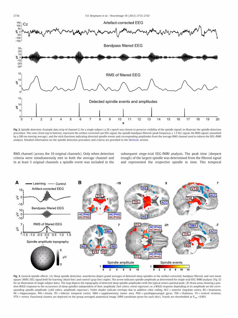
Fig. 2. Spindle detection. Example data strip of channel Cz for a single subject (a 20 s epoch was chosen to preserve visibility of the spindle signal) to illustrate the spindle detectionprocedure. The rows (from top to bottom) represent the artifact corrected raw EEG signal, the spindle bandpass filtered (peak frequency±1.5 Hz) signal, the RMS signal (smoothedby a 200 ms moving average), and the stick functions indicating detected spindle events and corresponding amplitudes from the average RMS channel used to inform the EEG-fMRIanalysis. Detailed information on the spindle detection procedure and criteria are provided in the Methods section.
2736 T.O. Bergmann et al. / NeuroImage 59 (2012) 2733–2742
RMS channel (across the 10 original channels). Only when detectioncriteria were simultaneously met in both the average channel andin at least 5 original channels a spindle event was included in the
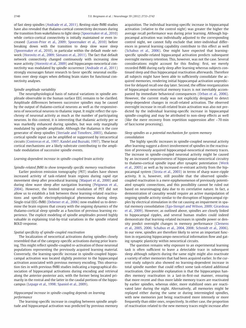
Fig. 3. General spindle effects. (A) Sleep spindle detection: waveforms depict grand averagesquare (RMS) EEG signal both for learning (black line) and control (gray line) nights. The arrfor an illustration of single subject data). The map depicts the topography of detected sleep sitive BOLD response to the occurrence of sleep spindles independent of their amplitude (hosponding spindle amplitude (cold colors; amplitude regressor). Violet shades indicate ovHC=hippocampus, INS=Insula, ITC=Inferior temporal cortex, SMA=supplementaryVTX=vertex. Functional clusters are depicted on the group averaged anatomical image (M
subsequent singe-trial EEG-fMRI analysis. The peak time (deepesttrough) of the largest spindle was determined from the filtered signaland represented the respective spindle in time. This temporal
s of detected sleep spindles in the artifact-corrected, bandpass filtered, and root meanow indicates spindle amplitude as determined for single-trial EEG-fMRI analysis (Fig. S2pindle amplitudes with the typical centro-parietal peak. (B) Brain areas showing a pos-t colors; event regressor) or a BOLD response depending in its amplitude on the corre-erlaps due to additive color coding. ACC=anterior cingulate cortex, BS=brainstem,motor area, PHG=parahippocampal gyrus, THL=thalamus, VS=ventral striatum,NI coordinate given for each slice). Voxels are thresholded at Puncb0.001.
2737T.O. Bergmann et al. / NeuroImage 59 (2012) 2733–2742
information was used to define spindle events in the fMRI analysiswhereas the corresponding RMS maximum from the average channelwas used as an index of spindle amplitude modulation over time. Theamplitude modulation was implemented in subsequent event-relatedfMRI analysis as an additional parametric modulator of the spindle re-lated BOLD response (Figs. 2 and 3A). In addition, also SOs were in-cluded in the fMRI analysis (see Supplementary Methods for detailson SO detection) to partial out the impact of SOs on the BOLD signal(Dang-Vu et al., 2008; Schabus et al., 2007).
fMRI data acquisition
fMRI was performed on a 3 Tesla MR scanner (Philips Achieva; Phi-lips Medical Systems, Best, The Netherlands). High-resolution T1-weighted anatomical images were obtained using a standard MPRAGEsequence (TR=7.7 ms, TE=3.6 ms, flip angle=8°, 170 sagittal slices,1×1×1 mm voxel size, field of view=224×224 mm). For task-fMRI,we used an echo planar imaging (EPI) sequence (TR=2500 ms,TE=37ms, flip angle=90°, FOV=216×216 mm, 38 transversalslices, slice thickness=3 mm, gap=10%, in plane voxel size=3.38×3.38 mm, continuous bottom-up slice acquisition order). Sleep-fMRI parameters were slightly modified (TR=2240 ms, TE=30ms)to ensure that residual gradient artifacts (in the slice repetition rate of~17 Hz) did not compromise the sleep spindle frequency.
fMRI data analyses
PreprocessingfMRI data were analyzed using SPM5 (www.fil.ion.ucl.ac.uk/spm)
running on Matlab 7.7 (The MathWorks, Natick, MA, USA). EPI vol-umes were slice time corrected (with reference to the middle sliceof each volume) and realigned to the mean image (4th order b-spline interpolation). Individual T1-weighted images were coregis-tered to the mean EPI, segmented using the tissue probability mapsprovided by SPM5, and concurrently transformed into the standardMontreal Neurological Institute (MNI) space. EPI volumes were thennormalized with the same spatial transformation and smoothedusing an 8 mm full-width at half-maximum isotropic Gaussian kernel.
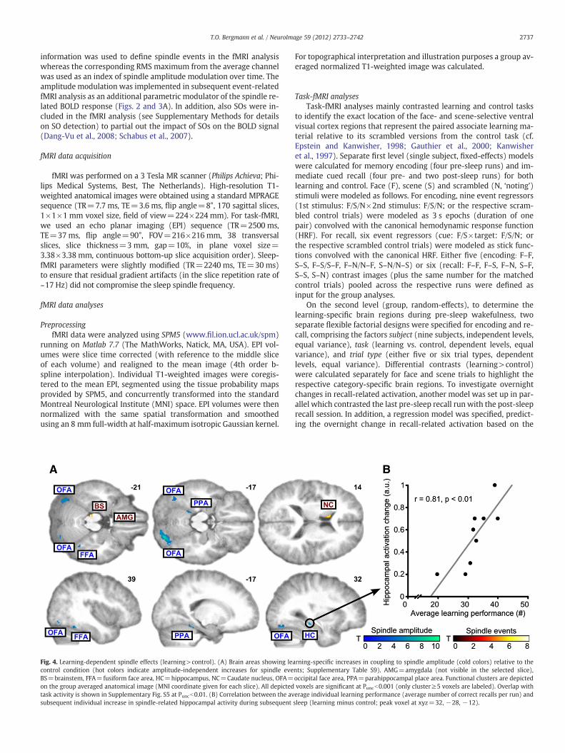
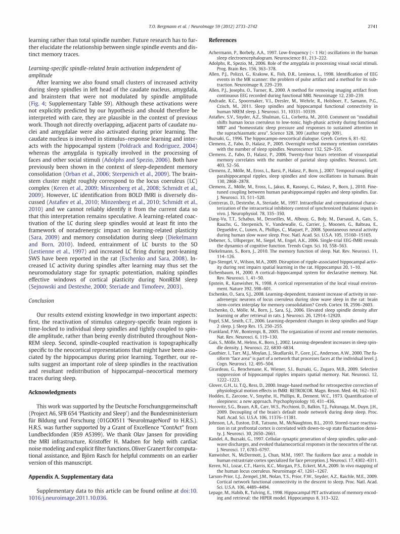
Fig. 4. Learning-dependent spindle effects (learning>control). (A) Brain areas showing leacontrol condition (hot colors indicate amplitude-independent increases for spindle eveBS=brainstem, FFA=fusiform face area, HC=hippocampus, NC=Caudate nucleus, OFA=on the group averaged anatomical image (MNI coordinate given for each slice). All depictedtask activity is shown in Supplementary Fig. S5 at Puncb0.01. (B) Correlation between the avsubsequent individual increase in spindle-related hippocampal activity during subsequent
For topographical interpretation and illustration purposes a group av-eraged normalized T1-weighted image was calculated.
Task-fMRI analysesTask-fMRI analyses mainly contrasted learning and control tasks
to identify the exact location of the face- and scene-selective ventralvisual cortex regions that represent the paired associate learning ma-terial relative to its scrambled versions from the control task (cf.Epstein and Kanwisher, 1998; Gauthier et al., 2000; Kanwisheret al., 1997). Separate first level (single subject, fixed-effects) modelswere calculated for memory encoding (four pre-sleep runs) and im-mediate cued recall (four pre- and two post-sleep runs) for bothlearning and control. Face (F), scene (S) and scrambled (N, ‘noting’)stimuli were modeled as follows. For encoding, nine event regressors(1st stimulus: F/S/N×2nd stimulus: F/S/N; or the respective scram-bled control trials) were modeled as 3 s epochs (duration of onepair) convolved with the canonical hemodynamic response function(HRF). For recall, six event regressors (cue: F/S×target: F/S/N; orthe respective scrambled control trials) were modeled as stick func-tions convolved with the canonical HRF. Either five (encoding: F–F,S–S, F–S/S–F, F–N/N–F, S–N/N–S) or six (recall: F–F, F–S, F–N, S–F,S–S, S–N) contrast images (plus the same number for the matchedcontrol trials) pooled across the respective runs were defined asinput for the group analyses.
On the second level (group, random-effects), to determine thelearning-specific brain regions during pre-sleep wakefulness, twoseparate flexible factorial designs were specified for encoding and re-call, comprising the factors subject (nine subjects, independent levels,equal variance), task (learning vs. control, dependent levels, equalvariance), and trial type (either five or six trial types, dependentlevels, equal variance). Differential contrasts (learning>control)were calculated separately for face and scene trials to highlight therespective category-specific brain regions. To investigate overnightchanges in recall-related activation, another model was set up in par-allel which contrasted the last pre-sleep recall run with the post-sleeprecall session. In addition, a regression model was specified, predict-ing the overnight change in recall-related activation based on the
rning-specific increases in coupling to spindle amplitude (cold colors) relative to thents; Supplementary Table S9). AMG=amygdala (not visible in the selected slice),occipital face area, PPA=parahippocampal place area. Functional clusters are depictedvoxels are significant at Puncb0.001 (only cluster≥5 voxels are labeled). Overlap witherage individual learning performance (average number of correct recalls per run) andsleep (learning minus control; peak voxel at xyz=32, −28, −12).
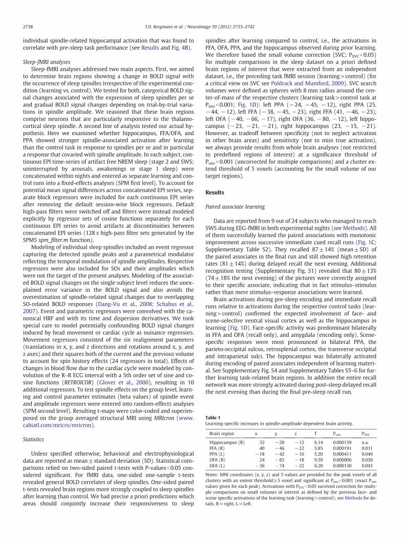
Table 1Learning-specific increases in spindle-amplitude dependent brain activity.
Brain region x y z T Punc PSVC
Hippocampus (R) 32 −28 −12 6.14 0.000139 n.a.FFA (R) 40 −46 −22 5.85 0.000191 0.031PPA (L) −18 −42 −16 5.20 0.000411 0.049OFA (R) 24 −82 −18 9.59 0.000006 0.030OFA (L) −36 −74 −22 6.20 0.000130 0.043
Notes: MNI coordinates (x, y, z) and T-values are provided for the peak voxels of allclusters with an extent threshold≥5 voxel and significant at Puncb0.001 (exact Puncvalues given for each peak). Activations with PSVCb0.05 survived correction for multi-ple comparisons on small volumes of interest as defined by the previous face- andscene specific activations of the learning task (learning>control); see Methods for de-tails. R=right, L=Left.
2738 T.O. Bergmann et al. / NeuroImage 59 (2012) 2733–2742
individual spindle-related hippocampal activation that was found tocorrelate with pre-sleep task performance (see Results and Fig. 4B).
Sleep-fMRI analysesSleep-fMRI analyses addressed two main aspects. First, we aimed
to determine brain regions showing a change in BOLD signal withthe occurrence of sleep spindles irrespective of the experimental con-dition (learning vs. control). We tested for both, categorical BOLD sig-nal changes associated with the expression of sleep spindles per seand gradual BOLD signal changes depending on trial-by-trial varia-tions in spindle amplitude. We reasoned that these brain regionscomprise neurons that are particularly responsive to the thalamo-cortical sleep spindle. A second line of analysis tested our actual hy-pothesis. Here we examined whether hippocampus, FFA/OFA, andPPA showed stronger spindle-associated activation after learningthan the control task in response to spindles per se and in particulara response that covaried with spindle amplitude. In each subject, con-tinuous EPI time-series of artifact free NREM sleep (stage 2 and SWS;uninterrupted by arousals, awakenings or stage 1 sleep) wereconcatenated within nights and entered as separate learning and con-trol runs into a fixed-effects analyses (SPM first level). To account forpotential mean signal differences across concatenated EPI series, sep-arate block regressors were included for each continuous EPI seriesafter removing the default session-wise block regressors. Defaulthigh-pass filters were switched off and filters were instead modeledexplicitly by regressor sets of cosine functions separately for eachcontinuous EPI series to avoid artifacts at discontinuities betweenconcatenated EPI series (128 s high-pass filter sets generated by theSPM5 spm_filter.m function).
Modeling of individual sleep spindles included an event regressorcapturing the detected spindle peaks and a parametrical modulatorreflecting the temporal modulation of spindle amplitudes. Respectiveregressors were also included for SOs and their amplitudes whichwere not the target of the present analyses. Modeling of the associat-ed BOLD signal changes on the single subject level reduces the unex-plained error variance in the BOLD signal and also avoids theoverestimation of spindle-related signal changes due to overlappingSO-related BOLD responses (Dang-Vu et al., 2008; Schabus et al.,2007). Event and parametric regressors were convolved with the ca-nonical HRF and with its time and dispersion derivatives. We tookspecial care to model potentially confounding BOLD signal changesinduced by head movement or cardiac cycle as nuisance regressors.Movement regressors consisted of the six realignment parameters(translations in x, y, and z directions and rotations around x, y, andz axes) and their squares both of the current and the previous volumeto account for spin history effects (24 regressors in total). Effects ofchanges in blood flow due to the cardiac cycle were modeled by con-volution of the R–R ECG interval with a 5th order set of sine and co-sine functions (RETROICOR) (Glover et al., 2000), resulting in 10additional regressors. To test spindle effects on the group level, learn-ing and control parameter estimates (beta values) of spindle eventand amplitude regressors were entered into random-effects analyses(SPM second level). Resulting t-maps were color-coded and superim-posed on the group averaged structural MRI using MRIcron (www.cabiatl.com/mricro/mricron).
Statistics
Unless specified otherwise, behavioral and electrophysiologicaldata are reported as mean±standard deviation (SD). Statistical com-parisons relied on two-sided paired t-tests with P-valuesb0.05 con-sidered significant. For fMRI data, one-sided one-sample t-testsrevealed general BOLD correlates of sleep spindles. One-sided pairedt-tests revealed brain regions more strongly coupled to sleep spindlesafter learning than control. We had precise a priori predictions whichareas should conjointly increase their responsiveness to sleep
spindles after learning compared to control, i.e., the activations inFFA, OFA, PPA, and the hippocampus observed during prior learning.We therefore based the small volume correction (SVC; PSVCb0.05)for multiple comparisons in the sleep dataset on a priori definedbrain regions of interest that were extracted from an independentdataset, i.e., the preceding task fMRI session (learning>control) (fora critical view on SVC see Poldrack and Mumford, 2009). SVC searchvolumes were defined as spheres with 8 mm radius around the cen-ter-of-mass of the respective clusters (learning task>control task atPuncb0.001; Fig. 1D): left PPA (−24, −45, −12), right PPA (25,−44, −12), left FFA (−38, −45, −23), right FFA (41, −46, −23),left OFA (−40, −66, −17), right OFA (36, −80, −12), left hippo-campus (−23, −21, −21), right hippocampus (23, −15, −21).However, as tradeoff between specificity (not to neglect activationin other brain areas) and sensitivity (not to miss true activation),we always provide results from whole brain analyses (not restrictedto predefined regions of interest) at a significance threshold ofPuncb0.001 (uncorrected for multiple comparisons) and a cluster ex-tend threshold of 5 voxels (accounting for the small volume of ourtarget regions).
Results
Paired associate learning
Data are reported from 9 out of 24 subjects who managed to reachSWS during EEG-fMRI in both experimental nights (see Methods). Allof them successfully learned the paired associations with monotonicimprovement across successive immediate cued recall runs (Fig. 1C;Supplementary Table S2). They recalled 87±14% (mean±SD) ofthe paired associates in the final run and still showed high retentionrates (81±14%) during delayed recall the next evening. Additionalrecognition testing (Supplementary Fig. S1) revealed that 80±13%(74±18% the next evening) of the pictures were correctly assignedto their specific associate, indicating that in fact stimulus–stimulusrather than mere stimulus–response associations were learned.
Brain activations during pre-sleep encoding and immediate recallruns relative to activations during the respective control tasks (lear-ning>control) confirmed the expected involvement of face- andscene-selective ventral visual cortex as well as the hippocampus inlearning (Fig. 1D). Face-specific activity was predominant bilaterallyin FFA and OFA (recall only), and amygdala (encoding only). Scene-specific responses were most pronounced in bilateral PPA, theparieto-occipital sulcus, retrosplenial cortex, the transverse occipitaland intraparietal sulci. The hippocampus was bilaterally activatedduring encoding of paired associates independent of learning materi-al. See Supplementary Fig. S4 and Supplementary Tables S5–6 for fur-ther learning task-related brain regions. In addition the entire recallnetwork wasmore strongly activated during post-sleep delayed recallthe next evening than during the final pre-sleep recall run.
2739T.O. Bergmann et al. / NeuroImage 59 (2012) 2733–2742
Spindle-coupled brain activity independent of experimental condition
Sleep spindles detected in the surface EEG to inform single-trialEEG-fMRI analyses by their corresponding amplitude showed the typ-ical waxing and waning pattern (Fig. 3A; Supplementary Fig. S3). In-dependent of the experimental learning vs. control condition, theoccurrence of spindle events per se (i.e., irrespective its correspond-ing amplitude) was associated with BOLD responses in the bilateralthalamus, hippocampus and adjacent parahippocampal gyrus, ventralstriatum, brainstem, inferior temporal cortex and cortical regionsaround the vertex (Fig. 3B; Supplementary Table S7). Of note, theBOLD response in bilateral hippocampus was clearly modulated byspindle amplitude, i.e., the higher the spindle amplitude the strongerwas the activity in these hippocampal regions. The same amplitude-dependent modulation of the BOLD signal was observed in bilateralinsula, anterior cingulate cortex, supplementary motor area, ventralstriatum, and a small thalamic cluster (Supplementary Table S8). Nei-ther the occurrence nor the amplitude of sleep spindles were associ-ated with any decrease in BOLD signal.
Learning-dependent increase in spindle-coupled brain activity
Compared to the visuomotor control task, paired associate learn-ing before sleep increased coupling of neuronal activity to spindleamplitude in a distinct set of brain regions (learning>control;Fig. 4A). The learning-specific increase in coupling between spindleamplitude and brain activity was restricted to clusters in the leftPPA, the right FFA, bilateral OFA (all PSVCb0.05) as well as the righthippocampus (Puncb0.001) (Table 1; see Supplementary Table S9for additional amplitude-independent results). The location of neo-cortical clusters matched the face- and scene-specific activations ob-served during the prior learning task (overlap maps are provided inSupplementary Fig. S5). For PPA and FFA, activations were located al-most entirely within the corresponding encoding and retrieval task-related clusters, and for the OFA there was a considerable overlapwith previous recall task-related activations.
In the hippocampus, the increase in spindle-coupled activity waslocated in the middle of its longitudinal axis, slightly posterior to thehippocampal activations observed during memory encoding. Corre-lational analyses linked this activation to individual learning perfor-mance: the better a subject performed on the learning task (averagenumber of correctly recalled paired associates) before sleep, themore pronounced was the learning-specific increase (relative tothe control night) in coupling between spindle amplitude and hip-pocampal activity (r=0.81, Pb0.01; Fig. 4B). The same holds truefor the learning slope (average increase per run; r=0.67, Pb0.05).However, the increase in spindle-related hippocampal activationdid neither predict one-day retention of the learned associates (in-dicated by the ratio of delayed to immediate cued recall or recogni-tion) nor the observed overnight increase in recall-related BOLDresponse.
Spindle-informed fMRI comparisons between sleep after learningand control sleep were not biased by general differences betweenthe sessions. Sleep architecture was closely comparable (Supplemen-tary Table S3) and none of the relevant parameters differed betweensessions, i.e., total scan time and analyzed sleep time, number of 30-sepochs in NonREM sleep stage 2 and slow wave sleep (SWS) includedin the fMRI analysis, as well as number, density and amplitude ofsleep spindles and SOs (all P>0.3; Supplementary Table S4). Subjectsalso reported similar levels of sleepiness prior to task performance(Stanford Sleepiness Scale: 4.8±1.2 vs. 5.2±1.3; P>0.3).
Discussion
Prior learning of face-scene associations gave rise to a strongercoupling between sleep spindles and neural activity in face- and
scene-selective regions of the ventral visual cortex as well as the hip-pocampus. Specifically, neural activity in these regions covaried moreclosely with ongoing variations in spindle amplitude after learningthan after a non-learning visuomotor control task. The increase incoupling between spindle amplitude and brain activity was clearly re-lated to previous learning. In the neocortex, increased coupling wastopographically restricted to face- and scene-specific regions thatwere active during prior learning. In the hippocampus, increased cou-pling was predicted by individual learning performance before sleep.Our findings are consistent with the idea that spindles represent a keymechanism for hippocampal-neocortical reprocessing of recently ac-quired memory traces during sleep, although in the light of the rela-tively small sample size only cautious conclusions can be drawn(Diekelmann and Born, 2010; Marshall and Born, 2007; Sejnowskiand Destexhe, 2000; Steriade and Timofeev, 2003).
Paired associate learning
We chose a paired associate learning task which not only relied onthe hippocampus, binding together neocortical representations(Eichenbaum, 2000; Frankland and Bontempi, 2005), but also strong-ly engaged category-specific neocortical regions of the ventral visualcortex, i.e., FFA (Kanwisher et al., 1997), OFA (Gauthier et al., 2000),and PPA (Epstein and Kanwisher, 1998) compared to a visuomotorcontrol task. This allowed us to identify neocortical regions as candi-date areas that would show learning-specific reactivation, conjointlywith the hippocampus, during subsequent sleep spindles. Althoughsleep deprivation or disrupted SWS can be detrimental to encodingperformance and related hippocampal activity (Van Der Werf et al.,2009; Yoo et al., 2007), our subjects still showed good learning per-formance together with distinct encoding-related hippocampal acti-vation, probably because the relatively mild sleep restriction in ourstudy did not affect the SWS rich first half of the night. Importantly,sleep restriction and accompanying tiredness did not differ betweenexperimental conditions.
Spindle-coupled brain activity independent of experimental condition
Hippocampal spindle couplingExtending previous findings (Schabus et al., 2007), we observed
task-independent spindle-related regional neuronal activity in vari-ous brain regions including – most importantly – the hippocampus.The pronounced bilateral activation of hippocampus and adjacentparahippocampal gyrus was not only associated with the mere oc-currence of sleep spindles but was also modulated by spindle ampli-tude. The hippocampal BOLD response was greater the larger thespindle amplitude. This observation suggests a tight link betweenhippocampal network activity and spindle input that goes beyond amere temporal synchrony of thalamo-cortical spindles and hippo-campal sharp-wave ripple activity as revealed by intracranial record-ings (Clemens et al., 2007; Mölle et al., 2009; Siapas and Wilson,1998; Sirota et al., 2003; Wierzynski et al., 2009). Within an interac-tive framework of neocortical–hippocampal loops, emergent spindleactivity might drive hippocampal ripples which in turn feed backinto ongoing cycles of the thalamo-cortical spindle to effectivelymodulate the input to neocortical neurons (Mölle et al., 2009; Sirotaet al., 2003).
Hippocampo-neocortical connectivityFurther support for a close sleep-dependent coupling between neo-
cortical regions and the hippocampus comes from recent resting-statefMRIwork showing increased hippocampo-fusiform functional connec-tivity during light sleep after face-location learning compared to post-learning wakefulness (van Dongen et al., 2011) and, learning-unrelated, a principal increase in functional connectivity between thehippocampal subiculum and neocortical sites related to the occurrence
2740 T.O. Bergmann et al. / NeuroImage 59 (2012) 2733–2742
of fast sleep spindles (Andrade et al., 2011). Resting-state fMRI studieshave also revealed that thalamo-cortical connectivity decreases duringthe transition fromwakefulness to light sleep (Spoormaker et al., 2010)while cortico-cortical connectivity is initially maintained or even in-creased (Larson-Prior et al., 2009; Spoormaker et al., 2010) beforebreaking down with the transition to deep slow wave sleep(Spoormaker et al., 2010), in particular within the default mode net-work (Horovitz et al., 2009; Sämann et al., 2011). The fact that defaultnetwork connectivity changed continuously with increasing slowwave activity (Horovitz et al., 2009) and hippocampo-neocortical con-nectivity was modulated by spindle occurrence (Andrade et al., 2011)strongly encourages future research to favor specific neuronal oscilla-tions over sleep stages when defining brain states for functional con-nectivity analyses.
Spindle amplitude variabilityThe neurophysiological basis of natural variations in spindle am-
plitude observable in the human surface EEG remains to be clarified.Amplitude differences between successive spindles may be causedby the output of thalamo-cortical neurons as well as the responsive-ness of neocortical neurons to thalamic input. It might reflect the syn-chrony of neuronal activity as much as the number of participatingneurons. In this context, it is interesting that thalamic activity per sewas markedly enhanced during spindles, but was only marginallymodulated by spindle amplitude. Although the thalamus is the coregenerator of sleep spindles (Steriade and Timofeev, 2003), thalamo-cortical spindle input can be amplified or suppressed by intracorticalcircuits (Contreras et al., 1997; Kandel and Buzsaki, 1997). These localcortical mechanisms are a likely substrate contributing to the ampli-tude modulation of successive spindle events.
Learning-dependent increase in spindle-coupled brain activity
Spindle-related fMRI to show temporally specific memory reactivationEarlier positron emission tomography (PET) studies have shown
increased activity of task-related brain regions during rapid eyemovement sleep after procedural learning (Maquet et al., 2000) andduring slow wave sleep after navigation learning (Peigneux et al.,2004). However, the limited temporal resolution of PET did notallow us to establish a link between these learning-related reactiva-tions and specific electrophysiological phenomena during sleep.Single-trial EEG-fMRI (Debener et al., 2006) now enabled us to deter-mine the brain regions that covary with the ongoing dynamics of thethalamo-cortical sleep spindle as a function of previous learning ex-perience. The explicit modeling of spindle amplitudes proved highlyvaluable in explaining trial-by-trial variations in the spindle relatedBOLD response.
Spatial specificity of spindle-coupled reactivationThe localization of neocortical activations during spindles closely
resembled that of the category-specific activations during prior learn-ing. This might reflect spindle-coupled re-activation of those neuronalpopulations representing the previously acquired learning material.Conversely, the learning-specific increase in spindle-coupled hippo-campal activation was located slightly posterior to the hippocampalactivation associated with previous memory encoding. This observa-tion ties in with previous fMRI studies indicating a topographical dis-sociation of hippocampal activations during encoding and retrievalalong the anterior-posterior axis, with the former being located pri-marily in the rostral and the latter in the caudal portions of the hippo-campus (Lepage et al., 1998; Spaniol et al., 2009).
Hippocampal increase in spindle-coupling depends on learningperformance
The learning-specific increase in coupling between spindle ampli-tude and hippocampal activation was predicted by previous memory
acquisition. The individual learning-specific increase in hippocampalactivation (relative to the control night) was greater the higher theaverage recall performance was during prior learning. Although hip-pocampal activation was individually adjusted to the correspondingcontrol night, we cannot fully exclude that inter-individual differ-ences in general learning capability contribute to this effect as well(Schabus et al., 2006). One might have expected that learning-specific spindle-related hippocampal activation predicts subsequentovernight memory retention. This, however, was not the case. Severalconsiderations might account for this finding: first, we merelyrecorded fMRI during early sleep after learning whereas subjects con-tinued sleep and thus hippocampal reactivation afterwards. Thereforeall subjects might have been able to sufficiently consolidate the ac-quired memories, rendering initial hippocampal activation unpredic-tive for delayed recall one day later. Second, the offline reorganizationof hippocampal–neocortical memory traces is not inevitably accom-panied by immediate behavioral consequences (Orban et al., 2006).However, the current study was not explicitly designed to detectsleep-dependent changes in recall-related activation. The observedovernight increase in recall-related brain activation was also not pre-dicted by the individual learning-specific increase in hippocampalspindle-coupling and may be attributed to non-sleep effects as well(like the mere recovery from repetition suppression after ~70 minof face/scene processing).
Sleep spindles as a potential mechanism for system memoryconsolidation
The highly specific increases in spindle-coupled neuronal activityafter learning suggest a direct involvement of spindles in the reactiva-tion of previously acquired hippocampal-neocortical memory traces.The increase in spindle-coupled neuronal activity might be causedby an increased responsiveness of hippocampal-neocortical circuitryto thalamo-cortical spindle input after synaptic potentiation (Werket al., 2005) as well as by increased re-entrant activity from the hip-pocampal system (Sirota et al., 2003) in terms of sharp wave-rippleactivity. It is, however, still possible that the observed spindle-coupled reactivation is a mere epiphenomenon of previously potenti-ated synaptic connections, and this possibility cannot be ruled outbased on neuroimaging data due to its correlative nature. In fact, ademonstration of causality would require an active manipulation ofongoing spindle activity similar to the disruption of hippocampal rip-ples by electrical stimulation in the rat causing an impairment in spa-tial memory consolidation (Ego-Stengel and Wilson, 2009; Girardeauet al., 2009). However, as outlined above, spindles are closely linkedto hippocampal ripples, and several human studies could indeeddemonstrate that learning-related increases in spindle power or den-sity predict overnight changes in memory performance (Clemenset al., 2005, 2006; Schabus et al., 2004, 2008; Schmidt et al., 2006).In our view, spindles are therefore likely to serve an important func-tion in organizing hippocampo-neocortical information flow and gat-ing synaptic plasticity within neocortical circuits.
The question remains why exposure to an experimental learningtask is often sufficient to leave a detectable trace in subsequentsleep although subjects during the same night might also reactivatea variety of other memories that had been acquired earlier. In the cur-rent study subjects also showed no learning-dependent increase intotal spindle number that could reflect some task-related additionalreactivation. One possible explanation is that the hippocampus han-dles memory reactivation in a last-in-first-out manner, ensuringthat more recent and thus more labile memory traces are reactivatedby earlier spindles, whereas older, more stabilized ones are reacti-vated later during the night. Alternatively, all memories might bereplayed either during the same or during intermingled spindles,with new memories just being reactivated more intensely or morefrequently than older ones, respectively. In either case, the proportionof reactivation related to the newmemory traces might increase after
2741T.O. Bergmann et al. / NeuroImage 59 (2012) 2733–2742
learning rather than total spindle number. Future research has to fur-ther elucidate the relationship between single spindle events and dis-tinct memory traces.
Learning-specific spindle-related brain activation independent ofamplitude
After learning we also found small clusters of increased activityduring sleep spindles in left head of the caudate nucleus, amygdala,and brainstem that were not modulated by spindle amplitude(Fig. 4; Supplementary Table S9). Although these activations werenot explicitly predicted by our hypothesis and should therefore beinterpreted with care, they are plausible in the context of previouswork. Though not directly overlapping, adjacent parts of caudate nu-clei and amygdalae were also activated during prior learning. Thecaudate nucleus is involved in stimulus–response learning and inter-acts with the hippocampal system (Poldrack and Rodriguez, 2004)whereas the amygdala is typically involved in the processing offaces and other social stimuli (Adolphs and Spezio, 2006). Both havepreviously been shown in the context of sleep-dependent memoryconsolidation (Orban et al., 2006; Sterpenich et al., 2009). The brain-stem cluster might roughly correspond to the locus coeruleus (LC)complex (Keren et al., 2009; Minzenberg et al., 2008; Schmidt et al.,2009). However, LC identification from BOLD fMRI is diversely dis-cussed (Astafiev et al., 2010; Minzenberg et al., 2010; Schmidt et al.,2010) and we cannot reliably identify it from the current data sothat this interpretation remains speculative. A learning-related coac-tivation of the LC during sleep spindles would at least fit into theframework of noradrenergic impact on learning-related plasticity(Sara, 2009) and memory consolidation during sleep (Diekelmannand Born, 2010). Indeed, entrainment of LC bursts to the SO(Lestienne et al., 1997) and increased LC firing during post-leaningSWS have been reported in the rat (Eschenko and Sara, 2008). In-creased LC activity during spindles after learning may thus set theneuromodulatory stage for synaptic potentiation, making spindleseffective windows of cortical plasticity during NonREM sleep(Sejnowski and Destexhe, 2000; Steriade and Timofeev, 2003).
Conclusion
Our results extend existing knowledge in two important aspects:first, the reactivation of stimulus category-specific brain regions istime-locked to individual sleep spindles and tightly coupled to spin-dle amplitude, rather than being evenly distributed throughout Non-REM sleep. Second, spindle-coupled reactivation is topographicallyspecific to the neocortical representations that might have been asso-ciated by the hippocampus during prior learning. Together, our re-sults suggest an important role of sleep spindles in the reactivationand resultant redistribution of hippocampal–neocortical memorytraces during sleep.
Acknowledgments
This work was supported by the Deutsche Forschungsgemeinschaft(Project A6, SFB 654 ‘Plasticity and Sleep’) and the Bundesministeriumfür Bildung und Forschung (01GO0511 ‘NeuroImageNord’ to H.R.S.).H.R.S. was further supported by a Grant of Excellence “ContAct” fromLundbeckfonden (R59 A5399). We thank Olav Jansen for providingthe MRI infrastructure, Kristoffer H. Madsen for help with cardiacnoisemodeling and explicit filter functions, Oliver Granert for computa-tional assistance, and Björn Rasch for helpful comments on an earlierversion of this manuscript.
Appendix A. Supplementary data
Supplementary data to this article can be found online at doi:10.1016/j.neuroimage.2011.10.036.
References
Achermann, P., Borbely, A.A., 1997. Low-frequency (b 1 Hz) oscillations in the humansleep electroencephalogram. Neuroscience 81, 213–222.
Adolphs, R., Spezio, M., 2006. Role of the amygdala in processing visual social stimuli.Prog. Brain Res. 156, 363–378.
Allen, P.J., Polizzi, G., Krakow, K., Fish, D.R., Lemieux, L., 1998. Identification of EEGevents in the MR scanner: the problem of pulse artifact and a method for its sub-traction. Neuroimage 8, 229–239.
Allen, P.J., Josephs, O., Turner, R., 2000. A method for removing imaging artifact fromcontinuous EEG recorded during functional MRI. Neuroimage 12, 230–239.
Andrade, K.C., Spoormaker, V.I., Dresler, M., Wehrle, R., Holsboer, F., Samann, P.G.,Czisch, M., 2011. Sleep spindles and hippocampal functional connectivity inhuman NREM sleep. J. Neurosci. 31, 10331–10339.
Astafiev, S.V., Snyder, A.Z., Shulman, G.L., Corbetta, M., 2010. Comment on “modafinilshifts human locus coeruleus to low-tonic, high-phasic activity during functionalMRI” and “homeostatic sleep pressure and responses to sustained attention inthe suprachiasmatic area”. Science 328, 309 (author reply 309).
Buzsaki, G., 1996. The hippocampo-neocortical dialogue. Cereb. Cortex 6, 81–92.Clemens, Z., Fabo, D., Halasz, P., 2005. Overnight verbal memory retention correlates
with the number of sleep spindles. Neuroscience 132, 529–535.Clemens, Z., Fabo, D., Halasz, P., 2006. Twenty-four hours retention of visuospatial
memory correlates with the number of parietal sleep spindles. Neurosci. Lett.403, 52–56.
Clemens, Z., Mölle, M., Eross, L., Barsi, P., Halasz, P., Born, J., 2007. Temporal coupling ofparahippocampal ripples, sleep spindles and slow oscillations in humans. Brain130, 2868–2878.
Clemens, Z., Mölle, M., Eross, L., Jakus, R., Rasonyi, G., Halasz, P., Born, J., 2010. Fine-tuned coupling between human parahippocampal ripples and sleep spindles. Eur.J. Neurosci. 33, 511–520.
Contreras, D., Destexhe, A., Steriade, M., 1997. Intracellular and computational charac-terization of the intracortical inhibitory control of synchronized thalamic inputs invivo. J. Neurophysiol. 78, 335–350.
Dang-Vu, T.T., Schabus, M., Desseilles, M., Albouy, G., Boly, M., Darsaud, A., Gais, S.,Rauchs, G., Sterpenich, V., Vandewalle, G., Carrier, J., Moonen, G., Balteau, E.,Degueldre, C., Luxen, A., Phillips, C., Maquet, P., 2008. Spontaneous neural activityduring human slow wave sleep. Proc. Natl. Acad. Sci. U.S.A. 105, 15160–15165.
Debener, S., Ullsperger, M., Siegel, M., Engel, A.K., 2006. Single-trial EEG-fMRI revealsthe dynamics of cognitive function. Trends Cogn. Sci. 10, 558–563.
Diekelmann, S., Born, J., 2010. The memory function of sleep. Nat. Rev. Neurosci. 11,114–126.
Ego-Stengel, V., Wilson, M.A., 2009. Disruption of ripple-associated hippocampal activ-ity during rest impairs spatial learning in the rat. Hippocampus 20, 1–10.
Eichenbaum, H., 2000. A cortical–hippocampal system for declarative memory. Nat.Rev. Neurosci. 1, 41–50.
Epstein, R., Kanwisher, N., 1998. A cortical representation of the local visual environ-ment. Nature 392, 598–601.
Eschenko, O., Sara, S.J., 2008. Learning-dependent, transient increase of activity in nor-adrenergic neurons of locus coeruleus during slow wave sleep in the rat: brainstem-cortex interplay for memory consolidation? Cereb. Cortex 18, 2596–2603.
Eschenko, O., Mölle, M., Born, J., Sara, S.J., 2006. Elevated sleep spindle density afterlearning or after retrieval in rats. J. Neurosci. 26, 12914–12920.
Fogel, S.M., Smith, C.T., 2006. Learning-dependent changes in sleep spindles and Stage2 sleep. J. Sleep Res. 15, 250–255.
Frankland, P.W., Bontempi, B., 2005. The organization of recent and remote memories.Nat. Rev. Neurosci. 6, 119–130.
Gais, S., Mölle, M., Helms, K., Born, J., 2002. Learning-dependent increases in sleep spin-dle density. J. Neurosci. 22, 6830–6834.
Gauthier, I., Tarr, M.J., Moylan, J., Skudlarski, P., Gore, J.C., Anderson, A.W., 2000. The fu-siform “face area” is part of a network that processes faces at the individual level. J.Cogn. Neurosci. 12, 495–504.
Girardeau, G., Benchenane, K., Wiener, S.I., Buzsaki, G., Zugaro, M.B., 2009. Selectivesuppression of hippocampal ripples impairs spatial memory. Nat. Neurosci. 12,1222–1223.
Glover, G.H., Li, T.Q., Ress, D., 2000. Image-based method for retrospective correction ofphysiological motion effects in fMRI: RETROICOR. Magn. Reson. Med. 44, 162–167.
Hoddes, E., Zarcone, V., Smythe, H., Phillips, R., Dement, W.C., 1973. Quantification ofsleepiness: a new approach. Psychophysiology 10, 431–436.
Horovitz, S.G., Braun, A.R., Carr, W.S., Picchioni, D., Balkin, T.J., Fukunaga, M., Duyn, J.H.,2009. Decoupling of the brain's default mode network during deep sleep. Proc.Natl. Acad. Sci. U.S.A. 106, 11376–11381.
Johnson, L.A., Euston, D.R., Tatsuno, M., McNaughton, B.L., 2010. Stored-trace reactiva-tion in rat prefrontal cortex is correlated with down-to-up state fluctuation densi-ty. J. Neurosci. 30, 2650–2661.
Kandel, A., Buzsaki, G., 1997. Cellular-synaptic generation of sleep spindles, spike-and-wave discharges, and evoked thalamocortical responses in the neocortex of the rat.J. Neurosci. 17, 6783–6797.
Kanwisher, N., McDermott, J., Chun, M.M., 1997. The fusiform face area: a module inhuman extrastriate cortex specialized for face perception. J. Neurosci. 17, 4302–4311.
Keren, N.I., Lozar, C.T., Harris, K.C., Morgan, P.S., Eckert, M.A., 2009. In vivo mapping ofthe human locus coeruleus. Neuroimage 47, 1261–1267.
Larson-Prior, L.J., Zempel, J.M., Nolan, T.S., Prior, F.W., Snyder, A.Z., Raichle, M.E., 2009.Cortical network functional connectivity in the descent to sleep. Proc. Natl. Acad.Sci. U.S.A. 106, 4489–4494.
Lepage, M., Habib, R., Tulving, E., 1998. Hippocampal PET activations of memory encod-ing and retrieval: the HIPER model. Hippocampus 8, 313–322.
2742 T.O. Bergmann et al. / NeuroImage 59 (2012) 2733–2742
Lestienne, R., Herve-Minvielle, A., Robinson, D., Briois, L., Sara, S.J., 1997. Slow oscilla-tions as a probe of the dynamics of the locus coeruleus-frontal cortex interactionin anesthetized rats. J. Physiol. Paris 91, 273–284.
Maquet, P., 2001. The role of sleep in learning and memory. Science 294, 1048–1052.Maquet, P., Laureys, S., Peigneux, P., Fuchs, S., Petiau, C., Phillips, C., Aerts, J., Del Fiore,
G., Degueldre, C., Meulemans, T., Luxen, A., Franck, G., Van Der Linden, M., Smith, C.,Cleeremans, A., 2000. Experience-dependent changes in cerebral activation duringhuman REM sleep. Nat. Neurosci. 3, 831–836.
Marshall, L., Born, J., 2007. The contribution of sleep to hippocampus-dependent mem-ory consolidation. Trends Cogn. Sci. 11, 442–450.
McClelland, J.L., McNaughton, B.L., O'Reilly, R.C., 1995. Why there are complementarylearning systems in the hippocampus and neocortex: insights from the successesand failures of connectionist models of learning and memory. Psychol. Rev. 102,419–457.
Minzenberg, M.J., Watrous, A.J., Yoon, J.H., Ursu, S., Carter, C.S., 2008. Modafinil shiftshuman locus coeruleus to low-tonic, high-phasic activity during functional MRI.Science 322, 1700–1702.
Minzenberg, M.J., Watrous, A.J., Yoon, J.H., La, C., Ursu, S., Carter, C.S., 2010. Response tocomment on “modafinil shifts human locus coeruleus to low-tonic, high-phasic ac-tivity during functional MRI”. Science 328, 309.
Mölle, M., Born, J., 2009. Hippocampus whispering in deep sleep to prefrontal cortex —for good memories? Neuron 61, 496–498.
Mölle, M., Marshall, L., Gais, S., Born, J., 2002. Grouping of spindle activity during slow os-cillations in human non-rapid eye movement sleep. J. Neurosci. 22, 10941–10947.
Mölle, M., Yeshenko, O., Marshall, L., Sara, S.J., Born, J., 2006. Hippocampal sharp wave-ripples linked to slow oscillations in rat slow-wave sleep. J. Neurophysiol. 96,62–70.
Mölle, M., Eschenko, O., Gais, S., Sara, S.J., Born, J., 2009. The influence of learning onsleep slow oscillations and associated spindles and ripples in humans and rats.Eur. J. Neurosci. 29, 1071–1081.
Orban, P., Rauchs, G., Balteau, E., Degueldre, C., Luxen, A., Maquet, P., Peigneux, P., 2006.Sleep after spatial learning promotes covert reorganization of brain activity. Proc.Natl. Acad. Sci. U.S.A. 103, 7124–7129.
Peigneux, P., Laureys, S., Fuchs, S., Collette, F., Perrin, F., Reggers, J., Phillips, C.,Degueldre, C., Del Fiore, G., Aerts, J., Luxen, A., Maquet, P., 2004. Are spatial mem-ories strengthened in the human hippocampus during slow wave sleep? Neuron44, 535–545.
Peyrache, A., Khamassi, M., Benchenane, K., Wiener, S.I., Battaglia, F.P., 2009. Replay ofrule-learning related neural patterns in the prefrontal cortex during sleep. Nat.Neurosci. 12, 919–926.
Poldrack, R.A., Mumford, J.A., 2009. Independence in ROI analysis: where is the voo-doo? Soc. Cogn. Affect. Neurosci. 4, 208–213.
Poldrack, R.A., Rodriguez, P., 2004. How do memory systems interact? Evidence fromhuman classification learning. Neurobiol. Learn. Mem. 82, 324–332.
Rasch, B., Born, J., 2007. Maintaining memories by reactivation. Curr. Opin. Neurobiol.17, 698–703.
Rechtschaffen, A., Kales, A., 1968. A Manual of Standardized Terminology, Techniquesand Scoring System for Sleep Stages of Human Subjects. United States GovernmentPrinting Office, Washington, DC.
Sämann, P.G., Wehrle, R., Hoehn, D., Spoormaker, V.I., Peters, H., Tully, C., Holsboer, F.,Czisch, M., 2011. Development of the Brain's default mode network from wakeful-ness to slow wave sleep. Cereb. Cortex 21, 2082–2093.
Sara, S.J., 2009. The locus coeruleus and noradrenergic modulation of cognition. Nat.Rev. Neurosci. 10, 211–223.
Schabus, M., Gruber, G., Parapatics, S., Sauter, C., Klosch, G., Anderer, P., Klimesch, W.,Saletu, B., Zeitlhofer, J., 2004. Sleep spindles and their significance for declarativememory consolidation. Sleep 27, 1479–1485.
Schabus, M., Hodlmoser, K., Gruber, G., Sauter, C., Anderer, P., Klosch, G., Parapatics, S.,Saletu, B., Klimesch, W., Zeitlhofer, J., 2006. Sleep spindle-related activity in thehuman EEG and its relation to general cognitive and learning abilities. Eur. J. Neu-rosci. 23, 1738–1746.
Schabus, M., Dang-Vu, T.T., Albouy, G., Balteau, E., Boly, M., Carrier, J., Darsaud, A.,Degueldre, C., Desseilles, M., Gais, S., Phillips, C., Rauchs, G., Schnakers, C., Sterpenich,V., Vandewalle, G., Luxen, A., Maquet, P., 2007. Hemodynamic cerebral correlates ofsleep spindles during human non-rapid eye movement sleep. Proc. Natl. Acad. Sci.U.S.A. 104, 13164–13169.
Schabus, M., Hoedlmoser, K., Pecherstorfer, T., Anderer, P., Gruber, G., Parapatics, S.,Sauter, C., Kloesch, G., Klimesch, W., Saletu, B., Zeitlhofer, J., 2008. Interindividualsleep spindle differences and their relation to learning-related enhancements.Brain Res. 1191, 127–135.
Schmidt, C., Peigneux, P., Muto, V., Schenkel, M., Knoblauch, V., Munch, M., deQuervain, D.J., Wirz-Justice, A., Cajochen, C., 2006. Encoding difficulty pro-motes postlearning changes in sleep spindle activity during napping. J. Neu-rosci. 26, 8976–8982.
Schmidt, C., Collette, F., Leclercq, Y., Sterpenich, V., Vandewalle, G., Berthomier, P.,Berthomier, C., Phillips, C., Tinguely, G., Darsaud, A., Gais, S., Schabus, M., Desseilles,M., Dang-Vu, T.T., Salmon, E., Balteau, E., Degueldre, C., Luxen, A., Maquet, P., Cajo-chen, C., Peigneux, P., 2009. Homeostatic sleep pressure and responses to sustainedattention in the suprachiasmatic area. Science 324, 516–519.
Schmidt, C., Peigneux, P., Maquet, P., Phillips, C., 2010. Response to comment on “ho-meostatic sleep pressure and responses to sustained attention in the suprachias-matic area”. Science 328, 309.
Sejnowski, T.J., Destexhe, A., 2000. Why do we sleep? Brain Res. 886, 208–223.Siapas, A.G., Wilson, M.A., 1998. Coordinated interactions between hippocampal rip-
ples and cortical spindles during slow-wave sleep. Neuron 21, 1123–1128.Sirota, A., Csicsvari, J., Buhl, D., Buzsaki, G., 2003. Communication between neocortex
and hippocampus during sleep in rodents. Proc. Natl. Acad. Sci. U.S.A. 100,2065–2069.
Spaniol, J., Davidson, P.S., Kim, A.S., Han, H., Moscovitch, M., Grady, C.L., 2009. Event-related fMRI studies of episodic encoding and retrieval: meta-analyses using ac-tivation likelihood estimation. Neuropsychologia 47, 1765–1779.
Spoormaker, V.I., Schroter, M.S., Gleiser, P.M., Andrade, K.C., Dresler, M., Wehrle, R.,Samann, P.G., Czisch, M., 2010. Development of a large-scale functional brain net-work during human non-rapid eye movement sleep. J. Neurosci. 30, 11379–11387.
Steriade, M., 2006. Grouping of brain rhythms in corticothalamic systems. Neurosci-ence 137, 1087–1106.
Steriade, M., Timofeev, I., 2003. Neuronal plasticity in thalamocortical networks duringsleep and waking oscillations. Neuron 37, 563–576.
Steriade, M., Nunez, A., Amzica, F., 1993. A novel slow (b 1 Hz) oscillation of neocorticalneurons in vivo: depolarizing and hyperpolarizing components. J. Neurosci. 13,3252–3265.
Sterpenich, V., Albouy, G., Darsaud, A., Schmidt, C., Vandewalle, G., Dang Vu, T.T., Des-seilles, M., Phillips, C., Degueldre, C., Balteau, E., Collette, F., Luxen, A., Maquet, P.,2009. Sleep promotes the neural reorganization of remote emotional memory. J.Neurosci. 29, 5143–5152.
Stickgold, R., 2005. Sleep-dependent memory consolidation. Nature 437, 1272–1278.Sutherland, G.R., McNaughton, B., 2000. Memory trace reactivation in hippocampal and
neocortical neuronal ensembles. Curr. Opin. Neurobiol. 10, 180–186.Van Der Werf, Y.D., Altena, E., Schoonheim, M.M., Sanz-Arigita, E.J., Vis, J.C., De Rijke,
W., Van Someren, E.J., 2009. Sleep benefits subsequent hippocampal functioning.Nat. Neurosci. 12, 122–123.
van Dongen, E.V., Takashima, A., Barth, M., Fernandez, G., 2011. Functional connectivityduring light sleep is correlated with memory performance for face-location associ-ations. Neuroimage 57, 262–270.
Werk, C.M., Harbour, V.L., Chapman, C.A., 2005. Induction of long-term potentiationleads to increased reliability of evoked neocortical spindles in vivo. Neuroscience131, 793–800.
Wierzynski, C.M., Lubenov, E.V., Gu, M., Siapas, A.G., 2009. State-dependent spike-timing relationships between hippocampal and prefrontal circuits during sleep.Neuron 61, 587–596.
Yoo, S.S., Hu, P.T., Gujar, N., Jolesz, F.A., Walker, M.P., 2007. A deficit in the ability toform new human memories without sleep. Nat. Neurosci. 10, 385–392.