Embed Size (px)
Citation preview

Journal of Case Reports and Images in Orthopedics and Rheumatology, Vol. 3, 2018.
J Case Rep Images Orthop Rheum 2018;3:100013Z14RM2018. www.edoriumjournals.com/ej/crj/jcrior
Magetsari et al. 1
CASE REPORT OPEN ACCESS
Benign fibrous histiocytoma of the right clavicle: A case report
Rahadyan Magetsari, Hendi Dwi Bharata
ABSTRACT
Benign Fibrous Histiocytoma (BFH) occurs approximately, 1% of all benign bone tumors and mostly BFH occurs in the long bones where femur and tibia are most frequently involved. Clavicle is not the common site for all bone tumors and majority of the clavicular tumor has malignant characteristics. BFH of the clavicle is rare, therefore to rule out the diagnosis we need to increase the awareness among clinician and multidisciplinary discussion. A 20-year-old male presented with chief complaint of mass in the right shoulder. Two years prior to admission, he had traffic accident and sustained right clavicle fracture. ORIF using plate and screw was carried out. 10 months prior to admission, he complained about cough but recovered after receiving treatment from pulmonologist. Four months later, the fracture had recovered and he underwent implant removal. He began to feel painless growing mass in the former site of surgery one month before admission. From physical examination, a mass size 8x8x2 cm with regular border and tenderness was found in his right shoulder without range of motion limitation. Laboratory results were normal. Plain radiograph suspected non union right clavicle.
Rahadyan Magetsari1, Hendi Dwi Bharata2
Affiliations: 1Staff Department of Orthopaedics and Trauma-tology, Sardjito General Hospital, Medical Faculty, Gadjah-Mada University, Yogyakarta, Indonesia; 2Resident, Sardjito General Hospital, Medical Faculty, GadjahMada University, Yogyakarta, Indonesia.Corresponding Author: Hendi Dwi Bharata, Resident, Sard-jito General Hospital, Medical Faculty, GadjahMada Uni-versity, Yogyakarta, Indonesia; Email: [email protected]
Received: 18 April 2018Accepted: 22 May 2018Published: 14 June 2018
CT of the right shoulder revealed soft tissue nodule 5.8x5.6x3 cm with clavicle destruction. It grew expansively became 9x6x4 cm in 2 months. A FNAB was performed with a result of benign fibrous histiocytoma. Unfortunately, the patient refused all medical procedures offered in the outpatient clinic.
Keywords: Benign fibrous histiocytoma, Clavicle, Soft tissue tumor
How to cite this article
Magetsari R, Bharata HD. Benign fibrous histiocytoma of the right clavicle: A case report. J Case Rep Images Orthop Rheum 2018;3:100013Z14RM2018.
Article ID: 100013Z14RM2018
*********
doi: 10.5348/100013Z14RM2018CR
INTRODUCTION
Benign Fibrous Histiocytoma (BFH) occurs approximately in 1% of all benign bone tumors and mostly BFH occurs in the long bones where femur and tibia are most frequently involved, but various authors reported BFH from pelvic, lumbar spine and rib [1]. In addition, the clavicle is not the common site for all bone tumors and majority of the clavicular tumors have malignant characteristics. It is the only long bone whose anatomical position is in the horizontal axis. It lacks a definite medullary cavity and ossifies by membranous ossification. It has two primary centres of ossification and only one secondary centre of ossification, the sternal end. It is the first bone to ossify in the embryo (fifth month). This bone is subcutaneous throughout its length, and it is
CASE REPORT PEER REVIEWED | OPEN ACCESS
Journal of Case Reports and Images in Orthopedics and Rheumatology, Vol. 3, 2018.
Magetsari et al. 2J Case Rep Images Orthop Rheum 2018;3:100013Z14RM2018. www.edoriumjournals.com/ej/crj/jcrior
occasionally pierced by the middle supraclavicular nerve. Fortunately, the clavicle is a bone that can be resected without causing significant disability [2]. The tumor can involve patients with a wide range of ages from 5 to 75 years. Usually involved patients are older than those with a non-ossifying fibroma. No sex predilection has been reported for this tumor [3].
Therefore, we presented a rare case of 20-year-old patient with benign fibrous histiocytoma on the right shoulder.
CASE REPORT
A 20-year-old male presented with chief complaint of mass in the right shoulder. Two years prior to admission, he had traffic accident and sustained right clavicle fracture. At that time, open reduction internal fixation using plate and screw was carried out. Ten months prior to admission, he complained about cough but recovered shortly afterwards after visiting pulmonologist and received treatment. Four months after, the fracture said to be recovered, therefore he underwent implant removal of the clavicle. He began to realized that there was painless growing mass on the former site of surgery one month before admission.
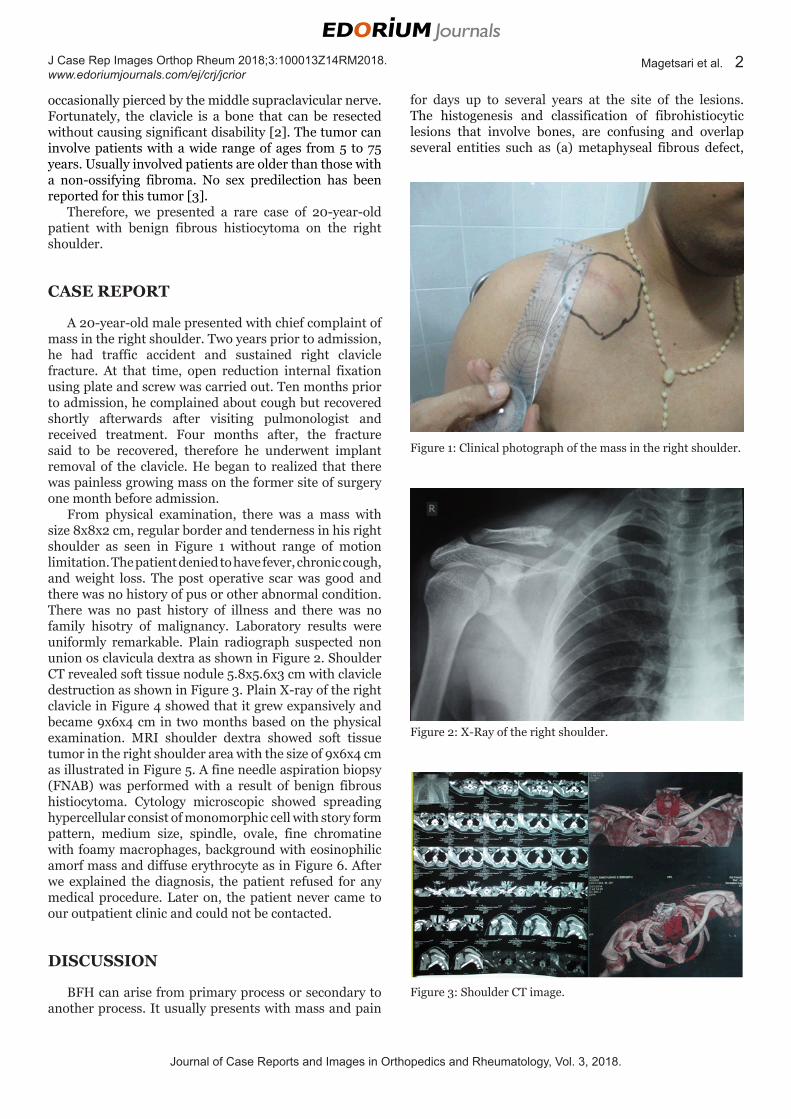
From physical examination, there was a mass with size 8x8x2 cm, regular border and tenderness in his right shoulder as seen in Figure 1 without range of motion limitation. The patient denied to have fever, chronic cough, and weight loss. The post operative scar was good and there was no history of pus or other abnormal condition. There was no past history of illness and there was no family hisotry of malignancy. Laboratory results were uniformly remarkable. Plain radiograph suspected non union os clavicula dextra as shown in Figure 2. Shoulder CT revealed soft tissue nodule 5.8x5.6x3 cm with clavicle destruction as shown in Figure 3. Plain X-ray of the right clavicle in Figure 4 showed that it grew expansively and became 9x6x4 cm in two months based on the physical examination. MRI shoulder dextra showed soft tissue tumor in the right shoulder area with the size of 9x6x4 cm as illustrated in Figure 5. A fine needle aspiration biopsy (FNAB) was performed with a result of benign fibrous histiocytoma. Cytology microscopic showed spreading hypercellular consist of monomorphic cell with story form pattern, medium size, spindle, ovale, fine chromatine with foamy macrophages, background with eosinophilic amorf mass and diffuse erythrocyte as in Figure 6. After we explained the diagnosis, the patient refused for any medical procedure. Later on, the patient never came to our outpatient clinic and could not be contacted.
DISCUSSION
BFH can arise from primary process or secondary to another process. It usually presents with mass and pain
for days up to several years at the site of the lesions. The histogenesis and classification of fibrohistiocytic lesions that involve bones, are confusing and overlap several entities such as (a) metaphyseal fibrous defect,
Figure 1: Clinical photograph of the mass in the right shoulder.
Figure 2: X-Ray of the right shoulder.
Figure 3: Shoulder CT image.

Journal of Case Reports and Images in Orthopedics and Rheumatology, Vol. 3, 2018.
Magetsari et al. 3J Case Rep Images Orthop Rheum 2018;3:100013Z14RM2018. www.edoriumjournals.com/ej/crj/jcrior
(b) nonossifying fibroma, (c) fibrous cortical defect, (d) fibrous xanthoma and (e) benign fibrous histiocytoma [4]. Local pain is the main symptom in benign fibrous histiocytoma in contrast to nonossifying fibroma and the X-ray shows an osteolytic lesion with sharply defined sclerotic borders in a typical case of benign fibrous histiocytoma [5]
Histological appearance of BFH of bone is characterized by proliferation of fibroblasts and histiocytes with many multinucleated giant cells. The fibroblasts are arranged in storiform pattern, the giant cells tend to have fewer nuclei than those found osteoclastoma or giant cell tumor [6]. Most neoplasms of the bone may contain giant cells but uniform spatial arrangement of osteoclastic giant cells with mononuclear stromal cells is critical in making a diagnosis of giant cell tumour. Lesions with osteoclastic giant cells and spindle cells if present in flat bones also need to be differentiated from hyperparathyroidism and aneurysmal bone cyst which have other distinctive histological features [5].
BFH may show indistinct borders with an aggressive pattern. It can be locally aggressive and amputation may be necessary to eliminate the tumor after recurrence. Suggested treatments for this tumor are curettage and filling of the defect with bone graft or cement. Recurrence is a risk in treatment and there are reports of recurrence and variable rate of amputation afterwards [3].
To rule out the diagnosis of BFH, complete history taking, physical examination, radiograph imaging, and histopathological examination were carried out.. BFH may recur after curettage and grew expansively at the local site as seen in a report where five out of eight patients had pain and three patients had recurrence and two patients had undergone amputation [7].
CONCLUSION
BFH of the right clavicle is a rare case, therefore to rule out the diagnosis we need to increase the awareness among clinician and need multidisciplinary discussion.
REFERENCES
1. Grohs JG, Nicolakis M, Kainberger F, Lang S, Kotz R. Benign fibrous histiocytoma of bone: A report of ten cases and review of literature. Wien Klin Wochenschr 2002 Jan 15;114(1–2):56–63.
2. Kapoor S, Tiwari A, Kapoor S. Primary tumours and tumorous lesions of clavicle. Int Orthop 2008 Dec;32(6):829–34.
3. Jafan D, Mazhar FN, Shoushtarizadeh T, Bagherifard A. Benign fibrous histiocytoma of ulnar bone: Rare tumor in a rare location. Shafa Ortho J 2014;1(1):30–2.
4. Kulkarni N. Benign fibrous histiocytoma of bone: A case report. Int J of Case Rep Images 2013;4(4):224–7.
5. Forrest M, Tomeno B, Vanel D. Orthopaedic Surgical Pathology: Diagnosis of Tumours and Pseudotumoral Lesions of Bone and Joint. Edinburg: Churchill livingstone; 1997. p. 317–21.
6. Azouz EM. Benign fibrous histiocytoma of the proximal tibial epiphysis in a 12-year-old girl. Skeletal Radiol 1995 Jul;24(5):375–8.
7. Clarke BE, Xipell JM, Thomas DP. Benign fibrous histiocytoma of bone. Am J Surg Pathol 1985 Nov;9(11):806–15.
*********
Author ContributionsRahadyan Magetsari – Substantial contributions to conception and design, Acquisition of data, Analysis and interpretation of data, Drafting the article, Revising it critically for important intellectual content, Final approval of the version to be publishedHendi Dwi Bharata – Substantial contributions to conception and design, Acquisition of data, Analysis and interpretation of data, Drafting the article, Revising
Figure 4: X-Ray of the right shoulder 2 months after.
Figure 5: MRI of the right shoulder.
Figure 6: Cytopathology of the patient.
Journal of Case Reports and Images in Orthopedics and Rheumatology, Vol. 3, 2018.
Magetsari et al. 4J Case Rep Images Orthop Rheum 2018;3:100013Z14RM2018. www.edoriumjournals.com/ej/crj/jcrior
it critically for important intellectual content, Final approval of the version to be published
Guarantor of SubmissionThe corresponding author is the guarantor of submission.
Source of SupportNone
Consent StatementWritten informed consent was obtained from the patient for publication of this case report.
Conflict of InterestAuthors declare no conflict of interest.
Copyright© 2018 Rahadyan Magetsari et al. This article is distributed under the terms of Creative Commons Attribution License which permits unrestricted use, distribution and reproduction in any medium provided the original author(s) and original publisher are properly credited. Please see the copyright policy on the journal website for more information.
ABOUT THE AUTHORS
Article citation: Magetsari R, Bharata HD. Benign fibrous histiocytoma of the right clavicle: A case report. J Case Rep Images Orthop Rheum 2018;3:100013Z14RM2018.
Rahadyan Magetsari is a Staff in the Department of Orthopaedics and Traumatology Sardjito General Hospital – Medical Faculty, Gadjah Mada University, Yogyakarta.
Hendi Dwi Bharata is a Resident in the Department of Orthopaedics and Traumatology Sardjito General Hospital – Medical Faculty, Gadjah Mada University, Yogyakarta.
Access full text article onother devices
Access PDF of article onother devices