Embed Size (px)
Citation preview
Chapter 9
Benign childhood seizure susceptibility syndromes MICHALIS KOUTROUMANIDIS and CHRYSOSTOMOS PANAYIOTOPOULOS Department of Clinical Neurophysiology and Epilepsies, St Thomas’ Hospital, Guy’s and St Thomas’ NHS Foundation Trust, London ___________________________________________________________________________ Introduction Benign childhood focal seizures and related idiopathic epileptic syndromes affect 25% of children with non-febrile seizures and constitute a significant part of the everyday practice of paediatricians, neurologists and electroencephalographers. They comprise three identifiable electroclinical syndromes recognised by the International League against Epilepsy (ILAE)1: rolandic epilepsy which is well known; Panayiotopoulos syndrome (PS), a common autonomic epilepsy, which is currently more readily diagnosed; and the idiopathic childhood occipital epilepsy of Gastaut (ICOE-G) including the idiopathic photosensitive occipital lobe epilepsy, a less common form with uncertain prognosis. There are also reports of children with benign focal seizures of predominantly affective symptoms, and claims have been made for other clinical phenotypes associated with specific inter-ictal EEG foci, such as frontal, midline or parietal, with or without giant somatosensory evoked spikes (GSES). Neurological and mental states and brain imaging are normal, though because of their high prevalence any type of benign childhood focal seizures may incidentally occur in children with neurocognitive deficits or abnormal brain scans. The most useful diagnostic test is the EEG. In clinical practice, the combination of a normal child with infrequent seizures and an EEG showing disproportionately severe spike activity is highly suggestive of these benign childhood syndromes2. All these conditions may be linked together in a broad, age-related and age-limited, benign childhood seizure susceptibility syndrome (BCSSS) which may be genetically determined3. Details of original studies, numerous case histories and published reports not cited here can be found in our previous reviews2,4-7. Rolandic epilepsy (benign childhood epilepsy with centrotemporal spikes)
Rolandic epilepsy is the best known and commoner benign childhood focal epilepsy2,8-11. The age of onset ranges from one to 14 years with 75% starting between 7–10 years. There is a 1:5 male predominance, prevalence is around 15% in children aged 1–15 years with non-febrile seizures and incidence is 10–20/100,000 children aged 0–15 years12-17. Clinical manifestations The cardinal features of rolandic epilepsy are focal seizures consisting of unilateral facial sensory-motor symptoms (30% of patients), oro-pharyngo-laryngeal symptoms (53%), speech arrest (40%) and hypersalivation (30%)2,8-11,18-20. Ictal manifestations indicative of temporal lobe involvement do not occur in rolandic epilepsy, and the term ‘centrotemporal’ refers only to the spike topography, partly a misnomer (see EEG section below). Hemifacial sensory-motor seizures are mainly localised in the lower lip and may spread to the ipsilateral hand. Motor manifestations are clonic contractions sometimes concurrent with ipsilateral tonic deviation of the mouth, and sensory symptoms consist of numbness in the corner of the mouth. Oro-pharyngo-laryngeal symptoms are unilateral sensory-motor symptoms of numbness or paraesthesias (tingling, prickling or freezing) inside the mouth, associated with strange sounds, such as death rattle, gargling, grunting and guttural sounds.
Hypersalivation, a prominent autonomic manifestation, is often associated with hemifacial seizures, oro-pharyngo-laryngeal symptoms and speech arrest. The child is actually anarthric, unable to utter a single intelligible word and attempts to communicate with gestures. Consciousness and recollection are fully retained in more than half (58%) of rolandic seizures. In the remainder, consciousness becomes impaired during the ictal progress and in one-third there is no recollection of ictal events. Progression to hemiconvulsions or generalised tonic–clonic seizures (GTCS) occurs in around half of children and hemiconvulsions may be followed by post-ictal Todd’s hemiparesis2. Consciousness and recollection are fully retained in more than half (58%) of rolandic seizures. In the remainder, consciousness becomes impaired during the ictal progress and in one-third there is no recollection of ictal events. Three-quarters of rolandic seizures occur during non-REM (rapid eye movement) sleep, mainly at sleep onset or just before awakening. Rolandic seizures are usually brief, lasting for 1–3 minutes. Focal motor, hemiconvulsive and generalised convulsive status epilepticus are rare at around 5%2,21,22. Opercular status epilepticus usually occurs in children with atypical evolution23-25 or may be induced by carbamazepine or lamotrigine26,27. This state lasts for hours to months and consists of ongoing unilateral or bilateral contractions of the mouth, tongue or eyelids, positive or negative subtle perioral or other myoclonus, dysarthria, speech arrest, difficulties in swallowing, buccofacial apraxia and hypersalivation. Other seizure types Despite prominent hypersalivation, focal seizures with primarily autonomic manifestations (autonomic seizures) are not considered part of the core clinical syndrome of rolandic epilepsy. However, some children may present with independent autonomic seizures or seizures with mixed rolandic-autonomic manifestations including emesis (see below, relations between rolandic epilepsy and PS). Primarily GTCS are considered part of rolandic epilepsy by the ILAE1 and their occurrence cannot be excluded. However, from the published ictal recordings2,10,28 and the electroclinically unequivocal focal nature of rolandic epilepsy, it can be inferred that at least the majority of the GTCS follow rolandic activation, and are therefore secondarily GTCS. Short-lived initial focal symptoms may pass unnoticed in daytime GTCS and are bound to be missed in nocturnal GTCS. Electroencephalography By definition, centrotemporal spikes (CTS) are the hallmark of benign childhood epilepsy with CTS. However, although called centrotemporal, these spikes are mainly localised in the C3 and C4 (high central) or C5 and C6 (low central) supra-sylvian and not temporal electrodes2,29. CTS are often bilateral and typically activated by drowsiness and slow (non-REM) sleep, but not by overbreathing. Rarely, children with rolandic epilepsy may have normal EEG, CTS may be very small or they may appear only during non-REM sleep (3–35%)2. In serial EEGs of the same child, CTS may occur right or left, infrequently or frequently, and appear small or giant, alone or with spikes in other locations. The incidence of extra-rolandic spikes in rolandic epilepsy is not precisely known but may be high when these are sought2. Dipole EEG30-32, magnetoencephalography (MEG)33,34 and functional MRI35 studies have demonstrated that the main negative spike component of CTS is usually modelled by a single and stable tangential dipole source with the negative pole maximum in the central region and the positive pole maximum in the frontal region.
Brief 1–3 second generalised bursts of 3–5 Hz slow waves with intermixed small spikes without associated overt clinical symptoms may occur in about 4% of patients with rolandic epilepsy2,36. Typical 3 Hz spike-wave discharges and absence seizures are rare2,36,37, though a high incidence of them has been reported38. CTS are diagnostic markers of benign rolandic epilepsy only in a suggestive clinical presentation. Their frequency, location and persistence do not determine the clinical manifestations, severity and frequency of seizures or the prognosis. It is well established that CTS are not specific to rolandic epilepsy2,39 as they:
occur in 2–3% of normal school-aged children, of whom less than 10% develop rolandic seizures40-43
are common among relatives of children with rolandic epilepsy44,45 may occur in a variety of organic brain diseases with or without seizures, such as
cerebral tumours, Rett syndrome, fragile X syndrome and focal cortical dysplasia2,39 may incidentally be found in non-epileptic children with various symptoms, such as
headache, speech, and behavioural and learning difficulties40.
Somatosensory stimulation is common form of activation of CTS (10–20%)2,46-49 and evokes GSES, which correspond to mid- or long-latency somatosensory evoked potentials50. GSES, like spontaneous CTS, occur in children with or without seizures and disappear with age. There have been around 20 reported ictal EEGs of rolandic seizures showing an initial paucity of spontaneous CTS before the onset of the ictal discharge, which appears contralateral to the clinical manifestations in the rolandic regions and consists of slow waves intermixed with spikes2,10,51. GTCS, when they occurred, were preceded by focal clinical and EEG features2,10,28. Aetiology Rolandic epilepsy is genetically determined although conventional genetic influences may be less important than other mechanisms52,53. There is evidence of linkage with chromosome 15q1454. Autosomal dominant inheritance with age-dependent penetrance refers to the EEG CTS and not to the clinical syndrome of rolandic epilepsy44,45. Siblings or parents of patients with rolandic epilepsy may rarely have the same type of seizures or other phenotypes of BCSSS, such as PS. Reported occurrence of febrile seizures ranges from 10–20%55. Pathophysiology As indicated by the distribution of centrotemporal spikes, the epileptogenic zone in rolandic epilepsy involves neuronal networks within the rolandic cortex surrounding the central fissure bilaterally. This is congruent with the seizure symptomalogy (symptomatogenic zone) and in agreement with those described by Penfield and Rasmussen56 during electrical stimulation of the lower part of the precentral and postcentral gyrus in man. The speech arrest is due to anarthria attributed to loss of the power and co-ordination of the musculature responsible for the articulation of words. There is no impairment of the cortical language networks. Hypersalivation most probably relates to the involvement of the superior bank of the sylvian fissure57, but defining ictal symptomatogenesis by plotting the simple topographic co-ordinates of an ictal discharge can hardly explain the high prevalence of hypersalivation in benign rolandic epilepsy compared to its exceptional only occurrence in adults with symptomatic foci of similar topography. Nor can it explain the opercular status epilepticus, with the speech arrest lasting several hours, drooling and bilateral regional twitching that is associated with diffuse or bilateral rolandic spike-wave activity, but does not propagate in a conventional way and does not involve other systems like, for instance, the motor strip or the language function. Therefore, at variance with the symptomatic adult focal epilepsies of comparable but more discretely localised topography, rolandic epilepsy reflects
an age-related maturational instability of the lower rolandic (somatosensory) cortex that represents the face and the oropharynx bilaterally7. Evolution and prognosis The prognosis for rolandic seizures is invariably excellent, with probably less than 2% risk of developing absence seizures and less often GTCS in adult life2,20,38,58-62. Remission occurs within 2–4 years from onset and before the age of 16 years. The total number of seizures is low, the majority of patients having fewer than 10 seizures; 10–20% have just a single seizure. About 10–20% may have frequent seizures, but these also remit with age. Children with rolandic seizures may develop usually mild and reversible linguistic, cognitive and behavioural abnormalities during the active phase of the disease63-68. These may be worse in children with onset of seizures before eight years of age, high rate of occurrence and multifocal EEG spikes69-71. The effect of antiepileptic drugs (AEDs), the impact of stigmatising because of epilepsy, bias in selection of the most serious cases and other factors have not been excluded in most of these studies. The development, social adaptation and occupations of adults with a previous history of rolandic seizures was found normal58,59. Rarely (<1%) rolandic epilepsy may evolve to more severe syndromes with linguistic, behavioural and neuropsychological deficits, such as Landau-Kleffner syndrome, atypical focal epilepsy of childhood or epilepsy with continuous spike and wave during sleep (CSWS)25, as explained later in this assessment.
Panayiotopoulos syndrome
Panayiotopoulos syndrome (PS) is a common, childhood-related, susceptibility to autonomic seizures confirmed in long-term studies of over 1000 children worldwide4,72-82. PS is defined as ‘benign age-related focal seizure disorder occurring in early and mid-childhood. It is characterised by seizures, often prolonged, with predominantly autonomic symptoms, and by an EEG that shows shifting and/or multiple foci, often with occipital predominance’83. ‘Early onset benign childhood occipital epilepsy’, often used as synonymous with PS1,84, does not represent the wide clinical, EEG and pathophysiological spectrum of PS which is far beyond the occipital neocortex85. Onset is from age 1–14 years with 76% starting between 3–6 years. Both sexes are equally affected. Prevalence of PS may be high, though this is practically absent in designed controlled epidemiological studies16,86-89 which is understandable as this syndrome was only recently formally recognised, its features imitate many other conditions, and it often manifests with a single seizure only. In the original cohort of Panayiotopoulos in 1988, prevalence was around 13% in children aged 3–6 years with one or more non-febrile seizures, and 6% in the age group 1–15 years. These figures may be higher if children who are currently considered to have atypical clinical presentation are included in the syndrome4,90. A recent study found that PS is the most common specific cause of non-febrile status epilepticus in childhood91. Clinical manifestations The hallmark of PS is ictal autonomic aberrations that may involve any function of the autonomic system and mainly emesis (70–80% of seizures). The following description of clinical manifestations of PS is based on a synthetic analysis of available clinical historical data as perceived by patients and witnessed by observers from our records and those provided in the literature4. Therefore, they may not accurately represent their true prevalence and sequence in PS.
Ictal autonomic symptoms Seizures commonly commence with autonomic manifestations (80–90%) while consciousness and speech, as a rule, are preserved. Ictus emeticus (nausea, retching, vomiting) culminates in vomiting in 74–82% of seizures; in others, only nausea or retching occurs and, in a quarter, emesis may not be apparent. Emesis is usually the first apparent ictal symptom, but it may also occur long after the onset of other manifestations. Other autonomic manifestations include pallor (93%), incontinence of urine (19%) and faeces (3%), hypersalivation (10%), mydriasis (7%) and less often miosis (2%), coughing and abnormalities of intestinal motility (3%). Breathing (7%) and cardiac irregularities are rarely reported though they may be common in mild forms. Tachycardia is a common finding, sometimes at the onset of ictal EEG75,92-94. Cardiorespiratory arrest is rare, probably occurring in 1 per 200 individuals (four possible cases out of around 1000 patients with PS have been reported)4,83,95. Raised temperature has been documented in a few cases after seizure onset. Cephalic auras of discomfort and odd sensations or headache commonly occur with other autonomic symptoms at seizure onset. Syncope-like manifestations occur in at least one-fifth of seizures4,83,90,96. The child becomes ‘completely unresponsive and flaccid like a rag doll’ which may precede, be concurrent with other seizure symptoms, or be the sole manifestation of a seizure4,75. They may occur while the patient is standing, sitting, lying down or asleep and last from 1–2 minutes to half an hour195. Ictal behavioural changes Restlessness, agitation, terror or quietness, may appear at the onset of seizures, often in combination with other autonomic manifestations. Ictal non-autonomic symptoms Pure autonomic seizures and pure autonomic status epilepticus appear to occur in 10% of patients. They commence and terminate solely with autonomic symptoms. In all other seizures, autonomic manifestations are followed or occasionally start with conventional seizure symptoms. The child gradually or suddenly becomes confused and unresponsive. Unilateral deviation of the eyes is common (60–83%), occur with or without vomiting, seldom happens at onset and may be brief or lengthy. In some patients eyes open widely and remain in mid-position instead of deviating to one side. Other ictal symptoms include speech arrest (8–13%), hemifacial convulsions (6–13%), visual hallucinations (6–10%), oro-pharyngo-laryngeal symptoms (3%), unilateral drooping of the mouth (3%) and rarely (1%) eyelid or limb jerks, nystagmus and automatisms. The seizures may end with hemiconvulsions often with jacksonian marching (19–30%), or generalised convulsions (21–36%). Duration of seizures and precipitating factors The seizures are usually lengthy of over six minutes and almost half of them last for more than 30 minutes to many hours, thus constituting autonomic status epilepticus4,96. Lengthy seizures are equally common in sleep and wakefulness. Even after the most severe seizures and status, the patient is normal after a few hours’ sleep. There is no record of residual neurological abnormalities. Hemiconvulsive or convulsive status epilepticus is rare (4%). Two-thirds of seizures start in sleep. Many seizures have been witnessed while travelling in a car, boat or aeroplane. The reason for this may be because in these circumstances the child easily falls asleep, seizures are more likely to be witnessed and because travelling also precipitates motion sickness, to which children are particularly susceptible.
Intra-individual seizure variability The same child may have brief and lengthy seizures, diurnal and nocturnal, with marked, inconspicuous, or even without any autonomic changes4,74-80,82. Even cardinal symptoms (such as vomiting or eye deviation) may be present in one but absent in another seizure. Seizures without autonomic manifestations are rare (7%) and occur in patients who also have additional autonomic seizures4. Ictal video EEG recordings have documented that autonomic symptoms and signs may vary between seizures of the same child93. There is no correlation between ictal semiology and topography of inter-ictal spikes. Aetiology PS, like rolandic epilepsy, is probably genetically determined. Usually, there is no family history of similar seizures, although siblings with PS or PS and rolandic epilepsy have been reported74,77,79,80,97. There is a high prevalence of febrile seizures (about 17%)4. SCN1A mutations have been recently reported in a child98 and two siblings97 with relatively early onset of seizures, prolonged time over which many seizures have occurred and strong association with febrile precipitants even after the age of five years. This is an area that needs further attention but may indicate that SCN1A mutations contribute to a more severe phenotype of PS. Pathophysiology Autonomic symptoms of any type are often encountered in seizures, whether focal or generalised, in adults or children96,99,100. They are generated by activation or inhibition of parts of the central autonomic network that involves the insular cortex, medial prefrontal cortex, amygdala, hypothalamus, and ventrolateral medulla100. The resultant autonomic disturbances depend on the brain areas involved in seizure onset or propagation, and appear as single or multiple symptoms, some of which may be of localising value101. In PS, the neuroanatomical and neurophysiological underpinnings of autonomic manifestations are unknown. Any explanation of the pathophysiology of PS should take into account two pieces of evidence that converge from clinical, EEG and magneto-encephalographic studies: first, the epileptogenic zone in PS is wide and bilateral with multifocal pockets in cortical areas surrounding major fissures such as calcarine, central or sylvian102-105; second, ictal autonomic symptomatology appears to pertain to any epileptogenic cortical onset zone, be this occipital, frontotemporal or frontal75,92-94,106. Autonomic seizures and autonomic status epilepticus with the symptomatology and sequence detailed in PS, appear to be specific for childhood96,107. For example, in adults ictal vomiting occurs scarcely, and as a rule when consciousness is impaired following other focal mainly temporal lobe symptoms, and is attributed to non-dominant mesial temporal lobe involvement108-111. In contrast, ictal vomiting in children is common, usually occurs when consciousness is intact without preceding focal cortical symptoms, and probably has no localising or lateralising value (see Electroencephalography, below). A possible explanation for these discrepancies may relate to the fact that children are constitutionally more vulnerable to emetic disturbances as exemplified by the ‘cyclic vomiting syndrome’, a non-seizure disorder of unknown aetiology that is also specific to childhood112 and associated with autonomic dysfunction113. Thus, the preferential involvement of emetic and other autonomic manifestations in PS may be attributed to a maturation-related susceptibility of the central autonomic network4,107. This is compounded by a multifocal cortical epileptogenic hyperexcitability that is also maturation related and may predominate in one brain area, which is often posterior. It is likely that central autonomic networks have a lower threshold to epileptogenic activation than those producing focal cortical semiology (occipital, frontal, central, parietal and less often temporal). Irrespective of the localisation of their onset, ictal discharges may activate the lower-threshold autonomic centres (and therefore produce
autonomic manifestations) before other cortical regions of relatively higher threshold that generate focal cortical symptoms (sensory, motor, visual or other). Seizures remain purely autonomic if ictal neuronal activation of non-autonomic cortical areas fails to reach threshold; otherwise they consist of autonomic and localisation-related cortical symptoms and signs that may only rarely occur from onset. This hypothesis may explain why similar autonomic manifestations may appear from anterior or posterior, right or left brain onsets. As seizures primarily involve a particular system (the autonomic), PS may be considered as an electroclinical example of ‘system epilepsy’7. Syncopal-like attacks may be difficult to explain in individual cases. They may be a distinct seizure type similar to atonic seizures, but on some occasions they may be due to cardiac asystole (ictal syncope) generated by the seizure discharge195. Electroencephalography Inter-ictal EEG findings show great variability4,6,73,75,77-80,82,83. In about 90% of cases, the EEG reveals mainly multifocal, high amplitude, sharp slow wave complexes that may appear in any area, often shifting from one region to another in the same or the contralateral hemisphere in sequential EEGs of the same child. Occipital spikes predominate but they do not occur in one-third of patients. Occipital paroxysms in their classical form with fixation off sensitivity (FOS) are even rarer. Clone-like, repetitive, multifocal spike-wave complexes may be characteristic features when they occur (19%)4. Brief generalised discharges of slow waves, intermixed with small spikes, may occur either alone (4%) or more often with focal spikes (15%). A single routine EEG may be normal in 10% of patients, and a few children have consistently normal wake EEGs before a diagnostic sleep recording. Sleep typically accentuates the spike abnormalities, and photosensitivity is practically absent. As in benign rolandic epilepsy, the frequency, location and persistence of spikes do not determine the clinical manifestations, the duration, the severity and frequency of seizures or their prognosis. For instance, spikes may persist for many years after clinical remission or appear only once despite multiple EEGs. The multifocal potential for epileptogenesis in PS has also been documented by EEG dipole analysis104 and magnetoencephalography, which have implicated areas along the parieto-occipital, calcarine and central sulci or in the frontal lobes102,103,105. In the few reported ictal EEGs, the discharges consist mainly of unilateral rhythmic slow activity, usually intermixed with fast rhythms and small spikes. They start in wider more often in posterior than anterior regions, quickly become diffuse and last for many minutes75,92-94,106. The first ictal clinical symptoms become apparent long after the onset of the electrical discharge and present as tachycardia, breathing irregularities, coughing or emesis, which would be impossible to consider as seizure events without an EEG. Differential diagnosis PS is easy to diagnose because of the characteristic clustering of clinical seizure semiology, which is often supported by inter-ictal EEG findings. The main problem is to recognise emetic and other autonomic manifestations as seizure events, and not to dismiss them or erroneously consider them as unrelated to the ictus and as a feature of encephalitis, migraine, syncope or gastroenteritis, which is the reason for the belated recognition of this common syndrome4,73,114,115. A most difficult situation that demands experienced evaluation is when a child is seen at the acute stage of a seizure when symptoms may dramatically accumulate in succession and the diagnosis of true encephalitis is possible. A history of a previous similar seizure or full recovery after a few hours of sleep is reassuring and may help to avoid unnecessary investigations and promote withdrawal of any medication that may have been initiated6,116.
Approximately 10–20% of autonomic seizures and autonomic status epilepticus in children is due to heterogeneous cerebral pathology4,73. These symptomatic cases are betrayed by abnormal neurological or mental state, abnormal brain imaging and background EEG abnormalities. PS is significantly different from rolandic epilepsy and ICOE-G, despite some overlapping clinical and/or EEG features and these are detailed in the relevant section of this paper. Prognosis PS is remarkably benign in terms of its evolution4,73-80 but autonomic seizures are of concern in the rare context of cardiorespiratory arrest4,83,5,96. The majority of patients have a single or less than five seizures until remission. Only one-quarter have multiple and sometimes very frequent and prolonged seizures that may be resistant to treatment. Remission often occurs within 1–2 years of onset but probably 10% may have more protracted active seizure periods. One-fifth of patients develop rolandic and less often occipital or other seizures but these are also age related and remit4. Atypical evolution of PS similar to those described in rolandic epilepsy is rare probably less than 3%80,117-119. The risk of epilepsy in adult life appears to be no higher than in the general population4,80,83. Subtle neuropsychological deficits in some children during the active phase120 may be syndrome-related symptoms in PS, but may also reflect effects of AEDs (most of the children were on AEDs including phenobarbital and vigabatrin) and/or other contributing factors. Prognosis of cognitive function is good even for patients with atypical evolutions80.
Idiopathic childhood occipital epilepsy of Gastaut
ICOE-G is a relatively rare form of pure occipital epilepsy accounting for about 2–7% of benign childhood focal seizures2,74,76,121-130. Age at onset ranges from 3–15 years, but most frequently it starts between 8–11 years. Both sexes are equally affected. Clinical manifestations Seizures are occipital and primarily manifest with elementary visual hallucinations, blindness or both2,121-124,126,127. They are usually frequent, brief and diurnal. Visual ictal symptoms Elementary visual hallucinations are the commonest and most characteristic ictal symptom of ICOE-G. They are frequently the first and often the only seizure symptom. They develop rapidly within seconds and consist mainly of small multicoloured circular patterns that often appear in the periphery of a visual field, becoming larger and multiplying during the course of the seizure, frequently moving towards the other side. Ictal blindness is probably the second most common symptom after visual hallucinations. It is sudden, usually total and it is frequently the first and often the only seizure symptom in patients who may also have other visual seizures without blindness. Impairment of visual awareness is consistently reported by some patients before the appearance of visual hallucinations. Complex visual hallucinations such as faces and figures and visual illusions such as micropsia, palinopsia and metamorphopsia occur in less than 10% of patients and mainly after the appearance of other visual symptoms122. Non-visual ictal occipital lobe symptoms Non-visual occipital symptoms usually appear after the elementary visual hallucinations and these, in order of prevalence, are deviation of the eyes, eyelid fluttering or repetitive eye closures and sensory hallucinations of ocular movements2,121,122,124,126,127. Deviation of the eyes, often associated with ipsilateral turning of the head, is the most common (in about 70% of cases) non-visual ictal symptom. It usually starts after the commencement of visual hallucinations and may be mild, but more often it is forceful tonic and may progress to
hemiconvulsions and GTCS. Some children may have seizures of eye deviation from the start without visual hallucinations and it is likely that these cases have a better prognosis74,130. Other ocular manifestations may include unidirectional ocular clonic seizures (oculoclonic seizures) that are rare, and eyelid fluttering or repetitive eye closures that occur in about 10% of patients, usually at a later stage when consciousness is impaired. They signal an impending secondary GTCS. Ictal headache, or mainly orbital pain, is a common ictal symptom, and in a small number of patients it may start before the first visual or other ictal occipital symptoms. Consciousness is intact during the visual symptoms (simple focal seizures), but may be disturbed or lost in the course of the seizure, usually before or at the time of eye deviation or convulsions. Syncopal-like attacks are rare4. Extra-occipital seizure progression Elementary visual hallucinations or other ictal symptoms may progress to complex focal seizures (14%), hemiconvulsions (43%) or GTCS (13%)122. Complex focal seizures of temporal lobe symptomatology are extremely rare and may indicate a symptomatic cause124. Ictal vomiting may occur with progression to the non-dominant temporal lobe131. Post-ictal headache Post-ictal headache, mainly diffuse, but also severe, unilateral, pulsating and indistinguishable from migraine headache, occurs in half the patients, in 10% of whom it may be associated with nausea and vomiting2,22,124,126. This occurs immediately, or 5–10 minutes after the end of the visual hallucinations. The duration and severity of the headache appears to be proportional to the duration and severity of the preceding seizure, although it may also occur after brief simple visual seizures. Seizure stereotype For any one patient, in every seizure the elementary visual hallucinations have a fingerprint with a stereotypic appearance regarding morphology, colours, location, movement and other characteristics. Most patients also know at what stage of their ictal manifestations a secondarily GTCS may occur. Duration and circadian distribution Visual seizures are usually brief, lasting from a few seconds to 1–3 minutes if they occur alone without other occipital or extra-occipital spreading2,121,122,124-127. However, a few patients with brief visual seizures may later develop lengthy visual seizures lasting for 10–20 minutes. Visual seizures are predominantly diurnal and occur at any time of the day but some patients may also have infrequent seizures in sleep or on awakening. Frequency of seizures If untreated, the majority of patients experience frequent brief visual seizures ranging from several every day to one per week or month. However, propagation to other seizure manifestations, such as focal or generalised convulsions, is much less frequent occurring once per month, year or even rarer. Precipitating factors and idiopathic photosensitive occipital epilepsy This is a matter of inclusion criteria. Gastaut considers photosensitivity as part of ICOE-G121,122, while the ILAE Task Force recognises ‘idiopathic photosensitive occipital lobe epilepsy’ as a syndrome of reflex epilepsy with age-related onset1,132. Reflex occipital seizures induced by television, video games, and intermittent photic stimulation (IPS) manifest with similar semiology as the spontaneous visual seizures5,131,133-136. Deviation of the eyes, epigastric discomfort and vomiting, headache, and generalised convulsions may follow. Prognosis is
uncertain. Some children may have only one or two seizures, but others may not remit. Inter-ictal EEG shows spontaneous and photically induced occipital spikes. Centrotemporal spikes may coexist. Ictal EEG documented the occipital origin and the spreading of the discharges to the temporal regions131,135. There remain no other significant precipitating factors in ICOE-G if photosensitive patients are excluded. Despite FOS in EEG, only a few patients report seizure precipitation by going from bright light to darkness or by darkness itself137. Aetiology There is an increased family history of epilepsies (21–37%) or migraine (9–16%)122,126,138 but familial ICOE-G appears to be rare139,140. Pathophysiology The seizures are purely of occipital lobe origin. The epileptogenic zone involves wide and bilateral networks within the occipital lobes and this localisation is congruent with the symptomatogenic zone. Elementary visual hallucinations originate from the visual cortex, complex visual hallucinations from the junction of the occipital with the parietal and temporal lobes, formed visual illusions from the lateral occipital-posterior temporal junction and tonic deviation of the eyes from the medial occipital cortex, above or below the calcarine sulcus. Ictal blindness may reflect bi-occipital seizure spreading but this may not explain its sudden onset, without any other preceding manifestations. From the EEG standpoint, the occipital paroxysms are usually bilateral and synchronous because they are activated in both occipital regions by the elimination of fixation (FOS) and central vision72 and not by thalamocortical activation proposed by Gastaut and Zifkin122. The mechanisms for post-ictal headache are unknown. It is likely that the occipital seizure discharge triggers a genuine migraine headache through trigeminovascular or brain-stem mechanisms124,141. Diagnostic procedures By definition, all tests other than the EEG are normal. However, high-resolution MRI is mandatory, because symptomatic occipital epilepsy present with the same clinical-EEG manifestations. Electroencephalography The inter-ictal EEG shows occipital paroxysms121,122, often demonstrating FOS72,142. Because terminology is often unclear and FOS is not always tested, the prevalence of classical occipital paroxysms with FOS is uncertain and ranges between 100%122, 88%126 and 19%2. Some patients may have only random occipital spikes, whereas others may have occipital spikes only in sleep EEG and some may have a consistently normal EEG124. Centrotemporal, frontal and GSES occur together with occipital spikes in around 20% of patients122,143. IPS consistently elicits occipital spikes and/or generalised discharges in photosensitive patients. As happens with the rolandic spikes, occipital spikes are not pathognomonic of any particular syndrome, because they also occur in a variety of organic brain diseases with or without seizures, in children with congenital or early onset visual and ocular deficits, and even in 0.5–1.2 % of normal pre-school age children39,40,144. They are common in young children with a peak age at first discovery of 4–5 years, and ‘tend to disappear in adult life, and the subsidence of the EEG abnormality is usually accompanied by a cessation of seizures’40,144. There are many reported ictal EEGs92,121,122,133,145-148. Seizure onset is preceded by regression of occipital paroxysms, and is characterised by the sudden appearance of an occipital discharge that consists of fast rhythms, fast spikes or both and is of much lower amplitude than the occipital paroxysms. Elementary visual hallucinations relate to the initially fast spike activity and complex visual hallucinations may occur when the ictal discharge is slower. In oculoclonic
seizures, spikes and spike-wave complexes are slower, and a localised ictal fast spike rhythm may occur before deviation of the eyes. Ictal EEG during blindness is characterised by pseudo-periodic slow waves and spikes, which differ from those seen in ictal visual hallucinations. There are usually no post-ictal abnormalities. Differential diagnosis The differential diagnosis of ICOE-G is mainly from symptomatic occipital epilepsy, migraine with aura, acephalgic and basilar migraine where misdiagnosis is very high2,124. Patients with symptomatic occipital epilepsy may often have symptoms identical to those of ICOE-G with normal neuro-ophthalmological examination and routine brain imaging. Thus, high-resolution MRI is required to detect subtle lesions149. Occipital seizures of mitochondrial disorders, Lafora disease and coeliac disease should be considered2,84. The differential diagnosis of ICOE-G from migraine is usually easy if all clinical elements are properly assessed and synthesised. Contrary to visual seizures, visual aura of migraine develops slowly within minutes, lasts for 10–20 minutes and consists of mainly achromatic and linear patterns150-152. Illustration of the visual symptoms of the attacks by the patient is a powerful tool in differential diagnosis and to inform objective analysis. Orbital pain in the ictal phase of visual hallucinations is typical of occipital seizures and does not occur in migraine. However, post-attack headache is common and similar for both occipital epilepsy and migraine. Basilar migraine attacks also develop slowly within minutes, last for 30–60 minutes and consist of mainly bilateral impairment of vision associated with, or followed by, neurological symptoms such as vertigo, tinnitus, ataxia, bilateral weakness and dysaesthesiae which do not occur in occipital lobe epilepsy141. Mistaking visual seizures for migraine attacks may be common in publications referring to controversial diagnostic terms such as ‘migralepsy’ and ‘basilar migraine with occipital paroxysms’. A critical review of such reported cases indicates that these are likely to be genuine occipital seizures imitating migraine141. ICOE-G is distinctive from PS (Table 1) and the differences have been statistically validated2 despite some overlapping features. A key point in the differential diagnosis is that seizure onset is primarily with visual symptoms in ICOE-G and with autonomic manifestations in PS. Prognosis The prognosis of ICOE-G is unclear, although available data indicate that remission occurs in 50–60% of patients within 2–4 years of onset122,124,126. Seizures show a dramatically good response to carbamazepine in more than 90% of patients. However, 40–50% of patients may continue having visual seizures and infrequent secondarily GTCS. Rarely, atypical evolutions to epilepsy with CSWS and cognitive deterioration have been reported153. Also rarely, children with ICOE-G may manifest with typical absence seizures, which usually appear after the onset of occipital seizures154. The performance scores for attention, memory and intellectual functioning were lower in patients with ICOE-G than control subjects though basic neurophysiological functions did not differ significantly155. Other phenotypes of BCSSS
There are reports of children suffering from benign childhood focal seizures with clinical-EEG manifestations that cannot be classified as rolandic epilepsy, PS or ICOE-G. They may represent rare, atypical or overlapping presentations of BCSSS.
Benign childhood seizures with affective symptoms Benign childhood epilepsy with affective symptoms, reported in less than 40 patients, is a clinical phenotype of BCSSS with features common in both PS (behavioural and autonomic symptoms) and rolandic epilepsy (speech arrest and hypersalivation)156,157. Onset is between 2–9 years of age and both sexes are equally affected. Seizures manifest with terror and screaming, autonomic disturbances (pallor, sweating, abdominal pain, hypersalivation), chewing and other automatisms, speech arrest and mild impairment of consciousness. These are usually brief for 1–2 minutes, frequently occurring several times a day in wakefulness or sleep. One-fifth of patients have febrile seizures and some may also have infrequent rolandic seizures. Generalised seizures do not occur. The inter-ictal EEG shows high-amplitude frontotemporal and parietotemporal spikes that are exaggerated by sleep. Ictal EEG discharges are mainly localised in the frontotemporal, centrotemporal or parietal regions and are stereotypical for each patient. The response to treatment is excellent and remission occurs within 1–2 years from onset. Behavioural problems may be prominent during the active stage of the disease, but subside later with seizure remittance.
Benign childhood epilepsy with parietal spikes and frequent extreme somatosensory-evoked spikes Benign childhood epilepsy with parietal spikes and frequent GSES46,47,158 has been proposed as another phenotype of BCSSS. The defining features are EEG spikes in the parietal regions, which are often elicited by tactile stimulation. However, GSES are not specific for any syndrome because they also occur in 10–20% of children with rolandic seizures47, in a few patients with PS2,4 and in children with no seizures159. Versive seizures of the head and body, often without impairment of consciousness, are mainly diurnal and infrequent. Frequent seizures and focal status epilepticus are exceptional. Remission usually occurs within one year from seizure onset, but EEG abnormalities may persist for longer. Benign childhood focal seizures associated with frontal or midline spikes Benign childhood focal seizures associated with frontal2,160,161 or midline spikes2,162 have been described and long follow-up reports have confirmed a benign course, although no systematic studies have been published. However, EEG spike foci specificity is questionable, as EEG spike foci of various locations (including frontal and midline) are also seen in rolandic epilepsy and more commonly in PS, and midline spikes are more common in children than in adults163,164. Recently ‘benign infantile focal epilepsy with midline spikes during sleep’ has been described as a new syndrome of BCSSS165,166. Age at onset is in the first three years of life and both sexes are equally affected. Seizures consist mainly of staring, motion arrest, cyanosis, loss of consciousness and stiffening of the arms. Clonic convulsions and automatisms are rare. Seizures are brief from 1–5 minutes, mainly diurnal and are generally infrequent from 1–3 per year. There is a strong family history of undefined types of epileptic seizures with benign epilepsies prevailing. Inter-ictal EEG abnormalities are seen only in non-REM sleep and consist of small, mostly singular, midline spikes. The prognosis is excellent, with remission of seizures, normal development and normalisation of the EEG before the age of four years.
Differential diagnosis between seizures and syndromes of BCSSS
The differential diagnosis between the main phenotypes of BCSSS is easy in their typical presentations (Table 1). Problems may arise in children with clinical symptoms that fall into two (or more) phenotypes or from overemphasising on EEG localisation. As in any other medical condition, a single symptom is of limited syndromic significance. The differential diagnosis requires that symptoms are meaningfully synthesised in regard to quality and quantity, chronological sequence, consistency, relation to other seizure manifestations, the circumstances of their appearance and the overall clustering of clinical-EEG manifestations. Rolandic epilepsy vs Panayiotopoulos syndrome Their differential diagnosis is usually easy (Table 1). However, there are some cases with overlapping features: (a) One-tenth of children with PS often have typical and lengthy autonomic seizures with concurrent rolandic features such as speech arrest, hemi-facial convulsions, hypersalivation and OPS but these appear after the onset of autonomic symptoms and emesis4,74-77,80. Conversely, these ictal symptoms occur at onset and usually without autonomic symptoms in rolandic epilepsy. (b) One-tenth of children with PS develop pure rolandic seizures, either in parallel with autonomic seizures, or at a later age prior to final remission4,75,77,80. (c) The topography of inter-ictal spikes may overlap. Covanis et al79 studied 24 otherwise normal children with focal non-febrile seizures who had emetic manifestations in at least one seizure and CTS in at least one EEG; 21 (83%) had ictal semiology typical of PS but five also had concurrent rolandic symptoms and four later developed pure rolandic seizures. The other four children (17%) had typical rolandic seizures with concurrent ictus emeticus. Ohtsu et al82 found that in early-onset rolandic epilepsy vomiting usually happened in the middle of the ictus, seizures, neurocognitive and behavioural abnormalities were more frequent while focal status epilepticus and prolonged seizures were less common that in PS. (d) Of siblings one may have rolandic seizures and another PS and there is a high prevalence of febrile seizures in both4,74,75,0. Idiopathic childhood occipital epilepsy of Gastaut vs PS The differentiation here is more straightforward (Table 1). The seizures of ICOE-G are purely occipital and as such start and often end only with occipital lobe symptomatology. Further, seizures are mainly brief, frequent and diurnal. Rarely, seizures may be longer and also occur in sleep but these are also fundamentally different to the rolandic epilepsy or the autonomic seizures and autonomic status epilepticus of PS. Exceptionally ictal vomiting may occur in ICOE-G but this follows the appearance of visual symptomatology as it happens with reflex photosensitive occipital seizures131,135 and the same patient usually has frequent brief occipital seizures. Conversely, visual symptoms in PS, when present, are not prominent or stereotypic and by rule occur concurrently with other salient clinical manifestations after the seizure has started4,74,80,167,168. From the EEG standpoint, occipital paroxysms or occipital spikes which characterise ICOE-G are also common in PS but these often occur with extra-occipital spikes and with shifting locations in sequential EEG. Further, ictal EEG is markedly different in these syndromes. Reported difficulties in the differential diagnosis of ICOE-G and PS169 may arise when assessing them on individual symptoms and features without considering quality, chronological sequence from onset, stereotypical appearance or not that may be common even amongst
different disorders including migraine with aura (visual hallucinations, lengthy durations, vomiting and headache). Further, the commonly quoted argument that PS is not essentially different from ICOE-G considering that ‘the younger the children are, the less likely they are to describe visual symptoms’138 is not tenable: a) more than two-thirds of children with PS are older than four years and therefore able to describe their visual experiences; b) there is no difference in seizure presentation between younger and older children with PS. A few patients with either PS or rolandic epilepsy may later develop purely occipital seizures as of ICOE-G4,70,171. These cases are easy to diagnose and indicate the intimate links of these disorders within the framework of BCSSS.
BCSSS: a unified concept of benign childhood focal seizures
Rolandic epilepsy, PS, ICOE-G and other possible clinical phenotypes of benign childhood focal seizures are likely to be linked together by a genetically determined, functional derangement of the systemic brain maturation that is mild and age related (BCSSS)3,4. They have distinctive characteristics but they also share common clinical and EEG features: seizures are infrequent, usually nocturnal and remit within a few years from onset. Brief or prolonged seizures, even focal status epilepticus, may occur only once in the patient’s lifetime. Despite the distinctiveness of their core clinical and EEG features, the natural histories of these syndromes may show significant reciprocity: some children with rolandic epilepsy may present autonomic seizures referable to PS (and vice versa) before remittance, while other may have alternate autonomic and rolandic seizures. Some seizures may be of mixed character, and certainly ictal autonomic manifestations, such as hypersalivation, emesis, headache and syncopal-like attacks that are unusual in other epileptic syndromes in children or adults, are frequent in BCSSS, and may predominate. Affected siblings may have the same or another type of benign childhood focal seizures, and febrile seizures are common. EEG spikes are regional (bilateral and multifocal) than focal – and as a rule disproportionately abundant to the frequency of seizures – and there is a significant overlap of inter-ictal topographies. There is no reason to suggest that these syndromes differ merely because an ‘epileptogenic’ focus is slightly anterior or posterior, lateral or medial to the central regions. The relevant ictal semiologies and EEG findings suggest that each one of these forms reflects constitutional hyperexcitability of a particular functional brain area or system: the lower rolandic (somatosensory) cortex that represents the face and the oropharynx bilaterally in benign rolandic epilepsy, the occipital areas (cortical visual system) in ICOE-G and of the central autonomic network bilaterally and diffusely in PS7. Therefore, all these conditions are linked together by a genetically determined, functional derangement of the systemic brain maturation that is mild and age related2,3. This derangement is often clinically silent and presents in more than 90% of the susceptible children only with – also age-related – EEG sharp and slow waves; the remaining tenth of these children have infrequent focal seizures. A small number of susceptible children, with or without seizures, may also have minor and fully reversible neuropsychological symptoms that are rarely clinically overt and can be detected only by formal neuropsychological testing. Finally, in a very small number of patients (probably <1%) this disturbance of brain maturation may further evolve into a more aggressive clinical state with seizures, neuropsychological manifestations and EEG abnormalities of various combinations and severity, such as atypical benign focal epilepsy of childhood, Landau–Kleffner syndrome and epilepsy with CSWS. This concept of BCSSS is in agreement with previously expressed views of ‘functional epilepsies of maturation’172, ‘multifactorial pathogenesis of epilepsies with benign focal
epileptiform sharp waves’173,174, ‘selective rates of maturation of the different cortical areas’57 and more recently of possible ‘neurobiological relationships’ between BCSSS and IGE84. BCSSS, febrile and other idiopathic focal seizures in neonates and infants One of the most interesting aspects of benign childhood seizures is their striking age-related sequence that appears to reflect enhanced epileptogenicity of the developing brain, as a whole and also of its functional systems, in different stages of maturation. Benign neonatal and infantile seizures, rolandic epilepsy, PS, ICOE-G and other clinical phenotypes of BCSSS are specific to early life and do not occur in adults. This is also the case with most febrile seizures whose different genetic influences may explain their high prevalence amongst patients with BCSSS and other more severe types of epilepsy, including the febrile plus phenotypes and genotypes175,176. It appears that there are three main periods of age-related childhood susceptibility to benign seizures: febrile, mainly generalised, convulsions first appear in early childhood at a peak age of around 18–22 months; rolandic epilepsy and ICOE-G manifest with purely focal seizures and occur in late childhood age; PS presents with mainly autonomic seizures and covers the intermediate early childhood period with peak at four or five years. The neonatal and early infantile periods are not immune to focal seizure susceptibility either, as indicated by the benign neonatal seizures of the first few days of life177, and the benign infantile focal seizures of Watanabe and Vigevano178. This point is exemplified by reports of children with neonatal seizures who later developed rolandic epilepsy179 or PS5,77. BCSSS, Landau–Kleffner syndrome, epilepsy with CSWS and atypical benign partial epilepsy of childhood Landau–Kleffner syndrome and epilepsy with CSWS180 are now considered by the ILAE as an entity named ‘epileptic encephalopathy of CSWS including Landau–Kleffner syndrome’ with a common pathophysiological mechanism1. Atypical benign partial epilepsy of childhood181,82 may be a mild form of epilepsy with CSWS183. Epileptic encephalopathy of CSWS including Landau–Kleffner syndrome are functional disorders occurring at an age where cortical synaptogenesis with abundant axonal sprouting and elemental functional network is being established in the brain. Aggressive epileptogenic activity at this active period of brain organisation is detrimental for the establishment of appropriate neuronal connections, normal brain development and functioning184. All these disorders may constitute a rare and extreme derailment of BCSSS. EEG manifests with abundant and often continuous high amplitude sharp waves morphologically similar to the centrotemporal spikes and occipital paroxysms. Seizures are predominantly nocturnal and often resemble rolandic seizures. Otherwise typical rolandic epilepsy25, Panayiotopoulos syndrome117,118 evolve to clinical and EEG features of epileptic encephalopathy of CSWS including Landau–Kleffner syndrome and atypical partial epilepsy of childhood185. Atypical benign partial epilepsy of childhood probably is of intermediate severity between the epileptic encephalopathy of CSWS and BCSSS. The reason for this derailment of BCSSS is unknown, but may be related to location, epileptogenic threshold and other intrinsic and external superimposed factors. Intense epileptic activity in the dominant temporal region would affect linguistic capabilities as in Landau–Kleffner syndrome186. Conversely, the mainly frontal localisation of CSWS primarily affects higher cognitive and executive functioning184,187,188. BCSSS and idiopathic generalised epilepsies The majority of BCSSS if properly diagnosed do not have any clinical or EEG resemblance to idiopathic generalised epilepsies though others may disagree84. Overlap of BCSSS with IGE is limited (see above). However, a possible link, the type and extent of which should be explored further with clinical and genetic studies may be suggested by: (a) the occurrence of EEG generalised discharges in BCSSS (though these are markedly different from the classical generalised spike or polyspike discharges of IGE) and
(b) an undetermined but probably small proportion of patients with any type of BCSSS that may also suffer typical generalized convulsive or absence seizures either during the active phase of BCSSS or more often at a later stage (c) an undetermined but probably small proportion of patients with syndromes of IGE including childhood absence epilepsy that may also have focal spikes or typical seizures of BCSSS38,62,84. Management of benign childhood focal seizures
Short- and long-term treatment strategies of benign childhood focal seizures are empirical. In the acute stage, control of the seizure is paramount. On the rare occasions that the child is febrile, treatment of the underlying illness is also important. Long-lasting convulsive seizures (>10 minutes) or convulsive status epilepticus (>30 minutes), although rare, constitute a genuine paediatric emergency that demands appropriate and vigorous treatment – as for prolonged febrile seizures and febrile status epilepticus. Benzodiazepines, in intravenous, rectal or buccal preparations, are used to terminate status epilepticus. Early parental intervention is more effective than late emergency treatment. Autonomic status epilepticus needs thorough evaluation for proper diagnosis and assessment of the neurological/autonomic state of the child. Aggressive treatment should be avoided because of the risk of iatrogenic complications83. Continuous antiepileptic medication is not usually recommended. Although there are effective therapies that could prevent the occurrence of additional seizures, potential adverse effects may not commensurate with the benefit. The risks of recurrent seizures are small, the potential side effects of drugs appear to outweigh the benefits and there is no convincing evidence that any therapy will alleviate the possibility of recurrences. Decisions on management must take into account the following: (a) Most children have excellent prognosis: 10–30% may have only a single seizure and 60–70% may have less than 10 in total. However, 10–20% of children may have frequent seizures, which are sometimes resistant to treatment. (b) Remission of benign childhood focal seizures is expected in all patients by the age of 15–16 years at the latest. (c) There is no evidence that the long-term prognosis is worse in untreated children, although they may not be protected against seizure recurrences. (d) Some children become frightened, even by simple focal seizures, and some parents are unable to cope with the possibility of another fit despite firm reassurances. (e) Persistence and frequency of EEG functional spikes do not predict clinical severity, frequency or degree of liability to seizures (f) In contrast to the other phenotypes of BCSSS, patients with ICOE-G often suffer from frequent seizures and therefore prophylactic AED treatment may be mandatory. Secondarily GTCS are probably unavoidable without medication. Continuous prophylaxis consists of daily monotherapy using any AED that has proven efficacy in focal seizures. Most authorities recommend carbamazepine though this drug may exaggerate seizures in a minority of children with BCSSS including PS119. Recently, sulthiame has been revived as an excellent drug for the treatment of benign childhood epilepsy with centrotemporal spikes with EEG normalisation189,190 but this may be associated with cognitive abnormalities191. Of the newer
drugs, levetiracetam has been reported as effective and safe192,193,66. Lamotrigine on rare occasions may cause seizure exacerbation and cognitive deterioration194. When to withdraw medication differs among experts, although all agree that there is no need to continue with AEDS 1–3 years after the last seizure and certainly not after age 14 when most benign childhood focal seizures remit or 16 when they are practically non-existent. References 1. Engel J, Jr. Report of the ILAE classification core group. Epilepsia 2006;47:1558-68. 2. Panayiotopoulos CP. Benign Childhood Partial Seizures and Related Epileptic Syndromes. London: John Libbey
& Company Ltd, 1999. 3. Panayiotopoulos CP. Benign childhood partial epilepsies: benign childhood seizure susceptibility syndromes
[editorial]. J Neurol Neurosurg Psychiatry 1993;56:2-5. 4. Panayiotopoulos CP. Panayiotopoulos Syndrome: A Common and Benign Childhood Epileptic Syndrome.
London: John Libbey & Company, 2002. 5. Panayiotopoulos CP. A Clinical Guide to Epileptic Syndromes and their Treatment (2nd edition). London:
Springer, 2007. 6. Sanders S, Rowlinson S, Manidakis I, Ferrie CD, Koutroumanidis M. The contribution of the EEG technologists
in the diagnosis of Panayiotopoulos syndrome (susceptibility to early onset benign childhood autonomic seizures). Seizure 2004;13:565-73.
7. Koutroumanidis M. Panayiotopoulos Syndrome: An Important Electroclinical Example of Benign Childhood System Epilepsy. Epilepsia 2007;48:1044-53.
8. Bouma PA, Bovenkerk AC, Westendorp RG, Brouwer OF. The course of benign partial epilepsy of childhood with centrotemporal spikes: a meta-analysis. Neurology 1997;48:430-7.
9. Dalla Bernardina B, Sgro M, Fejerman N. Epilepsy with centro-temporal spikes and related syndromes. In: Roger J, Bureau M, Dravet C, Genton P, Tassinari CA, Wolf P, eds. Epileptic Syndromes in Infancy, Childhood and Adolescence (4th Edition). Montrouge, France: John Libbey Eurotext, 2005:203-25.
10. Wirrell E, Camfield CS, Camfield PR. Idiopathic and bening partial epilepsies of childhood. In: Wyllie E, Gupta A, Lachhwani D, eds. The Treatment of Epilepsy. Philadelphia: Lippincott Williams & Wilkins, 2006:373-89.
11. Fejerman N. Benign childhood epilepsy with centrotemporal spikes. In: Engel JJr, Pedley TA, eds. Epilepsy: A Comprehensive Textbook. Philadelphia: Lippincott Williams & Wilkins, A Wolters Kluwer Business, 2008:2369-77.
12. Heijbel J, Blom S, Bergfors PG. Benign epilepsy of children with centrotemporal EEG foci. A study of incidence rate in outpatient care. Epilepsia 1975;16:657-64.
13. Cavazzuti GB. Epidemiology of different types of epilepsy in school age children of Modena, Italy. Epilepsia 1980;21:57-62.
14. Sidenvall R, Forsgren L, Heijbel J. Prevalence and characteristics of epilepsy in children in northern Sweden. Seizure 1996;5:139-46.
15. Astradsson A, Olafsson E, Ludvigsson P, Bjorgvinsson H, Hauser WA. Rolandic epilepsy: an incidence study in Iceland. Epilepsia 1998;39:884-6.
16. Berg AT, Shinnar S, Levy SR, Testa FM. Newly diagnosed epilepsy in children: presentation at diagnosis. Epilepsia 1999;40:445-52.
17. Larsson K, Eeg-Olofsson O. A population based study of epilepsy in children from a Swedish county. Eur J Paediatr Neurol 2006.
18. Beaussart M. Benign epilepsy of children with Rolandic (centro-temporal) paroxysmal foci. A clinical entity. Study of 221 cases. Epilepsia 1972;13:795-911.
19. Loiseau P, Duche B. Benign childhood epilepsy with centrotemporal spikes. Cleve Clin J Med 1989;56 Suppl 1:S17-S22.
20. Lerman P, Kivity S. Benign focal epilepsy of childhood. A follow-up study of 100 recovered patients. Arch Neurol 1975;32:261-4.
21. Deonna T, Ziegler AL, Despland PA, van Melle G. Partial epilepsy in neurologically normal children: clinical syndromes and prognosis. Epilepsia 1986;27:241-7.
22. Wirrell EC, Camfield PR, Gordon KE, Dooley JM, Camfield CS. Benign rolandic epilepsy: atypical features are very common. J Child Neurol 1995;10:455-8.
23. Colamaria V, Sgro V, Caraballo R et al. Status epilepticus in benign rolandic epilepsy manifesting as anterior operculum syndrome. Epilepsia 1991;32:329-34.
24. Deonna TW, Roulet E, Fontan D, Marcoz JP. Speech and oromotor deficits of epileptic origin in benign partial epilepsy of childhood with rolandic spikes (BPERS). Relationship to the acquired aphasia-epilepsy syndrome. Neuropediatrics 1993;24:83-7.
25. Fejerman N, Caraballo R, Tenembaum SN. Atypical evolutions of benign localization-related epilepsies in children: are they predictable? Epilepsia 2000;41:380-90.
26. Caraballo R, Fontana E, Michelizza B et al. Carbamazepina, 'assenze atipiche', 'crisi atoniche', 'crisi atoniche' e stato di PO continua del sonno. Boll Lega It Epil 1989;66/67:379-81.
27. Parmeggiani L, Seri S, Bonanni P, Guerrini R. Electrophysiological characterization of spontaneous and carbamazepine-induced epileptic negative myoclonus in benign childhood epilepsy with centro-temporal spikes. Clin Neurophysiol 2004;115:50-8.
28. Watanabe K. Benign partial epilepsies. In: Wallace S, ed. Epilepsy in Children. London: Chapman & Hall, 1996:293-313.
29. Legarda S, Jayakar P, Duchowny M, Alvarez L, Resnick T. Benign rolandic epilepsy: high central and low central subgroups. Epilepsia 1994;35:1125-9.
30. Gregory DL, Wong PK. Clinical relevance of a dipole field in rolandic spikes. Epilepsia 1992;33:36-44. 31. Tsai ML, Hung KL. Topographic mapping and clinical analysis of benign childhood epilepsy with
centrotemporal spikes. Brain Dev 1998;20:27-32. 32. Jung KY, Kim JM, Kim DW. Patterns of interictal spike propagation across the central sulcus in benign rolandic
epilepsy. Clin Electroencephalogr 2003;34:153-7. 33. Minami T, Gondo K, Yamamoto T, Yanai S, Tasaki K, Ueda K. Magnetoencephalographic analysis of rolandic
discharges in benign childhood epilepsy. Ann Neurol 1996;39:326-34. 34. Huiskamp G, Van der MW, Van Huffelen A, Van Nieuwenhuizen O. High resolution spatio-temporal EEG-
MEG analysis of rolandic spikes. J Clin Neurophysiol 2004;21:84-95. 35. Boor R, Jacobs J, Hinzmann A et al. Combined spike-related functional MRI and multiple source analysis in the
non-invasive spike localization of benign rolandic epilepsy. Clin Neurophysiol 2007. 36. Gelisse P, Genton P, Bureau M et al. Are there generalised spike waves and typical absences in benign rolandic
epilepsy? Brain Dev 1999;21:390-6. 37. Beydoun A, Garofalo EA, Drury I. Generalized spike-waves, multiple loci, and clinical course in children with
EEG features of benign epilepsy of childhood with centrotemporal spikes. Epilepsia 1992;33:1091-6. 38. Beaumanoir A, Ballis T, Varfis G, Ansari K. Benign epilepsy of childhood with Rolandic spikes. A clinical,
electroencephalographic, and telencephalographic study. Epilepsia 1974;15:301-15. 39. Kellaway P. The incidence, significance and natural history of spike foci in children. In: Henry CE, ed. Current
clinical neurophysiology. Update on EEG and Evoked Potentials. New York: Elsevire/North Holland, 1980:151-75.
40. Gibbs FA, Gibbs EL. Medical Electroencephalography. 1967. Reading, Massachusetts, Addison-Wesley Publishing Company.
41. Petersen I, Eeg-Olofsson O. The development of the electroencephalogram in normal children from the age of 1 through 15 years. Non-paroxysmal activity. Neuropadiatrie 1971;2:247-304.
42. Cavazzuti GB, Cappella L, Nalin A. Longitudinal study of epileptiform EEG patterns in normal children. Epilepsia 1980;21:43-55.
43. Okubo Y, Matsuura M, Asai T et al. Epileptiform EEG discharges in healthy children: prevalence, emotional and behavioral correlates, and genetic influences. Epilepsia 1994;35:832-41.
44. Bray PF, Wiser WC. Hereditary characteristics of familial temporal-central focal epilepsy. Pediatrics 1965;36:207-21.
45. Bali B, Kull LL, Strug LJ et al. Autosomal dominant inheritance of centrotemporal sharp waves in rolandic epilepsy families. Epilepsia 2007;48:2266-72.
46. De Marco P, Tassinari CA. Extreme somatosensory evoked potential (ESEP): an EEG sign forecasting the possible occurrence of seizures in children. Epilepsia 1981;22:569-75.
47. Fonseca LC, Tedrus GM. Somatosensory evoked spikes and epileptic seizures: a study of 385 cases. Clin Electroencephalogr 2000;31:71-5.
48. Kubota M, Takeshita K, Sakakihara Y, Yanagisawa M. Magnetoencephalographic study of giant somatosensory evoked responses in patients with rolandic epilepsy. J Child Neurol 2000;15:370-9.
49. Langill L, Wong PK. Tactile-evoked rolandic discharges: a benign finding? Epilepsia 2003;44:221-7. 50. Manganotti P, Miniussi C, Santorum E et al. Influence of somatosensory input on paroxysmal activity in benign
rolandic epilepsy with 'extreme somatosensory evoked potentials'. Brain 1998;121:647-58. 51. Fejerman N, Caraballo RH, eds. Benign Focal Epilepsies in Infancy, Childhood and Adolescence. John Libbey
Eurotext, 2007. 52. Vadlamudi L, Harvey AS, Connellan MM et al. Is benign rolandic epilepsy genetically determined? Ann Neurol
2004;56:129-32. 53. Vadlamudi L, Kjeldsen MJ, Corey LA et al. Analyzing the etiology of benign rolandic epilepsy: a multicenter
twin collaboration. Epilepsia 2006;47:550-5. 54. Neubauer BA, Fiedler B, Himmelein B et al. Centrotemporal spikes in families with rolandic epilepsy: linkage
to chromosome 15q14. Neurology 1998;51:1608-12. 55. Kajitani T, Kimura T, Sumita M, Kaneko M. Relationship between benign epilepsy of children with centro-
temporal EEG foci and febrile convulsions. Brain Dev 1992;14:230-4. 56. Penfield W, Rasmussen T. The Cerebral Cortex of Man: A Clinical Study of Localisation of Function. 4th
Edition. New York: The Macmillan Company, 1957. 57. Luders HO, Lesser RP, Dinner DS, Morris III HH. Benign focal epilepsy of childhood. In: Luders H, Lesser RP,
eds. Epilepsy. Electroclinical Syndromes. Berlin Heidelberg: Springer-Verlag, 1987:303-46. 58. Blom S, Heijbel J. Benign epilepsy of children with centrotemporal EEG foci: a follow-up study in adulthood of
patients initially studied as children. Epilepsia 1982;23:629-32. 59. Loiseau P, Pestre M, Dartigues JF, Commenges D, Barberger-Gateau C, Cohadon S. Long-term prognosis in
two forms of childhood epilepsy: typical absence seizures and epilepsy with rolandic (centrotemporal) EEG foci. Ann Neurol 1983;13:642-8.
60. Datta A, Sinclair DB. Benign epilepsy of childhood with rolandic spikes: typical and atypical variants. Pediatr Neurol 2007;36:141-5.
61. Koutroumanidis M, Aggelakis K, Panayiotopoulos CP. Idiopathic epilepsy with generalized tonic-clonic seizures only versus idiopathic epilepsy with phantom absences and generalized tonic-clonic seizures: one or two syndromes? Epilepsia 2008;49:2050-62.
62. Caraballo RH, Fontana E, Darra F et al. Childhood absence epilepsy and electroencephalographic focal abnormalities with or without clinical manifestations. Seizure 2008;17(7):617-24.
63. Giordani B, Caveney AF, Laughrin D et al. Cognition and behavior in children with benign epilepsy with centrotemporal spikes (BECTS). Epilepsy Res 2006;70:89-94.
64. Nicolai J, Aldenkamp AP, Arends J, Weber JW, Vles JS. Cognitive and behavioral effects of nocturnal epileptiform discharges in children with benign childhood epilepsy with centrotemporal spikes. Epilepsy Behav 2006;8:56-70.
65. Riva D, Vago C, Franceschetti S et al. Intellectual and language findings and their relationship to EEG characteristics in benign childhood epilepsy with centrotemporal spikes. Epilepsy Behav 2007;10(2):278-85.
66. Kossoff EH, Los JG, Boatman DF. A pilot study transitioning children onto levetiracetam monotherapy to improve language dysfunction associated with benign rolandic epilepsy. Epilepsy Behav 2007;11:514-7.
67. Perkins FF, Jr., Breier J, McManis MH et al. Benign rolandic epilepsy – perhaps not so benign: use of magnetic source imaging as a predictor of outcome. J Child Neurol 2008;23:389-93.
68. Kavros PM, Clarke T, Strug LJ, Halperin JM, Dorta NJ, Pal DK. Attention impairment in rolandic epilepsy: Systematic review. Epilepsia 2008;49(9):1570-80.
69. Boatman DF, Trescher WH, Smith C et al. Cortical auditory dysfunction in benign rolandic epilepsy. Epilepsia 2008;49(6):1018-26.
70. Piccinelli P, Borgatti R, Aldini A et al. Academic performance in children with rolandic epilepsy. Dev Med Child Neurol 2008;50:353-6.
71. Bulgheroni S, Franceschetti S, Vago C et al. Verbal dichotic listening performance and its relationship with EEG features in benign childhood epilepsy with centrotemporal spikes. Epilepsy Res 2008;79:31-8.
72. Panayiotopoulos CP. Inhibitory effect of central vision on occipital lobe seizures. Neurology 1981;31:1330-3. 73. Panayiotopoulos CP. Vomiting as an ictal manifestation of epileptic seizures and syndromes. J Neurol Neurosurg
Psychiatr 1988;51:1448-51. 74. Ferrie CD, Beaumanoir A, Guerrini R et al. Early-onset benign occipital seizure susceptibility syndrome.
Epilepsia 1997;38:285-93. 75. Oguni H, Hayashi K, Imai K, Hirano Y, Mutoh A, Osawa M. Study on the early-onset variant of benign
childhood epilepsy with occipital paroxysms otherwise described as early-onset benign occipital seizure susceptibility syndrome. Epilepsia 1999;40:1020-30.
76. Kivity S, Ephraim T, Weitz R, Tamir A. Childhood epilepsy with occipital paroxysms:Clinical variants in 134 patients. Epilepsia 2000;41:1522-3.
77. Lada C, Skiadas K, Theodorou V, Covanis A. A study of 43 patients with Panayiotopoulos syndrome: A common and benign childhood seizure suceptibility. Epilepsia 2003;44:81-8.
78. Ohtsu M, Oguni H, Hayashi K, Funatsuka M, Imai K, Osawa M. EEG in Children with Early-onset Benign Occipital Seizure Susceptibility Syndrome: Panayiotopoulos Syndrome. Epilepsia 2003;44:435-42.
79. Covanis A, Lada C, Skiadas K. Children with rolandic spikes and ictus emeticus: Rolandic epilepsy or Panayiotopoulos syndrome? Epileptic Disord 2003;5:139-43.
80. Caraballo R, Cersosimo R, Fejerman N. Panayiotopoulos syndrome: a prospective study of 192 patients. Epilepsia 2007;48:1054-61.
81. Dura-Trave T, Yoldi-Petri ME, Gallinas-Victoriano F. Panayiotopoulos syndrome: epidemiological and clinical characteristics and outcome. Eur J Neurol 2008;15:336-41.
82. Ohtsu M, Oguni H, Imai K, Funatsuka M, Osawa M. Early-onset form of benign childhood epilepsy with centro-temporal EEG foci – a different nosological perspective from panayiotopoulos syndrome. Neuropediatrics 2008;39:14-9.
83. Ferrie C, Caraballo R, Covanis A et al. Panayiotopoulos syndrome: a consensus view. Dev Med Child Neurol 2006;48:236-40.
84. Taylor I, Scheffer IE, Berkovic SF. Occipital epilepsies: identification of specific and newly recognized syndromes. Brain 2003;126:753-69.
85. Martinovic Z. The new ILAE Report on Classification and Evidence-Based Commentary on Panayiotopoulos Syndrome and Autonomic Status Epilepticus. Epilepsia 2007;48:1215-6.
86. Jallon P, Loiseau P, Loiseau J. Newly diagnosed unprovoked epileptic seizures: presentation at diagnosis in CAROLE study. Coordination Active du Réseau Observatoire Longitudinal de l' Epilepsie. Epilepsia 2001;42:464-75.
87. Forsgren L, Beghi E, Oun A, Sillanpaa M. The epidemiology of epilepsy in Europe - a systematic review. Eur J Neurol 2005;12:245-53.
88. Cowan LD. The epidemiology of the epilepsies in children. Ment Retard Dev Disabil Res Rev 2002;8:171-81. 89. Shinnar S, Pellock JM. Update on the epidemiology and prognosis of pediatric epilepsy. J Child Neurol 2002;17
Suppl 1:S4-17. 90. Covanis A. Panayiotopoulos Syndrome: A Benign Childhood Autonomic Epilepsy Frequently Imitating
Encephalitis, Syncope, Migraine, Sleep Disorder, or Gastroenteritis. Pediatrics 2006;118:e1237-e1243 (doi:10.1542/peds.2006-0623).
91. Okanishi T, Maegaki Y, Ohno K, Togari H. Underlying neurologic disorders and recurrence rates of status epilepticus in childhood. Brain Dev 2008;30(10):624-8
92. Beaumanoir A. Semiology of occipital seizures in infants and children. In: Andermann F, Beaumanoir A, Mira L, Roger J, Tassinari CA, eds. Occipital Seizures and Epilepsies in Children. London: John Libbey and Company Ltd, 1993:71-86.
93. Koutroumanidis M, Rowlinson S, Sanders S. Recurrent autonomic status epilepticus in Panayiotopoulos syndrome: Video/EEG studies. Epilepsy Behav 2005;7:543-7.
94. Parisi P, Ferri R, Pagani J, Cecili M, Montemitro E, Villa MP. Ictal video-polysomnography and EEG spectral analysis in a child with severe Panayiotopoulos syndrome. Epileptic Disord 2005;7:333-9.
95. Verrotti A, Salladini C, Trotta D, di Corcia G, Chiarelli F. Ictal cardiorespiratory arrest in Panayiotopoulos syndrome. Neurology 2005;64:1816-7.
96. Ferrie CD, Caraballo R, Covanis A et al. Autonomic status epilepticus in panayiotopoulos syndrome and other childhood and adult epilepsies: a consensus view. Epilepsia 2007;48:1165-72.
97. Livingston J, Cross H, Birch R, Zuberi S. Inherited SCN1A mutation in siblings with Panayiotopoulos syndrome. Dev Med Child Neurol 2008;50 (Suppl 112):3.
98. Grosso S, Orrico A, Galli L, Di BR, Sorrentino V, Balestri P. SCN1A mutation associated with atypical Panayiotopoulos syndrome. Neurology 2007;69:609-11.
99. Freeman R. Cardiovascular manifestations of autonomic epilepsy. Clin Auton Res 2006;16:12-7. 100. Goodman JH, Stewart M, Drislane FW. Autonomic disturbances. In: Engel JJr, Pedley TA, eds. Epilepsy: A
Comprehensive Textbook. Philadelphia: Lippincott Williams & Wilkins, A Wolters Kluwer Business, 2008:1999-2005.
101. Baumgartner C, Lurger S, Leutmezer F. Autonomic symptoms during epileptic seizures. Epileptic Disord 2001;3:103-16.
102. Saito N, Kanazawa O, Tohyama J et al. Brain maturation-related spike localization in Panayiotopoulos syndrome: magnetoencephalographic study. Pediatr Neurol 2008;38:104-10.
103. Saitoh N, Kanazawa O, Toyama J, Akasaka N, Kamimura T. Magnetoencephalographic findings of Panayiotopoulos syndrome with frontal epileptic discharges. Pediatr Neurol 2007;36:190-4.
104. Yoshinaga H, Koutroumanidis M, Kobayashi K et al. EEG dipole characteristics in Panayiotopoulos syndrome. Epilepsia 2006;47:781-7.
105. Kanazawa O, Tohyama J, Akasaka N, Kamimura T. A magnetoencephalographic study of patients with Panayiotopoulos syndrome. Epilepsia 2005;46:1106-13.
106. Demirbilek V, Dervent A. Panayiotopoulos syndrome: video-EEG illustration of a typical seizure. Epileptic Disord 2004;6:121-4.
107. Panayiotopoulos CP. Autonomic seizures and autonomic status epilepticus peculiar to childhood: diagnosis and management. Epilepsy Behav 2004;5:286-95.
108. Kramer RE, Luders H, Goldstick LP et al. Ictus emeticus: an electroclinical analysis. Neurology 1988;38:1048-52.
109. Luders HO, Noachtar S, Burgess RC. Semiologic classification of epileptic seizures. In: Luders HO, Noachtar S, eds. Epileptic Seizures. Pathophysiology and Clinical Semiology. New York: Churchill Livingstone, 2000:263-85.
110. Schauble B, Britton JW, Mullan BP, Watson J, Sharbrough FW, Marsh WR. Ictal vomiting in association with left temporal lobe seizures in a left hemisphere language-dominant patient. Epilepsia 2002;43:1432-5.
111. Koutroumanidis M. Ictal vomiting in association with left temporal lobe seizures in a left hemisphere language-dominant patient. Epilepsia 2003;44:1259.
112. Li BU, Issenman RM, Sarna SK. Consensus statement--2nd International Scientific Symposium on CVS. The Faculty of The 2nd International Scientific Symposium on Cyclic Vomiting Syndrome. Dig Dis Sci 1999;44:9S-11S.
113. Chelimsky TC, Chelimsky GG. Autonomic abnormalities in cyclic vomiting syndrome. J Pediatr Gastroenterol Nutr 2007;44:326-30.
114. Kivity S, Lerman P. Stormy onset with prolonged loss of consciousness in benign childhood epilepsy with occipital paroxysms. J Neurol Neurosurg Psychiatr 1992;55:45-8.
115. Nahum E, Kivity S, Ben Ari J, Weisman Y, Schonfeld T. Benign occipital epilepsy mimicking a catastrophic intracranial event. Pediatr Emerg Care 2001;17:196-8.
116. Kivity S, Lerman P. Benign partial epilepsy of childhood with occipital discharges. In: Manelis J, Bental E, Loeber JN, Dreifuss FE, eds. Advances in Epileptology. XVIIth Epilepsy International Symposium. New York: Raven Press, 1989:371-3.
117. Caraballo RH, Astorino F, Cersosimo R, Soprano AM, Fejerman N. Atypical evolution in childhood epilepsy with occipital paroxysms (Panayiotopoulos type). Epileptic Disord 2001;3:157-62.
118. Ferrie CD, Koutroumanidis M, Rowlinson S, Sanders S, Panayiotopoulos CP. Atypical evolution of Panayiotopoulos syndrome: a case report [published with video- sequences]. Epileptic Disord 2002;4:35-42.
119. Kikumoto K, Yoshinaga H, Oka M et al. EEG and seizure exacerbation induced by carbamazepine in Panayiotopoulos syndrome. Epileptic Disord 2006;8:53-6.
120. Germano E, Gagliano A, Magazu A et al. Benign childhood epilepsy with occipital paroxysms: Neuropsychological findings. Epilepsy Res 2005;64:137-50.
121. Gastaut H. A new type of epilepsy: benign partial epilepsy of childhood with occipital spike-waves. Clin Electroencephalogr 1982;13:13-22.
122. Gastaut H, Zifkin BG. Benign epilepsy of childhood with occipital spike and wave complexes. In: Andermann F, Lugaresi E, eds. Migraine and Epilepsy. Boston: Butterworths, 1987:47-81.
123. Gastaut H, Roger J, Bureau M. Benign epilepsy of childhood with occipital paroxysms. Up-date. In: Roger J, Bureau M, Dravet C, Dreifuss FE, Perret A, Wolf P, eds. Epileptic Syndromes in Infancy, Childhood and Adolescence. London: John Libbey & Company Ltd, 1992:201-17.
124. Panayiotopoulos CP. Elementary visual hallucinations, blindness, and headache in idiopathic occipital epilepsy: differentiation from migraine. J Neurol Neurosurg Psychiatry 1999;66:536-40.
125. Covanis A, Ferrie CD, Koutroumanidis M, Oguni H, Panayiotopoulos CP. Panayiotopoulos syndrome and Gastaut type idiopathic childhood occipital epilepsy. In: Roger J, Bureau M, Dravet C, Genton P, Tassinari CA, Wolf P, eds. Epileptic Syndromes in Infancy, Childhood and Adolescence (Fourth Edition with video). Montrouge, France: John Libbey Eurotext, 2005:227-53.
126. Caraballo RH, Cersosimo RO, Fejerman N. Childhood occipital epilepsy of Gastaut: A study of 33 patients. Epilepsia 2008;49:288-97.
127. Gobbi G, Guerrini R, Grosso S. Late onset childhood occipital epilepsy (Gastaut type). In: Engel JJr, Pedley TA, eds. Epilepsy: A Comprehensive Textbook. Philadelphia: Lippincott Williams & Wilkins, A Wolters Kluwer Business, 2008:2387-95.
128. Gastaut H. [Benign epilepsy with occipital spike waves in children]. [French]. Bull Mem Acad R Med Belg 1981;136:540-55.
129. Gastaut H. [The benign epilepsy in childhood with occipital spike wave complexes (author's transl)]. [German]. EEG-EMG Zeitschr Elektroenzephalogr Elektromyogr Verwandte Geb 1982;13:3-8.
130. Beaumanoir A. Infantile epilepsy with occipital focus and good prognosis. European Neurology 1983;22:43-52. 131. Guerrini R, Dravet C, Genton P et al. Idiopathic photosensitive occipital lobe epilepsy [published erratum
appears in Epilepsia 1996 Mar;37(3):310]. Epilepsia 1995;36:883-91. 132. Engel J, Jr. A proposed diagnostic scheme for people with epileptic seizures and with epilepsy: Report of the
ILAE Task Force on Classification and Terminology. Epilepsia 2001;42:796-803. 133. Aso K, Watanabe K, Negoro T et al. Visual seizures in children. Epilepsy Res 1987;1:246-53. 134. Michelucci R, Tassinari CA. Television-induced occipital seizures. In: Andermann F, Beaumanoir A, Mira L,
Roger J, Tassinari CA, eds. Occipital Seizures and Epilepsies in Children. London: John Libbey & Company Ltd, 1993:141-4.
135. Guerrini R, Bonanni P, Parmeggiani L et al. Induction of partial seizures by visual stimulation. Clinical and electroencephalographic features and evoked potential studies. Adv Neurol 1998;75:159-78.
136. Yalcin AD, Kaymaz A, Forta H. Reflex occipital lobe epilepsy. Seizure 2000;9:436-41. 137. Beaumanoir A, Capizzi G, Nahory A, Yousfi Y. Scotogenic seizures. In: Beaumanoir A, Gastaut H, Roger J,
eds. Reflex Seizures and Reflex Epilepsies. Geneve: Medecine and Hygiene 1989:219-23. 138. Andermann F. The benign occipital epilepsies of childhood: How many syndromes? Annual Course Handbook.
December 1997. Boston: American Epilepsy Society, 1997:B1-B13. 139. Nagendran K, Prior PF, Rossiter MA. Benign occipital epilepsy of childhood: a family study. J R Soc Med
1990;83:804-5. 140. Grosso S, Vivarelli R, Gobbi G, Bartolo RD, Berardi R, Balestri P. Late-onset childhood occipital epilepsy
(Gastaut type): A family study. Eur J Paediatr Neurol 2008. 141. Panayiotopoulos CP. Visual phenomena and headache in occipital epilepsy: a review, a systematic study and
differentiation from migraine. Epileptic Disord 1999;1:205-16. 142. Panayiotopoulos CP. Basilar migraine? Seizures, and severe epileptic EEG abnormalities. Neurology
1980;30:1122-5. 143. Herranz Tanarro FJ, Saenz Lope E, Cristobal Sassot S. La pointe-onde occipitale avec et sans epilepsie benigne
chez l'enfant. Rev Electroencephalogr Neurophysiol Clin 1984;14:1-7. 144. Gibbs FA, Gibbs EL. Atlas of Electroencephalography, Vol 2. Epilepsy. [2], 214-290. 1952. Reading, MA.,
Addison-Wesley. 145. De Romanis F, Feliciani M, Cerbo R. Migraine and other clinical syndromes in children affected by EEG
occipital spike-wave complexes. Funct Neurol 1988;3:187-203. 146. De Romanis F, Buzzi MG, Cerbo R, Feliciani M, Assenza S, Agnoli A. Migraine and epilepsy with infantile
onset and electroencephalographic findings of occipital spike-wave complexes. Headache 1991;31:378-83. 147. Beaumanoir A. An EEG contribution to the study of migraine and of the association between migraine and
epilepsy in childhood. In: Andermann F, Beaumanoir A, Mira L, Roger J, Tassinari CA, eds. Occipital Seizures and Epilepsy in Children. London: John Libbey & company Ltd, 1993:101-10.
148. Thomas P, Arzimanoglou A, Aicardi J. Benign idiopathic occipital epilepsy: report of a case of the late (Gastaut) type. Epileptic Disord 2003;5:57-9.
149. Kuzniecky R, Gilliam F, Morawetz R, Faught E, Palmer C, Black L. Occipital lobe developmental malformations and epilepsy: clinical spectrum, treatment, and outcome. Epilepsia 1997;38:175-81.
150. Panayiotopoulos CP. Elementary visual hallucinations in migraine and epilepsy. J Neurol Neurosurg Psychiatr 1994;57:1371-4.
151. Russell MB, Olesen J. A nosographic analysis of the migraine aura in a general population. Brain 1996;119:355-61.
152. Schott GD. Exploring the visual hallucinations of migraine aura: the tacit contribution of illustration. Brain 2007;130:1690-703.
153. Tenembaum S, Deonna T, Fejerman N, Medina C, Ingvar-Maeder M, Gubser-Mercati D. Continuous spike-waves and dementia in childhood epilepsy with occipital paroxysms. J Epilepsy 1997;10:139-45.
154. Caraballo RH, Cersosimo RO, Fejerman N. Late-onset, 'Gastaut type', childhood occipital epilepsy: an unusual evolution. Epileptic Disord 2005;7:341-6.
155. Gulgonen S, Demirbilek V, Korkmaz B, Dervent A, Townes BD. Neuropsychological functions in idiopathic occipital lobe epilepsy. Epilepsia 2000;41:405-11.
156. Dalla Bernardina B, Colamaria V, Chiamenti C, Capovilla G, Trevisan E, Tassinari CA. Benign partial epilepsy with affective symptoms ('benign psychomotor epilepsy'). In: Roger J, Bureau M, Dravet C, Dreifuss FE, Perret A, Wolf P, eds. Epileptic Syndromes in Infancy, Childhood and Adolescence. London: John Libbey & Company Ltd, 1992:219-23.
157. Dalla Bernardina B, Fontana E, Darra F. Are there other types of benign focal epilepsies in childhood? In: Fejerman N, Caraballo RH, eds. Benign Focal Epilepsies in Infancy, Childhood and Adolescence. John Libbey Eurotext, 2007:169-78.
158. Tassinari CA, De Marco P. Benign partial epilepsy with extreme somato-sensory evoked potentials. In: Roger J, Bureau M, Dravet C, Dreifuss FE, Wolf P, Perret A, eds. Epileptic Syndromes in Infancy, Childhood and Adolescense. London: John Libbey, 1992:225-9.
159. Negrin P, De Marco P. Parietal focal spikes evoked by tactile somatotopic stimulation in sixty non-epileptic children: the nocturnal sleep and clinical and EEG evolution. Electroencephalogr Clin Neurophysiol 1977;43:312-6.
160. Beaumanoir A, Nahory A. [Benign partial epilepsies: 11 cases of frontal partial epilepsy with favorable prognosis]. [French]. Rev Electroencephalogr Neurophysiol Clin 1983;13:207-11.
161. Martin-Santidrian MA, Garaizar C, Prats-Vinas JM. Frontal lobe epilepsy in infancy: is there a benign partial frontal lobe epilepsy? Rev Neurol 1998;26:919-23.
162. Bagdorf R, Lee SI. Midline spikes: is it another benign EEG pattern of childhood? Epilepsia 1993;34:271-4. 163. Kutluay E, Passaro EA, Gomez-Hassan D, Beydoun A. Seizure semiology and neuroimaging findings in patients
with midline spikes. Epilepsia 2001;42:1563-8. 164. Sanders S, Rowlinson S, Koutroumanidis M, Ferrie CD, Panayiotopoulos CP. Midline spikes in children and
clinical correlations. Epilepsia 2002;43:1436-9. 165. Bureau M, Cokar O, Maton B, Genton P, Dravet C. Sleep-related, low voltage Rolandic and vertex spikes: an
EEG marker of benignity in infancy-onset focal epilepsies. Epileptic Disord 2002;4:15-22. 166. Capovilla G, Beccaria F, Montagnini A. 'Benign focal epilepsy in infancy with vertex spikes and waves during
sleep'. Delineation of the syndrome and recalling as 'benign infantile focal epilepsy with midline spikes and waves during sleep' (BIMSE). Brain Dev 2006;28:85-91.
167. Panayiotopoulos CP. Benign childhood epilepsy with occipital paroxysms: a 15-year prospective study. Ann Neurol 1989;26:51-6.
168. Yalcin AD, Kaymaz A, Forta H. Childhood occipital epilepsy: seizure manifestations and electroencephalographic features. Brain Dev 1997;19:408-13.
169. Genizi J, Zelnik N, Ravid S, Shahar E. Childhood epilepsy with occipital paroxysms: difficulties in distinct segregation into either the early-onset or late-onset epilepsy subtypes. J Child Neurol 2007;22:588-92.
170. Guerrini R, Bonanni P, Parmeggiani L, Belmonte A. Adolescent onset of idiopathic photosensitive occipital epilepsy after remission of benign Rolandic epilepsy. Epilepsia 1997;38:777-81.
171. Parmeggiani L, Guerrini R. Idiopathic partial epilepsy: electroclinical demonstration of a prolonged seizure with sequential rolandic and occipital involvement. Seizure spread due to regional susceptibility? Epileptic Disord 1999;1:35-40.
172. Sorel L, Rucquoy-Ponsar M. L'epilepsie functionelle de maturation. Apport des montages verticaux en E.E.G. dans le diagnostic de cette forme d' epilepsie. Rev Neurol 1969;121:289-97.
173. Doose H, Petersen B, Neubauer BA. Occipital sharp waves in idiopathic partial epilepsies-clinical and genetic aspects. Epilepsy Res 2002;48:121-30.
174. Doose H, Brigger-Heuer B, Neubauer B. Children with focal sharp waves: clinical and genetic aspects. Epilepsia 1997;38:788-96.
175. Harkin LA, McMahon JM, Iona X et al. The spectrum of SCN1A-related infantile epileptic encephalopathies. Brain 2007;130:843-52.
176. Scheffer IE, Harkin LA, Grinton BE et al. Temporal lobe epilepsy and GEFS+ phenotypes associated with SCN1B mutations. Brain 2007;130:100-9.
177. PLOUIN P, ANDERSON VE. Benign familial and non-familial neonatal seizures. In: Roger J, Bureau M, Dravet C, Genton P, Tassinari CA, Wolf P, eds. Epileptic Syndromes In infancy, Childhood and Adolescence (4th edn). Montrouge, France: John Libbey Eurotext Ltd, 2005:3-15.
178. Vigevano F, Specchio N, Caraballo R, Watanabe K. Benign familial and nonfamilial seizures. In: Engel JJr, Pedley TA, eds. Epilepsy: A Comprehensive Textbook. Philadelphia: Lippincott Williams & Wilkins, A Wolters Kluwer Business, 2008:2313-21.
179. Maihara T, Tsuji M, Higuchi Y, Hattori H. Benign familial neonatal convulsions followed by benign epilepsy with centrotemporal spikes in two siblings. Epilepsia 1999;40:110-3.
180. Beaumanoir A, Bureau M, Deonna T, Mira L, Tassinari CA. Continuous Spike and Waves during Slow Sleep. Electrical Status Epilepticus during Slow Sleep. 1995, London, John Libbey & Company.
181. Aicardi J. Epilepsy in Children. New York: Raven Press, 1994. 182. Doose H, Hahn A, Neubauer BA, Pistohl J, Stephani U. Atypical 'benign' partial epilepsy of childhood or pseudo-
lennox syndrome. Part II: family study. Neuropediatrics 2001;32:9-13. 183. Arzimanoglou A, Guerrini R, Aicardi J. Aicardi's epilepsy in children. Philadelphia: Lippincott Williams &
Wilkins, 2004. 184. Smith MC, Hoeppner TJ. Epileptic encephalopathy of late childhood: Landau-Kleffner syndrome and the
syndrome of continuous spikes and waves during slow-wave sleep. J Clin Neurophysiol 2003;20:462-72. 185. Saltik S, Uluduz D, Cokar O, Demirbilek V, Dervent A. A clinical and EEG study on idiopathic partial epilepsies
with evolution into ESES spectrum disorders. Epilepsia 2005;46:524-33. 186. Morrell F, Whisler WW, Smith MC et al. Landau-Kleffner syndrome. Treatment with subpial intracortical
transection. Brain 1995;118:1529-46. 187. Galanopoulou AS, Bojko A, Lado F, Moshe SL. The spectrum of neuropsychiatric abnormalities associated with
electrical status epilepticus in sleep. Brain Dev 2000;22:279-95. 188. Tassinari CA, Rubboli G, Volpi L, Billard C, Bureau M. Electrical status epilepticus during slow sleep (ESES
or CSWS) including acquired epileptic aphasia (Landau-Kleffner syndrome). In: Roger J, Bureau M, Dravet C, Genton P, Tassinari CA, Wolf P, eds. Epileptic Syndromes in Infancy, Childhood and Adolescence (4th edn). London: John Libbey & Co Ltd, 2005:295-314.
189. Ben Zeev B, Watemberg N, Lerman P, Barash I, Brand N, Lerman-Sagie T. Sulthiame in childhood epilepsy. Pediatr Int 2004;46:521-4.
190. Bast T, Volp A, Wolf C, Rating D. The influence of sulthiame on EEG in children with benign childhood epilepsy with centrotemporal spikes (BECTS). Epilepsia 2003;44:215-20.
191. Wirrell E, Sherman EM, Vanmastrigt R, Hamiwka L. Deterioration in cognitive function in children with benign epilepsy of childhood with central temporal spikes treated with sulthiame. J Child Neurol 2008;23:14-21.
192. Kossoff EH, Los JG, Boatman DF. A pilot study transitioning children onto levetiracetam monotherapy to improve language dysfunction associated with benign rolandic epilepsy. Epilepsy Behav 2007;11(4):514-7.
193. Verrotti A, Coppola G, Manco R et al. Levetiracetam monotherapy for children and adolescents with benign rolandic seizures. Seizure 2007;16(3):271-5.

194. Cerminara C, Montanaro ML, Curatolo P, Seri S. Lamotrigine-induced seizure aggravation and negative myoclonus in idiopathic rolandic epilepsy. Neurology 2004;63:373-5.
195. Koutroumanidis M, Ferrie CD, Valeta T, Sanders S, Michael M, Panayiotopoulos CP. Syncope-like epileptic seizures in Panayiotopoulos syndrome. Neurology 2012; 31;79(5):463-7
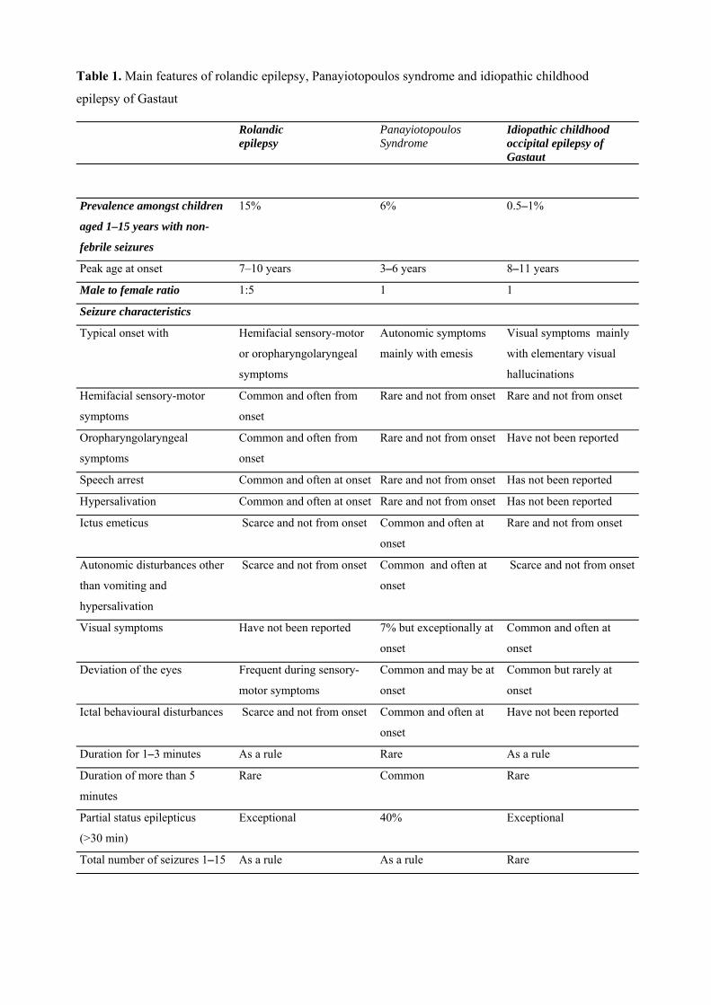
Table 1. Main features of rolandic epilepsy, Panayiotopoulos syndrome and idiopathic childhood
epilepsy of Gastaut
Rolandic epilepsy
Panayiotopoulos Syndrome
Idiopathic childhood occipital epilepsy of Gastaut
Prevalence amongst children
aged 1–15 years with non-
febrile seizures
15% 6% 0.5–1%
Peak age at onset 7–10 years 3–6 years 8–11 years
Male to female ratio 1:5 1 1
Seizure characteristics
Typical onset with Hemifacial sensory-motor
or oropharyngolaryngeal
symptoms
Autonomic symptoms
mainly with emesis
Visual symptoms mainly
with elementary visual
hallucinations
Hemifacial sensory-motor
symptoms
Common and often from
onset
Rare and not from onset Rare and not from onset
Oropharyngolaryngeal
symptoms
Common and often from
onset
Rare and not from onset Have not been reported
Speech arrest Common and often at onset Rare and not from onset Has not been reported
Hypersalivation Common and often at onset Rare and not from onset Has not been reported
Ictus emeticus Scarce and not from onset Common and often at
onset
Rare and not from onset
Autonomic disturbances other
than vomiting and
hypersalivation
Scarce and not from onset Common and often at
onset
Scarce and not from onset
Visual symptoms Have not been reported 7% but exceptionally at
onset
Common and often at
onset
Deviation of the eyes Frequent during sensory-
motor symptoms
Common and may be at
onset
Common but rarely at
onset
Ictal behavioural disturbances Scarce and not from onset Common and often at
onset
Have not been reported
Duration for 1–3 minutes As a rule Rare As a rule
Duration of more than 5
minutes
Rare Common Rare
Partial status epilepticus
(>30 min)
Exceptional 40% Exceptional
Total number of seizures 1–15 As a rule As a rule Rare
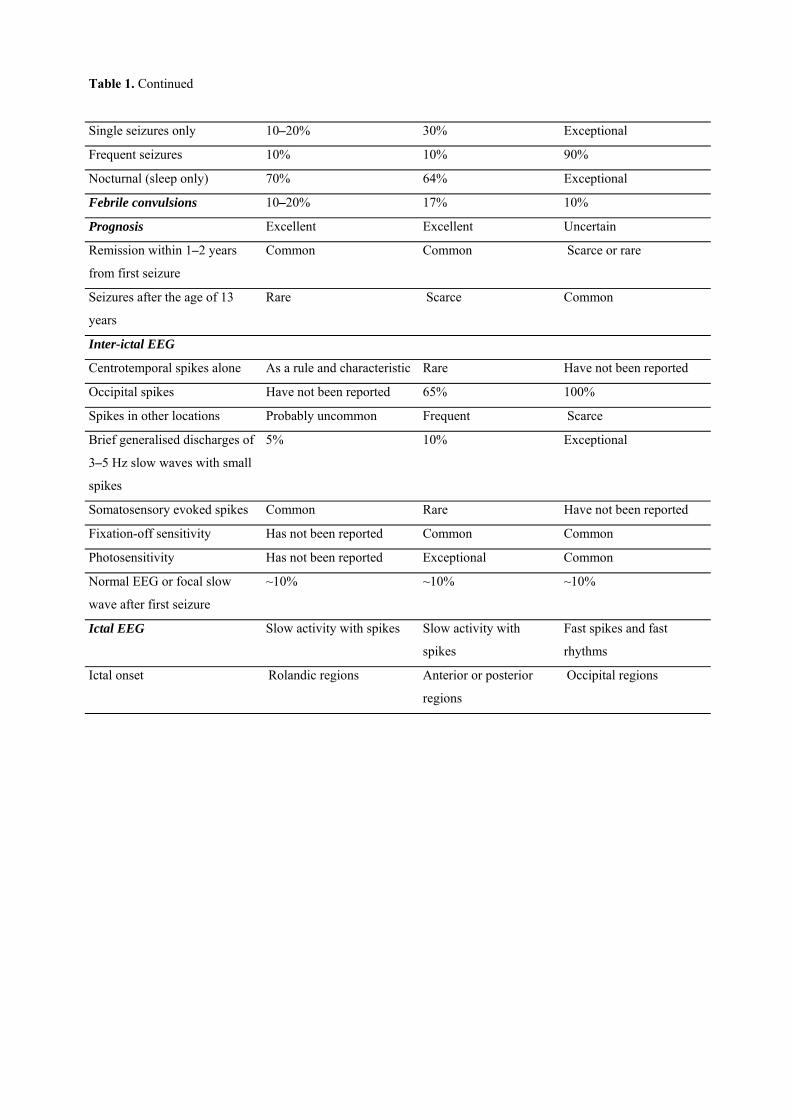
Table 1. Continued
Single seizures only 10–20% 30% Exceptional
Frequent seizures 10% 10% 90%
Nocturnal (sleep only) 70% 64% Exceptional
Febrile convulsions 10–20% 17% 10%
Prognosis Excellent Excellent Uncertain
Remission within 1–2 years
from first seizure
Common Common Scarce or rare
Seizures after the age of 13
years
Rare Scarce Common
Inter-ictal EEG
Centrotemporal spikes alone As a rule and characteristic Rare Have not been reported
Occipital spikes Have not been reported 65% 100%
Spikes in other locations Probably uncommon Frequent Scarce
Brief generalised discharges of
3–5 Hz slow waves with small
spikes
5% 10% Exceptional
Somatosensory evoked spikes Common Rare Have not been reported
Fixation-off sensitivity Has not been reported Common Common
Photosensitivity Has not been reported Exceptional Common
Normal EEG or focal slow
wave after first seizure
~10% ~10% ~10%
Ictal EEG Slow activity with spikes Slow activity with
spikes
Fast spikes and fast
rhythms
Ictal onset Rolandic regions Anterior or posterior
regions
Occipital regions