Embed Size (px)
Citation preview

Beneficial Effects of Autologous Bone Marrow-DerivedMesenchymal Stem Cells in Naturally OccurringTendinopathyRoger Kenneth Whealands Smith1, Natalie Jayne Werling2, Stephanie Georgina Dakin1, Rafiqul Alam1,Allen E. Goodship3, Jayesh Dudhia1*
1 Department of Clinical Sciences and Services, the Royal Veterinary College, University of London, Hatfield, United Kingdom, 2 Department of Biotherapeutics,National Institute for Biological Standards and Control, South Mimms, United Kingdom, 3 Institute of Orthopaedics & Musculo-Skeletal Science, UniversityCollege London, Stanmore, United Kingdom
Abstract
Tendon injuries are a common age-related degenerative condition where current treatment strategies fail to restorefunctionality and normal quality of life. This disease also occurs naturally in horses, with many similarities to humantendinopathy making it an ideal large animal model for human disease. Regenerative approaches are increasinglyused to improve outcome involving mesenchymal stem cells (MSCs), supported by clinical data where injection ofautologous bone marrow derived MSCs (BM-MSCs) suspended in marrow supernatant into injured tendons hashalved the re-injury rate in racehorses. We hypothesized that stem cell therapy induces a matrix more closelyresembling normal tendon than the fibrous scar tissue formed by natural repair. Twelve horses with career-endingnaturally-occurring superficial digital flexor tendon injury were allocated randomly to treatment and control groups.1X107 autologous BM-MSCs suspended in 2 ml of marrow supernatant were implanted into the damaged tendon ofthe treated group. The control group received the same volume of saline. Following a 6 month exercise programmehorses were euthanized and tendons assessed for structural stiffness by non-destructive mechanical testing and formorphological and molecular composition.
BM-MSC treated tendons exhibited statistically significant improvements in key parameters compared to saline-injected control tendons towards that of normal tendons and those in the contralateral limbs. Specifically, treatedtendons had lower structural stiffness (p<0.05) although no significant difference in calculated modulus of elasticity,lower (improved) histological scoring of organisation (p<0.003) and crimp pattern (p<0.05), lower cellularity(p<0.007), DNA content (p<0.05), vascularity (p<0.03), water content (p<0.05), GAG content (p<0.05), and MMP-13activity (p<0.02).
Treatment with autologous MSCs in marrow supernatant therefore provides significant benefits compared tountreated tendon repair in enhancing normalisation of biomechanical, morphological, and compositional parameters.These data in natural disease, with no adverse findings, support the use of this treatment for human tendon injuries.
Citation: Smith RKW, Werling NJ, Dakin SG, Alam R, Goodship AE, et al. (2013) Beneficial Effects of Autologous Bone Marrow-Derived MesenchymalStem Cells in Naturally Occurring Tendinopathy. PLoS ONE 8(9): e75697. doi:10.1371/journal.pone.0075697
Editor: Elizabeth G Laird, University of Liverpool, United Kingdom
Received May 10, 2013; Accepted August 20, 2013; Published September 25, 2013
Copyright: © 2013 Smith et al. This is an open-access article distributed under the terms of the Creative Commons Attribution License, which permitsunrestricted use, distribution, and reproduction in any medium, provided the original author and source are credited.
Funding: This study was funded by the Horserace Betting Levy Board, UK, the Royal Veterinary College, UK (http://www.hblb.org.uk) and VetCellBioscience Ltd, UK (http://www.vetcell.com). SGD was funded by a Biotechnology and Biological Sciences Research Council (www.bbsrc.ac.uk). Thefunders had no role in study design, data collection and analysis, decision to publish, or preparation of the manuscript.
Competing interests: The authors have the following interests. This study was partly funded by VetCell Bioscience Ltd. RKS was a director of VetCellBioscience Ltd, UK from 2002 - 2010. There are no patents, products in development or marketed products to declare. This does not alter their adherenceto all the PLOS ONE policies on sharing data and materials, as detailed online in the guide for authors.
* E-mail: [email protected]
Introduction
Tendon disease is a common age-related degenerativecondition where current treatment strategies frequently deliverineffective results in terms of functionality which impacts onquality of life, frequently with a recurrence of injury. Tendondisorders are a major direct and indirect financial burden on the
NHS with more than 85,000 patients presenting to GPs eachyear with symptomatic Achilles tendinopathy [1] and 50,735operations on tendon disorders being performed on the NHS in2005/6 [2]. There are a multitude of therapies, ranging fromsurgical repair, rest alone and physiotherapy to corticosteroidinjections, extracorporeal shockwave therapy, platelet richplasma injections and eccentric loading exercises. However,
PLOS ONE | www.plosone.org 1 September 2013 | Volume 8 | Issue 9 | e75697

apart from eccentric exercises for Achilles tendinopathy, nonehave been shown to be any more effective than placebo [3]and there is a need for improved non-surgical treatments.
Equine and human tendinopathy have many similarities intheir pathophysiology [4]. Thus, both the superficial digitalflexor tendon (SDFT) in the horse and the Achilles tendon inthe human have similar structure and function, being elasticweight-bearing tendons having an energy-storing capacity forefficient locomotion. Both appear to accumulate degeneratechange with age and exercise, which precede a high frequencyof overstrain injury. Overstrain injury of the SDFT in the equineathlete remains devastating and often career ending.Epidemiological studies have demonstrated that these injuriesaccount for 46% of all limb injuries on the racecourse andaffect 24% of racehorses in training [5,6].
Tendons heal slowly with fibrosis following the initialinflammation leading to the formation of disorganised scartissue. While the initial injury causes a reduction in structuralstiffness, fibrosis frequently results in the tendon as a structurebeing becoming stiffer than normal tendon in the chronicstages [7,8]. This increased stiffness compromises theessential function of the SDFT as an elastic energy store forefficient locomotion [9] and results in a horse withcompromised locomotor function and prone to re-injury [10]. Anoptimal treatment should, therefore, aim to restore normalstructure and function. Consequently, there has beenconsiderable interest in the potential therapeutic benefits ofmesenchymal stem cells (MSCs) in regeneration of functionaltendon and ligament. These cells reside in small numbers,within niches in all mesenchymal tissues, usually closely
associated with blood vessels and are capable of differentiatinginto a number of different cell types [11,12]. As in otherspecies, the ability of MSCs derived from both bone marrowand fat to differentiate into one [13] or more lineages has alsobeen demonstrated for equine MSCs [14-19]. These cells mayregenerate tendon tissue by differentiation into tendon cells orby modulating the inflammatory response to injury andconsequent repair process [20,21].
Bone marrow (BM)-derived MSCs have been used in a largenumber of experimental laboratory animal models of acutetendon transection and have demonstrated continued viabilityand significantly improved outcomes over controls [22-27]supporting the translation of the technology into clinical use. Inhorses, Herthel [28] reported success with the direct injectionof BM into damaged suspensory ligaments. However, injectionof large volumes of BM (30-50 ml) causes further disruption ofthe healing tendon; additionally aspirated BM contains very fewMSCs (~1 in 104 nucleated cells) [18], bone spicules and fatcells which may be deleterious to tendon healing. Furthermore,there was no assessment as to whether functional tendon orscar tissue is formed after this treatment.
In an attempt to avoid the above concerns, Smith et al. [29]demonstrated the feasibility of in vitro isolation and expansionof equine BM-derived MSCs, with re-implantation of largenumbers of autologous MSCs suspended in bone marrowsupernatant into damaged equine SDFTs. The MSCs werecombined with bone marrow supernatant so that thepreparation would be completely autologous and also becausebone marrow supernatant had been shown to provide asignificant cellular anabolic stimulus [30,31]. Since this original
Table 1. Details of the horses in each treatment group and characteristics of equine bone marrow derived stem cells.
Horse numberand treatmentgroup Age (years) Limb affected
Injury toimplantationinterval (days)
Analysis site(maximum injuryzone) PDT (hours) CFU assay
Oestogenicpotential
Chondrogenicpotential
Lipogenicpotential
Stem cell 1 6 RF 38 4 45 1.3 Positive Moderate Moderate2 9 LF 75 4 38 1.1 Positive Positive Moderate3 4 LF 33 4 44 0.8 Positive Moderate Positive4 8 RF 47 4 50 1.0 Moderate Positive Poor5 8 RF 51 3 nd nd nd nd nd6 7 LF 52 3 nd nd nd nd nd
Mean 7.0 49.3 44.3 1.1
SD± 1.8 14.6 4.9 0.2
Saline 1 15 RF 66 4 48 1.1 Positive Moderate Positive2 11 LF 63 4 nd nd nd nd nd3 9 LF 53 3 46 0.8 Positive Positive Positive4 5 RF 49 3 53 0.6 Positive Moderate Poor5 7 LF 39 4 nd nd nd nd nd6 4 LF 59 4 43 0.9 Positive Positive Moderate
Mean 8.5 54.8 47.5 0.9
SD± 4.1 10.0 4.20 0.21
There were no significant differences between stem cell and saline groups for age, injury to implantation interval, CFU or PDT. LF, left forelimb; RF, right forelimb; CFU,colony forming unit; PDT, population doubling time; SD±, standard deviation; nd, not determined.doi: 10.1371/journal.pone.0075697.t001
Mesenchymal Stem Cell Treatment of Tendon Injuries
PLOS ONE | www.plosone.org 2 September 2013 | Volume 8 | Issue 9 | e75697
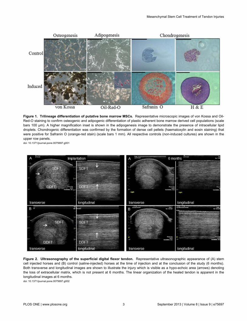
Figure 1. Trilineage differentiation of putative bone marrow MSCs. Representative microscopic images of von Kossa and Oil-Red-O staining to confirm osteogenic and adipogenic differentiation of plastic adherent bone marrow derived cell populations (scalebars 100 µm). A higher magnification inset is shown in the adipogenesis image to demonstrate the presence of intracellular lipiddroplets. Chondrogenic differentiation was confirmed by the formation of dense cell pellets (haematoxylin and eosin staining) thatwere positive for Safranin O (orange-red stain) (scale bars 1 mm). All respective controls (non-induced cultures) are shown in theupper row panels.doi: 10.1371/journal.pone.0075697.g001
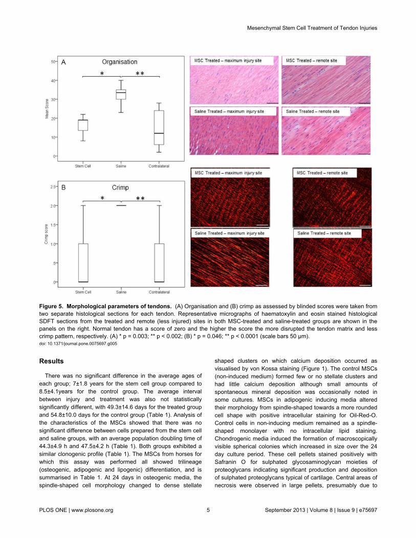
Figure 2. Ultrasonography of the superficial digital flexor tendon. Representative ultrasonographic appearance of (A) stemcell injected horses and (B) control (saline-injected) horses at the time of injection and at the conclusion of the study (6 months).Both transverse and longitudinal images are shown to illustrate the injury which is visible as a hypo-echoic area (arrows) denotingthe loss of extracellular matrix, which is not present at 6 months. The linear organization of the healed tendon is apparent in thelongitudinal images at 6 months.doi: 10.1371/journal.pone.0075697.g002
Mesenchymal Stem Cell Treatment of Tendon Injuries
PLOS ONE | www.plosone.org 3 September 2013 | Volume 8 | Issue 9 | e75697

publication, the technique has been widely adopted in manycountries for the treatment of tendon (and ligament) overstraininjuries in horses. Experimental studies have been conducted,using collagenase tendon injury models in large animalspecies, which have demonstrated significantly improvedoutcomes with MSCs compared to saline injected controls[32,33]. In contrast, a different model based on acutemechanical disruption [34] failed to show the benefit ofimplanted stem cells over controls of BM supernatant [35],although this study only evaluated ultrastructure at one early(12 week) time-point with no mechanical or functionalevaluation.
However, experimental models using induced acute injurieshave limitations as they do not reflect all the features of clinicaldisease which commonly has a preceding phase of age-related
degeneration [4]. In contrast, an adequately powered largeclinical study of SDFT injuries in racehorses treated withautologous MSCs in bone marrow supernatant [36]demonstrated significantly reduced re-injury rates compared totwo published case series of horses which had undergone avariety of other treatments [10,37]. However, there was nocontemporaneous control population and no indication of amechanism for the action of implanted MSCs on the healing ofthe damaged tendon matrix. Therefore, this study was initiatedto explore the hypothesis that implantation of autologous BM-MSCs suspended in marrow supernatant into the site of injuryin naturally injured SDFTs will induce a tissue more closelyresembling normal tendon matrix than the fibrous scar tissueformed subsequent to natural repair in saline injected controls.
Figure 3. Cross-sectional area of the superficial digital flexor tendon. (A) Cross-sectional areas were measuredultrasonographically at the time of implantation (Stem Cell 1, Saline 1 and Contra 1) and at 6 months (Stem Cell 6, Saline 6 andContra 6). (B) Relative change in cross-sectional area between groups. The Wilcoxon Signed-Rank test showed that there was nodifference in cross-sectional area between 1 and 6 months for either the stem cell treated group or the saline group, or between thecontralateral limbs in each group. Contralateral limbs were therefore combined from both groups for statistical analysis. The valuesfor normal horses are shown as mean (thick dashed line) with two standard deviations (thin dashed lines). * p = 0.006, ** p= 0.003,***p<0.045.doi: 10.1371/journal.pone.0075697.g003
Figure 4. Biomechanical properties of tendons. SDFTs were subjected to mechanical testing within 2 h of post euthanasia todetermine (A) structural stiffness and (B) material stiffness (modulus), which was calculated by expressing the ratio of the structuralstiffness to the ultrasonographically-determined cross-sectional area. Structural stiffness was significantly reduced in stem celltreated tendons and closer to that of the uninjured (contralateral) tendons. * p=0.015.doi: 10.1371/journal.pone.0075697.g004
Mesenchymal Stem Cell Treatment of Tendon Injuries
PLOS ONE | www.plosone.org 4 September 2013 | Volume 8 | Issue 9 | e75697
Results
There was no significant difference in the average ages ofeach group; 7±1.8 years for the stem cell group compared to8.5±4.1years for the control group. The average intervalbetween injury and treatment was also not statisticallysignificantly different, with 49.3±14.6 days for the treated groupand 54.8±10.0 days for the control group (Table 1). Analysis ofthe characteristics of the MSCs showed that there was nosignificant difference between cells prepared from the stem celland saline groups, with an average population doubling time of44.3±4.9 h and 47.5±4.2 h (Table 1). Both groups exhibited asimilar clonogenic profile (Table 1). The MSCs from horses forwhich this assay was performed all showed trilineage(osteogenic, adipogenic and lipogenic) differentiation, and issummarised in Table 1. At 24 days in osteogenic media, thespindle-shaped cell morphology changed to dense stellate
shaped clusters on which calcium deposition occurred asvisualised by von Kossa staining (Figure 1). The control MSCs(non-induced medium) formed few or no stellate clusters andhad little calcium deposition although small amounts ofspontaneous mineral deposition was occasionally noted insome cultures. MSCs in adipogenic inducing media alteredtheir morphology from spindle-shaped towards a more roundedcell shape with positive intracellular staining for Oil-Red-O.Control cells in non-inducing medium remained as a spindle-shaped monolayer with no intracellular lipid staining.Chondrogenic media induced the formation of macroscopicallyvisible spherical colonies which increased in size over the 24day culture period. These cell pellets stained positively withSafranin O for sulphated glycosaminoglycan moieties ofproteoglycans indicating significant production and depositionof sulphated proteoglycans typical of cartilage. Central areas ofnecrosis were observed in large pellets, presumably due to
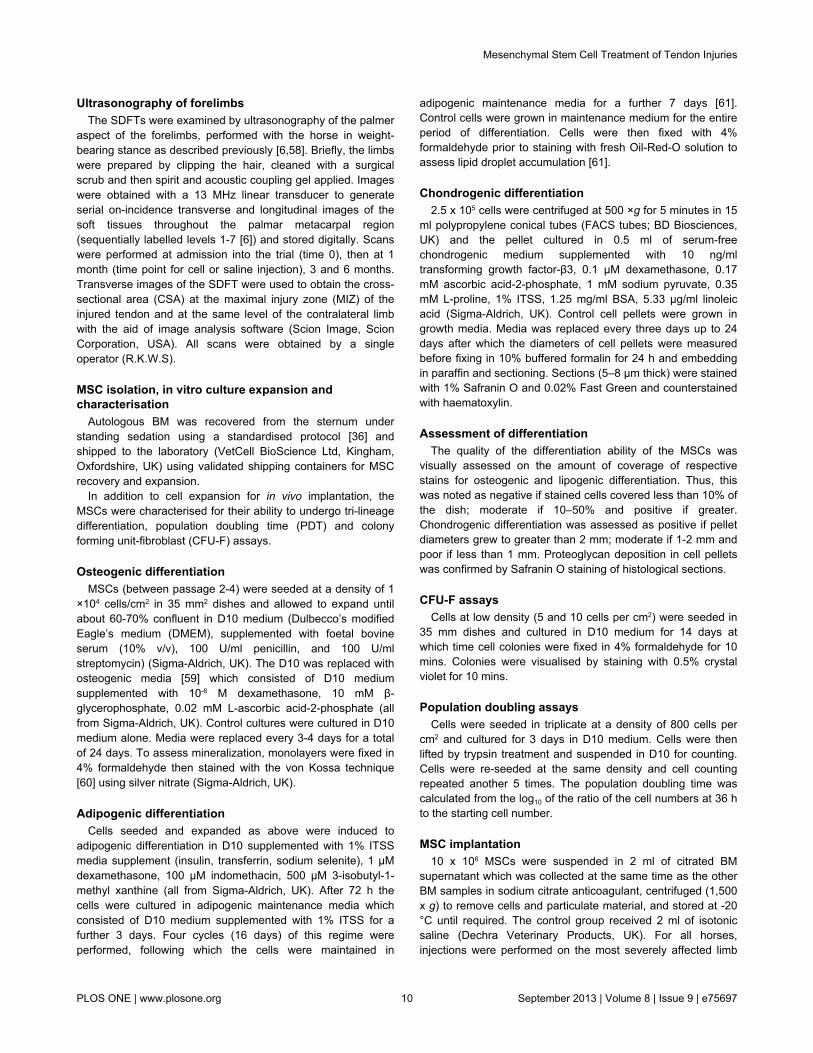
Figure 5. Morphological parameters of tendons. (A) Organisation and (B) crimp as assessed by blinded scores were taken fromtwo separate histological sections for each tendon. Representative micrographs of haematoxylin and eosin stained histologicalSDFT sections from the treated and remote (less injured) sites in both MSC-treated and saline-treated groups are shown in thepanels on the right. Normal tendon has a score of zero and the higher the score the more disrupted the tendon matrix and lesscrimp pattern, respectively. (A) * p = 0.003; ** p < 0.002; (B) * p = 0.046; ** p < 0.0001 (scale bars 50 µm).doi: 10.1371/journal.pone.0075697.g005
Mesenchymal Stem Cell Treatment of Tendon Injuries
PLOS ONE | www.plosone.org 5 September 2013 | Volume 8 | Issue 9 | e75697

limiting nutrient diffusion. Non-induced control cell pelletsremained unchanged or decreased in size and in some caseswere no longer visible at the end of the culture period. Wherecontrol pellets could be recovered, these showed poor stainingfor proteoglycans. There was intra-horse and inter-horsevariability for each assay but in general osteogenesis was goodacross all horses whereas lipogenesis varied from positive tomoderate (two horses). Chondrogenesis measured by pelletdiameter was consistent in all horses albeit with somevariability in the intensity for proteoglycan staining.
In vivo analysisAll horses had moderate to marked ultrasonographic
changes in the tendons (Figure 2). Most had generalisedlesions with mixed central hypoechogenicity. Only three horses(2 stem cell and 1 saline control) had more focal hypoechoiccore lesions. In all cases the contralateral limb had either nosignificant abnormalities on ultrasound (8/13 horses) or milderpathology (5/13 horses). No horses exhibited adverse effectsafter the intra-tendinous injections and all horses underwentthe same post-injection exercise programme on the horse-walker (walking and trotting only) without lameness in 12horses. One horse treated with stem cells was eliminated infurther analysis due to sudden onset lameness during therehabilitation phase of the study, which was associated withrecurrent injury of a very severe tendon injury in the treatedlimb.
Cross-sectional area of the affected SDFT at the time oftreatment (cell or saline injection) was larger than the normalcross-sectional area, consistent with injury [6], but was notsignificantly different between the groups (Figure 3A). After 6months, however, there was no significant difference in thecross-sectional area in the stem cell treated group compared tothe contralateral tendons, however the cross-sectional area inthe saline control group remained significantly larger comparedto the contralateral (p=0.006) and to stem cell treated(p=0.045) tendons. This was supported by the analysis of thechange in cross-sectional area as a percentage of the cross-sectional area at the time of implantation, which showed that
the treated group had a reduction in area while the controlgroup increased although this was not statistically significant(Figure 3B). The cross-sectional area of the SDFT in thecontralateral limbs was smaller than the affected tendons andinside the normal range, confirming the mild nature ofpathology in the contralateral limbs. There was no statisticallysignificant change in the cross-sectional area of the untreatedcontralateral limbs over the period of the rehabilitation. As therewas no significant difference in the contralateral limbs betweengroups, the data for the contralateral limbs were pooled for theanalysis.
In vitro analysisThe hydration of the SDFT at the injury site of the treated
group (65.63±3.78%) was not significantly different to the
Figure 7. Vascularity of tendons. There was reducedvascularity in the MSC-treated tendons, similar to normaltendon, compared to saline-injected controls. * p = 0.026; ** p =0.004.doi: 10.1371/journal.pone.0075697.g007
Figure 6. Cellularity of tendons. Both (A) cellularity scores and (B) DNA content demonstrated reduced cellularity in MSC-treatedtendons. The values for normal horses are shown as mean (thick dashed line) with two standard deviations (thin dashed lines). (A) *p=0.007; ** p=0.001; (B) * p=0.037; ** p<0.001.doi: 10.1371/journal.pone.0075697.g006
Mesenchymal Stem Cell Treatment of Tendon Injuries
PLOS ONE | www.plosone.org 6 September 2013 | Volume 8 | Issue 9 | e75697

contralateral SDFT (62.73±2.2%) but both were significantlydifferent to the saline treated control group (69.55±3.6%),(p=0.04 compared to the stem cell group; p=0.01 compared tothe contralateral SDFT).
A similar pattern was seen for tendon stiffness with thetreated SDFT having significantly lower overall tendonstructural stiffness than the control SDFT (by ~25%; p=0.015)and was similar to the stiffness of the contralateral tendon(2.2% different) (Figure 4). When a measure of the modulus ofelasticity (tissue material property) was calculated using thecross-sectional area measured ultrasonographically at themaximum injury zone immediately prior to euthanasia, therewas a higher modulus for the treated tendons compared tocontrols, although this was not statistically significant (Figure4). In both cases, the modulus was not significantly different tothe contralateral tendons.
Histologically, treated tendons had lower (indicatingimproved) organisational scores at the injured site compared tothe control group, which were also significantly different to thecontralateral tendons (Figure 5A; p=0.003 and p=0.002,respectively). The contralateral tendons had a score of14.2±9.5 and so were not histologically normal reflecting thehigh frequency of bilateral (but milder) disease and there wasno significant difference in scores between the contralaterallimbs of both groups. This improved histological score in thetreated tendons was supported by similar significantdifferences in the crimp scoring from polarised light microscopy(Figure 5B). It was also evident that the MSC-treated grouphad low cellularity scores (Figure 6; p=0.007), which againwere comparable to remote, less injured sites. The lowercellularity was supported by lower DNA content (Figure 6;p=0.037). The treated group had significantly lower vascularitycompared to the controls, which was similar to the contralaterallimbs (Figure 7, p=0.026).
Total collagen (as hydroxyproline) content in MSC-treatedSDFTs was unaltered between groups (Figure 8), while sGAGcontent was reduced and closer to the contralateral tendon,which was in the normal range published previously for normaltendons [38]. The sGAG content of the control group remainedsignificantly elevated (p=0.005). There was no significantdifference in the tissue-linked fluorescence between treatmentand control groups although both were significantly lower thanthe contralateral limb (p=0.039 for treated and p=0.003 forcontrols) (Figure 9), which suggested similar amounts of newcollagen in the injured limbs. A higher remodelling rate,exemplified by MMP-13 activity, was also demonstrated for thecontrol group compared to the treated group (p=0.045) andcontralateral limbs (p=0.015) (Figure 9).
Discussion
The data presented here have supported the hypothesis thatMSC implantation with BM supernatant results in a tissue morelike normal tendon matrix rather than the fibrous scar tissueformed after natural inflammation and repair. For mostparameters, the values for the treated group were closer to thecontralateral, relatively uninjured, tendon than the control groupand closer to the normal values available for some of the
compositional parameters. This was further supported bysignificant differences in the organisational scores betweeninjured and remote, less injured sites.
Many experimental models of induced acute tendon injury inlaboratory animal models report increased material propertiesof the tissue (ultimate tensile stress and elastic modulus)[23,27] associated with stem cell treatment. In this study wereport the overall structural properties of the tendon, which donot take account of the cross-sectional area. We havedemonstrated a reduction in structural stiffness, which confersimproved functional outcome as it will improve elastic energystorage and therefore locomotor efficiency. In addition, thereduced stiffness of the healed area minimises the abruptstrain gradient at adjacent, relatively normal tendon tissue,which is hypothesised to result in the high frequency of re-injury when the horse returns to high level exercise, seenclinically as recurrent tearing of the tissue at these ‘transitionsites’ [39]. When the structural stiffness was corrected for thecross-sectional area at the maximum injury zone to give anapproximate value for the modulus of elasticity, the modulus ofthe stem cell treated horses was not significantly different fromthe contralateral tendons, while the saline treated tendons hada lower modulus, although not statistically different from thetreated group.
In addition to the mechanical properties, both organisationaland compositional analyses reflected the same trend tonormalisation of tissue parameters towards that of thecontralateral limb, which, while not completely normal onultrasonography and histology, were closer to normal tissue,supported by DNA content and sGAGs being within the rangeof normal tendons. In an experimental model study, thesebiochemical parameters remained elevated compared tonormal at the end of the study at 24 weeks [40] although inboth studies it is possible that they may have continued tochange or remained stable with further rehabilitation as theusual length of rehabilitation for equine SDFT tendinopathy isapproximately 12 months prior to a return to full athleticism.
The mechanism of action of MSCs with marrow supernatantafter implantation has not yet been clarified. It has beenproposed that they act either by inducing a true regenerativeeffect or by an immunomodulatory response by modifying theinflammatory response and indirectly modulating thesubsequent fibrous repair processes of the host cells.Inflammation is often cited to be absent in tendon injuries,however we have recently shown the presence of a robustinflammatory response in natural disease and the presence ofactivated macrophages in the damaged tissue which appear toswitch to a pro-resolving phenotype with disease phase[41,42]. It is tempting to speculate that MSCs play a role inmodulating the pro-resolution phenotype of activatedmacrophages however it is not possible to determine which ofthese two actions (regeneration or immunomodulation) hadoccurred in this study. There is still persistent abnormalorganisation and composition, which suggests that the effectdoes not involve a fully regenerative mechanism. This is inkeeping with the findings in experimental studies using surgicalor chemical models of tendon injury in horses where there hasbeen no effect on ultrastructure in a surgical model [35] and
Mesenchymal Stem Cell Treatment of Tendon Injuries
PLOS ONE | www.plosone.org 7 September 2013 | Volume 8 | Issue 9 | e75697

only significant improvements in organisational scores in achemically induced model [32]. The limited effects seen inthese studies may be due to the differences betweenexperimental models and naturally occurring injury. The natureof the tissue into which the cells were deposited was a naturallyoccurring injury with a preceding age-related degenerationwhich is very different from that induced acutely inexperimental models. There was also a markedly differentinterval between treatment and when the tissue was harvestedand analysed. The experimental studies evaluated the tendons6-12 weeks after MSC implantation, while in this study, thereparative tissue was analysed after 24 weeks (6 months), at atime when many horses would be progressing to higher-leveltraining.
It was intriguing to observe a lower cell count in the tendonsthat had received cell treatment. However, it is known fromother studies that a large proportion of the implanted cells arelost [43,44], and this loss appears to occur early afterimplantation [45]. As the dynamics of the implanted cells havebeen evaluated, and because we have shown labels used forlong term tracking of cells alter the viability and behaviour ofimplanted cells (data not shown), additional horses were notrecruited to track the fate of the implanted cells in the currentstudy. However, the number of implanted cells persisting to 6months would be expected to be low and the overall lower cellnumbers could also reflect an immunomodulatory effect inreducing hyperplasia of the tendon and the subsequent amountof fibrosis.
The preparation of stem cells used in this study consisted ofa combination of MSCs and bone marrow supernatant, thelatter of which has been shown to exhibit anabolic propertiesacting on tenocytes [30]. The treatment used was that found toreduce re-injury in a large equine clinical cohort study [36]. Inthis study it was not possible to determine whether the effectseen was caused by the MSCs, the supernatant, or both.Although we did not measure the biochemical composition ofthe bone marrow supernatants, its composition is similarbetween horses [46] which suggests a minimal variability in its
contribution to the repair process. It is possible that growthfactors in the bone marrow [30,46,47] could contribute to thebeneficial effects of the implanted stem cells which is anadditional possible explanation for the lack of response seen ina previous acute induced experimental model [35], where thecontrol horses received bone marrow supernatant only and nosignificant effects were seen on ultrastructure (collagen fibrildiameter) alone. Further work is needed to determine whetherboth components are necessary and act synergistically.
The higher MMP-13 activity seen in the saline treated groupis consistent with the lower tissue-linked fluorescence,indicative of greater collagen turnover. However, the differencein tissue-linked fluorescence, may have been influenced by thedifferences in ages between the two groups (saline group wereolder on average although not significantly) as intrinsicfluorescence of long-lived tissue components, such ascollagen, increases linearly with age by the accumulation ofage-related advanced glycation end-products [48]. Whenplotted against age, the injured limbs had lower tissue-linkedfluorescence than the contralateral limbs (data not shown)reflecting their acute pathology and greater matrix remodellingwith old collagen replaced with new. The small differences intissue-linked fluorescence most likely reflect the mixture oftissues present within the analysed sample. Given the severityof the recent injury, and the possible presence of previouspathology and fibrosis, it would not be surprising to encounter amixture of newly formed collagen, normal aged collagen andolder collagen from previous injury.
The significant reduction in sGAG levels in the treated groupcompared to the controls, which was only just outside thenormal range, was considered a positive outcome, againrepresentative at least in part, of a regenerative effect ornormalisation of the healed tendon tissue closer to that ofnormal tendon. GAG levels are consistently elevated withpathology in tendons across many injuries in many species[49-54] and most treatments that enhance fibrosis tend toincrease sGAG levels. The current study did not includeanalysis of the proteoglycan degrading enzymes ADAMTS4
Figure 8. Compositional analysis of tendons. Concentrations for (A) hydroxyproline and (B) sGAGs are shown. The values fornormal horses are shown as mean (thick dashed line) with two standard deviations (thin dashed lines). Note that the sGAG contentof MSC-treated tendons was closer to the normal range and not significantly different from the relatively uninjured contralaterallimbs (p = 0.24). * p = 0.05.doi: 10.1371/journal.pone.0075697.g008
Mesenchymal Stem Cell Treatment of Tendon Injuries
PLOS ONE | www.plosone.org 8 September 2013 | Volume 8 | Issue 9 | e75697

and ADAMTS5 to determine if the accumulation of sGAGscorrelated with altered expression and/or activity of theseenzymes. Increased proteoglycan deposition in tendon diseasecorrelates with ADAMTS5 knockout [55] and with its reducedexpression in human tendinopathy [56]. It is intriguing that inequine suspensory ligament desmitis, chondroprogenitor cellsappear responsible for elevated aggrecan synthesis and itsaccumulation may arise due to the inactivation of ADAMSTS5despite its increased expression [57]. The role of implantedMSCs in modulating proteoglycan synthesis and the activity ofthese enzymes during tendon healing requires furtherinvestigation. The absence of a significant change inhydroxyproline levels between groups is not surprising as thisparameter does not distinguish between different collagens innormal and fibrotic tendons. A more detailed analysis ofchanges in the expression of specific collagens is required toaddress this.
ConclusionThis study has demonstrated that the technique of MSC
implantation with marrow supernatant is not only safe but alsoindicates significant benefits to the healing tendon innormalising biomechanical (reduced stiffness), histological(better organisation) and compositional parameters (lowerGAG content) towards those levels in normal tendon, whichcould be considered surrogate indicators of regeneration. Thefindings provide evidence that the implantation of autologousMSCs has beneficial therapeutic effects on the healing ofsuperficial digital flexor tendon over-strain injuries whichsupports the reduction in re-injury rate seen clinically with thistreatment [36]. In addition, these data support the potential useof MSCs in bone marrow supernatant for treating humantendon injuries.
Materials and Methods
Ethical Statement and recruitment of horsesThe study was carried out following informed consent from
the owners for horses to be donated to this study and underapproval from the Ethics and Welfare Committee of the RoyalVeterinary College (URN 2013 1230R 2005) and under UKHome Office Licences. Thirteen Thoroughbred andThoroughbred cross horses (all male castrates), aged between5 and 15 years of age (7.8±3.0 years), and suffering career-ending severe superficial digital flexor tendonitis within themetacarpal region were recruited for this study (Table 1). Theselection criteria were that each horse had a recent injury tothe SDFT in the mid-metacarpal region of less than 2 months(average 30 days between injury and entry into the study).Additional requirements were that the paratenon of the SDFThad to be intact and no horse had received previous intra-tendinous injections, thereby reducing potential loss of cellsafter implantation. Horses could have received anti-inflammatory agents but all systemically administered drugshad been cleared by the time of implantation because of theinterval between entry into the study and treatment. It was notpossible to determine with confidence if a previous injury hadbeen present, although, as the horses had suffered career-ending injury which rendered them uneconomic to attempt toreturn them to racing, the injuries were severe, in many casescompletely disrupting the entire tendon matrix (Figure 2).Naturally-occurring tendinopathy frequently manifests withbilateral involvement (a comprehensive epidemiological studydocumented a 35% frequency of bilateral injuries [6]) althoughthe contralateral limb usually has milder pathology or else wasinjured previously (e.g. chronic disease). For the horsesrecruited for this study, pathology in the contralateral limbs wasrecorded but the limbs were not treated and were recovered forcomparative evaluation.
Figure 9. Collagen re-modelling in injured and stem cells treated tendons. (A) Tissue-linked fluorescence and (B) MMP-13activity. The increased MMP-13 activity seen in the saline-treated group matched the lower tissue-linked fluorescence suggestinggreater remodelling of the collagen matrix in the saline-treated compared to the MSC-treated tendons. (A) * p = 0.039; ** p = 0.003;(B) * p = 0.045; ** p = 0.015.doi: 10.1371/journal.pone.0075697.g009
Mesenchymal Stem Cell Treatment of Tendon Injuries
PLOS ONE | www.plosone.org 9 September 2013 | Volume 8 | Issue 9 | e75697
Ultrasonography of forelimbsThe SDFTs were examined by ultrasonography of the palmer
aspect of the forelimbs, performed with the horse in weight-bearing stance as described previously [6,58]. Briefly, the limbswere prepared by clipping the hair, cleaned with a surgicalscrub and then spirit and acoustic coupling gel applied. Imageswere obtained with a 13 MHz linear transducer to generateserial on-incidence transverse and longitudinal images of thesoft tissues throughout the palmar metacarpal region(sequentially labelled levels 1-7 [6]) and stored digitally. Scanswere performed at admission into the trial (time 0), then at 1month (time point for cell or saline injection), 3 and 6 months.Transverse images of the SDFT were used to obtain the cross-sectional area (CSA) at the maximal injury zone (MIZ) of theinjured tendon and at the same level of the contralateral limbwith the aid of image analysis software (Scion Image, ScionCorporation, USA). All scans were obtained by a singleoperator (R.K.W.S).
MSC isolation, in vitro culture expansion andcharacterisation
Autologous BM was recovered from the sternum understanding sedation using a standardised protocol [36] andshipped to the laboratory (VetCell BioScience Ltd, Kingham,Oxfordshire, UK) using validated shipping containers for MSCrecovery and expansion.
In addition to cell expansion for in vivo implantation, theMSCs were characterised for their ability to undergo tri-lineagedifferentiation, population doubling time (PDT) and colonyforming unit-fibroblast (CFU-F) assays.
Osteogenic differentiationMSCs (between passage 2-4) were seeded at a density of 1
×104 cells/cm2 in 35 mm2 dishes and allowed to expand untilabout 60-70% confluent in D10 medium (Dulbecco’s modifiedEagle’s medium (DMEM), supplemented with foetal bovineserum (10% v/v), 100 U/ml penicillin, and 100 U/mlstreptomycin) (Sigma-Aldrich, UK). The D10 was replaced withosteogenic media [59] which consisted of D10 mediumsupplemented with 10-6 M dexamethasone, 10 mM β-glycerophosphate, 0.02 mM L-ascorbic acid-2-phosphate (allfrom Sigma-Aldrich, UK). Control cultures were cultured in D10medium alone. Media were replaced every 3-4 days for a totalof 24 days. To assess mineralization, monolayers were fixed in4% formaldehyde then stained with the von Kossa technique[60] using silver nitrate (Sigma-Aldrich, UK).
Adipogenic differentiationCells seeded and expanded as above were induced to
adipogenic differentiation in D10 supplemented with 1% ITSSmedia supplement (insulin, transferrin, sodium selenite), 1 µMdexamethasone, 100 µM indomethacin, 500 µM 3-isobutyl-1-methyl xanthine (all from Sigma-Aldrich, UK). After 72 h thecells were cultured in adipogenic maintenance media whichconsisted of D10 medium supplemented with 1% ITSS for afurther 3 days. Four cycles (16 days) of this regime wereperformed, following which the cells were maintained in
adipogenic maintenance media for a further 7 days [61].Control cells were grown in maintenance medium for the entireperiod of differentiation. Cells were then fixed with 4%formaldehyde prior to staining with fresh Oil-Red-O solution toassess lipid droplet accumulation [61].
Chondrogenic differentiation2.5 x 105 cells were centrifuged at 500 ×g for 5 minutes in 15
ml polypropylene conical tubes (FACS tubes; BD Biosciences,UK) and the pellet cultured in 0.5 ml of serum-freechondrogenic medium supplemented with 10 ng/mltransforming growth factor-β3, 0.1 µM dexamethasone, 0.17mM ascorbic acid-2-phosphate, 1 mM sodium pyruvate, 0.35mM L-proline, 1% ITSS, 1.25 mg/ml BSA, 5.33 µg/ml linoleicacid (Sigma-Aldrich, UK). Control cell pellets were grown ingrowth media. Media was replaced every three days up to 24days after which the diameters of cell pellets were measuredbefore fixing in 10% buffered formalin for 24 h and embeddingin paraffin and sectioning. Sections (5–8 µm thick) were stainedwith 1% Safranin O and 0.02% Fast Green and counterstainedwith haematoxylin.
Assessment of differentiationThe quality of the differentiation ability of the MSCs was
visually assessed on the amount of coverage of respectivestains for osteogenic and lipogenic differentiation. Thus, thiswas noted as negative if stained cells covered less than 10% ofthe dish; moderate if 10–50% and positive if greater.Chondrogenic differentiation was assessed as positive if pelletdiameters grew to greater than 2 mm; moderate if 1-2 mm andpoor if less than 1 mm. Proteoglycan deposition in cell pelletswas confirmed by Safranin O staining of histological sections.
CFU-F assaysCells at low density (5 and 10 cells per cm2) were seeded in
35 mm dishes and cultured in D10 medium for 14 days atwhich time cell colonies were fixed in 4% formaldehyde for 10mins. Colonies were visualised by staining with 0.5% crystalviolet for 10 mins.
Population doubling assaysCells were seeded in triplicate at a density of 800 cells per
cm2 and cultured for 3 days in D10 medium. Cells were thenlifted by trypsin treatment and suspended in D10 for counting.Cells were re-seeded at the same density and cell countingrepeated another 5 times. The population doubling time wascalculated from the log10 of the ratio of the cell numbers at 36 hto the starting cell number.
MSC implantation10 x 106 MSCs were suspended in 2 ml of citrated BM
supernatant which was collected at the same time as the otherBM samples in sodium citrate anticoagulant, centrifuged (1,500x g) to remove cells and particulate material, and stored at -20°C until required. The control group received 2 ml of isotonicsaline (Dechra Veterinary Products, UK). For all horses,injections were performed on the most severely affected limb
Mesenchymal Stem Cell Treatment of Tendon Injuries
PLOS ONE | www.plosone.org 10 September 2013 | Volume 8 | Issue 9 | e75697
3-4 weeks after bone marrow aspiration. A 2 inch 19G needlewas guided under ultrasound guidance into the middle of thelesion (maximum injury zone) at 2-4 sites to ensure adequatespread throughout the lesion (as assessedultrasonographically). The limb was then immediatelybandaged and a single peri-operative dose of intra-muscularantibiotics (Norocillin, Norbrook Laboratories (GB) Ltd, Corby,UK) administered.
Post-implantation exercise regimeThe horses were box-rested with the limb bandaged for 7
days after which the horses began a standardised ascendingexercise rehabilitation regimen of walking (increasing from10-45 mins over the first 3 months) and then combined withtrotting (increasing from 5-20 mins) in the second 3 monthsusing a horse-walker. Horses were trotted in a straight line atthe time of BM aspiration, pre-implantation and 1, 3 and 6months after implantation to assess for obvious lameness(tendinopathy of the SDFT characteristically only causessignificant lameness during the acute inflammatory stages(typically 1-2 weeks). At the same time, the palmar aspect ofthe metacarpus was evaluated ultrasonographically usingstandard transverse and longitudinal images of the SDFT asdetailed above.
Tendon analysisHorses were euthanized after 6 months post-implantation
and tendons were harvested from both forelimbs from eachhorse and analysed as follows
Mechanical propertiesThe SDFT was removed in its entirety from both forelimbs
and mounted in freezer clamps within 2 h in a 50 kN electro-hydraulic material testing machine (Dartec HA50; Zwick RoellLtd, UK; retrofitted with an Instron 8800 control system; Instron,High Wycombe, UK) to give an inter-clamp (gauge) length ofbetween 120 and 250 mm consisting of the damaged area ofthe SDFT. Non-destructive testing was performed on thetendon with loading up to 4 kN (one quarter of the averagefailure load found in similar equine tendons) for 20 cycles (toallow for pre-conditioning) at 0.5 Hz. The stiffness wascalculated from between 3 and 4kN on the last loading cyclewhere the points lay on a linear part of the force-deformationcurve. The gradient of the best-fit line (R2>0.99 in all cases)was used as the stiffness (N/%).
After mechanical testing the SDFT was divided into seven 4cm sections matching the extent of the ultrasonographicexamination from proximal to the distal extent of the SDFT.Each 4 cm section was sub-divided into four 1 cm sections.Peripheral tissue was discarded before the samples wereprepared for histology and compositional analysis.
Histological examinationTendon sections were prepared for histology as previously
described [58]. Briefly, tendon sections were embedded inoptimal cutting temperature compound (OCT, Sakura Tissue-Tek®, The Netherlands), and snap frozen in pre-chilled (-80
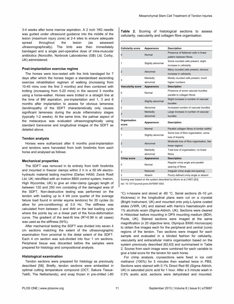
°C) n-hexane and stored at -80 °C. Serial sections (8–10 µmthickness) in the longitudinal plane were cut on a cryostat(Bright Instrument, UK) and mounted onto poly-L-lysine coatedslides (VWR, UK) and stained with Harris’s haematoxylin and1% alcoholic eosin (Sigma-Aldrich, UK). Sections were clearedin Histoclear before mounting in DPX mounting medium (BDH,Poole, UK). Stained sections were imaged at the samemagnification (x 20 objective lens; Olympus BX60 microscope)to obtain five images each for the peripheral and central (core)regions of the tendon. Two sections were imaged for eachsample and evaluated in a blinded fashion for cellularity,vascularity and extracellular matrix organisation based on thesystem previously described [62,63] and summarised in Table2. Scores from each image were combined for each variable togive a total score for the tendon for each horse.
For crimp analysis, cryosections were fixed in ice coldmethanol (100%) for 5 minutes then washed twice in PBS.Sections were stained with 0.1% Direct Red 80 (Sigma Aldrich,UK) in saturated picric acid for 1 hour. After a 5 minute wash in0.5% acetic acid, sections were dehydrated and mounted.
Table 2. Scoring of histological sections to assesscellularity, vascularity and collagen fibre organisation.
Cellularity score Appearance Description
0 NormalPresence of flattened cells in linearpattern between fibres
1 Slightly abnormalSome rounded cells present, slightincrease in cellularity
2 AbnormalMany rounded cells present, obviousincrease in cellularity
3Markedlyabnormal
Mostly rounded cells present, muchhigher numbers
Vascularity score Appearance Description
0 NormalPresence of some vascular bundlesparallel to collagen fibres
1 Slightly abnormalSlight increase in number of vascularbundles
2 Abnormal Increased number of vascular bundles
3Markedlyabnormal
Large increase in number of vascularbundles
Organisationscore
Appearance Description
0 Normal Parallel collagen fibres of similar widths
1 Slightly abnormalSome loss of fibre organisation, someloss of linearity
2 AbnormalModerate loss of fibre organisation, fewlinear regions
3Markedlyabnormal
Total loss of organisation, no linearfibres
Crimp score Appearance Description
0 NormalRegular crimp angle and parallelspacing of fibres
1 Reduced Irregular crimp angle and spacing2 Absent Poorly defined crimp angle or absent
Scoring was based on the system described by Movin et al (1997) [63].doi: 10.1371/journal.pone.0075697.t002
Mesenchymal Stem Cell Treatment of Tendon Injuries
PLOS ONE | www.plosone.org 11 September 2013 | Volume 8 | Issue 9 | e75697

Sections were imaged under polarised light (Olympus CX31-Pmicroscope) and were scored in a blinded fashion for bandingpatterns indicative of crimp. A score of 0 indicated a normalbanding pattern, a score of 1 indicated a reduced bandingpattern, and a score of 2 indicated no banding (Table 2).
Compositional analysisOne tendon section (1 cm section from each level) was
freeze-dried and ground to a powder prior to analysis of tissuecontent. Water content was measured from the weight of thetissue section before and after freeze-drying.
Collagen content was determined by a measure ofhydroxyproline content. Aliquots (10 mg) of ground tissue weresuspended in papain digestion buffer (PBS containing 5 mMEDTA, 5 mM L-cysteine HCl and 125 µg/ml papain (all fromSigma-Aldrich, UK) and digested at 60 °C for 24 h and thesolubilised collagen hydrolysed with 6 N HCl at 105 °C for 24hours in sealed tubes. The aqueous phase was then removedby freeze-drying and the solid residue reconstituted in distilledwater. Aliquots in triplicate were then assayed forhydroxyproline in a microtitre plate assay. Briefly, the samplewas made to 35% isopropanol, 12 mM Chloramine T, 0.06 Msodium acetate and 0.05 M trisodium citrate and 4 mM citricacid. This was then incubated at 70 °C in 0.3 M 4-dimethylaminobenzaldehyde, 7% Perchloric acid and 40%isopropanol. The absorbance of the samples at 540 nm wasmeasured after 20 mins. Absorbance values were quantifiedusing a standard curve consisting of hydroxyproline (Sigma-Aldrich, UK) dilutions (50 to 0.78125 µg/ml) treated as above.
Glycosaminoglycan (sGAG) content of papain digestedtissue was determined by the dimethylmethylene blue (DMMB)assay [64]. Standards were made from bovine tracheachondroitin sulphate (Sigma-Aldrich, UK). 10 µL aliquots ofstandards or media samples were added to a 96 well flat-bottomed plate in triplicate. 100 µL of DMMB (Sigma-Aldrich,UK) solution consisting of 46 µM DMMB, 0.2% methanol, 40µM sodium chloride, 40 µM glycine and 10 mM HCl (pH 3.5)was then added to each well and the absorbance measured at530 nm within 15 minutes on a spectrophotometer (AppliedBiosystems UK). sGAG concentration of samples was derivedfrom the standard curve expressed as µg/ml and subsequentlyadjusted to represent sGAG per mg of tissue.
DNA and tissue linked fluorescence assaysDNA as a measure of tissue cellularity and tissue-linked
fluorescence as a measure of the age of tissue componentswere determined on aliquots of papain digested tissue extractsprepared as described above. DNA was measured usingbisBenzimide (Hoechst 33258, Sigma Aldrich, UK) and tissue-linked fluorescence was measured in the absence ofbisBenzimide [62]. Samples were corrected for backgroundfluorescence and the DNA concentration calculated from astandard curve prepared with calf thymus DNA (Sigma Aldrich,UK). DNA content of tendon samples was expressed asmicrograms of DNA per mg of dry weight tissue and tissuelinked fluorescence as arbitrary units per milligram of collagen(measured as hydroxyproline content).
MMP-13 assayA sample of freeze-dried powdered tendon was mixed with
100 volumes (w/v) of extraction buffer (50 mM Tris pH 7.5, 150mM NaCl, 5 mM CaCl2, 1 µM ZnCl2, 0.01% Brij 35) at roomtemperature for 90 mins. Supernatants were recovered bycentrifugation (20,000 x g for 10 mins at room temperature andfrozen at -20 °C until further analysis. MMP-13 in thesupernatants was measured by a fluorogenic substrate assay(Calbiochem, UK) according to manufacturer’s instructions.Briefly, 1 µl of fluorogenic substrate was added to 25 µl oftissue extract and 74 µl of assay buffer (50 mM HEPES, 200mM NaCl, 1 mM CaCl2, 0.01% Brij 35) in a 96-well plate. Plateswere incubated at 37 °C in the dark for 2 h and plates read atexcitation wavelength 325 nm and emission wavelength 393nm. Plates were incubated a further 6 h at 37 °C in the darkand measured every hour. A standard curve was constructedusing human recombinant active MMP-13 (Calbiochem, UK).Standard and samples were assayed in duplicate and theresults expressed as µg active MMP-13 per mg dry weighttissue.
Statistical analysisHistology scores and biochemical data were analysed by a
non-parametric Mann-Whitney U-test as described for Movinscores of tendon by Maffulli et al. [65] Tendon cross-sectionalareas were subjected by Kruskal-Wallis non-parametric oneway ANOVA to identify differences between groups (stem celltreated, saline treated and contralateral) followed by a post-hocMann-Whitney test with Bonferroni correction. A detailedcomparison of the contralateral limbs within and between thegroups (stem cell or saline) using an independent samples orpaired T-test (or their non-parametric equivalent) showed thatthere was no difference between the groups or with time(implantation time compared to 6 months). Therefore theresults are shown with the contralateral limbs as a single grouppooled from both treatment groups. The n values therefore aren = 6 each for stem cell and saline injection groups and n = 12for the contralateral group. Significance level was set at 5%(p≤0.05). Data are expressed as mean ± standard deviation(SD).
Acknowledgements
The authors would like to acknowledge the Racehorse OwnersAssociation for their assistance in recruiting cases and theowners and veterinary surgeons of the horses that weredonated to the study. We would also like to thank Dr BrendanJackson for critical review of the statistical analysis, and MrTom Hughes, Mr Andrew Crawford and Mr Andrew Fiske-Jackson for their assistance with the clinical management ofthe horses.
Author Contributions
Conceived and designed the experiments: RKWS AEG JD.Performed the experiments: RKWS NJW SGD RA JD.Analyzed the data: RKWS NJW SGD JD. Contributedreagents/materials/analysis tools: RKWS AEG JD. Wrote themanuscript: RKWS NJW JD.
Mesenchymal Stem Cell Treatment of Tendon Injuries
PLOS ONE | www.plosone.org 12 September 2013 | Volume 8 | Issue 9 | e75697

References
1. The Clinical Practice Research Datalink website. Available: http://www.cprd.com. Accessed 2013 February 10
2. Health and Social Care Information Centre, Hospital Episode Statisticswebsite. Available: http://www.hscic.gov.uk/hes. Accessed 2013February 10.
3. Magnussen RA, Dunn WR, Thomson AB (2009) Nonoperativetreatment of midportion Achilles tendinopathy: a systematic review. ClinJ Sport Med 19: 54-64. doi:10.1097/JSM.0b013e31818ef090. PubMed:19124985.
4. Lui PP, Maffulli N, Rolf C, Smith RK (2011) What are the validatedanimal models for tendinopathy? Scand J Med Sci Sports 21: 3-17. doi:10.1111/j.1600-0838.2010.01164.x. PubMed: 20673247.
5. Williams RB, Harkins LS, Hammond CJ, Wood JL (2001) Racehorseinjuries, clinical problems and fatalities recorded on British racecoursesfrom flat racing and National Hunt racing during 1996, 1997 and 1998.Equine Vet J 33: 478-486. PubMed: 11558743.
6. Avella CS, Ely ER, Verheyen KL, Price JS, Wood JL et al. (2009)Ultrasonographic assessment of the superficial digital flexor tendons ofNational Hunt racehorses in training over two racing seasons. EquineVet J 41: 449-454. doi:10.2746/042516409X391042. PubMed:19642404.
7. Crevier-Denoix N, Collobert C, Pourcelot P, Denoix JM, Sanaa M et al.(1997) Mechanical properties of pathological equine superficial digitalflexor tendons. Equine Vet J Suppl: 23-26. PubMed: 9354282.
8. Dakin SG, Jespers K, Warner S, O’Hara LK, Dudhia J et al. (2011) Therelationship between in vivo limb and in vitro tendon mechanics afterinjury: a potential novel clinical tool for monitoring tendon repair. EquineVet J 43: 418-423. doi:10.1111/j.2042-3306.2010.00303.x. PubMed:21496076.
9. Wilson AM, McGuigan MP, Su A, van den Bogert AJ (2001) Horsesdamp the spring in their step. Nature 414: 895-899. doi:10.1038/414895a. PubMed: 11780059.
10. Dyson SJ (2004) Medical management of superficial digital flexortendonitis: a comparative study in 219 horses (1992-2000). Equine VetJ 36: 415-419. PubMed: 15253082.
11. Caplan AI (1991) Mesenchymal stem cells. J Orthop Res 9: 641-650.doi:10.1002/jor.1100090504. PubMed: 1870029.
12. Liechty KW, MacKenzie TC, Shaaban AF, Radu A, Moseley AM et al.(2000) Human mesenchymal stem cells engraft and demonstrate site-specific differentiation after in utero transplantation in sheep. Nat Med6: 1282-1286. doi:10.1038/81395. PubMed: 11062543.
13. Crovace A, Lacitignola L, De Siena R, Rossi G, Francioso E (2007) Celltherapy for tendon repair in horses: an experimental study. Vet ResCommun 31 Suppl 1: 281-283. doi:10.1007/s11259-007-0047-y.PubMed: 17682895.
14. Vidal MA, Robinson SO, Lopez MJ, Paulsen DB, Borkhsenious O et al.(2008) Comparison of chondrogenic potential in equine mesenchymalstromal cells derived from adipose tissue and bone marrow. Vet Surg37: 713-724. doi:10.1111/j.1532-950X.2008.00462.x. PubMed:19121166.
15. Vidal MA, Kilroy GE, Lopez MJ, Johnson JR, Moore RM et al. (2007)Characterization of equine adipose tissue-derived stromal cells:adipogenic and osteogenic capacity and comparison with bonemarrow-derived mesenchymal stromal cells. Vet Surg 36: 613-622. doi:10.1111/j.1532-950X.2007.00313.x. PubMed: 17894587.
16. Vidal MA, Kilroy GE, Johnson JR, Lopez MJ, Moore RM et al. (2006)Cell growth characteristics and differentiation frequency of adherentequine bone marrow-derived mesenchymal stromal cells: adipogenicand osteogenic capacity. Vet Surg 35: 601-610. doi:10.1111/j.1532-950X.2006.00197.x. PubMed: 17026544.
17. Strassburg S, [!(surname)!], Goodship AE, Hardingham TE, Clegg PD(2006) Adult and late foetal equine tendon contain cell populations withweak progenitor properties in comparison to bone marrow derivedmesenchymal stem cells. Trans Orthop Res Soc Annu Meet 31: 1113
18. Alves AG, Stewart AA, Dudhia J, Kasashima Y, Goodship AE et al.(2011) Cell-based therapies for tendon and ligament injuries. Vet ClinNorth Am Equine Pract 27: 315-333. doi:10.1016/j.cveq.2011.06.001.PubMed: 21872761.
19. Fortier LA, Nixon AJ, Williams J, Cable CS (1998) Isolation andchondrocytic differentiation of equine bone marrow-derivedmesenchymal stem cells. Am J Vet Res 59: 1182-1187. PubMed:9736400.
20. Schu S, Nosov M, O’Flynn L, Shaw G, Treacy O et al. (2012)Immunogenicity of allogeneic mesenchymal stem cells. J Cell Mol Med16: 2094-2103. doi:10.1111/j.1582-4934.2011.01509.x. PubMed:22151542.
21. Duffy MM, Pindjakova J, Hanley SA, McCarthy C, Weidhofer GA et al.(2011) Mesenchymal stem cell inhibition of T-helper 17 cell-differentiation is triggered by cell-cell contact and mediated byprostaglandin E2 via the EP4 receptor. Eur J Immunol 41: 2840-2851.doi:10.1002/eji.201141499. PubMed: 21710489.
22. Young RG, Butler DL, Weber W, Caplan AI, Gordon SL et al. (1998)Use of mesenchymal stem cells in a collagen matrix for Achilles tendonrepair. J Orthop Res 16: 406-413. doi:10.1002/jor.1100160403.PubMed: 9747780.
23. Awad HA, Butler DL, Boivin GP, Smith FN, Malaviya P et al. (1999)Autologous mesenchymal stem cell-mediated repair of tendon. TissueEng 5: 267-277. doi:10.1089/ten.1999.5.267. PubMed: 10434073.
24. Awad HA, Boivin GP, Dressler MR, Smith FN, Young RG et al. (2003)Repair of patellar tendon injuries using a cell-collagen composite. JOrthop Res 21: 420-431. doi:10.1016/S0736-0266(02)00163-8.PubMed: 12706014.
25. Hildebrand KA, Jia F, Woo SL (2002) Response of donor and recipientcells after transplantation of cells to the ligament and tendon. MicroscRes Tech 58: 34-38. doi:10.1002/jemt.10114. PubMed: 12112420.
26. Butler DL, Juncosa-Melvin N, Boivin GP, Galloway MT, Shearn JT et al.(2008) Functional tissue engineering for tendon repair: Amultidisciplinary strategy using mesenchymal stem cells, bioscaffolds,and mechanical stimulation. J Orthop Res 26: 1-9. doi:10.1002/jor.20456. PubMed: 17676628.
27. Ouyang HW, Goh JC, Thambyah A, Teoh SH, Lee EH (2003) Knittedpoly-lactide-co-glycolide scaffold loaded with bone marrow stromal cellsin repair and regeneration of rabbit Achilles tendon. Tissue Eng 9:431-439. doi:10.1089/107632703322066615. PubMed: 12857411.
28. Herthel D (2001) Enhanced suspensory ligament healing in 100 horsesby stem cells and other bone marrow components. Proc Am AssocEquine Practice 47. p. 319.
29. Smith RK, Korda M, Blunn GW, Goodship AE (2003) Isolation andimplantation of autologous equine mesenchymal stem cells from bonemarrow into the superficial digital flexor tendon as a potential noveltreatment. Equine Vet J 35: 99-102. PubMed: 12553472.
30. Smith JJ, Ross MW, Smith RK (2006) Anabolic effects of acellular bonemarrow, platelet rich plasma, and serum on equine suspensoryligament fibroblasts in vitro. Vet Comp Orthop Traumatol 19: 43-47.PubMed: 16594543.
31. Schnabel LV, Sonea HO, Jacobson MS, Fortier LA (2008) Effects ofplatelet rich plasma and acellular bone marrow on gene expressionpatterns and DNA content of equine suspensory ligament explantcultures. Equine Vet J 40: 260-265. doi:10.2746/042516408X278030.PubMed: 18267879.
32. Schnabel LV, Lynch ME, van der Meulen MC, Yeager AE, KornatowskiMA et al. (2009) Mesenchymal stem cells and insulin-like growth factor-I gene-enhanced mesenchymal stem cells improve structural aspectsof healing in equine flexor digitorum superficialis tendons. J Orthop Res27: 1392-1398. doi:10.1002/jor.20887. PubMed: 19350658.
33. Crovace A, Lacitignola L, Rossi G, Francioso E (2010) Histological andimmunohistochemical evaluation of autologous cultured bone marrowmesenchymal stem cells and bone marrow mononucleated cells incollagenase-induced tendinitis of equine superficial digital flexortendon. Vetmed Int, 2010: 2010: 250978. PubMed: 20445779
34. Schramme M, Hunter S, Campbell N, Blikslager A, Smith R (2010) Asurgical tendonitis model in horses: technique, clinical,ultrasonographic and histological characterisation. Vet Comp OrthopTraumatol 23: 231-239. doi:10.3415/VCOT-09-10-0106. PubMed:20585715.
35. Caniglia CJ, Schramme MC, Smith RK (2012) The effect ofintralesional injection of bone marrow derived mesenchymal stem cellsand bone marrow supernatant on collagen fibril size in a surgical modelof equine superficial digital flexor tendonitis. Equine Vet J 44: 587-593.doi:10.1111/j.2042-3306.2011.00514.x. PubMed: 22150794.
36. Godwin EE, Young NJ, Dudhia J, Beamish IC, Smith RK (2012)Implantation of bone marrow-derived mesenchymal stem cellsdemonstrates improved outcome in horses with overstrain injury of thesuperficial digital flexor tendon. Equine Vet J 44: 25-32. doi:10.1111/j.2042-3306.2011.00363.x. PubMed: 21615465.
37. O’Meara B, Bladon B, Parkin TD, Fraser B, Lischer CJ (2010) Aninvestigation of the relationship between race performance andsuperficial digital flexor tendonitis in the Thoroughbred racehorse.Equine Vet J 42: 322-326. doi:10.1111/j.2042-3306.2009.00021.x.PubMed: 20525050.
38. Batson EL, Paramour RJ, Smith TJ, Birch HL, Patterson-Kane JC et al.(2003) Are the material properties and matrix composition of equine
Mesenchymal Stem Cell Treatment of Tendon Injuries
PLOS ONE | www.plosone.org 13 September 2013 | Volume 8 | Issue 9 | e75697

flexor and extensor tendons determined by their functions? Equine VetJ 35: 314-318. PubMed: 12755437.
39. Smith RWK (2010) Pathophysiology of tendon injury. In: MW RossSDyson. Diagnosis and Management of Lameness in the Horse. 2nd ed.St. Louis: W.B. Saunders Co.
40. Dahlgren LA, Mohammed HO, Nixon AJ (2005) Temporal expression ofgrowth factors and matrix molecules in healing tendon lesions. J OrthopRes 23: 84-92. doi:10.1016/j.orthres.2004.05.007. PubMed: 15607879.
41. Dakin SG, Werling D, Hibbert A, Abayasekara DR, Young NJ et al.(2012) Macrophage sub-populations and the lipoxin A4 receptorimplicate active inflammation during equine tendon repair. PLOS ONE7: e32333. doi:10.1371/journal.pone.0032333. PubMed: 22384219.
42. Dakin SG, Dudhia J, Werling NJ, Werling D, Abayasekara DR et al.(2012) Inflamm-aging and arachadonic acid metabolite differences withstage of tendon disease. PLOS ONE 7: e48978. doi:10.1371/journal.pone.0048978. PubMed: 23155437.
43. Guest DJ, Smith MR, Allen WR (2008) Monitoring the fate ofautologous and allogeneic mesenchymal progenitor cells injected intothe superficial digital flexor tendon of horses: preliminary study. EquineVet J 40: 178-181. doi:10.2746/042516408X276942. PubMed:18267891.
44. Guest DJ, Smith MR, Allen WR (2010) Equine embryonic stem-likecells and mesenchymal stromal cells have different survival rates andmigration patterns following their injection into damaged superficialdigital flexor tendon. Equine Vet J 42: 636-642. doi:10.1111/j.2042-3306.2010.00112.x. PubMed: 20840579.
45. Becerra P, Valdes Vazquez MA, Dudhia J, Fiske-Jackson AR, Neves Fet al. (2013) Distribution of injected technetium (99m)-labeledmesenchymal stem cells in horses with naturally occurringtendinopathy. J Orthop Res 31: 1096-1102. doi:10.1002/jor.22338.PubMed: 23508674.
46. McCarrel T, Fortier L (2009) Temporal growth factor release fromplatelet-rich plasma, trehalose lyophilized platelets, and bone marrowaspirate and their effect on tendon and ligament gene expression. JOrthop Res 27: 1033-1042. doi:10.1002/jor.20853. PubMed: 19170097.
47. Schnabel LV, Mohammed HO, Miller BJ, McDermott WG, JacobsonMS et al. (2007) Platelet rich plasma (PRP) enhances anabolic geneexpression patterns in flexor digitorum superficialis tendons. J OrthopRes 25: 230-240. doi:10.1002/jor.20278. PubMed: 17106885.
48. Birch HL, Bailey JV, Bailey AJ, Goodship AE (1999) Age-relatedchanges to the molecular and cellular components of equine flexortendons. Equine Vet J 31: 391-396. doi:10.1111/j.2042-3306.1999.tb03838.x. PubMed: 10505954.
49. Cadby JA, David F, van de Lest C, Bosch G, van Weeren PR et al.(2013) Further characterisation of an experimental model oftendinopathy in the horse. Equine Vet J 45: 642-648. doi:10.1111/evj.12035. PubMed: 23448172.
50. Samiric T, Parkinson J, Ilic MZ, Cook J, Feller JA et al. (2009) Changesin the composition of the extracellular matrix in patellar tendinopathy.Matrix Biol 28: 230-236. doi:10.1016/j.matbio.2009.04.001. PubMed:19371780.
51. Smith MM, Sakurai G, Smith SM, Young AA, Melrose J et al. (2008)Modulation of aggrecan and ADAMTS expression in ovine tendinopathyinduced by altered strain. Arthritis Rheum 58: 1055-1066. doi:10.1002/art.23388. PubMed: 18383380.
52. Fu SC, Chan KM, Rolf CG (2007) Increased deposition of sulfatedglycosaminoglycans in human patellar tendinopathy. Clin J Sport Med
17: 129-134. doi:10.1097/JSM.0b013e318037998f. PubMed:17414481.
53. Corps AN, Robinson AH, Movin T, Costa ML, Hazleman BL et al.(2006) Increased expression of aggrecan and biglycan mRNA inAchilles tendinopathy. Rheumatology (Oxf) 45: 291-294. PubMed:16219640.
54. Riley GP, Harrall RL, Constant CR, Chard MD, Cawston TE et al.(1994) Glycosaminoglycans of human rotator cuff tendons: changeswith age and in chronic rotator cuff tendinitis. Ann Rheum Dis 53:367-376. doi:10.1136/ard.53.6.367. PubMed: 8037495.
55. Wang VM, Bell RM, Thakore R, Eyre DR, Galante JO et al. (2012)Murine tendon function is adversely affected by aggrecan accumulationdue to the knockout of ADAMTS5. J Orthop Res 30: 620-626. doi:10.1002/jor.21558. PubMed: 21928430.
56. Corps AN, Robinson AH, Harrall RL, Avery NC, Curry VA et al. (2012)Changes in matrix protein biochemistry and the expression of mRNAencoding matrix proteins and metalloproteinases in posterior tibialistendinopathy. Ann Rheum Dis 71: 746-752. doi:10.1136/annrheumdis-2011-200391. PubMed: 22241901.
57. Plaas A, Sandy JD, Liu H, Diaz MA, Schenkman D et al. (2011)Biochemical identification and immunolocalizaton of aggrecan,ADAMTS5 and inter-alpha-trypsin-inhibitor in equine degenerativesuspensory ligament desmitis. J Orthop Res 29: 900-906. doi:10.1002/jor.21332. PubMed: 21246622.
58. Smith RK, Jones R, Webbon PM (1994) The cross-sectional areas ofnormal equine digital flexor tendons determined ultrasonographically.Equine Vet J 26: 460-465. doi:10.1111/j.2042-3306.1994.tb04050.x.PubMed: 7889919.
59. Jaiswal N, Haynesworth SE, Caplan AI, Bruder SP (1997) Osteogenicdifferentiation of purified, culture-expanded human mesenchymal stemcells in vitro. J Cell Biochem 64: 295-312. doi:10.1002/(SICI)1097-4644(199702)64:2. PubMed: 9027589.
60. Bancroft JD, Gamble M (2007) Theory and practice of histologicaltechniques. 6th ed. London/GB:. Elsevier Health Sciences.
61. Barbero A, Ploegert S, Heberer M, Martin I (2003) Plasticity of clonalpopulations of dedifferentiated adult human articular chondrocytes.Arthritis Rheum 48: 1315-1325. doi:10.1002/art.10950. PubMed:12746904.
62. Young NJ, Becker DL, Fleck RA, Goodship AE, Patterson-Kane JC(2009) Maturational alterations in gap junction expression andassociated collagen synthesis in response to tendon function. MatrixBiol 28: 311-323. doi:10.1016/j.matbio.2009.05.002. PubMed:19481603.
63. Movin T, Gad A, Reinholt FP, Rolf C (1997) Tendon pathology in long-standing achillodynia. Biopsy findings in 40 patients. Acta OrthopScand 68: 170-175. doi:10.3109/17453679709004002. PubMed:9174456.
64. Farndale RW, Buttle DJ, Barrett AJ (1986) Improved quantitation anddiscrimination of sulphated glycosaminoglycans by use ofdimethylmethylene blue. Biochim Biophys Acta 883: 173-177. doi:10.1016/0304-4165(86)90306-5. PubMed: 3091074.
65. Maffulli N, Longo UG, Franceschi F, Rabitti C, Denaro V (2008) Movinand Bonar scores assess the same characteristics of tendon histology.Clin Orthop Relat Res 466: 1605-1611. doi:10.1007/s11999-008-0261-0. PubMed: 18437501.
Mesenchymal Stem Cell Treatment of Tendon Injuries
PLOS ONE | www.plosone.org 14 September 2013 | Volume 8 | Issue 9 | e75697