Embed Size (px)
Citation preview
BELGIAN SOCIETY OF FUNDAMENTAL AND CLINICAL
PHYSIOLOGY AND PHARMACOLOGY
Autumn Meeting
Saturday, October 22 2011
A B S T R A C T B O O K
Organisation
Prof. Dr. Dirk Snyders Departement Biomedische Wetenschappen
Universiteit Antwerpen Campus Drie Eiken – D.T.421
Universiteitsplein 1 2610 Wilrijk-Antwerpen
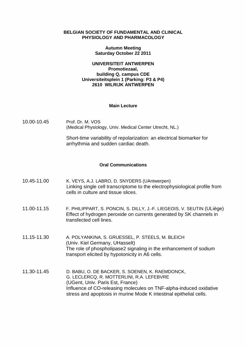
BELGIAN SOCIETY OF FUNDAMENTAL AND CLINICAL PHYSIOLOGY AND PHARMACOLOGY
Autumn Meeting
Saturday October 22 2011
UNIVERSITEIT ANTWERPEN Promotiezaal,
building Q, campus CDE Universiteitsplein 1 (Parking: P3 & P4)
2610 WILRIJK ANTWERPEN
Main Lecture
10.00-10.45 Prof. Dr. M. VOS (Medical Physiology, Univ. Medical Center Utrecht, NL.) Short-time variability of repolarization: an electrical biomarker for arrhythmia and sudden cardiac death.
Oral Communications
10.45-11.00 K. VEYS, A.J. LABRO, D. SNYDERS (UAntwerpen) Linking single cell transcriptome to the electrophysiological profile from cells in culture and tissue slices.
11.00-11.15 F. PHILIPPART, S. PONCIN, S. DILLY, J.-F. LIEGEOIS, V. SEUTIN (ULiège)
Effect of hydrogen peroxide on currents generated by SK channels in transfected cell lines.
11.15-11.30 A. POLYANKINA, S. GRUESSEL, P. STEELS, M. BLEICH
(Univ. Kiel Germany, UHasselt) The role of phospholipase2 signaling in the enhancement of sodium transport elicited by hypotonicity in A6 cells.
11.30-11.45 D. BABU, O. DE BACKER, S. SOENEN, K. RAEMDONCK,
G. LECLERCQ, R. MOTTERLINI, R.A. LEFEBVRE (UGent, Univ. Paris Est, France) Influence of CO-releasing molecules on TNF-alpha-induced oxidative stress and apoptosis in murine Mode K intestinal epithelial cells.
11.45-12.00 B. BOUTIN, N. TAJEDDINE, P. VANDERSMISSEN, N. ZANOU, L. MONDIN, B. TOMBAL, P.J. COURTOY. P. GAILLY (UCLouvain)
Androgen deprivation and bicalutamide induce autophagy of hormone-resistant prostate cancer cells, a process which increases their resistance to apoptosis.
12.00-12.15 C. VAN DER DONCKT, J.L. VAN HERCK, G. VANHOUTTE,
W. MARTINET, M. VERHOYE, A. VAN DER LINDEN, D.M. SCHRIJVERS, H. BULT, A.G. HERMAN, G.R.Y. DE MEYER (UAntwerpen) A mouse model of atherosclerotic plaque rupture leading to myocardial infarction, atheroembolic stroke and sudden death.
12.15-12.30 K. DECALUWE, B. PAUWELS, S. VERPOEST, J. VAN DE VOORDE (UGent)
The molecular mechanisms underlying CO and CORM-2 induced corporal relaxation.
12.30-14.00 Lunch Poster Session General Assembly of the Society and the National Committee of Physiology and Pharmacology
Oral Communications 14.00-14.15 N. WANG, K. SIPIDO, F. BUKAUSKAS, L. LEYBAERT (UGent, KULeuven,
Albert Einstein College of Medicine, New York USA). The connexin mimetic peptides Gap26 and Gap27 inhibit connexin hemichannel unitary current activities.
14.15-14.30 S.M.R. COSYNS, R.A. LEFEBVRE (UGent)
Mechanisms of action of BAY 41-2272 in gastrointestinal relaxation. 14.30-14.45 G. CALJON, V. CAVELIERS, T. LAHOUTTE, P. DE BAETSELIER,
J. VAN DEN ABBEELE, I. SMOLDERS, Y. MICHOTTE, S. MUYLDERMANS, S. MAGEZ, R. CLINCKERS (VUBrussel, VIB/UGent, UAntwerpen) Analysis of blood-brain barrier permeability for nanobodies using microdialysis.
14.45-15.00 K. VENKATESAN, P. ALIX, A. MARQUET, M. DOUPAGNE, I. NIESPODZIANY, B. ROGISTER, V. SEUTIN (ULiège & UCB Pharma –
Braine l’Alleud) Altered GABAergic inhibitory tone in the CA1 region of SV2A-deficient mice.
15.00-15.15 I. VAN BRUSSEL, I. PINTELON, D.M. SCHRIJVERS, J.M. BOSMANS, W.
MARTINET, G.R.Y. DE MEYER, C.J. VRINTS, D. ADRIAENSEN, H. BULT (UAntwerpen) Responses of human monocyte-derived dentritic cells and monocytes to oxidative stress.
15.15-15.30 C. BOUCKAERT, A. PALEM, B. DHOOGHE, P. WALLEMACQ, T.
LEAL (UCLouvain), introduced by J. LEBACQ (UCLouvain) Resveratrol corrects refractory adrenergic pathway in saliva secretion of cystic fibrosis mice.
Posters (dimensions: height 120 cm – width 120 cm)
1. S. WENINGER, J.H. DE MAEYER, R.A. LEFEBVRE (UGent, Shire-Movetis NV,
Turnhout) Phosphorylation of troponin I and phospholamban, induced by 5-HT4 receptor stimulation in pig left atrium, is regulated by phosphodiesterases.
2. N. ZANOU, O. SCHAKMAN, A. DIETRICH, L. BIRNBAUMER, P. GAILLY (UCLouvain,
Univ. Marburg Germany, NIEHS, Nort Carolina USA) Lack of TRPC1 channels impairs skeletal muscle regeneration.
3. K. LOUCHAMI, P.D. BROWN, W.J. MALAISSE, L. BEST, R. BEAUWENS,
A. SENER (ULBruxelles, Univ. Manchester UK) Studies on the role of the glyceroaquaporin AQP7 in mouse pancreatic β-cells.
4. B. DHOOGHE, C. BOUCKAERT, P. WALLEMACQ, T. LEAL (UCLouvain), introduced by
J. LEBACQ (UCLouvain) Vardenafil corrects transrectal chloride transport in cystic fibrosis mice.
5. L. ROTH, D. VAN DAM, C. VAN DER DONCKT, D.M. SCHRIJVERS,
W. MARTINET, P.P. DE DEYN, H. BULT, G.R.Y. DE MEYER (UAntwerpen) A new mouse model of atherosclerotic plaque rupture: insights in motor coordination and spatial learning.
6. C. MICHIELS, G.R.Y. DE MEYER, D.M. SCHRIJVERS, H. BULT, W. MARTINET
(UAntwerpen) Effect of autophagy induction by spermidine on atherosclerotic plaque size and composition.

7. T. WYCKHUYS, N. DE GEETER, S. DELEYE, J. PARTHOENS, W. FIAS, S. STROOBANTS,
S. STAELENS (UAntwerpen, UGent) Use of microSPECT in visualizing the effect of repetitive transcranial magnetic stimulation in the rat.
8. A.J. LABRO, F. BEZANILLA, D.J. SNYDERS (UAntwerpen, Univ. Chicago USA) K+ permeation in Kv channels is under strict control of the voltage-sensor as evidenced by voltage-clamp fluorimetry.
legend
O – oral communications numbered
P – Posters numbered
O-01
LINKING SINGLE CELL TRANSCRIPTOME TO THE ELECTROPHY SIOLOGICAL PROFILE
FROM CELLS IN CULTURE AND TISSUE SLICES
Veys K., Labro A.J., Snyders D.J.
Department of Biomedical Sciences, University of Antwerp, Antwerp, Belgium
Cellular excitability originates from a concerted technique to study the electrophysiology of excitability at the single cell level. At present only few attempts have been made to link electrophysiological profiling to mRNA transcript levels and most suffered from experimental noise making correlations virtually impossible. To collate the patch-clamp analysis with quantitative real-time action of different ion channels and over a 100 different genes have been characterized that underlie the vast diversity in the central nervous system. Patch-clamp is a powerful (qRT) PCR we optimized the technique such that the qRT-PCR essay could be done in twofold and the expression of a housekeeping gene could be used to correct for cell-to-cell variability in mRNA isolation. The technique was validated on a stable Ltk-cell line expressing the Kv2.1 channel. A nice correlation (R2 = 0.9) between current density and Kv2.1 transcript quantity was only obtained when the data were normalized to the transcript level of the housekeeping gene GAPD, indicating that the mRNA isolation step represents most of the experimental noise. Subsequently, we correlated the properties of the complex spike (CS) to the expression level of Kv3.3 in the cerebellar Purkinje Cells of acute brain slices. The data show that variance in Kv3.3 expression level underlies the amount of spikelets in the CS (R2 = 0.65) and shape the complexity (R2 = 0.9). This improved technique should prove very valuable for studying the molecular background of diversity in excitability in normal and disease conditions.
O-02 EFFECT OF HYDROGEN PEROXIDE ON CURRENTS GENERATED BY SK CHANNELS IN TRANSFECTED CELL LINES Philippart F.1, Poncin S.1, Dilly S.2, Liégeois J.-F.2, Seutin V.1
1Laboratory of Pharmacology and Electrophysiology Unit, GIGA-Neurosciences, 2Laboratory of Medicinal Chemistry and CIRM, University of Liège, B-4000 Sart Tilman/Liège, Belgium
Abstract withdrawn
O-03 THE ROLE OF PHOSPHOLIPASE2 SIGNALING IN THE ENHANCE MENT OF SODIUM TRANSPORT ELICITED BY HYPOTONICITY IN A6 CELLS Polyankina A. 1, Gruessel S. 1, Steels P. 2, Bleich M. 1 1Physiologisches Institut der Christian-Albrechts-Universität zu Kiel, Germany; 2BIOMED, Physiology, University Hasselt, Belgium Na+ channel activity in cultured renal (A6) epithelium from Xenopus laevis is regulated by the solution osmolarity. In the present study we examined which pathway downstream of phospholipase A2 (PLA2) is involved. A6 cells were grown on permeable supports and mounted into a water-jacketed (28°C) Ussing chamber . Transepithelial electrical measurements were made under open circuit conditions. The transepithelial conductance (Gt) was determined by pulsed current injections (∆I =1µA) and Isc was calculated. A hypo-osmotic shock (from 260 to 160 mosm/kg H2O) was induced after an equilibration period of 30 minutes by decreasing the NaCl concentration of the mucosal and serosal solutions. Within 40 minutes exposure to hypotonicity Isc increased from 1.4±0.4 µA/cm2 to 7.6±1.5 µA/cm2 (n=7). In the presence of the PLA2 blocker quinacrine (100 µmol/l) Isc did not increase (1.9±0.4 µA/cm2 vs 1.8±0.6 µA/cm2 (n=5)). PLA2 inhibition by Aristolochic acid (100 µmol/l) diminished the increase significantly. In contrast, the inhibitor of the Ca2+-insensitive PLA2, bromoenol lactone (10 µmol/l), did not inhibit the hypo-induced increase in Isc. The COX-1 inhibitor resveratrol (10 µmol/l) inhibited and the COX-2 inhibitor celecoxib (20 µmol/l) increased the hypo-induced increase in Isc, whereas indomethacin (10 µmol/l) was not effective. In the presence of the 5-lipoxygenase inhibitor nordihydroguaiaretic acid (20 µmol/l) Isc increased from 2.7±0.3 µA/cm2 to 5.8±0.8 µA/cm2 (n=7), which was significantly less than in control hypotonic conditions. The data suggest that the leukotriene and COX1 pathways contribute to the osmotic regulation of the sodium channel activity in A 6 cells.
O-04 INFLUENCE OF CO-RELEASING MOLECULES ON TNF-ALPHA-IN DUCED OXIDATIVE STRESS AND APOPTOSIS IN MURINE MODE-K INTESTINAL EP ITHELIAL CELLS Babu D.1, De Backer O.1, Soenen S.2, Raemdonck K.2, Leclercq G.3, Motterlini R.4, Lefebvre R.A.1
1Heymans Institute of Pharmacology and 3Department of Clinical Chemistry, Microbiology and Immunology, Faculty of Medicine and Health Sciences; 2Laboratory of General Biochemistry and Physical Pharmacy, Faculty of Pharmaceutical Sciences, Ghent University, Belgium; 4INSERM U955, University Paris-Est, France In the mouse postoperative ileus model, our group has previously shown an increase in oxidative stress after intestinal manipulation occurring earlier in the mucosal layer than in the muscular layer; this might lead to epithelial barrier dysfunction and intestinal inflammation, contributing to ileus (De Backer et al., 2009). The carbon monoxide (CO)-releasing molecule, CORM-3, was shown to markedly reduce oxidative stress and to restore intestinal motility. We therefore investigated the effect of CO-releasing molecules on TNF-alpha-induced oxidative stress and apoptosis in murine Mode-K intestinal epithelial cells. Mode-K cells (passage 10-35) were grown for 36h and then serum starved overnight; they were then exposed to TNF-α/cycloheximide (CHX) for up to 6h. TNF-α/CHX increased reactive oxygen species (ROS) production (carboxy-H2DCFDA fluorescence intensity measurement), reduced the level of the antioxidant glutathione (bioluminescent assay), increased caspase-3/7 activity (bioluminescent assay), and induced apoptosis (subdiploid DNA analysis by flow cytometry) in a concentration-dependent manner in Mode-K cells. All these effects of TNF-α/CHX were partially prevented by pretreatment with CORM-A1. The effect of CORM-A1 was not mimicked by treating the cells with cobalt protoporphyrin, an inducer of heme oxygenase-1, an endogenous source of CO, although the expression of heme oxygenase-1 was confirmed by Western blotting. CO-RMs might thus protect the epithelial barrier from leaking, by reduction of oxidative stress.
O-05 ANDROGEN DEPRIVATION AND BICALUTAMIDE INDUCE AUTOPH AGY OF HORMONE-RESISTANT PROSTATE CANCER CELLS, A PROCESS WHICH INCREASES THEIR RESISTANCE TO APOPTOSIS Boutin B., Tajeddine N., Vandersmissen P., Zanou N., Mondin L., Tombal B., Courtoy P.J., Gailly P.
Lab of Cell Physiology, University of Louvain, 55, av. Hippocrate, 1200 Brussels, Belgium. Treatment of advanced PCa consists in pharmacological or surgical androgen deprivation. However, androgen deprivation is temporarily efficient. After a few months or years, the tumor relapses despite the absence of androgenic stimulation: it becomes hormone-refractory (HRPCa). Despite the fact that autophagy is involved in chemoresistance in some cancers, its role remains unknown in the development of HRPCa. In this study, androgen deprivation or treatment with the anti-androgen bicalutamide increased the on rate of autophagy as demonstrated by detecting GFP-LC3 relocalization and LC3 conversion. This effect was dramatically reduced after depletion of Atg5, Beclin-1 and ULK1, three canonical autophagic genes. Moreover, autophagy induced by androgen deprivation and bicalutamide depends of the inhibition of the PI3-K/Akt/mTOR that is mediated by a disruption of the complex formed by AR and the regulatory subunit of PI3-K p85. siRNA-mediated depletion of Atg5 and Beclin-1 significantly increased the level of cell death induced by bicalutamide or androgen deprivation. Finally, the safe anti-malarial drug chloroquine, an inhibitor of autophagy, dramatically increased cell death after androgen deprivation and bicalutamide treatment. Taken together, our data suggest that autophagy is a protective mechanism against androgen deprivation in HRPCa cells and that restoration of hormone sensitivity in LNCaP cells could be achieved by concomitant treatment with chloroquine.
O-06 A MOUSE MODEL OF ATHEROSCLEROTIC PLAQUE RUPTURE LEA DING TO MYOCARDIAL INFARCTION, ATHEROEMBOLIC STROKE AND SUD DEN DEATH Van der Donckt C.1, Van Herck J.L.2, Vanhoutte G.3, Martinet W.1, Verhoye M.3, Van der Linden A.3, Schrijvers D.M.1, Bult H.1, Herman A.G.1, De Meyer G.R.Y.1
1Division of Pharmacology, 2Division of Cardiology and 3Bio-Imaging Lab, University of Antwerp, B-2610 Antwerp, Belgium We have previously shown that impaired elastin function, as a result of a mutation in the fibrillin-1 (Fbn1C1039G+/-) gene, led to destabilization of atherosclerotic plaques with sporadic plaque ruptures in apolipoprotein E (ApoE-/-) -deficient mice fed a Western type diet. In this study, we investigated whether ApoE-/- Fbn1C1039G+/- mice can be used as an animal model of atherosclerotic plaque rupture with clinical end points. Female ApoE-/- Fbn1C1039G+/- (n=20) and ApoE-/- mice (n=20) were fed a Western type diet for up to 35 weeks. 15 of 20 ApoE-/- Fbn1C1039G+/- mice (75%) died suddenly, of which fifty percent after 20 weeks. Sudden death was associated with acute plaque rupture in the proximal ascending aorta (seven out of ten examined animals). Premature death did not occur in ApoE-/- mice or ApoE-/- Fbn1C1039G+/- mice on normal chow (n=8). Magnetic resonance imaging of the brain showed the presence of one or more lesions in all ApoE-/- Fbn1C1039G+/- mice on Western type diet. Brain lesions were characterized by the presence of cholesterol clefts, foam cells, macrophages and smooth muscle cells, suggesting that these lesions arose from embolization of plaque debris after rupture. Furthermore, acute myocardial infarction was observed in three out of fifteen ApoE-/- Fbn1C1039G+/- mice (20%) that died suddenly. It is concluded that ApoE-/- Fbn1C1039G+/- mice represent a unique reproducible model of acute plaque rupture with clinical end-points as atheroembolic stroke, myocardial infarction and sudden death. This animal model offers the possibility to further investigate the mechanisms underlying plaque rupture and enables the investigation of novel plaque stabilizing therapies.
O-07
THE MOLECULAR MECHANISMS UNDERLYING CO AND CORM-2 I NDUCED CORPORAL RELAXATION Decaluwé K., Pauwels B., Verpoest S., Van de Voorde J. Department of Pharmacology, Ghent University, Ghent, Belgium CO has comparable to NO, the principal mediator of penile erection, vasodilator capacities. However, research on whether CO could be a new therapeutic target for treating erectile dysfunction remains scarce. The danger associated with the systemic administration of CO has led to the development of CO-releasing molecules, releasing CO in a local, safe and controlled way. These CO-releasing molecules have shown positive outcomes in cardiovascular studies. More knowledge on the (patho)physiological functions of CO in erectile function and the potential therapeutic role of CO-releasing molecules is required. The present study assessed the effects of CO and the CO-donor CORM2 on the corporal tension and investigated the underlying molecular mechanisms. Organ bath studies were performed measuring isometric tension on isolated mice corpora cavernosa strips. Responses to CO (10 – 300 µmol/L) and CORM2 (10 – 100 µmol/L) were evaluated in the presence/absence of activators/inhibitors of different molecular pathways. While both CO and CORM2 relax corpora cavernosa, their molecular mechanisms seem to differ completely. CO induces corporal relaxation by activating soluble guanylyl cyclase, increasing cGMP concentrations. The molecular mechanism involved in CORM2 induced corporal relaxation is not related to cGMP and remains obscure. However the inhibitory influence of 4-aminopyridine indicates a small contribution of voltage-dependent potassium channel opening. It is concluded that both CO and CORM2 induce corporal relaxation, but that the underlying molecular mechanisms are different. That CO induces corporal relaxation through a mechanism similar to that of NO is finding that may be of importance, indirectly displaying the possibility that endogenous CO might serve as a back-up system for insufficient NO availability in cases of erectile dysfunction. Whether CORM2 possesses the same capacity is questionable and requires further research.
O-08 THE CONNEXIN-MIMETIC-PEPTIDES GAP26 AND GAP27 INHIB IT CONNEXIN-HEMICHANNEL UNITARY CURRENT ACTIVITIES Wang N.1, Sipido K.R.2, Bukauskas F.F.3, Leybaert L.1
1Department of Basic Medical Sciences, Physiology Group, Faculty of Medicine and Health Sciences, De Pintelaan 185 (Block B, 3rd Floor), Ghent University, B-9000 Ghent, Belgium 2Department for Experimental Cardiology, O & N1, K.U.Leuven, Leuven, Belgium 3Dominick P. Purpura Department of Neuroscience, Albert Einstein College of Medicine, Bronx, NY 10461, USA Gap26 and Gap27 are two connexin-mimetic peptides that correspond to sequences in the first and second extracellular loop regions of Cx43 respectively. They were initially developed as gap junction inhibitors but were later found to also inhibit ATP release and dye uptake suggesting block of unapposed connexin-hemichannels (CxHCs). However, up to now, no conclusive arguments are available to support HC block by these peptides. The purpose of the present work was to investigate the effect of Gap26 and Gap27 on unitary current activity through Cx43 HCs. Unitary-currents were recorded using whole-cell voltage-clamp in Hela cells stably transfected with Cx43 (Hela-Cx43) in the presence of K+-channel blockers and current activity was quantified by integrating unitary events over time, yielding the charge transfer via HCs. Hela-Cx43 cells exhibited HC unitary gating events of ~220 pS in response to depolarization from -30 to +70 mV. Thirty minutes exposure to Gap26 (160 µM) or Gap27 (200 µM) reduced the HC charge transfer by ~70%. HC charge transfer declined with a time constant of ~2.5 min for Gap26 and ~3.7 min for Gap27 and steady-state inhibition was estimated to be in the order of ~65 % inhibition for Gap26 and ~85 % for Gap27. Half-maximal inhibition, determined at t = time constant, occurred at 81 µM for Gap26 and at 161 µM for Gap27. Analysis of the voltage-dependency of unitary current activation demonstrated that Gap26 and gap27 shifted the I-V relation in positive direction. The peptides had no apparent effects at the level of the single channel conductance. Importantly, peptides with scrambled Gap26 or Gap27 sequences exhibited no inhibitory effect on HC charge transfer unless they were applied at 1 mM, at which point steric pore occlusion presumably starts to play a role. We conclude that Gap26 and Gap27, at concentrations in the hundreds of micromolar range, inhibit depolarization-induced Cx43 HC unitary currents within 4 min, by shifting the voltage-dependency of channel opening to positive potentials. Peptides of scrambled sequences exerted no inhibitory action at concentrations where Gap26/27 was active, suggesting sequence-specific effects of the two peptides. Gap26 has been demonstrated to interact with the extracellular loops of Cx43 and therefore, our results indicate a role of the extracellular loops in the gating of unapposed HCs.
O-09 MECHANISM OF ACTION OF BAY 41-2272 IN GASTROINTESTI NAL RELAXATION Cosyns S., Lefebvre R.
Heymans Institute of Pharmacology, Ghent University, Ghent, 9000, Belgium. BAY 41-2272 is a heme-dependent NO-independent soluble guanylate cyclase (sGC) stimulator. Still, its relaxant effect in respiratory and urogenital tissue is only partially dependent on sGC activation; its mechanism of action has not been studied in the gastrointestinal tract. We therefore investigated the effect of BAY 41-2272 in mouse gastric fundus and colon. Circular smooth muscle strips from the gastric fundus and distal colon (after removal of the mucosa) of male C57Bl/6J mice (11-15 weeks) were mounted in organ baths under NANC conditions for isometric force recording. cGMP levels were determined using an enzyme immunoassay kit after clamping of the tissues. BAY 41-2272 induced concentration-dependent relaxation in both tissues (1-10 µM [fundus]; 0.3-3 µM [colon]). BAY 41-2272 (10 µM [fundus], 3 µM [colon]) increased basal cGMP levels by 3-fold in the fundus strips and by 4.5-fold in the colonic strips. The sGC inhibitor ODQ (30 µM) totally inhibited this BAY 41-2272-induced increase of cGMP, but only partially reduced the corresponding relaxation. The relaxant responses to BAY 41-2272 (10 µM [fundus]); 3 µM [colon]) were not significantly influenced by apamin (0.3 µM), charybdotoxin (0.1 µM), or ouabain (10 µM) excluding interaction with small, intermediate and large conductance Ca2+-activated K+-channels and with Na+/K+-ATPase. Under conditions where all intracellular calcium was depleted from the Ca2+-stores, CaCl2-induced contractions (0.01-100 mM) were significantly reduced by BAY 41-2272 in fundus and colon strips. Pre-treatment with ODQ (30 µM) did not influence the reduction in CaCl2-induced contractions by BAY 41-2272. The present study demonstrates that BAY 41-2272 exerts its relaxing effect in the mouse gastric fundus and distal colon partially through a cGMP-dependent mechanism and by at least one additional cGMP-independent mechanism involving Ca2+entry blockade.
O-10 ANALYSIS OF BLOOD-BRAIN BARRIER PERMEABILITY FOR NA NOBODIES USING MICRODIALYSIS Caljon G.2,3,5, Caveliers V.4, Lahoutte T.4, De Baetselier P.2,3, Van Den Abbeele J.5, Smolders I.1, Michotte Y.1, Muyldermans S.2,3, Magez S.2,3, Clinckers R.1*
1 Department of Pharmaceutical Chemistry and Drug Analysis, Research Group Experimental Pharmacology, Vrije Universiteit Brussel, Brussels, Belgium. 2 Unit of Cellular and Molecular Immunology, Vrije Universiteit Brussel, Brussels, Belgium. 3 Department of Molecular and Cellular Interactions, VIB, Ghent, Belgium. 4 In Vivo Cellular and Molecular Imaging, ICMI, Vrije Universiteit Brussel, Brussels, Belgium. 5 Department of Animal Health, Unit of Veterinary Protozoology, Institute of Tropical Medicine Antwerp, Antwerp, Belgium. Nanobodies are considered as very promising antigen-binding moieties for molecular imaging and therapeutic purposes due to their favorable pharmacological and pharmacokinetic properties. However, the possibility for monovalent Nanobodies to reach targets in the central nervous system (CNS) remains to be demonstrated. We have assessed the blood-brain barrier permeability for Nb_An33, a Nanobody against the Trypanosoma brucei Variant-specific Surface Glycoprotein (VSG), in healthy rats and rats that are in the encephalitic stage of African trypanosomiasis and suffer from acute brain inflammation. To assess the integrity of the blood-brain barrier, brain disposition of 2-Methoxy-IsoButyl-Isonitrile (MIBI) and Evans Blue was monitored in both experimental groups of animals. Perfusion of the different compounds was analyzed by intracerebral microdialysis, Single Photon Emission Computed Tomography (SPECT) or a combination of both methodologies. This enabled the quantification of unlabeled and (99m)Tc-labeled Nanobodies using respectively a sensitive VSG-based Nanobody-detection ELISA, radioactivity measurement in collected microdialysates and SPECT image analysis. As the ELISA analysis provided direct evidence for antigen-binding potential, this approach allowed to differentiate between active Nanobody and fragments that might be generated during the (99m)Tc-labeling process or in vivo by proteolytic processing. The combined read-out methodologies illustrate that, while brain disposition of Nb_An33 upon intravenous injection is at the limit of detection in healthy rats, inflammation-induced damage to the blood-brain barrier significantly increases (approximately 20 times) the Nanobody perfusion efficiency. Despite the enhanced brain disposition, calculated intrahippocampal Nb_An33 concentrations remained very low (131 ± 63 ng/ml with a dose of 4 mg/kg), likely resulting from the observed high clearance rates of Nanobodies in general. Collectively, we illustrate the advantage of complementing SPECT analyses with intracerebral microdialysis in brain disposition studies and suggest that it is of interest to evaluate the blood-brain barrier penetrating potential of monovalent Nanobodies in models of CNS-inflammation. However, the pharmacokinetic properties that are considered favorable for peripheral imaging and therapeutic approaches might be key factors that prelude efficient application of Nanobodies to target the CNS. O-11
ALTERED GABAERGIC INHIBITORY TONE IN THE CA1 REGION OF SV2A-DEFICIENT MICE Venkatesan K.1, Alix P.1, Marquet A.2, Doupagne M. 2, Niespodziany I.4, Rogister B.2, 3, Seutin V.1
1Laboratory of Pharmacology and Electrophysiology Unit, GIGA-Neurosciences, University of Liège 2Laboratory of Developmental Neurobiology, GIGA-Neurosciences, University of Liège 3Department of Neurology, CHU of Liège, all at B-4000 Sart Tilman, Belgium; 4UCB Pharma, CNS Research, B-1420 Braine-l’Alleud, Belgium. GABAergic synaptic transmission plays a critical role in the control of neuronal excitability in the hippocampus. Synaptic vesicle protein 2 (SV2), a glycoprotein common to most synaptic vesicles, exists in three highly homologous isoforms, SV2A, SV2B, and SV2C. SV2A knock out (KO) mice and SV2A/SV2B double KO (DKO) mice, but not SV2B KO animals, show a clear seizure phenotype compared to wild type animals. SV2A-deficient mice start to experience severe seizures 7-11 days after birth and die between P14 and P23. Because GABAA inhibitory neurotransmission is altered in the CA3 region of SV2A KO mice, it seemed worthwhile to study whether this finding can be extended to other hippocampal areas. We therefore examined the effects of a SV2A and/or SV2B deletion on GABAA receptor-mediated inhibitory neurotransmission in CA1. Spontaneous and miniature GABAergic inhibitory postsynaptic currents (sIPSCs and mIPSCs) were recorded in the whole-cell configuration ex vivo. Experiments were performed in the presence of the glutamate receptor antagonists D-AP5 and CNQX. mIPSCs were recorded in the additional presence of TTX (500 nM). We found that the frequency and amplitude of sIPSCs were decreased in SV2A KO and SV2A/SV2B DKO mice, but not in SV2B KO animals. However, neither the amplitude nor the frequency of mIPSCs were altered in SV2A KO and SV2A/SV2B DKO mice. These results confirm that action potential-dependent GABAergic transmission is altered in the hippocampus of SV2A-deficient mice, whereas the mechanism of exocytosis by itself is not altered.
O-12 RESPONSES OF HUMAN MONOCYTE-DERIVED DENDRITIC CELLS AND MONOCYTES TO OXIDATIVE STRESS Van Brussel I.1, Pintelon I.2, Schrijvers D.M.3, Bosmans J.M.1, Martinet W.3, De Meyer G.R.Y.3, Vrints C.J.1, Adriaensen D.2, Bult H.3
1Dept. of Cardiology, 2Lab. of Cell Biology and Histology, 3Div. of Pharmacology, University of Antwerp, B-2610 Wilrijk, Belgium. Dendritic cells (DCs) are professional antigen-presenting cells with the unique ability to initiate primary T-cell responses. They are present in atherosclerotic lesions where they are exposed to oxidative stress. Therefore, we examined the function of antioxidant defence systems in human monocyte-derived (mo)DCs in comparison to monocytes. Monocytes were purified from peripheral blood mononuclear cells by using anti-CD14-conjugated magnetic microbeads and cultured for 1 day in RPMI (monocytes), or for 6 days in RPMI containing GM-CSF and interleukin-4 (moDCs). Cells were exposed to tertiary-butylhydro-peroxide (tert-BHP 200µM, 30 min) followed by evaluation of intracellular reactive oxygen species (ROS, 5-(and-6)-chloromethyl-2,7-dichlorodihydrofluorescein diacetate (CM-H2DCFDA) fluorescence) and viability (neutral red assay). Total RNA (5 105 cells) was extracted for a PCR profiling array and for confirmation by Taqman gene expression assays. Results: tert-BHP increased CM-H2DCFDA fluorescence and caused cell death; both processes occurred more slowly in moDCs than in monocytes. The mRNA profiling array showed more than 2-fold differential expression of 32 ROS–related genes, including peroxiredoxin-2 (PRDX2), an enzyme reducing hydrogen peroxide and lipid peroxides. PRDX2 upregulation was confirmed by Taqman assays, immunoblotting and immunohistochemistry. Silencing PRDX2 in moDCs by means of siRNA significantly increased CM-DCF fluorescence and cell death in tert-BHP-stimulated samples. The results indicate that moDCs exhibit higher intracellular antioxidant capacities and are better equipped to resist ROS than monocytes. Upregulation of PRDX2 is involved in the neutralization of ROS in moDCs. Taken together, this points to better functionality and survival chances of DCs in oxidative stress environments, such as atherosclerotic plaques.
O-13 RESVERATROL CORRECTS REFRACTORY ADRENERGIC PATHWAY IN SALIVA SECRETION OF CYSTIC FIBROSIS MICE Bouckaert C., Palem A., Dhooghe B., Wallemacq P., Leal T.
Louvain Centre for Toxicology and Applied Pharmacology; Université catholique de Louvain, Brussels - Belgium In vitro studies have suggested that resveratrol, a polyphenol compound found in red wine peanuts, soy beans, and pomegranates, for which antioxidant and anti-inflammatory properties have being claimed, would have beneficial effects in cystic fibrosis (CF). It has also been characterized that CF exocrine glands, such as salivary glands, are refractory to β-adrenergic stimulation. We tested the effect of resveratrol on the constitutive contribution and on the β-agonist sensitivity of the adrenergic pathway of saliva secretion in CF female mice homozygous for the F508del mutation as compared to wild-type (WT) homozygous mice built in the 159/FVB genetic background. Resveratrol (50mg/kg) or vehicle (1:1 DMSO/NaCl 0.9%) treatment was applied once-daily, by ip administration, for 4 days. A salivary secretion test was performed, in CF and WT mice, 2 h after treatment on days 2 and 4 (n=3 for each duration time). Control experiments were performed in animals from both genotypes in the absence of any vehicle or resveratrol treatment (n=5 in each group). The basal, constitutive contribution of β-adrenergic pathway of saliva secretion flow (SSF; µg/min/g body weight) was assessed after blocking the cholinergic pathway with local injection of 10-3 M atropine, by weighing saliva collected onto filter papers placed on the inner face of the tested cheek. The β-agonist sensitivity of the adrenergic pathway was then examined after topical application of 10-4 M isoprenaline. DMSO did not affect the degree of atropine or isoprenaline responses in any experimental group. The constitutive β-adrenergic pathway of saliva secretion was significantly reduced in untreated F508del-CF mice as compared to WT mice (n=5 in each group). Indeed, median [range] values of SSF in CF mice were 4.8 [2.4-8.1] µg/min/g while the values in WT mice were at least twice as high (p=0.037; Wilcoxon test). β-agonist stimulation produced a dramatic increase (p=0.012) in WT SSF which changed from 8.5 [5.7-10.9] to 27.9 [19.5-63.5] µg/min/g while no detectable change was seen in CF. Resveratrol treatment removed the refractory effect of CF β-agonist response by increasing SSF by at least 3-fold; as similar magnitude of responses was observed on days 2 and 4, data were pooled. These preliminary data confirmed that the β-adrenergic pathway of saliva secretion is reduced in CF and indicated that resveratrol rescues CFTR-dependent refractory adrenergic pathway of saliva secretion in F508del-CF mice.
P-01 PHOSPHORYLATION OF TROPONIN I AND PHOSPHOLAMBAN, IN DUCED BY 5-HT4 RECEPTOR STIMULATION IN PIG LEFT ATRIUM, IS REGULA TED BY PHOSPHODIESTERASES Weninger S.1, De Maeyer J.H.2, Lefebvre R.A.1
1Heymans Institute of Pharmacology, UG, Gent, 9000, Belgium; 2Shire-Movetis NV, Turnhout, 2300, Belgium The concerted action of phosphodiesterase (PDE) 3 and PDE4 subtypes, which degrade the second messenger cAMP, is responsible for the fade of inotropic responses to 5-HT4 receptor activation in porcine left atrium. We now further assessed this controlling role of PDE3 and PDE4 by determining the phosphorylation of troponin I (TnI) and phospholamban (PLB), both targets of protein kinase A, involved in the inotropic response to 5-HT. Left atrial pectinate muscles from male pigs (15-25 kg) were mounted into tissue baths, where they received either the non-selective PDE-inhibitor IBMX or the combination of the PDE3 inhibitor cilostamide and the PDE4 inhibitor rolipram. Tissues were freeze-clamped 2, 10 and 30 min after 5-HT4 receptor stimulation with 5-HT or prucalopride and homogenized. Proteins were separated by SDS/PAGE, transferred to nitrocellulose and analysed by immunoblotting using antibodies against TnI, phospho-TnI Ser23/24, PLB and phospho-PLB Ser16. The PDE inhibitors per se had no clear-cut effect on PLB or TnI phosphorylation. PLB Ser16 phosphorylation was significantly increased after addition of 5-HT or prucalopride. This was significantly further enhanced by IBMX, or cilostamide plus rolipram. Phosphorylation of TnI Ser23/24 tended to increase after addition of 5-HT, but in the presence of IBMX, or cilostamide plus rolipram a clear-cut increase in phosphorylation of TnI was obtained. In contrast prucalopride alone significantly enhanced TnI Ser23/24 phosphorylation, while IBMX, or cilostamide plus rolipram only tended to further increase phosphorylation. These results demonstrate that the control of the cAMP signal upon 5-HT4 receptor stimulation in pig left atrium by PDE3 and PDE4 indeed leads to concomitant dampening of downstream mediators of the cAMP-protein kinase A pathway.
P-02 LACK OF TRPC1 CHANNELS IMPAIRS SKELETAL MUSCLE REGE NERATION Zanou N.1, Schakman O.1, Dietrich A.2, Birnbaumer L.3, Gailly P.1 1University of Louvain, 55 av. Hippocrate, 1200 Brussels, Belgium, 2University of Marburg, Germany. 3. NIEHS, North Carolina USA. We previously showed in vitro that calcium entry through TRPC1 ion channels regulates myoblasts migration and differentiation by activating calpain, a calcium-dependent protease which in its turn is able to cleave MARCKS protein (myristoylated alanine-rich C-kinase substrate), an actin binding protein involved in focal adhesion. To explore, in vivo, whether the absence of TRPC1 channel impairs skeletal muscle regeneration, we used cardiotoxin injections to induce muscle injury in adult TRPC1+/+ and TRPC1-/- mice. Interestingly, we observed that TRPC1-/- muscles showed a smaller fibre size and a decreased muscle specific force after 10 days and 14 days of regeneration respectively. Moreover, we observed an increased proportion of centrally nucleated fibres at day 14 of regeneration in TRPC1-/-. These observations pointed out a delay of muscle regeneration in TRPC1-/- mice in comparison with their controls. This was corroborated by the fact that, in comparison with their controls, TRPC1-/- muscles showed a decrease in myogenic transcription factors activity and, in particular, a decrease of MyoD, Myf5 and myogenin expression. As expected, the developmental Myosin Heavy Chain, a well known downstream target of MyoD during muscle regeneration, was more expressed in TRPC1+/+ than in TRPC1-/- muscles at day 3 of regeneration. Moreover, the Akt/mTOR/P70S6k pathway which is involved in regeneration and in regulation of protein synthesis and muscles fibre size, was also down regulated in TRPC1-/- muscles. Indeed, we observed a decrease of phosphorylation of both Akt and P70S6k in TRPC1-/- muscles than in TRPC1+/+ muscles. Altogether, our results emphasize the involvement of TRPC1 channels in skeletal muscles development and identify a calcium-dependent activation of the Akt /mTOR/P70S6k pathway during muscles regeneration which is decreased in TRPC1-/- mice.
P-03 STUDIES ON THE ROLE OF THE GLYCEROAQUAPORIN AQP7 IN MOUSE PANCREATIC Β-CELLS Louchami K.1, Brown P.D.2, Malaisse W.J.1, Best L.2, Beauwens R.3, Sener A.1
1Laboratory of Experimental Hormonology, Université Libre de Bruxelles, Brussels, Belgium, 2School of Medicine and Life sciences, University of Manchester, Manchester, UK, 3Laboratory of cell Physiology, Université Libre de Bruxelles, Brussels, Belgium.
We have proposed that increased β-cell volume in response to uptake and metabolism of nutrient stimuli such as glucose represents an important event in the regulation of insulin release. We hypothesise that cell swelling results in the activation of the volume-regulated anion channel (VRAC) which generates an inward (depolarizing) current, resulting in electrical and secretory activity. Cell swelling could, in turn, depend on water transport via aquaporins (AQPs) such as AQP7, known to be expressed in the β-cell. In the present study, we have investigated pancreatic β-cell function in AQP7 knockout mice in order to elucidate the role of this glyceroaquaporin in insulin-secreting cells. Insulin release was measured from intact mouse islets. Relative cell volume was measured using a video-imaging technique and electrical activity by means of the perforated patch technique, using isolated β-cells in both cases. No significant difference was observed between AQP7+/+ and -/- mice in terms of either plasma D-glucose or insulin concentration. The basal release of insulin at 2.8 mM D-glucose did not differ significantly between AQP7+/+ (12.0 ± 1.1 µU/islet; n = 60) or AQP7-/- (13.0 ± 1.3 µU/islet; n = 60) mouse islets. Exposure of AQP7+/+ islets to a 33% hypotonic medium (omission of 50 mM NaCl) caused a 4-fold stimulation of insulin release (P<0.01). In contrast, insulin release from AQP7-/- islets was not significantly affected by hypotonic medium. Similarly, substituting 50 mM NaCl by the permeable osmolyte glycerol (100 mM) significantly (P<0.01) augmented insulin release in AQP7+/+ but not -/- islets. Incubation of islets at raised glucose concentrations (8.3 or 16.7 mM) stimulated insulin release to a significantly greater extent (P<0.05 and 0.001 respectively) in AQP7+/+ compared to -/- mouse islets. Exposure of β-cells from AQP7 +/+ mice to a 33% hypotonic solution caused a rapid and pronounced increase in cell volume followed by a gradual regulatory volume decrease. Virtually identical responses were observed in cells isolated from AQP7 -/- mice. A rise in glucose concentration from 4 to 20 mM also caused a similar degree of swelling in +/+ and -/- cells. However, when exposed to 50 mM glycerol (substituted for mannitol), the increase in cell volume was significantly lower (P<0.02) in the AQP7-/- than in AQP7+/+ cells. Electrical activity could be evoked in β-cells from both AQP7+/+ and -/- mice by a rise in glucose concentration to 8.3, 11.1 or 16.7 mM, by exposure to a 33% hypotonic medium or to 50 µM tolbutamide. However, in sharp contrast to the β-cells from AQP7+/+ mice, cells from AQP7-/- mice failed to display any significant change in membrane potential when exposed to 50 mM glycerol. Activation of the VRAC by swelling the cells using a hypertonic intracellular solution displayed similar kinetics in terms of rate and extent of activation in cells from AQP7+/+ and -/- mice. The present study revealed that insulin release evoked by a rise in glucose concentration, by extracellular hypotonicity or by the isosmotic addition of glycerol in isolated pancreatic islets was always lower in AQP7-/- than in AQP7+/+ mice. β-cells from AQP+/+ and -/- mice both showed comparable rates and degrees of cell swelling
when exposed to a hypotonic solution or raised glucose concentration, implying the existing of at least one water transport pathway in mouse β-cells in addition to AQP7. In contrast, the addition of glycerol caused a significantly smaller increase in cell volume in the AQP-/- cells compared to those from wild type mice, presumably reflecting the activity of AQP7 as a glycerol transport pathway. Similarly, whilst β-cell electrical activity in response to glucose, hypotonic solutions or tolbutamide was essentially unaltered in AQP-/- β-cells, little or no response to glycerol was observed in β-cells from AQP7-/- mice. The finding that secretory activity in AQP7-/- cells was impaired, despite apparently normal volume and electrical responses to various stimuli, suggests that AQP7 could, directly or indirectly, play a role at a ‘distal’ site of the exocytotic pathway.
P-04 VARDENAFIL CORRECTS TRANSRECTAL CHLORIDE TRANSPORT IN CYSTIC FIBROSIS MICE Dhooghe B., Bouckaert C., Wallemacq P., Leal T. Louvain Centre for Toxicology and Applied Pharmacology, Université catholique de Louvain, Brussels, 1200, Belgium We have previously shown that characteristic transepithelial ion transport defects in cystic fibrosis (CF), namely increased sodium absorption and reduced or abolished chloride transport, can be assessed at the intestinal level, a clinically relevant CF target organ, by means of the rectal potential difference test (RPD). The aim of this work is to test the efficacy of vardenafil, an inhibitor of phosphodiesterase type 5, to correct CFTR-dependent transrectal ion transport in CF mice homozygous for the F508del mutation as compared to heterozygous (HTZ) and wild-type homozygous (WT) animals from the same 129/FVB genetic background. RPD was performed 1 h after a single ip injection of vardenafil (0.14 mg/kg). Total chloride secretion across the rectal mucosa (TCS) was monitored under perfusion of a Ringer’s solution without chloride (replaced by gluconate) and with 10- 5 M forskolin, and containing 10-4 M amiloride and 5 x 10⁻³ M barium, to block sodium and potassium movement. Vardenafil significantly stimulated TCS in CF mice when values obtained in the absence of vardenafil (-4.2 ± 0.5 mV; mean ± SEM; n:5) reached -9.3 ± 1.1mV (n:7; p:0.0054) after vardenafil treatment. Intermediately reduced TCS values in the HTZ group were also significantly increased by vardenafil treatment. The corrector effect of vardenafil was mainly due to the contribution of forskolin sensitivity that was increased by 4-fold in the HTZ group and by 5-fold in the CF group. These data indicate that RPD is a powerful tool to assess efficacy of therapeutic strategies in CF. Our findings confirm that vardenafil corrects CFTR-dependent chloride transport in CF intestinal tissue.
P-05 A NEW MOUSE MODEL OF ATHEROSCLEROTIC PLAQUE RUPTURE : INSIGHTS IN MOTOR COORDINATION AND SPATIAL LEARNING Roth L.1, Van Dam D.2, Van der Donckt C.1, Schrijvers D.M.1, Martinet W.1, De Deyn P.P.2, Bult H.1, De Meyer G.R.Y.1
1 Laboratory of Pharmacology and 2 Laboratory of Neurochemistry and Behaviour, Institute Born-Bunge, University of Antwerp, B-2610 Wilrijk, Belgium
Previously, we showed that apolipoprotein E deficient (ApoE-/-) mice with a heterozygous mutation in the fibrillin-1 gene (Fbn1C1039G+/-
, Marfan phenotype) that leads to fragmentation of the elastin fibres, show accelerated progression of atherosclerosis and spontaneous occurrence of plaque ruptures on a Western type diet. Consequently, athero-embolic stroke occurs, accompanied by neurological symptoms (head tilt) and sudden death. In the present study, we focused on motor coordination and spatial learning of the ApoE-/- Fbn1C1039G+/- mice, which might provide useful information on the neurological aspects of this mouse model. Female ApoE-/- (control) and ApoE-/- Fbn1C1039G+/- mice (6 weeks old) were fed a Western type diet for up to 20 weeks (n=10 mice in each group). Motor coordination was assessed every 2 weeks starting at 10 weeks of Western type diet by the following tests: stationary beam, wire suspension and accelerating rotarod. The stationary beam was the only test that revealed a significant decrease in motor coordination of the ApoE-/- Fbn1C1039G+/- mice compared to the controls (crossed segments: 23±5 vs. 49±8 at week 16, 17±9 vs. 39±6 at week 18, 20±8 vs. 48±7 at week 20). Moreover, this decline was seen at a time point before head tilt occurred, indicating that the stationary beam test might detect neurological symptoms at an early stage. Spatial learning was evaluated by the Morris water maze test, but did not show significant differences. In conclusion, the stationary beam is the only test that showed a difference in motor coordination between ApoE-/- and ApoE-/- Fbn1C1039G+/- mice and can therefore be used to further characterize the ApoE-/- Fbn1C1039G+/- model. However, additional experiments are needed to evaluate whether the Marfan phenotype as such also affects motor performance.
P-06 EFFECT OF AUTOPHAGY INDUCTION BY SPERMIDINE ON ATHE ROSCLEROTIC PLAQUE SIZE AND COMPOSITION Michiels C., De Meyer G.R.Y., Schrijvers D.M., Bult H., Martinet W. Division of Pharmacology, University of Antwerp, B-2610 Wilrijk, Belgium. Atherosclerosis is one of the main underlying causes of death in the Western world. Despite the existing therapies, morbidity and mortality due to plaque rupture remains considerable. Recent research provides evidence that cell death is a strong modulating factor in the progression of atherosclerosis. Along these lines, it has been suggested that autophagy, a cellular housekeeping mechanism that leads to cell death under certain conditions, could play an important role in atherosclerosis. Therefore, we assessed in the present study the effect of the autophagy inducer spermidine on atherogenesis and plaque stability. In vitro, smooth muscle cells (SMCs) were sensitive to cell death following spermidine treatment, while macrophages were resistant up to 100µM spermidine. Treatment with 10µM spermidine for 24 h did not significantly affect SMC viability (95±5%; n=3), but nearly all SMCs died after treatment with 100µM spermidine (5±1%; n=3). SMC death was characterised by an increased processing of the cytosolic active microtubule-associated protein 1 light chain 3 (LC3-I) to the autophagosome specific LC3-II, as assessed by Western blotting, indicating autophagy induction. Furthermore, ApoE deficient mice on a Western type diet were treated with 3mM spermidine in drinking water ad libitum or plain drinking water for 20 weeks (n=12 per group). Spermidine did not significantly reduce plaque size compared to control group (aortic valves: 1081±57 vs. 1109±61 103µm2; proximal ascending aorta: 219±24 vs. 216±29 103µm2; brachiocephalic artery: 209±14 vs. 195±14 103µm2; thoracic aorta: 124±27 vs. 191±90 103µm2). Moreover, spermidine did not change the cellular content of the plaque (SMC, macrophages, collagen) in the different sections of the aorta. Overall, our study indicates that although spermidine induces autophagy in isolated cells, this induction does not alter plaque size and composition.
P-07 USE OF MICROSPECT IN VISUALIZING THE EFFECT OF REPETITIVE TRANS-CRANIAL MAGNETIC STIMULATION IN THE RAT Wyckhuys T.1, De Geeter N.2, Deleye S.1, Parthoens J.1, Fias W.3, Stroobants S.1, Staelens S.1
1Molecular Imaging Center Antwerp (MICA), Antwerp, 2016, Belgium; 2Department of Electrical Energy, Systems and Automation (EESA), Ghent, 9000, Belgium; 3Department of Experimental Psychology, Ghent, 9000, Belgium. Repetitive Transcranial Magnetic Stimulation (rTMS) is a promising experimental approach to treat various neurological and psychiatric disorders. However, the optimal stimulation paradigm and coil design are unknown. Neuro-imaging by means of Single Photon Emission Computed Tomography (SPECT) is a non-invasive manner of evaluating regional cerebral blood flow (rCBF) changes, which are assumed to reflect changes in neural activity. Here, changes induced by rTMS are evaluated by comparing stimulation on/off using small animal SPECT of the rat brain. Rats (n=6) were injected with 10mCi of HMPAO-Tc99m during application of rTMS (1Hz and 10Hz, 50% machine output) or sham, using a 20mm figure-of-eight coil. MicroSPECT images were coregistered and spatially normalized on a 9T brain MR template prior to SPM- and VOI-based analysis. SPM-analysis revealed widespread significant hypoperfusion throughout the entire rat brain due to rTMS: 1Hz caused significant hypoperfusion (p<0.05) in 11.6% of rat brain volume while 10Hz caused this in 23.5%; the minimal average t-value induced by 1Hz was -3.01 while this was -2.75 due to 10Hz. Maximal percentage of hypoperfused volume and minimal t-values due to 1Hz were reached at tissue experiencing 189-231 mTesla, while this was 105-147 mTesla for 10Hz. We demonstrate the feasibility of small animal SPECT to draw conslusions on the location, intensity and spatial distribution of changes in rCBF induced by rTMS in the rat brain. 1Hz induced less widespread but more intense hypoperfusion in comparison with 10Hz rTMS. The effect of rTMS is determined by the magnetic field distribution in the tissue. This innovative approach can be used as a fast screening tool in evaluating the effect of various stimulation paradigms and coil designs for rTMS.
P-08
K+ PERMEATION IN Kv CHANNELS IS UNDER STRICT CONTROL OF THE VOLTAGE-
SENSOR AS EVIDENCED BY VOLTAGE-CLAMP FLUORIMETRY
Labro A.J. 1, Bezanilla F.2, Snyders D.J.1
1Department of Biomedical Sciences, University of Antwerp, Antwerp, B-2610, Belgium
2 Department of Biochemistry and Molecular Biology, University of Chicago, Chicago, IL
60637, USA
Kv channels are voltage-dependent potassium pores that shape the action potential duration and are essential for cell excitability. Detection of membrane potential is done by a charged voltage sensing domain (VSD) that can adopt at least 3 different conformational states; (1) closed, (2) activated, and (3) relaxed. The entry into this relaxed state occurs when membrane depolarization is maintained and manifests itself in the Shaker K+ channel by slowing down the return of the VSD to its resting conformation upon repolarization. K+ permeation through the pore is actively controlled by the S6 bundle crossing (BC) gate that is under direct control of the VSD. To monitor VSD and BC gate movement in Shaker simultaneously residue M356 was mutated to a cysteine that could then be labeled with tetramethyl-rhodamine-5-maleimide. Using voltage clamp fluorometry both the time dependent changes in fluorescence (∆F) that report on the VSD movement and ionic current (BC gate rearrangements) were recorded simultaneously. The results indicate that the time constants of both Ideac and ∆F are fully synchronized and show an identical biphasic slowing of the kinetics as a function of membrane depolarization. This indicates that the BC gate is constantly under strict control of the VSD and remains open even when the VSD relaxes.