Embed Size (px)
Citation preview

Behavioral/Systems/Cognitive
Differential Roles of Orexin Receptor-1 and -2 in theRegulation of Non-REM and REM Sleep
Michihiro Mieda,1,2,4 Emi Hasegawa,1 Yaz Y. Kisanuki,2,4,5 Christopher M. Sinton,3 Masashi Yanagisawa,2,4
and Takeshi Sakurai1
1Department of Molecular Neuroscience and Integrative Physiology, Faculty of Medicine, Kanazawa University, Kanazawa, Ishikawa 920-8640, Japan,Departments of 2Molecular Genetics and 3Pathology, 4Howard Hughes Medical Institute, University of Texas Southwestern Medical Center, Dallas, Texas75390-9050, and 5Department of Neurology, The Ohio State University Medical Center, Columbus, Ohio 43210
Orexin-A and orexin-B are hypothalamic neuropeptides that play critical roles in the maintenance of wakefulness. Intracerebroventric-ular (ICV) administration of orexin-A has been shown to promote wakefulness and suppress both rapid eye movement (REM) sleep andnon-REM (NREM) sleep through the orexin receptor-1 (OX1R) and orexin receptor-2 (OX2R). Here, we elucidated the differential roles oforexin receptors in the regulation of sleep and wakefulness by comparing the effects of ICV orexin-A administration in wild-type,OX1R�/�, and OX2R�/� mice. The effects of orexin-A on wakefulness and NREM sleep were significantly attenuated in both knock-outmice as compared with wild-type mice, with substantially larger attenuation in OX2R�/� mice than in OX1R�/� mice. These resultssuggest that although the OX2R-mediated pathway has a pivotal role in the promotion of wakefulness, OX1R also plays additional roles inpromoting arousal. In contrast, suppression of REM sleep by orexin-A administration was slightly and similarly attenuated in bothOX1R�/� and OX2R�/� mice, suggesting a comparable contribution of the two receptors to REM sleep suppression. Histological studiesdemonstrated differential distributions of each receptor subtype in distinct neuronal populations with specific neurotransmitter iden-tities in brainstem cholinergic/monoaminergic neurons. In the laterodorsal tegmental and pedunculopontine tegmental nuclei especially,cholinergic neurons exclusively expressed OX1R mRNA, but OX2R mRNA was expressed mainly in GABAergic putative interneurons. Thus, eachorexin receptor subtype plays differential roles in gating NREM and REM sleep through distinct neuronal pathways.
IntroductionNeurons expressing orexins (hypocretins) are distributed withinthe perifornical lateral hypothalamus and send projectionsthroughout the brain and spinal cord, with particularly denseinnervations to nuclei containing monoaminergic and cholin-ergic neurons constituting the ascending activating system in thebrainstem (de Lecea et al., 1998; Sakurai et al., 1998; Peyron et al.,1998; Chemelli et al., 1999; Date et al., 1999; Nambu et al., 1999;van den Pol, 1999). Two subtypes of orexin receptors were alsofound to be abundantly expressed in these monoaminergic/cho-linergic nuclei with distinct distributions (Trivedi et al., 1998;
Marcus et al., 2001). ICV injection of orexin in mice and rats hasbeen shown to increase wakefulness potently and suppress bothnon-REM (NREM) and REM sleep (Hagan et al., 1999; Piper etal., 2000), at least partly by acting on these monoaminergic/cho-linergic neurons. Loss of orexin neurons is associated with nar-colepsy in the human, a condition characterized by excessivedaytime sleepiness, sleep-onset REM periods, and cataplexy(sudden bilateral skeletal muscle weakness without impairmentof consciousness) (Nishino et al., 2000; Peyron et al., 2000; Th-annickal et al., 2000), highlighting a critical role of orexins in themaintenance of wakefulness.
Mice with targeted deletion of the prepro-orexin gene (orexin�/�
mice) display a phenotype strikingly similar to narcolepsy:brief, abrupt behavioral arrests with muscle atonia (i.e., po-tentially cataplexy), fragmented wakefulness, and direct tran-sitions from wakefulness to REM sleep (Chemelli et al., 1999).In addition, functionally null mutations in the OX2R genewere found in familial narcoleptic dogs (Lin et al., 1999). Con-sistently, OX2R�/� mice are also narcoleptic, although theirphenotype is significantly milder than that of orexin�/� mice(Willie et al., 2003). In contrast, OX1R�/� mice do not exhibitany overt behavioral abnormalities (Sakurai, 2007; Hondo etal., 2010). These observations suggest that the OX2R-mediatedpathway has a pivotal role, although OX1R has an additionalrole in the regulation of sleep/wake states.
The discovery of a causal link between loss of orexin signalingand narcolepsy has brought about the possibility of novel thera-
Received Dec. 13, 2010; revised Feb. 25, 2011; accepted March 9, 2011.Author contributions: M.M., M.Y., and T.S. designed research; M.M. and E.H. performed research; Y.Y.K. and M.Y.
contributed unpublished reagents/analytic tools; M.M. and C.M.S. analyzed data; M.M., C.M.S., and T.S. wrote thepaper.
This study was supported in part by the Career Development Award from the Human Frontier Science Program,grants-in-aid for scientific research from the Ministry of Education, Culture, Sports, Science, and Technology (MEXT)of Japan, and the Cabinet Office, Government of Japan through its “Funding Program for Next Generation World-Leading Researchers.” M.Y. is an investigator at the Howard Hughes Medical Institute. We thank S. A. Dixon and S.Sawada for technical assistance, S. Takamori for VMAT2 and VAChT cDNAs, Y. Yanagawa for Gad1 cDNA, and GlaxoSmithKline Pharmaceuticals for synthetic orexin-A.
Correspondence should be addressed to either of the following: Dr. Michihiro Mieda, Department of MolecularNeuroscience and Integrative Physiology, Faculty of Medicine, Kanazawa University, Kanazawa, Ishikawa 920-8640,Japan; E-mail: [email protected]; or Dr. Takeshi Sakurai, Department of Molecular Neuroscience andIntegrative Physiology, Faculty of Medicine, Kanazawa University, Kanazawa, Ishikawa 920-8640, Japan; E-mail:[email protected].
DOI:10.1523/JNEUROSCI.6506-10.2011Copyright © 2011 the authors 0270-6474/11/316518-09$15.00/0
6518 • The Journal of Neuroscience, April 27, 2011 • 31(17):6518 – 6526
pies for the disorder. Indeed, we previously demonstrated thatacute ICV administration of orexin-A maintained wakefulness,suppressed sleep, and inhibited cataplectic attacks in a murinemodel of narcolepsy (Mieda et al., 2004). Thus, orexin receptoragonists would be of potential value for treating narcolepsy, aswell as other conditions of excessive daytime sleepiness in hu-mans. Likewise, dual orexin receptor antagonists, which antago-nize both receptors with similar affinity, have been shown to havepotential as new medications for the treatment of insomnia(Brisbare-Roch et al., 2007), although single selective OX2R an-tagonists have been reported to be more effective for sleep pro-motion than dual antagonists in rats (Dugovic et al., 2009).Therefore, analysis of the physiological roles of each receptor insleep/wakefulness regulation is particularly important to under-stand the mechanisms of the action of these drugs targeted toorexin receptors.
In the present study, we compared the effects of ICV orexin-Aadministration on sleep/wakefulness states in wild-type mice,OX1R�/� mice, and OX2R�/� mice. In addition, we histologicallydetermined the subtypes of orexin receptors expressed in neu-rons implicated in sleep/wakefulness regulation.
Materials and MethodsAnimals. Fourteen to 20 week-old male mice from four genotypes (wild-type, OX1R�/�, OX2R�/�, and OX1R�/�;OX2R�/� mice, N5–N6 back-cross to C57BL/6J, generated by crosses between homozygous mice) wereused (Willie et al., 2003; Sakurai, 2007). All experimental proceduresinvolving animals were approved by the appropriate institutional animalcare and use committees of the University of Texas Southwestern Medi-cal Center at Dallas or Kanazawa University. All efforts were made tominimize animal suffering and discomfort and to reduce the number ofanimals used.
EEG/EMG recordings following intracerebroventricular administrationof orexin-A. Mice were anesthetized and implanted with an EEG/EMGimplant and a guide cannula as described previously (Mieda et al., 2004).All animals were allowed to recover for at least 10 d before experiments;mice used for further study had regained their preoperative body weightand exhibited no obvious sign of infection. Before the sleep/wakefulnessevaluation, mice were habituated to the experimental procedure by gen-tle restraint once a day for 2 d before the experiment.
Each mouse was administered various doses of synthetic orexin-A(0.3, 1, and 3 nmol in 1 �l of sterile artificial CSF per mouse ICV, GlaxoSmithKline Pharmaceuticals) or vehicle at 3 h into the light phase [zeit-geber time (ZT) 3] using a randomized crossover design at an interval of3– 4 d (Mieda et al., 2004). Mice were then returned to their home cagesfor immediate recording of EEG/EMG for the subsequent 8 h. Mice weretreated with penicillin-G benzathine/penicillin-G procaine suspension(100,000 U/kg, s.c.) after the surgery and each recording session.
EEG/EMG records were visually scored according to standard criteriaof rodent sleep and further analyzed using custom software (Chemelli etal., 1999; Willie et al., 2003). Latencies to NREM sleep and REM sleepwere defined as the intervals between the ICV administration and theinitiation of the first episodes of NREM sleep and REM sleep, respec-tively, which continued for at least one epoch (i.e., 20 s).
In situ hybridization. Preparation of coronal brain sections and singlein situ hybridization were performed according to procedures previouslydescribed (Mieda et al., 2006). For double in situ hybridization, eachcombination of two antisense riboprobes labeled with either fluorescein-UTP (for VMAT2, VAChT, or Gad1) or digoxygenin-UTP (for OX1R orOX2R) was hybridized to sections simultaneously. Following the chro-mogen reaction of the first color (blue) obtained with anti-digoxygenin-alkaline phosphatase (AP) Fab fragments, 5-bromo-4-chloro-3-indolylphosphate (Roche) and nitroblue tetrazolium (Roche), sections wererinsed three times with TBS, treated twice with 0.1 M glycine, pH 2.2,0.1% Tween 20 for 5 min, washed, and then incubated with anti-fluorescein-alkaline phosphatase (AP) Fab fragments. For the chromo-gen reaction of the second color (orange), 5-bromo-4-chloro-3-indolyl
phosphate (Roche) and 2-[4-iodophenyl]-3-[4-nitrophenyl]-5-phenyl-tetrazolium chloride (Roche) were used.
Antisense riboprobes were synthesized from plasmids containing ratOX1R (NM_013064, nucleotides 824-1813), rat OX2R (NM_013074, nu-cleotides 23-1439), rat VMAT2 (NM_013031, nucleotides 28-762), ratVAChT (NM_031663, nucleotides 65-2587), and mouse Gad1(NM_008077, nucleotides 281-821) cDNAs.
Statistical analysis. For the hourly distribution of vigilance states (seeFig. 1), data collected from each strain of mice were analyzed by one-wayrepeated-measures ANOVA and Tukey post hoc tests to detect significantdose effects. For dose responses of the time spent in each vigilance stateand latencies to NREM and REM sleep (see Fig. 2), data of every combi-nation of two mouse strains were analyzed by two-way repeated-measures ANOVA to detect significant effects of dose and genotype,followed by post hoc tests to detect significant effects of genotype withineach dose. For cumulative time in NREM sleep and REM sleep inOX2R�/� mice (see Fig. 3 A, B), data of every combination of two doseswere analyzed by two-way repeated-measures ANOVA to detect signifi-cant effects of dose. For percentage of time spent in NREM or REM sleepwithin 4 h following 1 or 3 nmol orexin-A administration relative tovehicle administration in OX2R�/� mice (see Fig. 3C), data were ana-
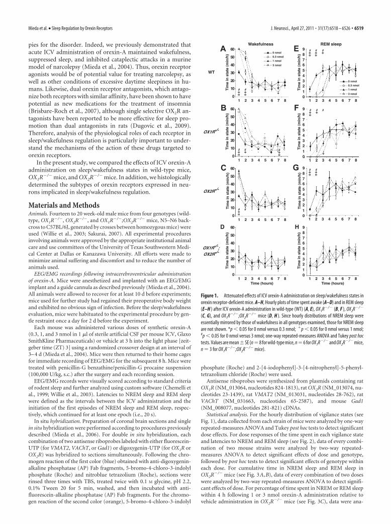
A E
B F
C G
Figure 1. Attenuated effects of ICV orexin-A administration on sleep/wakefulness states inorexin receptor-deficient mice. A–H, Hourly plots of time spent awake (A–D) and in REM sleep(E–H ) after ICV orexin-A administration in wild-type (WT) (A, E), OX1R�/� (B, F ), OX2R�/�
(C, G), and OX1R�/�;OX2R�/� mice (D, H ). Since hourly distributions of NREM sleep wereessentially mirrored by those of wakefulness in all genotypes examined, those for NREM sleepare not shown. *p � 0.05 for 0 nmol versus 0.3 nmol; �p � 0.05 for 0 nmol versus 1 nmol;#p � 0.05 for 0 nmol versus 3 nmol; one-way repeated-measures ANOVA and Tukey post hoctests. Values are mean�SE (n �8 for wild-type mice, n �6 for OX1R�/� and OX2R�/� mice,n � 3 for OX1R�/�;OX2R�/� mice).
Mieda et al. • Sleep Regulation by Orexin Receptors J. Neurosci., April 27, 2011 • 31(17):6518 – 6526 • 6519
lyzed by two-way repeated-measures ANOVAto detect significant effects of dose and vigi-lance states. For testing correlation betweenlatencies to NREM sleep and REM sleep, Pear-son’s correlation coefficients were calculatedfor OX1R�/� and OX2R�/� mice with data of 1and 3 nmol orexin-A administration. Proba-bility ( p) values �0.05 were considered statis-tically significant. Only relevant informationfrom the statistical analysis has been indicatedin the text and figures.
ResultsPromotion of wakefulness andsuppression of NREM sleep by ICVorexin-A administrationTo further elucidate the differential roles oforexin receptors in sleep/wakefulness regu-lation, we compared the wakefulness-promoting and sleep-suppressing effectsof ICV orexin-A administration in wild-type, OX1R�/�, OX2R�/�, and OX1R�/�;OX2R�/� mice. Synthetic orexin-A (0.3,1, and 3 nmol/mouse) or vehicle was ad-ministered into the lateral ventricle at 3 hinto the light phase (ZT3), and EEG/EMGsignals were recorded for the subsequent8 h. The doses and route of administrationchosen were comparable to those usedpreviously to examine the effects oforexin-A on behavior and metabolism inrodents (Lubkin and Stricker-Krongrad,1998; Hagan et al., 1999; Piper et al., 2000;Stricker-Krongrad et al., 2002). The specific action of orexin-Aadministration on orexin receptors was further confirmed by thelack of any effect when the neuropeptide was administered toOX1R�/�;OX2R�/� mice (Figs. 1D,H, 2).
As previously reported, orexin-A administration in wild-type mice increased wakefulness time in a dose-dependentmanner (n � 8, p � 0.0001) accompanied by a decrease inNREM sleep time (n � 8, p � 0.0001) (Figs. 1 A, 2 A, B,D).These effects continued for 2 h after administration at all dosesexamined (Fig. 1 A).
In OX1R�/� mice, the effects of orexin-A administration onwakefulness and NREM sleep were slightly but significantlysmaller than those in wild-type mice. The effect of the lowest doseof orexin-A (0.3 nmol) lasted for only 1 h in OX1R�/� mice (Fig.1B). During the first 2 h after administration, orexin-A increasedwakefulness time (n � 6, p � 0.0001) and suppressed NREMsleep time (n � 6, p � 0.0001) in a dose-dependent manner inOX1R�/� mice, but to a degree significantly less than that inwild-type mice (n � 8 and 6 for wild-type and OX1R�/� mice,respectively; p � 0.0078 and 0.0082 for wakefulness and NREMsleep, respectively) (Fig. 2A,B). However, the latency to the firstepisode of NREM sleep after administration in OX1R�/� micewas not significantly different from that in wild-type mice (n � 8and 6 for wild-type and OX1R�/� mice, respectively, p � 0.1229)(Fig. 2D).
In contrast, the effects of orexin-A administration in OX2R�/�
mice were considerably attenuated as compared with those inboth wild-type and OX1R�/� mice. Although all doses oforexin-A examined increased wakefulness in OX2R�/� mice inthe first hour following administration (n � 6, p � 0.0002 for all
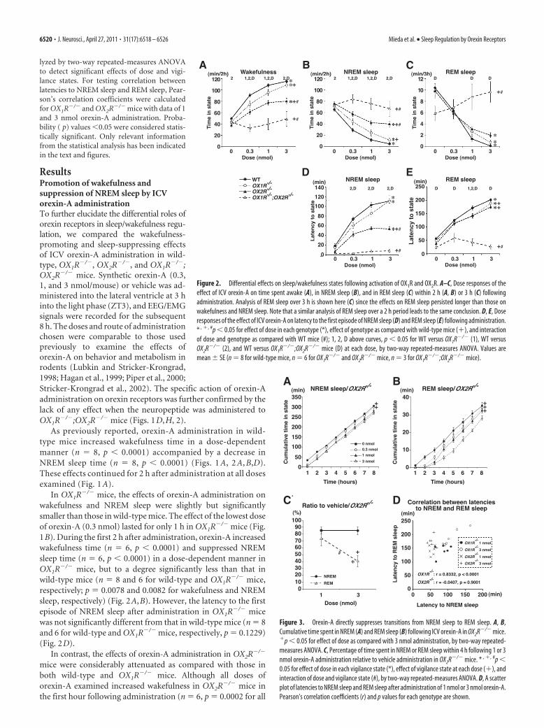
A B
D E
C
Dose (nmol) Dose (nmol)
Late
ncy
to s
tate
Figure 2. Differential effects on sleep/wakefulness states following activation of OX1R and OX2R. A–C, Dose responses of theeffect of ICV orexin-A on time spent awake (A), in NREM sleep (B), and in REM sleep (C) within 2 h (A, B) or 3 h (C) followingadministration. Analysis of REM sleep over 3 h is shown here (C) since the effects on REM sleep persisted longer than those onwakefulness and NREM sleep. Note that a similar analysis of REM sleep over a 2 h period leads to the same conclusion. D, E, Doseresponses of the effect of ICV orexin-A on latency to the first episode of NREM sleep (D) and REM sleep (E) following administration.* , �, #p � 0.05 for effect of dose in each genotype (*), effect of genotype as compared with wild-type mice (�), and interactionof dose and genotype as compared with WT mice (#); 1, 2, D above curves, p � 0.05 for WT versus OX1R�/� (1), WT versusOX2R�/� (2), and WT versus OX1R�/�;OX2R�/� mice (D) at each dose, by two-way repeated-measures ANOVA. Values aremean � SE (n � 8 for wild-type mice, n � 6 for OX1R�/� and OX2R�/� mice, n � 3 for OX1R�/�;OX2R�/� mice).
A B
C D
Figure 3. Orexin-A directly suppresses transitions from NREM sleep to REM sleep. A, B,Cumulative time spent in NREM (A) and REM sleep (B) following ICV orexin-A in OX2R�/� mice.�p � 0.05 for effect of dose as compared with 3 nmol administration, by two-way repeated-measures ANOVA. C, Percentage of time spent in NREM or REM sleep within 4 h following 1 or 3nmol orexin-A administration relative to vehicle administration in OX2R�/� mice. * , �, #p �0.05 for effect of dose in each vigilance state (*), effect of vigilance state at each dose (�), andinteraction of dose and vigilance state (#), by two-way repeated-measures ANOVA. D, A scatterplot of latencies to NREM sleep and REM sleep after administration of 1 nmol or 3 nmol orexin-A.Pearson’s correlation coefficients (r) and p values for each genotype are shown.
6520 • J. Neurosci., April 27, 2011 • 31(17):6518 – 6526 Mieda et al. • Sleep Regulation by Orexin Receptors

three doses as compared with vehicle), these effects dissipatedmore rapidly when compared with wild-type and OX1R�/� mice(Fig. 1C). Accordingly, the dose–response curve of 2 h wakeful-ness in OX2R�/� mice was below those of both wild-type (n � 8and 6 for wild-type and OX2R�/� mice, respectively, p � 0.0001)and OX1R�/� mice (n � 6, p � 0.0012), and the dose–responsecurve of 2 h NREM sleep in OX2R�/� mice was above those ofboth wild-type (n � 8 and 6 for wild-type and OX2R�/� mice,respectively, p � 0.0001) and OX1R�/� mice (n � 6, p � 0.0011)(Fig. 2A,B). In addition, the latency to NREM sleep afterorexin-A administration was significantly shorter than that inwild-type (n � 8 and 6 for wild-type and OX2R�/� mice, respec-tively, p � 0.0001) and OX1R�/� mice (n � 6, p � 0.0003) (Fig.2D). Importantly, although the effect was smaller than observedin the other genotypes, the fact that orexin-A administration in-creased wakefulness in OX2R
�/� mice implies that activation of OX1Rhas wakefulness-promoting effects.
Thus, activation of either of the two orexin receptors was suf-ficient to increase wakefulness, but to a degree significantly lessthan that with simultaneous activation of both receptors, suggestingthat both OX1R and OX2R mediate the wakefulness-promoting andNREM sleep-suppressing effects of ICV orexin-A administration.We also noted that the contributions of OX2R to these effects weresubstantially greater than those of OX1R.
Both OX1R and OX2R are similarlyinvolved in orexin-A-mediated REMsleep regulationAs previously reported, orexin-A ad-ministration in wild-type mice potentlysuppressed REM sleep in a dose-dependent manner (n � 8, p � 0.0001)(Figs. 1 E, 2C,E). REM sleep suppressionlasted for 3 h (3 nmol and 1 nmolorexin-A) or for 2 h (0.3 nmol orexin-A)after administration in wild-type mice(Fig. 1 E).
In both OX1R�/�and OX2R�/� mice,the effects of orexin-A on REM sleep sup-pression dissipated more rapidly than inwild-type mice. In OX1R�/�mice, thelower doses of orexin-A (0.3 and 1 nmol)suppressed REM sleep for 1 and 2 h afteradministration, respectively (Fig. 1F). InOX2R�/� mice, REM sleep suppressionlasted for only 2 h at all doses examined(Fig. 1G). Although the amount of REMsleep in the first 3 h after administrationwas not significantly different among thethree different genotypes ( p � 0.2835 forwild-type vs OX1R�/�mice, p � 0.6582for wild-type vs OX2R�/�mice, p �0.5677 for OX1R�/� vs OX2R�/�mice)(Fig. 2C), the latency to REM sleep afteradministration was slightly shorter inOX1R�/� (n � 6, p � 0.0308) andOX2R�/� mice (n � 6, p � 0.0004) than inwild-type mice (n � 8) (Fig. 2E). The la-tency to REM sleep was not different be-tween OX1R�/� and OX2R�/� mice ( p �0.3270). Thus, REM sleep suppressionfollowing orexin-A administration wasslightly and similarly attenuated inOX1R�/� and OX2R�/� mice when com-
pared with wild-type mice. These results suggest that both OX1Rand OX2R mediate REM sleep suppression with similar efficacythrough different and redundant pathways.
Since REM sleep episodes are only observed after a prolongedperiod of NREM sleep in normal mammals, it can be speculatedthat suppression of REM sleep by orexin-A administration mayoccur secondary to suppression of NREM sleep. If REM sleepsuppression by orexin-A resulted solely from indirect effects ofNREM sleep suppression, and orexin-A had no influence ontransitions from NREM to REM sleep, the emergence of REMsleep episodes would likely depend simply on the amount of pre-ceding NREM sleep. After orexin-A administration, OX2R�/� micespent significantly longer in NREM sleep than did OX1R�/�
mice, whereas OX1R�/� and OX2R�/� mice spent similar durationsin REM sleep (Figs. 1, 2). Thus, in addition to the indirect effects,ICV-administered orexin-A is likely to suppress transitions fromNREM sleep to REM sleep directly. In OX2R�/� mice, further-more, 1 and 3 nmol orexin-A administration suppressed NREMsleep to a similar degree, but orexin-A suppressed REM sleepmore potently at 3 nmol than 1 nmol (Fig. 3A–C) (n � 6, p �0.0252 for C). Consistently, latency to REM sleep was not corre-lated with latency to NREM sleep after administration of 1 or 3nmol of orexin-A in OX2R�/� mice (r � �0.0407, p � 0.9001),but these parameters were highly correlated in OX1R�/� mice
Figure 4. Similar distributions of OX1R and OX2R mRNA in wake-active nuclei of wild-type, OX1R�/�, and OX2R�/� mice. A–D,Coronal brain sections containing TMN (A), LC (B), DR (C), and LDT (D) from wild-type, OX1R�/�, and OX2R�/� mice werehybridized in situ to an OX1R or OX2R antisense probe. 3v, Third ventricle; 4v, fourth ventricle; Aq, aqueduct. Scale bar, 100 �m.
Mieda et al. • Sleep Regulation by Orexin Receptors J. Neurosci., April 27, 2011 • 31(17):6518 – 6526 • 6521
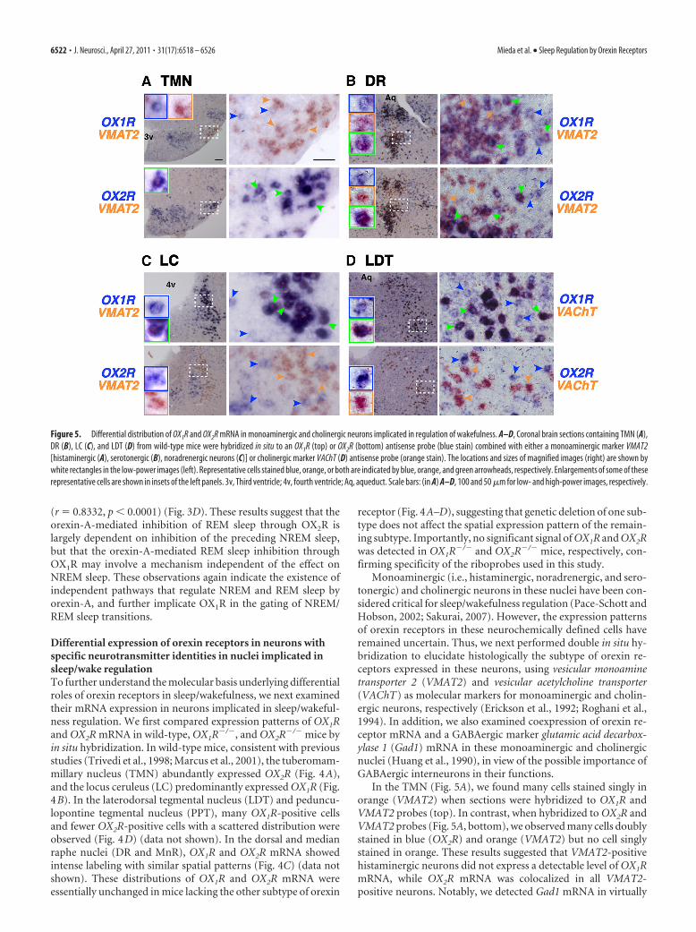
(r � 0.8332, p � 0.0001) (Fig. 3D). These results suggest that theorexin-A-mediated inhibition of REM sleep through OX2R islargely dependent on inhibition of the preceding NREM sleep,but that the orexin-A-mediated REM sleep inhibition throughOX1R may involve a mechanism independent of the effect onNREM sleep. These observations again indicate the existence ofindependent pathways that regulate NREM and REM sleep byorexin-A, and further implicate OX1R in the gating of NREM/REM sleep transitions.
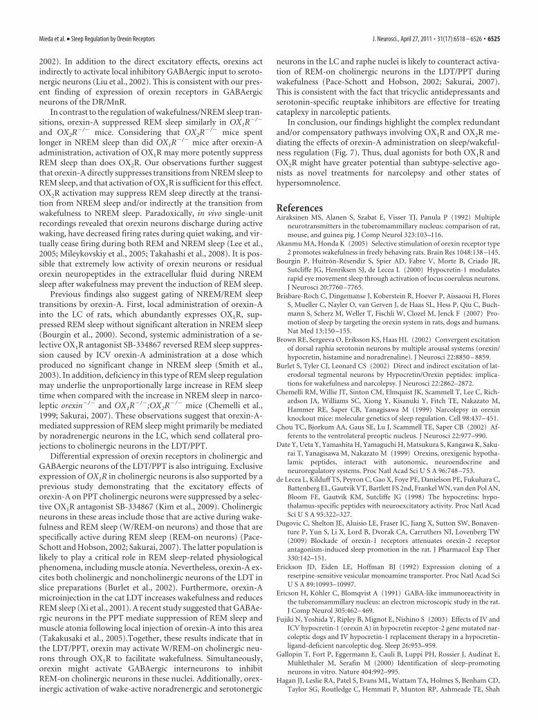
Differential expression of orexin receptors in neurons withspecific neurotransmitter identities in nuclei implicated insleep/wake regulationTo further understand the molecular basis underlying differentialroles of orexin receptors in sleep/wakefulness, we next examinedtheir mRNA expression in neurons implicated in sleep/wakeful-ness regulation. We first compared expression patterns of OX1Rand OX2R mRNA in wild-type, OX1R�/�, and OX2R�/� mice byin situ hybridization. In wild-type mice, consistent with previousstudies (Trivedi et al., 1998; Marcus et al., 2001), the tuberomam-millary nucleus (TMN) abundantly expressed OX2R (Fig. 4A),and the locus ceruleus (LC) predominantly expressed OX1R (Fig.4B). In the laterodorsal tegmental nucleus (LDT) and peduncu-lopontine tegmental nucleus (PPT), many OX1R-positive cellsand fewer OX2R-positive cells with a scattered distribution wereobserved (Fig. 4D) (data not shown). In the dorsal and medianraphe nuclei (DR and MnR), OX1R and OX2R mRNA showedintense labeling with similar spatial patterns (Fig. 4C) (data notshown). These distributions of OX1R and OX2R mRNA wereessentially unchanged in mice lacking the other subtype of orexin
receptor (Fig. 4A–D), suggesting that genetic deletion of one sub-type does not affect the spatial expression pattern of the remain-ing subtype. Importantly, no significant signal of OX1R and OX2Rwas detected in OX1R�/� and OX2R�/� mice, respectively, con-firming specificity of the riboprobes used in this study.
Monoaminergic (i.e., histaminergic, noradrenergic, and sero-tonergic) and cholinergic neurons in these nuclei have been con-sidered critical for sleep/wakefulness regulation (Pace-Schott andHobson, 2002; Sakurai, 2007). However, the expression patternsof orexin receptors in these neurochemically defined cells haveremained uncertain. Thus, we next performed double in situ hy-bridization to elucidate histologically the subtype of orexin re-ceptors expressed in these neurons, using vesicular monoaminetransporter 2 (VMAT2) and vesicular acetylcholine transporter(VAChT) as molecular markers for monoaminergic and cholin-ergic neurons, respectively (Erickson et al., 1992; Roghani et al.,1994). In addition, we also examined coexpression of orexin re-ceptor mRNA and a GABAergic marker glutamic acid decarbox-ylase 1 (Gad1) mRNA in these monoaminergic and cholinergicnuclei (Huang et al., 1990), in view of the possible importance ofGABAergic interneurons in their functions.
In the TMN (Fig. 5A), we found many cells stained singly inorange (VMAT2) when sections were hybridized to OX1R andVMAT2 probes (top). In contrast, when hybridized to OX2R andVMAT2 probes (Fig. 5A, bottom), we observed many cells doublystained in blue (OX2R) and orange (VMAT2) but no cell singlystained in orange. These results suggested that VMAT2-positivehistaminergic neurons did not express a detectable level of OX1RmRNA, while OX2R mRNA was colocalized in all VMAT2-positive neurons. Notably, we detected Gad1 mRNA in virtually
Figure 5. Differential distribution of OX1R and OX2R mRNA in monoaminergic and cholinergic neurons implicated in regulation of wakefulness. A–D, Coronal brain sections containing TMN (A),DR (B), LC (C), and LDT (D) from wild-type mice were hybridized in situ to an OX1R (top) or OX2R (bottom) antisense probe (blue stain) combined with either a monoaminergic marker VMAT2[histaminergic (A), serotonergic (B), noradrenergic neurons (C)] or cholinergic marker VAChT (D) antisense probe (orange stain). The locations and sizes of magnified images (right) are shown bywhite rectangles in the low-power images (left). Representative cells stained blue, orange, or both are indicated by blue, orange, and green arrowheads, respectively. Enlargements of some of theserepresentative cells are shown in insets of the left panels. 3v, Third ventricle; 4v, fourth ventricle; Aq, aqueduct. Scale bars: (in A) A–D, 100 and 50 �m for low- and high-power images, respectively.
6522 • J. Neurosci., April 27, 2011 • 31(17):6518 – 6526 Mieda et al. • Sleep Regulation by Orexin Receptors

all OX2R-expressing TMN neurons, suggesting that histaminer-gic neurons are also GABAergic (Fig. 6A). In addition, we founda few VMAT2-negative/Gad1-negative cells expressing OX1R mRNA inthe same area.
In the DR (Fig. 5B) and MnR (data not shown), we detectedboth OX1R and OX2R mRNA in �90% of VMAT2-positive sero-tonergic neurons (DR: 87.4% and 89.5%, MnR: 96.6% and 92.2%for OX1R and OX2R, respectively), suggesting that the majority ofserotonergic neurons in these areas express both OX1R andOX2R. Many VMAT2-negative nonserotonergic cells in the DR/MnR also expressed OX1R or OX2R mRNA. At least some popu-lations of these cells were likely to be GABAergic, since apopulation of Gad1-positive cells demonstrated detectable OX1Ror OX2R mRNA (Fig. 6B). We could not conclude whether asingle population of GABAergic neurons expressed both recep-tors or whether different populations expressed either OX1R orOX2R.
In the LC (Fig. 5C), all VMAT2-positive noradrenergic neu-rons exhibited intense OX1R expression, whereas OX2R mRNAwas exclusively detected in VMAT2-negative nonnoradrenergicneurons. Only some of these OX2R-expressing cells exhibitedGad1 expression, leaving the remaining cells neurochemicallyunidentified (Fig. 6C).
In the LDT (Fig. 5D) and PPT (data not shown), all VAChT-positive cholinergic neurons expressed OX1R but not OX2RmRNA, but many OX1R-positive and/or OX2R-positive noncho-linergic neurons were intermingled with cholinergic neurons inthe area. Gad1 mRNA staining further revealed that OX1R- orOX2R-expressing cells included both GABAergic and non-
GABAergic cells (Fig. 6D). A previous study showed that �50%of ChAT-immunoreactive neurons in the LDT/PPT also con-tained GABA (Jia et al., 2003), and our double in situ hybridiza-tion using VAChT and Gad1 probes confirmed at least theexistence of such cholinergic/GABAergic neurons (data notshown). Thus, in the LDT/PPT, these results suggest that: (1)OX1R mRNA is expressed in all cholinergic neurons and possiblyin GABAergic and neurochemically unidentified neurons, and(2) OX2R mRNA is expressed in GABAergic and neurochemicallyunidentified neurons but not in cholinergic neurons. Whetherboth OX1R and OX2R are expressed in the same populations ofGABAergic or unidentified neurons remains unknown.
This differential expression of OX1R and OX2R is summarizedin Figure 7.
DiscussionIn the present study, we compared the contribution of OX1R andOX2R to the effects of ICV orexin-A administration on sleep andwakefulness using mice lacking either OX1R or OX2R. We admin-istered orexin-A, the dual OX1R/OX2R agonist, in all mice. Fur-thermore, we used knock-out mice to assure specific andcomplete deletion of orexin receptor genes. Hence our strategyeliminated confounding factors that could affect results fromstudies that use subtype-selective orexin agonists and/or antago-nists, such as specificity, potency, efficacy, occupancy, and stabil-ity of the administered drugs. In addition, the current lack ofavailability of an OX1R-selective agonist supports our strategy. Itis possible that the lack of one receptor subtype might be com-pensated by enhanced expression of the other subtype. However,
Figure 6. Differential distributions of OX1R and OX2R mRNA in GABAergic neurons in wake-promoting nuclei. A–D, Coronal brain sections containing TMN (A), DR (B), LC (C), and LDT (D) fromwild-type mice were hybridized in situ to an OX1R (top) or OX2R (bottom) antisense probe (blue stain) combined with a GABAergic marker Gad1 (orange stain). The locations and sizes of magnifiedimages (right) are shown by white rectangles in the low-power images (left). Representative cells stained blue, orange, or both are indicated by blue, orange, green arrowheads, respectively.Enlargements of some of these representative cells are shown in insets of the left panels. 3v, Third ventricle; 4v, fourth ventricle; Aq, aqueduct. Scale bars: (in A) A–D, 100 and 50 �m for low- andhigh-power images, respectively.
Mieda et al. • Sleep Regulation by Orexin Receptors J. Neurosci., April 27, 2011 • 31(17):6518 – 6526 • 6523

we found that genetic deletion of OX1R orOX2R did not result in an overt change inthe distribution of the other subtypemRNA (Fig. 4). However, whether the in-tensity of expression of the other receptoris altered remains unknown.
In this study, we demonstrated that ac-tivation of OX2R promotes wakefulnessand suppresses NREM sleep with substan-tially higher efficacy than that of OX1R. Incontrast, both OX1R and OX2R appear tobe involved in the suppression of REMsleep by orexin-A administration to a sim-ilar degree. These findings are consistentwith the conclusion derived from behav-ioral studies and baseline sleep/wake-fulness recordings of OX1R�/� andOX2R�/� mice: the normal regulation ofwakefulness/NREM sleep transitions de-pends critically on OX2R, but the pro-found dysregulation of REM sleep controlunique to narcolepsy-cataplexy syn-dromes emerges from loss of signalingthrough both OX1R and OX2R (Willie etal., 2003; Sakurai, 2007). A previous phar-macological study using OX1R- andOX2R-selective antagonists in rats alsodemonstrated a principal role of OX2R insuppression of NREM sleep (Dugovic etal., 2009). Furthermore, ICV administra-tion of an OX2R-selective agonist[Ala 11]orexin-B in rats was sufficient topromote wakefulness and suppress NREM and REM sleep(Akanmu and Honda, 2005).
Histaminergic neurons in the TMN are believed to play animportant role in the wake-promoting effect of orexin, sincethe effects of ICV orexin-A administration are markedly at-tenuated by the histamine H1 receptor (H1R) antagonist py-rilamine and are absent in H1R�/� mice (Huang et al., 2001;Yamanaka et al., 2002). The TMN abundantly expresses OX2R,supporting a critical contribution of TMN histaminergic neu-rons (Marcus et al., 2001; Yamanaka et al., 2002). Here, and inagreement with the previous studies, we showed marked at-tenuation of orexin-A-induced wakefulness in OX2R�/� mice.Additionally, we demonstrated expression of OX2R mRNA inalmost all TMN histaminergic neurons, which is consistentwith a previous immunohistochemical study (Yamanaka et al.,2002). Interestingly, our results suggested coexpression of hista-minergic and GABAergic molecular markers with OX2R in theTMN, consistent with previous studies showing that TMN hista-minergic neurons contain GABA (Ericson et al., 1991; Airaksinenet al., 1992). Despite dense histaminergic innervation to the ven-trolateral preoptic area (VLPO) from TMN, histamine reportedlyhas no effect on sleep-promoting neurons of this area, which is innotable contrast to the inhibitory effects of noradrenaline, sero-tonin, and acetylcholine (Gallopin et al., 2000; Chou et al., 2002).In addition, histidine decarboxylase�/� and H1R�/� mice showmild or subtle phenotypes concerning sleep/wakefulness regula-tion under baseline conditions, clearly contrasting with OX2R�/�
mice, which show marked abnormalities in sleep and wakefulness(Parmentier et al., 2002; Willie et al., 2003; Huang et al., 2006;Hondo et al., 2010). Furthermore, OX1R�/�;H1R�/� mice alsoshow no detectable abnormality in sleep/wakefulness states
(Hondo et al., 2010). Thus, GABAergic transmission by TMNhistaminergic neurons, driven by the OX2R (Willie et al., 2003),may play a critical role, including suppression of sleep-promoting neurons in the VLPO.
However, orexin-A administration in OX2R�/� mice stillcaused an increase in wakefulness. Thus, OX1R-expressing neu-rons may also play a role in the effects of orexin-A. Consistentwith this thesis, fragmentation of wakefulness in OX2R�/� mice ismilder than that in OX1R�/�;OX2R�/� mice (Sakurai, 2007). Inaddition, ICV administration of an OX2R-selective agonist[Ala 11]orexin-B seemed less effective when compared withorexin-A (Akanmu and Honda, 2005). However, ICV adminis-tration of orexin-A in OX2R-deficient dogs has been reported tohave no effect on time spent in wakefulness (Fujiki et al., 2003).This discrepancy between the studies with OX2R-defficient miceand dogs may be explained by differences in doses, time windowsof analyses, numbers of animals examined, and species (Willie etal., 2003).
LC noradrenergic neurons, which abundantly express OX1R,are one of the candidate systems to mediate the arousal effectmediated by OX1R. Indeed, microinjection of orexin-A into theLC increases wakefulness (Bourgin et al., 2000). Other regionsthat could mediate OX1R-induced arousal effects include cholin-ergic neurons in the LDT/PPT and serotonergic neurons in theraphe nuclei, both of which are thought to be critically involvedin the regulation of sleep/wakefulness states (Pace-Schott andHobson, 2002; Sakurai, 2007). Redundant expression of bothOX1R and OX2R mRNA was reported in raphe nuclei (Marcus etal., 2001). We showed that most serotonergic neurons in the DRand MnR are positive for OX1R and/or OX2R mRNA, which is inaccordance with a previous single-cell PCR study (Brown et al.,
Figure 7. Schematic illustration of presumed pathways underlying orexin actions on NREM and REM sleep. Orexins activatehistaminergic (His)/GABAergic (GA), serotonergic (5HT), noradrenergic (NA), and cholinergic (ACh) neurons, as well as GABAergicand neurochemically unidentified (not shown in this figure for simplicity) putative interneurons, in wake-promoting nuclei,including the TMN, DR/MnR, LDT/PPT, and LC. These neurons differentially express OX1R and/or OX2R and regulate wakefulness/NREM sleep and NREM/REM sleep transitions. OX1R and OX2R may be expressed in the same populations of GABAergic neurons, asshown in the figure, or may be expressed in distinct populations of these neurons in each area. Wake/REM-on cholinergic neurons(ACh/W) are likely to suppress NREM sleep but REM-on cholinergic neurons (ACh/R) are likely to induce REM sleep. Wake-activeserotonergic and noradrenergic neurons in the DR/MnR and LC, respectively, counteract activation of REM-on cholinergic neuronsin the LDT/PPT, as well as REM-on neurons in the brainstem reticular formation (Sakurai et al., 1998; Pace-Schott and Hobson,2002). Previous reports have suggested contributions of GABAergic interneurons inhibiting PPT cholinergic and raphe serotonergicneurons (Liu et al., 2002; Takakusaki et al., 2005). LHA, Lateral hypothalamic area; PH, posterior hypothalamus.
6524 • J. Neurosci., April 27, 2011 • 31(17):6518 – 6526 Mieda et al. • Sleep Regulation by Orexin Receptors
2002). In addition to the direct excitatory effects, orexins actindirectly to activate local inhibitory GABAergic input to seroto-nergic neurons (Liu et al., 2002). This is consistent with our pres-ent finding of expression of orexin receptors in GABAergicneurons of the DR/MnR.
In contrast to the regulation of wakefulness/NREM sleep tran-sitions, orexin-A suppressed REM sleep similarly in OX1R�/�
and OX2R�/� mice. Considering that OX2R�/� mice spentlonger in NREM sleep than did OX1R�/� mice after orexin-Aadministration, activation of OX1R may more potently suppressREM sleep than does OX2R. Our observations further suggestthat orexin-A directly suppresses transitions from NREM sleep toREM sleep, and that activation of OX1R is sufficient for this effect.OX2R activation may suppress REM sleep directly at the transi-tion from NREM sleep and/or indirectly at the transition fromwakefulness to NREM sleep. Paradoxically, in vivo single-unitrecordings revealed that orexin neurons discharge during activewaking, have decreased firing rates during quiet waking, and vir-tually cease firing during both REM and NREM sleep (Lee et al.,2005; Mileykovskiy et al., 2005; Takahashi et al., 2008). It is pos-sible that extremely low activity of orexin neurons or residualorexin neuropeptides in the extracellular fluid during NREMsleep after wakefulness may prevent the induction of REM sleep.
Previous findings also suggest gating of NREM/REM sleeptransitions by orexin-A. First, local administration of orexin-Ainto the LC of rats, which abundantly expresses OX1R, sup-pressed REM sleep without significant alteration in NREM sleep(Bourgin et al., 2000). Second, systemic administration of a se-lective OX1R antagonist SB-334867 reversed REM sleep suppres-sion caused by ICV orexin-A administration at a dose whichproduced no significant change in NREM sleep (Smith et al.,2003). In addition, deficiency in this type of REM sleep regulationmay underlie the unproportionally large increase in REM sleeptime when compared with the increase in NREM sleep in narco-leptic orexin�/� and OX1R�/�;OX2R�/� mice (Chemelli et al.,1999; Sakurai, 2007). These observations suggest that orexin-A-mediated suppression of REM sleep might primarily be mediatedby noradrenergic neurons in the LC, which send collateral pro-jections to cholinergic neurons in the LDT/PPT.
Differential expression of orexin receptors in cholinergic andGABAergic neurons of the LDT/PPT is also intriguing. Exclusiveexpression of OX1R in cholinergic neurons is also supported by aprevious study demonstrating that the excitatory effects oforexin-A on PPT cholinergic neurons were suppressed by a selec-tive OX1R antagonist SB-334867 (Kim et al., 2009). Cholinergicneurons in these areas include those that are active during wake-fulness and REM sleep (W/REM-on neurons) and those that arespecifically active during REM sleep (REM-on neurons) (Pace-Schott and Hobson, 2002; Sakurai, 2007). The latter population islikely to play a critical role in REM sleep-related physiologicalphenomena, including muscle atonia. Nevertheless, orexin-A ex-cites both cholinergic and noncholinergic neurons of the LDT inslice preparations (Burlet et al., 2002). Furthermore, orexin-Amicroinjection in the cat LDT increases wakefulness and reducesREM sleep (Xi et al., 2001). A recent study suggested that GABAe-rgic neurons in the PPT mediate suppression of REM sleep andmuscle atonia following local injection of orexin-A into this area(Takakusaki et al., 2005).Together, these results indicate that inthe LDT/PPT, orexin may activate W/REM-on cholinergic neu-rons through OX1R to facilitate wakefulness. Simultaneously,orexin might activate GABAergic interneurons to inhibitREM-on cholinergic neurons in these nuclei. Additionally, orex-inergic activation of wake-active noradrenergic and serotonergic
neurons in the LC and raphe nuclei is likely to counteract activa-tion of REM-on cholinergic neurons in the LDT/PPT duringwakefulness (Pace-Schott and Hobson, 2002; Sakurai, 2007).This is consistent with the fact that tricyclic antidepressants andserotonin-specific reuptake inhibitors are effective for treatingcataplexy in narcoleptic patients.
In conclusion, our findings highlight the complex redundantand/or compensatory pathways involving OX1R and OX2R me-diating the effects of orexin-A administration on sleep/wakeful-ness regulation (Fig. 7). Thus, dual agonists for both OX1R andOX2R might have greater potential than subtype-selective ago-nists as novel treatments for narcolepsy and other states ofhypersomnolence.
ReferencesAiraksinen MS, Alanen S, Szabat E, Visser TJ, Panula P (1992) Multiple
neurotransmitters in the tuberomammillary nucleus: comparison of rat,mouse, and guinea pig. J Comp Neurol 323:103–116.
Akanmu MA, Honda K (2005) Selective stimulation of orexin receptor type2 promotes wakefulness in freely behaving rats. Brain Res 1048:138 –145.
Bourgin P, Huitron-Resendiz S, Spier AD, Fabre V, Morte B, Criado JR,Sutcliffe JG, Henriksen SJ, de Lecea L (2000) Hypocretin-1 modulatesrapid eye movement sleep through activation of locus coeruleus neurons.J Neurosci 20:7760 –7765.
Brisbare-Roch C, Dingemanse J, Koberstein R, Hoever P, Aissaoui H, FloresS, Mueller C, Nayler O, van Gerven J, de Haas SL, Hess P, Qiu C, Buch-mann S, Scherz M, Weller T, Fischli W, Clozel M, Jenck F (2007) Pro-motion of sleep by targeting the orexin system in rats, dogs and humans.Nat Med 13:150 –155.
Brown RE, Sergeeva O, Eriksson KS, Haas HL (2002) Convergent excitationof dorsal raphia serotonin neurons by multiple arousal systems (orexin/hypocretin, histamine and noradrenaline). J Neurosci 22:8850 – 8859.
Burlet S, Tyler CJ, Leonard CS (2002) Direct and indirect excitation of lat-erodorsal tegmental neurons by Hypocretin/Orexin peptides: implica-tions for wakefulness and narcolepsy. J Neurosci 22:2862–2872.
Chemelli RM, Willie JT, Sinton CM, Elmquist JK, Scammell T, Lee C, Rich-ardson JA, Williams SC, Xiong Y, Kisanuki Y, Fitch TE, Nakazato M,Hammer RE, Saper CB, Yanagisawa M (1999) Narcolepsy in orexinknockout mice: molecular genetics of sleep regulation. Cell 98:437– 451.
Chou TC, Bjorkum AA, Gaus SE, Lu J, Scammell TE, Saper CB (2002) Af-ferents to the ventrolateral preoptic nucleus. J Neurosci 22:977–990.
Date Y, Ueta Y, Yamashita H, Yamaguchi H, Matsukura S, Kangawa K, Saku-rai T, Yanagisawa M, Nakazato M (1999) Orexins, orexigenic hypotha-lamic peptides, interact with autonomic, neuroendocrine andneuroregulatory systems. Proc Natl Acad Sci U S A 96:748 –753.
de Lecea L, Kilduff TS, Peyron C, Gao X, Foye PE, Danielson PE, Fukuhara C,Battenberg EL, Gautvik VT, Bartlett FS 2nd, Frankel WN, van den Pol AN,Bloom FE, Gautvik KM, Sutcliffe JG (1998) The hypocretins: hypo-thalamus-specific peptides with neuroexcitatory activity. Proc Natl AcadSci U S A 95:322–327.
Dugovic C, Shelton JE, Aluisio LE, Fraser IC, Jiang X, Sutton SW, Bonaven-ture P, Yun S, Li X, Lord B, Dvorak CA, Carruthers NI, Lovenberg TW(2009) Blockade of orexin-1 receptors attenuates orexin-2 receptorantagonism-induced sleep promotion in the rat. J Pharmacol Exp Ther330:142–151.
Erickson JD, Eiden LE, Hoffman BJ (1992) Expression cloning of areserpine-sensitive vesicular monoamine transporter. Proc Natl Acad SciU S A 89:10993–10997.
Ericson H, Kohler C, Blomqvist A (1991) GABA-like immunoreactivity inthe tuberomammillary nucleus: an electron microscopic study in the rat.J Comp Neurol 305:462– 469.
Fujiki N, Yoshida Y, Ripley B, Mignot E, Nishino S (2003) Effects of IV andICV hypocretin-1 (orexin A) in hypocretin receptor-2 gene mutated nar-coleptic dogs and IV hypocretin-1 replacement therapy in a hypocretin-ligand-deficient narcoleptic dog. Sleep 26:953–959.
Gallopin T, Fort P, Eggermann E, Cauli B, Luppi PH, Rossier J, Audinat E,Muhlethaler M, Serafin M (2000) Identification of sleep-promotingneurons in vitro. Nature 404:992–995.
Hagan JJ, Leslie RA, Patel S, Evans ML, Wattam TA, Holmes S, Benham CD,Taylor SG, Routledge C, Hemmati P, Munton RP, Ashmeade TE, Shah
Mieda et al. • Sleep Regulation by Orexin Receptors J. Neurosci., April 27, 2011 • 31(17):6518 – 6526 • 6525

AS, Hatcher JP, Hatcher PD, Jones DN, Smith MI, Piper DC, Hunter AJ,Porter RA, Upton N (1999) Orexin A activates locus coeruleus cell firingand increases arousal in the rat. Proc Natl Acad Sci U S A96:10911–10916.
Hondo M, Nagai K, Ohno K, Kisanuki Y, Willie JT, Watanabe T, YanagisawaM, Sakurai T (2010) Histamine-1 receptor is not required as a down-stream effector of orexin-2 receptor in maintenance of basal sleep/wakestates. Acta Physiol (Oxf) 198:287–294.
Huang WM, Reed-Fourquet L, Wu E, Wu JY (1990) Molecular cloning andamino acid sequence of brain L-glutamate decarboxylase. Proc Natl AcadSci U S A 87:8491– 8495.
Huang ZL, Qu WM, Li WD, Mochizuki T, Eguchi N, Watanabe T, Urade Y,Hayaishi O (2001) Arousal effect of orexin A depends on activation ofthe histaminergic system. Proc Natl Acad Sci U S A 98:9965–9970.
Huang ZL, Mochizuki T, Qu WM, Hong ZY, Watanabe T, Urade Y, HayaishiO (2006) Altered sleep-wake characteristics and lack of arousal responseto H3 receptor antagonist in histamine H1 receptor knockout mice. ProcNatl Acad Sci U S A 103:4687– 4692.
Jia HG, Yamuy J, Sampogna S, Morales FR, Chase MH (2003) Colocaliza-tion of gamma-aminobutyric acid and acetylcholine in neurons in thelaterodorsal and pedunculopontine tegmental nuclei in the cat: a light andelectron microscopic study. Brain Res 992:205–219.
Kim J, Nakajima K, Oomura Y, Wayner MJ, Sasaki K (2009) Electrophysi-ological effects of orexins/hypocretins on pedunculopontine tegmentalneurons in rats: an in vitro study. Peptides 30:191–209.
Lee MG, Hassani OK, Jones BE (2005) Discharge of identified orexin/hypo-cretin neurons across the sleep-waking cycle. J Neurosci 25:6716 – 6720.
Lin L, Faraco J, Li R, Kadotani H, Rogers W, Lin X, Qiu X, de Jong PJ, NishinoS, Mignot E (1999) The sleep disorder canine narcolepsy is caused by amutation in the hypocretin (orexin) receptor 2 gene. Cell 98:365–376.
Liu RJ, van den Pol AN, Aghajanian GK (2002) Hypocretins (orexins) reg-ulate serotonin neurons in the dorsal raphe nucleus by excitatory directand inhibitory indirect actions. J Neurosci 22:9453–9464.
Lubkin M, Stricker-Krongrad A (1998) Independent feeding and metabolicactions of orexins in mice. Biochem Biophys Res Commun 253:241–245.
Marcus JN, Aschkenasi CJ, Lee CE, Chemelli RM, Saper CB, Yanagisawa M,Elmquist JK (2001) Differential expression of orexin receptors 1 and 2 inthe rat brain. J Comp Neurol 435:6 –25.
Mieda M, Willie JT, Hara J, Sinton CM, Sakurai T, Yanagisawa M (2004)Orexin peptides prevent cataplexy and improve wakefulness in an orexinneuron-ablated model of narcolepsy in mice. Proc Natl Acad Sci U S A101:4649 – 4654.
Mieda M, Williams SC, Richardson JA, Tanaka K, Yanagisawa M (2006)The dorsomedial hypothalamic nucleus as a putative food-entrainablecircadian pacemaker. Proc Natl Acad Sci U S A 103:12150 –12155.
Mileykovskiy BY, Kiyashchenko LI, Siegel JM (2005) Behavioral correlatesof activity in identified hypocretin/orexin neurons. Neuron 46:787–798.
Nambu T, Sakurai T, Mizukami K, Hosoya Y, Yanagisawa M, Goto K (1999)Distribution of orexin neurons in the adult rat brain. Brain Res827:243–260.
Nishino S, Ripley B, Overeem S, Lammers GJ, Mignot E (2000) Hypocretin(orexin) deficiency in human narcolepsy. Lancet 355:39 – 40.
Pace-Schott EF, Hobson JA (2002) The neurobiology of sleep: genetics, cel-lular physiology and subcortical networks. Nat Rev Neurosci 3:591– 605.
Parmentier R, Ohtsu H, Djebbara-Hannas Z, Valatx JL, Watanabe T, Lin JS(2002) Anatomical, physiological, and pharmacological characteristicsof histidine decarboxylase knock-out mice: evidence for the role of brain
histamine in behavioral and sleep-wake control. J Neurosci 22:7695–7711.
Peyron C, Tighe DK, van den Pol AN, de Lecea L, Heller HC, Sutcliffe JG,Kilduff TS (1998) Neurons containing hypocretin (orexin) project tomultiple neuronal systems. J Neurosci 18:9996 –10015.
Peyron C, Faraco J, Rogers W, Ripley B, Overeem S, Charnay Y, NevsimalovaS, Aldrich M, Reynolds D, Albin R, Li R, Hungs M, Pedrazzoli M, Padi-garu M, Kucherlapati M, Fan J, Maki R, Lammers GJ, Bouras C, Kucher-lapati R, Nishino S, Mignot E (2000) A mutation in a case of early onsetnarcolepsy and a generalized absence of hypocretin peptides in humannarcoleptic brains. Nat Med 6:991–997.
Piper DC, Upton N, Smith MI, Hunter AJ (2000) The novel brain neuro-peptide, orexin-A, modulates the sleep-wake cycle of rats. Eur J Neurosci12:726 –730.
Roghani A, Feldman J, Kohan SA, Shirzadi A, Gundersen CB, Brecha N,Edwards RH (1994) Molecular cloning of a putative vesicular trans-porter for acetylcholine. Proc Natl Acad Sci U S A 91:10620 –10624.
Sakurai T (2007) The neural circuit of orexin (hypocretin): maintainingsleep and wakefulness. Nat Rev Neurosci 8:171–181.
Sakurai T, Amemiya A, Ishii M, Matsuzaki I, Chemelli RM, Tanaka H, Wil-liams SC, Richardson JA, Kozlowski GP, Wilson S, Arch JR, BuckinghamRE, Haynes AC, Carr SA, Annan RS, McNulty DE, Liu WS, Terrett JA,Elshourbagy NA, Bergsma DJ, Yanagisawa M (1998) Orexins and orexinreceptors: a family of hypothalamic neuropeptides and G protein-coupled receptors that regulate feeding behavior. Cell 92:573–585.
Smith MI, Piper DC, Duxon MS, Upton N (2003) Evidence implicating arole for orexin-1 receptor modulation of paradoxical sleep in the rat.Neurosci Lett 341:256 –258.
Stricker-Krongrad A, Richy S, Beck B (2002) Orexins/hypocretins in theob/ob mouse: hypothalamic gene expression, peptide content and meta-bolic effects. Regul Pept 104:11–20.
Takahashi K, Lin JS, Sakai K (2008) Neuronal activity of orexin and non-orexin waking-active neurons during wake-sleep states in the mouse.Neuroscience 153:860 – 870.
Takakusaki K, Takahashi K, Saitoh K, Harada H, Okumura T, Kayama Y,Koyama Y (2005) Orexinergic projections to the cat midbrain mediatealternation of emotional behavioural states from locomotion to cata-plexy. J Physiol 568:1003–1020.
Thannickal TC, Moore RY, Nienhuis R, Ramanathan L, Gulyani S, Aldrich M,Cornford M, Siegel JM (2000) Reduced number of hypocretin neuronsin human narcolepsy. Neuron 27:469 – 474.
Trivedi P, Yu H, MacNeil DJ, Van der Ploeg LH, Guan XM (1998) Distri-bution of orexin receptor mRNA in the rat brain. FEBS Lett 438:71–75.
van den Pol AN (1999) Hypothalamic hypocretin (orexin): robust innerva-tion of the spinal cord. J Neurosci 19:3171–3182.
Willie JT, Chemelli RM, Sinton CM, Tokita S, Williams SC, Kisanuki YY,Marcus JN, Lee C, Elmquist JK, Kohlmeier KA, Leonard CS, RichardsonJA, Hammer RE, Yanagisawa M (2003) Distinct narcolepsy syndromesin Orexin receptor-2 and Orexin null mice: molecular genetic dissectionof non-REM and REM sleep regulatory processes. Neuron 38:715–730.
Xi MC, Morales FR, Chase MH (2001) Effects on sleep and wakefulness ofthe injection of hypocretin-1 (orexin-A) into the laterodorsal tegmentalnucleus of the cat. Brain Res 901:259 –264.
Yamanaka A, Tsujino N, Funahashi H, Honda K, Guan JL, Wang QP, Tomi-naga M, Goto K, Shioda S, Sakurai T (2002) Orexins activate histamin-ergic neurons via the orexin 2 receptor. Biochem Biophys Res Commun290:1237–1245.
6526 • J. Neurosci., April 27, 2011 • 31(17):6518 – 6526 Mieda et al. • Sleep Regulation by Orexin Receptors


![Behavioral/Systems/Cognitive ... · Behavioral/Systems/Cognitive AcuteCocaineInducesFastActivationofD1Receptorand ProgressiveDeactivationofD2ReceptorStriatalNeurons: InVivoOpticalMicroprobe[Ca2]](https://img.pdfslide.us/doc/110x75/6013f75e26e57852b94803cb/behavioralsystemscognitive-behavioralsystemscognitive-acutecocaineinducesfastactivationofd1receptorand.jpg)