Embed Size (px)
Citation preview
References and Notes
1. J. A. Campbell, J. Physiol. 90, 91P (1937). 2. C. W. Smith, J. W. Bean, R. Bauer, Am.
J. Physiol. 199, 883 (1960). 3. J. W. Bean and R. Bauer, Proc. Soc. Exptl.
Biol. Med. 81, 693 (1952). 4. P. Felig and W. L. Lee, Jr., Ann. N.Y. Acad.
Sci., in press.
References and Notes
1. J. A. Campbell, J. Physiol. 90, 91P (1937). 2. C. W. Smith, J. W. Bean, R. Bauer, Am.
J. Physiol. 199, 883 (1960). 3. J. W. Bean and R. Bauer, Proc. Soc. Exptl.
Biol. Med. 81, 693 (1952). 4. P. Felig and W. L. Lee, Jr., Ann. N.Y. Acad.
Sci., in press.
There has been renewed interest in the feeding reflex of hydra, stemming from the contention of Loomis (1) that reduced glutathione (GSH) specifically controls the feeding re- flex of Hydra littoralis. Lenhoff (2) has systematically studied the role played by GSH in activating the feed-
ing response in this species, and pro- posed a quantitative assay for the reflex based on the time of mouth opening of the animal. The uniqueness of GSH as an initiator of feeding in hydra has been questioned by Forrest (3) and Burnett et al. (4). Indeed, Forrest maintains not only that a
large class of apparently unrelated sub- strates induce feeding in hydra, but also that the GSH-induced reaction is not a true feeding response of the animal.
In studies of the contraction re- sponses of Hydra pirardi to stimuli of light and mechanical agitation, we observed that the characteristic con- tractions of the body column which occur in response to these stimuli (5) were inhibited while the animal was feeding on Artemia salina. The normal, spontaneous contractions of the animal were also temporarily eliminated dur- ing feeding. In this report we describe experiments on the inhibition of these contraction responses by feeding H.
pirardi on A. salina and by adding reduced glutathione to the culture medium.
The animals we used in this study were from a clone of H. pirardi. The hydra were cultured by the method of L oomis and Lenhoff (6), except that distilled water was substituted for tap water in the culture medium. Arteemia salina larvae were fed to the animals daily. The experimental ani-
602
5. J. J. Moran, Anal. Chem. 24, 378 (1952). 6. J. W. Bean, Physiol. Rev. 25, 1 (1945). 7. Supported in part by contract R-87 of the
NASA. We thank Dr. B. Katchman and the Clinical Laboratories, Miami Valley Hospital, Dayton, Ohio, for the determinations of protein-bound iodine.
27 March 1964 a
5. J. J. Moran, Anal. Chem. 24, 378 (1952). 6. J. W. Bean, Physiol. Rev. 25, 1 (1945). 7. Supported in part by contract R-87 of the
NASA. We thank Dr. B. Katchman and the Clinical Laboratories, Miami Valley Hospital, Dayton, Ohio, for the determinations of protein-bound iodine.
27 March 1964 a
mals were starved for 24 hours before being tested at 21 C. In experiments to determine the inhibition of the con- traction response to light, two groups of ten hydra were exposed to a regime of 75 seconds of light followed by 75 seconds of dark (7). Each group of animals was placed in a petri dish containing 9 ml of culture water. Be- fore each test, there was a control
period of 15 minutes during which the numbers of animals in each dish con- tracting to five successive light periods were recorded. Substances were then
pipetted into the dishes and the num- bers of animals contracting to sub- sequent light periods were recorded. By this means, the effect of GSH and other chemicals of known concentra- tions were tested in addition to live A. salina or homogenates of A. salina
(3). Almost all animals contracted in
response to the light stimulus in the control period before each test (Fig. 1). In marked contrast A. salina, both alive and in homogenate form, and 1 0-'M GSH initially inhibited these contractions almost completely. The
proportion of animals contracting to
light gradually increased, however, until it became comparable to that of control animals. Thus, the original light response was restored after 50 to 65 minutes.
The behavior of the animal when
exposed to light alone is markedly different from the response to light stimulation in the presence of A. salina or GSH. The usual contraction response to light alone consists of a successive series of partial body con- tractions culminating in the animal's
forming a tight ball with contracted tentacles. The time between onset of
the light and completed contraction of the animal (the reaction time) is a function of the intensity of the light and its spectral composition (9). The reaction time is inversely proportional to temperature (10). Under the present experimental conditions, the mean re- action time was 42.5 + 10.2 seconds, based on 50 animals. In contrast to this light-induced contraction response H. pirardi did not exhibit the total contraction to light in the presence of A. salina or GSH. In this situation the tentacles writhed and twisted to- ward the mouth and the mouth itself opened widely (11). The movements of the animal were similar to those described by Ewer (12) and Loomis (1). After ingestion of A. salina circular contractions of the distal portion of the body column forcing the food down into the gastrovascular cavity of the animal, described by Forrest (see 3) were frequently ob- served.
A second series of experiments was devised to demonstrate the inhibitory effect of A. salina and GSH on the contractional response to mechanical agitation. Two groups of five animals were placed in 48 ml of culture fluid in Stender preparation dishes. After the animals had attached themselves to the bottom of the dishes, they were shaken (13) for periods of 2 seconds every minute. During a control period before each test, the numbers of ani- mals in the two groups contracting to the pulses of 2-second shaking were recorded for five trials. In experiments with live A. salina larvae, the hydra were provided with excess larvae and allowed to feed for 5 minutes. Then, the numbers of animals contracting to successive shaking periods were re- corded. A similar procedure was used to study the effect of GSH. Figure 2 shows the results of inhibition of the contraction response to such mechanical agitation (14), both by live A. salina and by 10-5M GSH. An effect similar to that with light stimulation was ob- served here, although inhibition was shorter, lasting for 25 to 30 minutes.
Live Artemia salina and GSH also inhibited the rhythmic spontaneous con- tractions of the animal. In a typical experiment, the numbers of total body contractions per 30-minute period for groups of ten animals in (i) culture
the light and completed contraction of the animal (the reaction time) is a function of the intensity of the light and its spectral composition (9). The reaction time is inversely proportional to temperature (10). Under the present experimental conditions, the mean re- action time was 42.5 + 10.2 seconds, based on 50 animals. In contrast to this light-induced contraction response H. pirardi did not exhibit the total contraction to light in the presence of A. salina or GSH. In this situation the tentacles writhed and twisted to- ward the mouth and the mouth itself opened widely (11). The movements of the animal were similar to those described by Ewer (12) and Loomis (1). After ingestion of A. salina circular contractions of the distal portion of the body column forcing the food down into the gastrovascular cavity of the animal, described by Forrest (see 3) were frequently ob- served.
A second series of experiments was devised to demonstrate the inhibitory effect of A. salina and GSH on the contractional response to mechanical agitation. Two groups of five animals were placed in 48 ml of culture fluid in Stender preparation dishes. After the animals had attached themselves to the bottom of the dishes, they were shaken (13) for periods of 2 seconds every minute. During a control period before each test, the numbers of ani- mals in the two groups contracting to the pulses of 2-second shaking were recorded for five trials. In experiments with live A. salina larvae, the hydra were provided with excess larvae and allowed to feed for 5 minutes. Then, the numbers of animals contracting to successive shaking periods were re- corded. A similar procedure was used to study the effect of GSH. Figure 2 shows the results of inhibition of the contraction response to such mechanical agitation (14), both by live A. salina and by 10-5M GSH. An effect similar to that with light stimulation was ob- served here, although inhibition was shorter, lasting for 25 to 30 minutes.
Live Artemia salina and GSH also inhibited the rhythmic spontaneous con- tractions of the animal. In a typical experiment, the numbers of total body contractions per 30-minute period for groups of ten animals in (i) culture fluid alone, (ii) live A. salina, and (iii) 10-5M GSH were recorded. The mean number of completed contractions of
hydra after providing excess numbers
SCIENCE, VOL. 145
fluid alone, (ii) live A. salina, and (iii) 10-5M GSH were recorded. The mean number of completed contractions of
hydra after providing excess numbers
SCIENCE, VOL. 145
Behavior in Hydra: Inhibition of the Contraction
Responses of Hydra pirardi
Abstract. Hydra pirardi contracts spontaneously and in response to external stimuli of light and mechanical agitation. Inhibition of these contractions occurs when the animal feeds on Artemia salina or when reduced glutathione is present in the environment. Such inhibition demonstrates the control of one receptor-
e/Jector system by another in hydra.
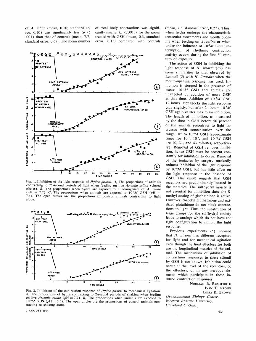
of A. salina (mean, 0.10; standard er- ror, 0.10) was significantly less (p <
.001) than that of controls (mean, 7.7; standard error, 0.62). The mean number
I- : .84
o .6
Z 4 I-
I.- .2 R2 z 0 O
0
4_ ! .8' 4.
? .6
.4 I
0 .2 G.
of total bod,
cantly smalle treated -with error, 0.15)
I . -
PRE-TEST NO GSH
IoDSM GSH ADDED ,
-- ..... . I I
5 10 15 20 25 30 35 TIME (MINS.)
Fig. 1. Inhibition of the light response of Hydra pirardi. A contracting to 75-second periods of light when feeding o circles). B, The proportions when hydra are exposed tc (pH - 7.7). C, The proportions when animals are exp 7.6). The open circles are the proportions of control alone.
.6
a .4 -
40
oz
a6
o0
._J 1.0.
~.6
2 .4
z
o .6
0
2
S
0~~~~ S .5
I 5 10 1I
TIME (MINS.)
Fig. 2. Inhibition of the contraction response of Hydra p A, The proportions of hydra contracting to 2-second per on live Artemia salina (pH = 7.7). B, The proportions 10-5M GHS (pH = 7.5). The open circles are the propo tracting to shaking alone.
7 AUGUST 1964
y contractions was signifi- (mean, 7.3; standard error, 0.27). Thus, r (p < .001) for the group when hydra undergo the characteristic GSH (mean, 0.3, standard tentacular movements and mouth open-
compared with controls ing when feeding on A. salina or when under the influence of 10-5M GSH, in-
terruption of rhythmic contraction . .O-_o_ activity occurs during the first 30 min-
o0 o utes of exposure. X ,,{ * The action of GSH in inhibiting the
light response of H. pirardi (15) has LIVE ARTEMIA some similarities to that observed by
ADDED (nx40) Lenhoff (2) with H. littoralis when the
mouth-opening response was used. In- O hibition is stopped in the presence of
excess 10-"M GSH and animals are unaffected by addition of more GSH at that time. Addition of 10-'M GSH 1.2 hours later blocks the light response
ARRTEMIA only slightly, but after 24 hours 10-"M * HOMOGENATE (n80)o OMOGEE GSH again causes maximum inhibition.
The length of inhibition, as measured
by the time in GSH before 50 percent ()O of the animals recontract to light in-
creases with concentration over the
range 10-7 to 10-5M GSH (approximate times for 10-, 10-", and 10-5M GSH are 10, 31, and 43 minutes, respective-
? * 1- * ly). Removal of GSH removes inhibi- *^ Io-~ GSH tion, hence GSH must be present con-
ADDED (n5O0) stantly for inhibition to occur. Removal of the tentacles by surgery markedly
Q reduces inhibition of the light response *
10.... I by 10-'M GSH, but has little effect on 40 45 50 55 60 65 the light response in the absence of
GSH. This result suggests that GSH 1, The proportions of animals receptors are predominantly located in n live Artemia salina (closed the tentacles. The sulfhydryl moiety is ) a homogenate of A. salina i r inibii i )osed to 10-'~M GSH (pH =u osed to 10-5M GSH (pH = not essential for inhibition since the S- animals contracting to light methyl analog of gluthathione is active.
However, S-acetyl gluthathione and oxi- dized glutathione do not block contrac- tions to light. Thus the substitution of
o CONTROL (nO50) ^ large groups for the sulfhydryl moiety o. ^ leads to analogs which do not have the
LV A A* right configuration to inhibit the light LIVE ARTEMIA(n.t50) response.
Previous experiments (5) showed that H. pirardi has different receptors for light and for mechanical agitation
______- _____. even though the final effectors for both 20 25 30 are the longitudinal muscles of the ani-
_? . mal. The mechanism of inhibition of
'~'~ * v contractions responses to these stimuli
* ^ -5M GSH (ns50) by GSH is not known. Inhibition could occur at the level of the receptors, or the effectors, or in any nervous ele- ments which participate in these in-
^ _.:___ ^ _ (i duced contraction responses. 20 25 30 NORMAN B. RUSHFORTH
IVAN T. KROHN irardi to mechanical agitation. LOMA K. BROWN riods of shaking when feeding Developmental Biology Center, when animals are exposed to rtions of control animals con- Western Reserve University
Cleveland 6, Ohio
603
References and Notes
1. W. F. Loomis, Ann. N.Y. Acad. Sci. 62, 209 (1955).
2. H. M. Lenhoff, J. Gen. Physiol. 45, 33 (1961); Biology of Hydra (Univ. of Miami Press, Coral Gables, Fla., 1961), pp. 203- 232.
3. H. Forrest, Biol. Bull. 122, 343 (1962). 4. A. L. Burnett, R. Davidson, M. Warren,
ibid. 125, 226 (1963). 5. N. B. Rushforth, A. L. Burnett, R. May-
nard, Science 139, 760 (1963). 6. W. F. Loomis and H. M. Lenhoff, J. Exptl.
Zool. 132, 555 (1956). 7. Identical cardboard disks from which ap-
propriate portions had been cut and re- moved were placed above and below two petri dishes. The disks were attached to a rotatory motor producing one revolution every five minutes. Unfiltered light from two American Optical illuminators (both housing a General Electric bulb model 1594 operated at 7.5 v) provided the illumination for each dish of animals during the 75- second light period. The light sources were placed 20 cm above and below each of the dishes. The dishes were placed so that the cut-out disks exposed one dish to the light leaving the other in relative darkness (ap- prox. 55 lumen/m2).
8. The homogenate of A. salina was prepared from a dense 1-ml suspension of A. salina in 5 ml of distilled water. The solution was treated for 1 minute with sonic oscillations
During division of egg cells, a cell
junction forms at the plane of cleavage. The present study concerns the electri-
cal intercellular coupling as a mem-
brane barrier to ions develops at the
junction.
During division of egg cells, a cell
junction forms at the plane of cleavage. The present study concerns the electri-
cal intercellular coupling as a mem-
brane barrier to ions develops at the
junction.
and centrifuged at 3000 rev/min for 5 minutes. Samples of 1 ml of the super- natants having a total protein content of 5.8 mg/ml were diluted tenfold and samples of 1 ml of this solution were administered to the hydra.
9. R. H. Singer, N. B. Rushforth, A. L. Burnett, J. Exptl. Zool. 154, 169 (1963).
10. N. B. Rushforth, unpublished observations. 11. In H. pirardi the mouth intermittently opens
and closes in the presence of 10-5M GSH. The duration of mouth opening in this species may be used as a reliable measurement of the feeding reflex (Lenhoff, personal communication). However, it pro- vides a more tedious assay than with H. littoralis.
12. R. F. Ewer, Proc. Zool. Soc. Lonldon, 117, 365 (1947).
13. The animals were shaken at 105 rotations per minute on a Clay Adams Model A 2273 Yankee Variable Speed Rotator.
14. Inhibition of contractions to mechanical agi- tation also occurs if the stimulus consists of poking the hydra with a dissecting needle. Inhibition of contractions is thus not specific to the mechanical stimulation used, although that applied by means of a rotator provides a more uniformly reproducible stimulus.
15. N. B. Rushforth, in preparation. 16. This research was supported in part by
grants from the National Science Foundation and National Institutes of Health.
26 May 1964
We selected for this study the divid-
ing egg cell of the starfish (Asterias forbesi) which offers several advan- tages. Its plane of cleavage can be pre- dicted at an early stage of cell division; its plasma membrane can be impaled
We selected for this study the divid-
ing egg cell of the starfish (Asterias forbesi) which offers several advan- tages. Its plane of cleavage can be pre- dicted at an early stage of cell division; its plasma membrane can be impaled
without apparent injury with micro- electrodes; and it is large and trans-
parent, so that the position of a micro- electrode inside the cell can be clearly viewed and controlled under a micro-
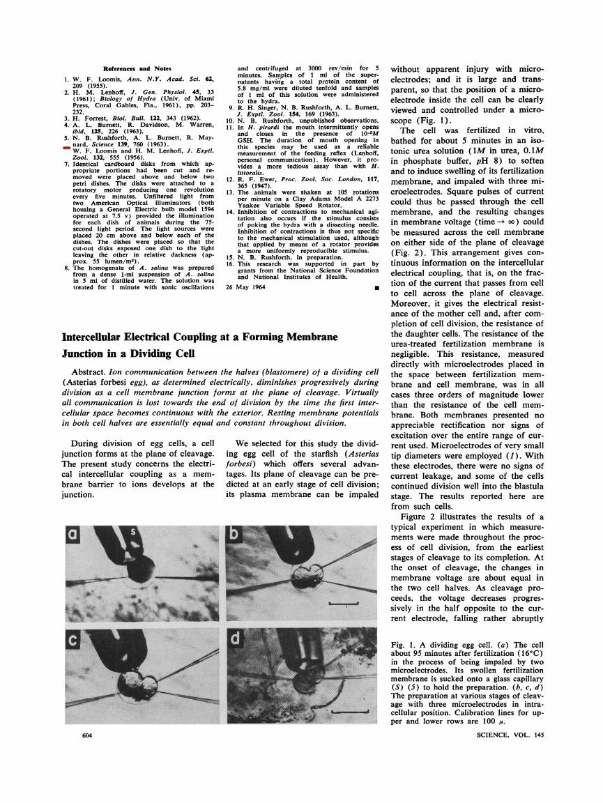
scope (Fig. 1). The cell was fertilized in vitro,
bathed for about 5 minutes in an iso- tonic urea solution (1M in urea, 0.1M in phosphate buffer, pH 8) to soften and to induce swelling of its fertilization membrane, and impaled with three mi- croelectrodes. Square pulses of current could thus be passed through the cell membrane, and the resulting changes in membrane voltage (time - oo) could
be measured across the cell membrane on either side of the plane of cleavage (Fig. 2). This arrangement gives con- tinuous information on the intercellular electrical coupling, that is, on the frac- tion of the current that passes from cell to cell across the plane of cleavage. Moreover, it gives the electrical resist- ance of the mother cell and, after com-
pletion of cell division, the resistance of the daughter cells. The resistance of the urea-treated fertilization membrane is
negligible. This resistance, measured
directly with microelectrodes placed in the space between fertilization mem- brane and cell membrane, was in all cases three orders of magnitude lower than the resistance of the cell mem- brane. Both membranes presented no
appreciable rectification nor signs of excitation over the entire range of cur- rent used. Microelectrodes of very small
tip diameters were employed (1). With these electrodes, there were no signs of current leakage, and some of the cells continued division well into the blastula
stage. The results reported here are from such cells.
Figure 2 illustrates the results of a
typical experiment in which measure- ments were made throughout the proc- ess of cell division, from the earliest
stages of cleavage to its completion. At the onset of cleavage, the changes in membrane voltage are about equal in the two cell halves. As cleavage pro- ceeds, the voltage decreases progres- sively in the half opposite to the cur- rent electrode, falling rather abruptly
without apparent injury with micro- electrodes; and it is large and trans-
parent, so that the position of a micro- electrode inside the cell can be clearly viewed and controlled under a micro-
scope (Fig. 1). The cell was fertilized in vitro,
bathed for about 5 minutes in an iso- tonic urea solution (1M in urea, 0.1M in phosphate buffer, pH 8) to soften and to induce swelling of its fertilization membrane, and impaled with three mi- croelectrodes. Square pulses of current could thus be passed through the cell membrane, and the resulting changes in membrane voltage (time - oo) could
be measured across the cell membrane on either side of the plane of cleavage (Fig. 2). This arrangement gives con- tinuous information on the intercellular electrical coupling, that is, on the frac- tion of the current that passes from cell to cell across the plane of cleavage. Moreover, it gives the electrical resist- ance of the mother cell and, after com-
pletion of cell division, the resistance of the daughter cells. The resistance of the urea-treated fertilization membrane is
negligible. This resistance, measured
directly with microelectrodes placed in the space between fertilization mem- brane and cell membrane, was in all cases three orders of magnitude lower than the resistance of the cell mem- brane. Both membranes presented no
appreciable rectification nor signs of excitation over the entire range of cur- rent used. Microelectrodes of very small
tip diameters were employed (1). With these electrodes, there were no signs of current leakage, and some of the cells continued division well into the blastula
stage. The results reported here are from such cells.
Figure 2 illustrates the results of a
typical experiment in which measure- ments were made throughout the proc- ess of cell division, from the earliest
stages of cleavage to its completion. At the onset of cleavage, the changes in membrane voltage are about equal in the two cell halves. As cleavage pro- ceeds, the voltage decreases progres- sively in the half opposite to the cur- rent electrode, falling rather abruptly
Fig. 1. A dividing egg cell. (a) The cell about 95 minutes after fertilization (16?C) in the process of being impaled by two microelectrodes. Its swollen fertilization membrane is sucked onto a glass capillary (S) (5) to hold the preparation. (b, c, d) The preparation at various stages of cleav- age with three microelectrodes in intra- cellular position. Calibration lines for up- per and lower rows are 100 ,.
SCIENCE, VOL. 145
Fig. 1. A dividing egg cell. (a) The cell about 95 minutes after fertilization (16?C) in the process of being impaled by two microelectrodes. Its swollen fertilization membrane is sucked onto a glass capillary (S) (5) to hold the preparation. (b, c, d) The preparation at various stages of cleav- age with three microelectrodes in intra- cellular position. Calibration lines for up- per and lower rows are 100 ,.
SCIENCE, VOL. 145 604 604
Intercellular Electrical Coupling at a Forming Membrane
Junction in a Dividing Cell
Abstract. Ion communication between the halves (blastomere) of a dividing cell
(Asterias forbesi egg), as determined electrically, diminishes progressively during division as a cell memibrane junction forms at the plane of cleavage. Virtually all communication is lost towards the end of division by the time the first inter- cellular space becomes continuous with the exterior. Resting membrane potentials in both cell halves are essentially equal and constant throughout division.