Embed Size (px)
Citation preview

“Be a lamp, a lifeboat, or a ladder.”
Cardiovascular System 1
-Rumi
Lesson Plan: Cardiovascular System 1
5 minutes: Breath of Arrival and Attendance
50 minutes: Cardiovascular System 1

Classroom Rules
Punctuality- everybody's time is precious:
Be ready to learn by 9:00, we'll have you out of here by 1:30
Tardiness: arriving late, late return after breaks, leaving early
The following are not allowed:
Bare feet
Side talking
Lying down
Inappropriate clothing
Food or drink except water
Phones in classrooms, clinic or bathrooms
You will receive one verbal warning, then you'll have to leave the room.
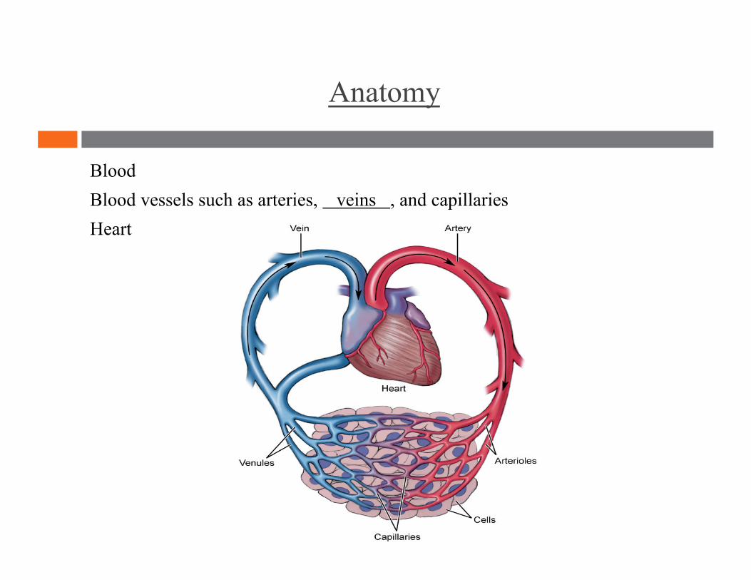
Anatomy
Blood Blood vessels such as arteries, veins , and capillaries Heart
Transportation Protection Combat hemorrhage
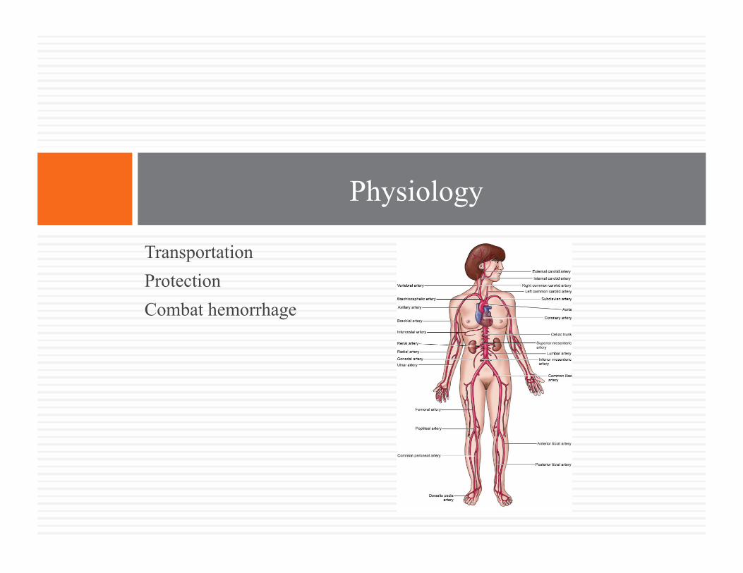
Physiology
Physiology
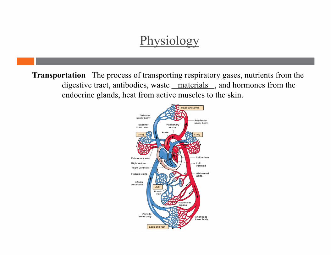
Transportation The process of transporting respiratory gases, nutrients from the digestive tract, antibodies, waste materials , and hormones from the endocrine glands, heat from active muscles to the skin.

Physiology
Protection The process of protecting the body through disease-fighting white blood cells and the removal of impurities and pathogens
Physiology
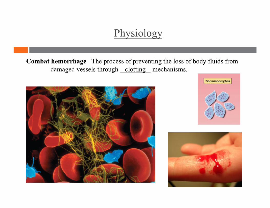
Combat hemorrhage The process of preventing the loss of body fluids from damaged vessels through clotting mechanisms.
Formed elements (blood cells) Plasma (liquid portion)
Blood
Blood
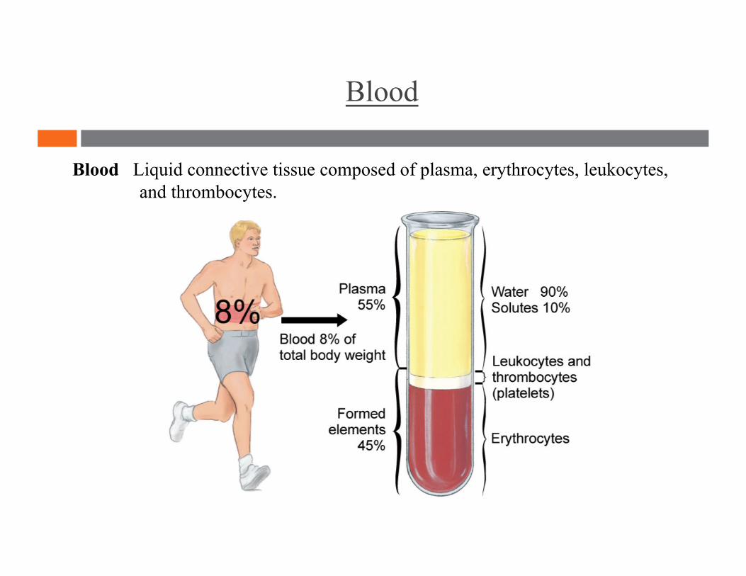
Blood Liquid connective tissue composed of plasma, erythrocytes, leukocytes, and thrombocytes.
Blood
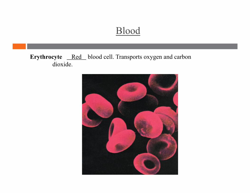
Erythrocyte Red blood cell. Transports oxygen and carbon dioxide.
Blood
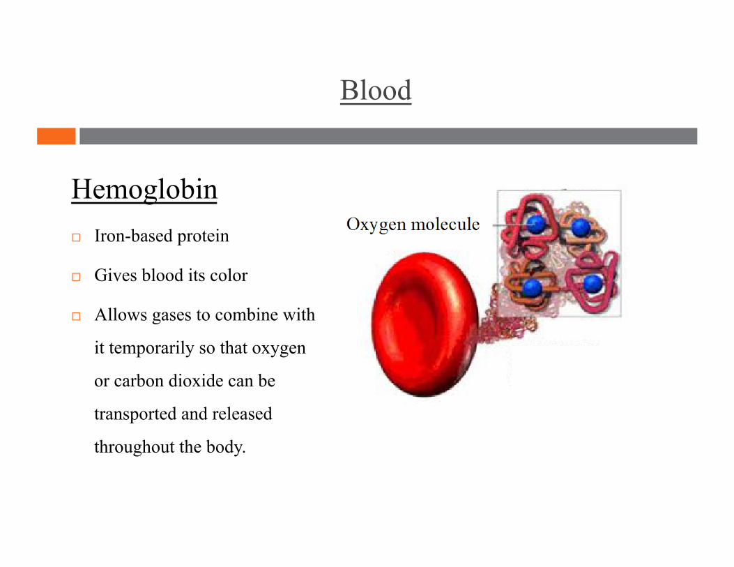
Hemoglobin
Iron-based protein
Gives blood its color
Allows gases to combine with
it temporarily so that oxygen
or carbon dioxide can be
transported and released
throughout the body.
Blood
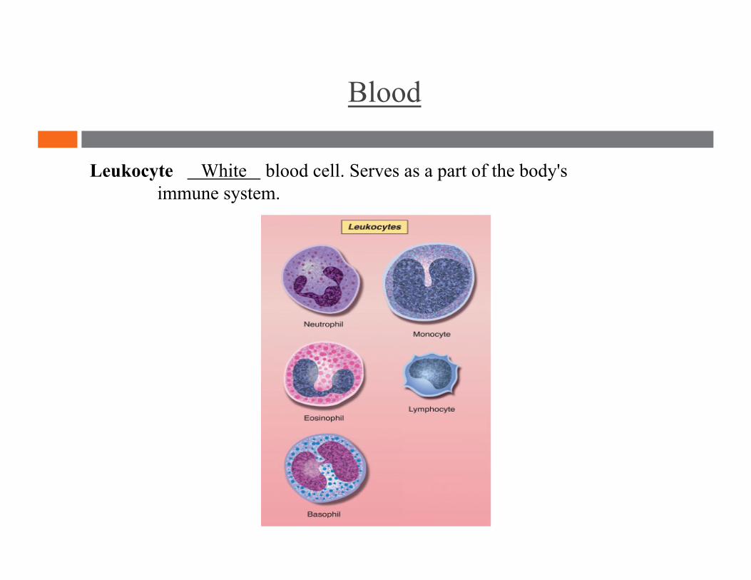
Leukocyte White blood cell. Serves as a part of the body's immune system.
Blood
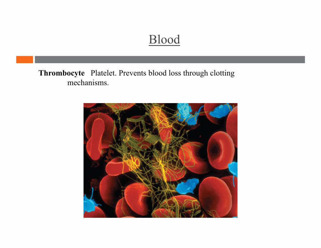
Thrombocyte Platelet. Prevents blood loss through clotting mechanisms.
Blood
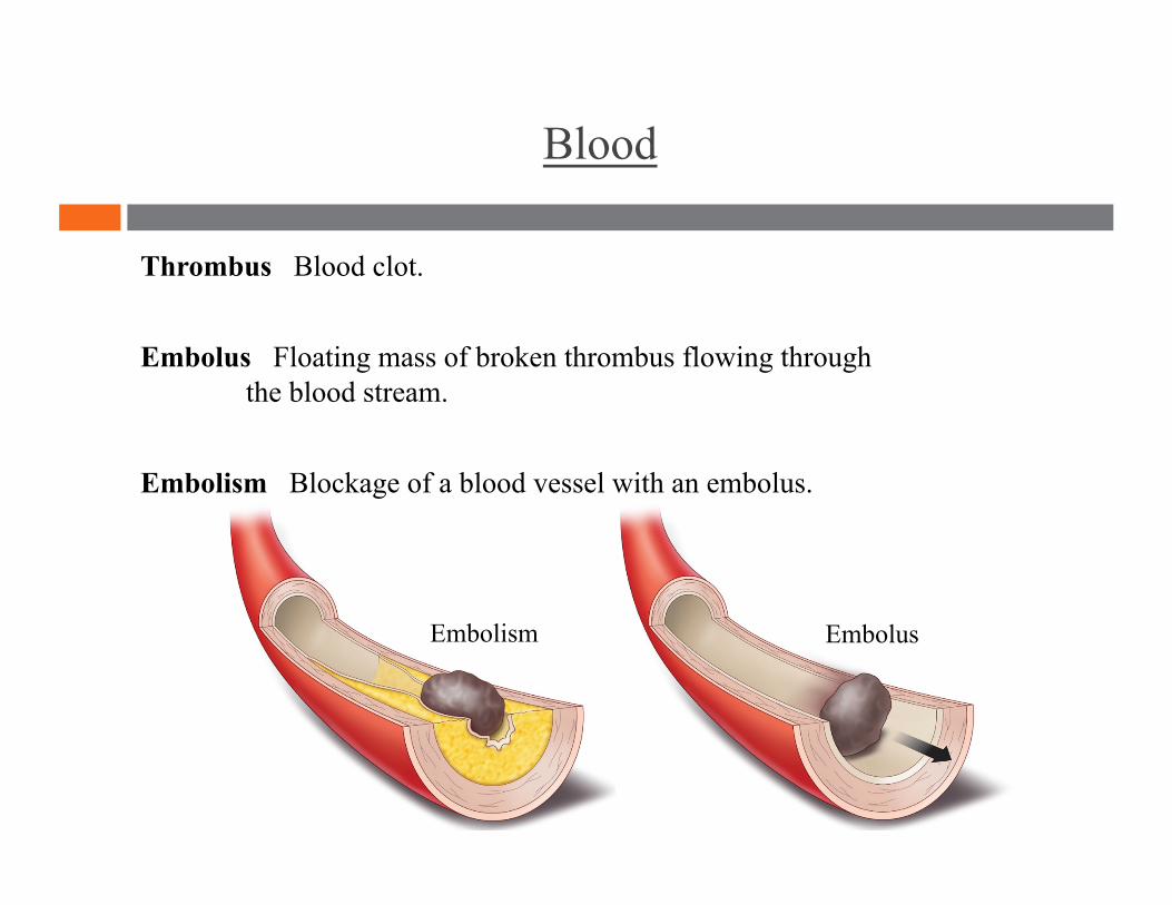
Thrombus Blood clot.
Embolus Floating mass of broken thrombus flowing through the blood stream.
Embolism Blockage of a blood vessel with an embolus.
Embolism Embolus
Blood
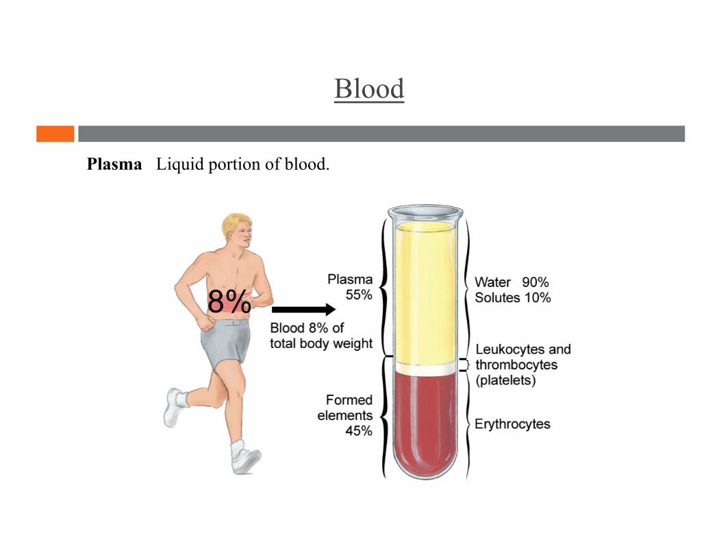
Plasma Liquid portion of blood.
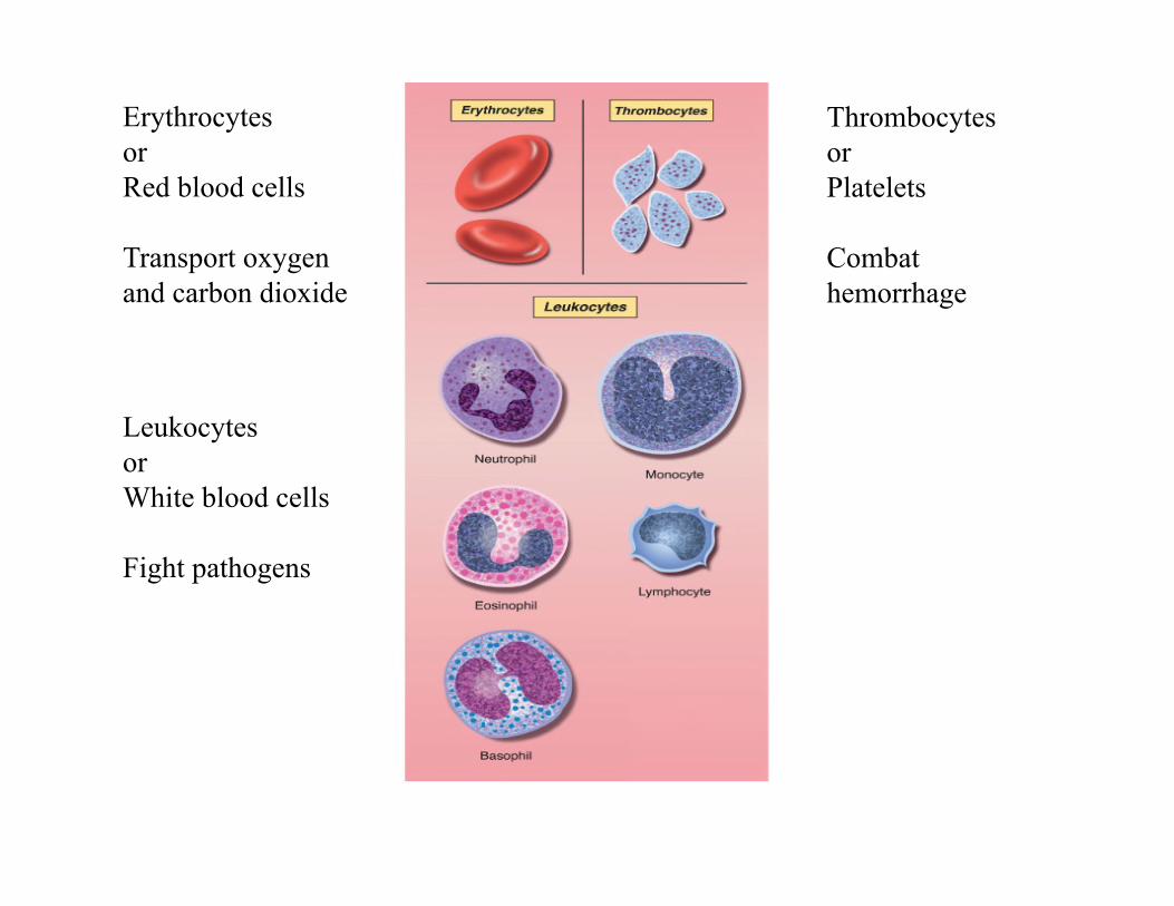
Erythrocytes or Red blood cells
Transport oxygen and carbon dioxide
Leukocytes or White blood cells
Fight pathogens
Thrombocytes or Platelets
Combat hemorrhage

Heart
Wall Chambers Valves Blood flow
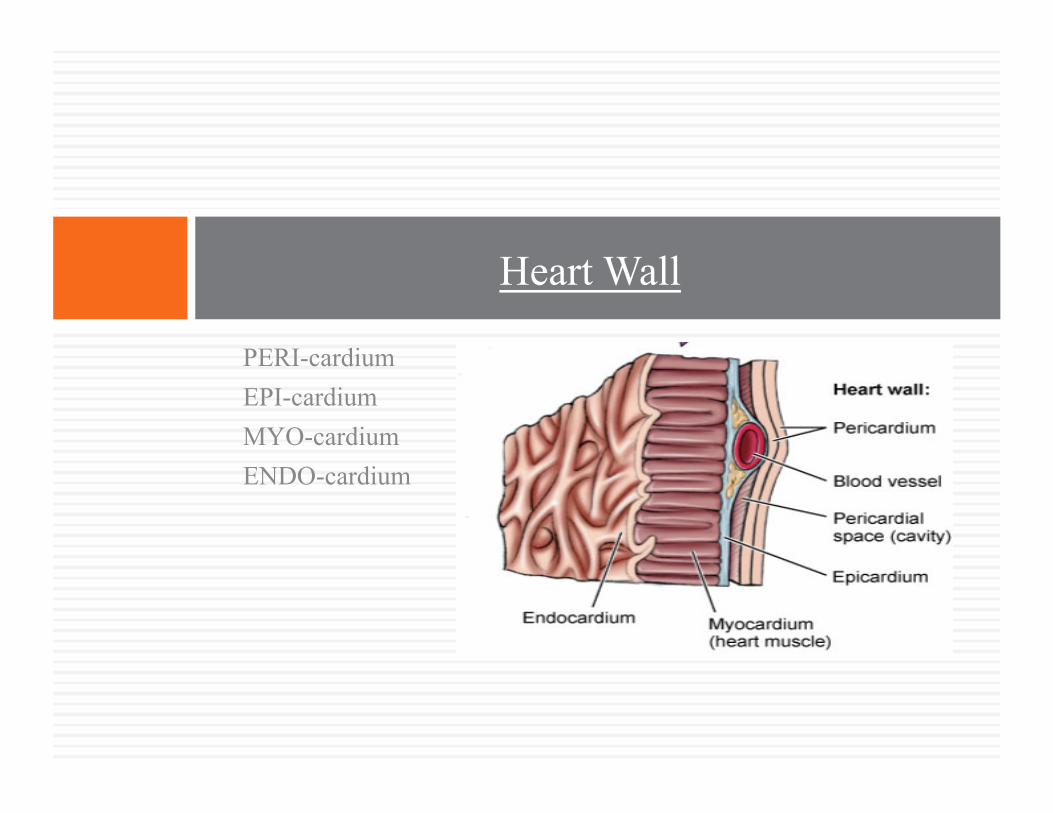
PERI-cardium EPI-cardium MYO-cardium ENDO-cardium
Heart Wall
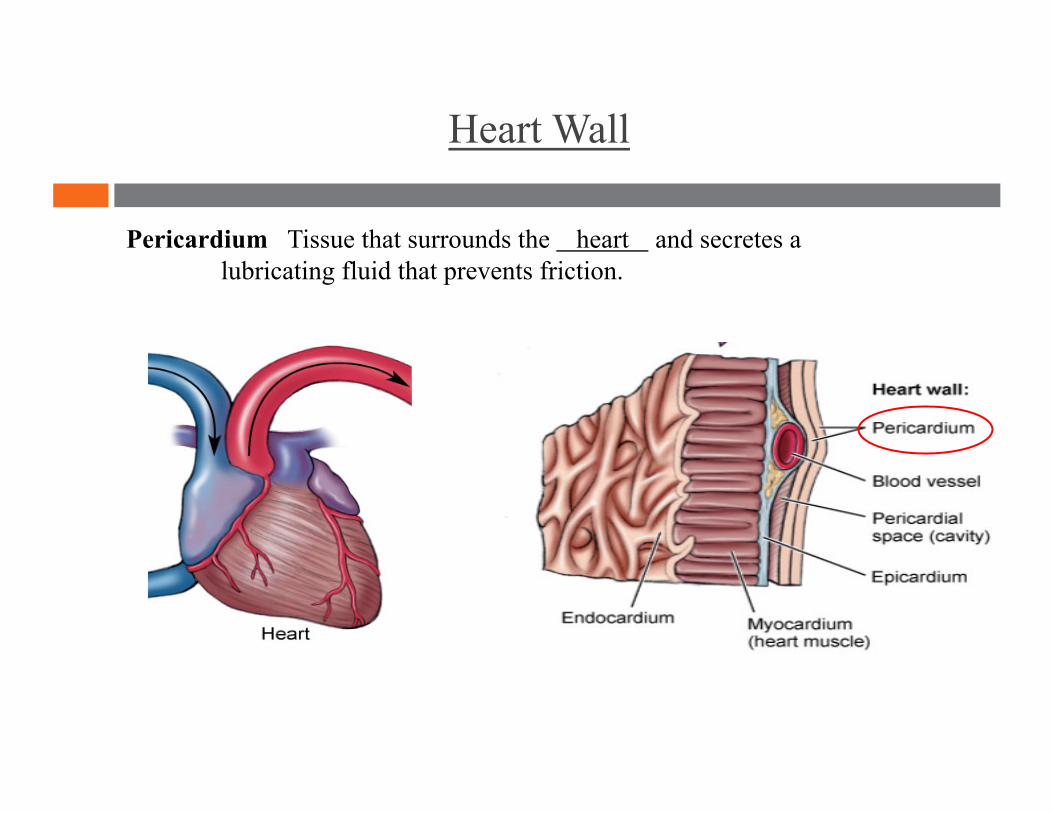
Heart Wall
Pericardium Tissue that surrounds the heart and secretes a lubricating fluid that prevents friction.
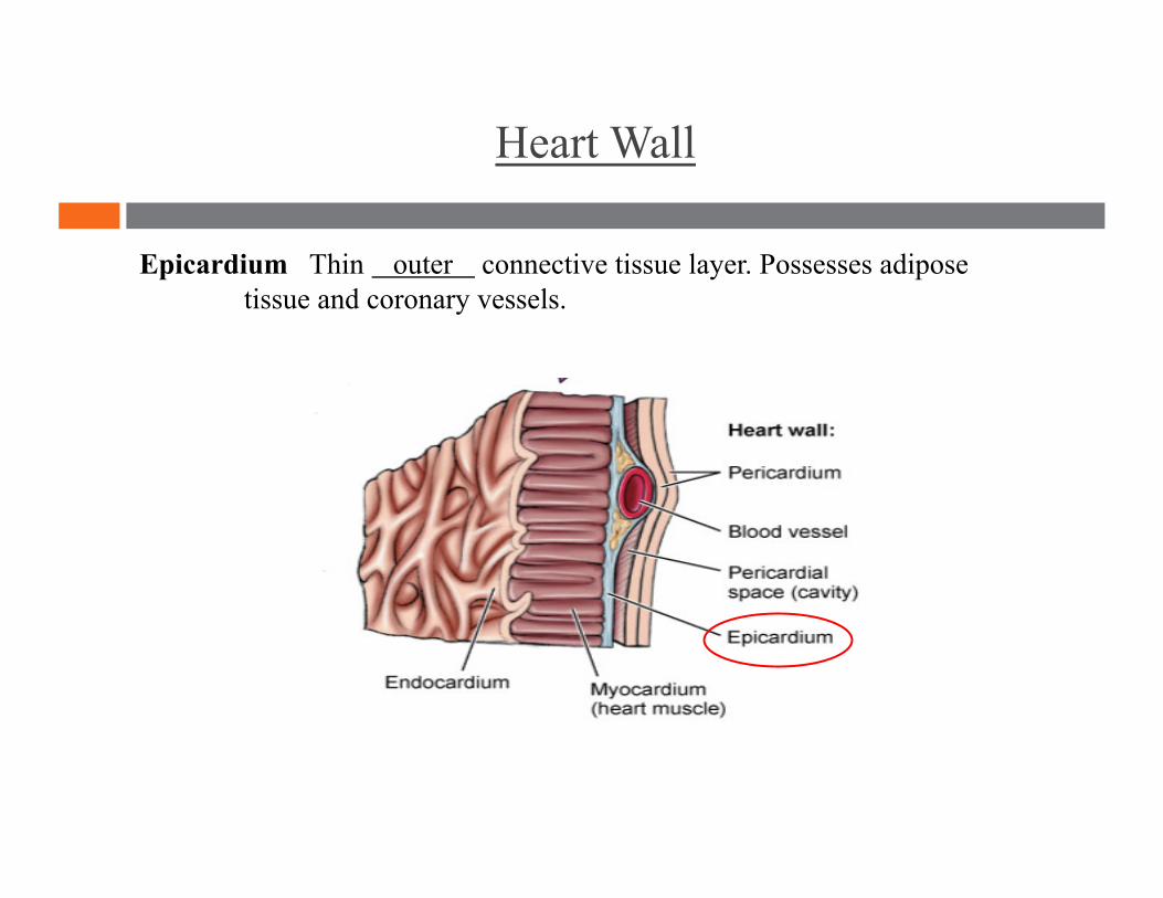
Heart Wall
Epicardium Thin outer connective tissue layer. Possesses adipose tissue and coronary vessels.

Heart Wall
Myocardium Thick muscular layer that makes up the bulk of the heart wall. Its contraction forces blood out of the ventricles.

Heart Wall
Endocardium Thin, inner lining of the heart. Continuous with the endothelial lining of the heart chambers and blood vessels, as well as the valves of the heart.
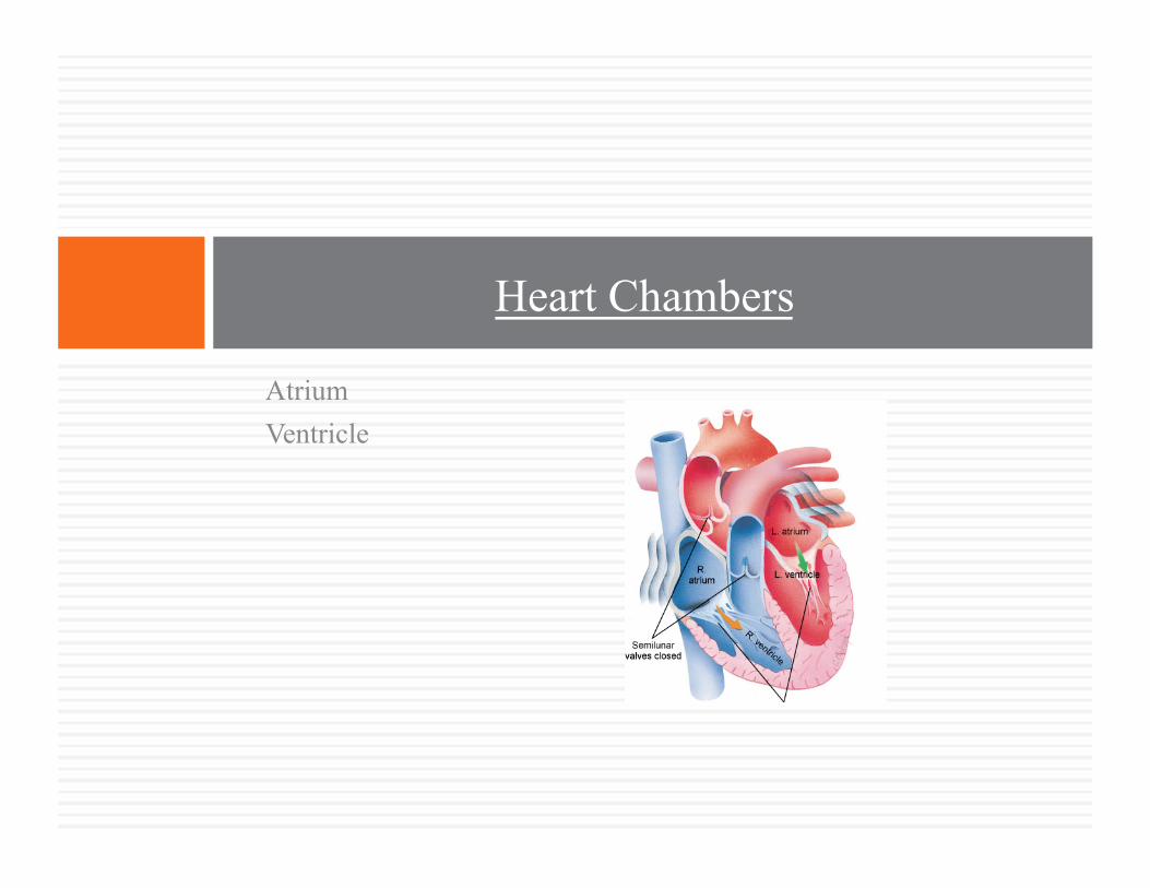
Atrium Ventricle
Heart Chambers
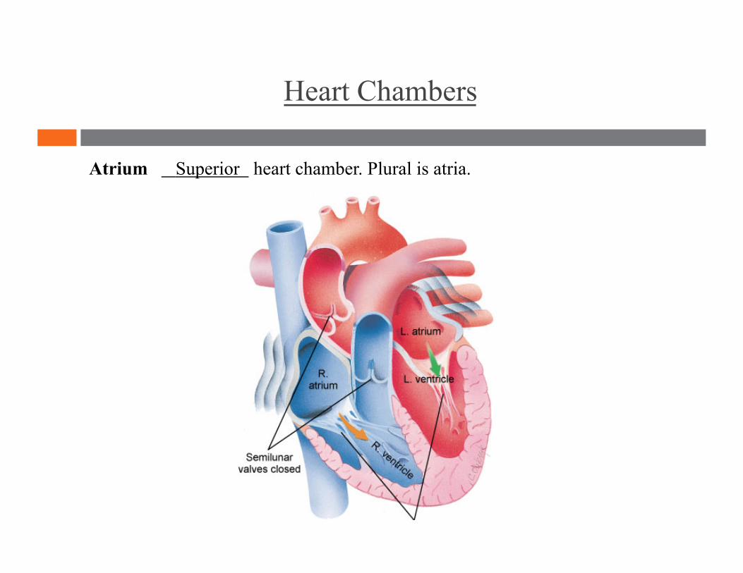
Heart Chambers
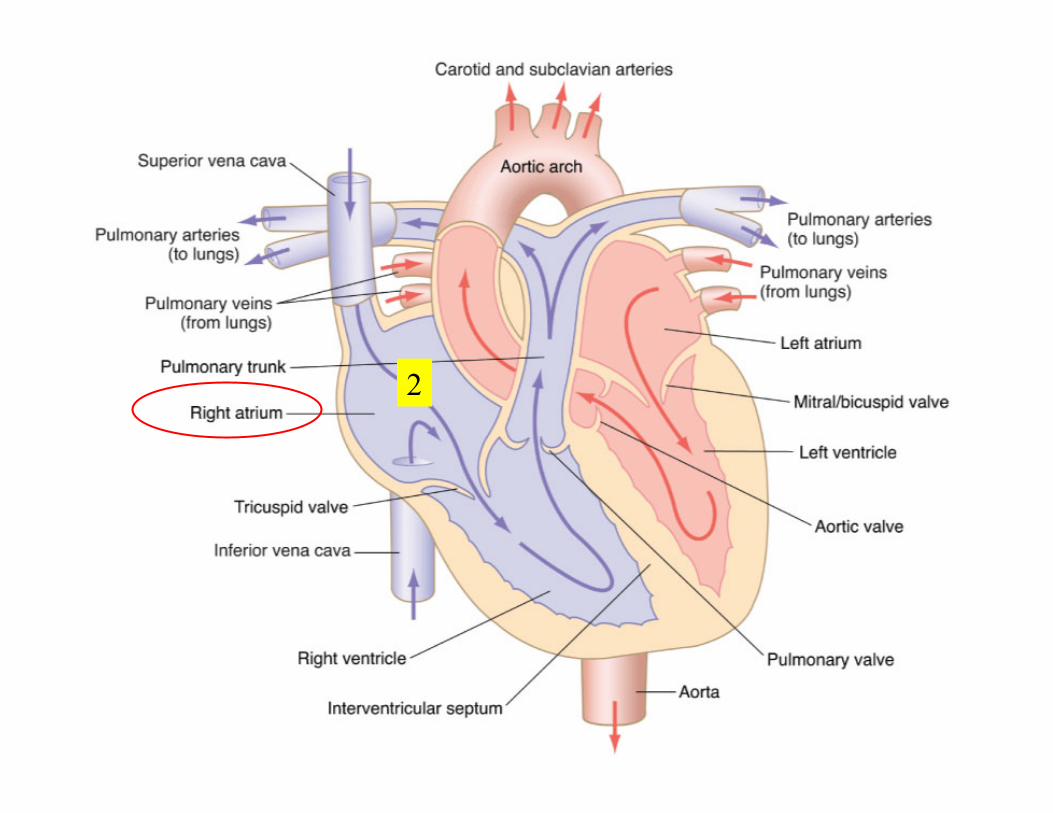
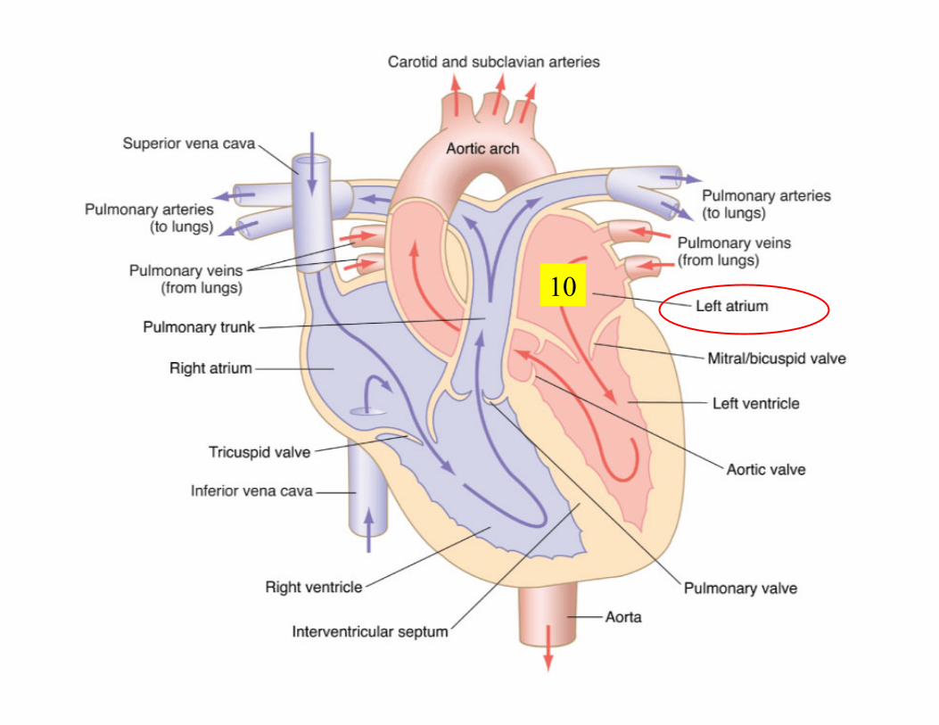
Atrium Superior heart chamber. Plural is atria.
Heart Chambers
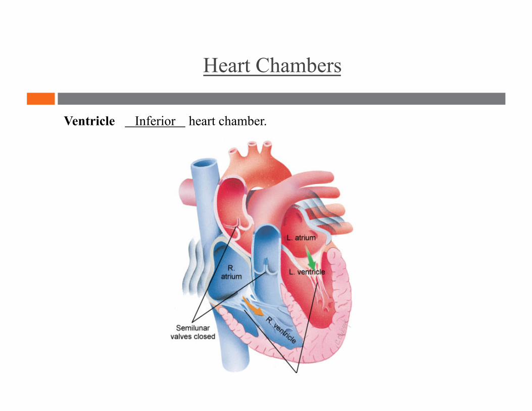
Ventricle Inferior heart chamber.
Atrioventricular (A-V valve) Semilunar
Heart Valves
Heart Valves
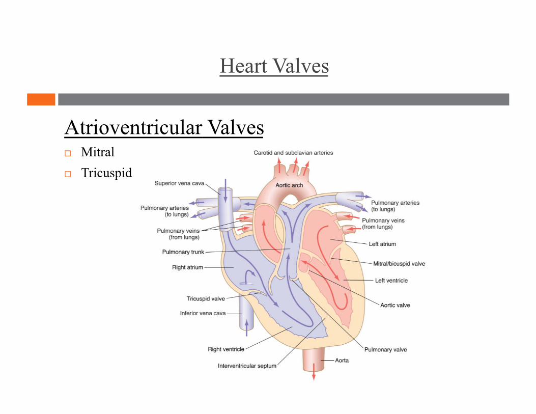
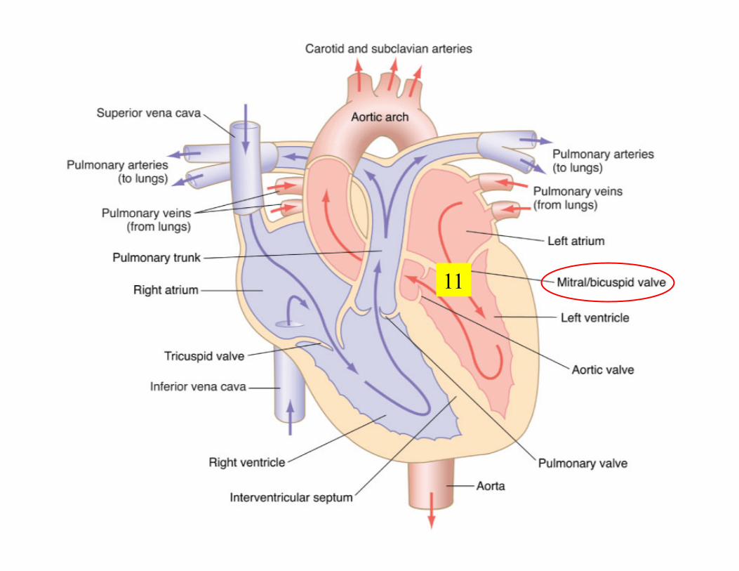
Atrioventricular Valves Mitral Tricuspid
Heart Valves
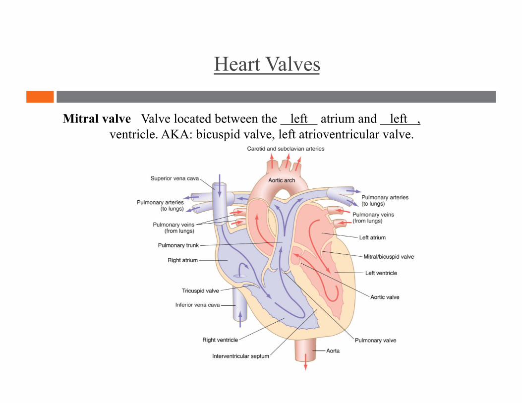
Mitral valve Valve located between the left atrium and left , ventricle. AKA: bicuspid valve, left atrioventricular valve.

Heart Valves
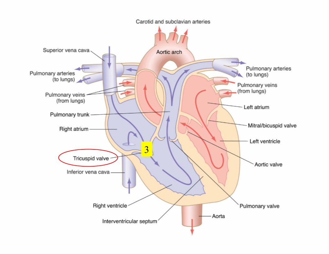
Tricuspid valve Valve located between the right atrium and right ventricle. AKA: right atrioventricular valve
Heart Valves
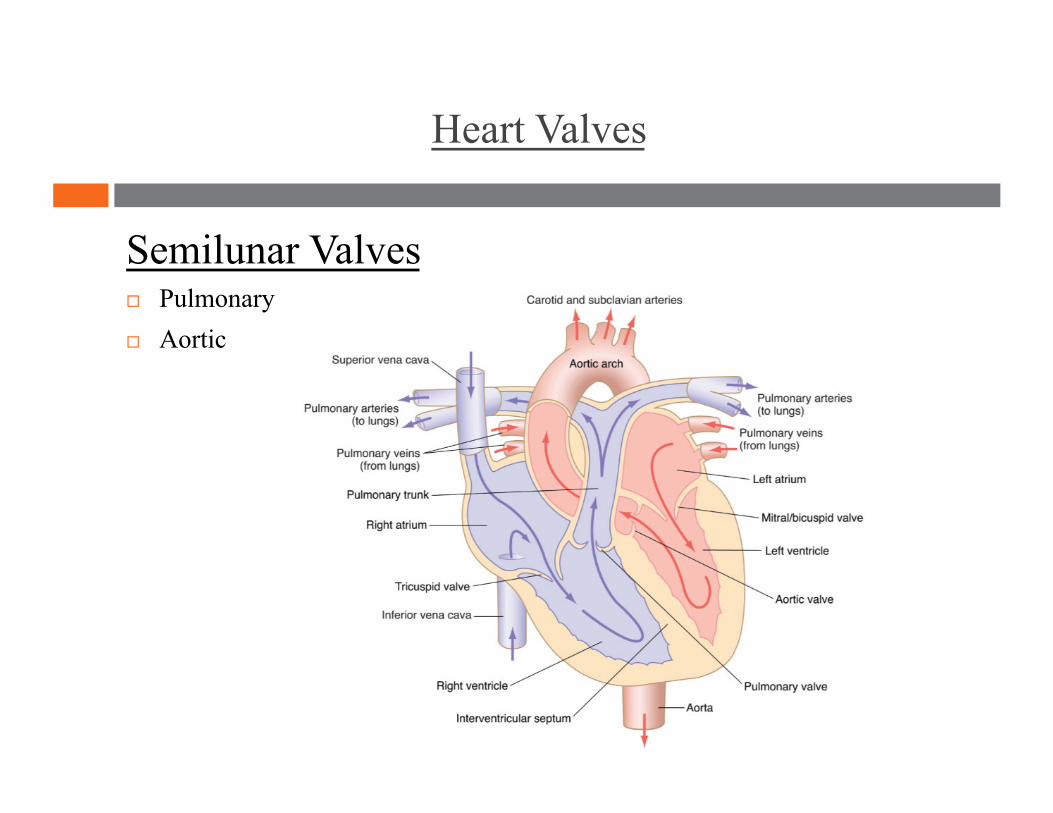
Semilunar Valves Pulmonary Aortic
Heart Valves
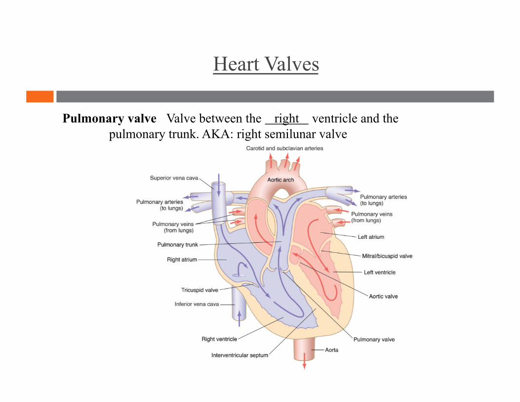
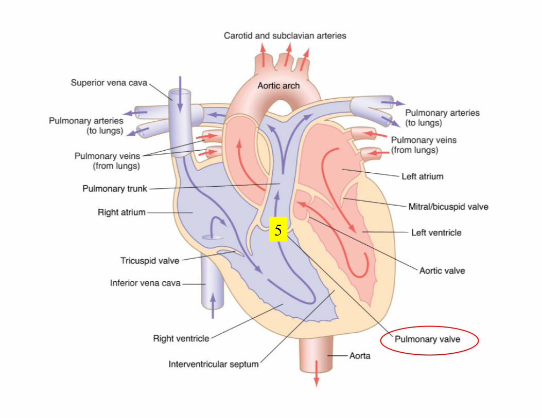
Pulmonary valve Valve between the right ventricle and the pulmonary trunk. AKA: right semilunar valve
Heart Valves
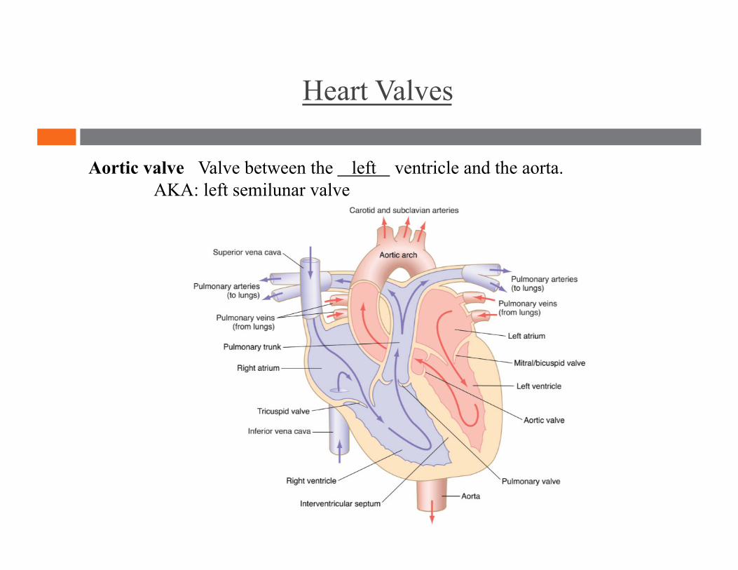
Aortic valve Valve between the left ventricle and the aorta. AKA: left semilunar valve
Coronary Vessels
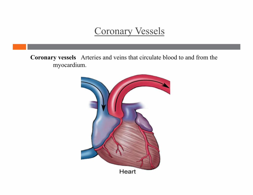
Coronary vessels Arteries and veins that circulate blood to and from the myocardium.
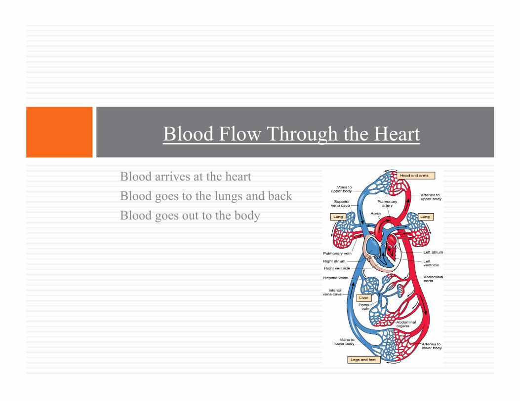
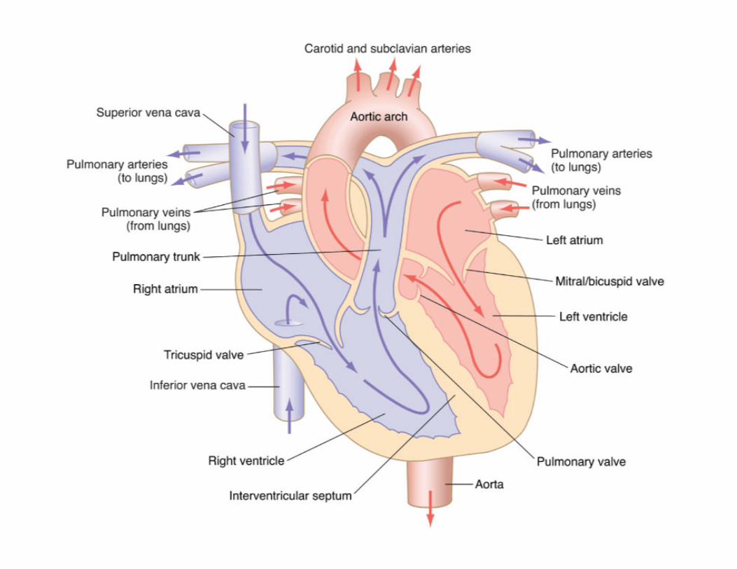
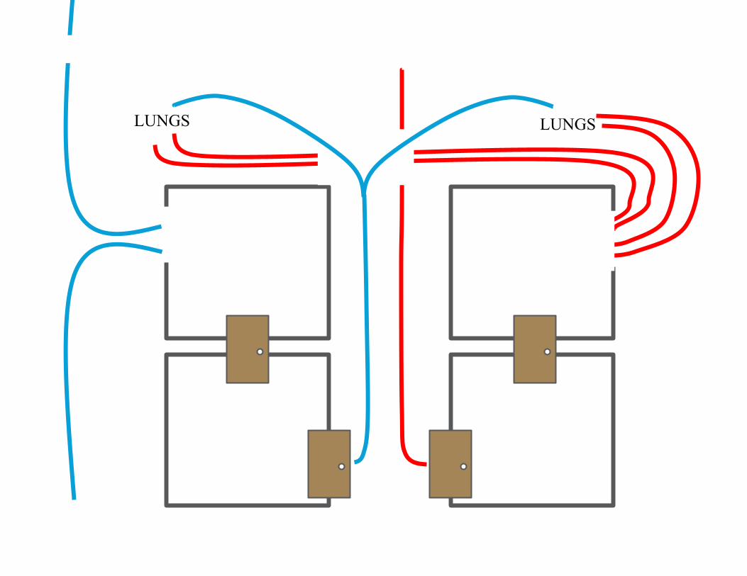
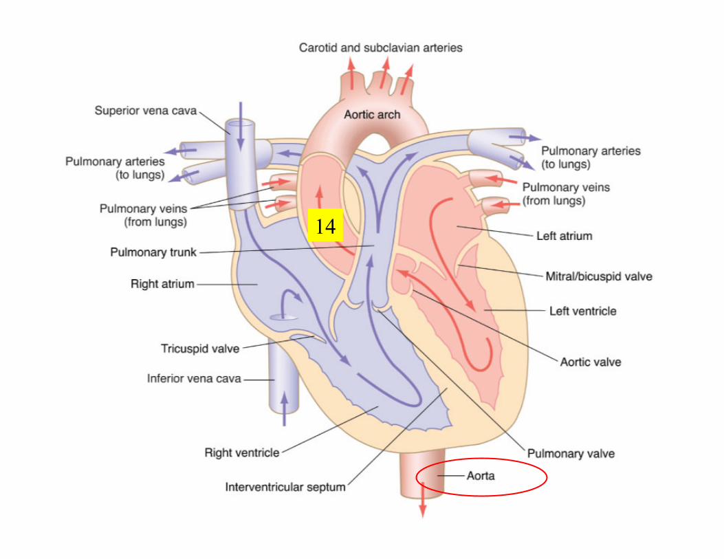
Blood arrives at the heart Blood goes to the lungs and back Blood goes out to the body
Blood Flow Through the Heart
Blood Flow Through the Heart
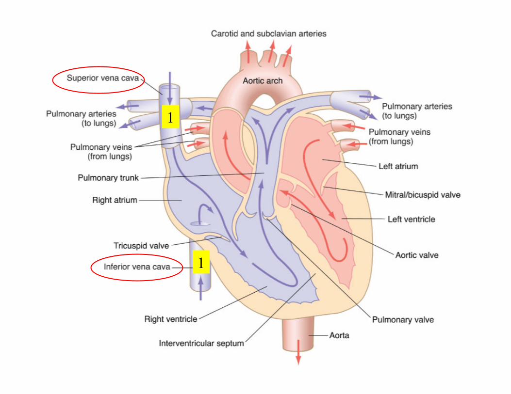
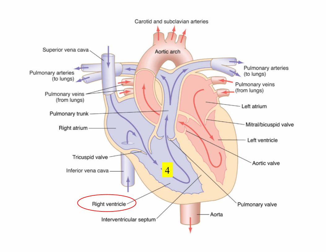
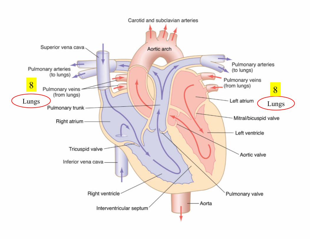
Stage 1 Oxygen-depleted blood enters the superior and inferior vena cava
and flows into the right atrium. When the right atrium is full, it
empties through the tricuspid valve into the right ventricle.
Occurs at the same time as Stage 3.
Blood Flow Through the Heart
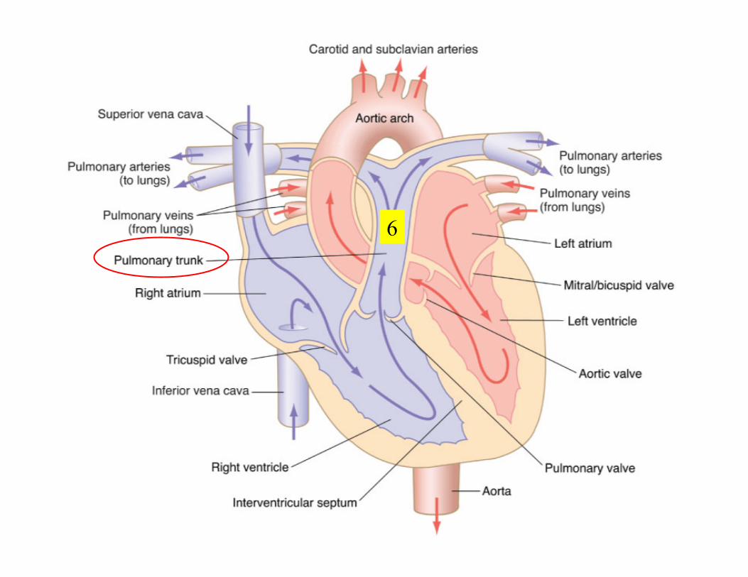
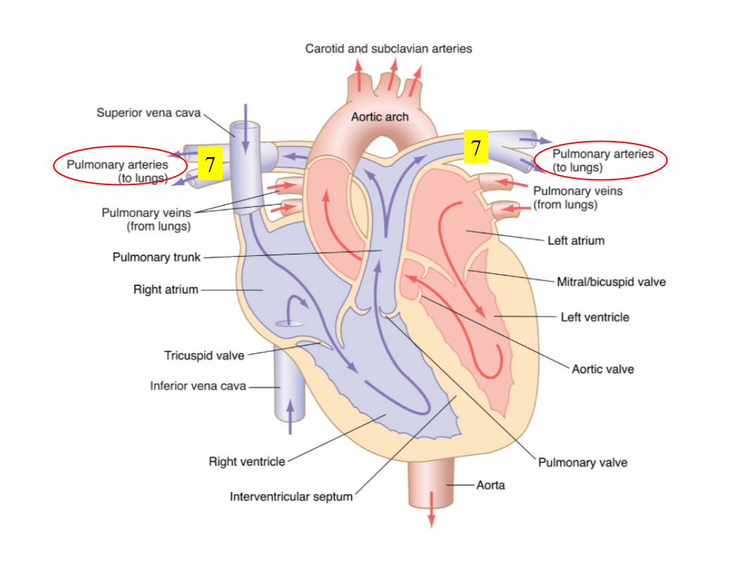
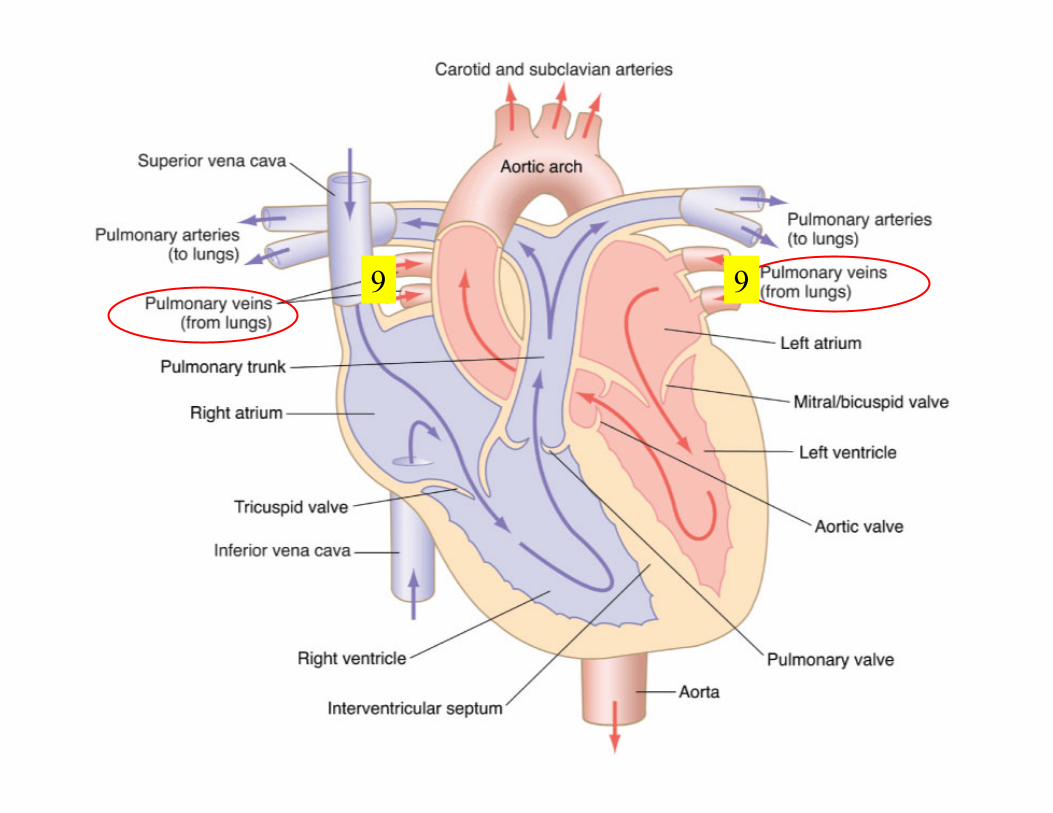
Stage 2 The right ventricle contracts and pushes blood through the
pulmonary valve into the pulmonary trunk. The pulmonary trunk then
divides into left and right pulmonary arteries which take blood to
each lung. Four pulmonary veins leave the lungs and carry oxygen-
rich blood back to the left atrium.
Blood Flow Through the Heart
Stage 3 Blood leaves the left atrium and passes through the left
ventricle through the mitral valve. The left ventricle contracts and
pushes blood through the aortic valve into the aorta and descending
aorta and to all parts of the body except the lungs. Occurs at the same
time as Stage 1.
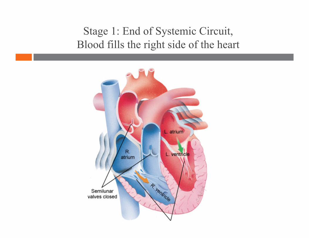
Stage 1: End of Systemic Circuit, Blood fills the right side of the heart
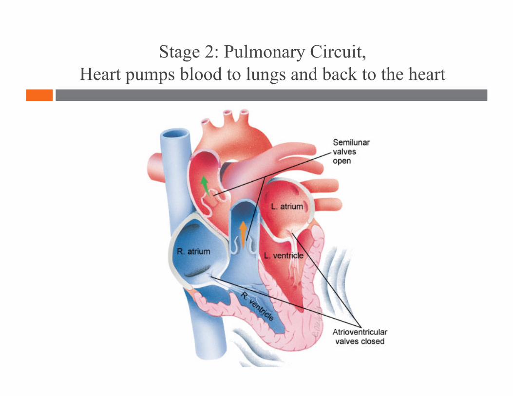
Stage 2: Pulmonary Circuit, Heart pumps blood to lungs and back to the heart
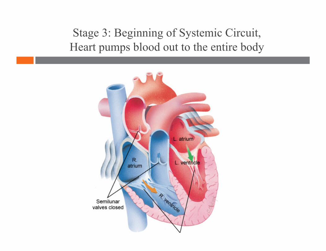
Stage 3: Beginning of Systemic Circuit, Heart pumps blood out to the entire body
LUNGS LUNGS
1
1
2
3
4
5
6
7 7
Lungs Lungs
8 8
9 9
10
11

12

13
14

“Be a lamp, a lifeboat, or a ladder.”
Cardiovascular System 1
-Rumi

“Be a lamp, a lifeboat, or a ladder.”
Cardiovascular System 2
-Rumi
Lesson Plan: Cardiovascular System 2
5 minutes: Breath of Arrival and Attendance
5 minutes: Trapezius OIA
45 minutes: Cardiovascular System 2

Classroom Rules
Punctuality- everybody's time is precious:
Be ready to learn by 9:00, we'll have you out of here by 1:30
Tardiness: arriving late, late return after breaks, leaving early
The following are not allowed:
Bare feet
Side talking
Lying down
Inappropriate clothing
Food or drink except water
Phones in classrooms, clinic or bathrooms
You will receive one verbal warning, then you'll have to leave the room.
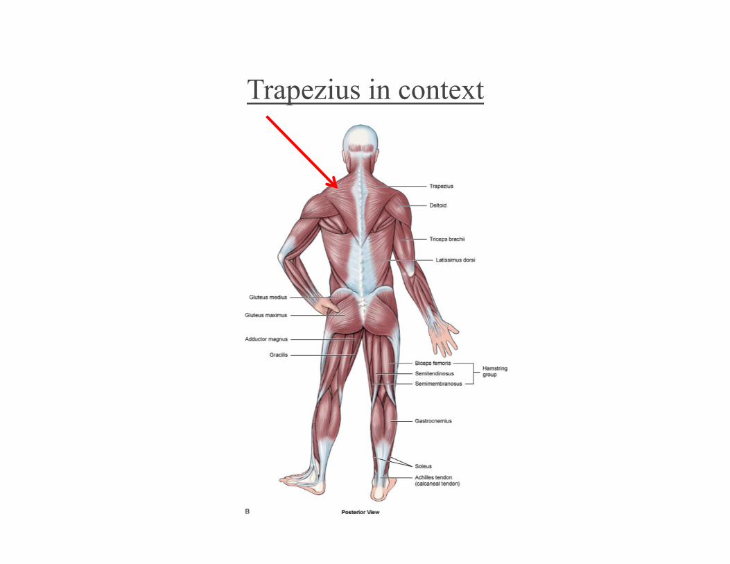
Trapezius in context
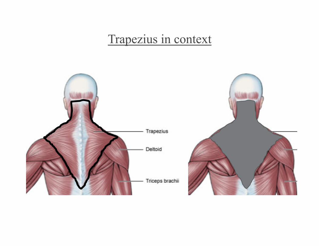
Trapezius in context
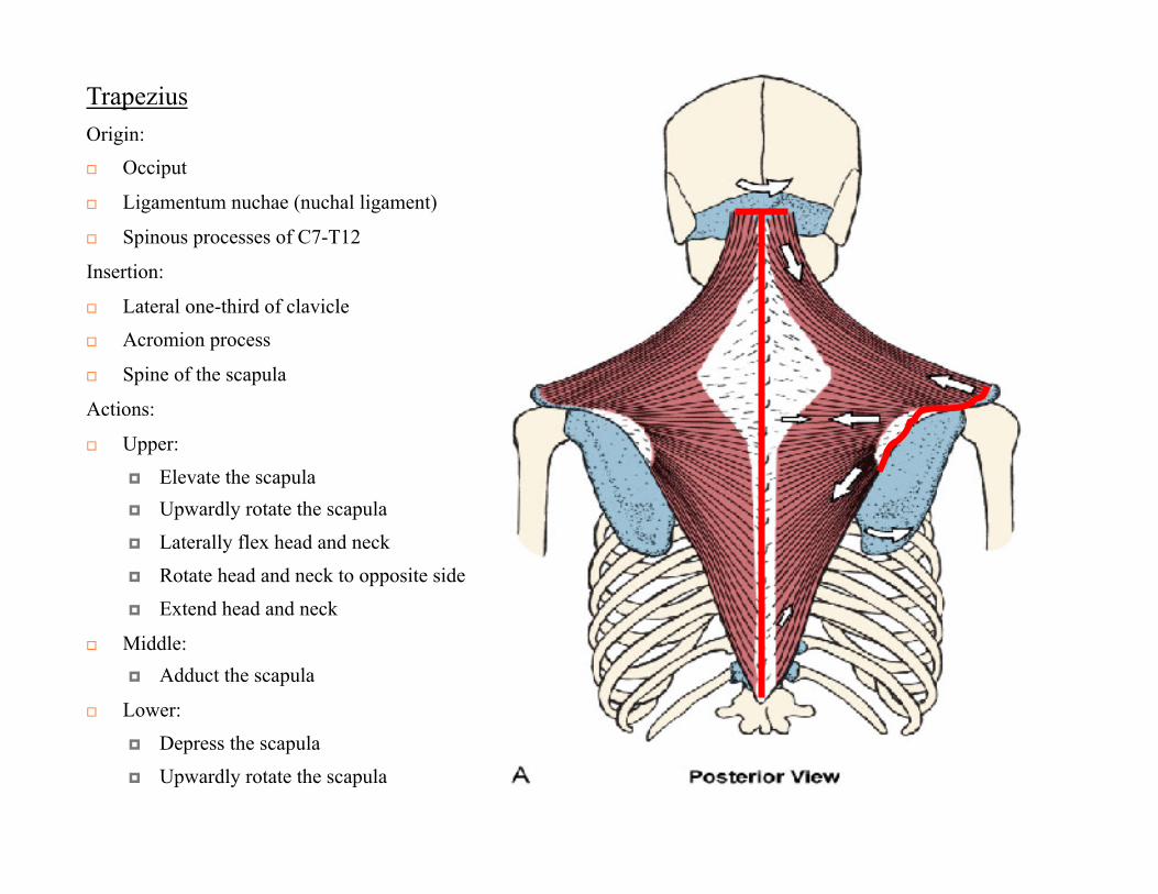
Trapezius
Origin:
Occiput
Ligamentum nuchae (nuchal ligament)
Spinous processes of C7-T12
Insertion:
Lateral one-third of clavicle
Acromion process
Spine of the scapula
Actions:
Upper:
Elevate the scapula Upwardly rotate the scapula
Laterally flex head and neck
Rotate head and neck to opposite side
Extend head and neck
Middle: Adduct the scapula
Lower:
Depress the scapula
Upwardly rotate the scapula
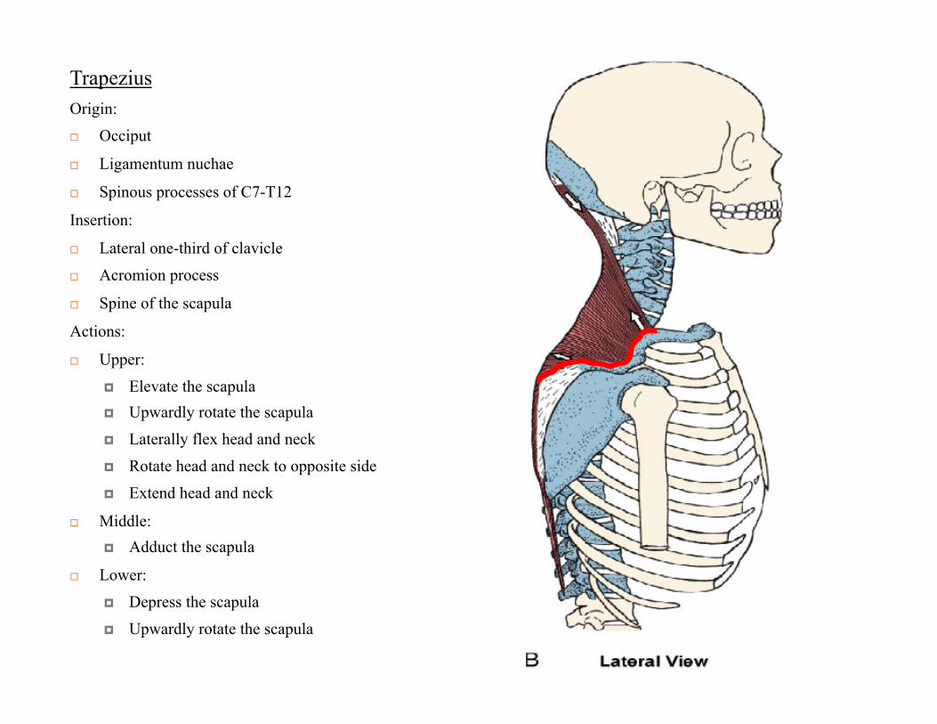
Trapezius
Origin:
Occiput
Ligamentum nuchae
Spinous processes of C7-T12
Insertion:
Lateral one-third of clavicle
Acromion process
Spine of the scapula
Actions:
Upper:
Elevate the scapula Upwardly rotate the scapula
Laterally flex head and neck
Rotate head and neck to opposite side
Extend head and neck
Middle: Adduct the scapula
Lower:
Depress the scapula
Upwardly rotate the scapula
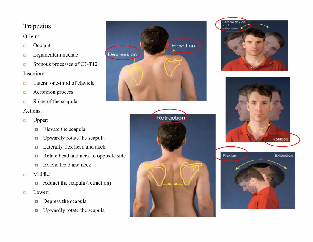
Trapezius
Origin:
Occiput
Ligamentum nuchae
Spinous processes of C7-T12
Insertion:
Lateral one-third of clavicle
Acromion process
Spine of the scapula
Actions:
Upper:
Elevate the scapula Upwardly rotate the scapula
Laterally flex head and neck
Rotate head and neck to opposite side
Extend head and neck
Middle: Adduct the scapula (retraction)
Lower:
Depress the scapula
Upwardly rotate the scapula
Walls of Arteries and Veins Arteries Pulse Capillary Veins Venous Return
Blood Vessels
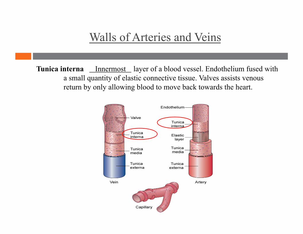
Walls of Arteries and Veins
Tunica interna Innermost layer of a blood vessel. Endothelium fused with a small quantity of elastic connective tissue. Valves assists venous return by only allowing blood to move back towards the heart.

Walls of Arteries and Veins
Tunica media Middle layer of a blood vessel. Contains both connective tissue and smooth muscle.
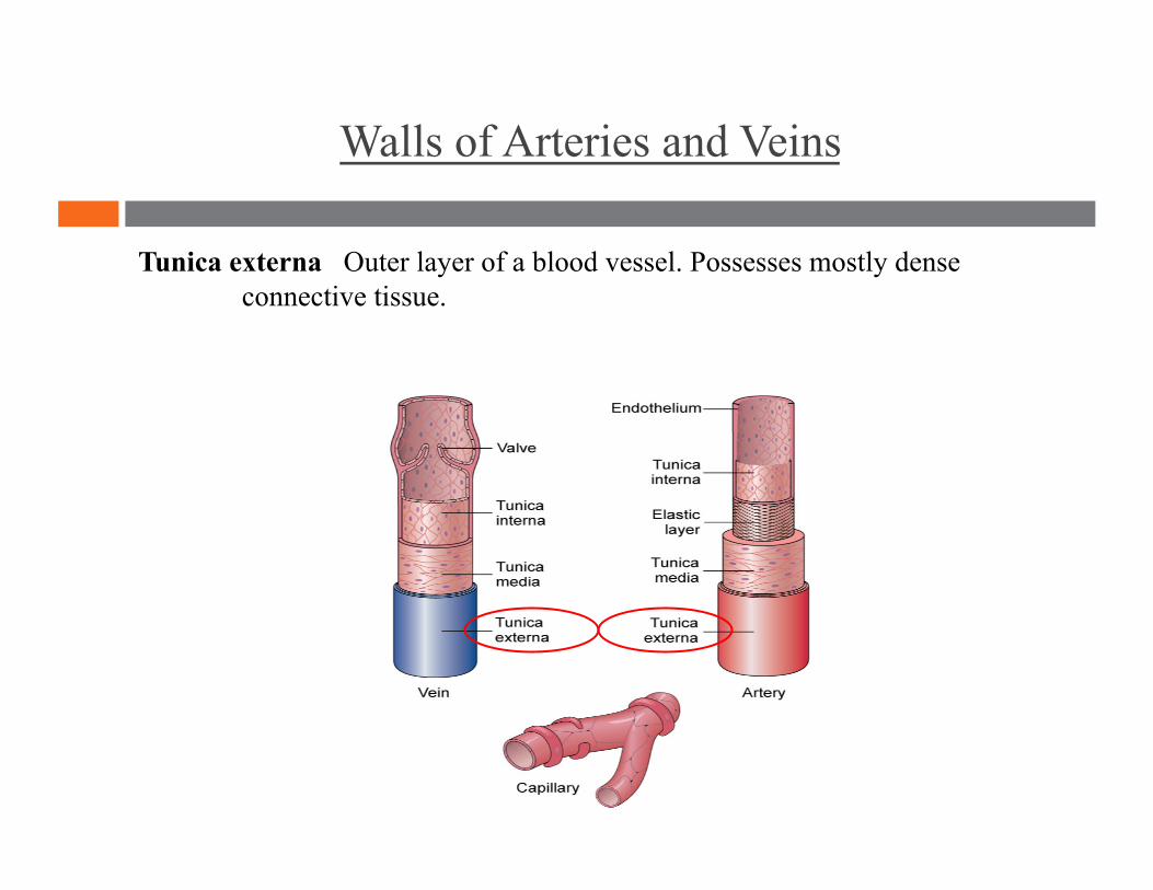
Walls of Arteries and Veins
Tunica externa Outer layer of a blood vessel. Possesses mostly dense connective tissue.
Walls of Arteries and Veins
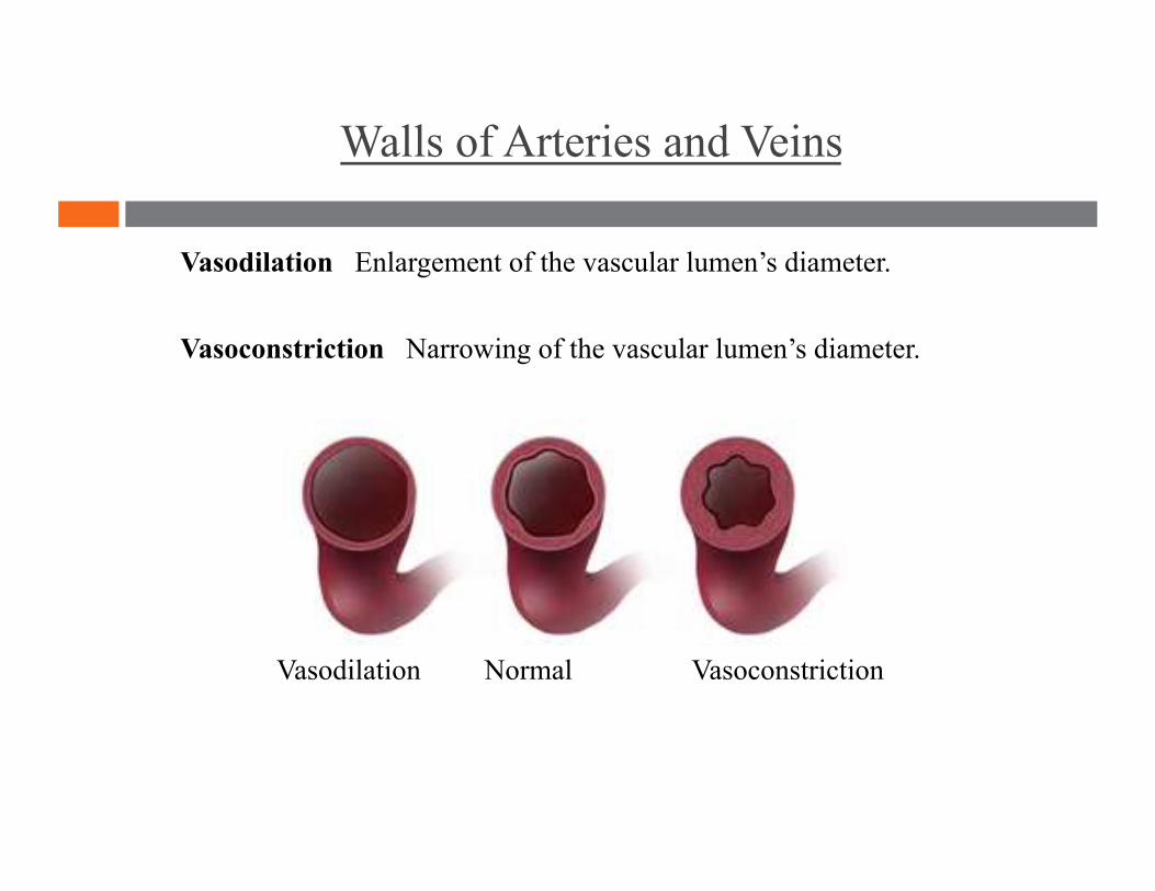
Vasodilation Enlargement of the vascular lumen’s diameter.
Vasoconstriction Narrowing of the vascular lumen’s diameter.
Vasodilation Normal Vasoconstriction
Walls of Arteries and Veins
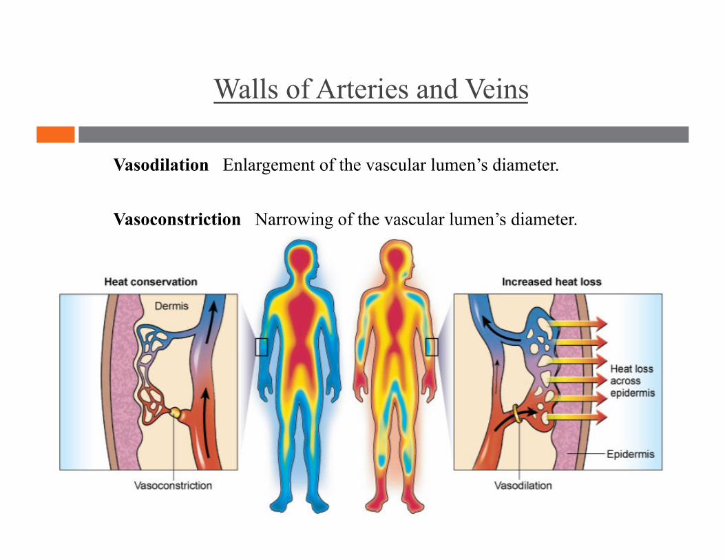
Vasodilation Enlargement of the vascular lumen’s diameter.
Vasoconstriction Narrowing of the vascular lumen’s diameter.
Walls of Arteries and Veins
Hyperemia Increased local blood flow causing the skin to become reddened and warm.
Ischemia Local abnormal decrease in blood flow. Often marked by pain and tissue dysfunction.
Arteries
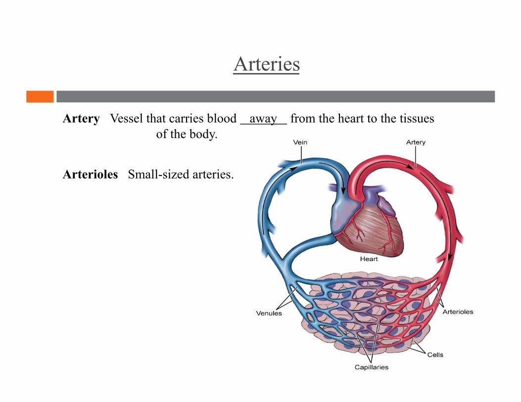
Artery Vessel that carries blood away from the heart to the tissues of the body.
Arterioles Small-sized arteries.
Arteries
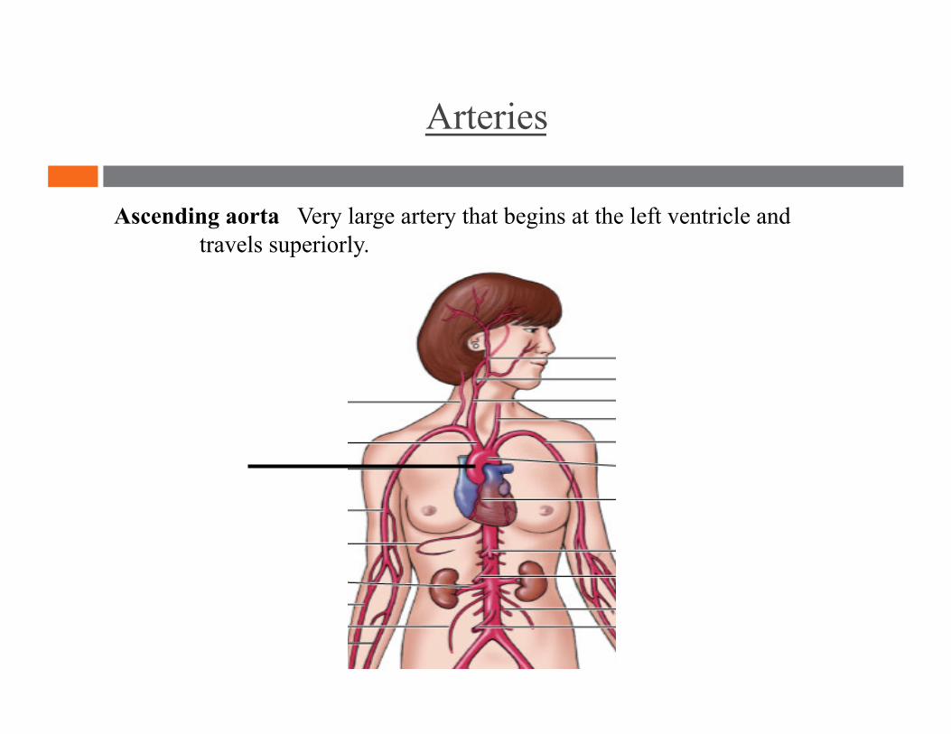
Ascending aorta Very large artery that begins at the left ventricle and travels superiorly.
Arteries
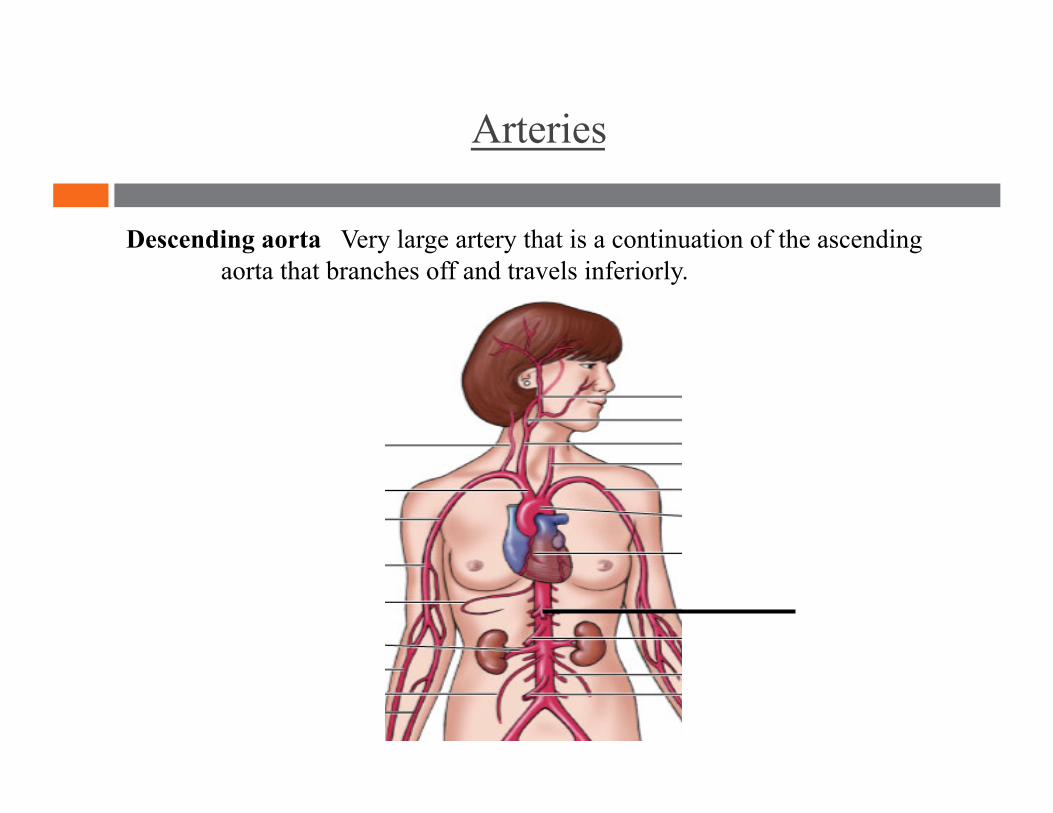
Descending aorta Very large artery that is a continuation of the ascending aorta that branches off and travels inferiorly.

Arteries
Common carotid arteries Two arteries located in the throat.
Right Carotid Artery Left Carotid Artery
Arteries
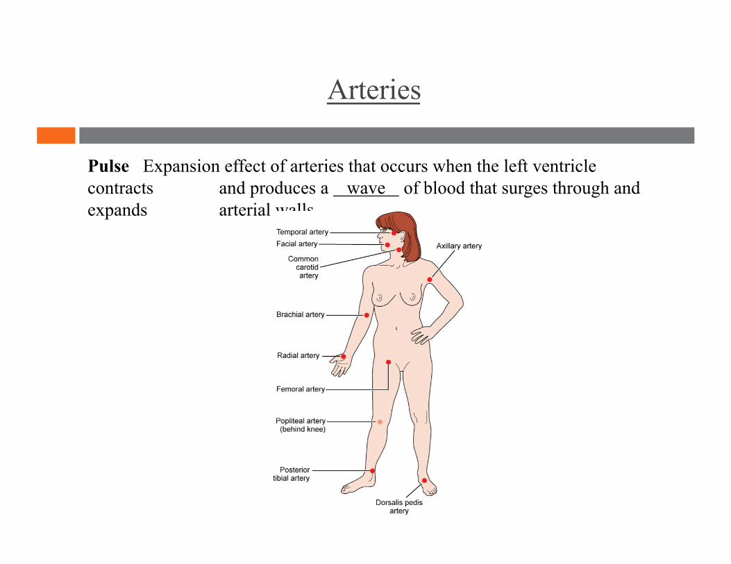
Pulse Expansion effect of arteries that occurs when the left ventricle contracts and produces a wave of blood that surges through and expands arterial walls.
Capillaries
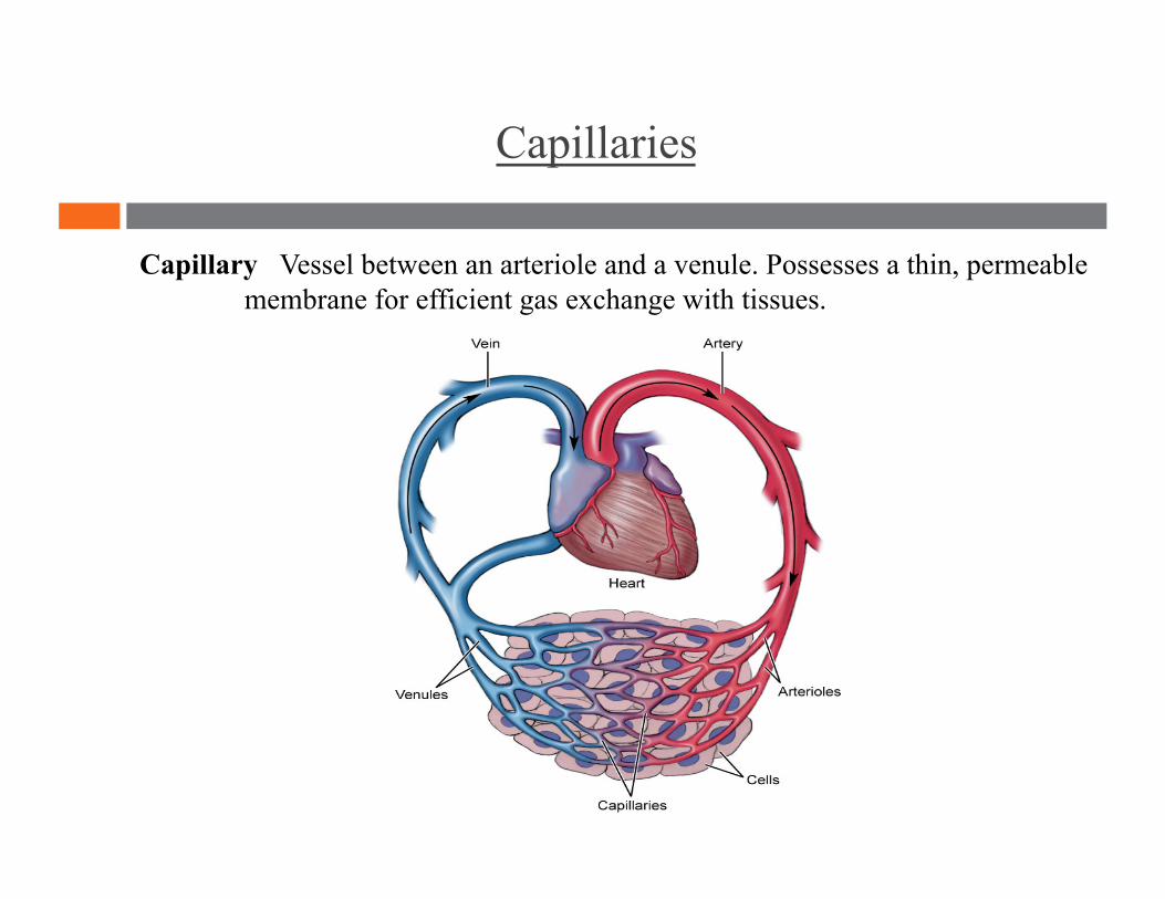
Capillary Vessel between an arteriole and a venule. Possesses a thin, permeable membrane for efficient gas exchange with tissues.
Capillaries
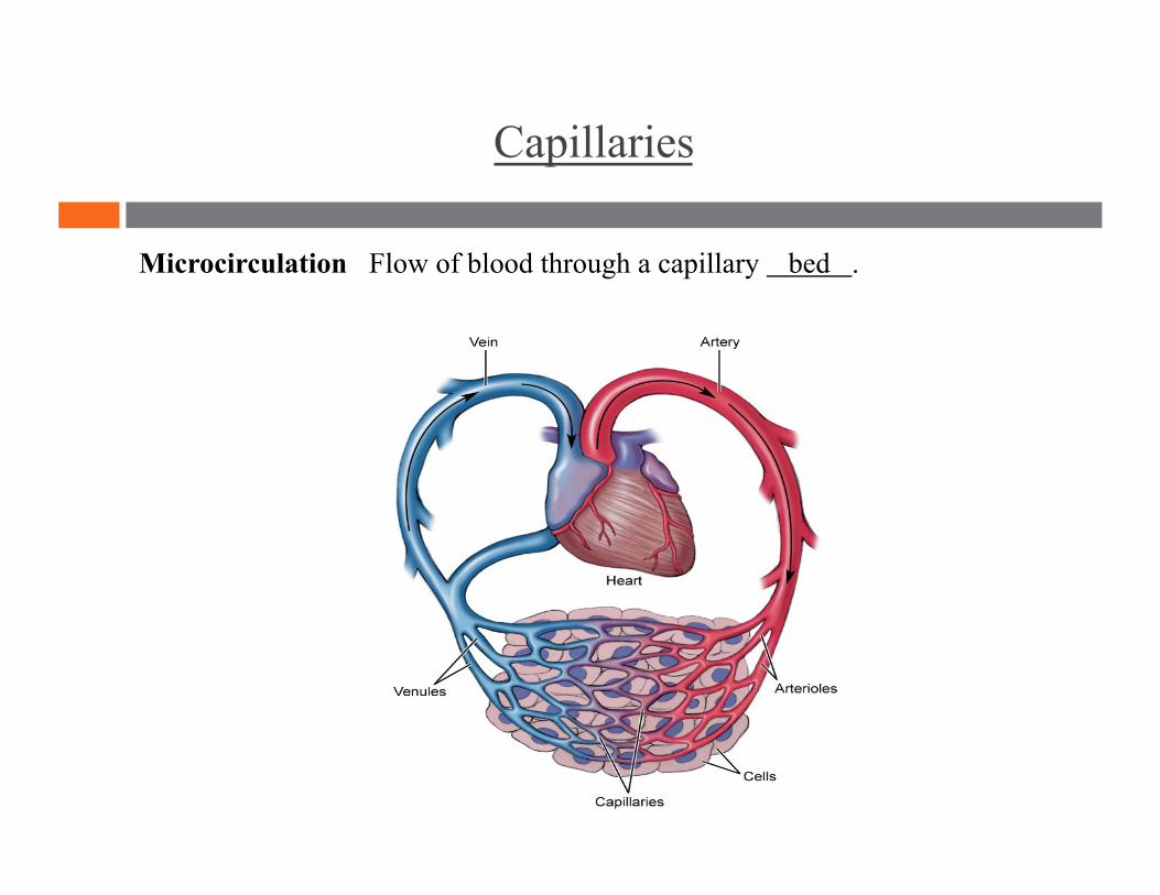
Microcirculation Flow of blood through a capillary bed .
Veins
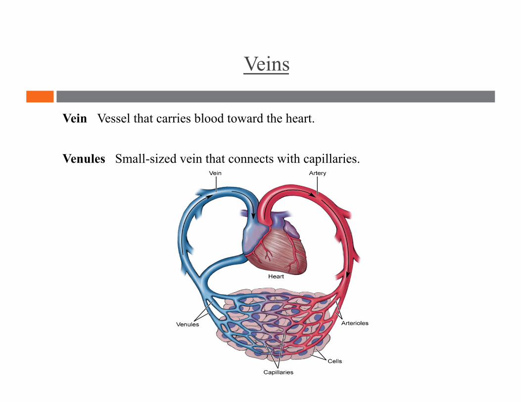
Vein Vessel that carries blood toward the heart.
Venules Small-sized vein that connects with capillaries.

Veins
Superior vena cava Very large vein that empties blood from the head and arms into the right atrium.
Veins
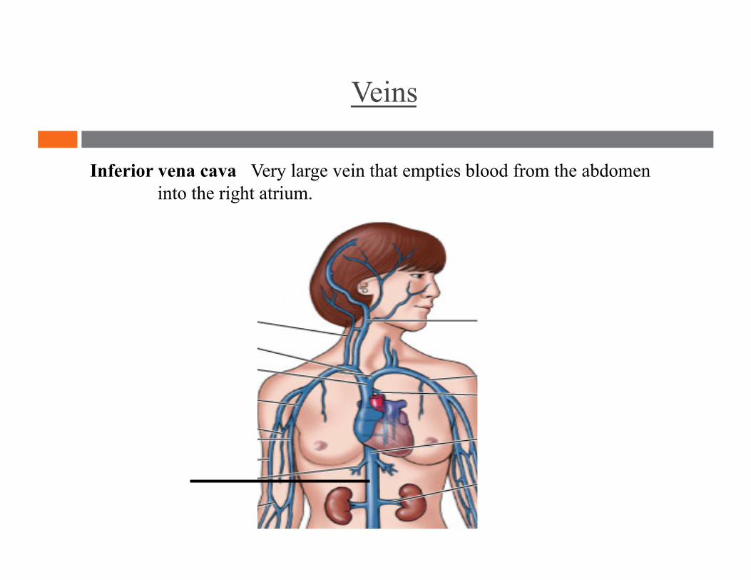
Inferior vena cava Very large vein that empties blood from the abdomen into the right atrium.
Veins
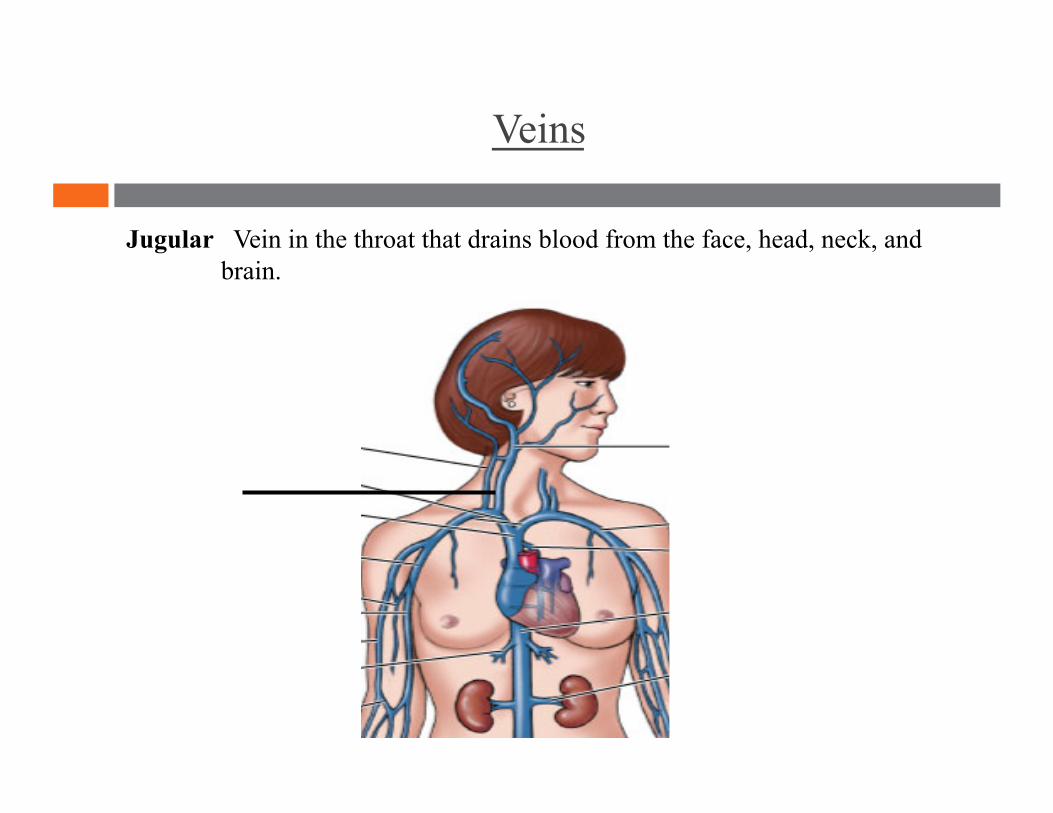
Jugular Vein in the throat that drains blood from the face, head, neck, and brain.
Veins
Avascular Lacking blood vessels.
Venous Return
Venous Return
Venous return Veins return blood to the heart passively.
Venomotor tone
Skeletal muscle pump
Respiratory pump
Venous Return
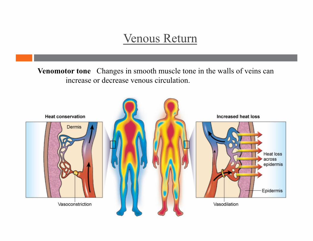
Venomotor tone Changes in smooth muscle tone in the walls of veins can increase or decrease venous circulation.
Venous Return
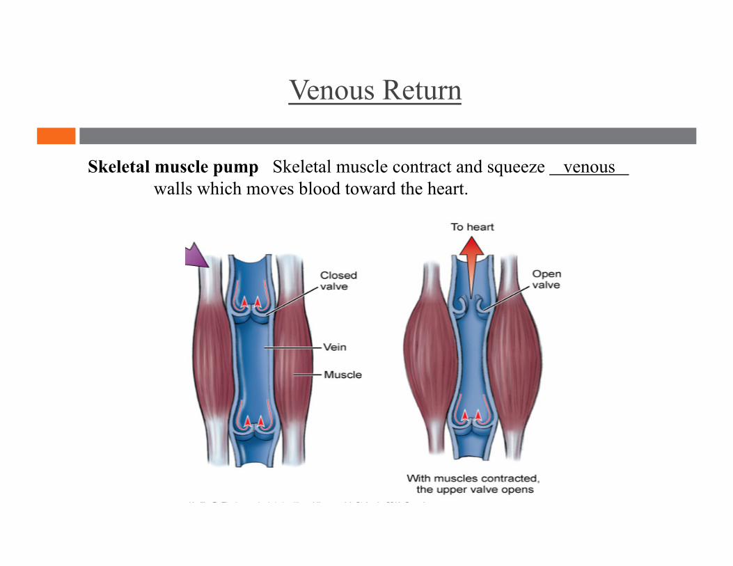
Skeletal muscle pump Skeletal muscle contract and squeeze venous walls which moves blood toward the heart.
Venous Return
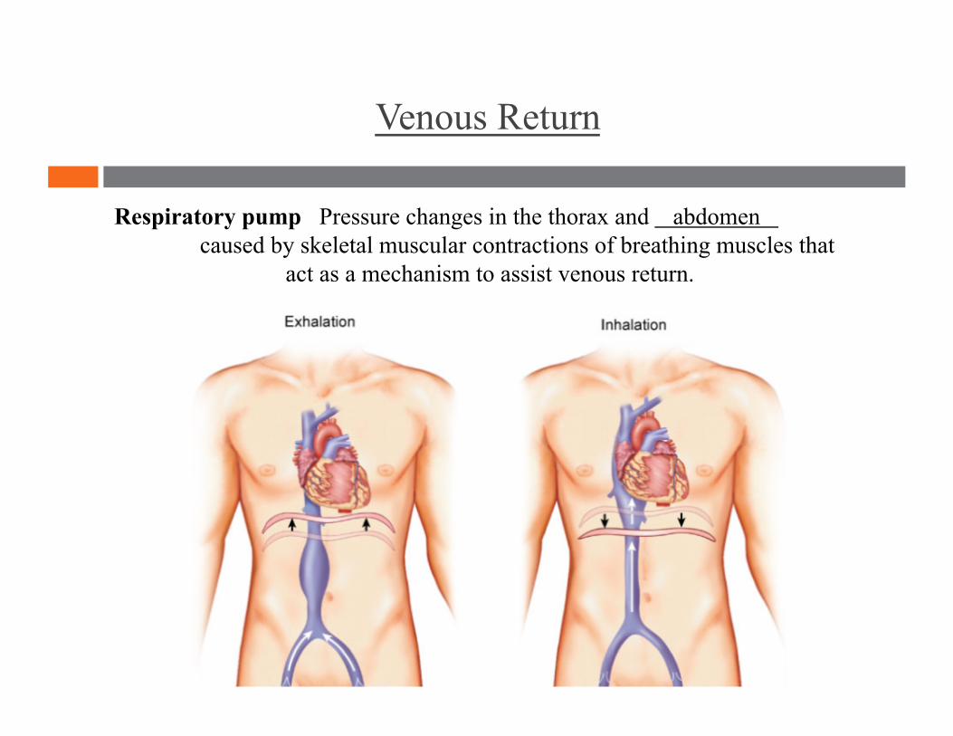
Respiratory pump Pressure changes in the thorax and abdomen caused by skeletal muscular contractions of breathing muscles that act as a mechanism to assist venous return.
Blood Pressure
Systolic pressure Diastolic pressure
High blood pressure Average blood pressure Low blood pressure
Blood Pressure
Blood pressure Pressure exerted by blood on the blood vessel walls.
Systolic pressure Maximal pressure in blood pressure measurement. Occurs when the left ventricle contracts.
Diastolic pressure Lowest pressure in blood pressure measurement. Occurs when the left ventricle relaxes.
Blood Pressure
High blood pressure Persistently more than 140/90 mm Hg. AKA: hypertension.
Average blood pressure 120/80 mm Hg.
Low blood pressure Persistently less than 90/60 mm Hg. AKA: hypotension
Paths of Circulation
Pulmonary circuit Systemic circuit Microcirculation
Paths of Circulation
Pulmonary circuit Circuit that brings de-oxygenated blood from the right
ventricle of the heart to the lungs to release carbon dioxide and regain
oxygen, then transports the oxygenated blood to the left atrium.
Paths of Circulation
Systemic circuit Circuit that brings oxygenated blood from the left
ventricle of the heart through numerous arteries into the capillaries, then
moves it through the veins and returns the now de-oxygenated blood to
the right atrium of the heart.
Paths of Circulation
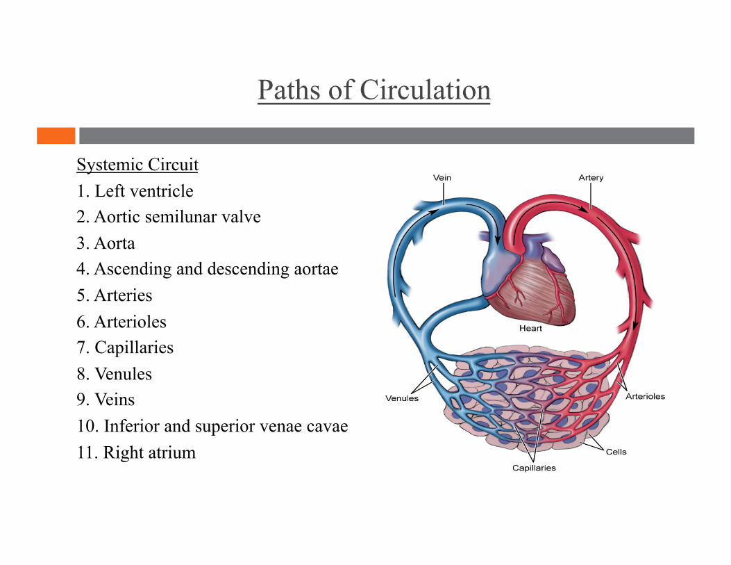
Systemic Circuit 1. Left ventricle 2. Aortic semilunar valve 3. Aorta 4. Ascending and descending aortae 5. Arteries 6. Arterioles 7. Capillaries 8. Venules 9. Veins 10. Inferior and superior venae cavae 11. Right atrium
Paths of Circulation
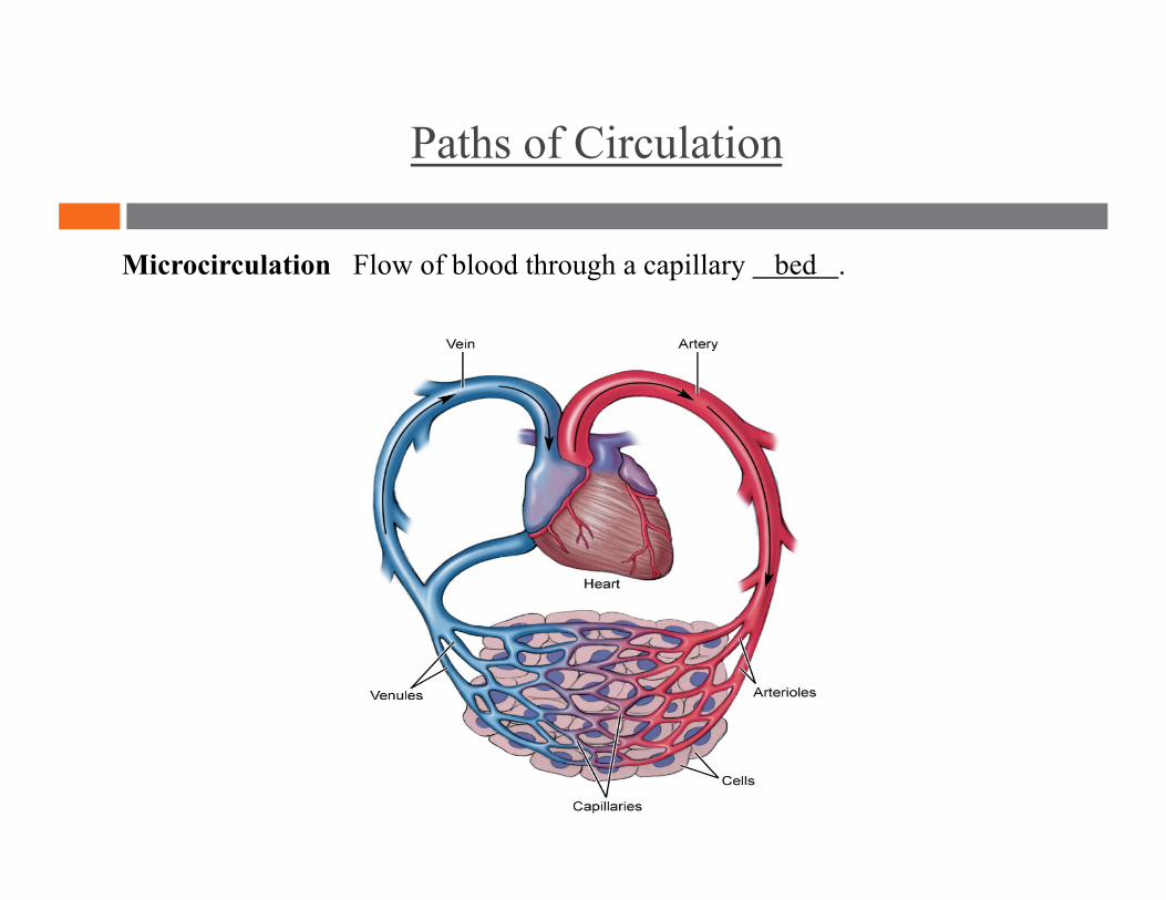
Microcirculation Flow of blood through a capillary bed .
“Be a lamp, a lifeboat, or a ladder.”
Cardiovascular System 2
-Rumi

“When an ordinary man attains knowledge, he becomes a sage. When a sage attains knowledge, he becomes an ordinary man.”
Lymphatic System and Immunity
-Zen saying
Lesson Plan: Lymphatic System and Immunity
5 minutes: Breath of Arrival and Attendance
5 minutes: Rhomboids and Levator Scapula
45 minutes: Lymphatic System and Immunity
Classroom Rules
Punctuality- everybody's time is precious:
Be ready to learn by 9:00, we'll have you out of here by 1:30
Tardiness: arriving late, late return after breaks, leaving early
The following are not allowed:
Bare feet
Side talking
Lying down
Inappropriate clothing
Food or drink except water
Phones in classrooms, clinic or bathrooms
You will receive one verbal warning, then you'll have to leave the room.

Rhomboids in context
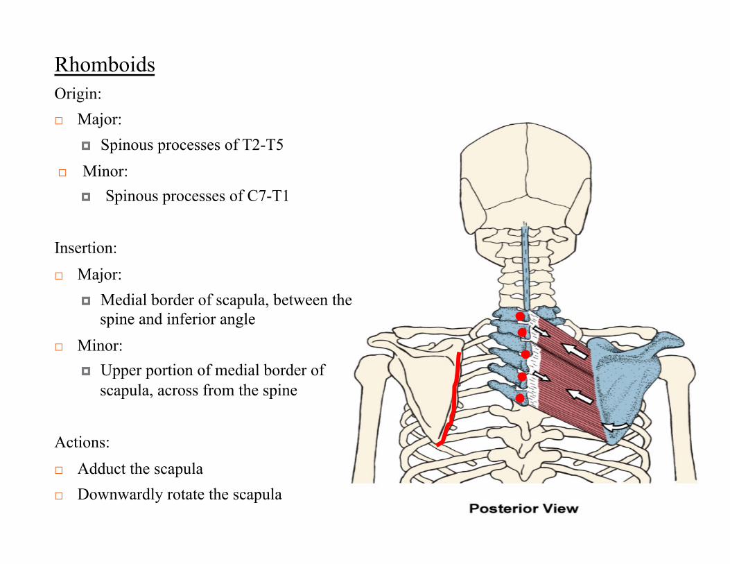
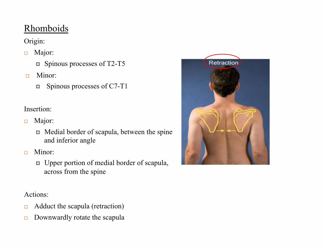
Rhomboids
Origin: Major:
Spinous processes of T2-T5 Minor:
Spinous processes of C7-T1
Insertion: Major:
Medial border of scapula, between the spine and inferior angle
Minor: Upper portion of medial border of
scapula, across from the spine
Actions: Adduct the scapula Downwardly rotate the scapula
Rhomboids
Origin: Major:
Spinous processes of T2-T5 Minor:
Spinous processes of C7-T1
Insertion: Major:
Medial border of scapula, between the spine and inferior angle
Minor: Upper portion of medial border of scapula,
across from the spine
Actions: Adduct the scapula (retraction) Downwardly rotate the scapula
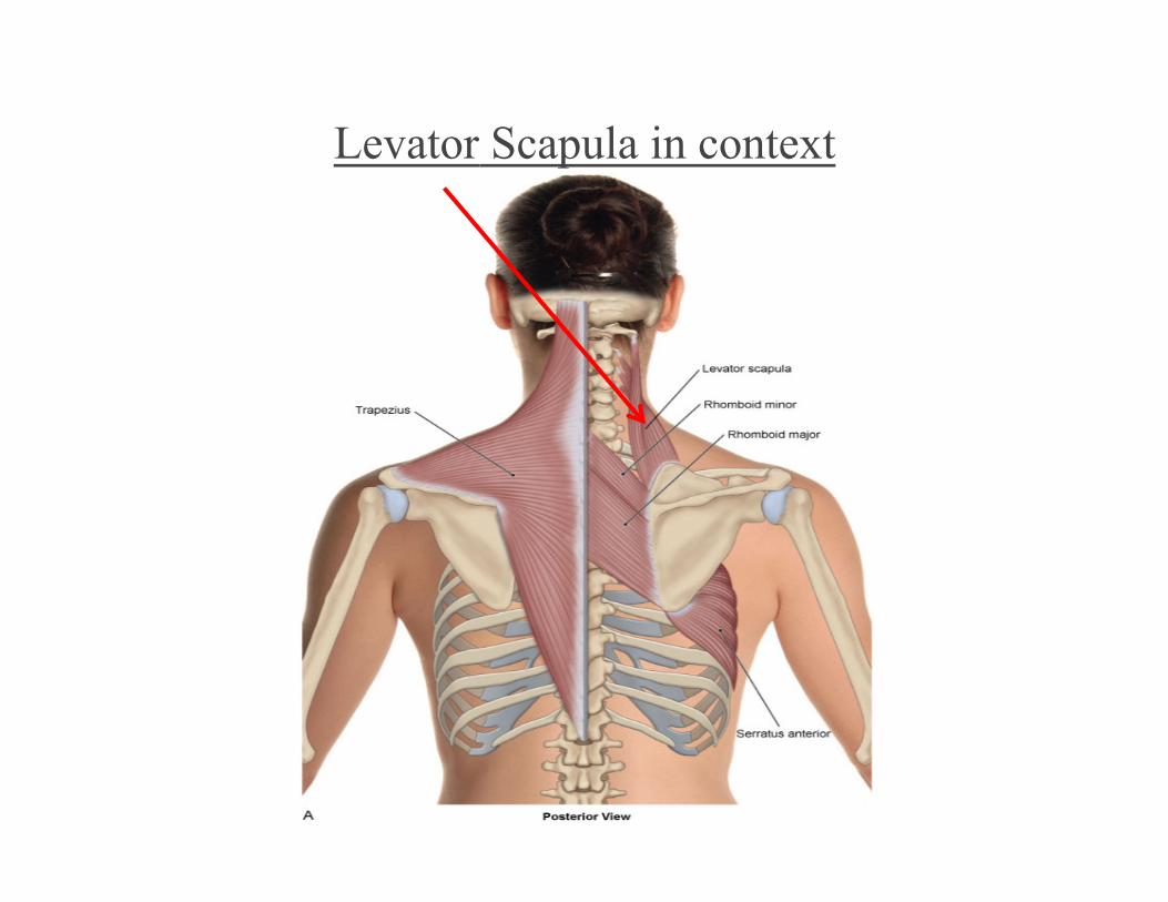
Levator Scapula in context
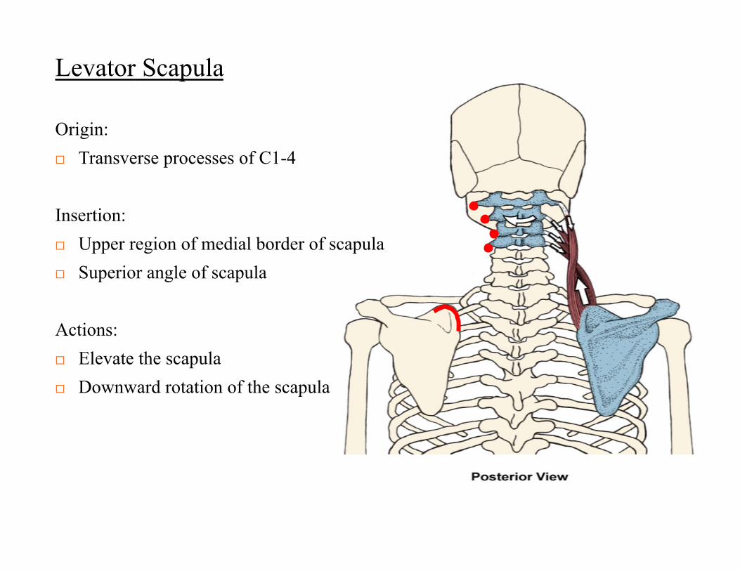
Levator Scapula
Origin: Transverse processes of C1-4
Insertion: Upper region of medial border of scapula Superior angle of scapula
Actions: Elevate the scapula Downward rotation of the scapula
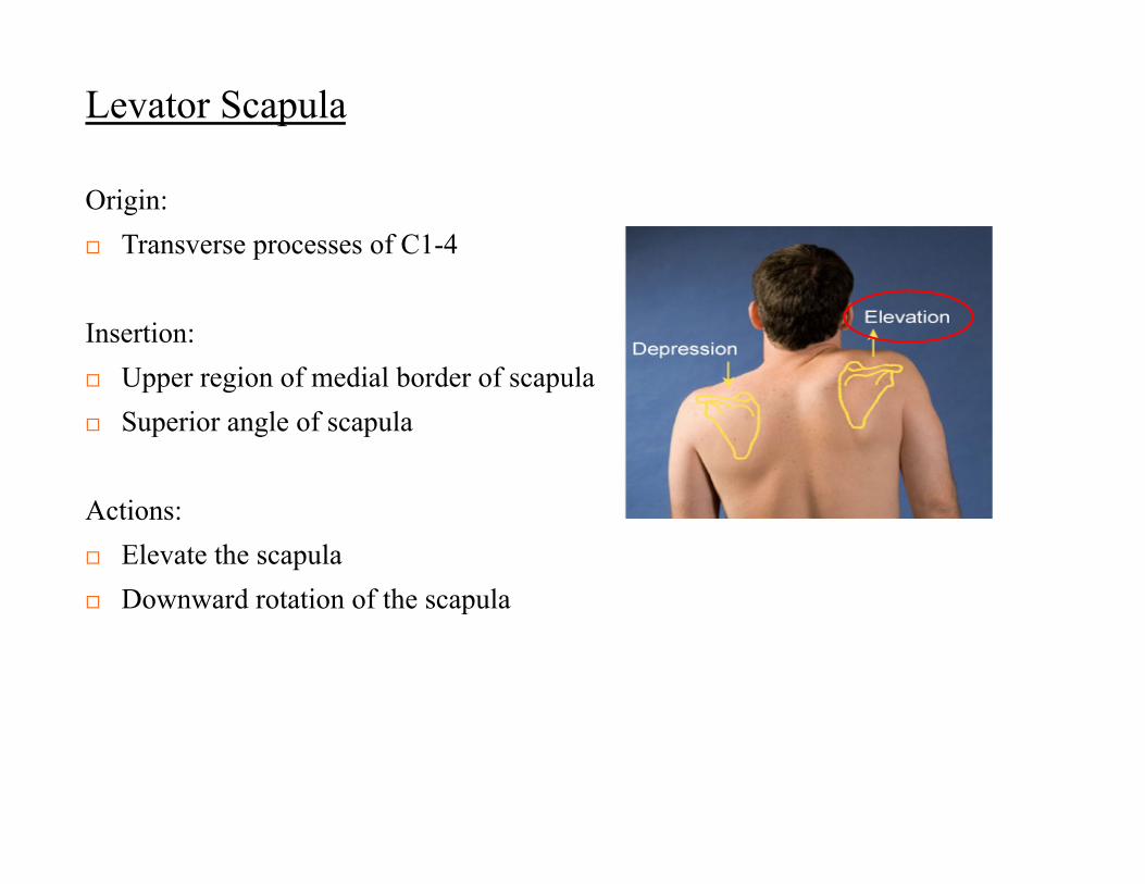
Levator Scapula
Origin: Transverse processes of C1-4
Insertion: Upper region of medial border of scapula Superior angle of scapula
Actions: Elevate the scapula Downward rotation of the scapula

Paths of Circulation
Pulmonary circuit Systemic circuit Microcirculation
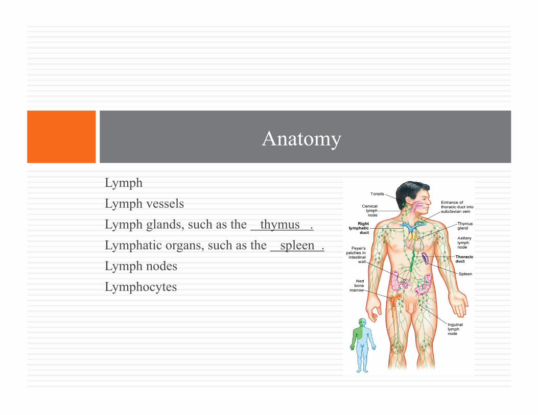
Lymph Lymph vessels Lymph glands, such as the thymus . Lymphatic organs, such as the spleen . Lymph nodes Lymphocytes
Anatomy
Transportation Immune response Maintain homeostasis
Physiology
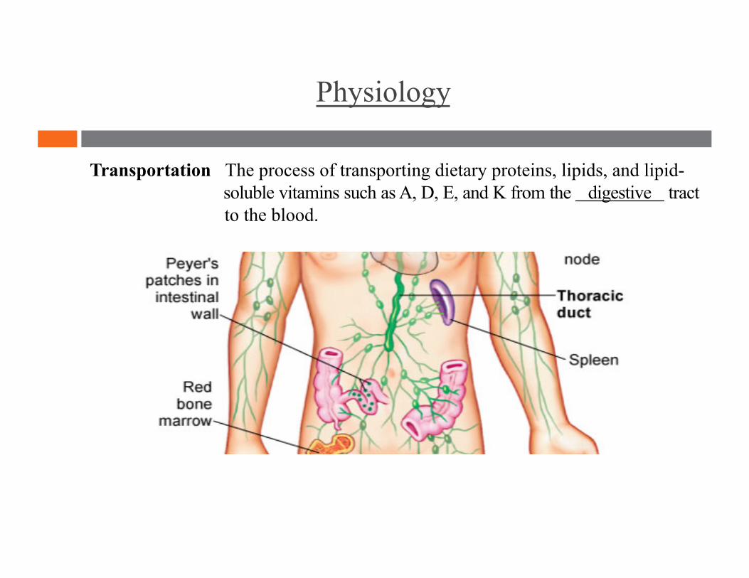
Physiology
Transportation The process of transporting dietary proteins, lipids, and lipid- soluble vitamins such as A, D, E, and K from the digestive tract to the blood.
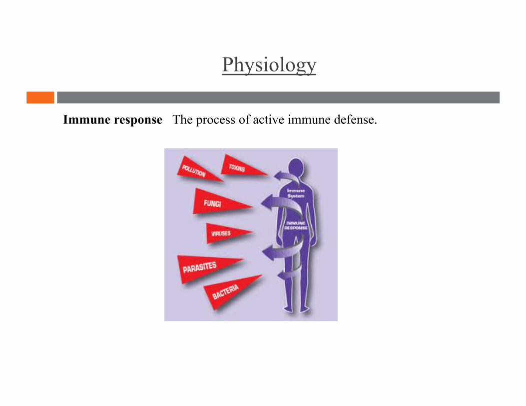
Physiology
Immune response The process of active immune defense.
Physiology
Maintains homeostasis The process of collecting accumulated tissue fluid and returning it to blood circulation. This maintains blood volume, blood pressure, and prevents edema (swelling).

Lymph
Lymph
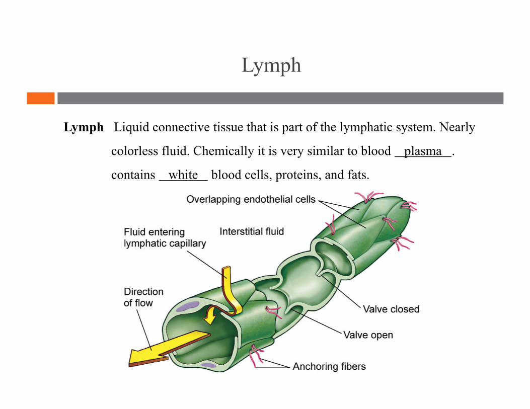
Lymph Liquid connective tissue that is part of the lymphatic system. Nearly
colorless fluid. Chemically it is very similar to blood plasma .
contains white blood cells, proteins, and fats.
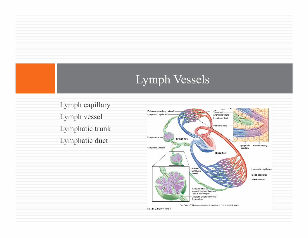
Lymph capillary Lymph vessel Lymphatic trunk Lymphatic duct
Lymph Vessels
Lymph Vessels
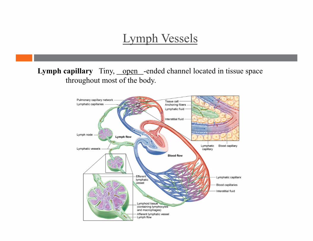
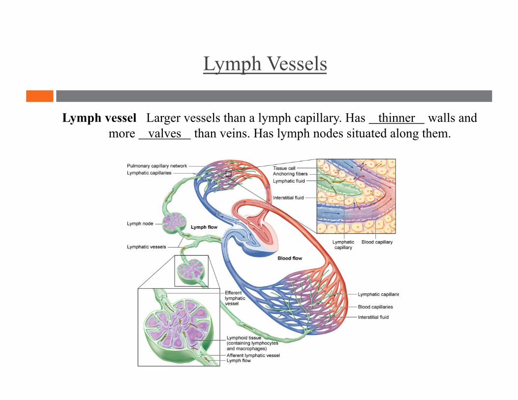
Lymph capillary Tiny, open -ended channel located in tissue space throughout most of the body.
Lymph Vessels
Lymph vessel Larger vessels than a lymph capillary. Has thinner walls and more valves than veins. Has lymph nodes situated along them.

Lymph Vessels
Lymphatic trunk Made up of large vessels into which lymph is drained from the lymph vessels.
Lymph Vessels
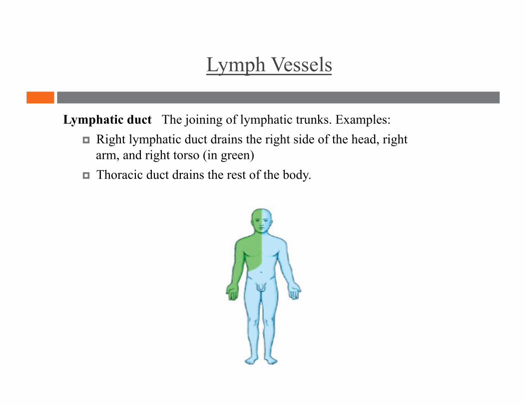
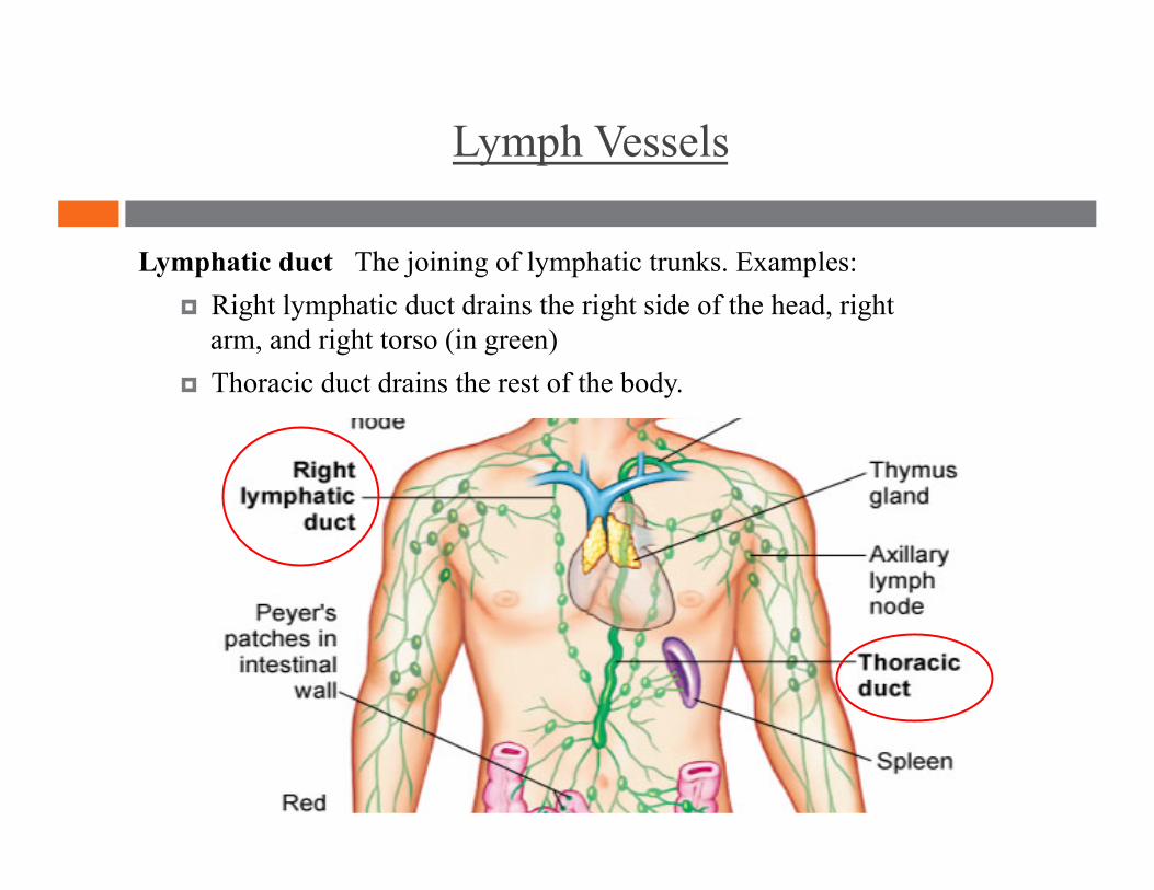
Lymphatic duct The joining of lymphatic trunks. Examples: Right lymphatic duct drains the right side of the head, right
arm, and right torso (in green) Thoracic duct drains the rest of the body.
Lymph Vessels
Lymphatic duct The joining of lymphatic trunks. Examples: Right lymphatic duct drains the right side of the head, right
arm, and right torso (in green) Thoracic duct drains the rest of the body.
Red bone marrow Lymphocyte Thymus Spleen Lymph node Mucosa-associated lymphoid tissue
Lymphatic Structures
Lymphatic Structures
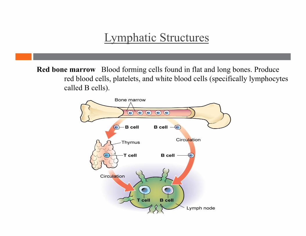
Red bone marrow Blood forming cells found in flat and long bones. Produce red blood cells, platelets, and white blood cells (specifically lymphocytes called B cells).
Lymphatic Structures
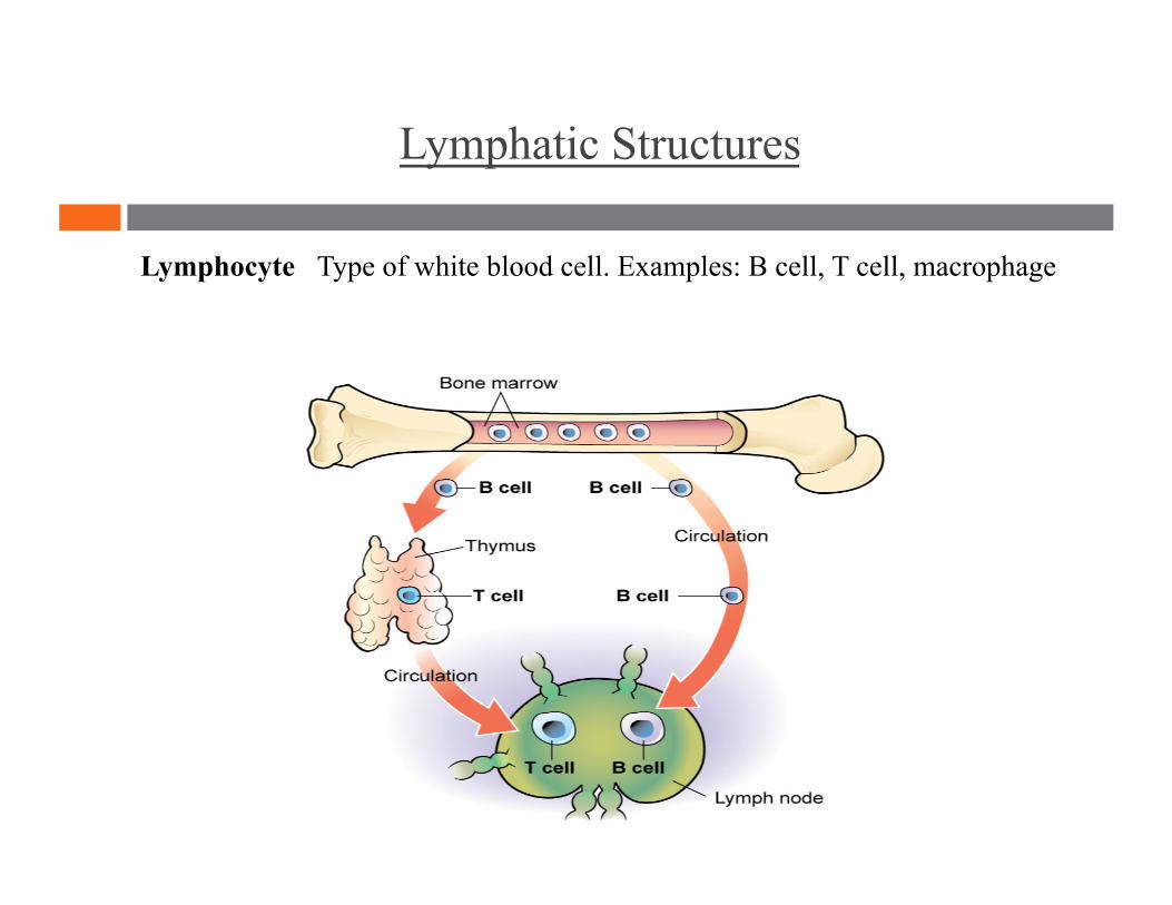
Lymphocyte Type of white blood cell. Examples: B cell, T cell, macrophage
Lymphatic Structures
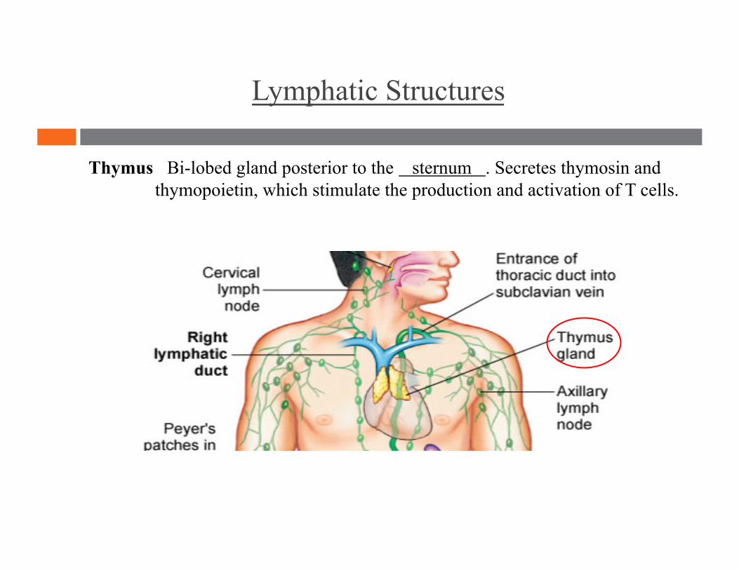
Thymus Bi-lobed gland posterior to the sternum . Secretes thymosin and thymopoietin, which stimulate the production and activation of T cells.
Lymphatic Structures
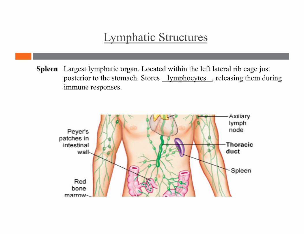
Spleen Largest lymphatic organ. Located within the left lateral rib cage just posterior to the stomach. Stores lymphocytes , releasing them during immune responses.
Lymphatic Structures
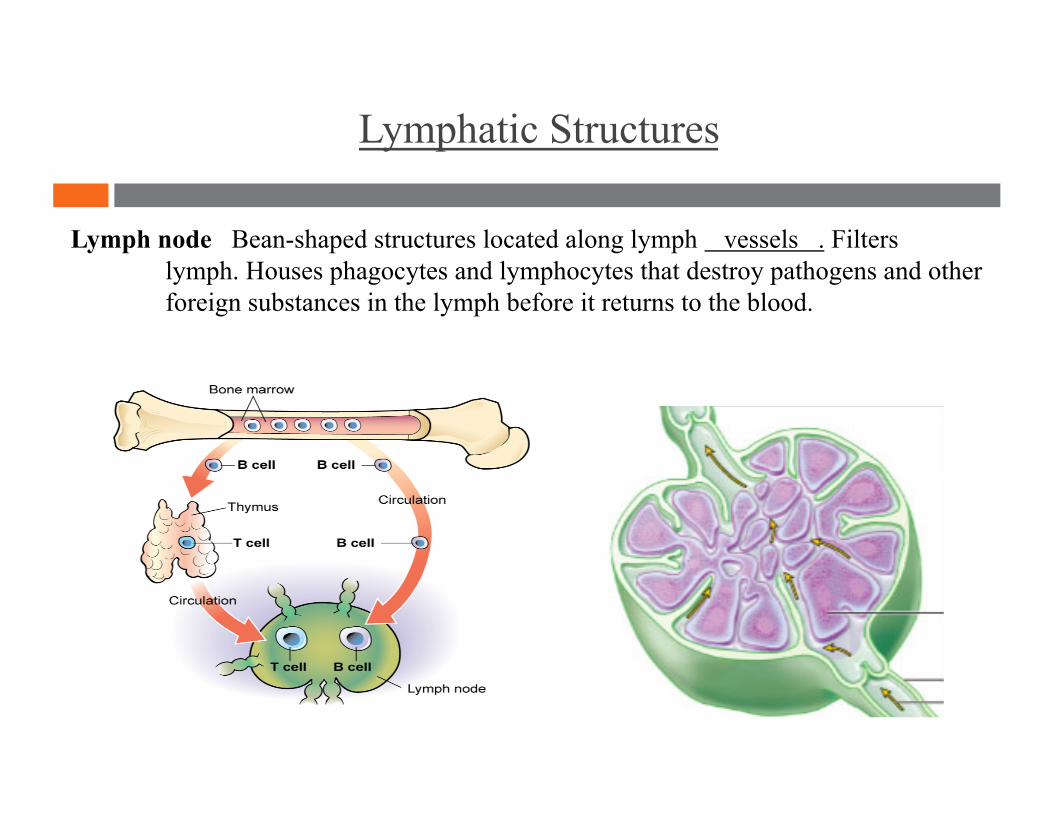
Lymph node Bean-shaped structures located along lymph vessels . Filters lymph. Houses phagocytes and lymphocytes that destroy pathogens and other foreign substances in the lymph before it returns to the blood.
Lymphatic Structures
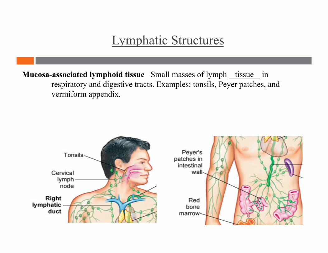
Mucosa-associated lymphoid tissue Small masses of lymph tissue in respiratory and digestive tracts. Examples: tonsils, Peyer patches, and vermiform appendix.
Lymphatic drainage Lymphatic pump
Lymph Flow

Lymph Flow
Lymphatic drainage The movement of lymph.
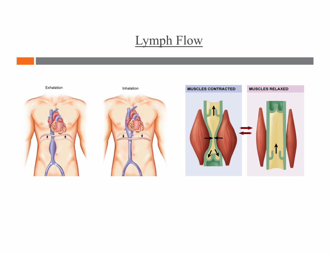
Lymph Flow
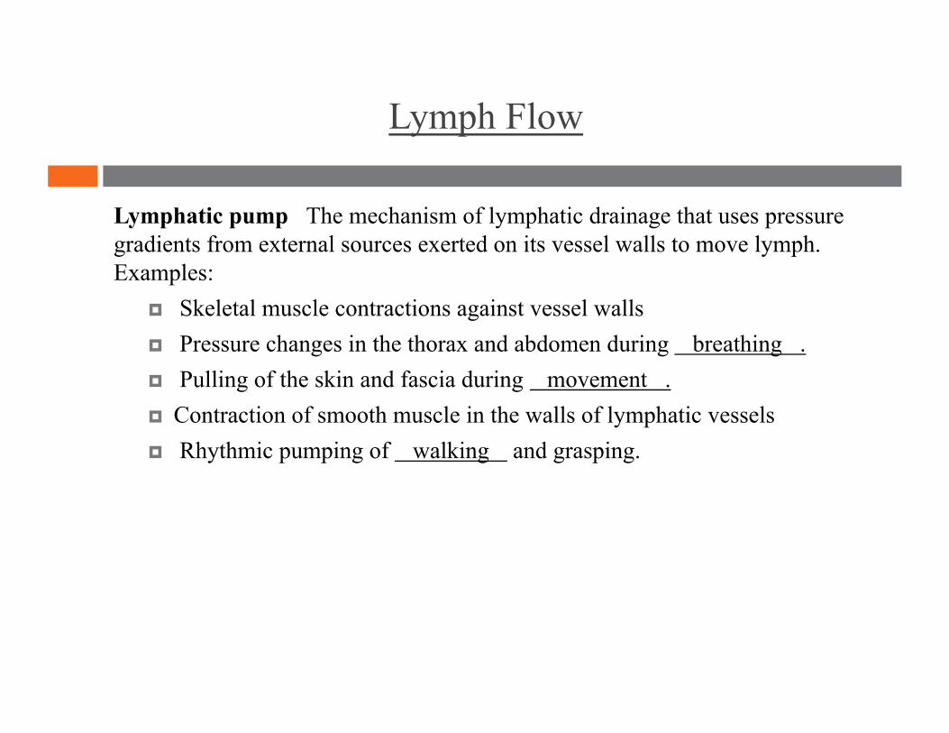
Lymphatic pump The mechanism of lymphatic drainage that uses pressure gradients from external sources exerted on its vessel walls to move lymph. Examples:
Skeletal muscle contractions against vessel walls Pressure changes in the thorax and abdomen during breathing . Pulling of the skin and fascia during movement . Contraction of smooth muscle in the walls of lymphatic vessels Rhythmic pumping of walking and grasping.
Lymph Flow
Non-specific immunity Infection Inflammation
Specific immunity T cells B cells
Immunity
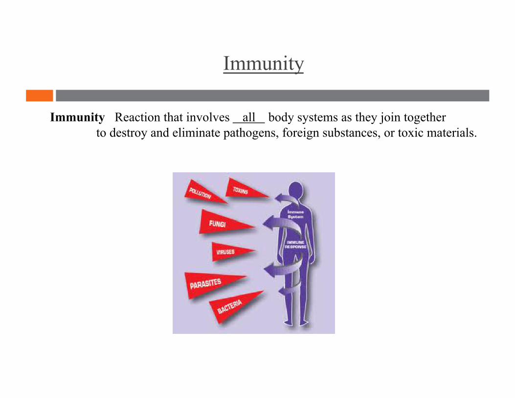
Immunity
Immunity Reaction that involves all body systems as they join together to destroy and eliminate pathogens, foreign substances, or toxic materials.
Immunity
Non-specific immunity Non-specific response to invading pathogens. Includes intact skin and mucous membranes, saliva, gastric juices, vomiting, urine flow, certain white blood cells, fever, and inflammation. AKA: innate immunity.
Immunity
Infection The period after disease transmission. Pathogens use host resources to multiply.
Immunity
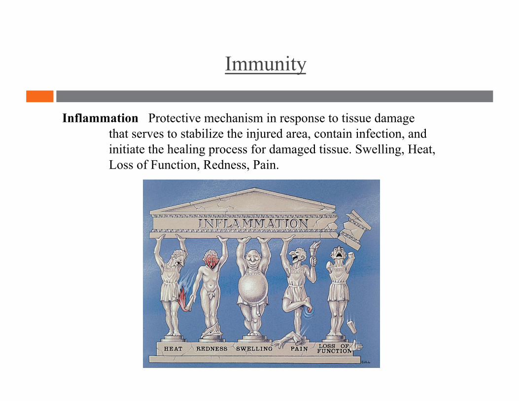
Inflammation Protective mechanism in response to tissue damage that serves to stabilize the injured area, contain infection, and initiate the healing process for damaged tissue. Swelling, Heat, Loss of Function, Redness, Pain.
Immunity
Specific immunity Body's response to invaders. T cells and B cells become activated for a specific pathogen after they come into contact with it and then destroy it. AKA: adaptive immunity.
Immunity
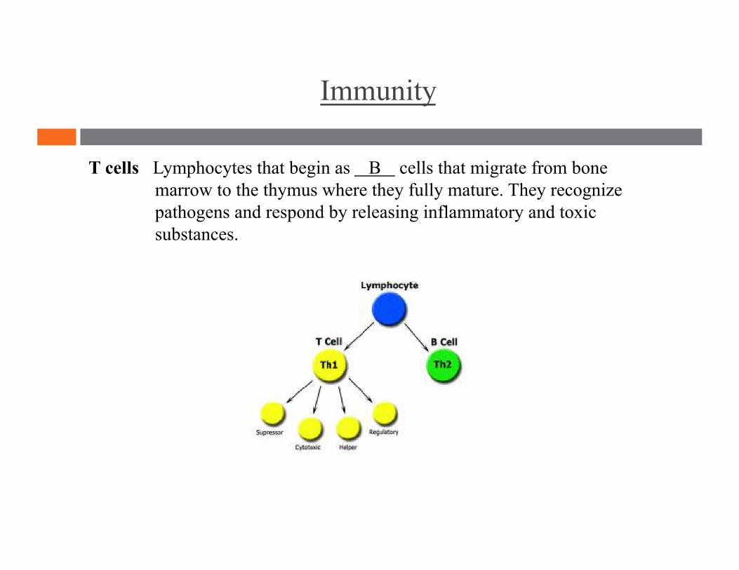
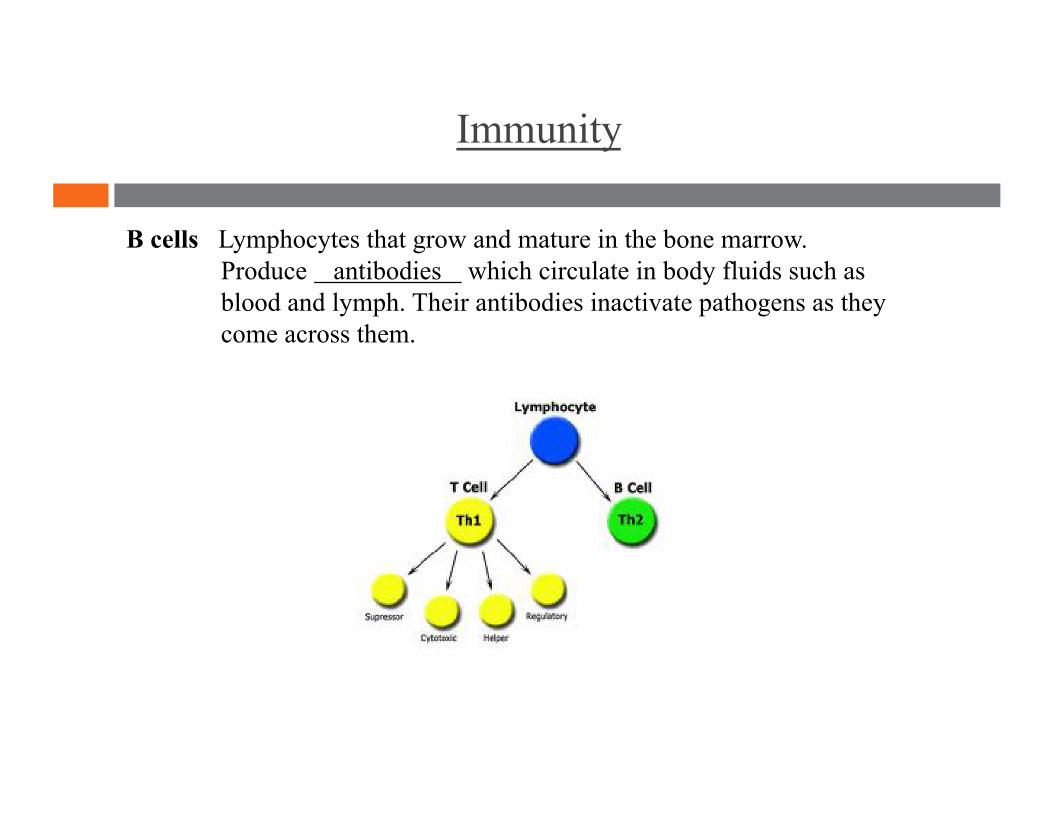
T cells Lymphocytes that begin as B cells that migrate from bone marrow to the thymus where they fully mature. They recognize pathogens and respond by releasing inflammatory and toxic substances.
Immunity
B cells Lymphocytes that grow and mature in the bone marrow. Produce antibodies which circulate in body fluids such as blood and lymph. Their antibodies inactivate pathogens as they come across them.
“When an ordinary man attains knowledge, he becomes a sage. When a sage attains knowledge, he becomes an ordinary man.”
Lymphatic System and Immunity
-Zen saying