Embed Size (px)
Citation preview

Correction
MEDICAL SCIENCESCorrection for “BCL6 promotes glioma and serves as a therapeutictarget,” by Liang Xu, Ye Chen, Marina Dutra-Clarke, AnandMayakonda, Masaharu Hazawa, Steve E. Savinoff, Ngan Doan,Jonathan W. Said, William H. Yong, Ashley Watkins, HenryYang, Ling-Wen Ding, Yan-Yi Jiang, Jeffrey W. Tyner, JianhongChing, Jean-Paul Kovalik, Vikas Madan, Shing-Leng Chan, MarkusMüschen, Joshua J. Breunig, De-Chen Lin, and H. Phillip Koeffler,which appeared in issue 15, April 11, 2017, of Proc Natl Acad SciUSA (114:3981–3986; first published March 29, 2017; 10.1073/pnas.1609758114).The authors note that the following statement should be added
to the Acknowledgments: “This work is also supported byphilanthropic donations from Valerie Baker Fairbank, who alsodeeply inspires our work.”
www.pnas.org/cgi/doi/10.1073/pnas.1706421114
www.pnas.org PNAS | May 23, 2017 | vol. 114 | no. 21 | E4317
CORR
ECTION

BCL6 promotes glioma and serves as atherapeutic targetLiang Xua,1, Ye Chena,1, Marina Dutra-Clarkeb,1, Anand Mayakondaa, Masaharu Hazawaa, Steve E. Savinoffb,Ngan Doanc, Jonathan W. Saidc, William H. Yongc, Ashley Watkinsb, Henry Yanga, Ling-Wen Dinga, Yan-Yi Jianga,Jeffrey W. Tynerd, Jianhong Chinge, Jean-Paul Kovalike, Vikas Madana, Shing-Leng Chana, Markus Müschenf,Joshua J. Breunigb,g,h,2, De-Chen Lina,i,2, and H. Phillip Koefflera,i,j
aCancer Science Institute of Singapore, National University of Singapore, 117599, Singapore; bBoard of Governors Regenerative Medicine Institute andDepartment of Biomedical Sciences, Cedars-Sinai Medical Center, Los Angeles, CA 90048; cDepartment of Pathology and Laboratory Medicine, University ofCalifornia, Los Angeles, and David Geffen School of Medicine, Los Angeles, CA 90095; dDepartment of Cell & Developmental Biology, Oregon Health &Science University, Portland, OR 97239; eCardiovascular and Metabolic Disorders Program, Duke-NUS Graduate Medical School, 169857, Singapore;fDepartment of Laboratory Medicine, University of California, San Francisco, CA 94143; gDepartment of Medicine, University of California, Los AngelesSchool of Medicine, Los Angeles, CA 90095; hSamuel Oschin Comprehensive Cancer Institute, Cedars-Sinai Medical Center, Los Angeles, CA 90048; iDivisionof Hematology/Oncology, Cedars-Sinai Medical Center, University of California, Los Angeles School of Medicine, Los Angeles, CA 90048; and jNationalUniversity Cancer Institute, National University Hospital, Singapore, 119074, Singapore
Edited by Webster K. Cavenee, Ludwig Institute for Cancer Research, University of California, San Diego, La Jolla, CA, and approved March 8, 2017 (receivedfor review June 17, 2016)
ZBTB transcription factors orchestrate gene transcription duringtissue development. However, their roles in glioblastoma (GBM)remain unexplored. Here, through a functional screening of ZBTBgenes, we identify that BCL6 is required for GBM cell viability andthat BCL6 overexpression is associated with worse prognosis. In asomatic transgenic mouse model, depletion of Bcl6 inhibits theprogression of KrasG12V-driven high-grade glioma. Transcriptomeanalysis demonstrates the involvement of BCL6 in tumor proteinp53 (TP53), erythroblastic leukemia viral oncogene homolog(ErbB), and MAPK signaling pathways. Indeed, BCL6 repressesthe expression of wild-type p53 and its target genes in GBM cells.Knockdown of BCL6 augments the activation of TP53 pathway inresponse to radiation. Importantly, we discover that receptor ty-rosine kinase AXL is a transcriptional target of BCL6 in GBM andmediates partially the regulatory effects of BCL6 on both MEK-ERK(mitogen-activated protein/extracellular signal-regulated kinasekinase–extracellular signal-regulated kinase) and S6K-RPS6 (ribosomalprotein S6 kinase–ribosomal protein S6) axes. Similar to BCL6 silenc-ing, depletion of AXL profoundly attenuates GBM proliferation bothin vitro and in vivo. Moreover, targeted inhibition of BCL6/nuclearreceptor corepressor 1 (NCoR) complex by peptidomimetic inhibitornot only significantly decreases AXL expression and the activity ofMEK-ERK and S6K-RPS6 cascades but also displays a potent antipro-liferative effect against GBM cells. Together, these findings uncovera glioma-promoting role of BCL6 and provide the rationale of tar-geting BCL6 as a potential therapeutic approach.
ZBTB | BCL6 | AXL | NCoR | glioblastoma multiforme
Human ZBTB family genes consist of 49 members who encodetranscription factors that modulate the transcription of genes
involved in cell fate decision and lineage commitment (1). BCL6,also known as ZBTB27, is a master regulator of germinal centerresponse and a proto-oncogene in diffuse large B-cell lymphoma(DLBCL) (2–4). Recent studies also unveiled the critical functionsof BCL6 in pre–B-cell development, acute lymphoblastic leukemia,and chronic myeloid leukemia (5–7). In addition to hematopoieticcell lineages, BCL6 is involved in mammary epithelial differentia-tion and neurogenesis (8, 9). To execute its transcriptional activity,BCL6 requires homodimerization and forming a complex withcofactors including BCL6 corepressor (BCoR), nuclear receptorcorepressor 1 (NCoR), and silencing mediator of retinoic acid andthyroid hormone receptor (SMRT) (10–12). BCL6 inhibitors arecapable of blocking the interaction between BCL6 and BCoR/NCoR/SMRT and selectively killing BCL6-addicted cancer cells(13, 14), thus holding a promise for BCL6-targeted therapy. No-tably, aberrant genomic or expressional changes of BCL6 have
been reported in solid tumors, including breast cancer, ovariancancer, and glioblastoma (GBM) (15–17). Intriguingly, althoughBCL6 confers growth advantages to breast cancer and ovariancancer, it was found to suppress the development of medulloblas-toma (18). Therefore, more efforts are required to decipher thelineage/tissue-specific networks orchestrated by BCL6.GBM remains the most aggressive brain malignancy, with little
improvement in prognosis or therapy for decades. Genomic analysisof GBM highlights the dysregulations of receptor tyrosine kinase(RTK), retinoblastoma protein (RB), and tumor protein p53(TP53) pathways (19, 20). AXL is an oncogenic RTK that co-overexpresses frequently with its ligand GAS6 in glioma (21), yetthe mechanism promoting its overexpression is unclear. As an im-portant effector of many RTKs, the RAS pathway is hyperactivatedin GBM. In addition, driver mutations of KRAS and NRAS havebeen identified in human diffuse gliomas (22). Of note, BCL6mitigates Ras-induced senescence in mouse embryonic fibroblastsand human B cells (23). However, the functional relevance of BCL6in GBM biology remains unknown. Here, with comprehensive
Significance
Glioblastoma (GBM) is the most lethal brain malignancy lackingeffective treatment. In this study, we demonstrate that BCL6 is aprognostic marker and a targetable GBM-promoting factor. Si-lencing of BCL6 inhibits the malignant phenotype of GBM cellsand triggers cellular senescence. We also identify AXL as animportant BCL6 transcriptional target, the expression of which isalso regulated positively by NCoR, a BCL6 cofactor. Either si-lencing of BCL6 or targeted disruption of the BCL6/NCoR com-plex diminishes AXL expression and inhibits GBM growth. Thisstudy elucidates a crucial BCL6-mediated signaling pathway inGBM biology. More importantly, our results highlight thepromise and merit of targeting BCL6 for treating this deadlydisease.
Author contributions: J.J.B., D.-C.L., and H.P.K. designed research; L.X., Y.C., M.D.-C., S.E.S.,N.D., A.W., Y.-Y.J., J.C., V.M., and S.-L.C. performed research; W.H.Y., L.-W.D., and J.W.T.contributed new reagents/analytic tools; L.X., Y.C., M.D.-C., A.M., M.H., J.W.S., H.Y., J.-P.K.,M.M., J.J.B., D.-C.L., and H.P.K. analyzed data; and L.X., D.-C.L., and H.P.K. wrote the paper.
The authors declare no conflict of interest.
Data deposition: The data reported in this paper have been deposited in the Gene Ex-pression Omnibus (GEO) database, www.ncbi.nlm.nih.gov/geo (accession no. GSE77053).1L.X., Y.C., and M.D.-C. contributed equally to this work.2To whom correspondence may be addressed. Email: [email protected] or [email protected].
This article contains supporting information online at www.pnas.org/lookup/suppl/doi:10.1073/pnas.1609758114/-/DCSupplemental.
www.pnas.org/cgi/doi/10.1073/pnas.1609758114 PNAS | April 11, 2017 | vol. 114 | no. 15 | 3981–3986
MED
ICALSC
IENCE
S

molecular, cellular, and animal studies, we revealed the biologicalrole of BCL6 in gliomagenesis and resolved a complex and criticalBCL6-mediated signaling network in GBM cells.
ResultsIdentification of BCL6 as a Progrowth Factor in GBM. To explore therole of ZBTB family genes in GBM cell growth, a customizedshRNA library was constructed to silence the expression of 49ZBTB genes (SI Appendix, Table S1). This screen identified a listof progrowth genes including ZBTB5, PATZ1, ZBTB20, BCL6,BCL6B, and ZBTB42 (Fig. 1A). Among those, BCL6 andZBTB20, both indispensable for cell viability, stood out as twotop hits, as they were also upregulated in GBM (Fig. 1 B and C
and SI Appendix, Fig. S1 A and B and Table S2) (19, 24–27).However, only BCL6 protein, not ZBTB20, was low or un-detectable in normal brain tissues (SI Appendix, Fig. S1 C and D)(28). The expression of BCL6 protein was verified in bothestablished GBM cell lines and primary explants (Fig. 1D) (29).Of note, GBM cells expressed a similar level of BCL6 protein asBCL6-positive breast cancer cells, albeit lower than BCL6-dependent DLBCL cells (SI Appendix, Fig. S1E). Next, wesought to examine the potential involvement of BCL6 in gliomaprogression. Through immunohistochemistry (IHC) analysis of153 primary glioma specimens and 8 normal brain samples,BCL6 expression was robustly elevated in tumor samples andpositively correlated with glioma pathological grade (Fig. 1E).Moreover, high BCL6 expression strongly predicted a worseprognosis (Fig. 1F). Hence, these results uncover BCL6 as aglioma-promoting gene and a biomarker whose up-regulationcorrelates with clinical grades.
BCL6 Expression Is a Functional Requisite for Glioma Cell Growth. Toverify the involvement of BCL6 in human GBM cell growth, wefirst rescreened the effects of BCL6 knockdown in five establishedGBM cell lines and two primary explant lines by shRNAs (Fig. 2 Aand B). Depletion of endogenous BCL6 markedly reduced GBMcell viability, BrdU incorporation, and colony-forming ability (Fig.2 A–C and SI Appendix, Fig. S2 A and B). Meanwhile, BCL6knockdown triggered cellular senescence and G0/G1 arrest, butdid not induce either sub-G1 cell population or caspase 3/7 activity(Fig. 2D and SI Appendix, Fig. S2 and C and D). Moreover, bothsiRNA- and CRISPR/Cas9-mediated BCL6 silencing showedsimilar antiproliferation effects (Fig. 2 E and F and SI Appendix,Fig. S2 E–G) (30). Importantly, depletion of endogenous BCL6 inGBM cells powerfully suppressed xenograft tumor growth in im-munodeficient mice (Fig. 2 G and H). Collectively, our loss-of-function analyses strongly suggest that BCL6 is required forhuman GBM cell growth, both in vitro and in vivo.Next, to investigate the role of Bcl6 during gliomagenesis, we
used a murine model of postnatal electroporation coupled withPiggyBac transposition for stable transgenesis of KrasG12V andshRNA targeting Bcl6 (31–33). Wild-type CD-1 mice were elec-troporated at postnatal day 2 with plasmids harboring eitherEGFP-KrasG12V (control) or EGFP-KrasG12V/miR-E–basedshRNA against Bcl6 #275 (shBcl6.275) (Fig. 3 A and B and SIAppendix, Fig. S3 A–D). A TagBFP2 blue fluorescent protein witha hemagglutinin (HA) epitope tag (TagBFP-HA)-nuclear locali-zation sequence reporter and pBase were coelectroporated to fa-cilitate counting of tumor cells and for transposition, respectively.Neural precursor cells along the rostro-caudal axis of the left lateralventricle where plasmids were delivered were susceptible to elec-troporation and subsequently transformed by KrasG12V (31, 33).Mouse brains were analyzed at 4 and 8 wk postelectroporationto monitor tumor growth. Mice electroporated with KrasG12V/shBcl6 showed a clear reduction in glioma size and constrictedtumor margins compared with the KrasG12V control group (Fig. 3C and D and SI Appendix, Fig. S3 E and F). In accordance, tumorcells from the KrasG12V/shBcl6 group tended to be less pro-liferative, as evidenced by the decreased Ki67 positivity at theventral margin of tumors (Fig. 3 E–G). There was a clear trendtoward increased tumor cell apoptosis in KrasG12V/shBcl6 micerelative to KrasG12V control, although because of intertumorheterogeneity, this result did not achieve statistical significance (SIAppendix, Fig. S3 G–I). Most importantly, KrasG12V/shBcl6 ani-mals survived much longer than KrasG12V control group animals(Fig. 3H; P = 0.0006), strongly suggesting that Bcl6 promotes theprogression of KrasG12V-driven glioma.
BCL6 Represses the TP53 Pathway. To dissect the GBM-associatedroles of BCL6, a cDNA microarray analysis was performed in U87cells. About 800 genes were differentially expressed following BCL6
Fig. 1. Identification of BCL6 as a progrowth factor in glioma. (A) Relativeviable number of U87 GBM cells after infection with lentiviral particlesencoding indicated shRNAs. SHC002 and pLKO-scramble were used as neg-ative controls (Negative Ctrl), and shRNAs targeting MYC and AKT1 wereused as positive controls (Positive Ctrl). Candidate genes (marked by redarrows) were selected only if all independent shRNAs and all repeats showedconsistent increase or decrease (>20%) in relative viable cell number com-pared with negative controls. (B) Plot showing the correlation between av-erage effect of ZBTB depletion on U87 cell growth and differential geneexpression (GBM vs. epilepsy; GSE4290). BCL6 and ZBTB20 were highlightedin red as candidates for growth-promoting genes. MYC and AKT1 weremarked in blue as positive controls for oncogenes. (C) Heat map showing theexpression of BCL6 and ZBTB20 in nontumor brain tissues (epilepsy), GBMcell lines, and primary GBM samples. G2, G3, and G4 denote World HealthOrganization grade II, III, and IV, respectively. NSC, neural stem cells.(D) Western blot result showing the endogenous BCL6 expression among6 established GBM cell lines and 4 primary GBM explants. (E) IHC results andrepresentative staining images showing BCL6 expression in normal brain,LGG (lower-grade glioma), and GBM samples. (Scale bars, 100 μm). IHC scoreswere compared using a nonparametric statistical test. (F) Survival curves forpatients with GBM with differential BCL6 expression (Low, IHC score 0 or 1;Intermediate, IHC score 2; High, IHC score 3). Log-rank test was applied.
3982 | www.pnas.org/cgi/doi/10.1073/pnas.1609758114 Xu et al.

depletion (SI Appendix, Table S3, false discovery rate ≤ 0.05) (34,35). Remarkably, pathway enrichment analysis of these genesidentified TP53, erythroblastic leukemia viral oncogene homolog(ErbB), and MAPK pathways among the top hits (SI Appendix, Fig.S4A), prompting us to investigate the functional relevance ofBCL6 in these pathways.First, we examined the effect of BCL6 on the TP53 pathway. In
BCL6-silenced U87, DBTRG, and U343 cells (harboring wild-typeTP53 and CDKN2A deletion), both transcript and protein levels ofTP53 (p53), CDKN1A (p21), and CDKN1B (p27) were markedlyelevated (SI Appendix, Fig. S4 B and C). Not surprisingly, p53protein level in U251 and T98G cells (harboring mutant TP53and CDKN2A deletion) was not altered after BCL6 knockdown(SI Appendix, Fig. S4D). In addition, while ectopic expression ofBCL6 suppressed the TP53 expression, doxycycline-induced de-pletion of BCL6 in U87 cells increased p53 protein (SI Appendix,Fig. S4 E–G) (36). Further, TP53 knockdown alleviated cell cycledysregulation caused by BCL6 depletion (SI Appendix, Fig. S4 Hand I), suggesting the involvement of TP53 pathway as a down-stream effector of BCL6 in controlling cell cycle progression.Next, we explored the effect of BCL6-p53 axis on radiation
response. BCL6 counteracted the p53 expression after radiationtreatment (SI Appendix, Fig. S5 A–E). Six-Gray radiation robustlyinduced p-p53S15, ac-p53K382, and p21, which could be elevatedfurther by BCL6 silencing (SI Appendix, Fig. S5 B–D). Simulta-neous knockdown of TP53 in BCL6-depleted cells abolished p21level (SI Appendix, Fig. S5E), confirming that p21 expression isdependent on p53. Moreover, BCL6 silencing exhibited an addi-tive effect with radiation in suppressing the viability of GBM cells,which was partially reversed by TP53 knockdown (SI Appendix,Fig. S5F), suggesting that BCL6-p53 axis plays a role in GBMradiotolerence.
Identification of AXL as a Target of BCL6. In addition to TP53 in-activation, dysregulation of RTK network is another major driverin GBM. The strong growth dependence of GBM on BCL6 ex-pression prompted us to examine whether BCL6 has a potentialrole in RTK signaling. Through a human phospho-RTK antibodyarray, we observed a marked down-regulation of AXL, MET,ROR2, and EGFR phosphorylation in BCL6-depleted cells (Fig.4A). Notably, high AXL expression was associated with worseoverall survival of patients with GBM (SI Appendix, Fig. S6A),indicating its crucial function in GBM progression. Furtheranalysis revealed that mRNA and protein levels of AXL, as wellas its phosphorylation, were all decreased following BCL6 si-lencing (Fig. 4 B and C and SI Appendix, Fig. S6 B and C andTable S3). BCL6 level was positively correlated with bothp-AXLY702 and AXL in BCL6-disrupted isogenic clones derivedfrom JM94 cells (SI Appendix, Fig. S6D). Moreover, gliomapopulations in KrasG12V/shBcl6 mice showed diminishedBcl6 and Axl expression compared with those in the KrasG12Vcontrol group (Fig. 4D and SI Appendix, Fig. S6E). Chromatinimmunoprecipitation (ChIP), coupled with quantitative real-time PCR (qPCR), revealed further a selective recruitment ofBCL6 to the intron 4 region of the AXL locus where ChIP-seqsignals of MED1, MYC, MAX, and H3K27ac were highlyenriched (Fig. 4 E and F and SI Appendix, Fig. S6F) (37). Onlywild-type BCL6, but not its Zinc-finger domain (BCL6-ZF), wasable to induce AXL expression (Fig. 4G), suggesting that thisregulation requires BCL6 cofactors. BCL6N21K H116A, a mutantdefective in recruitment of NCoR and BCoR (12), exhibited alower activity in promoting AXL expression than its wild-typecounterpart (SI Appendix, Fig. S6G). Depletion of NCoR, ratherthan BCoR, partially resembled the effect of BCL6 knockdown
Fig. 3. Bcl6 silencing inhibits glioma progression in mice. (A) Schematic ofbicistronic KrasG12V/shBcl6.275 miR-E knockdown construct. (B) Postnatal day2 electroporation of wild-type CD-1 mice targeting left lateral ventricle of thebrain with pBase, TagBFP-HA-nuclear localization sequence, and EGFP-KrasG12V/shBcl6 (or EGFP-KrasG12V) expressing plasmids. (C and D) Stitchedimages of tumors at 4- and 8-wk times in KrasG12V and KrasG12V/shBcl6 mice(n = 3 per time). CC, Ctx, and Str denote corpus callosum, cerebral cortex, andstriatum, respectively. (E and F) Coronal section of 8-wk tumors from KrasG12V(E) and KrasG12V/shBcl6 (F) mice stained with Ki67, EGFP, and HA (TagBFP).(E2 and E3) Colocalization of Ki67 (E2) and TagBFP signals (E3). (F2 and F3) Ki67
+
cells (F2) largely did not colocalize with TagBFP+ cells (F3). (G) Quantification ofpercentage of Ki67+ cells among the TagBFP-expressing cells at the ventralmargin of tumors from indicated groups. Data represent means ± SEM (n = 3).(H) Kaplan-Meier survival curves for KrasG12V (n = 14) and KrasG12V/shBcl6(n = 17) mice. Log-rank test was applied.
Fig. 2. BCL6 expression is a functional requisite for GBM cell growth. (A)Effect of shRNA-mediated depletion of BCL6 on GBM cell viability. In eachcell line, sh-BCL6 groups were significantly different from the control (sh-Ctrl)group. (B) Western blot results showing the efficiency of BCL6 knockdown. (Cand D) Effect of shRNA-mediated depletion of BCL6 on BrdU incorporation (C)and senescence-associated β-galactosidase (SA-β-Gal) activity (D). (E) Schematicrepresentation of potential BCL6 editing sites by four individual sgRNAs. (F)Western blot results showing the efficiency of each sgRNA against BCL6 and theeffect of BCL6 silencing by CRISPR/Cas9 system on cell viability. (G and H) Effectof BCL6 depletion by CRISPR/Cas9 system on tumor growth. U87 cells stablyexpressing either sg-BCL6-4 or sg-Ctrl were s.c. injected into NOD scid gammamice. Tumors were harvested 30 d after implantation and weighed.
Xu et al. PNAS | April 11, 2017 | vol. 114 | no. 15 | 3983
MED
ICALSC
IENCE
S
on AXL expression (Fig. 4H). Indeed, NCoR and BCL6 co-occupied the intron 4 region of AXL locus (Fig. 4I), and theirconcordant expression was significantly associated with AXLlevel in GBM samples (SI Appendix, Fig. S6H). Collectively,these data suggest AXL as a downstream target of BCL6 inGBM.
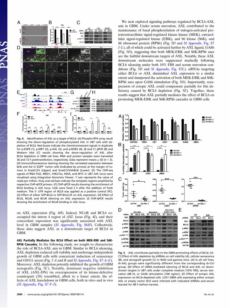
AXL Partially Mediates the BCL6 Effect on both MEK-ERK and S6K-RPS6 Cascades. In the following study, we sought to characterizethe role of BCL6-AXL axis in GBM. Similar to BCL6 silencing,AXL depletion reduced cell viability and anchorage-independentgrowth of GBM cells with concurrent induction of senescenceand G0/G1 arrest (Fig. 5 A and B and SI Appendix, Fig. S7 A–E).Moreover, AXL depletion potently inhibited the growth of GBMxenografts (Fig. 5C). Notably, dominant negative inhibitionof AXL (AXL-DN) via overexpression of its kinase-defectivecounterpart (38) resembled, albeit to a lesser extent, the ef-fects of AXL knockdown in GBM cells, both in vitro and in vivo(SI Appendix, Fig. S7 F–I).
We next explored signaling pathways regulated by BCL6-AXLaxis in GBM. Under serum starvation, AXL contributed to themaintenance of basal phosphorylation of mitogen-activated pro-tein/extracellular signal-regulated kinase kinase (MEK), extracel-lular signal-regulated kinase (ERK), and S6 kinase (S6K), andS6 ribosomal protein (RPS6) (Fig. 5D and SI Appendix, Fig. S7J–L), all of which could be activated further by AXL ligand, GAS6(Fig. 5D), suggesting that both MEK-ERK and S6K-RPS6 axesare the faithful downstream targets of AXL. Notably, these AXLdownstream molecules were suppressed markedly followingBCL6 silencing under both 10% FBS and serum starvation con-ditions (Fig. 5D and SI Appendix, Fig. S7L). siRNAs targetingeither BCL6 or AXL diminished AXL expression to a similarextent and dampened the activation of both MEK-ERK and S6K-RPS6 axes upon GAS6 stimulation (Fig. 5D). Importantly, reex-pression of ectopic AXL could compensate partially for this de-ficiency caused by BCL6 depletion (Fig. 5E). Together, theseresults suggest that AXL partially mediates the effect of BCL6 onpromoting MEK-ERK and S6K-RPS6 cascades in GBM cells.
Fig. 4. Identification of AXL as a target of BCL6. (A) Phospho-RTK array resultshowing the down-regulation of phosphorylated AXL in U87 cells with de-pletion of BCL6. Red boxes indicate the chemiluminescent signals in duplicatefor p-EGFR (1), p-MET (2), p-AXL (3), and p-ROR2 (4). (B and C) qPCR (B) andWestern blot (C) results showing the down-regulation of AXL afterBCL6 depletion in GBM cell lines. RNA and protein samples were harvested36 and 72 h posttransfection, respectively. Data represent means ± SD (n = 3).(D) Immunofluorescence staining showing the correlated expression betweenBcl6 and Axl in EGFP+ tumor cells (indicated by arrows) at the margin of tu-mors in KrasG12V (Upper) and KrasG12V/shBcl6 (Lower). (E) The ChIP-seqsignals of RNA Pol2, MED1, H3K27ac, MAX, and MYC in U87 AXL locus werevisualized using Integrative Genomics Viewer. Y axis represents the value ofreads per million. Gray and red bars indicate the template regions amplified byrespective ChIP-qPCR primers. (F) ChIP-qPCR results showing the enrichment ofBCL6 binding in AXL locus. Cells were fixed 2 h after the addition of freshmedium. The 5′ UTR region of BCL6 was applied as a positive control (PC).(G) Effect of either GFP-BCL6 or GFP-BCL6-ZF on AXL expression. (H) Effect ofBCL6, NCoR, and BCoR silencing on AXL expression. (I) ChIP-qPCR resultsshowing the enrichment of NCoR binding in AXL locus.
Fig. 5. AXL contributes partially to the GBM-promoting effects of BCL6. (A–C) Effect of AXL depletion by shRNAs on cell viability (A), cellular senescence(B), and xenograft growth (C) in NOD scid gamma mice. (A) In all cell lines,sh-AXL groups were significantly different from the corresponding sh-Ctrlgroup. (D) Effect of siRNA-mediated silencing of BCL6 and AXL on down-stream targets in U87 cells under complete medium (10% FBS), serum star-vation (48 h), or GAS6 stimulation (100 ng/mL). (E) Effect of ectopic AXLexpression on BCL6-depleted cells. U251 GBM cells expressing either ectopicAXL or empty vector (EV) were infected with indicated shRNAs and serumstarved for 48 h before harvest.
3984 | www.pnas.org/cgi/doi/10.1073/pnas.1609758114 Xu et al.

Targeting BCL6 in GBM. Considering the glioma-promoting func-tion of BCL6 described earlier, targeted inhibition of BCL6serves as an attractive therapeutic strategy. To test this, we ex-amined the anti-GBM effects of a peptidomimetic inhibitor (RI-BPI), which specifically blocks the interaction between BCL6and its cofactors (13). RI-BPI treatment potently suppressedGBM cell growth both in vitro and in vivo (Fig. 6 A–C). Im-portantly, about half of GBM cells displayed similar response toRI-BPI compared with BCL6-dependent DLBCL cells (OCI-LY3 and SU-DHL-4), which are known to be highly sensitive tothis inhibitor (Fig. 6A). RI-BPI also substantially reduced thelevels of AXL, MET, p-MEK, p-ERK, p-S6K, and p-RPS6 inGBM cells compared with empty peptide or vehicle control (Fig.6D), echoing the effects of BCL6 depletion described earlier. Inaddition, isogenic JM94 cells that were deficient for BCL6 ex-pression showed improved responses to EGFR inhibitors (gefi-tinib and erlotinib), but moderate or unchanged sensitivity tocisplatin or temozolomide treatment (SI Appendix, Fig. S8),suggesting that BCL6 inhibition may provide additional benefitfor combinational targeted therapy. Collectively, our data dem-onstrate that BCL6 can serve as a therapeutic target in GBM.
DiscussionIn this study, we identify a critical role of BCL6 in promotingglioma cell proliferation and glioma progression, as well as theprognostic value of BCL6 overexpression in patients with GBM.Our data suggest that BCL6 expression is progressively up-regulated from lower-grade glioma to GBM. Translocation ofBCL6 was reported in more than 36% of patients with GBM(17); however, genomic rearrangement of BCL6 locus is sur-prisingly not identified in the Cancer Genome Atlas (TCGA)glioma cohort (n > 1,100) (22), indicating that BCL6 trans-location in GBM remains elusive. In addition, copy numbervariation and somatic mutation of BCL6 are infrequent in GBM(19, 20). Therefore, nongenomic mechanism is likely involved inBCL6 overexpression in GBM. Notably, our analysis showed thatprimary GBM samples expressed a higher level of BCL6 than exvivo cultured GBM cells (SI Appendix, Fig. S1A), indicating
that GBM microenvironment or extrinsic factors may modulateBCL6 expression.Although the mechanism of BCL6 overexpression in GBM is
unclear, our loss-of-function studies strongly support a glioma-promoting role of BCL6 in both human GBM cell lines andmouse glioma models. Depletion of BCL6 in GBM cells inducedG0/G1 arrest and reduced BrdU incorporation. In line with thisobservation, BCL6 overexpression promoted G1-S transition ofovarian cancer cells (16). Our data also suggest that both TP53 andAXL pathways are functionally involved in BCL6-mediated cell cycleregulation. Moreover, silencing of BCL6 induced senescence inGBM cells, which was phenocopied by AXL knockdown. In-terestingly, this process appeared to be independent of known se-nescence regulators p53 and p16 (encoded by CDKN2A) (23, 39). Inaddition, GBM cells barely showed activation of apoptosis onBCL6 knockdown, in contrast to mammary epithelial cells (9, 15).In a somatic transgenic glioma model, silencing of Bcl6 in
neural precursor cells could suppress significantly, but not abolish,the formation of KrasG12V-driven glioma, suggesting that Bcl6 iscritical to promote glioma progression but is dispensable for itsinitiation. Taking advantage of EGFP labeling of KrasG12V-transformed cells that accurately separated tumor from stroma(Fig. 3 C and D and SI Appendix, Fig. S3 E and F), we observedthat glioma cells diffused extensively into the brain parenchyma inKrasG12V mice at 8 wk postelectroporation; in sharp contrast, allthe brains from KrasG12V/shBcl6 animals at the same point dis-played circumscribed tumor borders, little migration outside thetumor bulk, and virtually no cortical invasion. Thus, Bcl6 may alsofoster the invasive growth of glioma. Notably, we observed anincreased Ki67 positivity in nonglioma cells in KrasG12V/shBcl6glioma tissues compared with KrasG12V tumors, suggesting thatBcl6 may regulate glioma microenvironment.TP53 pathway is frequently attenuated in GBM (19, 20). In this
study, we report an additional layer of TP53 pathway repressionthrough BCL6 activation in GBM. Similar observations have beenreported in the context of germinal-center B cells, DLBCL, andchronic myeloid leukemia (4, 7), suggesting that the BCL6-TP53regulatory route is not tissue-specific. Interestingly, RI-BPI couldnot de-repress TP53 pathway in GBM cells (SI Appendix, Fig. S9),suggesting that the formation of BCL6/BCoR or BCL6/NCoRcomplex is dispensable for TP53 repression in this context. No-tably, after BCL6 silencing, the levels of ac-p53K382, p-p53S15, andp21 were augmented significantly in response to 6-Gy, but not 12-Gy, radiation, suggesting that the effect of BCL6 on p53 signalingmay be dose-dependent of radiation, and that alternative pathwaystriggered by high-dose radiation may compensate for BCL6 loss.In addition, quantitative analysis showed that p21 level corre-sponded more closely to ac-p53K382 and total p53 levels thanp-p53S15 upon BCL6 silencing and radiation (SI Appendix, Fig.S5B), indicating that acetylation of p53 at K382 potentiates p21expression in BCL6-depleted GBM cells (40).Overexpression of AXL is prevalent in glioma tissues and is
associated with worse prognosis of patients with glioma (21). Al-though genomic amplification of AXL was observed in a smallportion of GBM cases (19, 20), the mechanisms leading to AXLoverexpression remain largely unknown. Here, we report thatAXL is a transcriptional target of BCL6 in GBM. Both BCL6 andNCoR were recruited to the intron 4 region of AXL locus.However, P3 and P4 regions with high enrichment of both factorsdid not contain a consensus BCL6 binding motif, suggesting thatbinding of BCL6 over these segments may involve a noncanonicalmechanism. Notably, both RI-BPI treatment and NCoR knock-down achieved partial suppression of AXL, whereas BCL6knockdown completely abolished AXL expression, suggesting thatBCL6-mediated induction of AXL involves a NCoR-independentmechanism. Nevertheless, NCoR contributes to the full inductionof AXL by forming the BCL6/NCoR complex, supporting thenotion that NCoR can also function to enhance transcription (41).
Fig. 6. Targeting BCL6 in glioma. (A) Effect of RI-BPI on GBM cell viability.GBM cells were treated with indicated concentration of either RI-BPI or emptypeptide (EP) for 72 h before analysis. OCI-LY3 and SU-DHL-4 cells were used aspositive controls. (B and C) Effect of RI-BPI treatment on JM94 cell growthin vivo. Peptides were administrated intraperitoneally (50 mg·kg−1·day−1 for4 d). (D) Western blot results showing the effect of RI-BPI (20 μM, 24 h) onBCL6 downstream targets.
Xu et al. PNAS | April 11, 2017 | vol. 114 | no. 15 | 3985
MED
ICALSC
IENCE
S
Further, we demonstrated that both BCL6 and AXL positivelyregulated MEK-ERK and S6K-RPS6 cascades, and that theBCL6-dependent effect on these pathways was partially mediatedby AXL. Interestingly, the effects of BCL6 and AXL on GBM cellgrowth were more pronounced in anchorage-independent condi-tion and xenograft assay when compared with 2D monolayerculture, suggesting that microenvironmental factors may be in-volved in the progrowth function of BCL6 and AXL (42).In addition to AXL, we found that BCL6 knockdown reduced
the phosphorylation of EGFR, MET, and ROR2 in U87 cellsunder regular culture condition. However, upon acute EGFstimulation, BCL6 silencing either did not affect the induction ofp-EGFRY1068 (U87 cells) or even promoted it (JM94 cells; SIAppendix, Fig. S8), indicating that BCL6 may contribute tomaintaining the steady-state level of p-EGFRY1068, but its effecton p-EGFRY1068 during acute EGF response may vary accordingto cellular context. As AXL and MET have been shown to re-inforce EGFR signaling and to mediate the resistance to EGFRtyrosine kinase inhibitors (43–45), down-regulation of AXL andMET by BCL6 inhibition may exert a more profound effect onRTK pathway and overcoming therapeutic resistance of EGFRinhibitors. In support of this notion, BCL6 depletion sensitizedGBM cells to EGFR inhibitors (SI Appendix, Fig. S8). Togetherwith our finding that targeting BCL6 by RI-BPI effectivelyinhibited GBM cell viability with concurrent down-regulation ofAXL and MET proteins, these results not only identify BCL6 asa druggable candidate but also suggest a potential combinationaltherapeutic strategy for treating GBM.
Materials and MethodsExtended materials and methods are provided in SI Appendix. Either signedconsent forms or exemptions have been obtained for all human samples.The study of human samples was approved by the University of California,Los Angeles, Institutional Review Board. All murine xenograft experimentswere in compliance with ethical regulations of the Institutional Animal Careand Use Committee of National University of Singapore. Electroporation-based murine experiments were approved by the Institutional Animal Careand Use Committee of Cedars-Sinai Medical Center. Unless otherwise stated,two-tailed Student’s t test was used to analyze the potential statistical dif-ference between two groups. n.s., not significant; *P < 0.05; **P < 0.01;***P < 0.001. Log-rank test was used for survival analysis.
ACKNOWLEDGMENTS. We thank Sumiko Takao and Vaidehi Krishnan forhelp with γ-radiation experiments; Annouck Luyten, Yi-Ting Qiao, and Kol-Jia Yong for ChIP protocols and technical support; Hui-Min Geng, ChristianHurtz, Anand Jeyasekharan, and Shojiro Kitajima for reagent sharing; andHazimah Binte Mohd Nordin for help with mouse breeding. This work isfunded by the National Research Foundation Singapore under its SingaporeTranslational Research Investigator Award (to H.P.K.; NMRC/STaR/0021/2014), the Singapore Ministry of Education Academic Research Fund Tier 2(MOE2013-T2-2-150), the Singapore Ministry of Health’s National MedicalResearch Council Centre Grant awarded to National University Cancer Insti-tute of Singapore, the National Research Foundation Singapore, and theSingapore Ministry of Education under its Research Centres of Excellenceinitiatives, and is additionally supported by philanthropic donations fromthe Melamed family and Blanche and Steven Koegler, as well as Tom Collier“Regatta for Hope” Foundation. We also acknowledge support from theDonna and Jesse Garber Awards for Cancer Research (D.-C.L.), the SamuelOschin Comprehensive Cancer Institute Cancer Research Forum Award (J.J.B.),the Board of Governors RMI of Cedars-Sinai (J.J.B.), and National Institutes ofHealth Grant R33 CA202900 (to J.J.B.).
1. Siggs OM, Beutler B (2012) The BTB-ZF transcription factors. Cell Cycle 11:3358–3369.2. Basso K, Dalla-Favera R (2010) BCL6: Master regulator of the germinal center reaction
and key oncogene in B cell lymphomagenesis. Adv Immunol 105:193–210.3. Cattoretti G, et al. (2005) Deregulated BCL6 expression recapitulates the pathogenesis
of human diffuse large B cell lymphomas in mice. Cancer Cell 7:445–455.4. Phan RT, Dalla-Favera R (2004) The BCL6 proto-oncogene suppresses p53 expression in
germinal-centre B cells. Nature 432:635–639.5. Duy C, et al. (2010) BCL6 is critical for the development of a diverse primary B cell
repertoire. J Exp Med 207:1209–1221.6. Duy C, et al. (2011) BCL6 enables Ph+ acute lymphoblastic leukaemia cells to survive
BCR-ABL1 kinase inhibition. Nature 473:384–388.7. Hurtz C, et al. (2011) BCL6-mediated repression of p53 is critical for leukemia stem cell
survival in chronic myeloid leukemia. J Exp Med 208:2163–2174.8. Tiberi L, et al. (2012) BCL6 controls neurogenesis through Sirt1-dependent epigenetic
repression of selective Notch targets. Nat Neurosci 15:1627–1635.9. Logarajah S, et al. (2003) BCL-6 is expressed in breast cancer and prevents mammary
epithelial differentiation. Oncogene 22:5572–5578.10. Hatzi K, et al. (2013) A hybrid mechanism of action for BCL6 in B cells defined by formation
of functionally distinct complexes at enhancers and promoters. Cell Reports 4:578–588.11. Ghetu AF, et al. (2008) Structure of a BCOR corepressor peptide in complex with the
BCL6 BTB domain dimer. Mol Cell 29:384–391.12. Ahmad KF, et al. (2003) Mechanism of SMRT corepressor recruitment by the BCL6 BTB
domain. Mol Cell 12:1551–1564.13. Cerchietti LC, et al. (2009) A peptomimetic inhibitor of BCL6 with potent anti-
lymphoma effects in vitro and in vivo. Blood 113:3397–3405.14. Cerchietti LC, et al. (2010) A small-molecule inhibitor of BCL6 kills DLBCL cells in vitro
and in vivo. Cancer Cell 17:400–411.15. Walker SR, et al. (2015) The transcriptional modulator BCL6 as a molecular target for
breast cancer therapy. Oncogene 34:1073–1082.16. Wang YQ, et al. (2014) BCL6 is a negative prognostic factor and exhibits pro-
oncogenic activity in ovarian cancer. Am J Cancer Res 5:255–266.17. Ruggieri S, et al. (2014) Translocation of the proto-oncogene Bcl-6 in human glio-
blastoma multiforme. Cancer Lett 353:41–51.18. Tiberi L, et al. (2014) A BCL6/BCOR/SIRT1 complex triggers neurogenesis and suppresses
medulloblastoma by repressing Sonic Hedgehog signaling. Cancer Cell 26:797–812.19. Brennan CW, et al.; TCGA Research Network (2013) The somatic genomic landscape of
glioblastoma. Cell 155:462–477.20. Chin L, et al.; Cancer Genome Atlas Research Network (2008) Comprehensive genomic char-
acterization defines human glioblastoma genes and core pathways. Nature 455:1061–1068.21. Hutterer M, et al. (2008) Axl and growth arrest-specific gene 6 are frequently over-
expressed in human gliomas and predict poor prognosis in patients with glioblastomamultiforme. Clin Cancer Res 14:130–138.
22. Ceccarelli M, et al.; TCGA Research Network (2016) Molecular profiling reveals biologicallydiscrete subsets and pathways of progression in diffuse glioma. Cell 164:550–563.
23. Shvarts A, et al. (2002) A senescence rescue screen identifies BCL6 as an inhibitor ofanti-proliferative p19(ARF)-p53 signaling. Genes Dev 16:681–686.
24. Sun L, et al. (2006) Neuronal and glioma-derived stem cell factor induces angiogenesiswithin the brain. Cancer Cell 9:287–300.
25. Lee J, et al. (2006) Tumor stem cells derived from glioblastomas cultured in bFGF andEGF more closely mirror the phenotype and genotype of primary tumors than doserum-cultured cell lines. Cancer Cell 9:391–403.
26. Gautier L, Cope L, Bolstad BM, Irizarry RA (2004) affy–analysis of Affymetrix GeneChipdata at the probe level. Bioinformatics 20:307–315.
27. Irizarry RA, et al. (2003) Exploration, normalization, and summaries of high densityoligonucleotide array probe level data. Biostatistics 4:249–264.
28. Uhlén M, et al. (2015) Proteomics. Tissue-based map of the human proteome. Science347:1260419.
29. Yin D, et al. (2005) Proteasome inhibitor PS-341 causes cell growth arrest and apo-ptosis in human glioblastoma multiforme (GBM). Oncogene 24:344–354.
30. Sanjana NE, Shalem O, Zhang F (2014) Improved vectors and genome-wide librariesfor CRISPR screening. Nat Methods 11:783–784.
31. Breunig JJ, et al. (2015) Ets factors regulate neural stem cell depletion and gliogenesisin Ras pathway glioma. Cell Reports 12:258–271.
32. Fellmann C, et al. (2013) An optimized microRNA backbone for effective single-copyRNAi. Cell Reports 5:1704–1713.
33. Stancik EK, Navarro-Quiroga I, Sellke R, Haydar TF (2010) Heterogeneity in ventricularzone neural precursors contributes to neuronal fate diversity in the postnatal neo-cortex. J Neurosci 30:7028–7036.
34. Dunning MJ, Smith ML, Ritchie ME, Tavaré S (2007) beadarray: R classes and methodsfor Illumina bead-based data. Bioinformatics 23:2183–2184.
35. Ritchie ME, et al. (2015) limma powers differential expression analyses for RNA-sequencing and microarray studies. Nucleic Acids Res 43:e47.
36. Wiederschain D, et al. (2009) Single-vector inducible lentiviral RNAi system for on-cology target validation. Cell Cycle 8:498–504.
37. Lin CY, et al. (2012) Transcriptional amplification in tumor cells with elevated c-Myc.Cell 151:56–67.
38. Vajkoczy P, et al. (2006) Dominant-negative inhibition of the Axl receptor tyrosinekinase suppresses brain tumor cell growth and invasion and prolongs survival. ProcNatl Acad Sci USA 103:5799–5804.
39. Yu RY, et al. (2005) BCL-6 negatively regulates macrophage proliferation by sup-pressing autocrine IL-6 production. Blood 105:1777–1784.
40. Barlev NA, et al. (2001) Acetylation of p53 activates transcription through recruitmentof coactivators/histone acetyltransferases. Mol Cell 8:1243–1254.
41. Meyer MB, Pike JW (2013) Corepressors (NCoR and SMRT) as well as coactivators arerecruited to positively regulated 1α,25-dihydroxyvitamin D3-responsive genes.J Steroid Biochem Mol Biol 136:120–124.
42. Pampaloni F, Reynaud EG, Stelzer EH (2007) The third dimension bridges the gapbetween cell culture and live tissue. Nat Rev Mol Cell Biol 8:839–845.
43. Meyer AS, Miller MA, Gertler FB, Lauffenburger DA (2013) The receptor AXL diver-sifies EGFR signaling and limits the response to EGFR-targeted inhibitors in triple-negative breast cancer cells. Sci Signal 6:ra66.
44. Jun HJ, et al. (2012) Acquired MET expression confers resistance to EGFR inhibition ina mouse model of glioblastoma multiforme. Oncogene 31:3039–3050.
45. Zhang Z, et al. (2012) Activation of the AXL kinase causes resistance to EGFR-targetedtherapy in lung cancer. Nat Genet 44:852–860.
3986 | www.pnas.org/cgi/doi/10.1073/pnas.1609758114 Xu et al.