Embed Size (px)
Citation preview

1
BCHS 6229
Protein Structure and FunctionProtein Structure and Function
Lecture 4 (October 20, 2011)
From Structure to Function (I):
Flexibility and Protein Function
Intrinsically Unstructured Proteins and their
Functions

2
The folded state has a flexible structure
The protein molecule does not have a static rigid structure atnormal temperature. Instead, all the atoms are subject tosmall T-dependent fluctuation.
Breathing of the molecule - random or collective.
Motions are usually small (a few tenths of an Å); butsometimes large and very significant.
How such large collective movements are reflected in x-
ray studies?
Insight into these individual and collective motions has beenobtained by theoretical studies - MD ~picosecond for individual residues ~nanosecond for loop regions
Such movements: very important for the functions of manyproteins.

3
There can also be larger conformational changes betweendifferent functional states of the molecule.
(pH, ligands)
The flexibility of tertiary structure allows proteins to
adapt to their ligands.
Induced fit: originally, the change in the structure of anenzyme, induced by binding of the substrate, that bringsthe catalytic groups into proper alignment. Nowgeneralized to the idea that specific ligands can inducethe protein conformation that results in optimal bindinginteractions.

4
Haloperidol Crixivan Peptide analog
Inherent flexibility of proteins Conformational changes
HIV protease bound to three differentinhibitors.
Each inhibitor has a quite differentstructure, yet all bind tightly to the activesite and induce closure of a flap thatcovers it.

5
Protein flexibility is essential for biochemical function
In most proteins, binding is followed by some action(chemical transformation of L, conformational change inproteins, translocation of the protein, etc. )
In all cases, the structure of the protein must be flexibleenough that the net free energy released by binding and/orchemical transformation of the L can derive the requiredchanges in the protein’s structure and function
Binding: Kd = ~10-3 M (weak) to ~10-12 M(extremely strong)

6
Yeast MAITVGINGFGRIGRLVLRIALSRK--DIQIVAINDPFIAPEYASYMFKYDSTHGRYSGEVSHEGENIVIDGKKIRVYQERDPVNIPWGKDGVDYVIDSTTmari MAR-VAINGFGRIGRLVYRIIYERKNPDIEVVAIND-LTDTKTLAHLLKYDSVHKKFPGKVEYTENSLIVDGKEIKVFAEPDPSKLPWKDLGVDFVIEST ** *.*********** ** .** **::***** : .: ::::****.* ::.*:*.: :.:::***:*:*: * ** ::** . ***:**:**
110 120 130 140 150 160 170 180 190 200 | | | | | | | | | |Yeast GVFKELDSAQKHIDAGAKKVVITAPS-STAPMFVVGVNEDKYTPDLNIISNASCTTNCLAPLAKIINNKFGIEEGLMTTVHSITATQKTVDGPSHKDWRSTmari GVFRNREKAELHLQAGAKKVIITAPAKGEDITVVIGCNEDQLKPEHTIISCASCTTNSIAPIVKVLHEKFGIVSGMLTTVHSYTNDQRVLDLP-HKDLRR ***:: :.*: *::******:****: . .*:* ***: .*: .*** ******.:**:.*::::**** .*::***** * *:.:* * *** *
210 220 230 240 250 260 270 280 290 300 | | | | | | | | | |Yeast GPTASGNIIPSSTGAAKAVGKVIPELAGKLTGMSLRVPTVDVSVVDLTVKLLKDATYDEIKAAVKEAAEGPLKGVVGYTEDQVVSSDFLTDNRSSIFDAETmari ARAAAVNIIPTTTGAAKAVALVVPEVKGKLDGMAIRVPTPDGSITDLTVLVEKETTVEEVNAVMKEATEGRLKGIIGYNDEPIVSSDIIGTTFSGIFDAT . :*: ****::*******. *:**: *** **::**** * *:.**** : *::* :*::*.:***:** ***::**.:: :****:: . *.****
310 320 330 | | |Yeast AGIWLSPRFVKLIAWYDNEYGYSTRVVDLLEYVASKNTmari ITNVIGGKLVKVASWYDNEYGYSNRVVDTLELLLKM-
:. ::**: :*********.**** ** : .
Identity (*) : 170 is 50.45 %
Strongly similar (:) : 74 is 21.96 %
Weakly similar (.) : 27 is 8.01 %
Different : 66 is 19.58 %
yeast ( 334 residues).
Thermotoga maritima ( 333 residues).
Difference in T-dependence ofthe specific activity ofglyceraldehyde-3-phosphatedehydrogenase (GAPDH).
Protein flexibility is essential for biochemical function
Some mutant enzymes are stable at higher T thannormal T and rigid (can abolish functions).

7
The degree of flexibility varies in proteins with different
functions:
Not all proteins are equally flexible.
A number of proteins: relatively rigid (extracellular proteins)
Other proteins: very large shape changes when the correctligand binds.
w/ AMP w/ AMP + ATP (AMPPNP)
Adenylate kinase

8
Conformational changes in proteins:
• Small specific & localized changes: enzyme catalytic sites.
• Myoglobin: oxy- & deoxy forms are similar.
• Hemoglobin: oxygen binding leads to changes in 3° and 4°structure.
• Allosteric transitions involve long-range integratedconformational changes: Hb, aspartate carbamoyl-transferase, etc.
• Some very large proteins function as motors or pumpsthrough changes in conformation:

9
Hinge motions in proteins:
The simplest mechanism of conformational change in proteins
Hinge motion in lactoferrinResidues 89-92; 249-252, 54 Å rotations as rigid bodies
Open closed
Residues superposedD1 alone: 0.82 ÅD2 alone: 0.58 ÅBoth domains together: 6.1 Å
10
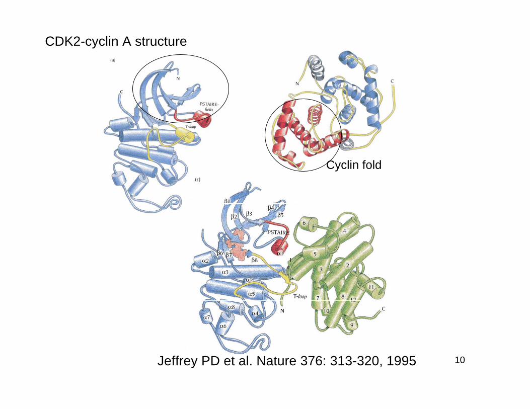
CDK2-cyclin A structure
Jeffrey PD et al. Nature 376: 313-320, 1995
Cyclin fold

11
Upon cyclin A binding, thePSTAIRE helix undergoes amajor conformational change90°.
Consequence?

12
Inactive active
Drastic change in T-loop:Substrate binding site open
T160

13
Peptide binding to calmodulin induces a largeinterdomain movement
Calmodulin: Ca- dep signaling pathway

14
Serpins (Serine protease inhibitors):
Spring-loaded safety catch mechanism
1-antitrypsin (a bloodplasma proteinase)
Serpins - antithrombin,plasminogen activatorinhibitor: all very similar 3Dstructures, and form tightcomplexes with theircorresponding S.P.
The lung health - neutrofil elastase
ovalbumin

15
Antithrombin 1-antitrypsin PAI
Which of these forms is most stable?In vivo PAI and antithrombin are stabilized in their activeforms by binding vitronectin and heparin.
“Spring-loaded safety catch mechanism” : revert totheir latent, stable form unless the catch is kept in a loadedposition by another molecule.

16
Intrinsically Unstructured Proteins and
their Functions
NATURE REVIEWS | MOLECULAR CELL BIOLOGY (2005)
H. Jane Dyson and Peter E.Wright
Many gene sequences in eukaryotic genomes encode entireproteins or large segments of proteins that lack a well-structuredthree-dimensional fold. Disordered regions can be highlyconserved between species in both composition and sequenceand, contrary to the traditional view that protein function equateswith a stable three-dimensional structure, disordered regions areoften functional, in ways that we are only beginning to discover.Many disordered segments fold on binding to their biologicaltargets (coupled folding and binding), whereas others constituteflexible linkers that have a role in the assembly of macromoleculararrays.

17
Why are we discovering unfolded proteins now?
Classic biochemical methods are strongly biased towards the productionand characterization of folded, active proteins.
The standard preparation methods:• Plant or animal tissues, or bacterial cells, are isolated and homogenized.• The homogenate is assayed for the activity of interest.• The homogenate is subjected to (NH4)2SO4 fractionation and
chromatography and/or gel filtration.• The fractions are assayed for activity and the active protein is purified.• The pure, active protein is sequenced.• The three-dimensional structure is determined.
This methodology automatically selects folded proteins, because theformation of a homogenate invariably releases proteases and unfoldedproteins are much more sensitive than folded proteins to degradationunder these conditions. Also, if the unfolded domains are part ofregulatory proteins, there might be only a few copies per cell, and theymight not have a convenient activity to assay.

18
So, why are we discovering unfolded proteins now?
We have only begun to understand the presence and roles of unfoldedproteins since the advent of new paradigms in biochemical methodology.
Instead of discovering a detectable activity and isolating it bypurifying the protein, we now have access to a vast library of genesequences. The use of genetic methods to isolate function, using mutantsand knockouts, has been the way that most unfolded proteins have beenidentified so far:• Formulating a function (for example, control of transcription).• Mapping the function to a particular gene and to a particular area in thegene.• Transcribing the gene, producing the protein and purifying the protein.• Examining its structure in solution by circular dichroism and NMRspectroscopy.• If the protein is unfolded, trying to co-express it with a binding partner(also identified through genetic mapping).

19
Will a domain be folded or unfolded?
Predicting the 3D structures of globular proteins fromsequence data alone - key challenge
Identifying sequences that are likely to be intrinsicallydisordered - relatively straightforward

20
The presence of low sequence complexity and amino-acidcompositional bias,with a low content of bulky hydrophobicamino acids (Val, Leu, Ile, Met, Phe,Trp, Tyr), anda high proportion of particular polar and charged aminoacids (Gln, Ser, Pro, Glu, Lys and, on occasion, Gly andAla).
Computer programs: prediction of unstructured regionsfrom amino acid sequences.PONDR, FoldIndex, DisEMBL, GLOBPLOT 2, andDISOPRED2
Sequence signatures of intrinsic disorder?

21
Intrinsically disordered proteins are highly prevalent
Proportion of proteins containing such segmentsincreases with the increasing complexity of an organism
Proteins involved in eukaryotic signal transduction orassociated with cancer: an increased propensity forintrinsic disorder

22
Experimental characterization of disordered proteins.
Nuclear magnetic resonance (NMR) spectroscopy
Information on the three dimensional structure anddynamics of biological molecules in solution.
CIRCULAR DICHROISM
The UV-CD spectrum uses the chirality or ‘handedness’of biological molecules to provide information onsecondary structure in solution.
Fluorescence spectroscopy
Information on the environment of aromatic rings, andcan be used in conjunction with external probes todetermine the distances between atoms in a molecule.

23
Experimental characterization of disordered proteins.
Vibrational CD Spectroscopy
the chiroptical version of infra-red spectroscopyinformation on the vibrations of individual bonds in amolecule.
Raman spectroscopy
Information on bond vibrations that is complementary tothat provided by infra-red spectroscopy.
Biochemical assays
Proteolysis susceptibility

24
Ward JJ, Sodhi JS, McGuffin LJ, Buxton BF and Jones DT (2004)Prediction and functional analysis of native disorder in proteins fromthe three kingdoms of lifeJournal of Molecular Biology, 337, 635-645.
The DISOPRED2 Prediction of Protein Disorder Server

25
The continuum of protein structure
Increasing content of stable 3-D structure
multi-domain proteins
Single domains
Compact but disordered

26
Functions of intrinsic disorder in proteins
• regulation of transcription and translation• cellular signal transduction,• protein phosphorylation,• storage of small molecules,• regulation of the self-assembly of large multiprotein
complexes

27
Coupled folding and bindingan intrinsically disordered protein (region) folds into anordered structure concomitant with binding to its target.
p-kinase inducible domain (pKID) of CREP (cAMP-response element binding P)
KIX of CBP
might involve ju
st a few residu
es

28
A case study: transcriptional co-activator CBP/p300

29
Domain structure of CBP

30
The biological ‘cost’ of disordered proteins
Intrinsically disordered regions are the sites of manychromosomal translocations that are associated withdisease.e.g.translocations that fuse regions of CBP or p300 tosegments of MOZ (monocytic zinc-finger leukaemiaprotein) associated with human leukaemias.
Disordered regions can also have a biological cost interms of the promotion and proliferation of protein foldingdiseases.Neurodegenerative diseases such as prion diseases orParkinson’s disease are associated with intrinsicallydisordered proteins (Q-rich motifs)

31
Cj0977 is highly soluble and a dimer in solution
His-Cj0977 crystalsobtained with (NH4)2SO4
But, no diffraction!
0.2 mm
M S 17 18 19 20 21 22 23
25 kDa
75 kDa50 kDa
29 kDa
A case study: a small C-ter tail of Cj0977
32
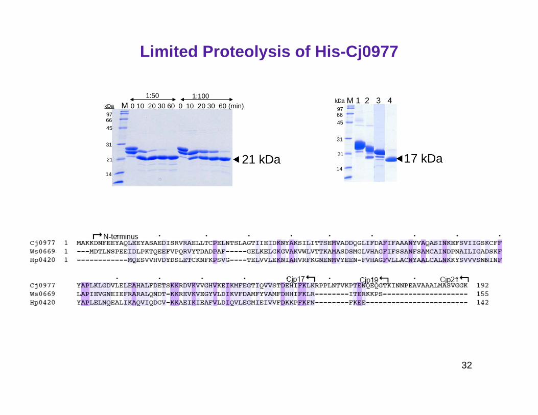
Limited Proteolysis of His-Cj0977
M 0 10 20 30 60 0 10 20 30 60 (min)9766
45
31
21
14
kDa
1:50 1:100M 1 2 3 4
9766
45
31
21
14
kDa
21 kDa 17 kDa

33
GST::Cj0977p17
0.1 mm
Crystallization of Cj0977p17
Cj0977p17
17 kDa
75 kDa50 kDa
29 kDa

34
MAD phasing at 2.8 Å.
Cj0977p17 electron density map.

35
Cj0977 adopts a Hotdog fold
180°

36
FapR: a structural homolog of Cj0977

37
Lipid Biosynthesis Regulation in Gram-negative Bacteria
1 43 67 77 188HTH L Hot dog domain
DNA binding
domain
Malonyl-CoA binding
domain
FapR
Schujman et al.EMBO J. (2006) 25(17):4074-83.

38
Function of Cj0977?
1 43 67 77 188HTH L Hot dog domain
DNA binding
domain
Malonyl-CoA binding
domain
FapR
1 10 155 192Hot dog domain
acyl-CoA binding
domain
?
Cj0977
Yokoyama et al.J. Mol. Biol. (2008) 384(2):364-76

39
Ligand recognition site of Hotdog fold proteins
SCoA
OO
-OMalonyl CoA
Acetyl CoASCoA
O

40
activation of effectors
cellular protein 1
acyl-CoA compound
C-ter
C-ter
Proposed model of coupled folding and binding of Cj0977
Dis
ord
ere
d p
robabili
ty
Sequence number
Hot dog domain
1 10 155 192